


Simple Summary
Intensive feeding with high-grain diets easily injures the rumen metabolism and immune function of sheep, which affects animal health and farming benefits. This study raised sheep with different diets, including a common high-grain diet and a diet mixed with Perilla frutescens seeds. We examined the genetic changes in sheep’s stomach tissue to explore the effects of Perilla frutescens seeds. The results showed that high-grain food weakened local immune defenses instead of causing widespread inflammation. Adding Perilla frutescens seeds effectively boosted the natural immune ability of sheep rumen tissue. This work provides transcriptomic evidence supporting Perilla frutescens seeds as a promising natural feed candidate for developing healthy feeding patterns in sustainable ruminant production.
Abstract
The ruminal epithelium plays a central role in nutrient absorption, epithelial barrier function, and mucosal immune defense. However, the molecular mechanisms by which dietary Perilla frutescens seeds (PFS) regulate ruminal epithelial immunity remain poorly understood. This study investigated the transcriptomic responses of the sheep ruminal papilla to dietary PFS supplementation. Forty-five male Tan sheep were assigned to three dietary treatments (n = 15 per group): a low-concentrate diet (LC), a high-concentrate diet (HC), and the LC diet supplemented with 3% PFS (LC + PFS). After an 84-day feeding trial, ruminal papilla tissues from six animals per group were randomly subjected to RNA sequencing and integrated bioinformatic analyses, covering all three pairwise comparisons (LC vs. HC, HC vs. LC + PFS, and LC vs. LC + PFS). The LC vs. LC + PFS comparison served as the direct assessment of PFS effects under the same basal diet, whereas HC vs. LC + PFS was treated as a cross-dietary comparison. LC vs. HC yielded 387 differentially expressed genes (DEGs), HC vs. LC + PFS yielded 104 DEGs, and LC vs. LC + PFS yielded 33 DEGs, with predominant enrichment in metabolic and stress-adaptive pathways (AMPK, MAPK, PI3K-Akt). The latter were mainly associated with metabolic and structural pathways. Propionate and valerate were negatively correlated with several interferon-signaling and antigen-presentation genes. Collectively, the direct LC vs. LC + PFS comparison demonstrates that PFS supplementation under physiological conditions is associated with targeted metabolic modulation. Meanwhile, the cross-dietary HC vs. LC + PFS comparison reveals that, relative to high-concentrate feeding, the PFS-supplemented low-concentrate feeding regimen is transcriptionally associated with enhanced immune surveillance capacity in a non-inflammatory manner. These findings provide transcriptomic evidence supporting the potential of PFS as a modulator of ruminal epithelial homeostasis and offer a foundation for further investigation into its application as a functional feed ingredient.
1. Introduction
Efficient production of ruminants heavily relies on a healthy rumen ecosystem. As the core organ for feed degradation and nutrient metabolism, the rumen undertakes microbial fermentation, volatile fatty acid (VFA) absorption and transformation, and acts as a vital immune barrier for ruminants [1]. The ruminal epithelium serves as a dynamic interface connecting rumen microbes and host tissues. Its structural integrity and normal physiological function are fundamental to guaranteeing nutrient utilization efficiency, animal health, growth performance and livestock product quality [1,2]. Ruminal epithelial homeostasis in this context is defined as a balanced state of structural integrity, efficient nutrient transport, appropriate metabolic activity, and calibrated immune responsiveness that effectively defends against pathogens without initiating overt, damaging inflammation.
Plant-derived lipids and phytochemicals have drawn extensive attention due to their potential to regulate rumen fermentation, enhance antioxidant capacity and modulate host immune responses [3]. Perilla frutescens seeds are a valuable oilseed resource abundant in α-linolenic acid (ALA), flavonoids, phenolic acids and terpenoids. These bioactive components confer strong antioxidant, anti-inflammatory, antibacterial, antiviral, and immunomodulatory properties [3,4]. Existing studies have verified that dietary supplementation with Perilla frutescens seeds optimizes the fatty acid profile of lamb meat, increases the deposition of n-3 polyunsaturated fatty acids (PUFAs) and improves meat antioxidant capacity [5]. In addition, Perilla frutescens seeds can enhance the growth performance of lambs: they raise the serum levels of immunoglobulin G (IgG), immunoglobulin M (IgM), superoxide dismutase (SOD) and glutathione peroxidase (GSH-Px), while reducing the contents of pro-inflammatory interleukin-1β (IL-1β), tumor necrosis factor-α (TNF-α) and malondialdehyde (MDA) [5,6]. For dairy goats, Perilla frutescens seeds or Perilla frutescens seed oil can enrich beneficial fatty acids such as conjugated linoleic acid (CLA), eicosapentaenoic acid (EPA) and docosahexaenoic acid (DHA) in milk, without compromising ruminal pH, VFA profiles or blood biochemical parameters [7]. Research on non-ruminant animals also demonstrates that Perilla seed oil ameliorates lipid metabolism and relieves inflammation, implying its regulatory effects on immunity and metabolism [8]. Overall, Perilla frutescens seeds are promising feed additives capable of improving livestock product quality and maintaining host health and immune function. Previous evidence showed that supplementation with 1% Perilla frutescens seed extract could mitigate ruminal methane production without adversely affecting VFA production [9], supporting the potential of PFS-derived bioactive components to modulate the rumen environment. However, the molecular mechanisms by which Perilla frutescens seeds regulate ruminal epithelial function remain poorly understood.
To address this gap, we established three dietary groups for Tan sheep: a low-concentrate diet (LC), a high-concentrate diet (HC), and the LC diet supplemented with Perilla frutescens seeds (LC + PFS). This design serves two complementary purposes. The direct LC vs. LC + PFS comparison isolates the transcriptional effects of PFS supplementation under a consistent dietary background. Concurrently, the HC group provides a reference framework representing the ruminal epithelial transcriptome under high-concentrate feeding—characterized by diminished expression of immune-related genes. Contrasting the LC + PFS profile against this HC reference allows us to assess whether PFS-supplemented low-concentrate feeding is associated with an enhanced immune surveillance signature relative to an HC-induced suppressed state. Notably, this cross-dietary comparison does not test a direct “rescue” effect; rather, it uses the HC transcriptional state as a contextual backdrop to interpret the LC + PFS-associated immune features.
Combining high-throughput RNA sequencing with multiple bioinformatics approaches, including weighted gene co-expression network analysis (WGCNA) and VFA-gene correlation network analysis, we systematically characterized the transcriptomic changes in the sheep ruminal epithelium across three dietary conditions. Our results revealed that, when contrasted against the HC-induced state of diminished immune gene expression, the LC + PFS transcriptional profile was associated with upregulation of RLR-interferon signaling and immune surveillance genes. Under physiological conditions (LC), PFS supplementation was associated with targeted changes in metabolic pathways without widespread immune activation. Collectively, these findings position PFS as a potential modulator of ruminal epithelial immune homeostasis rather than a conventional immune activator, providing transcriptomic evidence for its application as a functional feed ingredient in ruminant production.
2. Materials and Methods
2.1. Animals, Experimental Design, and Diets
All animal procedures were approved by the Animal Care and Use Committee of China Agricultural University, Approval No. AW30901202-1-1. The experimental animals were obtained from Tianyuan Liangzhong Sheep Farm in Wuzhong, Ningxia Hui Autonomous Region, China. A total of 45 healthy male Tan sheep (Ovis aries), approximately 6 months of age, with an initial body weight of 25.1 ± 1.23 kg, were selected for the experiment.
A completely randomized design was used. Sheep were balanced according to initial body weight and randomly assigned to three dietary treatments, with 15 animals per treatment. Each treatment included three replicate pens, with five sheep per pen. The dietary treatments were as follows: a low-concentrate control group (LC; concentrate-to-forage ratio of 55:45 on a dry matter basis), a high-concentrate control group (HC; concentrate-to-forage ratio of 80:20 on a dry matter basis), and a Perilla frutescens seed supplementation group (LC + PFS); LC diet was supplemented with 3% Perilla frutescens seeds on a dry matter basis. Therefore, the LC and LC + PFS diets were not formulated to be strictly isoenergetic or isonitrogenous. Based on the analyzed nutrient composition of PFS, the calculated crude protein concentrations of the LC and LC + PFS diets were 14.27% and 14.54% of dry matter, respectively, and the corresponding nitrogen concentrations were 2.283% and 2.326% DM. The ingredient composition and nutrient levels of the basal diets are presented in Table 1. The nutrient composition of Perilla frutescens seeds was as follows: ether extract, 40.9%; crude protein, 23.5%; neutral detergent fiber, 25.4%; acid detergent fiber, 19.9%; and ash, 3.9%, all expressed on a dry matter basis. The fatty acid profile was dominated by α-linolenic acid (C18:3n-3, 57.8%), oleic acid (C18:1n-9c, 19.9%), linoleic acid (C18:2n-6c, 11.9%), and palmitic acid (C16:0, 7.4%).
Table 1.
Ingredients and nutrient composition of the experimental diets.
The sheep were housed in group pens, with five animals per pen and three pens assigned to each dietary treatment. All pens were managed under the same routine farm conditions and were regularly cleaned and maintained in a clean and dry condition. All sheep were fed a total mixed ration (TMR). The Perilla frutescens seeds were thoroughly mixed with the concentrate before incorporation into the diet. Feed was offered twice daily at 09:00 and 17:00, and animals had free access to feed and water throughout the experiment. The amount of feed offered and refusals in each pen was recorded daily, and dry matter intake was calculated. The experiment lasted 84 days, including a 14-day adaptation period followed by a 70-day formal experimental period.
2.2. Sample Collection and Physiological and Biochemical Measurements
At the end of the 84-day feeding trial, six sheep were randomly selected from each dietary group for euthanasia. Ruminal papilla tissues from the ventral rumen sac were swiftly excised, rinsed thoroughly with pre-cooled sterile phosphate-buffered saline (PBS) to remove surface contaminants, and immediately snap-frozen in liquid nitrogen. All tissue samples were then stored at −80 °C for subsequent molecular analysis. The epithelial layer was not further separated from the underlying lamina propria or deeper tissue. Meanwhile, rumen fluid was collected to quantify volatile fatty acids (VFAs) including acetate, propionate, and butyrate, as well as ruminal ammonia nitrogen (NH3-N). VFAs were quantified via gas chromatography, and NH3-N concentrations were determined according to previous method [10,11].
Prior to morning feeding and slaughter, blood samples were harvested from the jugular vein of experimental animals. Serum was separated after centrifugation and used for subsequent detection. The measured indicators covered multiple categories: blood glucose (GLU), triglycerides (TG), high-density lipoprotein cholesterol (HDL-C), and low-density lipoprotein cholesterol (LDL-C) for glucose and lipid metabolism; alkaline phosphatase (ALP), alanine aminotransferase (ALT), aspartate aminotransferase (AST), and total bilirubin (TBIL) as indicators related to hepatic function; and blood urea nitrogen (BUN) as an indicator of nitrogen metabolism and renal function; MDA and SOD for antioxidant status; immunoglobulins (IgG, IgA, IgM); and pro-inflammatory cytokines including IL-1β and TNF-α. All biochemical parameters were detected using an automatic biochemical analyzer, while cytokine and immunoglobulin levels were determined via commercial enzyme-linked immunosorbent assay (ELISA), following the standard protocols provided by respective reagent manufacturers.
2.3. RNA Extraction, Library Construction and Transcriptome Sequencing
Total RNA was isolated from frozen ruminal papilla tissues using the EASYspin Total RNA Extraction Kit for fibrous tissues (Aidlab Biotechnologies, Beijing, China). Briefly, approximately 10 mg of tissue was fully ground into fine powder under liquid nitrogen and transferred to an RNase-free 1.5 mL centrifuge. A total of 350 μL RLT lysis buffer was added for tissue homogenization. Afterwards, 590 μL RNase-free water and 20 μL proteinase K were supplemented, and the mixture was incubated at 55 °C for 10 min in a metal bath. After centrifugation at 13,000 rpm for 5 min, the supernatant was transferred to a new RNase-free tube. An equal volume of absolute ethanol was added and mixed well, and the mixture was loaded onto an adsorption column in two separate steps for RNA purification. Column washing and RNA elution were performed strictly in accordance with the kit instructions.
RNA concentration and purity were determined using a NanoDrop 2000 spectrophotometer (Thermo Fisher Scientific, Waltham, MA, USA). Samples with RNA concentration ≥ 100 ng/μL and OD260/280 values ranging from 1.8 to 2.2 were regarded as qualified. RNA integrity was further evaluated before library preparation, and the RNA integrity number (RIN) values of the qualified samples ranged from 7.5 to 8.8. Qualified RNA was used for mRNA enrichment, fragmentation, cDNA synthesis, adapter ligation, and PCR amplification. After quality control, the libraries were sequenced on the DNBSEQ-T7 platform (MGI Tech Co., Ltd., Shenzhen, China) using paired-end 150-bp (PE150) sequencing.
2.4. Differentially Expressed Gene Analysis
The Differential Expression analysis for Sequence Counts 2 (DESeq2) package (v1.40.2) was adopted to screen DEGs across three pairwise comparison groups: LC vs. HC, HC vs. LC + PFS, and LC vs. LC + PFS. Genes with an absolute log2 fold change ≥ 1.0 and false discovery rate (FDR) < 0.05 were defined as significantly differentially expressed. Pen was not included as a blocking factor in the DESeq2 model, as all pens were managed under the same routine farm conditions. Initial body weight was used only to balance animal allocation before random assignment and was not treated as a formal blocking factor. For principal component analysis (PCA), gene expression values were transformed using log2(FPKM + 1) and mean-centered prior to dimensionality reduction. All other analyses were performed on normalized count data as described above.
2.5. KEGG Pathway and Gene Set Enrichment Analysis
Kyoto Encyclopedia of Genes and Genomes (KEGG) pathway enrichment was performed to identify functional pathways enriched in DEGs. Both analyses were implemented using the clusterProfiler package (v4.8.3) in R software (v4.3.2). Apart from DEG-based enrichment, gene set enrichment analysis (GSEA) was carried out based on the whole transcriptome expression matrix to comprehensively evaluate transcriptional variation and functional pathway changes among different dietary treatments. Gene sets with a normalized enrichment score (NES) > 1.5 and FDR < 0.05 were retained.
2.6. Weighted Gene Co-Expression Network Analysis
Weighted gene co-expression network analysis (WGCNA) was performed using the WGCNA R package (v1.72-5) within R v4.3.2. Prior to network construction, gene expression data normalized as fragments per kilobase of transcript per million mapped reads (FPKM) were processed. The top 5000 genes with the highest expression variation (calculated by median absolute deviation) were retained for subsequent analysis. A signed weighted co-expression network was built with the block-wise Modules function. The soft-thresholding power was set to 8 to ensure the scale-free topology fit (R2 > 0.85). Other key parameters were set as follows: TOMType = “unsigned”, minimum module size = 30, and module merging cut height = 0.25. Module eigengenes were calculated as the first principal component of each co-expression module. Pearson’s correlation combined with t-test approximation was used to analyze the associations between module eigengenes and phenotypic traits, including dietary groups, blood biochemical indices and rumen fermentation parameters.
2.7. Construction and Visualization of VFA-Gene Association Networks Based on Spearman Correlation
To visualize the associations between rumen fermentation traits and differentially expressed genes, we constructed a gene–phenotype association network based on the Spearman correlation results described above. Blood biochemical indicators were also included in the initial correlation screening; however, after FDR correction, no gene–blood indicator pairs met the significance threshold. Therefore, only the VFA-gene association network was constructed and visualized.
All Spearman correlations were recalculated, and the Benjamini–Hochberg (BH) procedure was applied to control the FDR across the entire set of gene–phenotype tests. Only correlation pairs with FDR < 0.05 were retained for network construction. In the network, red edges indicated positive correlations, while blue edges indicated negative correlations. Network visualization was performed using the OmicShare platform (https://www.omicshare.com/tools, accessed on 28 July 2026), an online bioinformatics data analysis platform. Hub genes with the highest connectivity within the network were identified based on degree centrality.
3. Results
3.1. Transcriptomic Profiling and Differential Gene Expression Analysis
3.1.1. Overall Transcriptomic Similarity via PCA
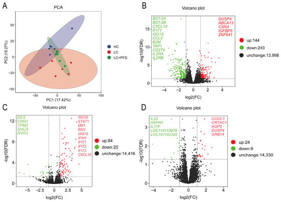
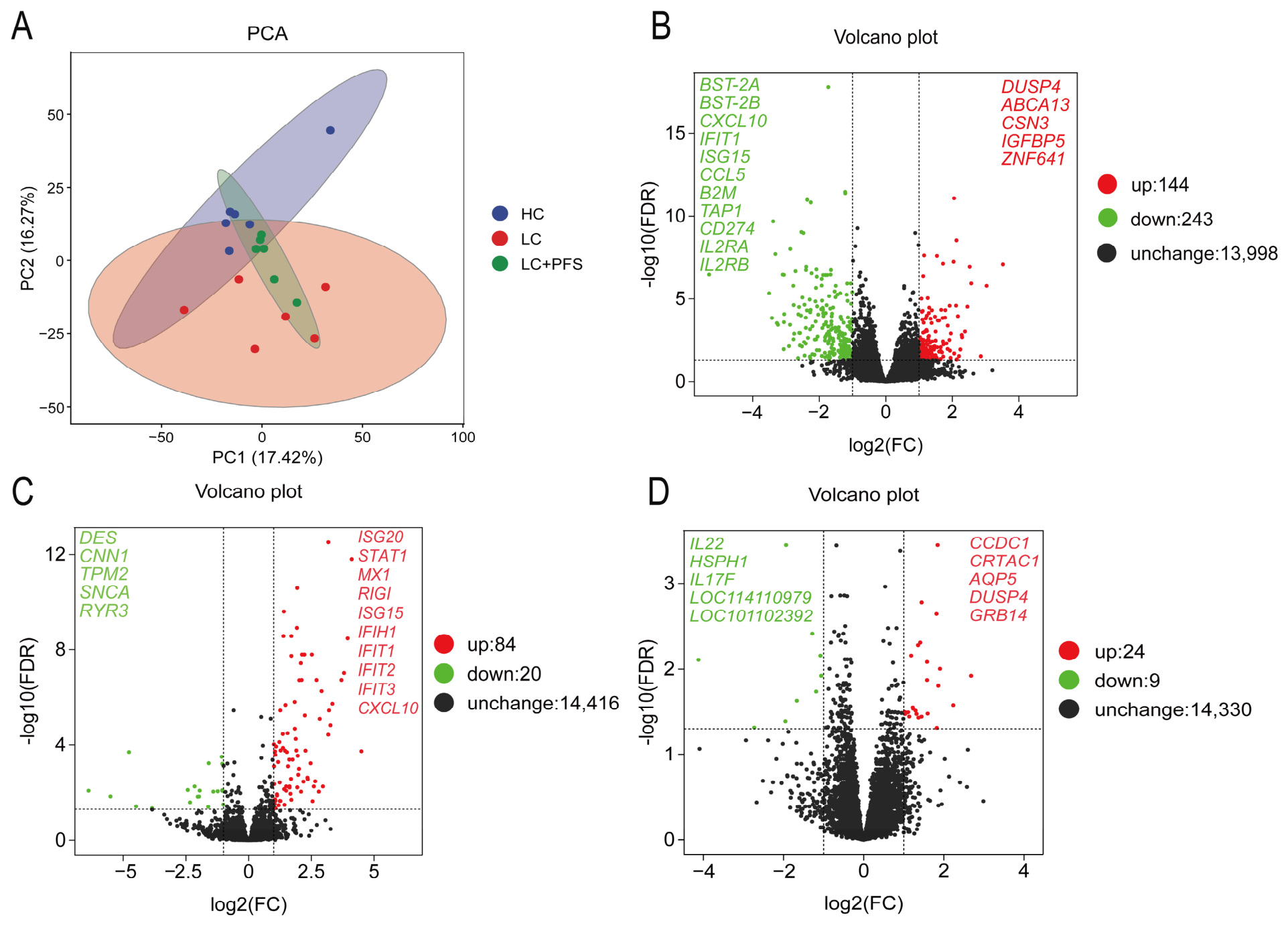
To assess the global similarity of transcriptomic profiles across dietary treatments, PCA was performed on the gene expression matrix of ruminal epithelial tissues (Figure 1A). The first two principal components (PC1 and PC2) explained 17.42% and 16.27% of the total variance, respectively, with a cumulative variance of 33.69%. Clear separation was observed between the HC group and the LC group, indicating a distinct transcriptomic response of the ruminal epithelium to high-concentrate feeding. Notably, the transcriptomic profiles of the LC + PFS group clustered closely with the LC group, forming a compact and distinct subgroup separate from the HC group. This pattern suggests that dietary PFS supplementation under low-concentrate conditions did not induce large-scale transcriptomic shifts relative to the LC baseline.
Figure 1.
Transcriptomic analysis of rumen papilla. (A) PCA of LC, HC, and LC + PFS groups after log2(FPKM + 1) transformation and mean-centering (PC1: 17.42%, PC2: 16.27%; cumulative: 33.69%). The ellipses represent the 95% confidence intervals for each group. (B–D) Volcano plots of DEGs in LC vs. HC (B), HC vs. LC + PFS (C), and LC vs. LC + PFS (D). The colored ellipses in the PCA plot represent the 95% confidence intervals for each dietary group. In the volcano plots, the vertical and horizontal dashed lines indicate the thresholds of |log2FC| = 1 and FDR = 0.05, respectively. Red dots: upregulated genes; green dots: downregulated genes; black dots: genes with no significant differential expression. Significance threshold: |log2FC| ≥ 1, FDR < 0.05.
3.1.2. Differentially Expressed Genes
A total of 387 DEGs were identified between the LC and HC groups, including 144 upregulated and 243 downregulated genes in the HC group (Figure 1B). Genes associated with cell adhesion and immune function, such as CXCL10, IFIT1, and ISG15, were significantly downregulated in the HC group, while genes related to cellular stress and metabolic adaptation, including DUSP4, ABCA13, and ZNF641, were upregulated. These expression changes indicate that high-concentrate feeding broadly suppressed immune-related transcriptional programs while triggering stress-responsive gene expression in the ruminal epithelium, establishing a background of diminished immune surveillance against which PFS effects could be evaluated.
In the HC vs. LC + PFS comparison, 104 DEGs were identified, with 84 upregulated and 20 downregulated genes in the LC + PFS group relative to HC (Figure 1C). Notably, multiple interferon-stimulated genes, such as ISG20, STAT1, MX1, RIGI (DDX58), and IFIT family members (IFIT1, IFIT2, IFIT3), were significantly upregulated in the LC + PFS group. In contrast, genes such as DES, CNN1, TPM2, SNCA, and RYR3 were downregulated in the LC + PFS group. These transcriptomic differences suggest that the LC + PFS regimen is associated with enhanced interferon-mediated immune signaling relative to the HC-induced suppressed state. A total of 33 differentially expressed genes were identified between the LC group and the LC + PFS group, 24 upregulated and 9 downregulated in LC + PFS group compared to LC group. Upregulated genes included DUSP4, GRB14, AQP5, and KRT35, while downregulated genes included NewGene_9611 and NewGene_6843 (Figure 1D). The limited transcriptomic alterations between the two groups indicated that adding Perilla frutescens seeds to the low-concentrate diet did not trigger large-scale gene expression reprogramming in ruminal epithelial cells.
3.2. Effects of Dietary Treatments on Signaling Pathways in the Ruminal Epithelium
Functional annotation and pathway enrichment analyses were performed to characterize the biological functions of DEGs across three pairwise comparisons. KEGG enrichment bubble plots were generated to visualize significantly enriched pathways, and key immune-related gene sets were further validated by GSEA.
3.2.1. LC vs. HC Comparison
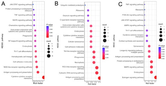
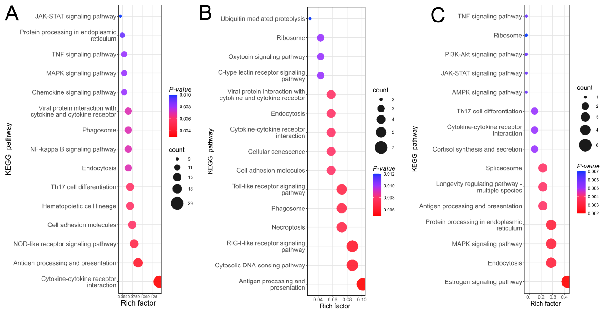
In the LC vs. HC comparison, KEGG pathway enrichment revealed that DEGs were mainly involved in immune regulation, with the most prominent pathways being antigen processing and presentation, cytokine–cytokine receptor interaction, TNF signaling, NF-κB signaling, and Th17 cell differentiation (Figure 2A). Notably, the HC group did not exhibit a classical transcriptional signature of widespread inflammation. Instead, multiple pathways related to antigen recognition and presentation were suppressed, indicating that high-concentrate feeding primarily impaired the ruminal epithelium’s local immune surveillance rather than triggering overt inflammatory activation. These findings establish the HC condition as a state of diminished immune surveillance, providing the reference context for evaluating PFS effects.
Figure 2.
KEGG enrichment of DEGs. (A–C) KEGG bubble plots for LC vs. HC, HC vs. LC + PFS and LC vs. LC + PFS, respectively.
3.2.2. HC vs. LC + PFS Comparison
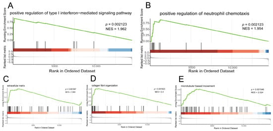
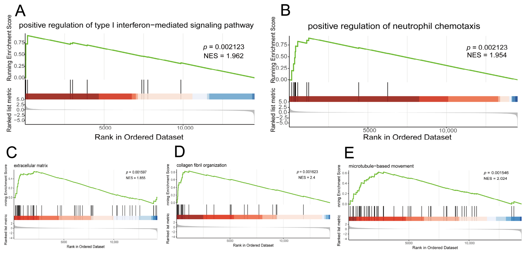
For the HC vs. LC + PFS comparison, KEGG enrichment analysis showed that DEGs were predominantly enriched in innate immune and pathogen-recognition pathways, including antigen processing and presentation, cytosolic DNA-sensing, RIG-I-like receptor signaling, and Toll-like receptor signaling (Figure 2B). To further validate these pathway-level changes, GSEA was performed on the whole transcriptome. The results confirmed significant upregulation of gene sets related to type I interferon-mediated signaling (Figure 3A) and neutrophil chemotaxis (Figure 3B) in the PFS group, with normalized enrichment scores (NES) of 1.96 and 1.95, respectively (p < 0.01). These patterns suggest that the LC + PFS regimen is associated with enhanced interferon-related immune surveillance relative to the HC-induced suppressed state.
Figure 3.
GSEA plots of pathway enrichment. (A,B) Type I interferon signaling (A) and neutrophil chemotaxis (B) in HC vs. LC + PFS. (C–E) Extracellular matrix organization (C), collagen fibril organization (D), and microtubule-based movement (E) in LC vs. LC + PFS. The green curve represents the running enrichment score, the black vertical bars indicate the positions of pathway genes in the ranked gene list, and the red-to-blue gradient represents positive-to-negative ranking metric values. Significance: NES > 1.5, FDR < 0.05.
3.2.3. LC vs. LC + PFS Comparison
In the LC vs. LC + PFS comparison, the number of DEGs was small, and KEGG enrichment revealed no strong immune-related signals. Instead, pathways such as estrogen signaling, endocytosis, MAPK and PI3K-Akt signaling, and protein processing in the endoplasmic reticulum were enriched (Figure 2C). GSEA further revealed subtle but coordinated enrichment of gene sets related to extracellular matrix organization (GO_0030198), collagen fibril organization (GO_0030199), and microtubule-based movement (GO_0007018) (Figure 3C–E). Collectively, these pathway-level shifts were mild and primarily involved extracellular matrix remodeling and cytoskeletal dynamics, with no significant enrichment of immune-related pathways detected (FDR < 0.05). This pattern indicates that PFS supplementation under physiological conditions was associated with subtle fine-tuning of epithelial structural homeostasis, rather than activation of a coordinated transcriptional immune program.
3.3. WGCNA Identified Treatment-Associated Immune Modules in the Ruminal Epithelium
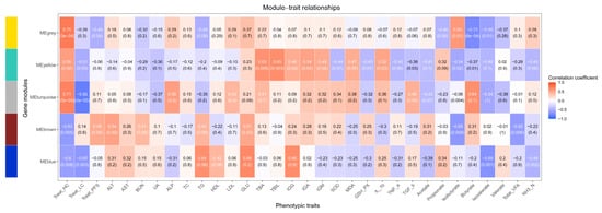
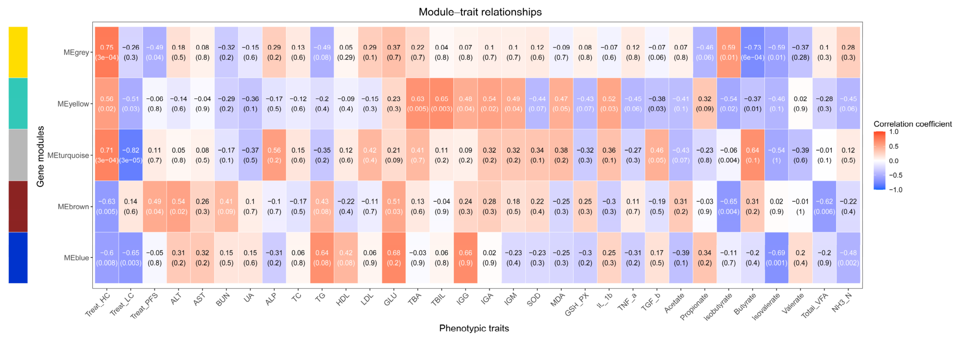
To identify co-expressed gene programs associated with PFS supplementation and to characterize their functional roles, we performed WGCNA on the 5000 most variable genes. This analysis produced several co-expression modules, of which the blue, brown, turquoise, and yellow modules showed the strongest correlations with treatments, blood parameters, and rumen fermentation traits (Figure 4). These modules displayed distinct association patterns, indicating functionally segregated gene programs that couple epithelial transcription to systemic metabolism and local immunity. Among these, the brown module was of particular interest as it was specifically associated with PFS supplementation.
Figure 4.
Correlation heatmap of WGCNA module eigengenes and phenotypic indicators. Correlation coefficients and p-values are displayed for the grey, yellow, turquoise, brown, and blue modules against dietary groups, rumen fermentation traits and blood parameters. The left color strip indicates module identity, while red and blue represent positive and negative correlations.
3.3.1. The Turquoise Module: A High-Concentrate-Associated Metabolic and Anti-Inflammatory Signature
The turquoise module correlated most strongly with HC (r = 0.71) and negatively with LC (r = −0.82), marking it as the transcriptomic signature most robustly induced by high-grain feeding. It also correlated with alkaline phosphatase (ALP) (r = 0.56) and propionate (r = 0.64), a key gluconeogenic precursor.
Immunologically, this module showed an anti-inflammatory and antioxidant pattern: positive correlations with IL-10 (r = 0.46), SOD (r = 0.38), and GSH-Px (r = 0.36), and negative correlations with TNF-α (r = −0.43) and MDA (r = −0.32). These results indicate that the turquoise module reflects an adaptive metabolic and redox-protective program co-regulated by propionate and other fermentation products, rather than a classical inflammatory response. Genes assigned to this module—S100A8, S100A9, HSPA8, CCL20, CXCL8, and NOD2—are typically linked to epithelial stress surveillance and homeostatic immune regulation, supporting this interpretation.
3.3.2. The Yellow Module: A Coordinated Antioxidant and Tissue-Repair Module
The yellow module showed a similar but weaker treatment correlation pattern (HC: r = 0.56; LC: r = −0.51), yet its physiological correlations were more strongly polarized toward antioxidant and repair phenotypes. It showed the strongest positive correlations with SOD (r = 0.49), GSH-Px (r = 0.47), IL-10 (r = 0.52), and TGF-β (r = 0.38), and the strongest negative correlations with MDA (r = −0.44) and TNF-α (r = −0.45). It also correlated positively with immunoglobulins (IgG: r = 0.48, IgA: r = 0.40, IgM: r = 0.54). Module genes included structural components such as POSTN, ACTG2, CNN1, TPM2, DES, and DAAM2, which are involved in tissue remodeling and cytoskeletal dynamics. The convergence of antioxidant, anti-inflammatory, and tissue-repair signatures implies that the yellow module represents an epithelial cytoprotective and remodeling program upregulated under HC, potentially as a compensatory response to mechanical or metabolic stress.
3.3.3. The Blue Module: A Low-Concentrate-Associated Immune Surveillance Program
In contrast to the turquoise and yellow modules, the blue module correlated positively with LC (r = 0.65) and negatively with HC (r = −0.60), indicating preferential expression under conventional low-concentrate feeding and suppression under high-grain challenge. It showed strong negative correlations with propionate (r = −0.69), valerate (r = −0.69), and TVFA (r = −0.40), suggesting that elevated VFA production under HC may drive its downregulation.
Immunologically, the blue module exhibited a pro-inflammatory and antigen-presentation profile: positive correlations with TNF-α (r = 0.34) and IL-1β (r = 0.17), and negative correlations with IL-10 (r = −0.39) and TGF-β (r = −0.11). Although modest, these correlations suggest a basal inflammatory-competent state. More importantly, the module was enriched for antigen processing and presentation genes and T-cell immunity markers, including B2M, TAP1, TAP2, CD3E, CD4, CD8A, IL7R, ICAM1, and CXCL13.
3.3.4. The Brown Module: A PFS-Associated Non-Inflammatory Immune Surveillance Network
The brown module displayed a unique association pattern: positive correlation with PFS (r = 0.49, p = 0.04), negative correlation with HC (r = −0.63, p = 0.005), and only a weak correlation with LC (r = 0.14). This pattern indicates that the brown module is specifically activated by PFS supplementation, rather than merely reflecting LC versus HC diet effects.
Physiologically, the brown module correlated positively with alanine aminotransferase (ALT) (r = 0.54, p = 0.02) and negatively with GLU (r = −0.51, p = 0.03), propionate (r = −0.65, p = 0.004), and valerate (r = −0.62, p = 0.006). These associations place module activation within a metabolic context of altered hepatic enzyme activity, reduced circulating glucose, and lower ruminal propionate, potentially reflecting a shift in systemic energy partitioning.
Functionally, the brown module was enriched for genes involved in cytosolic nucleic acid sensing, type I interferon signaling, and antiviral innate immunity. Hub genes included pattern recognition receptors IFIH1 (MDA5) and DDX58 (RIG-I); the regulatory adaptor DHX58 (LGP2); transcription factors IRF7, IRF9, and STAT1; and interferon-stimulated genes ISG15, MX1, MX2, IFIT1, IFIT2, IFIT3, and OAS family members. This gene set covered components of the RLR-IRF-ISG signaling cascade, including pattern recognition receptors (IFIH1 and DDX58), a regulatory adaptor (DHX58), transcription factors (IRF7, IRF9, and STAT1), and interferon-stimulated effectors (ISG15, MX1, MX2, IFIT1, IFIT2, IFIT3, and OAS family members). The module showed no significant correlation with classical inflammatory cytokines (IL-1β, TNF-α) or immunoglobulins.
3.4. VFA-Gene Association Network
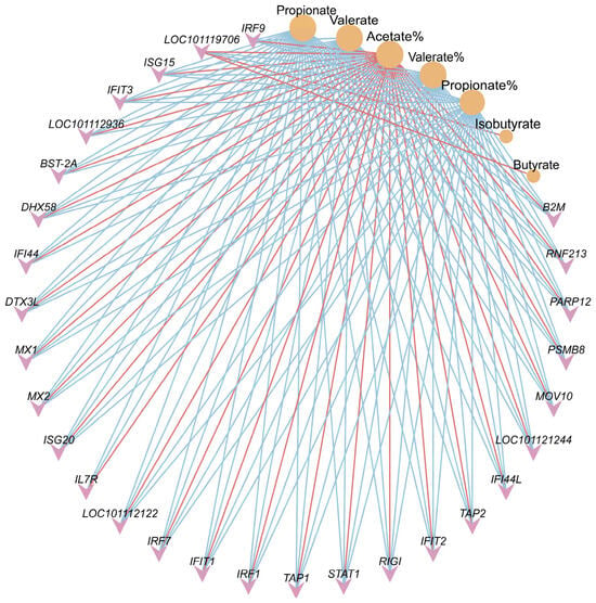
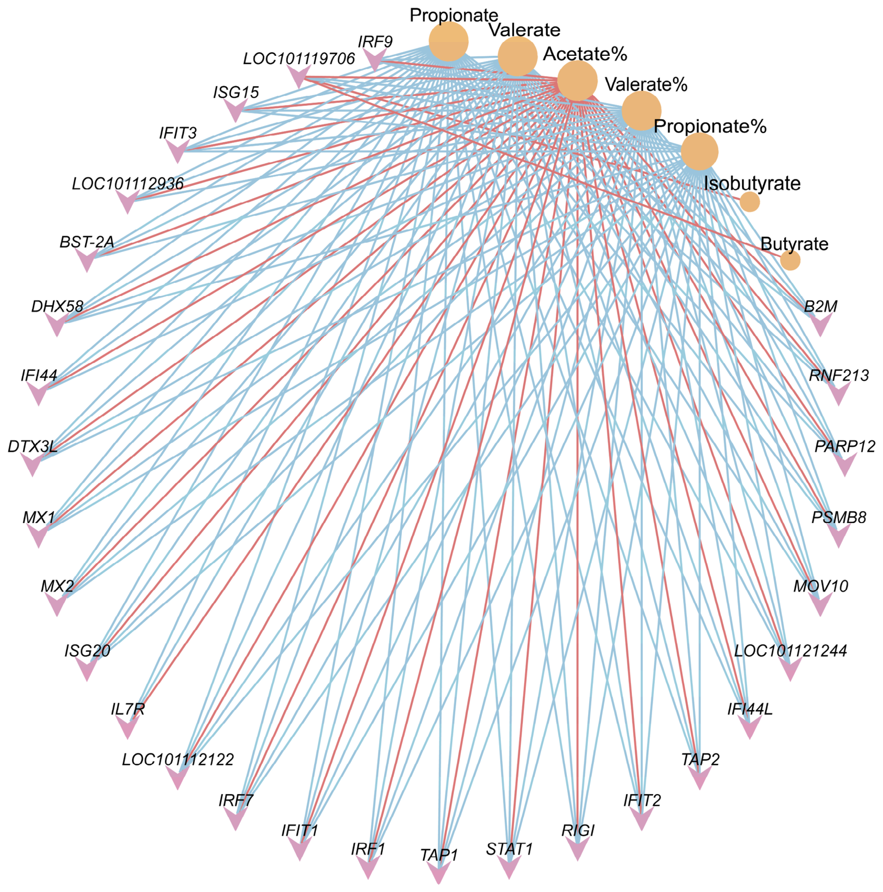
A VFA-gene association network based on Spearman correlation analysis. Only correlation pairs with |r| ≥ 0.5 and FDR < 0.05 were retained for network construction (Figure 5).
Figure 5.
VFA-gene association network based on Spearman correlation analysis. Red edges indicate positive correlations; blue edges indicate negative correlations. Only correlation pairs with FDR < 0.05 are shown. Pink inverted triangles represent genes, orange circles represent VFAs, and circle size reflects VFA connectivity (degree centrality). Propionate and valerate are highlighted as the central VFA nodes.
The network revealed that ruminal propionate and valerate were the primary VFA nodes connected to multiple immune-related genes. Specifically, propionate and valerate showed negative correlations with a cluster of interferon-stimulated genes, including IFI44, IFIT1, IFIT2, IFIT3, IRF7, ISG15, ISG20, MX1, MX2, RIGI, STAT1, DHX58, BST-2A, and MOV10. Additionally, genes involved in antigen processing and presentation—B2M, TAP1, TAP2, and PSMB8—as well as IL7R and DTX3L, were also negatively correlated with propionate and valerate.
In contrast, isobutyrate, butyrate, and acetate showed fewer and weaker connections to these immune-related genes. This pattern suggests that propionate and valerate are the dominant VFA species associated with the transcriptional immune signature in the ruminal epithelium, and that elevated concentrations of these VFAs may correlate with reduced expression of immune surveillance genes.
4. Discussion
The ruminal epithelium functions as a physical barrier and core mucosal immune interface, linking ruminant hosts with their ruminal microbiota. Its structural integrity and physiological homeostasis directly influence production efficiency and livestock health [1]. In the present study, we systematically characterized the transcriptomic landscape of the sheep ruminal epithelium across three dietary conditions, with a focus on understanding how PFS modulates epithelial function. The HC group exhibited a transcriptional state characterized by diminished expression of immune-related genes, which served as a reference framework for evaluating the LC + PFS-associated immune signature. Our results suggest that PFS is not a conventional immune activator, but rather a potential homeostatic modulator that fine-tunes metabolic pathways under physiological conditions and is transcriptionally associated with enhanced immune surveillance capacity relative to an HC-induced suppressed state.
Only 33 differentially expressed genes were identified between the LC and LC + PFS groups, and the enriched pathways contained relatively small gene sets. This limited transcriptional response is itself informative: biologically relevant functions can be enriched even with a modest number of DEGs, and the cumulative effect of small-magnitude changes can potentially have significant physiological consequences [12,13]. The modest number of DEGs indicates that PFS supplementation under physiological conditions does not trigger large-scale transcriptional reprogramming. Instead, the identity of the upregulated genes points to a targeted mode of action through key signaling checkpoints. DUSP4 encodes a dual-specificity phosphatase (MKP-2) that functions as a major negative regulator of MAPK signaling by dephosphorylating ERK and JNK, forming a transcriptional negative feedback loop that constrains stress and inflammatory signals [14,15]. GRB14 encodes an adaptor protein that acts as a negative regulator of insulin receptor signaling and has been shown to regulate hepatic lipogenesis through the p62/Nrf2 pathway [16,17,18,19]. As upstream regulatory nodes, even subtle changes in the expression of these genes can modulate downstream pathway activity without manifesting as broad transcriptional shifts. This pattern is consistent with the interpretation that PFS may act through targeted modulation of specific signaling checkpoints rather than through broad transcriptional activation.
The HC vs. LC + PFS comparison revealed marked upregulation of interferon-stimulated genes and enrichment of RIG-I-like receptor and type I interferon pathways. The brown module—specifically associated with LC + PFS—was enriched for genes spanning the RLR-IRF-ISG cascade. IFIH1 (MDA5) and DDX58 (RIG-I) encode cytosolic pattern recognition receptors that detect viral nucleic acids—the “sentinels” that initiate the innate immune response [20,21]. Their presence in the brown module suggests that PFS is associated with enhanced capacity to detect pathogenic threats. Downstream, IRF7 functions as a master transcriptional regulator of type I interferon-dependent immune responses, playing a critical role in the initiation and amplification of type I IFN responses [22,23]. IRF9 serves as an essential component of the ISGF3 transcription complex that, together with STAT1 and STAT2, binds to the IFN-stimulated response element (ISRE) to activate the transcription of interferon-stimulated genes, driving the cell into an antiviral state [24,25]. STAT1 is a signal transducer and activator of transcription that mediates cellular responses to interferons and plays an essential role in mediating responses to all types of interferons [26,27]. IFIT2 and IFIT3 form a stable heterodimeric complex that facilitates recognition and translation inhibition of viral mRNAs [28]. The coordinated enrichment of these genes, spanning from receptor to transcription factor to effector, indicates enhanced immune surveillance capacity across the entire RLR-IRF-ISG cascade in the LC + PFS group relative to HC. Importantly, the ruminal epithelium is intrinsically equipped with these innate immune sensing pathways. Transcriptomic profiling of bovine rumen epithelium revealed significant enrichment of interferon signaling pathways in differentially expressed gene sets, with many genes in these pathways downregulated in animals with liver abscesses, confirming that interferon-related transcriptional machinery is functionally present in ruminal epithelial tissue [29]. Moreover, the RIG-I-like receptor signaling pathway has been identified as one of the signaling pathways involved in rumen epithelium growth and host-microbiome crosstalk in a lamb model [30]. In sheep ruminal epithelial cells, transcriptomic analyses have demonstrated that the innate immune system actively responds to microbial challenges and plays a critical role in sustaining the balance between immune tolerance and inflammatory responses [31]. Collectively, these observations indicate that ruminal epithelial tissue is equipped with interferon-related transcriptional machinery, providing a molecular context for the interferon-associated gene expression patterns observed in the present study.
Beyond the direct comparison findings, our VFA-gene correlation network analysis revealed that ruminal propionate and valerate were negatively correlated with multiple interferon signaling and antigen presentation genes. This observation raises an important physiological consideration. Propionate is a major gluconeogenic precursor in ruminants, and its concentration reflects the extent of carbohydrate fermentation in the rumen. The inverse relationship between propionate/valerate and immune-related genes observed in our study is consistent with previous reports that changes in rumen microbial composition and epithelial TLR signaling are associated with the maintenance of rumen ecosystem stability. Moreover, propionate has been shown to exert anti-inflammatory effects in bovine rumen epithelial cells by suppressing NF-κB and MAPK signaling pathways [32,33]. Extrapolating from these findings, elevated VFA production—characteristic of high-concentrate feeding—may contribute to the suppression of epithelial immune surveillance, although this remains a hypothesis. This interpretation is consistent with the downregulation of antigen presentation and interferon pathway genes observed in the HC group, and is further supported by the module-trait correlations showing that the blue module (an LC-associated immune surveillance program) was negatively correlated with propionate and valerate. From a physiological perspective, this pattern may reflect a trade-off between metabolic and immune functions in the ruminal epithelium: high fermentative activity (elevated VFA) is associated with reduced expression of immune surveillance genes, whereas lower VFA production under low-concentrate feeding is associated with the maintenance of a relatively more active immune transcriptional program. However, these correlations do not establish causality, and the mechanistic basis of this metabolic-immune interplay warrants further investigation.
A key question is how PFS can be associated with enhanced epithelial immune surveillance despite reduced systemic IL-1β and TNF-α. The brown module showed no significant correlation with serum IL-1β or TNF-α levels, indicating that the interferon-related transcriptional upregulation in the epithelium is independent of systemic inflammatory markers. This dissociation can be explained by the differential engagement of two signaling arms downstream of RLR/MAVS: TBK1/IKKε → IRF3/7 (interferon axis) versus IKK → NF-κB (inflammatory axis). Importantly, Perilla frutescens compounds are known to suppress NF-κB activation. Thus, PFS may preferentially activate the interferon axis while restraining NF-κB-driven inflammation [34,35,36]. However, as MAVS was not directly detected as differentially expressed in our dataset, this mechanistic interpretation remains a hypothesis and requires functional validation.
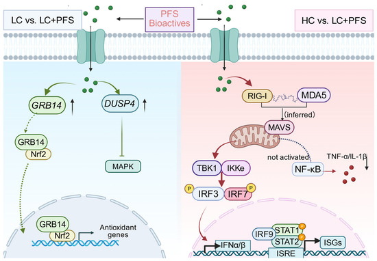
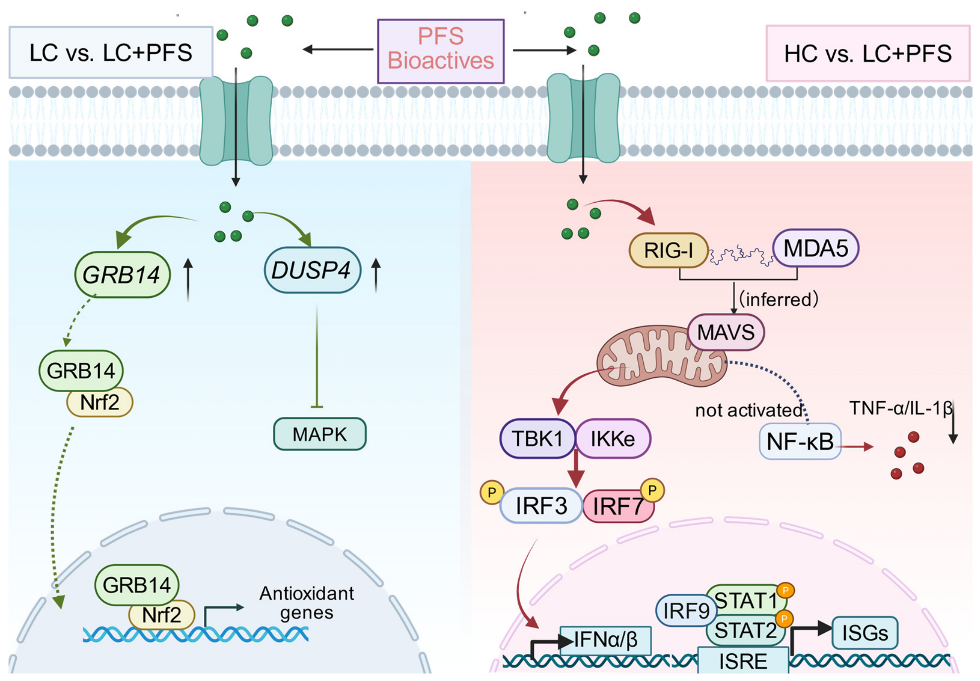
The high content of α-linolenic acid and bioactive compounds in Perilla frutescens seeds is presumed to be the main active substance. Relevant literature has shown that n-3 polyunsaturated fatty acids regulate immune cell activity via the GPR120-NLRP3 pathway [37]. The results from the current study are consistent with our previous findings of Perilla frutescens seeds in regulating lipid metabolism, antioxidant status, and immune function through the microbiota–gut–liver–muscle axis [6]. Collectively, these transcriptomic associations are consistent with a homeostatic modulatory role for PFS in ruminal epithelial immune function (Figure 6).
Figure 6.
Proposed model of PFS-associated ruminal epithelial transcriptional alterations. Left (LC vs. LC + PFS): PFS supplementation is associated with upregulation of DUSP4 and GRB14, which negatively modulate MAPK and insulin signaling. GRB14 correlates with systemic antioxidant, anti-inflammatory, and humoral immune parameters. Right (HC vs. LC + PFS): The LC + PFS regimen is associated with upregulation of RLR-IRF-ISG cascade genes (RIG-I, MDA5, IRF7, STAT1/STAT2, IRF9, ISGs). NF-κB is not activated, indicating non-inflammatory immune surveillance. Note: Green dots represent PFS bioactive compounds, while gray elements depict cellular structures shown for context. HC serves as a reference framework; HC vs. LC + PFS is a cross-dietary comparison. Solid arrows indicate transcriptomic associations, whereas dashed lines indicate inferred relationships requiring functional validation.
Several limitations should be acknowledged. First, the experimental design did not include an HC + PFS group. Therefore, although the comparison between the HC and LC + PFS groups revealed transcriptomic differences, it cannot determine whether PFS mitigates changes induced by an HC diet. Because these groups differed in both dietary concentrate level and PFS supplementation, the observed differences cannot be attributed solely to PFS. Second, only one PFS inclusion rate (3%) was evaluated. This design does not allow us to identify the dose that provides the best balance between biological effectiveness and economic feasibility. Further dose–response studies are therefore needed to determine the most cost-effective supplementation strategy. Third, transcriptomic associations alone cannot establish causal relationships or confirm the molecular mechanisms underlying the observed PFS-associated changes. Targeted functional studies, including in vitro epithelial cell models and gene-manipulation experiments, are needed to validate the biological roles of the identified transcriptional changes. Addressing these limitations will help clarify how PFS acts and support the development of practical feeding strategies.
5. Conclusions
This study provides transcriptomic evidence that dietary Perilla frutescens seeds are associated with context-dependent alterations in the sheep ruminal epithelial transcriptome. In the direct comparison between LC and LC + PFS, PFS supplementation was associated with targeted changes in metabolic pathways without widespread immune activation. In the cross-dietary comparison between HC and LC + PFS, the LC + PFS regimen was associated with upregulation of interferon-related genes relative to the HC-induced state of diminished immune gene expression. VFA-gene correlation network analysis further revealed that ruminal propionate and valerate were negatively correlated with multiple interferon signaling and antigen presentation genes, suggesting a potential metabolic-immune interplay in the ruminal epithelium that may be modulated by dietary PFS supplementation. These transcriptomic associations support the potential role of PFS as a modulator of ruminal epithelial immune function, but causal relationships and the underlying mechanisms require confirmation through targeted functional experiments.
Author Contributions
X.Z. and Y.C.: Writing—review & editing, Writing—original draft, Supervision, Conceptualization, Formal analysis, Visualization, Methodology, Data curation. Y.Z.: Formal analysis, Visualization, Methodology. B.W.: Writing—review & editing, Writing—original draft, Supervision, Investigation, Methodology, Project administration, Funding acquisition, Conceptualization. All authors have read and agreed to the published version of the manuscript.
Funding
This research was funded by China Agriculture Research System (CARS-37) and the Hainan Provincial Natural Science Foundation of China (326YXQN0947, 324QN289).
Institutional Review Board Statement
The animal study protocol was approved by the Animal Care and Use Committee of China Agricultural University (Approval No. AW30901202-1-1 on 12 April 2021).
Informed Consent Statement
Written informed consent was obtained from the owner of the animals involved in this study.
Data Availability Statement
The data presented in this study are openly available in NCBI at https://www.ncbi.nlm.nih.gov/, BioProject PRJNA1503609.
Acknowledgments
The authors thank the staff of Tianyuan Liangzhong Sheep Farm (Wuzhong, Ningxia Hui Autonomous Region, China) for their support during animal handling, slaughter operations, and sample collection. We also thank Beijing Biomarker Technologies Co., Ltd. (Beijing, China) for assisting in RNA-sequencing analysis.
Conflicts of Interest
The authors declare no conflicts of interest.
Abbreviations
The following abbreviations are used in this manuscript:
| ALA | Alpha-linolenic acid |
| ALP | Alkaline phosphatase |
| ALT | Alanine aminotransferase |
| AST | Aspartate aminotransferase |
| BUN | Blood urea nitrogen |
| CLA | Conjugated linoleic acid |
| DEG | Differentially expressed gene |
| DHA | Docosahexaenoic acid |
| DM | Dry matter |
| ELISA | Enzyme-linked immunosorbent assay |
| EPA | Eicosapentaenoic acid |
| FDR | False discovery rate |
| FPKM | Fragments per kilobase of transcript per million mapped reads |
| GLU | Glucose |
| GO | Gene Ontology |
| GSEA | Gene set enrichment analysis |
| GSH-Px | Glutathione peroxidase |
| HDL-C | High-density lipoprotein cholesterol |
| LDL-C | Low-density lipoprotein cholesterol |
| IgA | Immunoglobulin A |
| IgG | Immunoglobulin G |
| IgM | Immunoglobulin M |
| IL-1β | Interleukin-1β |
| IL-10 | Interleukin-10 |
| KEGG | Kyoto Encyclopedia of Genes and Genomes |
| MDA | Malondialdehyde |
| PBS | Phosphate-buffered saline |
| PCA | Principal component analysis |
| PFS | Perilla frutescens seeds |
| PUFA | Polyunsaturated fatty acid |
| SOD | Superoxide dismutase |
| TG | Triglycerides |
| TMR | Total mixed ration |
| TNF-α | Tumor necrosis factor-α |
| VFA | Volatile fatty acid |
| WGCNA | Weighted gene co-expression network analysis |
References
- Xiang, R.; Oddy, V.H.; Archibald, A.L.; Vercoe, P.E.; Dalrymple, B.P. Epithelial, metabolic and innate immunity transcriptomic signatures differentiating the rumen from other sheep and mammalian gastrointestinal tract tissues. PeerJ 2016, 4, e1762. [Google Scholar] [CrossRef] [Scilit] [PubMed]
- Pokhrel, B.; Jiang, H. Postnatal Growth and Development of the Rumen: Integrating Physiological and Molecular Insights. Biology 2024, 13, 269. [Google Scholar] [CrossRef] [Scilit] [PubMed]
- Oh, J.; Wall, E.H.; Bravo, D.M.; Hristov, A.N. Host-mediated effects of phytonutrients in ruminants: A review. J. Dairy Sci. 2017, 100, 5974–5983. [Google Scholar] [CrossRef] [Scilit] [PubMed]
- Ahmed, H.M. Ethnomedicinal, Phytochemical and Pharmacological Investigations of Perilla frutescens (L.) Britt. Molecules 2018, 24, 102. [Google Scholar] [CrossRef] [Scilit] [PubMed]
- Yu, Y.; Zhang, B.; Jiang, X.; Cui, Y.; Luo, H.; Stergiadis, S.; Wang, B. Exploring the metabolomic landscape: Perilla frutescens as a promising enhancer of production, flavor, and nutrition in Tan lamb meat. Meat Sci. 2024, 209, 109419. [Google Scholar] [CrossRef] [Scilit] [PubMed]
- Yu, Y.; Zhang, B.; Jiang, X.; Cui, Y.; Shang, Y.; Jin, Y.; Sun, T.; Yuan, Z.; Zhang, Z.; Luo, H.; et al. Perilla frutescens seeds enhance lamb immunity and antioxidant capacity via the microbiota-gut-liver-muscle axis. J. Anim. Sci. Biotechnol. 2026, 17, 1. [Google Scholar] [CrossRef] [Scilit] [PubMed]
- Beyzi, S.B.; Ulger, I.; Kaya, E.; Ocak, B.; Konca, Y. Effects of Different Technological Forms of the Perilla frutescens in the Diet on Ruminal Fermentation, Milk and Plasma Fatty Acid Composition, Ruminal Biohydrogenation and Milk Quality in Dairy Goats. Vet. Med. Sci. 2024, 10, e70087. [Google Scholar] [CrossRef] [Scilit] [PubMed]
- Hu, P.; Zhang, B.; Fatima, Z.; Liu, Y.; Li, L.; Ai, P.; Yang, X.; Li, L.; Chang, J. Physicochemical characterization, lipid-metabolism effects and transcriptomics insights of perilla seed oil in high-fat-diet induced rats. J. Ethnopharmacol. 2026, 363, 121442. [Google Scholar] [CrossRef] [Scilit] [PubMed]
- Wang, J.; Liu, M.; Wu, Y.; Wang, L.; Liu, J.; Jiang, L.; Yu, Z. Medicinal herbs as a potential strategy to decrease methane production by rumen microbiota: A systematic evaluation with a focus on Perilla frutescens seed extract. Appl. Microbiol. Biotechnol. 2016, 100, 9757–9771. [Google Scholar] [CrossRef] [Scilit] [PubMed]
- Cao, Y.C.; Yang, H.-J. Ruminal digestibility and fermentation characteristics in vitro of fenugreek and alfalfa hay combination with or without the inoculation of Neocallimastix sp. YAK11. Anim. Feed Sci. Technol. 2011, 169, 53–60. [Google Scholar] [CrossRef] [Scilit]
- Broderick, G.A.; Kang, J.H. Automated simultaneous determination of ammonia and total amino acids in ruminal fluid and in vitro media. J. Dairy Sci. 1980, 63, 64–75. [Google Scholar] [CrossRef] [Scilit] [PubMed]
- Maquat, L.E. The amazing web of post-transcriptional gene control: The sum of small changes can make for significant consequences. RNA 2015, 21, 488–489. [Google Scholar] [CrossRef] [Scilit] [PubMed]
- St Laurent, G.; Shtokalo, D.; Tackett, M.R.; Yang, Z.; Vyatkin, Y.; Milos, P.M.; Seilheimer, B.; McCaffrey, T.A.; Kapranov, P. On the importance of small changes in RNA expression. Methods 2013, 63, 18–24. [Google Scholar] [CrossRef] [Scilit] [PubMed]
- Jiao, H.; James, S.J.; Png, C.W.; Cui, C.; Li, H.; Li, L.; Chia, W.N.; Min, N.; Li, W.; Claser, C.; et al. DUSP4 modulates RIG-I- and STING-mediated IRF3-type I IFN response. Cell Death Differ. 2024, 31, 280–291. [Google Scholar] [CrossRef] [Scilit] [PubMed]
- Neamatallah, T.; Jabbar, S.; Tate, R.; Schroeder, J.; Shweash, M.; Alexander, J.; Plevin, R. Whole Genome Microarray Analysis of DUSP4-Deletion Reveals A Novel Role for MAP Kinase Phosphatase-2 (MKP-2) in Macrophage Gene Expression and Function. Int. J. Mol. Sci. 2019, 20, 3434. [Google Scholar] [CrossRef] [Scilit] [PubMed]
- Kopp, K.; Buntru, A.; Pils, S.; Zimmermann, T.; Frank, R.; Zumbusch, A.; Hauck, C.R. Grb14 is a negative regulator of CEACAM3-mediated phagocytosis of pathogenic bacteria. J. Biol. Chem. 2012, 287, 39158–39170. [Google Scholar] [CrossRef] [Scilit] [PubMed]
- Daly, R.J.; Sanderson, G.M.; Janes, P.W.; Sutherland, R.L. Cloning and characterization of GRB14, a novel member of the GRB7 gene family. J. Biol. Chem. 1996, 271, 12502–12510. [Google Scholar] [CrossRef] [Scilit] [PubMed]
- Popineau, L.; Morzyglod, L.; Carre, N.; Cauzac, M.; Bossard, P.; Prip-Buus, C.; Lenoir, V.; Ragazzon, B.; Fauveau, V.; Robert, L.; et al. Novel Grb14-Mediated Cross Talk between Insulin and p62/Nrf2 Pathways Regulates Liver Lipogenesis and Selective Insulin Resistance. Mol. Cell. Biol. 2016, 36, 2168–2181. [Google Scholar] [CrossRef] [Scilit] [PubMed]
- Carre, N.; Cauzac, M.; Girard, J.; Burnol, A.F. Dual effect of the adapter growth factor receptor-bound protein 14 (grb14) on insulin action in primary hepatocytes. Endocrinology 2008, 149, 3109–3117. [Google Scholar] [CrossRef] [Scilit] [PubMed]
- Solotchi, M.; Patel, S.S. Proofreading mechanisms of the innate immune receptor RIG-I: Distinguishing self and viral RNA. Biochem. Soc. Trans. 2024, 52, 1131–1148. [Google Scholar] [CrossRef] [Scilit] [PubMed]
- van Huizen, M.; Gack, M.U. The RIG-I-like receptor family of immune proteins. Mol. Cell 2025, 85, 3793–3806. [Google Scholar] [CrossRef] [Scilit] [PubMed]
- Gu, L.; Casserly, D.; Brady, G.; Carpenter, S.; Bracken, A.P.; Fitzgerald, K.A.; Unterholzner, L.; Bowie, A.G. Myeloid cell nuclear differentiation antigen controls the pathogen-stimulated type I interferon cascade in human monocytes by transcriptional regulation of IRF7. Nat. Commun. 2022, 13, 14. [Google Scholar] [CrossRef] [Scilit] [PubMed]
- Honda, K.; Yanai, H.; Negishi, H.; Asagiri, M.; Sato, M.; Mizutani, T.; Shimada, N.; Ohba, Y.; Takaoka, A.; Yoshida, N.; et al. IRF-7 is the master regulator of type-I interferon-dependent immune responses. Nature 2005, 434, 772–777. [Google Scholar] [CrossRef] [Scilit] [PubMed]
- Muhammad, I.; Contes, K.; Bility, M.T.; Tang, Q. Chasing Virus Replication and Infection: PAMP-PRR Interaction Drives Type I Interferon Production, Which in Turn Activates ISG Expression and ISGylation. Viruses 2025, 17, 528. [Google Scholar] [CrossRef] [Scilit] [PubMed]
- Ivashkiv, L.B.; Donlin, L.T. Regulation of type I interferon responses. Nat. Rev. Immunol. 2014, 14, 36–49. [Google Scholar] [CrossRef] [Scilit] [PubMed]
- Shi, T.; Jiang, W.; Li, R.; Liu, H.; Shao, S.; An, H.; Ma, X. Hippo kinase NDR1 crosstalks with GSK3 and positively regulates type I interferon-mediated antiviral innate immunity. Mol. Immunol. 2026, 192, 13–20. [Google Scholar] [CrossRef] [Scilit] [PubMed]
- Platanias, L.C. Mechanisms of type-I- and type-II-interferon-mediated signalling. Nat. Rev. Immunol. 2005, 5, 375–386. [Google Scholar] [CrossRef] [Scilit] [PubMed]
- Glasner, D.R.; Todd, C.; Cook, B.; D’Urso, A.; Khosla, S.; Estrada, E.; Wagner, J.D.; Bartels, M.D.; Ford, P.; Prych, J.; et al. Short 5’ UTRs serve as a marker for viral mRNA translation inhibition by the IFIT2-IFIT3 antiviral complex. bioRxiv 2025. [Google Scholar] [CrossRef] [Scilit] [PubMed]
- Abbas, W.; Keel, B.N.; Kachman, S.D.; Fernando, S.C.; Wells, J.E.; Hales, K.E.; Lindholm-Perry, A.K. Rumen epithelial transcriptome and microbiome profiles of rumen epithelium and contents of beef cattle with and without liver abscesses. J. Anim. Sci. 2020, 98, skaa359. [Google Scholar] [CrossRef] [Scilit] [PubMed]
- Lin, L.; Xie, F.; Sun, D.; Liu, J.; Zhu, W.; Mao, S. Ruminal microbiome-host crosstalk stimulates the development of the ruminal epithelium in a lamb model. Microbiome 2019, 7, 83. [Google Scholar] [CrossRef] [Scilit] [PubMed]
- Yang, C.; Lan, W.; Ye, S.; Zhu, B.; Fu, Z. Transcriptomic Analyses Reveal the Protective Immune Regulation of Conjugated Linoleic Acids in Sheep Ruminal Epithelial Cells. Front. Physiol. 2020, 11, 588082. [Google Scholar] [CrossRef] [Scilit] [PubMed]
- Shen, H.; Chen, Z.; Shen, Z.; Lu, Z. Maintaining stability of the rumen ecosystem is associated with changes of microbial composition and epithelial TLR signaling. Microbiologyopen 2017, 6, e00436. [Google Scholar] [CrossRef] [Scilit] [PubMed]
- Zhao, C.; Yi, F.; Wei, B.; Tan, P.; Huang, Y.; Zeng, F.; Wang, Y.; Xu, C.; Wang, J. Sodium Propionate Relieves LPS-Induced Inflammation by Suppressing the NF-ĸB and MAPK Signaling Pathways in Rumen Epithelial Cells of Holstein Cows. Toxins 2023, 15, 438. [Google Scholar] [CrossRef] [Scilit] [PubMed]
- Shi, Y.; Yuan, B.; Qi, N.; Zhu, W.; Su, J.; Li, X.; Qi, P.; Zhang, D.; Hou, F. An autoinhibitory mechanism modulates MAVS activity in antiviral innate immune response. Nat. Commun. 2015, 6, 7811. [Google Scholar] [CrossRef] [Scilit] [PubMed]
- Choi, J.; Yoo, E.B.; Park, J.; Fauziah, A.N.; Kim, H.; Oh, M.; Kang, M.; Kim, Y.J.; Kim, J.Y.; Lee, H.J.; et al. Perilla frutescens Britton var. acuta Kudo ameliorates OVA-induced allergic asthma via inhibiting NF-κB and p38 signaling pathways. Food Sci. Biotechnol. 2025, 34, 3669–3679. [Google Scholar] [CrossRef] [Scilit] [PubMed]
- Yang, Z.; Zhang, F.; Yang, S.; Anayyat, U.; Mo, Y.; Ying, Y.; Wang, X. Orally deliverable Perilla frutescens-derived nanovesicles as natural bioactive nanocarriers for colon-targeted colitis therapy via microenvironment reprogramming. Biomater. Adv. 2026, 184, 214832. [Google Scholar] [CrossRef] [Scilit] [PubMed]
- Yan, Y.; Jiang, W.; Spinetti, T.; Tardivel, A.; Castillo, R.; Bourquin, C.; Guarda, G.; Tian, Z.; Tschopp, J.; Zhou, R. Omega-3 fatty acids prevent inflammation and metabolic disorder through inhibition of NLRP3 inflammasome activation. Immunity 2013, 38, 1154–1163. [Google Scholar] [CrossRef] [Scilit] [PubMed]

Disclaimer/Publisher’s Note: The statements, opinions and data contained in all publications are solely those of the individual author(s) and contributor(s) and not of MDPI and/or the editor(s). MDPI and/or the editor(s) disclaim responsibility for any injury to people or property resulting from any ideas, methods, instructions or products referred to in the content. |
© 2026 by the authors. Licensee MDPI, Basel, Switzerland. This article is an open access article distributed under the terms and conditions of the Creative Commons Attribution (CC BY) license.