


Abstract
Antimicrobial nanomaterials have attracted increasing attention as potential alternatives for controlling pathogenic microorganisms. In this study, pure ZnO and Ag-modified ZnO nanoparticles prepared using nominal Ag contents of 1 and 5 wt.% were synthesized using a reflux-assisted polyol method and evaluated to determine the influence of Ag content on their structural characteristics and antibacterial activity. The synthesized materials were characterized by UV-Vis spectroscopy, Fourier-transform infrared spectroscopy (FTIR), X-ray diffraction (XRD), and high-resolution transmission electron microscopy (HRTEM). UV-Vis and FTIR analyses confirmed the characteristic optical response and chemical features of ZnO-based materials. XRD patterns revealed that all samples retained the hexagonal wurtzite structure of ZnO, while additional reflections corresponding to face-centered cubic (FCC) Ag were observed in the Ag-containing samples and increased in intensity with Ag content. Crystallite sizes estimated by the Scherrer equation were 16.9 ± 2.6 nm for ZnO, 12.9 ± 1.6 nm for ZnO-Ag 1%, and 32.6 ± 10.1 nm for ZnO-Ag 5%. HRTEM confirmed the formation of crystalline nanoparticles with average particle sizes of approximately 16 nm and 12 nm for ZnO and ZnO-Ag 1%, respectively. Antimicrobial activity was evaluated against the reference strains Escherichia coli ATCC 25922 and Salmonella typhimurium ATCC 14020. ZnO–Ag 5% exhibited the greatest antibacterial activity, with minimum inhibitory concentration (MIC) values of 250 ppm against E. coli and 750 ppm against S. typhimurium, and minimum bactericidal concentration (MBC) values of 750 and 1500 ppm, respectively. These findings demonstrate that increasing Ag content influences the structural properties of ZnO nanoparticles and enhances their antibacterial performance, highlighting the potential of ZnO-Ag nanomaterials for antimicrobial applications.
1. Introduction
Foodborne and waterborne bacterial pathogens are major public health concerns worldwide, causing millions of infections annually and generating significant economic losses from healthcare costs, food recalls, and reduced productivity [1,2]. Among the microorganisms frequently associated with food contamination, Escherichia coli and Salmonella typhimurium are of particular importance because of their widespread distribution, ability to survive under diverse environmental conditions, and involvement in numerous outbreaks linked to contaminated food and water sources [3,4,5,6,7,8,9].
The increasing prevalence of antimicrobial resistance has intensified the search for alternative strategies to control pathogenic microorganisms while reducing reliance on conventional antibiotics [10,11,12]. Nanotechnology has emerged as a promising approach to developing advanced antimicrobial materials due to the unique physicochemical properties of nanoparticles, including high surface-area-to-volume ratios, tunable surface chemistry, and enhanced interactions with microbial cells. Metal and metal oxide nanoparticles have attracted considerable attention because they can inhibit microbial growth through multiple mechanisms, including membrane disruption, oxidative stress generation, protein inactivation, and interference with essential cellular processes [11,13,14,15].
Among metal oxide nanomaterials, zinc oxide (ZnO) nanoparticles have been extensively investigated for their chemical stability, relatively low toxicity, cost-effectiveness, and broad-spectrum antimicrobial activity [15,16]. Previous studies have demonstrated that ZnO nanoparticles can inhibit both Gram-positive and Gram-negative bacteria through mechanisms involving the generation of reactive oxygen species (ROS), release of Zn2+ ions, and direct interactions with bacterial cell envelopes. In addition to their antimicrobial properties, ZnO nanomaterials exhibit attractive optical and electronic characteristics that support their use in environmental, biomedical, and food-related applications [17,18,19,20,21].
The incorporation of silver into ZnO nanostructures has emerged as an effective strategy to enhance the antibacterial performance of ZnO nanoparticles. Experimental studies have demonstrated that Ag-modified ZnO nanoparticles exhibit improved antimicrobial activity compared with pure ZnO due to the combined effects of Ag+ ion release, enhanced reactive oxygen species (ROS) generation, improved charge separation, and stronger interactions with bacterial membranes [10,17,22,23]. However, the magnitude of this enhancement depends on several factors, including the synthesis route, Ag loading, particle size, crystallinity, and the bacterial species evaluated. For example, Ag-doped ZnO nanoparticles synthesized by chemical and green synthetic approaches have shown lower MIC and MBC values against Escherichia coli, Salmonella spp., and Staphylococcus aureus, although considerable variability among studies has been reported because of differences in nanoparticle composition and preparation methods [24,25,26,27].
Despite the growing interest in ZnO–Ag systems, the relationship between silver loading, structural characteristics, and antimicrobial efficacy remains incompletely understood. In particular, determining the amount of silver required to produce meaningful improvements in bactericidal activity is essential for the rational design of nanomaterials intended for antimicrobial applications. Furthermore, correlating structural and optical modifications with biological performance may provide insight into the mechanisms underlying enhanced antibacterial activity [16,28].
Although previous studies have demonstrated the antimicrobial potential of Ag-modified ZnO nanomaterials, relatively few have systematically investigated the influence of nominal Ag loading using the same synthesis method while directly correlating structural, optical, and antibacterial properties against foodborne Gram-negative pathogens. Therefore, the objective of this study was to synthesize pure ZnO and Ag-modified ZnO nanoparticles using a reflux-assisted polyol method with nominal Ag contents of 1 and 5 wt.%, and to evaluate the influence of nominal Ag loading on their physicochemical and antimicrobial properties. All samples were prepared under identical synthesis conditions and characterized by UV–Vis spectroscopy, Fourier-transform infrared spectroscopy (FTIR), X-ray diffraction (XRD), and high-resolution transmission electron microscopy (HRTEM). Their antibacterial activity was assessed against Escherichia coli and Salmonella typhimurium by determining the minimum inhibitory concentration (MIC), minimum bactericidal concentration (MBC), and tolerance (MBC/MIC) values. By directly comparing pure ZnO with Ag-modified ZnO synthesized under identical experimental conditions, this study provides a clearer assessment of the relationship between nominal Ag loading, structural characteristics, and antibacterial performance, contributing to the rational design of ZnO-based antimicrobial nanomaterials.
2. Materials and Methods
2.1. Materials
Zinc acetate dihydrate (Zn(C2H3O2)2·2H2O, 98%, extra pure, MW = 219.50 g mol−1; Acros Organics, Fair Lawn, NJ, USA), sodium hydroxide (NaOH, ACS reagent grade, ≥98%, MW = 40.00 g mol−1; J.T. Avantor, Radnor, PA, USA), polyvinylpyrrolidone (PVP, (C6H9NO)n, MW = 360,000 g mol−1; Thermo Scientific, Ward Hill, MA, USA), ethylene glycol (C2H6O2, MW = 62.07 g mol−1; VWR BDH Chemicals, Radnor, PA, USA), and silver nitrate (AgNO3, ACS reagent, ≥99.9%, MW = 169.87 g mol−1; Sigma-Aldrich, St. Louis, MO, USA) were used in the synthesis of ZnO and Ag-doped ZnO nanoparticles.
The antibacterial activity of the synthesized nanoparticles was evaluated against Salmonella typhimurium (ATCC 14020) and Escherichia coli (ATCC 25922). Tryptic Soy Broth and Mueller–Hinton Broth (Hardy Diagnostics, Santa Maria, CA, USA) were used for bacterial cultivation and antimicrobial susceptibility testing. MacConkey Agar (Sigma-Aldrich, St. Louis, MO, USA) and Salmonella–Shigella Agar (Hardy Diagnostics, Santa Maria, CA, USA) were used as selective media. All reagents were of analytical grade and were used as received without further purification.
2.2. Production of ZnO and ZnO-Ag Nanoparticles
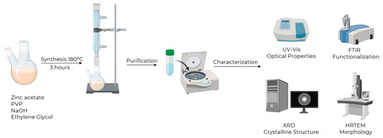
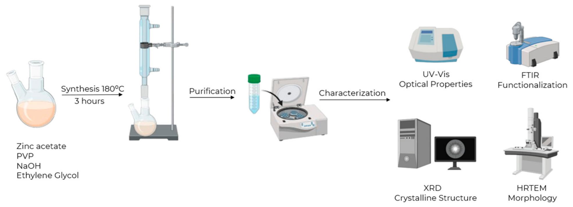
Figure 1 shows the synthesis route for pure and doped ZnO nanoparticles. Pure ZnO nanoparticles were synthesized using a reflux-assisted polyol method. Briefly, 1.0975 g of zinc acetate dihydrate, 0.400 g of sodium hydroxide, and 0.015 g of polyvinylpyrrolidone (PVP) were placed in a two-neck round-bottom flask. Subsequently, 20 mL of ethylene glycol was added, and the mixture was stirred until a homogeneous suspension was obtained.
Figure 1.
Synthesis routes of ZnO, ZnO-Ag 1%, and ZnO-Ag 5% nanoparticles.
The flask was connected to a reflux condenser and heated to approximately 180 °C. The reaction was maintained under reflux for 3 h to allow the formation and growth of ZnO nanoparticles. Upon completion of the reaction, the suspension was allowed to cool naturally to room temperature.
The Ag contents (1 and 5 wt.%) refer to the nominal amounts of silver precursor added during the synthesis. ZnO-Ag nanoparticles containing 1 and 5 wt.% of silver were prepared following the same procedure. The desired amount of silver nitrate was added together with the zinc precursor and other reagents before the addition of ethylene glycol. Specifically, 0.0109 g and 0.0548 g of AgNO3 were used to obtain the 1 wt.% and 5 wt.% solutions, respectively. The resulting mixtures were heated under reflux at approximately 180 °C for 3 h and then cooled to room temperature.
The synthesized nanoparticles were purified by centrifugation at 5000 rpm for 20 min. After the first centrifugation cycle, the supernatant was discarded, and the precipitate was redispersed in deionized water. This washing process was repeated three additional times to remove residual ethylene glycol and unreacted species. Finally, the purified nanoparticles were recovered and used for subsequent physicochemical characterization and antimicrobial activity studies [28].
2.3. Characterization of Nanoparticles
The optical properties of pure ZnO and ZnO–Ag nanoparticles (1 and 5 wt.% Ag) were evaluated by ultraviolet–visible (UV–Vis) spectroscopy using a UV-2700i spectrophotometer (Shimadzu, Columbia, MD, USA). Nanoparticle suspensions were prepared at a concentration of 100 ppm in deionized water. Deionized water was used as the reference (blank) for all measurements. Absorption spectra were recorded over the wavelength range of 200–800 nm, and three independent measurements were performed for each sample.
Fourier-transform infrared (FTIR) spectra were acquired using an IRSpirit spectrometer (Shimadzu, Columbia, MD, USA) equipped with an attenuated total reflectance (ATR) accessory. Spectra were collected over the 4000–450 cm−1 spectral range with a spectral resolution of 4 cm−1 using 40 accumulated scans. Three independent measurements were performed for each powder sample.
The crystalline structure of the nanoparticles was investigated by X-ray diffraction (XRD) using a SmartLab diffractometer (Rigaku Corporation, Tokyo, Japan) equipped with Cu Kα radiation (λ = 1.5406 Å). Diffraction patterns were collected over a 2θ range of 20–80° using an angular step size of 0.01° and a scan speed of 4.00° min−1. The diffraction patterns were analyzed to evaluate the phase composition and crystallographic characteristics of pure and ZnO–Ag samples.
High-resolution transmission electron microscopy (HRTEM) was performed using JEOL JEM-2011 and JEM-ARM200F microscopes (JEOL Ltd., Tokyo, Japan), both operated at an accelerating voltage of 200 kV. HRTEM analysis provided direct visualization of the nanoparticles and was used to evaluate their morphology, particle size, size distribution, and structural features at the nanoscale. Electron diffraction patterns obtained during HRTEM analysis were used to further assess the crystallinity of the synthesized materials.
2.4. Antimicrobial Assay Assessment
Antimicrobial susceptibility assays were performed in technical triplicate for each nanoparticle formulation and bacterial strain. The objective of these experiments was to determine the minimum inhibitory concentration (MIC) and minimum bactericidal concentration (MBC) using the broth microdilution method with two-fold serial dilutions. Because MIC and MBC represent discrete endpoint measurements rather than continuous quantitative variables, no inferential statistical analysis was performed. The reported MIC and MBC values correspond to the reproducible endpoints observed across the technical replicates.
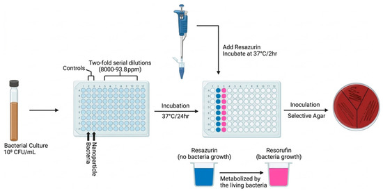
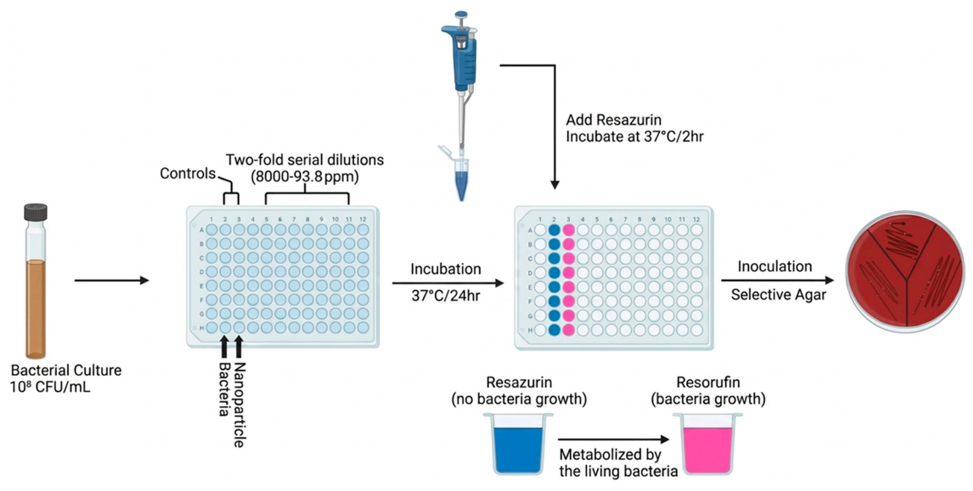
Figure 2 shows the antimicrobial activity assessment for pure and doped ZnO nanoparticles. The antimicrobial activity of pure ZnO and ZnO-Ag nanoparticles (1 and 5 wt.% Ag) was evaluated against Salmonella typhimurium (ATCC 14020) and Escherichia coli (ATCC 25922) using a colorimetric broth microdilution assay. Bacterial cultures were grown overnight in Tryptic Soy Broth (TSB) at 37 °C. The resulting suspensions were adjusted to approximately 1 × 10⁸ CFU mL−1 using a 0.5 McFarland standard to ensure a uniform inoculum throughout the study.
Figure 2.
Antimicrobial Activity Assessment of ZnO, ZnO-Ag 1%, and ZnO-Ag 5% nanoparticles.
Nanoparticle suspensions were prepared in Mueller–Hinton Broth (MHB) and serially diluted to obtain concentrations ranging from 93.8 to 8000 ppm. Antimicrobial assays were conducted in sterile 96-well microplates by combining 50 μL of the standardized bacterial suspension with 50 μL of the corresponding nanoparticle dilution, resulting in a final volume of 100 μL per well. The microplates were incubated at 37 °C with continuous agitation at 150 rpm for 24 h.
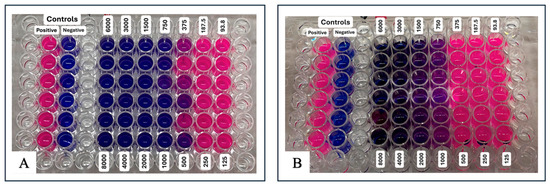
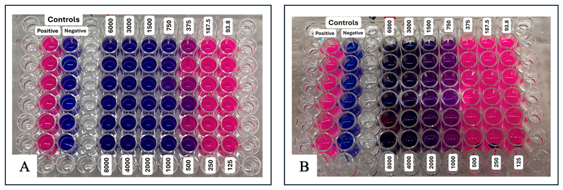
Following incubation, 30 µL of a 0.02% (w/v) resazurin solution was added to each well to indicate bacterial metabolic activity. The plates were further incubated at 37 °C for 2 h. Wells that remained blue were considered indicative of growth inhibition, whereas a color change from blue to pink indicated the presence of metabolically active bacteria. The MIC was defined as the lowest nanoparticle concentration at which the resazurin indicator remained completely blue after incubation, indicating the absence of detectable bacterial metabolic activity.
The minimum bactericidal concentration (MBC) was determined by subculturing aliquots from wells showing no visible bacterial growth onto selective agar media. Samples from S. typhimurium assays were streaked onto Salmonella–Shigella (SS) agar, whereas samples from E. coli assays were streaked onto MacConkey agar using a sterile 1 μL inoculating loop. The agar plates were incubated at 37 °C for 24 h and subsequently examined for the presence or absence of visible bacterial colonies. No quantitative colony counting was performed. The MBC was defined as the lowest nanoparticle concentration that produced no visible bacterial colony growth after subculture [23].
3. Results and Discussion
3.1. Morphological, Compositional, and Optical Characterization
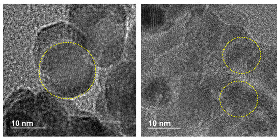
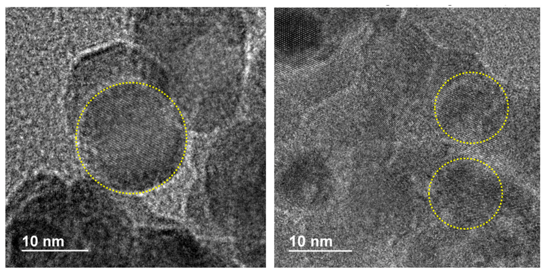
The HRTEM analysis focused on the pure ZnO and ZnO–Ag (1 wt.% Ag) samples, which were selected as representative materials to evaluate the effect of Ag addition on nanoparticle morphology and size. The structural characteristics of the ZnO–Ag (5 wt.% Ag) sample were assessed through complementary spectroscopic and diffraction techniques. Representative images of pure ZnO and ZnO–Ag (1 wt.% Ag) are presented in Figure 3. The pure ZnO sample consisted of well-defined nanoparticles with an average size of approximately 16 nm. Distinct lattice fringes were observed throughout the particles, indicating a high degree of crystallinity and confirming the formation of nanoscale ZnO domains. The nanoparticles tended to form aggregates, a common characteristic of metal oxide nanomaterials due to their high surface energy.
Figure 3.
High-resolution transmission electron microscopy (HRTEM) images of ZnO (left) and ZnO-Ag 1% (right) nanoparticles.
The ZnO–Ag (1 wt.% Ag) sample also displayed well-resolved lattice fringes, suggesting that the crystalline characteristic of the ZnO nanostructures was preserved after the introduction of Ag during synthesis. However, a reduction in particle size was observed, with average dimensions of approximately 12 nm. This decrease may indicate that the presence of Ag species influenced the nucleation and growth processes occurring during the reflux synthesis, leading to the formation of smaller crystalline domains. Similar effects have been reported in ZnO-Ag systems, where silver-containing precursors alter crystal growth kinetics and particle development [21,28].
The reduction in particle size observed for the Ag-containing sample may have important implications for its biological performance. Smaller nanoparticles generally exhibit a higher specific surface area and a larger proportion of surface atoms, which can increase interactions with bacterial cell envelopes and potentially enhance antimicrobial activity. Therefore, the morphological differences observed between pure ZnO and Ag-containing nanoparticles may contribute to the differences in antibacterial behavior evaluated against Escherichia coli and Salmonella typhimurium.
It should be noted that HRTEM provides direct information regarding nanoparticle morphology, size, and crystallinity, but does not independently confirm the structural location, distribution, or incorporation mechanism of Ag within the ZnO nanostructures. Consequently, the present observations are interpreted as evidence that the presence of Ag during synthesis modified nanoparticle growth behavior while maintaining the crystalline nature of the ZnO-based material. Additional compositional analyses would be required to determine the precise distribution of Ag within the nanoparticles.
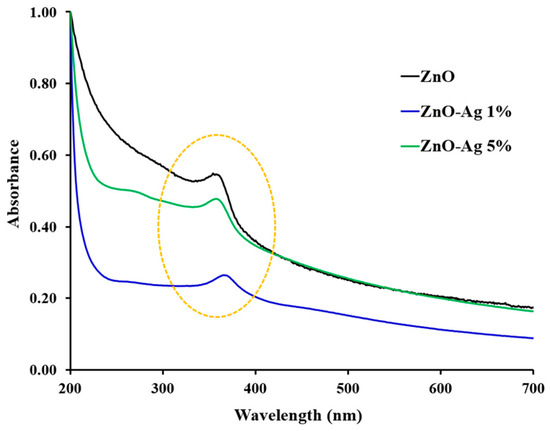
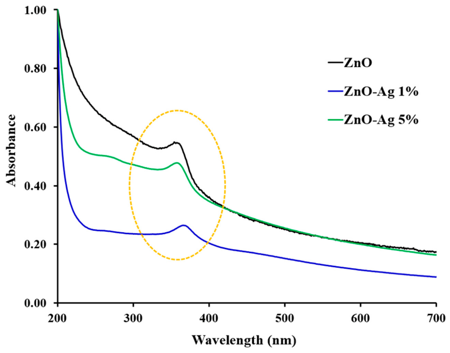
The UV–Vis absorption spectra of pure ZnO, ZnO–Ag (1 wt.% Ag), and ZnO–Ag (5 wt.% Ag) nanoparticles are shown in Figure 4. All samples exhibited strong absorption in the ultraviolet region, which is characteristic of ZnO-based nanomaterials. A well-defined absorption feature was observed between approximately 350 and 380 nm, corresponding to the characteristic absorption edge of ZnO. The presence of this band confirms the formation of ZnO nanoparticles and is consistent with previously reported values for nanocrystalline ZnO [20,21].
Figure 4.
UV-Vis spectra of ZnO, ZnO-Ag 1%, ZnO-Ag 5% nanoparticles.
The incorporation of Ag during synthesis resulted in noticeable changes in the absorption profiles. Pure ZnO exhibited the highest absorbance intensity throughout most of the analyzed spectral range. In contrast, the ZnO–Ag (1 wt.% Ag) sample displayed a substantial decrease in absorbance, while the ZnO–Ag (5 wt.% Ag) sample exhibited an intermediate behavior, maintaining a spectral profile similar to that of pure ZnO but with slightly lower intensity. Although the characteristic ZnO absorption edge remained present in all samples, variations in the intensity and shape of the absorption band were observed following the addition of Ag. These differences indicate that the presence of Ag influenced the optical properties of the synthesized materials, likely through modifications in their electronic environment and surface characteristics.
No distinct absorption band attributable to the surface plasmon resonance of metallic silver nanoparticles was observed within the visible region [29,30]. Therefore, the UV–Vis spectra do not provide direct evidence for the presence of separate plasmonic Ag nanoparticles. Instead, the results suggest that the addition of Ag altered the optical response of the ZnO-based materials while preserving the characteristic absorption behavior associated with ZnO. This behavior may be associated with a low effective Ag content, the small size or high dispersion of Ag species, or strong interactions between Ag and the ZnO matrix. Since no quantitative elemental analysis was performed, the contribution of each factor cannot be established conclusively.
Overall, the retention of the ZnO absorption edge in all samples (no monotonic variation in the absorption spectra) indicates that the fundamental optical characteristics of ZnO were maintained after Ag addition. At the same time, the observed spectral variations demonstrate that increasing Ag content influenced the absorption behavior of the nanoparticles, confirming that the synthesis conditions affected the optical properties of the resulting materials [21].
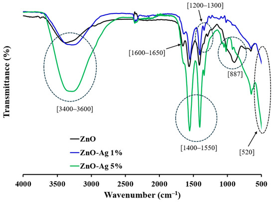
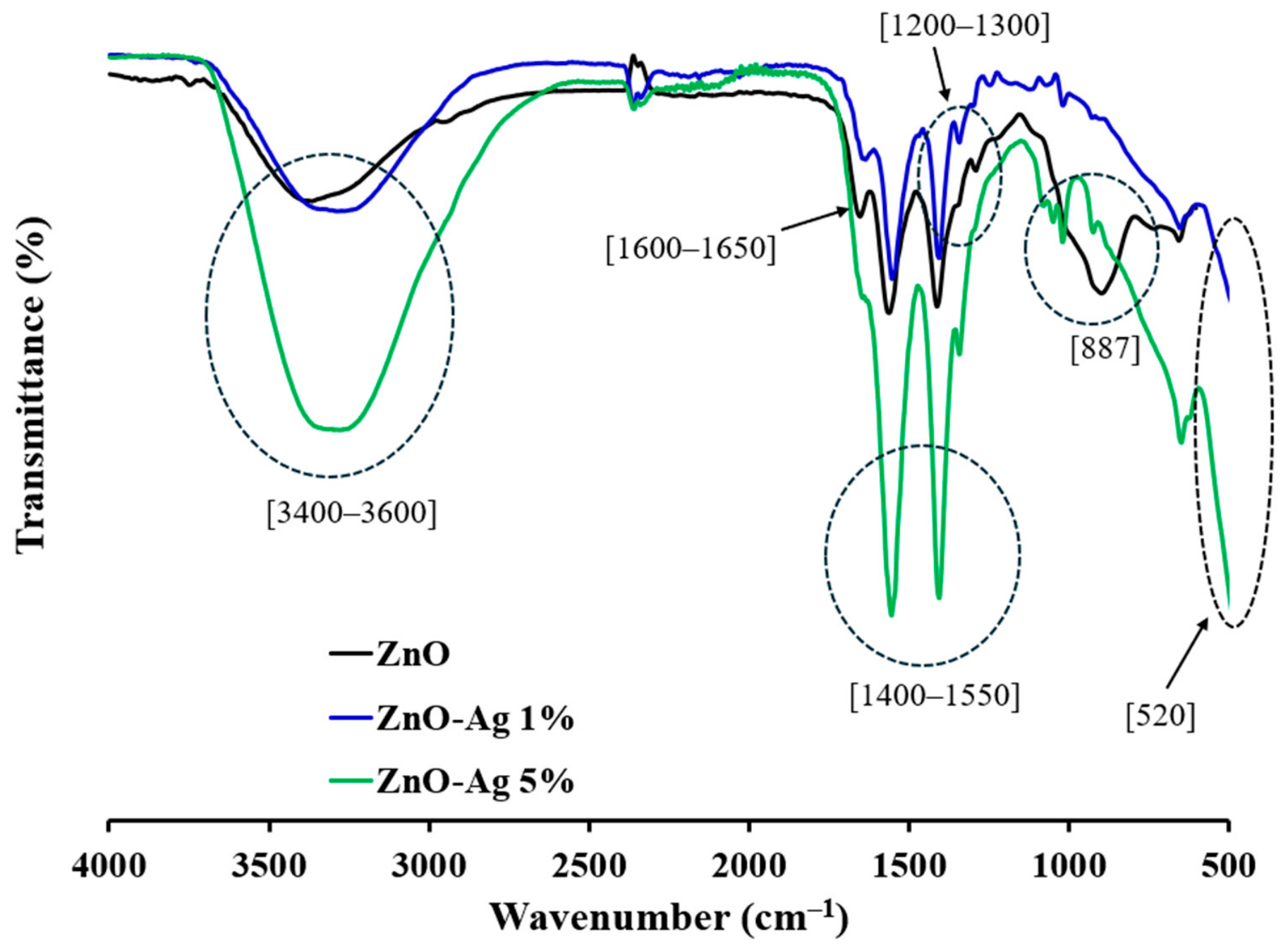
The FTIR spectra of pure ZnO, ZnO–Ag 1%, and ZnO–Ag 5% are shown in Figure 5. All samples exhibited absorption bands associated with surface hydroxyl groups, residual organic species from the synthesis process, and Zn–O vibrational modes.
Figure 5.
FTIR spectra of ZnO, ZnO-Ag 1%, ZnO-Ag 5% nanoparticles.
The broad band observed in the 3400–3600 cm−1 region is attributed to O–H stretching vibrations from surface hydroxyl groups and adsorbed water molecules. This band was more intense in the ZnO–Ag 5% sample, suggesting a greater contribution from hydroxylated surface species or adsorbed moisture in this material.
The band located around 1600–1650 cm−1 can be assigned to H–O–H bending vibrations of adsorbed water and may also include contributions from C=O-related vibrations associated with residual PVP or acetate species remaining after synthesis. The bands observed in the 1400–1550 cm−1 region are commonly associated with C–H bending and carboxylate-related vibrations from residual organic species, such as acetate or PVP fragments. These features were particularly pronounced in the ZnO–Ag 5% sample, indicating a stronger contribution from surface-bound organic residues or stabilizing molecules.
The absorption features between 1200 and 1300 cm−1 may be related to C–N stretching vibrations from PVP and/or C–O stretching vibrations from residual organic compounds. The band near 887 cm−1 can be attributed to C–H out-of-plane bending or to residual organic groups on the nanoparticle surface.
Importantly, the band observed near 520 cm−1 is assigned to Zn–O stretching vibrations, confirming the formation of ZnO-based nanoparticles. The presence of this band in all samples indicates that the ZnO framework was maintained after the addition of Ag during synthesis.
Overall, the FTIR results confirm the presence of Zn–O bonds and surface chemical groups associated with residual synthesis components. The changes in band intensity among pure ZnO, ZnO–Ag 1%, and ZnO–Ag 5% suggest that Ag addition influenced the surface chemistry of the nanoparticles, particularly in relation to hydroxyl groups and organic species retained after synthesis [31]. However, FTIR alone does not provide direct evidence of the exact location or coordination environment of Ag within the ZnO structure; therefore, these results are interpreted mainly in terms of surface functional groups and ZnO formation.
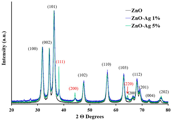
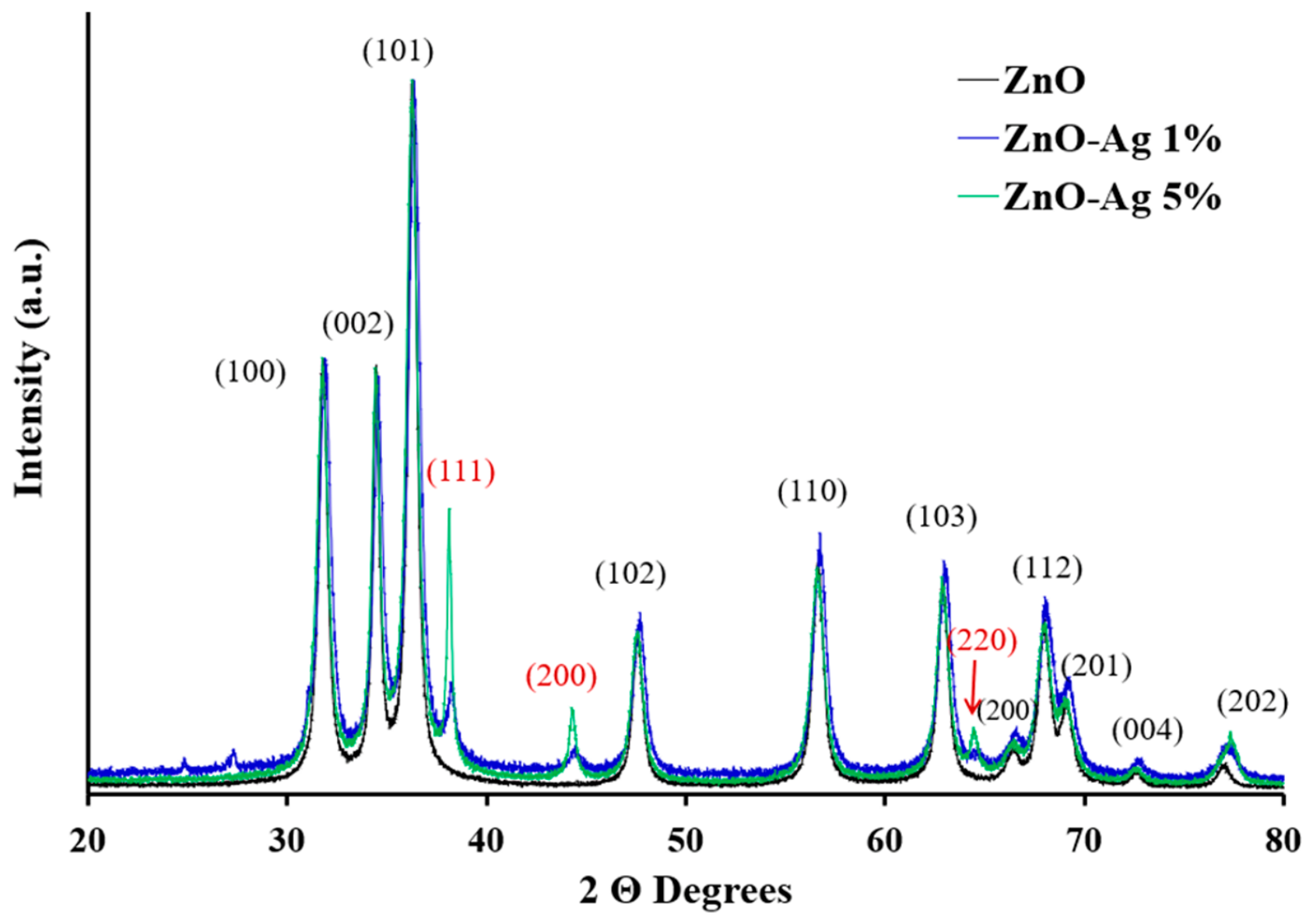
The X-ray diffraction patterns of pure ZnO, ZnO–Ag 1%, and ZnO–Ag 5% nanoparticles are presented in Figure 6. All samples exhibited the characteristic diffraction peaks of ZnO indexed to the (100), (002), (101), (102), (110), (103), (112), (201), (004), and (202) crystallographic planes. These reflections are consistent with the hexagonal wurtzite crystal structure of ZnO (JCPDS No. 36-1451), confirming the successful formation of crystalline ZnO in all synthesized materials. The similarity of the major ZnO diffraction peaks among the three samples indicates that the addition of Ag did not alter the fundamental ZnO crystal framework [32].
Figure 6.
X-ray diffraction pattern of ZnO, ZnO-Ag 1%, ZnO-Ag 5% nanoparticles.
In addition to the ZnO reflections, the Ag-containing samples exhibited diffraction peaks at approximately 38°, 44°, and 64°, which were assigned to the (111), (200), and (220) planes of metallic silver. These reflections are characteristic of face-centered cubic (FCC) Ag (JCPDS No. 04-0783). The Ag-related peaks were already detectable in the ZnO–Ag 1% sample and became more pronounced in the ZnO–Ag 5% sample, indicating that the contribution of crystalline Ag domains increased with increasing Ag content. The progressive increase in the intensity of these reflections suggests the formation of a ZnO/Ag composite system in which crystalline Ag is associated with the ZnO nanoparticles while the wurtzite ZnO phase remains dominant [28,33].
Crystallite sizes were estimated using the Scherrer equation by averaging values obtained from several diffraction peaks for each sample. Pure ZnO exhibited an average crystallite size of 16.9 ± 2.6 nm, while ZnO–Ag 1% showed a smaller average crystallite size of 12.9 ± 1.6 nm. The reduction in crystallite size at low Ag content suggests that Ag species present during synthesis may influence ZnO nucleation and crystal growth, resulting in smaller crystalline domains.
In contrast, the ZnO–Ag 5% sample exhibited a larger average crystallite size of 32.6 ± 10.1 nm. The increase in both the average crystallite size and standard deviation indicates greater heterogeneity in the crystalline domains present within the material. Because this sample exhibited more intense Ag-related FCC reflections, the Scherrer-derived value may reflect contributions from both ZnO and Ag-associated crystalline domains. Therefore, the larger apparent crystallite size should be interpreted with caution and is likely associated with the increasing contribution of crystalline Ag phases and the greater structural complexity of the ZnO–Ag 5% material.
Overall, the XRD results demonstrate that the addition of Ag influenced both the diffraction profile and the crystallite size of the synthesized nanoparticles. While the characteristic hexagonal wurtzite structure of ZnO was preserved in all samples, the appearance and progressive growth of reflections associated with FCC Ag indicate an increasing crystalline contribution from silver as its concentration increased from 1% to 5% [33].
Although XRD analysis confirmed the presence of crystalline Ag in the Ag-modified ZnO samples, no distinct surface plasmon resonance (SPR) band characteristic of Ag nanoparticles was observed in the UV–Vis spectra. These observations are not necessarily contradictory because XRD and UV–Vis probe different material properties. XRD identifies crystalline phases, whereas the SPR response depends on the optical properties of Ag, which are influenced by factors such as the amount, particle size, dispersion, and interaction of Ag species with the ZnO matrix. Furthermore, because no quantitative elemental analysis (e.g., ICP-OES or ICP-MS) was performed, the actual Ag content in the synthesized nanoparticles cannot be confirmed. Therefore, the absence of a well-defined SPR band should not be interpreted as evidence for the absence of crystalline Ag, but rather as an indication that the optical response of the Ag phase may be influenced by multiple factors.
3.2. Antimicrobial Properties
The antimicrobial activity of the synthesized nanoparticles was evaluated against E. coli and S. typhimurium using a resazurin-based broth microdilution assay. Antimicrobial activity was evaluated using technical triplicates for each nanoparticle concentration. The MIC and MBC values were reproducible across the replicate assays, and the values reported in Table 1 correspond to the consistent endpoint obtained for each nanoparticle formulation against each bacterial strain. Nanoparticle concentrations ranged from 8000 to 93.8 ppm. Metabolically active bacterial cells reduced blue resazurin to pink resorufin, whereas wells that remained blue indicated inhibition of bacterial growth due to a lack of detectable metabolic activity. Figure 7 presents representative microdilution plates illustrating the colorimetric response observed during the assay. The complete antimicrobial activity data, including the minimum inhibitory concentration (MIC), minimum bactericidal concentration (MBC), and tolerance (MBC/MIC) values for each nanoparticle formulation, are summarized in Table 1. The visual responses observed in the assay were consistent with the quantitative measurements obtained for both bacterial species. The MIC and MBC results demonstrate that increasing the Ag content from 1% to 5% substantially enhanced antibacterial efficacy, particularly by improving bactericidal activity.
Table 1.
Antimicrobial activity of pure ZnO and ZnO-Ag nanoparticles against Escherichia coli and Salmonella typhimurium expressed as MIC, MBC, and tolerance (MBC/MIC) values. Values were reproducible across the three technical replicates. MIC and MBC endpoints were identical in all replicate assays.
Figure 7.
Resazurin microdilution assay. ZnO nanoparticle suspensions were evaluated in triplicate using two-fold serial dilutions ranging from 8000 to 93.8 ppm against E. coli (A) and S. typhimurium (B).
Against E. coli, pure ZnO exhibited a MIC of 500 ppm and an MBC of 6000 ppm, resulting in a tolerance ratio of 12.0. Incorporation of 1% Ag did not improve performance; instead, the MIC increased to 750 ppm while the MBC remained unchanged at 6000 ppm, suggesting that this silver concentration was insufficient to generate a synergistic effect. In contrast, ZnO-Ag 5% reduced the MIC to 250 ppm and the MBC to 750 ppm, corresponding to reductions of 50% and 87.5%, respectively, relative to pure ZnO. The resulting MBC/MIC ratio of 3.0 indicates a transition from predominantly growth-inhibitory activity to a bactericidal effect.
A similar trend was observed for S. typhimurium. Pure ZnO produced a MIC of 750 ppm and an MBC of 8000 ppm, while ZnO-Ag 1% showed a further increase in MIC to 1500 ppm without improving the MBC. The highest Ag concentration markedly enhanced antimicrobial performance, decreasing the MIC to 750 ppm and the MBC to 1500 ppm. Compared with pure ZnO, this represents an 81.3% reduction in the bactericidal concentration and a fivefold decrease in the MBC/MIC ratio (10.6 to 2.0), indicating significantly greater killing efficiency [34].
The contrasting behavior of ZnO-Ag 1% and ZnO-Ag 5% suggests a minimum Ag loading is required to achieve effective antimicrobial synergy. At low concentrations, silver may be insufficient to significantly increase reactive oxygen species (ROS) production, membrane disruption, or Ag+ ion release. However, at 5% loading, the combined effects of ZnO and Ag likely enhance oxidative stress, protein inactivation, and damage to cellular membranes, resulting in substantially lower bactericidal concentrations [35].
Differences between the two bacterial species can be partially attributed to their cellular architecture. Although both E. coli and S. typhimurium are Gram-negative organisms, structural differences in the outer membrane composition, lipopolysaccharide organization, and stress-response systems influence susceptibility to metal-based nanomaterials. S. typhimurium consistently required higher bactericidal concentrations than E. coli for ZnO and ZnO-Ag 1%, suggesting greater intrinsic resistance. Specifically, the MBC values for S. typhimurium were 33% higher than those of E. coli for ZnO (8000 vs. 6000 ppm) and remained elevated for ZnO-Ag 1%. This observation is consistent with reports describing Salmonella’s ability to activate oxidative stress defense mechanisms, metal efflux systems, and membrane remodeling pathways that mitigate nanoparticle-induced damage [36].
Interestingly, incorporating 5% Ag largely eliminated these interspecies differences. While S. typhimurium maintained a higher MIC than E. coli (750 vs. 250 ppm), the bactericidal concentration decreased to only 1500 ppm, indicating that the enhanced Ag content was sufficient to overcome much of the intrinsic resistance associated with the Salmonella envelope. This result suggests that bactericidal activity depends more strongly on Ag-mediated mechanisms than on ZnO alone [37].
The tolerance analysis further supports these findings. None of the tested materials exhibited bacterial tolerance (MBC/MIC ≥ 16). However, ZnO and ZnO-Ag 1% produced intermediate MBC/MIC ratios ranging from 5.3 to 12.0, indicating that bacterial growth inhibition occurred at concentrations substantially lower than those required for cell death. In contrast, ZnO-Ag 5% generated ratios of 3.0 and 2.0 against E. coli and S. typhimurium, respectively, confirming true bactericidal activity according to accepted microbiological criteria. The lower ratio observed for S. typhimurium suggests that once growth inhibition was achieved, cell killing occurred more efficiently in this organism than in E. coli. Overall, the results demonstrate that silver concentration is the principal factor governing antimicrobial performance in the ZnO-Ag system. While ZnO and ZnO-Ag 1% primarily exerted inhibitory effects, increasing Ag loading to 5% transformed the nanocomposite into a bactericidal material, significantly reducing both MIC and MBC values and overcoming much of the intrinsic resistance associated with Gram-negative bacterial envelopes.
3.3. Study Limitations
One limitation of the present study is that antimicrobial activity was evaluated using technical triplicates without independent biological replicates. Consequently, the reproducibility of the results reflects assay repeatability rather than biological variability. Furthermore, MIC and MBC are discrete endpoints obtained from two-fold serial dilution assays and therefore are not readily amenable to conventional inferential statistical analyses. Future studies will include independent biological replicates and quantitative absorbance measurements using a multimode microplate reader, enabling statistical comparison of bacterial growth inhibition and a more comprehensive evaluation of antimicrobial performance.
4. Conclusions
Pure ZnO and ZnO-Ag nanoparticles containing 1 and 5 wt.% Ag were successfully synthesized using a reflux-assisted polyol method. UV–Vis spectroscopy confirmed the characteristic optical response of ZnO-based nanomaterials, while FTIR analysis revealed the presence of Zn–O bonds together with surface hydroxyl groups and residual organic species originating from the synthesis process.
XRD analysis demonstrated that all samples retained the hexagonal wurtzite structure of ZnO as the dominant crystalline phase. Additional diffraction peaks corresponding to FCC Ag became increasingly evident as the Ag content increased, indicating the presence of Ag-associated crystalline domains within the ZnO-based materials. Crystallite size calculations revealed that ZnO–Ag 1% exhibited smaller crystallites than pure ZnO, whereas ZnO–Ag 5% displayed larger and more heterogeneous crystalline domains. HRTEM observations further confirmed the formation of crystalline nanoparticles with average sizes in the nanometer range.
An antimicrobial evaluation demonstrated that Ag content strongly influenced antibacterial performance. ZnO–Ag 1% did not improve antimicrobial activity relative to pure ZnO and, in some cases, exhibited higher MIC values. In contrast, ZnO–Ag 5% showed the best antibacterial performance against both Escherichia coli and Salmonella typhimurium, exhibiting lower MIC and MBC values and substantially reduced tolerance ratios (MBC/MIC). For E. coli, ZnO–Ag 5% reduced the MIC and MBC to 250 ppm and 750 ppm, respectively, while for S. typhimurium, the MBC decreased from 8000 ppm for pure ZnO to 1500 ppm.
Overall, the results demonstrate that increasing Ag content modifies the structural and optical characteristics of ZnO-based nanoparticles and significantly enhances their bactericidal effectiveness. The superior antimicrobial performance of ZnO–Ag 5% suggests that the presence of Ag-associated crystalline domains enhances bacterial growth inhibition and cell inactivation, highlighting the potential of these nanostructured materials for antimicrobial applications.
Although the antimicrobial findings were highly reproducible across technical replicates, future investigations including independent biological replicates and quantitative absorbance-based analyses will provide a more robust statistical assessment of the antibacterial performance of ZnO-Ag nanomaterials.
Author Contributions
Conceptualization, M.R.-B.; Methodology, M.R.-B., K.T.-R., D.C.-L., P.B.-C. and I.P.-M.; Validation, M.R.-B.; Formal analysis, M.R.-B., K.T.-R. and S.J.B.-R.; Investigation, K.T.-R., D.C.-L., P.B.-C. and I.P.-M.; Resources, M.R.-B.; Writing—original draft, M.R.-B. and S.J.B.-R.; Writing—review and editing, M.R.-B. and S.J.B.-R.; Visualization, K.T.-R., D.C.-L., P.B.-C. and I.P.-M.; Supervision, M.R.-B. and S.J.B.-R.; Project administration, M.R.-B. and S.J.B.-R.; Funding acquisition, M.R.-B. and S.J.B.-R. All authors have read and agreed to the published version of the manuscript.
Funding
This research received external funding (NIFA/USDA 2026-67012-45562 Award).
Data Availability Statement
The data presented in this study are available in this article.
Acknowledgments
The authors gratefully acknowledge the Department of Chemistry and Physics at the University of Puerto Rico at Ponce for its support of this work. The authors also thank the Department of Mathematics and Physics at the University of Puerto Rico at Cayey, particularly Danilo Barrionuevo-Diestra, for his assistance with X-ray diffraction characterization (NSF grant 2215247).
Conflicts of Interest
The authors declare no conflicts of interest.
References
- Havelaar, A.H.; Kirk, M.D.; Torgerson, P.R.; Gibb, H.J.; Hald, T.; Lake, R.J.; Praet, N.; Bellinger, D.C.; de Silva, N.R.; Gargouri, N.; et al. World Health Organization Global Estimates and Regional Comparisons of the Burden of Foodborne Disease in 2010. PLoS Med. 2015, 12, e1001923. [Google Scholar] [CrossRef] [Scilit] [PubMed]
- Walter, E.J.S.; Cui, Z.; Tierney, R.; Griffin, P.M.; Hoekstra, R.M.; Payne, D.C.; Rose, E.B.; Devine, C.; Namwase, A.S.; Mirza, S.A.; et al. Foodborne Illness Acquired in the United States—Major Pathogens, 2019. Emerg. Infect. Dis. 2025, 31, 669–677. [Google Scholar] [CrossRef] [Scilit] [PubMed]
- Zizza, A.; Fallucca, A.; Guido, M.; Restivo, V.; Roveta, M.; Trucchi, C. Foodborne Infections and Salmonella: Current Primary Prevention Tools and Future Perspectives. Vaccines 2025, 13, 29. [Google Scholar] [CrossRef] [Scilit] [PubMed]
- Bintsis, T. Foodborne pathogens. AIMS Microbiol. 2017, 3, 529–563. [Google Scholar] [CrossRef] [Scilit] [PubMed]
- Popa, G.L.; Popa, M.I. Salmonella spp. Infection–A continuous threat worldwide. Germs 2021, 11, 88–96. [Google Scholar] [CrossRef] [Scilit] [PubMed]
- de Sousa, D.F.; Campos Filho, P.C.; da Conceição, A.O. Antibacterial activity of jackfruit leaves extracts and the interference on antimicrobial susceptibility of enteropathogen. Food Sci. Technol. 2022, 42, e49220. [Google Scholar] [CrossRef] [Scilit]
- Yang, S.-C.; Lin, C.-H.; Aljuffali, I.A.; Fang, J.-Y. Current pathogenic Escherichia coli foodborne outbreak cases and therapy development. Arch. Microbiol. 2017, 199, 811–825. [Google Scholar] [CrossRef] [Scilit] [PubMed]
- Omer, M.K.; Álvarez-Ordoñez, A.; Prieto, M.; Skjerve, E.; Asehun, T.; Alvseike, O.A. A Systematic Review of Bacterial Foodborne Outbreaks Related to Red Meat and Meat Products. Foodborne Pathog. Dis. 2018, 15, 598–611. [Google Scholar] [CrossRef] [Scilit] [PubMed]
- Jajere, S.M. A review of Salmonella enterica with particular focus on the pathogenicity and virulence factors, host specificity and antimicrobial resistance including multidrug resistance. Vet. World 2019, 12, 504–521. [Google Scholar] [CrossRef] [Scilit] [PubMed]
- Girma, A. Alternative mechanisms of action of metallic nanoparticles to mitigate the global spread of antibiotic-resistant bacteria. Cell Surf. 2023, 10, 100112. [Google Scholar] [CrossRef] [Scilit] [PubMed]
- Mouzakis, A.; Panagopoulos, P.; Papazoglou, D.; Petrakis, V. A Comprehensive Review of Nanoparticles in the Fight Against Antimicrobial Resistance. Pathogens 2025, 14, 1090. [Google Scholar] [CrossRef] [Scilit] [PubMed]
- Parvin, N.; Joo, S.W.; Mandal, T.K. Nanomaterial-Based Strategies to Combat Antibiotic Resistance: Mechanisms and Applications. Antibiotics 2025, 14, 207. [Google Scholar] [CrossRef] [Scilit] [PubMed]
- Generalova, A.N.; Dushina, A.O. Metal/metal oxide nanoparticles with antibacterial activity and their potential to disrupt bacterial biofilms: Recent advances with emphasis on the underlying mechanisms. Adv. Colloid Interface Sci. 2025, 345, 103626. [Google Scholar] [CrossRef] [Scilit] [PubMed]
- Wang, L.; Hu, C.; Shao, L. The antimicrobial activity of nanoparticles: Present situation and prospects for the future. Int. J. Nanomed. 2017, 12, 1227–1249. [Google Scholar] [CrossRef] [Scilit] [PubMed]
- Ozdal, M.; Gurkok, S. Recent advances in nanoparticles as antibacterial agent. ADMET DMPK 2022, 10, 115–129. [Google Scholar] [CrossRef] [Scilit] [PubMed]
- Baptista, P.V.; McCusker, M.P.; Carvalho, A.; Ferreira, D.A.; Mohan, N.M.; Martins, M.; Fernandes, A.R. Nano-strategies to fight multidrug resistant bacteria—“A Battle of the Titans”. Front. Microbiol. 2018, 9, 1441. [Google Scholar] [CrossRef] [Scilit] [PubMed]
- Slavin, Y.N.; Asnis, J.; Häfeli, U.O.; Bach, H. Metal nanoparticles: Understanding the mechanisms behind antibacterial activity. J. Nanobiotechnol. 2017, 15, 65. [Google Scholar] [CrossRef] [Scilit] [PubMed]
- Hetta, H.F.; Ramadan, Y.N.; Al-Harbi, A.I.; A. Ahmed, E.; Battah, B.; Abd Ellah, N.H.; Zanetti, S.; Donadu, M.G. Nanotechnology as a Promising Approach to Combat Multidrug Resistant Bacteria: A Comprehensive Review and Future Perspectives. Biomedicines 2023, 11, 413. [Google Scholar] [CrossRef] [Scilit] [PubMed]
- Gao, Q.; Feng, Z.; Wang, J.; Zhao, F.; Li, C.; Ju, J. Application of nano-ZnO in the food preservation industry: Antibacterial mechanisms, influencing factors, intelligent packaging, preservation film and safety. Crit. Rev. Food Sci. Nutr. 2025, 65, 4327–4353. [Google Scholar] [CrossRef] [Scilit] [PubMed]
- Kim, I.; Viswanathan, K.; Kasi, G.; Thanakkasaranee, S.; Sadeghi, K.; Seo, J. ZnO Nanostructures in Active Antibacterial Food Packaging: Preparation Methods, Antimicrobial Mechanisms, Safety Issues, Future Prospects, and Challenges. Food Rev. Int. 2022, 38, 537–565. [Google Scholar] [CrossRef] [Scilit]
- Lebaka, V.R.; Ravi, P.; Reddy, M.C.; Thummala, C.; Mandal, T.K. Zinc Oxide Nanoparticles in Modern Science and Technology: Multifunctional Roles in Healthcare, Environmental Remediation, and Industry. Nanomaterials 2025, 15, 754. [Google Scholar] [CrossRef] [Scilit] [PubMed]
- Khalifa, H.O.; Oreiby, A.; Mohammed, T.; Abdelhamid, M.A.A.; Sholkamy, E.N.; Hashem, H.; Fereig, R.M. Silver nanoparticles as next-generation antimicrobial agents: Mechanisms, challenges, and innovations against multidrug-resistant bacteria. Front. Cell Infect. Microbiol. 2025, 15, 1599113. [Google Scholar] [CrossRef] [Scilit] [PubMed]
- Reyes-Blas, M.; Maldonado-Luna, N.M.; Rivera-Quiñones, C.M.; Vega-Avila, A.L.; Roman-Velázquez, F.R.; Perales-Perez, O.J. Single Step Microwave Assisted Synthesis and Antimicrobial Activity of Silver, Copper and Silver-Copper Nanoparticles. J. Mater. Sci. Chem. Eng. 2020, 8, 13–29. [Google Scholar] [CrossRef]
- Panwar, A.; Yadav, K.L. Silver doped zinc oxide nanostructures with antibacterial properties against GFP-expressing antibiotic resistant Escherichia coli. Mater. Lett. 2022, 309, 131469. [Google Scholar] [CrossRef] [Scilit]
- El-Kattan, N.; Emam, A.N.; Mansour, A.S.; Ibrahim, M.A.; Abd El-Razik, A.B.; Allam, K.A.M.; Riad, N.Y.; Ibrahim, S.A. Curcumin assisted green synthesis of silver and zinc oxide nanostructures and their antibacterial activity against some clinical pathogenic multi-drug resistant bacteria. RSC Adv. 2022, 12, 18022–18038. [Google Scholar] [CrossRef] [Scilit] [PubMed]
- Dutta, G.; Chinnaiyan, S.K.; Sugumaran, A.; Narayanasamy, D. Sustainable bioactivity enhancement of ZnO-Ag nanoparticles in antimicrobial, antibiofilm, lung cancer, and photocatalytic applications. RSC Adv. 2023, 13, 26663–26682. [Google Scholar] [CrossRef] [Scilit] [PubMed]
- Naskar, A.; Shin, J.; Kim, K.S. A MoS2 based silver-doped ZnO nanocomposite and its antibacterial activity against β-lactamase expressing Escherichia coli. RSC Adv. 2022, 12, 7268–7275. [Google Scholar] [CrossRef] [Scilit] [PubMed]
- Sostre-Figueroa, J.; Rodríguez-Cadiz, A.; Bailón-Ruiz, S.J. Acute Toxicity of Pure and Silver-Doped ZnO Nanoparticles in Artemia salina Based on LC50 Determination. Micro 2025, 5, 58. [Google Scholar] [CrossRef] [Scilit]
- Sati, A.; Ranade, T.N.; Mali, S.N.; Ahmad Yasin, H.K.; Pratap, A. Silver Nanoparticles (AgNPs): Comprehensive Insights into Bio/Synthesis, Key Influencing Factors, Multifaceted Applications, and Toxicity—A 2024 Update. ACS Omega 2025, 10, 7549–7582. [Google Scholar] [CrossRef] [Scilit] [PubMed]
- Hosny, S.; Gaber, G.A.; Ragab, M.S.; Ragheb, M.A.; Anter, M.; Mohamed, L.Z. A Comprehensive Review of Silver Nanoparticles (AgNPs): Synthesis Strategies, Toxicity Concerns, Biomedical Applications, AI-Driven Advancements, Challenges, and Future Perspectives. Arab. J. Sci. Eng. 2025, 51, 13667–13714. [Google Scholar] [CrossRef] [Scilit]
- Matysiak, W.; Tański, T.; Zaborowska, M. Manufacturing process and characterization of electrospun PVP/ZnO NPs nanofibers. Bull. Pol. Acad. Sci. Tech. Sci. 2019, 67, 193–200. [Google Scholar] [CrossRef]
- Kumar, R.; Mushtaq, S.; Shweta, N.; Kumar, H.; Kulshrestha, S.; Teotia, S.; Singh, J.; Pandey, S.K. Synthesis and characterization of ZnO Nanoparticles and its application by Sol-Gel Method. Authorea 2023. [Google Scholar] [CrossRef] [Scilit] [PubMed]
- Amrute, V.; Monika, N.; Supin, K.K.; Vasundhara, M.; Chanda, A. Observation of excellent photocatalytic and antibacterial activity of Ag doped ZnO nanoparticles. RSC Adv. 2024, 14, 32786–32801. [Google Scholar] [CrossRef] [Scilit] [PubMed]
- Nan, J.; Chu, Y.; Guo, R.; Chen, P. Research on the antibacterial properties of nanoscale zinc oxide particles comprehensive review. Front. Mater. 2024, 11, 1449614. [Google Scholar] [CrossRef] [Scilit]
- Hao, Y.; Wang, Y.; Zhang, L.; Liu, F.; Jin, Y.; Long, J.; Chen, S.; Duan, G.; Yang, H. Advances in antibacterial activity of zinc oxide nanoparticles against Staphylococcus aureus. Biomed. Rep. 2024, 21, 161. [Google Scholar] [CrossRef] [Scilit] [PubMed]
- van der Heijden, J.; Reynolds, L.A.; Deng, W.; Mills, A.; Scholz, R.; Imami, K.; Foster, L.J.; Duong, F.; Finlay, B.B. Salmonella rapidly regulates membrane permeability to survive oxidative stress. mBio 2016, 7, e01238-16. [Google Scholar] [CrossRef] [Scilit] [PubMed]
- Suárez, D.M.; Colón, J.A.M.; García-Mercado, W.; Piñero-Cruz, D.; Bailón-Ruiz, S.J. Potential Bactericidal Activity of Silver Nanoparticles. MRS Adv. 2020, 5, 975–984. [Google Scholar] [CrossRef] [Scilit]

Disclaimer/Publisher’s Note: The statements, opinions and data contained in all publications are solely those of the individual author(s) and contributor(s) and not of MDPI and/or the editor(s). MDPI and/or the editor(s) disclaim responsibility for any injury to people or property resulting from any ideas, methods, instructions or products referred to in the content. |
© 2026 by the authors. Licensee MDPI, Basel, Switzerland. This article is an open access article distributed under the terms and conditions of the Creative Commons Attribution (CC BY) license.