


Abstract
Microplastics (MPs) and nanoplastics (NPs) are recognized as contaminants of relevance to human health. However, medical devices remain an insufficiently explored source of exposure. This review evaluates evidence on the release of MPs and NPs from polymer-based medical devices, with emphasis on particle-generation mechanisms, exposure pathways, and factors affecting release. The literature indicates that medical devices, including disposable masks and gloves, nasal irrigation devices, breastmilk storage bags, syringes, infusion bags, tubing and filters, extracorporeal circulation systems, ophthalmic surgical devices, orthodontic aligners and retainers, may release particles during manufacturing, storage, preparation, and clinical use, particularly under mechanical stress. Particle release is influenced by polymer type, device design, surface characteristics, temperature, contact time, fluid composition, storage conditions, mechanical abrasion, compression, bending, and repeated use. Of particular concern are medical applications involving direct access to the bloodstream, such as intravenous infusion and extracorporeal circulation, which may allow released particles to enter the circulation without passing through the gastrointestinal or respiratory tract. Thus, medical devices should be considered an emerging and distinct source of human exposure to MPs and NPs, while their potential risks must be interpreted in light of the available evidence and in the context of their essential clinical benefits.
1. Introduction
Microplastics (MPs) and nanoplastics (NPs) are now recognized as widespread environmental contaminants [1]. Research to date on human exposure to plastic particles has focused primarily on three exposure routes: dietary intake, drinking water consumption, and inhalation of airborne particles [2,3]. These particles have been detected in blood, lung tissue, the placenta, breast milk and urine, among other samples [4,5,6,7,8,9]. The potential health effects of MPs are therefore the subject of intensive research [10,11,12,13]. Studies in cellular and animal models have shown that exposure may induce oxidative stress, inflammatory responses, mitochondrial and endothelial dysfunction, damage to biological barriers, alterations in gut microbiota composition, and metabolic disturbances [10,14]. The biological effects of MPs and NPs depend not only on dose but also on particle size, shape and surface properties, polymer type, the presence of processing additives, and exposure duration [3].
Modern medicine relies heavily on polymeric materials. Polyethylene (PE), polypropylene (PP), poly(vinyl chloride) (PVC), polyurethane (PU), polyamide (PA), poly(methyl methacrylate) (PMMA), polytetrafluoroethylene (PTFE) and silicone elastomers are used in infusion bags and tubing, syringes, catheters, blood collection and transfusion systems, dialysis systems, extracorporeal circuits, respiratory equipment, dressings, prostheses and implants, among other applications [15]. The widespread use of these materials reflects the ability to modify their mechanical and chemical properties to meet specific clinical requirements, as well as advantages such as low weight, flexibility, transparency, resistance to biological fluids, and ease of molding. Polymers constitute a major class of materials used in single-use medical devices [15]. However, biocompatibility at the material or device level does not necessarily exclude the release of plastic particles during manufacturing, storage, preparation, or use.
Recent evidence indicates that medical devices may also contribute to human exposure to MPs and NPs. The release of PVC particles from intravenous infusion sets and tubing has been demonstrated under conditions simulating clinical procedures [16,17]. Microplastics have also been detected in fluids circulating in experimental extracorporeal circulation systems [18]. The presence of such contaminants has also been reported in fluids used in hemodialysis and peritoneal dialysis [19].
Exposure associated with medical devices [20,21,22] may differ substantially from conventional dietary exposure [23,24,25]. During intravenous infusion or extracorporeal circulation, particles released into fluids entering or returning to the bloodstream may bypass the biological barriers encountered during gastrointestinal, respiratory, or dermal exposure (Figure 1). Repeated or prolonged procedures and exposure to large volumes of processed fluids may result in greater cumulative exposure, particularly in patients undergoing intensive or long-term medical interventions, including neonates, intensive care patients, individuals receiving long-term dialysis or parenteral nutrition, and patients requiring extracorporeal circulation. In a preclinical study, MPs released from infusion sets under conditions simulating the administration of various solutions increased reactive oxygen species production activated the NLR family pyrin domain-containing 3 (NLRP3) inflammasome and inhibited angiogenesis in human endothelial cells, while also exerting toxic effects in a zebrafish model [10].
Figure 1.
Routes of human exposure to microplastics and nanoplastics. Created by the authors using Canva (Canva Pro) at https://www.canva.com/ (accessed on 19 October 2026). IV—intravenous.
However, the available literature remains fragmented, with limited systematic comparison of particle release across medical device categories, release mechanisms, exposure pathways, and potential clinical implications. It is essential to distinguish clearly between the presence of particles in the system under investigation, their release during use, their introduction into the body, the resulting biological response, and clinically significant harm. The aim of this review is therefore to critically evaluate medical devices as a potential source of human exposure to MPs and NPs, with particular attention to device categories, particle-generation mechanisms, exposure pathways, and factors affecting particle release. The review also considers these potential risks in the context of the essential clinical benefits provided by medical devices.
2. Materials and Methods
This critical narrative review was based on a literature search conducted using PubMed, Scopus, and ScienceDirect. Publications from 2021 to 2026 were considered in the primary literature search. However, older studies were also included when they provided relevant data on sources of exposure, processes leading to the release of plastic particles, or potential health effects associated with exposure to MPs and NPs.
The literature search was conducted using combinations of terms and phrases occurring in titles, abstracts, and keywords. The literature search used combinations of the following terms: (“microplastics” OR “nanoplastics”) AND (“medical devices” OR “medical equipment” OR “healthcare products” OR “infusion bag” OR “syringe” OR “infusion tubes” OR “infusion filters” OR “extracorporeal circulation systems” OR “masks” OR “gloves” OR “nasal irrigation devices” OR “breastmilk storage bags” OR “infant” OR “ophthalmic surgical devices” OR “orthodontic aligners” OR “dental retainers” OR “orthodontic appliances” OR “gynecological sampling devices”).
Both original research articles and review papers were considered eligible for inclusion. Review articles were used primarily to characterize the current state of knowledge, provide broader context for the topic, and identify potential sources of exposure. The inclusion criteria were as follows: availability of the full text in English, relevance to medical devices made wholly or partly of polymeric materials, and assessment of the potential for human exposure to MPs and NPs. Publications in languages other than English and materials available only as abstracts were excluded from the analysis.
Data from the included publications were compiled and analyzed. Particular attention was paid to the type of medical device, the polymer used, the mechanism of particle release, and the potential route of human exposure. Following the selection process, 89 publications were included in the final analysis.
3. Medical Devices
Polymeric materials constitute one of the main classes of materials used in modern medical devices. Their importance stems from the ability to tailor their chemical composition, structure, mechanical properties, transparency, and chemical resistance to specific applications [26].
Medical devices are often multi-material systems in which individual components are made from different polymers according to their function [26]. In the study by Li et al. [27], syringe barrels were made from PP, whereas infusion sets contained PVC in the tubing and drip chambers, acrylonitrile butadiene styrene (ABS) in the spikes and flow regulators, and PP in the filter membranes. Poly(methyl methacrylate) and PVC were used in filter housings, PE in protective components, polyoxymethylene (POM) in clamps, and polystyrene (PS) in Luer connectors [27]. Medical devices used for intravenous administration also contained components made from polycarbonate (PC) and PMMA [27].
The multi-material construction makes identifying the source of MPs considerably more difficult. Assessing particle origin therefore requires the characterization of all components in contact with the administered fluid. Furthermore, the material composition of devices may differ between manufacturers and models. Results obtained for one type of set should therefore not be automatically generalized to all products within the same category. The manufacturer, device specifications, and conditions of use may influence both the number of particles released and their properties [27].
4. Major Polymer Classes Used in Medical Devices
Polymers used in medical devices include PE, PP, PVC, ABS, PC, PMMA, POM, PS, poly(ethylene terephthalate) (PET), PA, polydimethylsiloxane (PDMS), PU, poly(lactic acid) (PLA), poly(glycolic acid) (PGA), and poly(ε-caprolactone) (PCL) [26]. The choice of polymer for a specific application depends on the required biocompatibility, durability, flexibility, and chemical resistance.
4.1. Polyolefins: Polypropylene and Polyethylene
Polyolefins are among the polymers most commonly used in single-use medical devices [28]. Polyethylene is found in fluid bags and bottles, needle covers, and other protective components. Polypropylene is used in syringe barrels, plungers, connectors, catheter components, and filter membranes, among other applications [27]. These polymers are also widely used in packaging materials, clothing, and laboratory equipment. Their widespread use is related to their chemical resistance and suitability for high-volume production [29].
4.2. Poly(vinyl chloride)
Poly(vinyl chloride) is one of the most important materials used in infusion tubing, drip chambers, catheters, fluid and blood bags, and extracorporeal circuits [27]. Depending on its plasticizer content, PVC can be either rigid or highly flexible. In medical applications, its flexibility and transparency allow tubing to be easily bent and visually monitored during fluid flow.
4.3. Thermoplastics
In addition to polyolefins and PVC, numerous engineering thermoplastics are used in medical devices, including ABS, PC, PMMA, POM, PS, PET, and PA [26]. These materials are used primarily where rigidity, dimensional stability, transparency, or mechanical strength is required [30]. They may be present in flow regulators, spikes, clamps, filter housings, connectors, chambers, and protective components [27].
4.4. Elastomers
Elastomers are used in devices requiring high flexibility, fatigue resistance, or prolonged contact with the body. This group includes PDMS and PU [31]. They are used in catheters, seals, pump tubing, prostheses, orthopedic implants, and vascular grafts [32].
4.5. Biodegradable Polymers
Biodegradable polymers are widely used in devices designed to degrade or resorb after serving a temporary function in the body. Common materials in this group include PLA, PGA, and PCL. These polymers are used in absorbable sutures, bone fixation implants, drug delivery systems, fillers, and tissue engineering scaffolds [26].
Biodegradability does not mean that a material disappears immediately after being placed in the body. Degradation is a gradual process whose course depends on the chemical structure, crystallinity, molecular weight, device geometry, and biological environment [33]. During degradation, the material may undergo fragmentation and progressive molecular-weight reduction, followed by further breakdown into smaller degradation products that may be metabolized or excreted, depending on the polymer and biological environment [34]. However, it is important to determine whether degradation generates persistent or transient particulate fragments within the micro- or nanoscale size range and how long such particles remain in the tissues. Relatively little is known about the direct release and characterization of MPs and NPs from biodegradable medical devices under clinically relevant conditions [34,35]. Consequently, the contribution of biodegradable medical devices to patient exposure to MPs and NPs remains poorly characterized and represents an important area for further research.
5. Factors Governing Microplastic Release
Polymer type is only one of several factors that influence the release of MPs and NPs (Figure 2). Other relevant factors include the degree of crystallinity, additive content, surface roughness, component thickness, manufacturing method, sterilization, and storage and use conditions [17,27,36,37,38].
Figure 2.
Factors affecting microplastic and nanoplastic release from medical devices. Created by the authors using Canva (Canva Pro) at https://www.canva.com/ (accessed on 21 September 2026). UV—ultraviolet.
Mechanical stresses are among the most important factors contributing to particle release. Friction between the plunger and syringe barrel, repeated opening and closing of clamps, bending of tubing, and piercing of stoppers may cause surface cracking and particle detachment. For example, simulated shaking of infusion bags increased the concentration of plastic particles from 522–5455 MPs/L initially to 4648–19,883 MPs/L after 15 days [39]. Complex medical devices can also generate MPs; for example, extracorporeal circulation systems release plastic particles through mechanical damage to tubing caused by roller pumps. Repeated compression can cause cracking and flaking of the inner surface of the tubing [18,40]. Furthermore, Li et al. [27] reported a significant increase in the release of MPs with repeated device use.
This process is also influenced by the composition of the solution in contact with the medical device, as well as by exposure time and temperature. In a study of PVC tubing, the extent of particle release differed between sodium chloride (NaCl), glucose, sodium bicarbonate (NaHCO3) and hydroxyethyl starch solutions, indicating that fluid composition may modify interactions at the material–solution interface [17]. Notably, infusion fluid composition had a greater effect on plastic release than temperature, storage time, or shaking [39]. This mechanism has been linked to the generation of reactive oxygen species (ROS). Solutions containing moxifloxacin hydrochloride or etimicin sulfate, as well as bicarbonate Ringer’s solution, generated greater amounts of ROS than NaCl or glucose solutions. The generation of hydroxyl and carbonate radicals and singlet oxygen promoted oxidation of the PP surface, formation of oxygen-containing groups and polymer chain scission, leading to material degradation and fragmentation [39]. This is important because it indicates that contact between a polymer and a pharmaceutical fluid is not necessarily chemically inert and that the propensity of the packaging to release MPs may depend on the specific preparation contained in the bag. Storage time was also shown to have a significant effect. After 15 days of storage at room temperature, the number of MPs released increased by more than 90%, whereas after 30 days, it was 5–10 times higher than the initial level. This increase was accompanied by greater surface roughness, the formation of cracks and pits, and progressive oxidation of PP [39]. Furthermore, after 30 days, bags stored at room temperature showed greater degradation than those stored at 4 °C, with surface roughness 1.41–1.52 times higher [39].
Other characteristics of the surrounding environment also influence the extent of plastic release. The release of fibers from surgical masks was influenced by pH, ionic strength, humic acid concentration, and shear stress in a study by Das et al. [41]. The greatest release was observed at pH 9, an ionic strength of 10 mM, and a humic acid concentration of 0.1 mg/L, with more vigorous mixing increasing fiber detachment. Release was lower at higher ionic strengths and at higher concentrations of humic substances [41]. These findings suggest that studies conducted solely in deionized water may not fully reflect plastic release under more complex environmental conditions.
The simultaneous action of several factors may be important for the release of MPs, as occurs, for example, in flow-through devices. Extracorporeal circulation tubing is simultaneously exposed to near-physiological temperature, flow, pressure, bending and cyclic compression [18]. In an infusion set, contact with the fluid may last for many hours, during which the fluid passes through or comes into contact with the tubing, regulator, membrane, chamber, connector, and needle [16,17,27,42]. The potential for the release of MPs should therefore be considered a characteristic of the entire system rather than of an individual component [18,27].
The mechanisms responsible for the generation of NPs may differ in scale from those leading to the release of MPs, although both processes are closely related. Mechanical abrasion, friction, and repeated deformation of polymer surfaces may directly generate small particles or promote the progressive fragmentation of larger plastic fragments. In addition, oxidative degradation and polymer chain scission may weaken the material structure, increasing its susceptibility to fragmentation and the release of nanoscale particles [17,39]. Another potential source of NPs is the detachment of pre-existing nanoscale particles from polymer surfaces during clinical use. The smaller size of NPs may facilitate their cellular uptake and interaction with biological barriers, including the blood–brain barrier. However, their actual translocation and health consequences following exposure through medical devices remain poorly understood.
6. Medical Devices as Sources of Microplastics
Modern medicine relies heavily on polymeric materials, which are used in both single-use and reusable medical devices [18,43,44,45]. A growing body of evidence indicates that these products may be a source of MPs and, in some cases, NPs released during manufacture, storage or use [16,27,39]. Unlike conventional exposure to plastic particles through ingestion, inhalation, or skin contact, the use of medical devices may result in exposure occurring directly during a diagnostic or therapeutic procedure [16,43]. Depending on the type of device, particles may be introduced into the nasal cavity with irrigation fluids or may come into direct contact with ocular tissues during ophthalmic procedures [43,44]. However, the intravenous route is particularly important because it allows these contaminants to be introduced directly into the circulation, bypassing the epithelial barriers associated with the gastrointestinal or respiratory route [16,27]. More complex medical systems also represent a potential source of intravascular exposure. Models of extracorporeal circulation have demonstrated the release of MPs during the operation of both conventional cardiopulmonary bypass (CPB) systems and minimally invasive extracorporeal circulation (MiECC) systems (Figure 3) [18]. These findings indicate that medical devices represent a potentially important and still insufficiently understood source of human exposure to MPs and NPs, including exposure through direct contact with body fluids and tissues during diagnostic or therapeutic procedures [16,18,27]. Selected representative studies on MPs and NPs released from different categories of medical devices are summarized in Table 1.
Figure 3.
Potential medical device sources of microplastics and nanoplastics. Created by the authors using Canva (Canva Pro) at https://www.canva.com/ (accessed on 19 October 2026).
Table 1.
Selected studies on microplastics and nanoplastics released from medical devices.
6.1. Disposable Masks
Single-use surgical masks are multilayer products composed mainly of nonwoven polymer fabrics. A standard surgical mask usually consists of three layers: an outer nonwoven layer that limits the penetration of moisture and larger particles, a middle filtration layer, and an inner layer in direct contact with the wearer’s face. Polypropylene is the predominant material, although PE, PA and PET are also used, depending on the manufacturer [52]. The multilayer fibrous structure improves filtration efficiency but also provides a surface from which individual fibers and fragments may detach during manufacture, transport, use, or improper handling of the mask [22,45,46,47,48,49,50,51].
The release of plastic particles while a mask is being worn may result from airflow, changes in humidity, bending of the material, friction against the skin and mechanical deformation of the nonwoven fabric. An eight-hour breathing simulation at an airflow rate of 15 L/min showed that the number of particles collected on a filter behind the mask ranged from 5421 ± 1417 to 15,113 ± 3532 per mask in a study conducted by Bhangare et al. [58]. The highest number was recorded for used two-layer surgical masks, and the lowest for new three-layer surgical masks [58]. These findings, together with evidence from other studies, indicate that material wear, repeated handling, and prolonged use may increase particle detachment.
The available evidence indicates that single-use masks can release MPs during use and after disposal. However, the extent of direct exposure of the wearer remains uncertain and depends on the type of mask, manufacturer, quality of the nonwoven fabric, wearing time, and intensity of friction. Data are also lacking to link the number of particles released from a mask to the dose deposited in different regions of the respiratory tract and to clinically significant health effects. Future studies should use intact masks, realistic airflow and humidity conditions, controls of environmental exposure, procedural blanks, and spectroscopic confirmation of the composition of all isolated particles. It is necessary to distinguish particles released by the mask from those previously captured from the air. Such an approach will make it possible to assess whether, under specific conditions, a mask increases total exposure to MPs and NPs or whether its filtration function outweighs emissions from the material itself. Risk assessment should therefore consider the balance between the retention of environmental particles and the release of particles from the mask itself, rather than merely the detection of MPs in the mask material.
6.2. Nasal Irrigation Devices
Nasal irrigation devices are widely used as an adjunct to the treatment of nasal and sinus conditions, including rhinitis and acute and chronic sinusitis. They primarily include nasal rinse bottles, seawater sprays, and syringes used to administer saline solutions. A substantial proportion of these products are made from plastics, and during fluid preparation and administration, the fluid comes into direct and sometimes prolonged contact with the polymer surface. Consequently, these devices may be a source of MPs introduced with the fluid directly into the nasal cavity, where they may come into contact with the respiratory mucosa [43].
A study comparing different nasal irrigation methods was conducted by Tuna and Taş [43]. The authors detected an average of 6.49 ± 13.08 MPs per product [43].
The importance of mechanical handling and repeated use of nasal irrigation devices was demonstrated more clearly in the study by Kim and Min [40]. The authors analyzed PP irrigation bottles, comparing new products with bottles subjected to procedures simulating 1, 3, and 6 months of use, respectively [40]. Even new bottles contained an average of 33.00 ± 20.42 MPs/300 mL, whereas after one simulated month of use, this number increased to 68.66 ± 30.07 MPs/300 mL. The greatest release was observed after three months, at 261.66 ± 20.59 MPs/300 mL, while after six months, it was 204.33 ± 52.16 MPs/300 mL. Most particles were 10–100 µm in size and occurred as fragments, while the predominant polymer was PP, the material from which the tested bottles were made.
The presence of MPs in nasal irrigation fluids was also confirmed, with PP among the most frequently identified polymers, in an earlier study by Kim et al. [59]. The correspondence between particle composition and the materials of the device components further indicates that at least some of the contaminants may arise from fragmentation of irrigation system components. This is particularly important for reusable bottles, since their surfaces are repeatedly squeezed, heated, washed, and refilled. However, not all particles present in the fluid after irrigation necessarily originate from the bottle itself. Other potential sources include the water, the salt preparation used to make the solution, and components of the closure and nozzle.
The possible contribution of the ingredients used to prepare the fluid was assessed by Kim and Min [60]. Water placed in PP bottles was compared with a solution prepared using sachets containing NaCl. The mean counts of MPs were 33.00 ± 20.42 and 15.66 ± 15.88 particles, respectively. Polypropylene predominated in both groups, while smaller amounts of PE and PET were detected. The authors therefore concluded that opening the sachet and adding NaCl powder did not, in itself, significantly increase the release of MPs into the fluid [60].
The available studies therefore indicate that nasal irrigation devices may be a source of exposure to MPs. Future studies should therefore include analyses of new and repeatedly used products from different manufacturers, characterization of NPs, and measurements of the fluid before and after passage through the device. Determining actual particle retention in the nasal cavity would also be particularly important, allowing the current assessment of plastic particle release from devices to progress towards a quantitative assessment of human exposure.
6.3. Disposable Gloves
Single-use gloves are one of the most commonly used categories of personal protective equipment in healthcare, where they serve as a barrier limiting staff contact with blood, body fluids, secretions, biological material, and chemicals. Depending on their intended use, they are manufactured from various polymeric materials, primarily natural rubber latex (NRL), nitrile butadiene rubber (NBR), and PVC, as well as PE, styrene-butadiene rubber, neoprene, or poly(vinyl alcohol) (PVA). The surface and mechanical properties of gloves depend not only on the base polymer but also on the plasticizers, cross-linking agents, stabilizers, mold-release agents, and other processing additives used [61]. During use, gloves are repeatedly stretched, bent, and subjected to friction, and their surfaces may remain in contact with water, biological fluids, lipids, disinfectants, and other chemicals. These factors may lead to changes in the material’s surface structure and the detachment of small particles [62].
One study addressing this issue, conducted by Wang et al. [53], assessed latex, nitrile, and vinyl gloves subjected to accelerated aging in an aqueous environment. The authors showed that ultraviolet (UV) radiation altered the physicochemical properties of all the materials analyzed, with nitrile gloves exhibiting greater stability than latex and vinyl gloves. Latex gloves showed the greatest propensity to release particles, and mechanical abrasion with sand further increased their release. These findings indicate that polymer type and mechanical and environmental aging of the material are important determinants of particle generation.
Further information on the release of MPs is provided by a study of gloves made from PE, thermoplastic elastomer (TPE), and PLA conducted by Gao et al. [63]. The authors demonstrated the release of plastic particles from all three types of material into both water and edible oil. In the oil-containing medium, the number of particles released was generally 2–3 times that in water. The highest value was found for PE gloves in oil (3183.33 ± 500.83 MPs/m2), and the lowest was found for TPE gloves in water (183.33 ± 28.87 MPs/m2). Fibers predominated among the released particles, while for PE, contact with oil also increased the proportion of small particles with a granular morphology. This study is particularly relevant to medical devices because it indicates that the type of medium in contact with a glove may significantly influence particle release. It may therefore be hypothesized that, in medical settings, contact with lipid-containing preparations, ointments, disinfectants or other substances may modify the properties of the glove surface.
The role of gloves as a source of plastic particles also extends to the post-use stage. Used gloves were shown to be susceptible to aging caused by UV exposure and abrasion in the study by Wang et al. [53]. In turn, burning disposable gloves was shown to generate MPs and NPs in the study by Luo et al. [64].
Current evidence supports considering single-use gloves as a potential source of MPs and, under certain conditions, NPs, although the importance of this route for patient exposure remains poorly understood. In particular, there is a lack of studies conducted under conditions replicating actual clinical activities, including repeated flexion and stretching of the hand, friction, contact with sweat, blood and other body fluids or disinfectants, and direct contact with wounds, mucous membranes, or instruments. It would be particularly important to determine whether released particles can be transferred from the glove surface directly to the patient or the procedural field, since there are currently insufficient data for a quantitative assessment of such exposure.
6.4. Breastmilk Storage Bags
Breastmilk storage bags (BSBs) are widely used for the temporary storage, refrigeration, and freezing of expressed milk, followed by thawing and warming before it is fed to the infant. The bags are made mainly from PE, and some products have a multilayer structure incorporating, for example, PET. Their use involves several successive stages that may affect the material surface: filling, storage for many hours or days, freezing, thawing, warming, and emptying. Direct contact between the plastic and milk means that released particles may subsequently be ingested by the infant [54,65,66]. Liu et al. [65] provided evidence of particle release from this type of product. The study mainly observed irregular agglomerates <300 µm in size and fragments in the range of 1–50 µm, as well as submicrometre particles with irregular, spherical, or oval shapes [65]. Micro-Raman spectroscopy (µ-Raman) analysis showed that PE predominated, followed by PET and PA. The mass of all particles and residues released from three randomly selected bags of the same type was 0.22–0.47 mg [65]. However, it should be emphasized that this study used a model fluid rather than actual breast milk, whose lipid and protein content may alter interactions at the polymer–fluid interface.
An important complementary study using actual breast milk as the medium was conducted by Susetio et al. [54]. The mean particle count in fresh milk stored in a glass was 1.41 ± 1.50 MPs/5 mL, whereas milk placed in bags contained 8.07 ± 6.36 MPs/5 mL after storage at −4 °C, 9.83 ± 10.12 MPs/5 mL after thawing at 4 °C and 6.07 ± 5.46 MPs/5 mL after thawing/warming at 40 °C. The particles were 10–680 μm in size and occurred mainly as fragments and fibers. The authors also reported that PA accounted for the largest proportion of the particles identified by micro-Fourier transform infrared spectroscopy (µ-FTIR) [54].
The available studies indicate that breastmilk storage bags may be an additional source of oral exposure to MPs and potentially to submicrometre particles in infants. However, the evidence base remains limited and is characterized by substantial methodological differences [54,65].
6.5. Syringes
Single-use syringes are among the most commonly used medical devices and represent a potentially important source of direct exposure to plastic particles. Modern syringes consist of several components made from polymeric materials. In the study by Li et al. [27], the analyzed syringe barrels were made from PP, while individual components of the plunger, seal, and needle cap included PP and elastomeric materials. During syringe filling and emptying, movement of the plunger against the inner surface of the barrel causes friction and mechanical stresses that may promote material detachment. Unlike exposure through ingestion or inhalation, injection may introduce particles released from the device directly into tissues and, in the case of intravenous administration, into the bloodstream [16,27].
Direct evidence of plastic release from syringes, with particular attention to PDMS used in sealing components and as a lubricant to reduce friction between the plunger and barrel, was provided by Fang et al. [67]. Movement of the plunger may cause both the detachment of solid PDMS fragments and the release of liquid PDMS droplets used as a lubricant. Based on the measurements performed, it was estimated that the use of a single 1 mL plastic syringe may be associated with the release of up to approximately 1.4 × 10⁵ PDMS microparticles and 2.1 × 10⁷ PDMS nanoparticles [67]. Improper handling of syringes may be an additional factor contributing to particle release. An increase in particle counts following mechanical shaking, dropping, and abrupt movement of syringes was demonstrated by Kim et al. [68]. These findings indicate that particle release is not solely a property of the material but may also depend on how the device is transported, stored, and used.
Syringes were shown to release 0.44 ± 0.90 particles per device in another study by Li et al. [27]. The number of MPs released depended on syringe size. For 10 mL syringes, 0.67 ± 0.87 particles per device were detected, whereas for 1 and 5 mL syringes, the values were 0.44 ± 0.89 and 0.22 ± 0.67 particles per device, respectively. Clear differences between manufacturers were also observed. No MPs were detected in one of the brands tested, whereas approximately 0.67 particles per device were found in each of the other two [27]. Repeated use proved to be a particularly important factor. After ten cycles of filling and emptying 10 mL syringes, the mean count of MPs increased to 2.67 ± 1.53 particles per device and was significantly higher than observed under standard conditions. The authors attributed the increase in MPs over successive cycles primarily to repeated friction, compression, and other mechanical stresses between device components [27]. Four types of polymers were identified in the syringes tested, with PP predominating, followed by PE. Substantial amounts of PDMS were also found, consistent with its use as a lubricant. Microplastics occurred mainly as fragments. Their size range in syringe samples was 12.6–285.9 µm, while the vast majority of all MPs from the devices analyzed were smaller than 50 µm [27].
The studies conducted indicate that syringes can be a source of plastic particles. Future studies should therefore include simultaneous analysis of MPs and NPs, different syringe types and capacities, products from different manufacturers, realistic numbers of plunger movements, a range of drug solutions, and actual administration conditions. It is also necessary to clearly distinguish between particles originating from PP or other structural materials, solid PDMS, and silicone oil droplets. Such an approach will allow the quantitative contribution of syringes to direct human exposure to plastic particles to be reliably determined.
6.6. Infusion Bags, Tubes, and Filters
Modern infusion fluid containers are made primarily from PP, PE, and PVC. The latter polymer also remains one of the main materials used to manufacture flexible infusion tubing [16]. The significance of this exposure differs from that of conventional oral and inhalation routes because it does not require passage across the intestinal or respiratory barrier, and contaminants present in the final infusion fluid may be administered directly into the bloodstream [20,21,69].
The presence of MPs in fluids stored in plastic containers has been confirmed in several independent studies, although the reported concentrations vary considerably [56,70]. More recently, Özçifçi et al. [70] analyzed 50 commercially available intravenous fluid samples in Turkey and detected MPs in 9 samples, with a mean concentration of 0.40 ± 0.90 MPs/L and an estimated single-use exposure of approximately 0.20 MPs per unit [70]. Analysis of infusion fluids in PP or PVC containers revealed MPs in all samples tested, at a mean concentration of 62.82 ± 72.38 MPs/L, in the study by Çağlayan et al. [56]. Concentrations of 50.74 ± 74.44 MPs/L were found in products packaged in PP and 90.00 ± 68.96 MPs/L in PVC containers. Fragments predominated, followed by fibers. Polyethylene was the most frequently identified polymer [56]. These findings confirm the presence of MPs in intravenously administered fluids, while the detection of polymers other than those used for the container suggests that additional sources, such as closure components, manufacturing processes, storage, or other parts of the infusion system, may contribute.
The potential contribution of the packaging material itself is suggested by findings from an analysis of products available in Spain, Italy, and Ecuador conducted by Casella et al. [71]. Concentrations in fluids contained in plastic bags were 166–299 MPs/L. Fragments and fibers were detected, and more than 60% of MPs were smaller than 100 µm [71]. Polypropylene, PE, PA, PU, and PET were the most frequently identified polymers [71].
Infusion tubing may also be an important source of plastic particles. The release of PVC particles during simulated infusion was demonstrated by Li et al. [16]. For a volume of 250 mL, the mass of PVC particles released was 0.36–0.72 μg. When both the bag and tubing were made from PVC, the mean value was approximately 0.67 μg, compared with approximately 0.44 μg in a system comprising a PP container and PVC tubing. In the same study, increasing the volume of fluid passed through the system from 250 to 1000 mL increased the amount of particles released approximately 3.7–7.6-fold, after which a further increase in volume to 3000 mL no longer produced a proportional increase. The authors interpreted this as the flushing of a substantial proportion of loosely bound particles from the system surface during the first liter of infusion. Furthermore, a 5% glucose solution caused greater PVC release than 0.9% NaCl [16].
A direct analysis of single-use PVC tubing was also conducted by Zheng et al. [17]. New, unused tubing contained an average of 931.4 MPs per set and approximately 0.040 µg of NPs. Under simulated infusion conditions for 4 h at room temperature, 1003.6–3494.6 MPs and 0.042–0.087 µg of NPs were released per set, depending on the fluid type. The greatest release of MPs was observed with 5% NaHCO3, followed by 5% glucose, hydroxyethyl starch solution, and 0.9% NaCl [17]. Time and temperature were also important factors. Compared with 4 h of contact with 0.9% NaCl, counts of MPs increased 1.9-fold after 24 h and 2.49-fold after 96 h. Raising the temperature from 25 to 37 °C during the 4-h experiment increased the number of particles released by approximately 39% [17].
However, not all studies confirm substantial release of plastic particles from the tubing itself. Zhu et al. [55] detected a total of eight MPs measuring 4–148 µm in infusion fluids from PE bags, PP bottles, and one glass bottle, while no MPs were detected in the infusion tubing analyzed [55]. The discrepancies between the findings of Li et al. [16] and Zhu et al. [55] may be largely attributed to differences in analytical methods and detection limits. Zhu et al. [55] employed µ-Raman spectroscopy following filtration through a 0.8 µm membrane. In contrast, Li et al. [16] used 20 nm filtration combined with scanning electron microscopy coupled with energy-dispersive X-ray spectroscopy (SEM-EDS) and pyrolysis–gas chromatography–mass spectrometry (Py-GC-MS), allowing the investigation of nanoscale PVC particles. While spectroscopic methods such as µ-FTIR typically have practical detection limits of approximately 10 µm, Py-GC-MS quantifies polymer mass without directly determining particle size or number. Importantly, the particle numbers reported by Li et al. were estimated from measured polymer mass using assumed particle diameters and density rather than being directly counted. Consequently, the results obtained using these different approaches are not directly comparable. These findings highlight the need for complementary analytical techniques and standardized reporting methods to enable reliable comparisons between studies.
In this context, in-line filters are an important component of the infusion system and may reduce the number of MPs reaching the patient, but they do not completely eliminate exposure. A mean concentration of 2.91 ± 3.91 MPs/L in the infusion products tested was reported by Cui et al. [72]. Polyethylene and PP fragments predominated, and most particles were within the range of 15–100 µm [72]. The use of a standard set with a 15 µm pore-size filter reduced counts of MPs only to a limited extent, whereas a finer 5 µm filter reduced their number by more than half [72]. Filters should therefore be regarded as a means of reducing exposure rather than as a barrier that ensures complete removal of plastic particles.
In summary, the available evidence indicates that both infusion bags and tubing may be sources of MPs and NPs, and that the extent of release depends on polymer type, fluid composition, storage time and temperature, infusion volume, mechanical stresses, and the design of the entire system. At the same time, discrepancies between studies are substantial. These discrepancies may partly reflect differences in filtration and identification methods, as well as differences in experimental conditions. Future studies should replicate the entire clinical infusion fluid delivery pathway and analyze MPs and NPs simultaneously using complementary methods. Such an approach is necessary to distinguish the contributions of individual system components and reliably estimate the number of plastic particles reaching the bloodstream.
6.7. Extracorporeal Circulation Systems and Minimally Invasive Extracorporeal Circulation Circuits
Extracorporeal circulation systems represent a distinct category of potential sources of plastic particles because, during their use, the patient’s blood remains in direct contact with an extensive system of polymeric components for a prolonged period. A conventional CPB system temporarily takes over the function of the heart and lungs, directing venous blood to a reservoir and oxygenator before returning it to the arterial circulation. Flow in conventional systems is usually generated by a roller pump, which operates through repeated mechanical deformation of the tubing. By contrast, MiECC uses a more closed and miniaturized circuit, with shorter tubing, a reduced priming volume, and a centrifugal rather than a roller pump [18]. Design differences between these systems may affect not only blood contact with synthetic materials but also the mechanisms of particle release and the properties of the MPs released.
A quantitative and qualitative assessment of MPs released from both conventional CPB and MiECC was conducted by Green et al. [18]. Both types of system generated substantial numbers of MPs [18]. Conventional CPB circuits yielded a mean of 60.4 ± 7.6 MPs/L/h, compared with 48.4 ± 31.3 MPs/L/h for MiECC. After correction for background contamination, the values were 58.58 ± 4.11 MPs/L/h for CPB and 46.58 ± 27.81 MPs/L/h for MiECC. The difference between conventional CPB and MiECC was not statistically significant. This is an important observation because, despite the simpler design and absence of a roller pump in MiECC, the available evidence does not currently establish that this system significantly reduces total exposure to MPs. More pronounced differences between the systems were observed for the polymer composition of the MPs released. A total of 20 different polymer types were identified. In conventional CPB systems, PDMS, poly(decyl methacrylate) (PDMA), and poly(N-butyl methacrylate) (PBMA) predominated, with concentrations of 19.6 ± 17.1, 18.7 ± 26.6, and 12.9 ± 7.3 particles/L/h, respectively. Polyethylene, PP, PTFE, PS, and other polymers were also identified. In MiECC systems, by contrast, PP, PE, and PA predominated, at 16.4 ± 17.8, 10.7 ± 1.3, and 7.1 ± 6.8 particles/L/h, respectively. These findings may be related to the distinct design of the two systems. Of particular interest is PDMS, which was one of the predominant polymers identified in conventional CPB but was not detected in MiECC. In turn, the high proportion of PP in MiECC may be associated, among other possible sources, with the soft-shell reservoir used during priming or with oxygenator components. Significant differences were also found in the size and morphology of MPs. Particles from conventional CPB were longer than those from MiECC. Their mean length was 93.5 µm, compared with 62.0 µm in MiECC. Their mean widths were 31.3 ± 18.1 and 28.0 ± 15.8 µm, respectively. Films predominated in conventional CPB, followed by fragments, irregular particles, and fibers. In MiECC, by contrast, fragments were most common, followed by films, irregular particles, and fibers. These findings suggest that system design may influence not only the type of polymer released but also the physical characteristics of the particles, which may be relevant from a toxicological perspective. The available evidence thus indicates that both conventional CPB systems and MiECC circuits can release MPs directly into the circulating fluid, although particle properties may depend on system design. The main priorities for further research should therefore be to identify the sources of individual polymers in specific components, determine their release dynamics during the procedure, and assess patient exposure through blood analysis before, during and after the procedure. It is also necessary to include the fraction of NPs in measurements and to assess whether modifications to circuit design, material selection, pre-flushing of the system, or filtration can reduce direct patient exposure to plastic particles.
6.8. Ophthalmic Surgical Devices
Ophthalmic procedures, particularly cataract surgery, require numerous single-use and reusable devices made from polymeric materials. Phacoemulsification uses, among other items, bags containing saline and balanced salt solution (BSS), syringes, ophthalmic viscosurgical devices (OVDs), irrigation–aspiration tubing, and intraocular lens implantation systems. Because fluids passing through these components are subsequently introduced directly into the anterior chamber of the eye, degradation and abrasion of polymer surfaces may lead to a distinct form of exposure in which MPs enter ocular structures directly [44].
A study specifically investigating the presence of MPs during cataract surgery was conducted by Choi et al. [44]. No MPs were detected in the tested samples of saline administered by syringe, BSS drawn into a syringe, or the material contained in the OVDs [44]. However, particles were found in the fluid after it had passed through the more complex system used during phacoemulsification. A mean concentration of 0.1 ± 0.1 MPs/mL was detected in the fluid entering the eye. All particles detected in this sample were 10–19 µm in size and consisted of PE or PP fragments [44]. This finding suggests that passage through the phacoemulsification system itself may contribute to additional contamination, even when the initial solutions and devices used for their manual administration contain no detectable plastic particles. However, the concentration of MPs in fluid leaving the eye after phacoemulsification was considerably higher than in the incoming fluid. In five samples collected directly into glass vessels, it ranged from 1.5 ± 1.4 to 7.2 ± 3.5 MPs/mL. In addition, the outgoing fluid contained smaller particles, including those in the 5–9 µm range, and the polymer composition was more diverse, comprising PE, PP, PET, and PS [44]. A substantial proportion of the MPs present in the outgoing fluid may already have been in the eye before surgery began [44].
The available findings therefore indicate that ophthalmic surgical devices may be a direct source of MPs during surgery. However, current evidence does not allow MPs released during the procedure to be distinguished with complete certainty from the particles already present in the eye. Further studies are therefore needed, including analysis of each successive component of the fluid pathway, collection of samples immediately before and after contact with the device, and simultaneous assessment of MPs and NPs. Such an approach would allow the particle dose introduced into the eye during ophthalmic procedures to be determined and help identify components whose material modification could reduce exposure.
6.9. Orthodontic Aligners and Retainers
Orthodontic aligners and removable dental retainers represent another potential source of human exposure to MPs and NPs. Clear aligners are typically worn for most of the day during orthodontic treatment, whereas removable retainers are frequently used every night for several years. Prolonged contact with saliva, repeated insertion and removal, mechanical friction, and forces generated during swallowing or clenching may contribute to progressive surface degradation and the detachment of plastic particles. These particles may subsequently be swallowed with saliva or remain in contact with the oral mucosa.
Experimental evidence that clear orthodontic aligners release particles was provided by Quinzi et al. [73], who investigated seven types of aligners subjected to mechanical agitation in artificial saliva. Using Raman microspectroscopy and scanning electron microscopy (SEM), the authors identified released MPs, predominantly ranging from 5 to 20 µm in size. In addition, Orilisi et al. [57] investigated ten types of aligners and demonstrated that MP release increased significantly after 14 days compared with seven days of simulated use, particularly for particles smaller than 5 µm. The extent of release was associated with polymer composition, whereas no significant relationship with the manufacturing process was observed. Importantly, Dobrowski et al. [74] extended the investigation to orthodontic retainers and nightguards. The authors evaluated five orthodontic appliances under conditions simulating nighttime (8 h) or full-time (20 h) wear over a 14-day period. Laser-directed infrared spectroscopy identified 11–16 particles in the analyzed aliquots from nighttime appliances, including retainers and a nightguard, whereas 341 and 1382 particles were recovered from two types of aligners tested under full-time wear conditions.
The potential biological relevance of particles released from orthodontic materials was also investigated by Warunek et al. [75]. The authors demonstrated the release of micro- and nanoscale particles from several polymeric materials used in orthodontic appliances and observed their uptake by macrophages under experimental conditions. Certain particle preparations also promoted pro-inflammatory macrophage responses.
Overall, the available evidence indicates that orthodontic aligners and retainers may contribute to prolonged oral exposure to MPs and potentially NPs. However, the actual quantities ingested during routine use, the contribution of nightly retainer wear to cumulative exposure, and the potential long-term health consequences remain insufficiently characterized. Further studies under realistic intraoral conditions, including repeated mechanical loading, saliva exposure, cleaning procedures, and prolonged use, are necessary to assess the significance of this exposure pathway.
6.10. Gynecological and Contraceptive Devices
Gynecological sampling devices, particularly vaginal and cervical swabs, represent a potential source of exposure to MPs and NPs that has received limited scientific attention. These devices are routinely used during diagnostic procedures and may contain synthetic polymers, including nylon fibers and plastic applicators. For example, flocked swabs consist of numerous polymer fibers attached to a plastic shaft, allowing efficient collection of biological material [76]. During sampling, direct contact with the vaginal mucosa, mechanical friction, and exposure to biological fluids may contribute to the detachment of plastic particles. In addition, condoms may represent another potential source of exposure to polymeric particles. Lambert et al. (2013) [77] demonstrated the formation of nanoscale particles during the environmental degradation of natural rubber latex condoms, although particle release during normal use was not investigated.
The relevance of the vaginal mucosa as a potential exposure site is supported by recent studies. Shim and Min (2025) [78] identified 91 MPs in cervicovaginal lavage fluids collected from 10 women, with PP and PS being the predominant polymers. Although these findings demonstrate the presence of MPs in the cervicovaginal environment, their origin could not be conclusively established. Furthermore, Pontecorvi et al. (2023) [79] demonstrated that PE nano- and microplastics could be internalized by human vaginal keratinocytes, inducing cytotoxic effects and alterations in epithelial barrier-related mechanisms. In addition, exposure to PE particles induced metabolic disturbances, oxidative stress-related responses, and inflammatory signaling in vaginal epithelial cells [80]. These findings indicate that the vaginal mucosa should be considered when evaluating potential exposure to MPs and NPs associated with medical devices.
7. Background Contamination and Analytical Detection Limitations
Background contamination represents a major challenge in the reliable identification and quantification of MPs and NPs released from medical devices. Airborne particles, laboratory equipment, reagents, and sample handling procedures may introduce additional plastic particles, potentially leading to an overestimation of exposure. Therefore, procedural blanks covering all analytical steps, appropriate environmental controls, and measures to minimize contamination are essential for distinguishing device-derived particles from background contamination.
Analytical detection limits further complicate the interpretation of the available findings, particularly for NPs. Conventional µ-FTIR typically detects particles approximately 10 µm or larger, depending on the analytical configuration, whereas µ-Raman spectroscopy can identify smaller particles but remains limited by spectral interference. In contrast, Py-GC-MS enables polymer mass quantification without directly determining particle size, morphology, or number [16,55]. Consequently, differences in analytical methods may substantially influence the reported number of particles and limit comparisons between studies. Although nanoscale particles have been investigated in selected studies [17,67], their detection and characterization remain challenging. Thus, the predominance of MPs in the available literature may partly reflect methodological limitations rather than the actual absence of NPs.
8. Future Directions
Future research on plastic particle release from medical devices should focus on standardizing analytical methods, assessing MPs and NPs simultaneously, identifying sources of contamination and replicating realistic clinical conditions. The main research priorities for individual categories of medical devices are summarized in Table 2. Analysis of samples before and after contact with the device and estimation of the dose introduced into the body are particularly important. It is also necessary to distinguish between particle release, introduction into the body, biological responses, and actual clinical risk. However, assessment of this issue should remain proportionate to the available evidence and take into account the fundamental therapeutic benefits provided by medical devices.
Table 2.
Research priorities for microplastics and nanoplastics released from medical devices.
Such assessments should also consider occupational exposure to MPs and NPs during medical device manufacturing, in addition to patient exposure during clinical use. Polymer processing, including injection molding, grinding, and material handling, may generate airborne plastic particles, potentially exposing manufacturing workers through inhalation. Studies conducted in plastic manufacturing facilities have confirmed occupational exposure to MPs, while industrial wastewater has been identified as another pathway for their release into the environment [81,82,83]. However, evidence specifically addressing emissions of MPs and NPs during medical device manufacturing remains limited. Therefore, improved ventilation, dust collection systems, and wastewater filtration should be considered to minimize particle release throughout the production process, alongside occupational exposure monitoring.
Beyond identifying sources and pathways of exposure, an additional research priority concerns the potential biological effects of MPs and NPs following human exposure. Casella and Ballaz [84] highlighted the potential genotoxic and neurotoxic effects of intracellular NPs, including oxidative stress, mitochondrial dysfunction, impaired autophagy, and inflammatory responses. Furthermore, Casella et al. [85] emphasized the analytical challenges associated with the detection and characterization of airborne MPs and NPs, as well as the need for further investigations into their long-term health effects. Although these studies did not specifically focus on medical devices, their findings underline the importance of combining reliable particle identification with assessments of exposure and potential biological effects when evaluating the safety of medical devices.
From a regulatory perspective, current frameworks address the safety of medical devices but provide limited requirements specifically targeting MPs and NPs. The Food and Drug Administration guidance emphasizes risk-based biocompatibility assessment, while Regulation (EU) 2017/745 requires minimizing, as far as possible, risks related to the size and properties of particles that are or can be released from medical devices into the patient’s or user’s body [86,87,88]. ISO 18562-2:2024 addresses particulate emissions from respiratory medical devices and provides acceptance criteria for particulate matter within its scope [89]. However, harmonized exposure limits specifically for MPs and NPs are still lacking. To minimize potential exposure, manufacturers should prioritize chemically and mechanically stable polymers, improve device design, and evaluate particle release under realistic clinical conditions. Proper storage, handling, and adherence to recommended usage procedures may further reduce particle release. Where appropriate, filtration systems may also limit exposure, although their effectiveness depends on particle size and does not guarantee the removal of NPs.
9. Conclusions
The available evidence indicates that medical devices may represent an important, hitherto insufficiently considered source of human exposure to MPs and, in some cases, NPs. Plastic particles have been detected in or released from single-use masks and gloves, nasal irrigation devices, BSBs, syringes, infusion bags and tubing, extracorporeal circulation systems, devices used during ophthalmic procedures, and orthodontic aligners and retainers, among others. Procedures in which these contaminants may be introduced directly into tissues, the eye, or the circulation, bypassing the barriers associated with oral and inhalation exposure, are particularly important.
The release of MPs and NPs depends not only on the type of polymer used but also on device design, manufacturing and storage conditions, temperature, contact time, fluid composition, and mechanical stresses occurring during use. Friction, compression, bending, repeated use and prolonged flow may cause progressive damage to material surfaces and increase particle release. In complex medical systems, several different components may simultaneously be sources of contamination. Exposure should therefore be assessed across the entire fluid contact pathway rather than for a single component alone.
At the same time, comparing the findings of available studies remains difficult because of substantial methodological differences, including differences in the size ranges of particles analyzed, filtration and identification techniques, and the way results are expressed. Knowledge concerning the fraction of NPs, the dose reaching the patient, and the clinical significance of the observed exposure is particularly limited. In many cases, merely detecting MPs in a fluid or biological material is not sufficient to attribute them unequivocally to a specific device or demonstrate a link with a particular health effect. The issue of MPs and NPs released from medical devices should therefore be incorporated into a broader assessment of material safety, while maintaining a balance between potential exposure-related risks and the indispensable clinical role of these devices.
Author Contributions
Conceptualization, K.K.-C. and M.G.; methodology, K.K.-C.; software, K.K.-C.; formal analysis, K.K.-C.; investigation, K.K.-C.; resources, K.K.-C.; data curation, K.K.-C.; writing—original draft preparation, K.K.-C., B.B. and M.G.; writing—review and editing, K.K.-C. and M.G.; visualization, K.K.-C.; supervision, K.K.-C. and M.G.; project administration, K.K.-C. and M.G. All authors have read and agreed to the published version of the manuscript.
Funding
This research received no external funding.
Institutional Review Board Statement
Not applicable.
Informed Consent Statement
Not applicable.
Data Availability Statement
No new data were created or analyzed in this study. Data sharing is not applicable to this article.
Acknowledgments
The authors gratefully acknowledge the support of their respective institutions.
Conflicts of Interest
The authors declare no conflicts of interest.
Abbreviations
The following abbreviations are used in this manuscript:
| MPs | Microplastics |
| NPs | Nanoplastics |
| PE | Polyethylene |
| PP | Polypropylene |
| PVC | Poly(vinyl chloride) |
| PU | Polyurethane |
| PA | Polyamide |
| PMMA | Poly(methyl methacrylate) |
| PTFE | Polytetrafluoroethylene |
| NLRP3 | NLR family pyrin domain-containing 3 |
| IV | Intravenous |
| ABS | Acrylonitrile butadiene styrene |
| POM | Polyoxymethylene (polyacetal) |
| PS | Polystyrene |
| PC | Polycarbonate |
| PET | Poly(ethylene terephthalate) |
| PDMS | Polydimethylsiloxane |
| PLA | Poly(lactic acid) |
| PGA | Poly(glycolic acid) |
| PCL | Poly(ε-caprolactone) |
| UV | Ultraviolet |
| ROS | Reactive oxygen species |
| CPB | Cardiopulmonary bypass |
| MiECC | Minimally invasive extracorporeal circulation |
| PEVA | Poly(ethylene-vinyl acetate) |
| PEMA | Poly(ethylene-co-methacrylic acid) |
| PES | Polyester |
| NBR | Nitrile butadiene rubber |
| PVA | Poly(vinyl alcohol) |
| PI | Polyimide |
| PVS | Poly(vinyl stearate) |
| VDN | Vein detained needle |
| PVDC | Poly(vinylidene chloride) |
| PDMA | Poly(decyl methacrylate) |
| PBMA | Poly(N-butyl methacrylate) |
| SR | Silicone rubber |
| SP | Silicone polymer |
| PMAA | Poly(N-methyl acrylamide) |
| PUR | Poly(ether urethane) |
| BSS | Balanced salt solution |
| NRL | Natural rubber latex |
| TPE | Thermoplastic elastomer |
| BSBs | Breastmilk storage bags |
| µ-Raman | Micro-Raman spectroscopy |
| µ-FTIR | Micro-Fourier transform infrared spectroscopy |
| SEM | Scanning electron microscopy |
| SEM-EDS | Scanning electron microscopy coupled with energy-dispersive X-ray spectroscopy |
| Py-GC-MS | Pyrolysis–gas chromatography–mass spectrometry |
| OVDs | Ophthalmic viscosurgical devices |
References
- Kadac-Czapska, K.; Jutrzenka Trzebiatowska, P.; Knez, E.; Zaleska-Medynska, A.; Grembecka, M. Microplastics in Food—A Critical Approach to Definition, Sample Preparation, and Characterisation. Food Chem. 2023, 418, 135985. [Google Scholar] [CrossRef] [Scilit]
- Cox, K.D.; Covernton, G.A.; Davies, H.L.; Dower, J.F.; Juanes, F.; Dudas, S.E. Human Consumption of Microplastics. Environ. Sci. Technol. 2019, 53, 7068–7074. [Google Scholar] [CrossRef] [Scilit]
- Kadac-Czapska, K.; Knez, E.; Grembecka, M. Food and Human Safety: The Impact of Microplastics. Crit. Rev. Food Sci. Nutr. 2024, 64, 3502–3521. [Google Scholar] [CrossRef] [Scilit]
- Leslie, H.A.; van Velzen, M.J.M.; Brandsma, S.H.; Vethaak, A.D.; Garcia-Vallejo, J.J.; Lamoree, M.H. Discovery and Quantification of Plastic Particle Pollution in Human Blood. Environ. Int. 2022, 163, 107199. [Google Scholar] [CrossRef] [Scilit]
- Ragusa, A.; Svelato, A.; Santacroce, C.; Catalano, P.; Notarstefano, V.; Carnevali, O.; Papa, F.; Rongioletti, M.C.A.; Baiocco, F.; Draghi, S.; et al. Plasticenta: First Evidence of Microplastics in Human Placenta. Environ. Int. 2021, 146, 106274. [Google Scholar] [CrossRef] [Scilit]
- Jenner, L.C.; Rotchell, J.M.; Bennett, R.T.; Cowen, M.; Tentzeris, V.; Sadofsky, L.R. Detection of Microplastics in Human Lung Tissue Using ΜFTIR Spectroscopy. Sci. Total Environ. 2022, 831, 154907. [Google Scholar] [CrossRef] [Scilit]
- Ragusa, A.; Notarstefano, V.; Svelato, A.; Belloni, A.; Gioacchini, G.; Blondeel, C.; Zucchelli, E.; De Luca, C.; D’Avino, S.; Gulotta, A.; et al. Raman Microspectroscopy Detection and Characterisation of Microplastics in Human Breastmilk. Polymers 2022, 14, 2700. [Google Scholar] [CrossRef] [Scilit]
- Pironti, C.; Notarstefano, V.; Ricciardi, M.; Motta, O.; Giorgini, E.; Montano, L. First Evidence of Microplastics in Human Urine, a Preliminary Study of Intake in the Human Body. Toxics 2023, 11, 40. [Google Scholar] [CrossRef] [Scilit]
- Kadac-Czapska, K.; Bukowska, B.; Sicińska, P.; Grembecka, M. Hidden Threats in Infant Diets and Environment—Risks of Microplastics and Nanoplastics in Food. Rev. Environ. Contam. Toxicol. 2025, 263, 24. [Google Scholar] [CrossRef] [Scilit]
- Yu, H.; Xiang, G.; He, S.; Zeng, Z.; Deng, A.; Yin, Y.; Wang, Y. Microplastics Release from Infusion Sets during Intravenous Infusion Induces Cardiovascular Toxicity. Environ. Int. 2026, 209, 110187. [Google Scholar] [CrossRef] [Scilit]
- Kadac-Czapska, K.; Ośko, J.; Knez, E.; Grembecka, M. Microplastics and Oxidative Stress—Current Problems and Prospects. Antioxidants 2024, 13, 579. [Google Scholar] [CrossRef] [Scilit]
- Kadac-Czapska, K.; Knez, E.; Gierszewska, M.; Olewnik-Kruszkowska, E.; Grembecka, M. Microplastics Derived from Food Packaging Waste—Their Origin and Health Risks. Materials 2023, 16, 674. [Google Scholar] [CrossRef] [Scilit]
- Kadac-Czapska, K.; Ośko, J.; Nowak, N.; Jażdżewska, K.; Kowalczyk, P.; Grembecka, M. Unseen Hazards—Toxicological Effects and Human Health Impacts of Nanoplastics and Microplastics. Appl. Sci. 2025, 15, 10146. [Google Scholar] [CrossRef] [Scilit]
- Jin, Y.; Lu, L.; Tu, W.; Luo, T.; Fu, Z. Impacts of Polystyrene Microplastic on the Gut Barrier, Microbiota and Metabolism of Mice. Sci. Total Environ. 2019, 649, 308–317. [Google Scholar] [CrossRef] [Scilit]
- Vienken, J.; Boccato, C. Do Medical Devices Contribute to Sustainability? The Role of Innovative Polymers and Device Design. Int. J. Artif. Organs 2024, 47, 240–250. [Google Scholar] [CrossRef] [Scilit]
- Li, P.; Li, Q.; Lai, Y.; Yang, S.; Yu, S.; Liu, R.; Jiang, G.; Liu, J. Direct Entry of Micro(Nano)Plastics into Human Blood Circulatory System by Intravenous Infusion. iScience 2023, 26, 108454. [Google Scholar] [CrossRef] [Scilit]
- Zheng, X.; Feng, Q.; Guo, L. Quantitative Analysis of Microplastics and Nanoplastics Released from Disposable PVC Infusion Tubes. J. Hazard. Mater. 2024, 465, 133246. [Google Scholar] [CrossRef] [Scilit]
- Green, J.L.; Field, D.T.; Bennett, R.; Jenner, L.C.; Chapman, E.C.; Sadofsky, L.R.; Rotchell, J.M.; Loubani, M. Microplastics in Cardiopulmonary Bypass: Quantification and Characterization of Particles across Systems. Interdiscip. Cardiovasc. Thorac. Surg. 2025, 40, ivaf080. [Google Scholar] [CrossRef] [Scilit]
- Kara, E.; Konur, K.; Koca, Y.Ş.; Aytan, Ü. Unveiling Hidden Contaminants: A Systematic Quantification and Characterization of Microplastics in Hemodialysis and Peritoneal Dialysis Fluids. BMC Nephrol. 2025, 26, 359. [Google Scholar] [CrossRef] [Scilit]
- Huang, T.; Liu, Y.; Wang, L.; Ruan, X.; Ge, Q.; Ma, M.; Wang, W.; You, W.; Zhang, L.; Valev, V.K.; et al. MPs Entering Human Circulation through Infusions: A Significant Pathway and Health Concern. Environ. Health 2025, 3, 551–559. [Google Scholar] [CrossRef] [Scilit]
- Vercauteren, M.; Panneel, L.; Jorens, P.G.; Covaci, A.; Cleys, P.; Mulder, A.; Janssen, C.R.; Asselman, J. An Ex Vivo Study Examining Migration of Microplastics from an Infused Neonatal Parenteral Nutrition Circuit. Environ. Health Perspect. 2024, 132, 037703. [Google Scholar] [CrossRef] [Scilit]
- Ma, M.; Xu, D.; Zhao, J.; Gao, B. Disposable Face Masks Release Micro Particles to the Aqueous Environment after Simulating Sunlight Aging: Microplastics or Non-Microplastics? J. Hazard. Mater. 2023, 443, 130146. [Google Scholar] [CrossRef] [Scilit]
- Kadac-Czapska, K.; Gadomska, K.; Szefer, P.; Grembecka, M. Quantification and Exposure Assessment of Microplastics in Water Used for Infant Feeding. J. Environ. Chem. Eng. 2026, 14, 124340. [Google Scholar] [CrossRef] [Scilit]
- Kadac-Czapska, K.; Sobczak, M.; Behrendt, M.; Mahlik, S.; Grembecka, M. Unmasking the Hidden Dangers: Microplastics Contamination Analysis of Follow-on Formulas. J. Food Compos. Anal. 2025, 144, 107707. [Google Scholar] [CrossRef] [Scilit]
- Kadac-Czapska, K.; Jutrzenka Trzebiatowska, P.; Mazurkiewicz, M.; Kowalczyk, P.; Knez, E.; Behrendt, M.; Mahlik, S.; Zaleska-Medynska, A.; Grembecka, M. Isolation and Identification of Microplastics in Infant Formulas—A Potential Health Risk for Children. Food Chem. 2024, 440, 138246. [Google Scholar] [CrossRef] [Scilit]
- Chen, W.-H.; Chen, Q.-W.; Chen, Q.; Cui, C.; Duan, S.; Kang, Y.; Liu, Y.; Liu, Y.; Muhammad, W.; Shao, S.; et al. Biomedical Polymers: Synthesis, Properties, and Applications. Sci. China Chem. 2022, 65, 1010–1075. [Google Scholar] [CrossRef] [Scilit]
- Li, B.; Li, M.; Du, D.; Tang, B.; Yi, W.; He, M.; Liu, R.; Yu, H.; Yu, Y.; Zheng, J. Characteristics and Influencing Factors of Microplastics Entering Human Blood through Intravenous Injection. Environ. Int. 2025, 198, 109377. [Google Scholar] [CrossRef] [Scilit]
- Gopanna, A.; Rajan, K.P.; Thomas, S.P.; Chavali, M. Polyethylene and Polypropylene Matrix Composites for Biomedical Applications. In Materials for Biomedical Engineering; Elsevier: Amsterdam, The Netherlands, 2019; pp. 175–216. [Google Scholar]
- Karaki, A.; Hammoud, A.; Masad, E.; Khraisheh, M.; Abdala, A.; Ouederni, M. A Review on Material Extrusion (MEX) of Polyethylene—Challenges, Opportunities, and Future Prospects. Polymer 2024, 307, 127333. [Google Scholar] [CrossRef] [Scilit]
- Shakiba, M.; Rezvani Ghomi, E.; Khosravi, F.; Jouybar, S.; Bigham, A.; Zare, M.; Abdouss, M.; Moaref, R.; Ramakrishna, S. Nylon—A Material Introduction and Overview for Biomedical Applications. Polym. Adv. Technol. 2021, 32, 3368–3383. [Google Scholar] [CrossRef] [Scilit]
- Zhang, S.; Staples, A.E. Natural Aging of Biomaterials in Ambient and Physiological Environments. Adv. Nanobiomed Res. 2026, 6, e202500198. [Google Scholar] [CrossRef] [Scilit]
- Drożdż, K.; Gołda-Cępa, M.; Brzychczy-Włoch, M. Polyurethanes as Biomaterials in Medicine: Advanced Applications, Infection Challenges, and Innovative Surface Modification Methods. Adv. Microbiol. 2024, 63, 223–238. [Google Scholar] [CrossRef] [Scilit]
- Mayer, J.B.; Patil, S.M.; Shin, S.-H.; Yoo, J.; Won, Y.-Y. Internal Water-Induced Acceleration, Chemical Pathways, and Contributing Factors in the Degradation of Poly(Lactic- Co -Glycolic Acid) (PLGA) Microparticles and Devices. ACS Biomater. Sci. Eng. 2025, 11, 3932–3948. [Google Scholar] [CrossRef] [Scilit]
- Shi, Y.; Li, D.; Hill, C.; Yang, L.; Sheerin, E.D.; Pilliadugula, R.; Wang, J.J.; Boland, J.; Xiao, L. Micro and Nano Plastics Release from a Single Absorbable Suture into Simulated Body Fluid. J. Hazard. Mater. 2024, 466, 133559. [Google Scholar] [CrossRef] [Scilit]
- Bedir, M.E.; Akbari Dana, P.; Ulusal, O.; Sönmez, M.E.; Aydın, Z.; Uğuz, A.C.; Gedik, K. Iatrogenic Micro(Nano)Plastics Exposure in Healthcare: A Review of Analytical Processes, Emerging Threats, and Perspectives. Regul. Toxicol. Pharmacol. 2026, 170, 106121. [Google Scholar] [CrossRef] [Scilit]
- Cui, Q.; Yang, X.; Li, J.; Miao, Y.; Zhang, X. Microplastics Generation Behavior of Polypropylene Films with Different Crystalline Structures under UV Irradiation. Polym. Degrad. Stab. 2022, 199, 109916. [Google Scholar] [CrossRef] [Scilit]
- Ouyang, D.; Peng, Y.; Li, B.; Shao, F.; Li, K.; Cai, Y.; Guo, T.; Zhang, H. Microplastic Formation and Simultaneous Release of Phthalic Acid Esters from Residual Mulch Film in Soil through Mechanical Abrasion. Sci. Total Environ. 2023, 893, 164821. [Google Scholar] [CrossRef] [Scilit]
- Hasan, M.K.; Staack, D.; Pillai, S.D.; Fifield, L.S.; Pharr, M. Connecting Radiation-Driven Changes in Structural, Thermal, and Mechanical Properties in Several Medical Device Polymers. Polym. Degrad. Stab. 2024, 221, 110677. [Google Scholar] [CrossRef] [Scilit]
- Zhang, X.; Li, N.; Li, X.; Liu, C.; Wang, M.; Zhang, S.; Dong, Z.; Ma, J.; Liu, S. Reactive Oxygen Species Drive Aging-Associated Microplastic Release in Diverse Infusion Ingredients. J. Hazard. Mater. 2025, 490, 137728. [Google Scholar] [CrossRef] [Scilit]
- Kim, K.S.; Min, H.J. Measurement of Microplastic Release After the Use of Polypropylene Nasal Irrigation Bottles. Clin. Exp. Otorhinolaryngol. 2024, 17, 310–316. [Google Scholar] [CrossRef] [Scilit]
- Das, S.; Shaw, A.; Sumaiya, M.R.; Jeeva, J.B.; Mukherjee, A. Release Study of Microplastic Fibres and Heavy Metals from Disposable Surgical Face Masks in Aqueous Medium: The Effect of Physio-Chemical Factors and Shear Forces. RSC Adv. 2025, 15, 38811–38821. [Google Scholar] [CrossRef] [Scilit]
- Chen, C.; Du, S.; Liu, Z.; Li, W.; Tao, F.; Qie, X. Systematic Characterisation of Microplastics Released from Disposable Medical Devices Using Laser Direct Infrared Spectroscopy. Anal. Chim. Acta 2025, 1355, 343982. [Google Scholar] [CrossRef] [Scilit]
- Tuna, A.; Taş, B.M. Microplastics in Different Nasal Irrigation Options. Eur. Arch. Otorhinolaryngol. 2025, 282, 273–278. [Google Scholar] [CrossRef] [Scilit]
- Choi, Y.-H.; Song, M.-S.; Park, N.; Park, S.-A.; Jung, J.; Choi, Y.-H.; Kim, D.H. Microplastics Exposures in Cataract Surgery and Potential Clinical Concerns. Sci. Rep. 2026, 16, 8898. [Google Scholar] [CrossRef] [Scilit]
- Ma, M.; Xu, D. Identification of Microplastics and Non-Microplastics Released from Masks under Environmental Conditions. E3S Web Conf. 2023, 406, 04020. [Google Scholar] [CrossRef] [Scilit]
- Neethudas, B.Y.; Rex, M.C.; Suresh, P.K.; Mukherjee, A. Toxicity Due to Release of Microplastic Fibres from Disposable Face Masks on Marine Diatom Chaetoceros sp. and the Role of EPS in Combating the Toxic Effects. Emerg. Contam. 2025, 11, 100481. [Google Scholar] [CrossRef] [Scilit]
- Liao, J.; Ji, S.; Chi, Y. Effects of Discarded Masks on the Offshore Microorganisms during the COVID-19 Pandemic. Toxics 2022, 10, 426. [Google Scholar] [CrossRef] [Scilit]
- Niari, M.H.; Ghobadi, H.; Aslani, M.R. Quantification of Microplastics and Phthalate Esters in Nasal Lavage Fluid of Hospital Employees after Face Mask Use. Water Air Soil Pollut. 2025, 236, 130. [Google Scholar] [CrossRef] [Scilit]
- Celik, S.O. The Release Potential of Microplastics from Face Masks into the Aquatic Environment. Sustainability 2023, 15, 14293. [Google Scholar] [CrossRef] [Scilit]
- Das, S.; Deb, S.; Mukherjee, A. Unveiling the Toxic Release: Kinetics and Comparative Effects of Microplastic Fibers, Organic, and Metal Leachates from Disposable Surgical Face Masks on Two Aquatic Trophic Levels: Algae and Crustaceans. Environ. Sci. Eur. 2026, 38, 31. [Google Scholar] [CrossRef] [Scilit]
- Das, S.; Mukherjee, A. UV-A Pre-Irradiation and Shear Stress Enhanced Microplastic Fibre Release from Disposable Surgical Facemasks and Its Toxicity towards Freshwater Microalgae Scenedesmus Obliquus. Environ. Pollut. Bioavailab. 2025, 37, 2475954. [Google Scholar] [CrossRef] [Scilit]
- Zuri, G.; Oró-Nolla, B.; Torres-Agulló, A.; Karanasiou, A.; Lacorte, S. Migration of Microplastics and Phthalates from Face Masks to Water. Molecules 2022, 27, 6859. [Google Scholar] [CrossRef] [Scilit]
- Wang, Z.; An, C.; Lee, K.; Chen, X.; Zhang, B.; Yin, J.; Feng, Q. Physicochemical Change and Microparticle Release from Disposable Gloves in the Aqueous Environment Impacted by Accelerated Weathering. Sci. Total Environ. 2022, 832, 154986. [Google Scholar] [CrossRef] [Scilit]
- Susetio, M.M.; Sincihu, Y.; Steven, S. Comparison of Microplastic Particle in Breastmilk Stored with Breastmilk Storage Bag on Different Temperature. J. Widya Med. 2025, 11, 24–30. [Google Scholar] [CrossRef] [Scilit]
- Zhu, L.; Ma, M.; Sun, X.; Wu, Z.; Yu, Y.; Kang, Y.; Liu, Z.; Xu, Q.; An, L. Microplastics Entry into the Blood by Infusion Therapy: Few but a Direct Pathway. Environ. Sci. Technol. Lett. 2024, 11, 67–72. [Google Scholar] [CrossRef] [Scilit]
- Çağlayan, U.; Gündoğdu, S.; Ramos, T.M.; Syberg, K. Intravenous Hypertonic Fluids as a Source of Human Microplastic Exposure. Environ. Toxicol. Pharmacol. 2024, 107, 104411. [Google Scholar] [CrossRef] [Scilit]
- Orilisi, G.; Vitiello, F.; Notarstefano, V.; Santoni, C.; Tosco, V.; Saccomanno, S.; Marzo, G.; Giorgini, E.; Orsini, G.; Quinzi, V. Can wear time, material composition, and manufacturing processes affect microplastic release from clear orthodontic aligners? A multidisciplinary in vitro study. Korean J. Orthod. 2026, 56, 92–103. [Google Scholar] [CrossRef] [Scilit]
- Bhangare, R.C.; Tiwari, M.; Ajmal, P.Y.; Rathod, T.D.; Sahu, S.K. Exudation of Microplastics from Commonly Used Face Masks in COVID-19 Pandemic. Environ. Sci. Pollut. Res. 2023, 30, 35258–35268. [Google Scholar] [CrossRef] [Scilit]
- Kim, S.H.; Kim, J.; Park, S.; Jung, J.; Kim, K.S.; Min, H.J. Identification and Characterization of Microplastics in Nasal Irrigation Fluids: A Preliminary Study. Int. Forum Allergy Rhinol. 2024, 14, 135–137. [Google Scholar] [CrossRef] [Scilit]
- Kim, K.S.; Min, H.J. The Use of Sodium Chloride Powder Sachets May Not Increase the Risk of Microplastic Emissions in Nasal Irrigation Fluids: A Preliminary Study. J. Rhinol. 2024, 31, 151–155. [Google Scholar] [CrossRef] [Scilit]
- Jędruchniewicz, K.; Ok, Y.S.; Oleszczuk, P. COVID-19 Discarded Disposable Gloves as a Source and a Vector of Pollutants in the Environment. J. Hazard. Mater. 2021, 417, 125938. [Google Scholar] [CrossRef] [Scilit]
- Clough, M.E.; Ochoa Rivera, E.; Ayala, A.M.; Parham, R.L.; Pennacchio, J.; Thurber, H.E.; Ault, A.P.; Tewari, A.; McNeil, A.J. Avoiding and Reducing Microplastic False Positives from Dry Glove Contact. Anal. Methods 2026, 18, 2914–2926. [Google Scholar] [CrossRef] [Scilit]
- Gao, Q.; Li, Z.; Liu, H.; Zhang, C. Microplastics Released from Disposable Food-Handling Gloves: Role of Material Type and Food Simulant. Materials 2026, 19, 2045. [Google Scholar] [CrossRef] [Scilit]
- Luo, Y.; Gibson, C.T.; Chuah, C.; Tang, Y.; Ruan, Y.; Naidu, R.; Fang, C. Fire Releases Micro- and Nanoplastics: Raman Imaging on Burned Disposable Gloves. Environ. Pollut. 2022, 312, 120073. [Google Scholar] [CrossRef] [Scilit]
- Liu, L.; Zhang, X.; Jia, P.; He, S.; Dai, H.; Deng, S.; Han, J. Release of Microplastics from Breastmilk Storage Bags and Assessment of Intake by Infants: A Preliminary Study. Environ. Pollut. 2023, 323, 121197. [Google Scholar] [CrossRef] [Scilit]
- Zhao, L.; Gu, Y.; Cai, H.; Xu, X.; Yu, J.; Li, J.; Shen, Y.; Zhu, L.; Jin, Y.; Zhang, M.; et al. Microplastics Release from Infant Feeding Bottles and Milk Storage Bags. Food Control 2025, 168, 110921. [Google Scholar] [CrossRef] [Scilit]
- Fang, C.; Zhao, L.; Pu, R.; Lei, Y.; Zhou, W.; Hu, J.; Zhang, X.; Naidu, R. Microplastics and Nanoplastics Released from Injection Syringe, Solid and Liquid Dimethylpolysiloxane (PDMS). J. Hazard. Mater. 2024, 474, 134782. [Google Scholar] [CrossRef] [Scilit]
- Kim, N.A.; Kim, D.J.; Jeong, S.H. Do Not Flick or Drop Off-Label Use Plastic Syringes in Handling Therapeutic Proteins before Administration. Int. J. Pharm. 2020, 587, 119704. [Google Scholar] [CrossRef] [Scilit]
- Pal, A.; Chakraborty, S. Hidden Hazards: Microplastics in Intravenous Admixtures and Their Path into the Body. Environ. Monit. Assess. 2025, 197, 400. [Google Scholar] [CrossRef] [Scilit]
- Özçifçi, Z.; Basaran, B.; Akçay, H.T. Microplastics in Intravenous Fluids: Occurrence, Characterization, and Risk Indicators. Regul. Toxicol. Pharmacol. 2026, 172, 106213. [Google Scholar] [CrossRef] [Scilit]
- Casella, C.; Cornelli, U.; Zanoni, G.; Moncayo, P.; Ramos-Guerrero, L. Health Risks from Microplastics in Intravenous Infusions: Evidence from Italy, Spain, and Ecuador. Toxics 2025, 13, 597. [Google Scholar] [CrossRef] [Scilit]
- Cui, T.; Liu, K.; Zhu, L.; Wang, X.; Dong, X.; Jabeen, K.; Zong, C.; Wang, X.; Wei, N.; He, Y.; et al. Is Intravenous Infusion an Unrecognized Route for Internal Microplastic Human Exposure? A General Assessment. J. Hazard. Mater. 2024, 480, 135769. [Google Scholar] [CrossRef] [Scilit]
- Quinzi, V.; Orilisi, G.; Vitiello, F.; Notarstefano, V.; Marzo, G.; Orsini, G. A Spectroscopic Study on Orthodontic Aligners: First Evidence of Secondary Microplastic Detachment after Seven Days of Artificial Saliva Exposure. Sci. Total Environ. 2023, 866, 161356. [Google Scholar] [CrossRef] [Scilit]
- Dobrowski, D.; Akyalcin, S.; Hartsfield, J.; Nadeau, K.C.; Johnson, M.M.; Cheong, A.; Ross, E.; Weissheimer, A. Microplastic Particle Recovery from Orthodontic Appliances: An In Vitro Pilot Study Using Laser-Directed Infrared Spectroscopy. Appl. Sci. 2026, 16, 7715. [Google Scholar] [CrossRef] [Scilit]
- Warunek, J.; Warunek, S.; Calderon, M.; Franks, J.; Watkins, S.; Turnquist, H.; Al-Jewair, T. Orthodontic Derived Microplastics Impact Macrophage Differentiation and Homeostasis. Prog. Orthod. 2026, 27, 3. [Google Scholar] [CrossRef] [Scilit]
- Mboumba Bouassa, R.-S.; Muwonga Tukisadila, J.; Belec, L. Differential Uptake and Release of Female Genital Secretions Components and HPV DNA by Veil, Swab, and Vaginal Tampon. Diagnostics 2026, 16, 380. [Google Scholar] [CrossRef] [Scilit]
- Lambert, S.; Johnson, C.; Keller, V.D.J.; Sinclair, C.J.; Williams, R.J.; Boxall, A.B.A. Do Natural Rubber Latex Condoms Pose a Risk to Aquatic Systems? Environ. Sci. Processes Impacts 2013, 15, 2312–2320. [Google Scholar] [CrossRef] [Scilit]
- Shim, Y.; Min, H. Identification and Characterization of Microplastics in Human Cervicovaginal Lavage Fluids Using Raman Spectroscopy: A Preliminary Study. Life 2025, 15, 357. [Google Scholar] [CrossRef] [Scilit]
- Pontecorvi, P.; Ceccarelli, S.; Cece, F.; Camero, S.; Lotti, L.V.; Niccolai, E.; Nannini, G.; Gerini, G.; Anastasiadou, E.; Scialis, E.S.; et al. Assessing the Impact of Polyethylene Nano/Microplastic Exposure on Human Vaginal Keratinocytes. Int. J. Mol. Sci. 2023, 24, 11379. [Google Scholar] [CrossRef] [Scilit]
- Pontecorvi, P.; Cassandri, M.; Gianoncelli, A.; Pascolo, L.; Cece, F.; Niccolai, E.; Camero, S.; Bonanni, V.; Bozzer, S.; Romano, E.; et al. Polyethylene Nano- and Microplastics Trigger Metabolic Stress Responses in Human Vaginal Epithelial Cells. Cell Death Discov. 2026, 12, 173. [Google Scholar] [CrossRef] [Scilit]
- Ding, R.; Xie, Y.; Xiao, T.; Wang, J.; Chen, Q.; Li, Y.; Qin, J.; Shen, H.; Bian, Q. Airborne Microplastics from Plastic Manufacturing Industry: Concentrations and Characterisation Using Py-GC/MS and Hyperspectral Analysis. Environ. Int. 2026, 207, 109936. [Google Scholar] [CrossRef] [Scilit]
- Shahsavaripour, M.; Abbasi, S.; Mirzaee, M.; Amiri, H. Human Occupational Exposure to Microplastics: A Cross-Sectional Study in a Plastic Products Manufacturing Plant. Sci. Total Environ. 2023, 882, 163576. [Google Scholar] [CrossRef] [Scilit]
- Barkmann-Metaj, L.; Steinfeld, F.; Bitter, H.; Wolff, S.; Lackner, S.; Engelhart, M. Industrial Wastewater Pretreatment Steps as an Important Barrier for Microplastics Emissions into the Aquatic Environment. J. Water Process Eng. 2026, 86, 109936. [Google Scholar] [CrossRef] [Scilit]
- Casella, C.; Ballaz, S.J. Genotoxic and Neurotoxic Potential of Intracellular Nanoplastics: A Review. J. Appl. Toxicol. 2024, 44, 1657–1678. [Google Scholar] [CrossRef] [Scilit]
- Casella, C.; Cornelli, U.; Ballaz, S.; Zanoni, G.; Merlo, G.; Ramos-Guerrero, L. Plastic Smell: A Review of the Hidden Threat of Airborne Micro and Nanoplastics to Human Health and the Environment. Toxics 2025, 13, 387. [Google Scholar] [CrossRef] [Scilit]
- ISO 10993-1; “Biological Evaluation of Medical Devices-Part 1: Evaluation and Testing Within a Risk Management Process”: Guidance for Industry and Food and Drug Administration Staff. FDA: Silver Spring, MD, USA, 2023.
- ISO 10993-1:2025; Biological Evaluation of Medical Devices-Part 1: Requirements and General Principles for the Evaluation of Biological Safety Within a Risk Management Process. International Organization for Standardization: Geneva, Switzerland, 2025.
- European Parliament; Council of the European Union. Regulation (EU) 2017/745 of the European Parliament and of the Council of 5 April 2017 on Medical Devices, Amending Directive 2001/83/EC, Regulation (EC) No 178/2002 and Regulation (EC) No 1223/2009 and Repealing Council Directives 90/385/EEC and 93/42/EEC. Off. J. Eur. Union 2017, L 117, 1–175. [Google Scholar]
- ISO 18562-2:2024; Biocompatibility Evaluation of Breathing Gas Pathways in Healthcare Applications-Part 2: Tests for Emissions of Particulate Matter. International Organization for Standardization: Geneva, Switzerland, 2024.
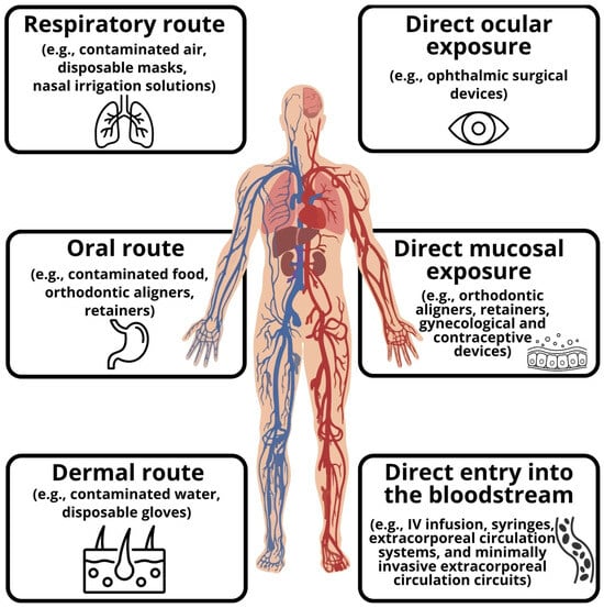
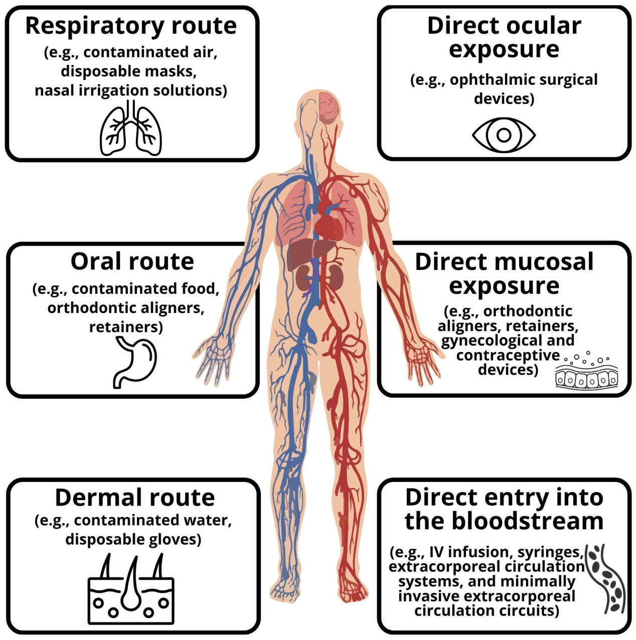
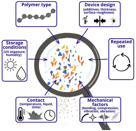
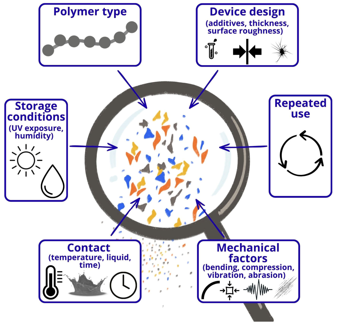
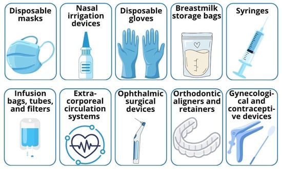
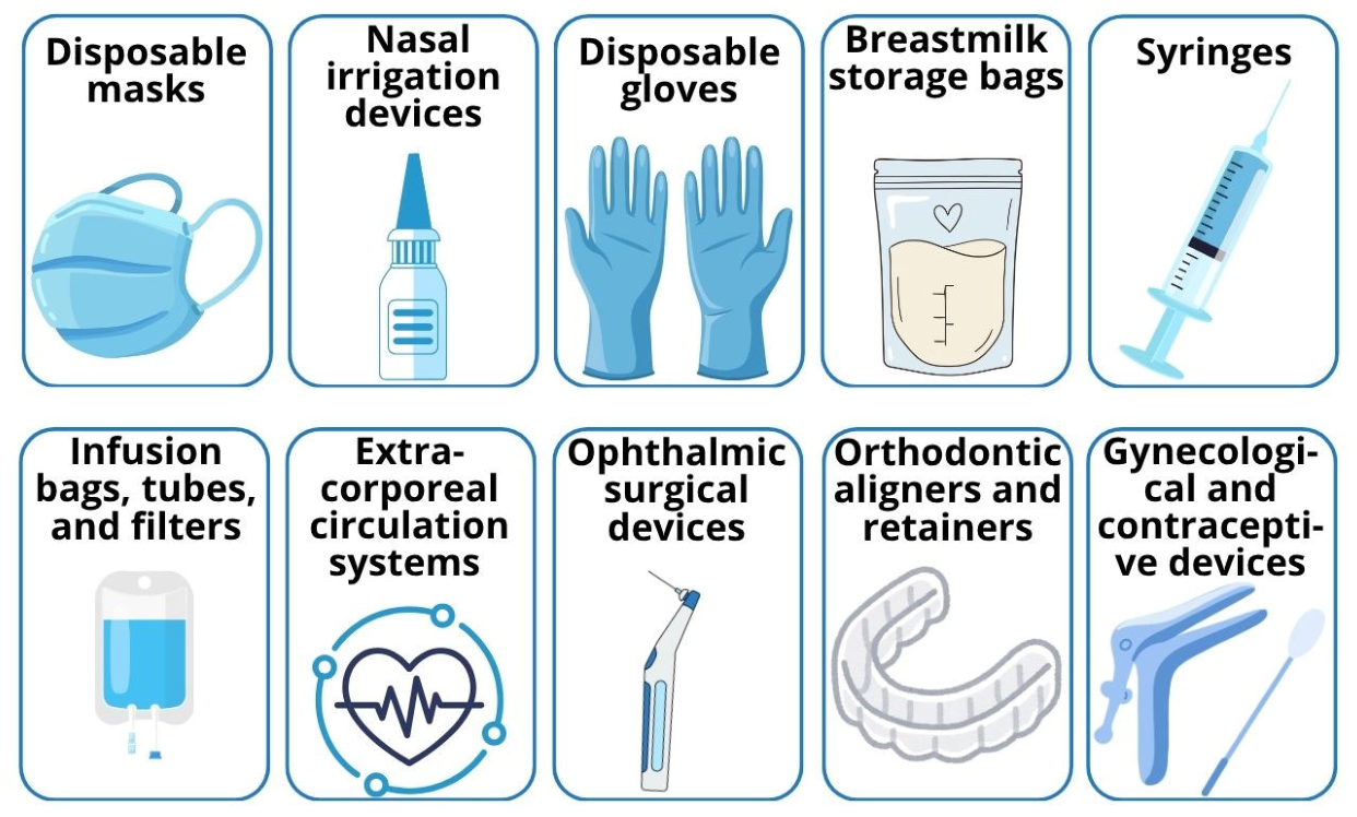

Disclaimer/Publisher’s Note: The statements, opinions and data contained in all publications are solely those of the individual author(s) and contributor(s) and not of MDPI and/or the editor(s). MDPI and/or the editor(s) disclaim responsibility for any injury to people or property resulting from any ideas, methods, instructions or products referred to in the content. |
© 2026 by the authors. Licensee MDPI, Basel, Switzerland. This article is an open access article distributed under the terms and conditions of the Creative Commons Attribution (CC BY) license.