


Abstract
The long-term biological fate of nanoplastics (NPs; <100 nm) in human tissues remains uncertain. Beyond acute toxicity, we propose a conceptual biological framework based on three interlinked pillars of host response: (1) biomolecular corona adaptation; (2) luminal biochemical–mechanical processing; and (3) a testable vascular-to-gastrointestinal (GI) recirculation hypothesis. This framework suggests that circulating NPs may undergo dynamic transit towards the GI tract, where mechanical shear and chemical degradation facilitate size reduction and surface erosion. Conversely, chronic entrapment in static compartments—such as the central nervous system parenchyma, fibrotic tissue, or atherosclerotic plaques—disrupts dynamic clearance. Luminal sequestration agents (i.e., Procambarus clarkii chitosan, PCC) represent potential exploratory proof-of-concept strategies to support elimination. This framework reframes systemic nanoplastic exposure from a static accumulation model to a dynamic physiological conversation. Specifically, we establish a conceptual biological framework that hypothesises three potential pillars of physiological response: (1) biocorona adaptability; (2) luminal chemical–mechanical processing capability; and (3) a testable vascular-to-GI recirculation hypothesis. According to this hypothesis, the migration of circulating particles in the path of the GI tract could act as an interacting physiological interface that promotes surface erosion and size reduction. This vascular-to-GI loop is proposed here as a viable hypothesis that requires quantitative mass-balance verification rather than an established physiological pathway. Conversely, this dynamic transit may be interfered with by chronic entrapment in static anatomical compartments, such as the brain parenchymal interstitium, fibrotic organ matrices, or atherosclerotic lesions.
1. Introduction
The long-term toxicokinetics and biological fate of <100 nm nanoplastics (NPs < 100 nm) in human systems are still not fully understood. A framework that takes into consideration physical changes, biomolecular interactions, and tissue clearance dynamics over long periods of time is essential to assess systemic effect, whereas conventional toxicology concentrates on acute intracellular reactions [1,2]. Conventional toxicology focuses predominantly on acute intracellular reactions to nanoplastics (NPs; <100 nm). However, understanding their long-term systemic impact requires evaluating dynamic interactions, biomolecular transformations, and tissue clearance kinetics over protracted timeframes. In this context, host ‘systemic resilience’ is defined as the physiological capacity to alter, redistribute, or delay xenobiotic-induced toxicity. Biomolecular corona formation represents the initial physicochemical phase of this response: plasma proteins rapidly coat circulating particles, redefining their biological identity and altering cellular endocytosis, immune recognition, and tissue uptake [3,4,5,6].
In vitro mammalian cell cultures and in vivo animal models demonstrate that sub-100 nm NPs are internalised by endothelial cells, epithelial cells, and macrophages via endocytosis, macropinocytosis, and passive membrane transport [4,5,7]. When particle aggregation or heavy biomolecular opsonisation surpasses intracellular degradation capacities, interstitial accumulation occurs, leading to prolonged tissue retention. Given the high cellular turnover of the gastrointestinal (GI) epithelium (approximately 300 g/day), the gut may function as a major clearance interface [8,9,10]. While it remains to be fully elucidated whether the biocorona merely attenuates acute reactivity or actively participates in size-reduction dynamics, it is hypothesised that circulating NPs may undergo dynamic vascular-to-gastrointestinal recirculation [1]. This perspective reframes exposure to NPs as a mechanistic, temporally protracted conversation between synthetic particles and human physiology rather than as a linear toxicological event. The human GI’s mechanical stressors—compression, shear forces, peristalsis, and abrasion—as well as chemical aggressors—acidic pH, bile salts, enzymes, reactive oxygen species (ROS), and hydrolytic processes—combine in order to trigger the fragmentation of NPs. To identify whether a material preferentially deforms, erodes, delaminates, or fractures into increasingly smaller particles, this interaction generates a polymer-specific “fragility signature” [11,12].
NP degradation is not limited to the lumen, but the GI tract is a main physiological location for initial mechanical fragmentation and biochemical surface erosion. Additionally, there is growing evidence that intracellular and extracellular processes may also result in systemic particle surface modifications, chemical degradation, and size modification. In particular, extracellular matrix-mediated degradation pathways, local production of ROS during phagocytic oxidative bursts, and endo-lysosomal acidification within macrophages can gradually alter synthetic polymer surfaces [13,14]. The ongoing development of the biomolecular corona, which continuously modifies surface reactivity, particle ageing, and intracellular destiny across biological compartments, operates in concert with these systemic biochemical processes [15].
Stiff, vitreous polymers including polystyrene (PS), polyvinyl chloride (PVC) and polymethyl methacrylate (PMMA) exhibit brittleness when undergoing compressive forces, rendering MPs susceptible to breaking into sub-micron particles. In contrast, semi-crystalline and more flexible polymers, such as polyethylene (PE) and polypropylene (PP), experience surface micro-abrasion, localised thinning, and progressive shedding instead of sudden fracturing. A third unique classification includes chemically hydrolysable polymers such as polyethylene terephthalate (PET), polyamide (PA), and polyurethane (PU), which exhibit moderate mechanical integrity but are susceptible to surface degradation, delamination, and backbone cleavage when exposed to gastric acidity (pH < 2.0), bile salts, and digestive enzymes [9,10,16]. Overall, polymer fragility in the GI tract and the heart’s cyclical pressure on the blood mass should be understood as a dynamic process of material deterioration, wherein chemical and mechanical stressors combine to reduce particle size and increase the potential for biological interaction [17,18].
Importantly, real sub-100 nm NPs exhibit significant and direct mechanical rupture from peristaltic compression or physiological shear forces alone. Conversely, further diminishment or deterioration at the nanoscale level is predominantly influenced by biological mechanisms, such as exposure to stomach acid (pH < 2.0), enzymatic hydrolysis, bile-induced solubilisation, and localised ROS-mediated polymer chain cleavage. In this nanoscale domain, physiological mechanical shear assumes a subordinate, non-fracturing function by aiding in the physical separation and degradation of chemically deteriorated surface layers. Differentiating between empirical findings from MPs (>1 µm) and real NPs (<100 nm) is crucial. Since surface chemistry, Brownian motion, interfacial energy, and aggregation dynamics mostly control nanoscale particles, direct mechanical breakage by physiological peristalsis is extremely rare. In order to explain where findings on NPs rely on actual nanoscale empirical data vs. theoretical extrapolation from MPs studies, Table 1 specifically classifies evidence according to particle size (<100 nm vs. >1 µm) and experimental model. Instead of generating primary mechanical micro-cleavage in this situation, physiological mechanical shear performs a secondary, non-fracturing function by encouraging the physical detachment and erosion of chemically damaged surface layers (Table 1) [19,20].
Table 1.
Proposed susceptibility and degradation characteristics of common plastic polymers under physiological GI conditions.
These polymer-specific fragmentation patterns have been progressively confirmed by current in vivo animal models and ex vivo physiological digestions. In particular, the combined effects of luminal shear stress, acidic gastric clearance, and intestinal peristalsis induce ingested stiff polymers such as PS and PVC to rapidly fracture into sub-micron particles [36,37]. Conversely, semi-crystalline and flexible polymers, including PE and PP, experience surface micro-abrasion, localised thinning, and gradual surface shedding instead of fast cleavage, demonstrating a strong resistance to catastrophic fracture [9,18,27]. When exposed to stomach fluid (pH < 2.0) and bile-mediated solubilisation, chemically sensitive polymers with hydrolysable ester or amide bonds (PET, PA, PU) exhibit rapid delamination [16,30].
Peristalsis and compression alone may not suffice to trigger further mechanical fracture in <100 nm NPs. Instead, a synergistic mechanism involving chemical hydrolysis, acidic pH, enzymatic digestion, bile-mediated solubilisation, and local ROS-mediated polymer chain scission is responsible for the size reduction and material degradation of NPs. Mechanical stress plays a secondary role in removing degraded surface layers [38].
Rigid polymers, such as PMMA, PS, and PVC, have the greatest probability of sharp fragmentation and the formation of NPs; elastic polymers, such as PE and PP, deform significantly more than they fracture, though long-term abrasion still reduces particle size; chemically vulnerable polymers, such as PA, PET, and PU, are subject to surface erosion and delamination, which promotes the release of smaller particles. Fragility is actually a consequence of developing “material fatigue” in the human environment rather than just mechanical stress. The aim of this study is to provide a mechanistic framework that defines systemic resistance to NPs as a time-dependent trait generated by three interconnected processes: vascular–gastrointestinal recirculation, organism-level fragmentation capability, and biomolecular corona creation. By combining data from human physiology and particle biology, we establish how these processes interact in order to determine NP fate and provide a framework explaining why only NPs trapped within the organ interstitial and atherosclerotic plaques are able to escape from this dynamic resilience cycle and maintain long-term persistence.
This article is not intended to be a systematic review or meta-analysis, but rather a biological conceptual framework. Since the main goal is to synthesise multidisciplinary mechanistic knowledge and suggest novel physiological hypotheses rather than to report on a comprehensive systematic extraction of data, no formal review methodology or PRISMA flowcharts were documented. Nevertheless, certain selection criteria applied to the literature were used to guarantee procedural transparency and scientific rigour. The wider physiological ability of the host system to modify, disperse, or postpone acute cellular damage caused by xenobiotic particles is the rigorous operationalisation of “systemic resilience” in this scenario. It is important to note that biomolecular corona formation is not an active host defence mechanism, but rather a spontaneous, passivity-driven physicochemical process controlled by surface thermodynamics and Vroman displacement. Furthermore, detoxification is not always indicated by particle redistribution, fragmentation, or surface changes; these changes might reduce or briefly increase biological reactivity and toxicity, depending on size, surface charge, and tissue microenvironment. Within this biological framework, resilience is defined as the host’s physiological ability to postpone acute toxicity by means of dynamic biocorona masking, localised material fragmentation, and ongoing tissue redistribution. As this study represents a narrative synthesis rather than a systematic review, a comprehensive multi-database search was implemented up to March 2026 to ensure full reproducibility. Primary records were retrieved from PubMed, Scopus, Web of Science, and Google Scholar using the exact database-specific Boolean syntax detailed herein: (“microplastics” OR “nanoplastics” OR “polymer particles”) AND (“protein corona” OR “biocorona” OR “ecocorona”) AND (“gastrointestinal” OR “intestinal barrier” OR “gut permeability”) AND (“recirculation” OR “clearance” OR “pharmacokinetics” OR “biodistribution”) AND (“atherosclerosis” OR “macrophage” OR “vascular endothelium”). Articles were screened based on relevance to systemic NP transport, physiological degradation, and tissue sequestration. Inclusion was limited to peer-reviewed English-language publications. To maintain terminological consistency throughout this manuscript, the term ‘biomolecular corona’ is adopted as the standard designation for the dynamic layer of biological macromolecules—comprising plasma proteins, lipids, metabolites, and active biomolecules—that spontaneously adsorb onto synthetic nanoparticle surfaces upon contact with biological fluids, thereby defining their biological identity [14,39]. Microplastics (MPs; particles > 1 µm to 5 mm) and NPs (particles < 100 nm or in the sub-micron range, depending on analytical context) are the standard abbreviations for MPs and NPs, respectively.
The literature identification and screening process was conducted in alignment with established methodological standards for narrative and scoping reviews [40,41].
To minimise selection bias, clear inclusion and exclusion criteria were established prior to narrative synthesis:
- Inclusion criteria: Peer-reviewed original research articles and authoritative reviews published in the English language focusing on the translocation, physical or chemical fragmentation/degradation, biomolecular corona formation, and pharmacokinetics of MPs and NPs (<100 nm and sub-micron range) across mammalian in vitro, ex vivo, and human in vivo models.
- Exclusion criteria: non-peer-reviewed preprints, conference proceedings lacking primary empirical data, opinion pieces without mechanistic evaluation, and studies exclusively investigating aquatic or non-mammalian organisms that are not directly applicable to human systemic physiology.
A summary of the record identification, deduplication, screening, and inclusion is provided in Table S1. Additionally, reference lists of the retrieved original articles and authoritative reviews were manually screened to identify further relevant studies published in peer-reviewed English-language journals.
2. Search Strategy and Narrative Synthesis Approach
2.1. Evidence Integration and Model Structuring
This article presents a conceptual narrative perspective to synthesise multidisciplinary knowledge and formulate a testable biological framework. The literature searches were conducted across PubMed, Scopus, and Web of Science up to March 2026 using structured search strings. Evidence was categorised into three core mechanistic domains: biomolecular corona dynamics, gastrointestinal fragmentation capacity, and vascular-to-GI recirculation. Physiological compartments were subsequently classified into dynamic routes (blood, lymph, GI lumen) and static sinks (atherosclerotic plaques, fibrotic interstitium, CNS parenchyma) based on clearance turnover. However, as this work is intended to formulate theoretical hypotheses rather than execute a formal systematic review, no systematic data extraction or PRISMA protocols were applied. Biomolecular corona formation, fragmentation capacity, and vascular-to-GI recirculation constituted the three mechanistic areas into which the data was classified. Consistency, mechanistic plausibility, and relevance to systemic particle fate have been evaluated for each field. To guarantee internal coherence and interoperability with recognised physiological principles such as immune cell trafficking, vascular disease, and epithelial turnover, the model was continually enhanced. Systemically circulating NPs reach the gastrointestinal wall via mesenteric and gastric arterial branches originating from the splanchnic circulation. Conversely, gut-absorbed particles enter the portal venous circulation to reach the liver, where hepatic processing may result in biliary secretion back into the duodenal lumen (true enterohepatic circulation). Vascular re-entry to intestinal tissues occurs specifically through mesenteric arterial beds rather than hepatic arterial routes.
The classification of biological sites into ‘dynamic’ and ‘static’ compartments serves as a theoretical framework rather than an absolute biological dichotomy. Compartments termed ‘static’—including vascular intima, renal interstice, and central nervous system parenchyma—reflect sites characterised by restricted lymphatic drainage, low turnover, or pronounced macrophage entrapment, where particle clearance kinetics are substantially prolonged. Sequestration within these sites should be understood as time-dependent and potentially reversible over extended periods, rather than permanent or absolute immobilisation. These physiological compartments could be identified for the fate of NPs based on convergence of standardised data: (a) a dynamic compartment including the gastrointestinal system, lymphatic circulation, and blood—sites with continuous flow, mechanical stress, and biochemical turnover; (b) a static compartment where macrophage-associated particles become stationary, characterised by the interstitial spaces of tissues and atherosclerotic plaques. NPs establish a process of chronic fibrotic remodelling that gradually affects tissue function after a crucial threshold of interstitial accumulation is exceeded. This behaviour is representative of a “metabolic scar”—defined here as a state of chronic, irreversible metabolic dysfunction and tissue remodelling that exacerbates over time. This classification establishes a mechanistic basis for differentiating between dynamic and static NP compartments and aligns with established pathways of macrophage retention, foam cell formation, and extracellular matrix entrapment observed in vascular and interstitial pathologies. In the context of this biological framework, ‘systemic resilience’ is operationalised strictly as the physiological capacity of the host to delay acute intracellular toxicity through dynamic biomolecular corona masking, localised material fragmentation, and continuous tissue redistribution. Furthermore, persistent non-resolving interstitial accumulation of NP aggregates is defined here as chronic tissue fibrosis or interstitial burden, replacing colloquial terms such as “metabolic scar”.
2.2. Fragmentation Pathway Characterisation
The primary fragmentation of NPs < 100 nm in the human GI system has not yet been confirmed by direct in vivo empirical evidence. MPs are the topic of the majority of the current studies on mechanical deterioration and size reduction. Our conceptual framework defines GI processing of NPs < 100 nm as a surface-dominated erosion process where shear-mediated surface detachment is employed in concert with enzymatic and oxidative chain scission, rather than as macroscopic mechanical shattering.
We combined data gathered from previous studies on: (a) mechanical degradation of polymers under shear and compression; (b) chemical modification by bile salts, pH gradients, and digestive enzymes; and (c) in vitro and in vivo models of NP exposure in GI settings in order to characterise the fragmentation pathway. The GI tract was determined as the main physiological site for NP fragmentation based on these findings. A structured “resilience loop” essentially represents the cyclical transport of NPs via circulation, tissues, and the GI tract and was established from the final conceptual framework. In comparison to previous studies, the framework had been evaluated for biological plausibility, consistency with established toxicokinetic principles, and explanation capacity. This study did not involve any animals, human participants, or experimental methods.
3. Evidence Synthesis and Proposed Conceptual Framework
3.1. Categorisation and Evidentiary Tiering of Model Core Tenets
The fundamental mechanistic pillars governing NP dynamic transit, transformation, and systemic fate are categorised into four formal evidentiary tiers in order to guarantee methodological rigour, transparent toxicological reporting, and a clear distinction between empirically validated bio-interactions and theoretical extrapolations within this biological framework:
- Established Evidence: The rapid adsorption of biomolecules to form a primary biomolecular corona upon contact with biological fluids, as well as intestinal epithelial uptake (via endocytosis or paracellular passage for sub-100 nm particles), represent well-documented processes validated in controlled mammalian in vitro and animal models, with preliminary structural observation in human tissues [13,42,43].
- Strongly Supported Inference: Based on bio-nano interface thermodynamics, soft-to-hard corona exchange dynamics, and the dynamic remodelling of the protein corona during systemic transit—which continuously redefines the biological identity of the particle, modifies pharmacokinetics, and sterically hinders or delays enzymatic matrix degradation—constitutes a strongly supported inference [43,44].
- Plausible Hypothesis: Based on in vitro polymer degradation studies and simulated physiological digestion models, the hypothesis of progressive mechanical–chemical fragmentation and superficial bio-degradation within the GI tract—driven by the synergistic action of stomach acidity, bile salt solubilisation, and local ROS generation—is supported by [14,45].
- Speculative Hypothesis: As a theoretical working framework, the systemic vascular-to-GI recirculation loop—which postulates that systemically circulating NPs undergo continuous retro-translocation back across the intestinal mucosa or biliary–enterohepatic interface into the GI lumen for shedding and faecal hypothesis—is a speculative working hypothesis requiring formal quantitative verification through mass-balance radio-labelled tracking [25,45].
3.2. Biomolecular Corona Formation: Physicochemical Masking and Biological Identity
Biomolecular corona formation is a passive physicochemical process governed by surface thermodynamics, hydrophobic interactions, and the Vroman effect. Plasma proteins quickly bind NPs when they enter biological fluids, altering their synthetic identity and affecting enzymatic accessibility and subsequent cellular interactions [1,6,38]. The particle’s biological identity is determined by its corona, which also influences immunological identification, cellular uptake, and biodistribution. The biomolecular corona is a spontaneous, passivity-driven process of protein adsorption dictated by surface free energy and Vroman displacement dynamics, which secondarily alters cellular uptake and circulation kinetics and is not a passive covering; it also extends circulation by stabilising the particle and minimising acute immunogenicity. The first phase of systemic resilience is represented by this process [46,47]. NPs are dynamic. Particles could be transported again into the interstitial space after penetrating tissues and inducing cellular stress and death. These particles eventually return to the GI system after re-entering the lymphatic or vascular circulation. The main mechanical processing setting is provided by the GI tract, which is characterised by the following factors: (a) peristaltic compression; (b) shear forces; (c) bile salts; (d) changing pH; (e) enzymatic activity; and (f) abrasive food materials. These pressures generate material fatigue, which culminates in gradual fragmentation [48,49,50]. These could be considered as a time-dependent resilience mechanism resulting from repeated exposure to GI.
3.3. Vascular-to-GI Recirculation: The Dynamic Loop
NPs are constantly transported by the organism from tissues to the bloodstream and subsequently into the GI tract. Epithelial turnover [38,51,52,53], mucosal barrier dynamics [54,55,56,57] lymphatic drainage, and macrophage trafficking all play a role in this loop (Figure 1).
Figure 1.
Mechanistic overview of static sequestration sinks and the disruption of the dynamic resilience loop. (a) Biomolecular corona-coated NPs penetrate the intima of the vascular wall through the damaged endothelium barrier and are phagocytosed by macrophages. Foam cell development, persistent oxidative stress (ROS generation), cell necrosis, and ultimately entrapment in the necrotic core of atherosclerotic plaques is caused by lysosomes’ inability to enzymatically break down synthetic polymers; (b) NPs migrate from the blood–brain barrier (BBB) into the neuronal parenchyma of the central nervous system (CNS). Due to limited glymphatic flow and ineffective parenchymal clearance pathways, they promote microglial activation and physically become trapped inside the interstitial space; (c) chronic injury-induced fibrosis and microvascular remodelling change the extracellular matrix (ECM) in parenchymal organs including the liver and kidneys. By immobilising NP aggregates and blocking their re-entry into the systemic circulation, this dense fibrotic network inhibits dynamic clearance.
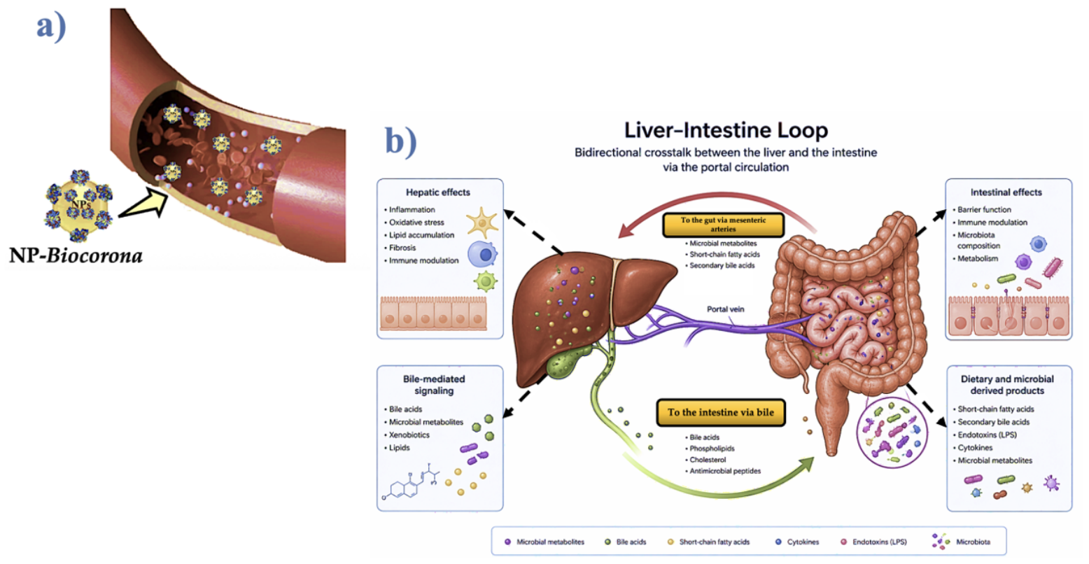
The mesenteric artery circulation is the way that systemic NPs get to the stomach. True enterohepatic circulation occurs when gut-absorbed NPs travel through the portal venous system to the liver, where hepatic processing may result in biliary excretion back into the GI tract. Mesenteric systemic arteries, not hepatic arteries, facilitate vascular re-entry to the gut [25,29]. The theoretical retro-translocation of systemically circulating NPs over the intestinal vascular–mucosal barrier is distinguished from proven hepatobiliary excretion in order to guarantee mechanistic clarity. Direct translocation from mesenteric artery blood back into the intestinal lumen is still a theoretical concept that has to be quantitatively verified using radio-labelled tracer kinetics, even though hepatobiliary transport of xenobiotics into the duodenum is a known clearance mechanism.
We suggest the vascular-to-GI recirculation loop as a biological theory based on integrative mucosal cell turnover, enterohepatic circulation, and macrophage trafficking. It is still necessary to provide solid evidence for the direct quantitative assessment of systemically absorbed NPs that return to the human GI lumen and are eliminated by faeces. Faecal excretion data from oral dosing studies primarily reflect unabsorbed passage through the gut lumen (>90–99%), while systemic-to-lumven retro-translocation via bile, biliary clearance, or desquamated enterocytes remains an unquantified hypothesis requiring mass-balance radio-labelling studies (Figure 1) [58,59]. Sequestration occurs primarily within immune-privileged anatomical sites or regions characterised by low lymphatic drainage, such as the central nervous system parenchyma, the renal interstitium, and the necrotic cores of vascular atherosclerotic plaques. Once phagocytosed by sub-endothelial macrophages within expanding atheromas, ingested synthetic polymers form sequestered, non-degradable polymer deposits due to the inability of lysosomal enzymes to cleave synthetic backbones [54,55,56,57,60,61,62,63]. Particles accumulate into the interstitial space in other organs such as the brain, kidney, liver [4,8,9], lung [64], or placenta [5]. These particles are incapable of undergoing further fragmentation and return to the GI tract. Long-term sequestration may be additionally facilitated by venous walls. In the systemic path of NPs, they ought to be the static compartments.
Permanent entrapment occurs in a number of anatomical static sites with low lymphatic clearance or high macrophage retention, such as: fibrotic renal and hepatic interstitial tissues; nervous system (CNS) parenchyma after blood–brain barrier passage; and atherosclerotic plaques within foam-cell-rich necrotic cores [1,9,61].
3.4. Conceptual Framework: The Resilience Loop
We conceptualise the resilience loop as a theoretical framework encompassing initial and lymphatic movement, enterocyte re-absorption, and final luminal discharge by physiological cell shedding into the faecal stream. Surface alteration of particles and incremental size diminution transpire during dynamic recirculation, propelled by the synergistic effects of gastrointestinal chemical hydrolysis and mechanical shear (Figure 2 and Figure 3).
Figure 2.
(a) Main potential pathways of NP intake and systemic distribution. The vast majority of ingested NPs transit through the stomach into the small intestine, where mucosal contact, endocytosis, and paracellular transport facilitate systemic uptake, whereas larger particles (>1 µm) are mostly retained or cleared by mucociliary elevation in the respiratory tract. (b) Translocation across epithelial barriers (enterocytes, pneumocytes) is governed by an interplay of particle size, shape, surface charge, and the composition of the acquired biomolecular corona. While particles smaller than 20–50 nm exhibit higher rates of endocytosis and paracellular transport, larger nanoparticles (up to 100–200 nm) can also cross mucosal barriers through receptor-mediated transcytosis, especially when opsonised by specific corona proteins; (c) NPs bigger than 20 nm up to 200–300 nm have been incorporated by reactive cells (such as macrophages) in the colon or airways; similarly, when circulating NPs reach the tissues, reactive cells are involved in their absorption process [65]. Solid arrows represent experimentally validated translocation pathways; dashed arrows represent hypothetical transport steps proposed in the vascular–gastrointestinal recirculation model (speculative hypotheses).
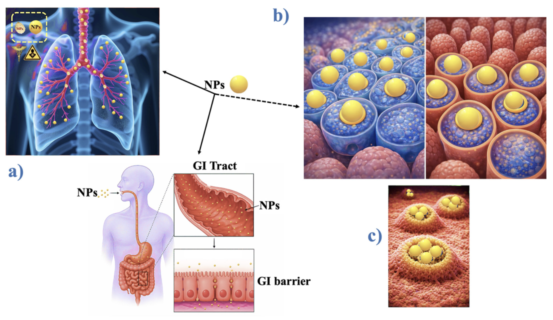
Figure 3.
(a) The NPs can be transported by the epithelial cell directly, by basolateral excretion, or after apoptosis to the vascular compartment (blood or lymphatic vessels). The NPs may return to the GI from the vascular compartment and enter the enterocyte. The enterocyte releases the NPs by apoptosis in the lumen, and the particle has been eliminated with stools; (b) the gut and liver loop. Systemic NPs reach the gut via the mesenteric arterial circulation. Gut-absorbed NPs enter the portal venous system to reach the liver, where hepatic processing can lead to biliary excretion back into the GI tract (true enterohepatic circulation). Vascular re-entry to the gut occurs via mesenteric systemic arteries, not hepatic arteries. Arrows indicate experimentally confirmed translocation pathways, while speculative transport stages suggested by the circulation model are represented by dashed arrows.
3.5. NP Pharmacokinetics
Several studies have been carried out to determine the presence of NPs in blood, and only a handful of them were observed (Table 2).
Analytical methods for MNP detection in human blood differ fundamentally in their physical principles. While µFTIR and µRaman provide spectroscopic particle counts but are blind to particles <300 nm or <1 µm respectively, Py-GC/MS quantifies total polymer mass concentration without resolving particle size or particle number. Therefore, human blood data reported in the literature must be parsed strictly by size class (MPs vs. <100 nm NPs) and interpreted as preliminary semi-quantitative estimates rather than absolute physiological steady-state levels (Table 2) [66]. Firm quantitative conclusions about concentrations, residence times, and clearance rates due to existing analytical methods (such as Py-GC/MS, µFTIR, and µRaman) cannot reliably quantify true sub-100 nm particles individually in complex human biological matrices. Thus, rather than being definitive measures of sub-100 nm NP toxicokinetics, findings reported in human blood and tissues should be considered semi-quantitative indicators of total exposure. Methodological heterogeneity is the primary cause of the large variation in reported circulating MNP concentrations among multi-group studies (from non-detectable to >1.5 µg/mL). While spectroscopic methods (µFTIR, µRaman) provide structural identity and particle counts but have strict lower size-resolution thresholds (~10 µm for standard µFTIR and ~1 µm for µRaman), Py-GC/MS measures total mass concentration without providing information on particle size, count, or morphology. As a result, <100 nm NPs are in an analytical “blind spot” for optical spectroscopy. Significant analytical noise is also introduced by the lack of standard reference materials, possible background aerosol contamination during blood collection and sample processing, and differences in matrix digestion procedures.
Until standardised, <100 nm quantitative techniques are devised, accurate toxicokinetic modelling is not possible due to these restrictions, which limit existing human blood data to preliminary semi-quantitative estimations [66].
Although it is difficult to compare the outcomes, the NP levels were determined as particles/mL [67,68] or in µg/mL [2,68,69,70]. The type of polymer remained nearly identical for most of the studies, including PE, PS, PVC, PP, and PMMA. The range of the blood levels was 0.12 µg/mL to 2.12 µg/mL. According to recent findings, human urine merely contains trace concentrations of NPs, indicating that urinary excretion of these xenobiotics is minimal [71,72]. This obviously indicates that systemic elimination is minimally enhanced by renal clearance.
A critical limitation in establishing quantitative toxicokinetic models for nanoplastics lies in the current analytical bottlenecks. Quantifying <100 nm particles in complex human biological matrices (such as whole blood, plasma, and tissue parenchymas) involves substantial measurement uncertainties. Techniques such as pyrolysis–gas chromatography/mass spectrometry (Py-GC/MS), micro-Raman spectroscopy (µRaman), micro-Fourier transform infrared spectrometry (µFTIR) and scanning electron microscopy (SEM) face trade-offs between chemical identification specificity, spatial resolution, and size detection limits. As highlighted by [66], method discrepancies, risk of background contamination during sampling, and lack of standardised reference materials introduce high variability across published blood and tissue concentration reports. Consequently, blood levels reported in Table 2 must be interpreted as qualitative or semi-quantitative indicators of systemic exposure rather than absolute steady-state concentrations [66].
The current analytical limitations around NPs < 100 nm represent a crucial theoretical limitation of our conceptual framework. Particles in the true nanometric regime (<100 nm) cannot be resolved by standard spectroscopic techniques (i.e., μFTIR, μRaman), whereas mass-based analytical procedures (i.e., Py-GC/MS) damage particle structural integrity, rendering it impossible to determine particle number, surface area, and aggregation state. Current analytical limitations impede accurate mass-balance measurement because biological barriers, endocytic transport, and biomolecular corona dynamic exchange are mostly determined by particle size and surface area in the <100 nm range. To verify the rate-limiting phases of systemic NP recirculation and tissue entrapment, high-resolution single-particle analytical methods (such as asymmetric flow field-flow fractionation combined with multi-angle light scattering and inductively coupled plasma mass spectrometry (AF4-MALS/ICP-MS), nano-Raman, or targeted radio-labelling) need to be established [73,74,75].
Although first exploratory studies have assessed luminal binding agents (such as certain cationic biopolymers; Procambarus clarkii chitosan–PCC) to modify faecal excretion kinetics [2,76,77], these approaches are still theoretical proof-of-concept models rather than proven therapeutic treatments (see Section 4.2). However, the clinical applicability and therapeutic efficacy of PCC remain speculative and require rigorous validation in independent, larger-scale clinical trials. Rather, the GI tract’s dynamics, which act as the main excretion interface involving enterohepatic recirculation and mucosal turnover, are represented in the substantial reduction in circulating NPs that was observed after a 15-day oral dose of PCC [2]. NPs accumulated within enterocytes are slowly eliminated by natural shedding given that the intestinal epithelium renews itself every 3 to 5 days, generating approximately 300 g of cells/day.
Table 2.
Studies on NP blood levels [28].
To construct a comprehensive toxicokinetic profile for NPs < 100 nm, current models frequently rely on empirical physiological data derived from MP exposure studies. However, critical size-dependent threshold differences must be maintained between micro- and nanoscale behaviours. Although derived from the microplastic literature (e.g., particles > 1 µm or >10 µm), these findings regarding biodistribution, tissue persistence, and mean residence time (MRT) are extrapolated cautiously to NPs (<100 nm), as smaller dimensions significantly increase surface-area-to-volume ratios, cellular uptake efficiency via M-cell-mediated transcellular transport, and systemic mobility across biological barriers [13,14].
NPs have unique pharmacokinetics controlled by instantaneous biomolecular corona dynamics, endo-lysosomal processing, and tissue-specific retention [15,43], whereas coarse MP fractions (>150 µm) are mainly confined to the intestinal lumen and subject to faecal excretion without significant mucosal uptake [79]. Therefore, direct translocation efficiency, intracellular accumulation, and long-term systemic recirculation loops continue to be exclusive operational properties of particles operating at the true nanoscale (<100 nm), even though MP observations provide crucial baseline parameters for initial biological clearance [14,25]. The framework developed from combining this biological turnover with the experimentally determined MRT (58 days) suggests that it will require approximately 4–5 months for circulating NPs to be almost entirely removed.
To formalise the resilience loop, the systemic transport, transformation, and clearance kinetics of NPs (<100 nm) are defined using a multicompartmental differential kinetic model:
where kdeg ≈ 0 and kclear ≈ 0, where CGI, Cblood, Ctissue, and Cstatic represent NP concentrations (μg/mL or particles/mL) in the GI lumen, vascular/lymphatic blood, mobile tissue/macrophages, and static sequestration compartments (i.e., vascular plaques, fibrotic tissue), respectively. The parameter I (t) (μg/mL⋅day or particles/mL⋅day) denotes the oral intake rate; while kabs, kfecal, kfrag, kvasc→GI, klymphj, ktissue, khepatic, kseq, kclear, and kdeg represent first-order rate constants (day−1) governing intestinal absorption, faecal elimination, luminal fragmentation, vascular-to-GI retro-translocation, lymphatic drainage, tissue uptake, hepatic clearance, static tissue sequestration, tissue clearance, and intra-tissue degradation, respectively. Under zero-input conditions (I (t) = 0) following complete absorption, and assuming negligible retro-translocation (kvasc → GI ≈ 0) and non-saturable systemic elimination, the vascular differential equation reduces to a classic single-compartment first-order kinetic decay model:
where C0 is the initial systemic concentration post-absorption, and k is the single-compartment first order elimination rate constant, defined as follows:
Assuming an operational MRT of approximately 58 days (k−1 = 58 days), an estimated total elimination window of 4 k−1 to 5 k−1 corresponds to approximately 232 to 290 days (7.6 to 9.5 months) to achieve >98% clearance (98.2% at 4 MRT and 99.3% at 5 MRT) of the absorbed internal dosage. Using <100 nm spherical PS particles under non-saturable physiological circumstances, this 58-day MRT value is purely obtained from mouse in vivo oral exposure data [13,70].
Importantly, the reduction of the multicompartmental system to this simplified mathematical decay relies on four explicit kinetic assumptions:
- First-order linear elimination kinetics: Assuming clearance rates remain directly proportional to systemic concentration.
- Single-compartment distribution: Assuming instantaneous, uniform vascular distribution without complex organ partitioning.
- Absence of deep-tissue re-uptake/sequestration: Neglecting irreversible organ trapping (kseq Ctissue) where kdeg ≈ 0.
- Non-saturable clearance pathways: Assuming macrophage uptake and biliary clearance mechanisms do not reach kinetic saturation.
The simplified single-compartment model’s projected 4–5 months clearance window is predicated on optimal first-order elimination kinetics (I (t) = 0, kvasc → GI ≈ 0) under non-saturable scenarios. For all NPs, this mathematical extrapolation does not represent an empirical toxicokinetic constant. Rather, it functions as a theoretical standard that requires formal empirical validation via mass-balance isotopic labelling studies. As a result, the correlation between computed decay rates and initial observational metrics is a simplified model-based prediction that has high uncertainty and does not indicate conclusive empirical verification. Polymer type, particle size distribution, surface functionalisation, biomolecular corona dynamics, and physiological species variations will all have a significant impact on the actual MRT and systemic clearance timeframe in vivo [13,70]. The exclusive compartment degradation assessment outlined in the present work employs an operational MRT of 58 days, exclusively obtained from rodent models subjected to unaltered PS-NPs under controlled experimental parameters. This parameter should not be extrapolated as a universal pharmacokinetic constant for human systemic clearance. Systemic residency durations in humans will significantly differ based on species-specific physiology, particle dimensions, polymer structure, corona behaviour, and tissue retention rates. According to previous studies, the mean around 58 days represents typical polymer populations (such as PE or PS) under specific intervention conditions. Nevertheless, this mathematical extrapolation cannot be applied to all NPs, independent of size, surface charge, or target tissue entrapment, as it presupposes a single compartment with continuous epithelium turnover. For MPs/NPs, a human MRT of approximately 58 days must be strictly treated as a first operational value determined under specific intervention scenarios (PCC co-administration). For synthetic or environmental NPs with different hydrodynamic diameters, surface charges, or tissue-specific retention profiles, it cannot act as a universal toxicokinetic constant.
The combination between kinetic modelling and physiological turnover emphasises the biological persistence of NPs in humans and provides significant internal validation for the estimated MRT. It is important to note that intestinal epithelial turnover does not automatically ensure the removal of NPs. Instead of luminal shedding, NPs internalised by enterocytes may proceed via intracellular disintegration, basal transcytosis into lymphatic/vascular beds, or transfer to sub-epithelial macrophages.
4. Discussion
4.1. Proposed Experimental Framework for Testing Biomolecular Corona-Driven Polymer Fragmentation
Future in vitro/in vivo studies must use standardised, multi-analytical procedures in order to transform the hypothetical association between biomolecular corona dynamics and polymer chain degradation from a theoretical theory to an experimentally verified mechanism [14,77]. Additionally, these studies should assess the following factors in order to empirically test the connection between biomolecular corona composition and polymer degradation:
- Soft vs. hard corona proteomic composition: Quantifying dynamic protein displacement (Vroman effect) and identifying selective adsorption of redox-active or hydrolytic enzymes using gel-based proteomics and liquid chromatography–tandem mass spectrometry (LC-MS/MS) after differential centrifugation [15,80].
- Surface oxidation and chemical transformation: Employing X-ray photoelectron spectroscopy (XPS) and μFTIR to monitor localised oxygen-to-carbon (O/C) ratios, carbonyl index increases, and functional group modifications along the polymer interface [19,27].
- Molecular weight distribution: Using gel permeation chromatography/size-exclusion chromatography (GPC/SEC) to identify backbone shortening, polymer chain scission, and variations in polydispersity indices carried on by enzymatic cleavage or biological oxidation [16,45].
- Particle size distribution: Tracking <100 nm size reduction, micro-cleavage, and shedding dynamics in real time under physiologically simulated conditions using asymmetric flow field-flow fractionation combined with multi-angle light scattering (AF4-MALS) or dynamic light scattering (DLS) [73,75,77,81].
4.2. Exploratory Proof-of-Concept: Luminal Sequestration Agents
Luminal sequestration approaches represent a preliminary exploratory avenue to intercept non-absorbed or recirculating MPs/NPs within the GI tract. Oral biopolymers, such as PCC, have shown an ability to bind luminal plastic particles via electrostatic and hydrophobic interactions in preliminary pilot observations. However, these findings should be interpreted strictly as an exploratory proof-of-concept rather than a validated clinical intervention. The core physiological framework proposed herein relies primarily on endogenous processes—such as biomolecular corona dynamic exchange, mucosal turnover, and biliary excretion. Any potential translational applications of luminal binding agents require independent, multi-centre validation regarding binding specificity, safety, and nutrient absorption impact [2,76,77]. Agents such as PCC need to be rigorously validated through independent, multi-centre clinical studies in order to determine their long-term binding selectivity, safety profile, influence on key nutrient absorption, and wide clinical efficacy [2,76,77]. Therefore, the core physiological resilience loop discussed here is largely dependent on endogenous biological processes, such as biomolecular corona dynamics, epithelial turnover, and enterohepatic recirculation, even though luminal binding agents provide an interesting experimental pathway for artificial clearance enhancement (Figures S1–S3) [1,15,25,51]. The observed decreases in circulating polymer signals after oral PCC treatment should be regarded with cautious consideration. These outcomes imply modified gastrointestinal processing, although they do not furnish conclusive evidence of improved systemic NP clearance through a vascular-to-GI loop. Alternative hypotheses—such as less luminal absorption, increased luminal aggregation, analytical inconsistencies, or variations in dietary exposure—cannot be dismissed without direct tracer-based mass-balance studies.
4.3. Testable and Falsifiable Predictions of the Resilience Loop
To transition the suggested resilience loop from a descriptive conceptual framework to a scientifically testable hypothesis, certain certain experimental methods, necessary controls, and the conclusive results must be delineated that would distinctly validate or refute each proposed pathway:
- Longitudinal Systemic-to-GI Tracing:
- ○
- Experimental Design: Intravenous or intra-intestinal administration of radio-labelled or fluorescently tagged <100 nm NPs in animal models [25,45,58,59].
- ○
- Required Controls: Biliary duct cannulation or ligation controls to distinguish direct vascular-to-intestinal mucosal retro-translocation from classical hepatobiliary excretion.
- ○
- Unambiguous Outcome: Detection of systemic NPs in luminal contents or faeces in biliary-ligated models would uniquely support direct vascular-to-intestinal retro-translocation; conversely, complete absence of faecal recovery following biliary blockade would falsify direct trans-mucosal transport.
- Lymphatic Blockade Impact:
- ○
- Experimental Design: Surgical disruption or pharmacological inhibition of mesenteric lymphatic drainage prior to systemic or luminal NP exposure [54,55,56,57].
- ○
- Required Controls: Sham-operated animals and non-targeted soluble macromolecular tracers to control for non-specific surgical trauma and baseline microvascular permeability.
- ○
- Unambiguous Outcome: A selective, significant reduction in intestinal particle re-entry and subsequent faecal elimination in lymph-blocked animals compared to sham controls would uniquely support lymphatic transport as a key component of the loop.
- Epithelial Turnover Modulation:
- ○
- Experimental Design: Pharmacological acceleration (i.e., via R-spondin 1) or inhibition (i.e., via chemotherapy agents or irradiation) of intestinal epithelial cell turnover [38,51,52,53].
- ○
- Required Controls: Vehicle-treated controls and assessment of mucosal tight-junction integrity (i.e., FITC-dextran flux) to ensure alterations in particle clearance are driven by cell shedding rather than non-specific barrier breakdown.
- ○
- Unambiguous Outcome: A proportional increase in particle faecal excretion during accelerated epithelial shedding—without increased paracellular leakiness—would uniquely confirm enterocyte shedding as a primary mucosal elimination pathway.
- Progressive Surface Oxidation:
- ○
- Experimental Design: Tracking molecular weight distributions and surface chemical states of recirculating NPs recovered from distal GI segments versus pristine ingested particles [16,19,27].
- ○
- Required Controls: Non-recirculating, luminal-only control particles incubated in simulated digestive fluids to isolate systemic biological oxidation from simple luminal chemical digestion.
- ○
- Unambiguous Outcome: A significant increase in oxygen-to-carbon (O/C) functional ratios and backbone chain scission uniquely observed in systemically recovered particles compared to luminal-only controls would confirm endogenous biological processing during recirculation [16,19,27].
- Sequestration vs. Health Kinetics:
- ○
- Experimental Design: Comparing tissue retention and clearance kinetics of injected NPs in animal models of advanced atherosclerosis or renal fibrosis versus healthy age-matched controls [1,4,9,61].
- ○
- Required Controls: Non-pathological, highly vascularised control tissue (i.e., healthy skeletal muscle) to rule out generalised hemodynamic alterations.
- ○
- Unambiguous Outcome: Exponentially prolonged tissue residence times (t1/2) restricted to fibrotic or atherosclerotic lesions would uniquely validate these pathological sites as static sequestration sinks that disrupt dynamic resilience.
4.4. Evolutionary Discrepancy, Anatomical Sequestration Sinks, and Toxicological Implications
As opposed to considering NP exposure as a static accumulation, this perspective reframes it as a dynamic biological process (Figure 1). The organism actively processes NPs via mechanical, metabolic, and immunological pathways instead of passively accumulating them. Particle size is reduced, surface chemistry is modified, and toxicity profiles might alter over time due to fragmentation, which probably occurs in the GI system. According to this perspective, the GI tract represents the main organ of resilience, serving as a metabolic interface and a physical barrier that may influence the fate of NPs that are absorbed. It is critical to note that particle fragmentation does not inherently equal detoxification. Decreasing particle size to the <50 nm regime drastically increases specific surface area, surface charge density, and potential ROS generation per unit mass. Consequently, while GI-mediated fragmentation may facilitate eventual chemical breakdown, it may transiently increase biological reactivity, cellular uptake, and systemic toxicity before complete clearance occurs [32]. Formation of the biomolecular corona, which is affected by the polymer’s type, size, shape, surface, and hydrophilic or hydrophobic nature, is the starting point subsequent to the absorption of NPs. NPs rapidly establish a dynamic protein corona when they enter biological fluids, which significantly alters their surface chemistry and, as a result, their fragmentation behaviour [1,80,82,83]. The biomolecular corona is able to attract redox-active or degradative proteins that locally enhance ROS fluxes and surface reactivity, accelerating polymer erosion, or it may protect the polymer surface from oxidative and enzymatic attack, inhibiting polymer oxidation and secondary fragmentation. The biomolecular corona indirectly affects where and how fragmentation occurs considering that it also influences the biological routing of the particle (i.e., toward acidic, enzymatically rich, or ROS-intense microenvironments). Thus, the biomolecular corona is not merely a passive coating but actually a key component in the physicochemical modification of the polymer, affecting the pathways and kinetics of NP removal in vivo.
In vivo, the biomolecular corona has a complicated, dual influence on the physical integrity of NPs as well as their dynamic changes. Conversely, the adsorption of high-affinity plasma proteins (also known as “hard corona”) produces a thick steric barrier that prevents direct enzymatic cleavage of the underlying polymer matrix, obscures surface reactive sites, and delays oxidative chain scission. Nevertheless, in some microenvironments (such acidic phagolysosomes or inflammatory tissues), dynamic corona development might encourage particle disintegration. Myeloperoxidase, transferrin, and redox-active proteins can be selectively enriched by corona formation. These proteins locally catalyse the production of hydroxyl radicals and ROS at the polymer interface. Whether a particle experiences rapid structural destruction or stays biologically disguised and prolonged in mesenteric arterial circulation is determined by this dynamic biomolecular corona remodelling (Figure 3) [1,80,82,83].
The composition of the biomolecular corona and the shape of the protein molecules also alter as NPs spread in the new biological environment, from the cellular to the blood or lymphatic compartments. These discrepancies could have a major effect on both the MRT and the hazardous activity of the NPs. The MRT of PP was higher than that of PE in volunteers that were considered healthy and in a healthy condition (mean values of 46 vs. 33, respectively), emphasising the importance of the polymer characteristics [2]. It is noteworthy to observe that PE’s reduced MRT compared with PP is in accordance with its widely recognised higher susceptibility to oxidative and mechanical fragmentation. This trend supports the concept that polymers more susceptible to in vivo fragmentation could undergo faster size reduction and clearance, even though the study was not designed to compare polymer fragility.
Although PP and PE exhibit a hydrophobic backbone, once a protein corona forms, their environmental and biological endurance diverges substantially. Even when the corona absorbs redox-active proteins, PP might possess a longer residence period due to its methyl-substituted backbone and tertiary carbon centres provide a surface that is less susceptible to oxidative activation. PE, conversely, exhibits faster hydrogen abstraction and ROS-driven oxidation due to its less complex, linear methylene structure, especially in microenvironments enriched with corona-associated enzymes or peroxidised proteins. This differentiation is further demonstrated by the protein corona: on PP, the adsorbed proteins often form a more stable, hydrophobic “hard” corona that preserves the polymer from oxidative or enzymatic access, postponing disintegration [1]. ROS, acids, and enzymes may interact with the polymer surface more easily on PE because the corona is typically more dynamic and permeable. Consequently, compared to PE, PP’s chemical structure and more protective corona phenotype result in lower oxidation, less chain scission, and subsequently a longer persistence. Discrepancies in the recorded residence durations of PE and PP therefore warrant caution in interpretation. Despite the extensive literature of variations in backbone structure and susceptibility to oxidation in polymer chemistry, direct experimental proof connecting in vivo polymer oxidation to diminished particle size and expedited clearance has yet to be confirmed.
From an evolutionary perspective, human physiology has adapted to natural particulate stress over millennia, whereas synthetic NPs represent a recent anthropogenic exposure (<100 years). Consequently, endogenous clearance pathways process synthetic polymers through an existing biological repertoire that did not evolve specifically for synthetic material degradation. Pathological sites, such as atherosclerotic plaques or fibrotic organs, act as static sequestration sinks where impaired vascular permeability and restricted lymphatic drainage disrupt dynamic recirculation, leading to permanent local entrapment. Their unique cellular environment, especially characterised by inadequate clearance, altered flow dynamics, and chronic inflammation, renders it common for NPs to accumulate and remain, thereby bypassing the typical efficient processing that occurs in healthy tissues. To prevent reverse causality, it is essential to distinguish between active illness causation and passive physical sequestration (particle entrapment). Pre-existing endothelial dysfunction, impaired lymphatic/glymphatic clearance, and increased macrophage recruitment may transform vascular atherosclerotic plaques or neurodegenerative parenchymal lesions into biological “sinks” that passively absorb circulating NPs. The presence of accumulating NPs in static lesions largely indicates a failure of physiological clearance pathways rather than a defined primary aetiology, even if they could worsen local inflammation as a secondary factor [58,61,62,78].
As a consequence, risk assessment should evolve from a static assessment of the burden of NPs to an assessment of their dynamic biological fate, considering clearance, fragmentation, translocation, and the distinctive vulnerabilities of pathological ecosystems. By recognising both the benefits and current limitations of human adaptability, this framework aligns exposure studies with the temporal and mechanical realities of human biology. MRTs, which generally represent substantial tissue loading and usually manifest clinically as organ-specific toxicity, are not neutral pharmacokinetic characteristics. Classical persistent agents provide a clear example of the following: amiodarone accumulates in lung, liver, skin, and cornea and damages those organs [84,85], desipramine exhibits prolonged persistence in the CNS and myocardium with corresponding neuropsychiatric and arrhythmogenic risks [39], chloroquine and hydroxychloroquine concentrate in lysosome-rich tissues and the retina, where their toxicity appears noticeable [86], and dioxins accumulate in the liver and adipose tissue, resulting in long-term metabolic and carcinogenic effects [87]. By comparison, a systemic MRT of several weeks for NPs clearly suggests that toxicity and accumulation at the organ level are not only feasible but likewise probable. High blood flow by itself does not always correspond to tissue toxicity, even though high-flow vascular beds the kidneys, liver, and GI system) invariably get a larger cumulative mass delivery of circulating NPs per unit time. Instead, a complex interaction between microvascular endothelial permeability, cell-specific absorption efficiency, local macrophage clearance activity, tissue retention kinetics, and particle physicochemistry determines final organ sensitivity [59,70]. High regional blood flow should therefore be interpreted as a physiological predictor of initial particle delivery and potential systemic interaction, rather than an automatic indicator of localised tissue damage. It is evident from this arrangement that, with the exception of the heart and lipids, each other organ is subjected to extremely high flow, especially the liver and gut, contributing to approximately 50% of the mesenteric arterial circulation. These organs ought to be more adaptable to a material with some degree of hazardous activity and an MRT of months (Table 3).
Table 3.
Blood flow at rest for 75 kg of body weight.
A broad spectrum of chronic and acute diseases (19 and 21, respectively) is associated with exposure to NPs, according to the Chicago Cluster, which identifies 26 signs and symptoms that correspond to the presence of NPs [88,89,90]. Oxidative stress and low-grade inflammation share a mechanistic axis, which is particularly represented in this correlation. This biological “irritating thorn” may aggravate the underlying disease and enhance pre-existing vulnerabilities, even at subclinical levels. GI disorders and inflammatory markers, particularly increased C-reactive protein (CRP), are among the most frequently observed symptoms correlated with NPs. The high-blood-flow complementarity across the gut and the liver, which increases the potential of NP translocation and systemic inflammatory signalling, is consistent with this pattern. Similar to how it occurs with blood glucose or cholesterol, every individual’s susceptibility to the toxicity of NPs is likely determined by a specific sensitivity threshold or biological cutoff.
Detectable circulating NPs may be found in apparently healthy subjects without apparent clinical symptoms as their levels could be elevated for a longer period of time before symptoms emerge. However, this threshold is still not well established at present.
The differential toxicity of different polymers is also undetermined. A key factor in opsonisation, aggregation, fragmentation, and MRT is the formation of the biomolecular corona that is defined by polymer characteristics such as hydrophobicity, polarity, and chemical inertness. The biological fate and pathogenicity are subsequently regulated by these corona-driven processes. Nevertheless, the target organ profile of NPs in humans remains mostly unidentified, compared to these well-characterised drugs and toxicological agents.
Vascular atherosclerotic plaques are not the only conditions that trigger NPs, which are failure sites of the dynamic recirculation loop, to get permanently immobilised. Instead, permanent entrapment occurs at a number of anatomical static sites with low lymphatic clearance or high macrophage retention, such as: fibrotic renal and hepatic interstitial tissues, CNS parenchyma after blood–brain barrier passage, and atherosclerotic plaques within foam-cell-rich necrotic cores [1,9,61].
While a fraction of NPs recirculates between the vascular system and mucosal interfaces, complete systemic clearance is prevented when particles become permanently sequestered in anatomical compartments with limited lymphatic drainage or impaired phagocytic clearance. Rather than atherosclerotic plaques being the sole point of failure, permanent entrapment occurs across multiple privileged or pathological sites, including:
- Atherosclerotic plaques: where macrophage foam cells engulf NPs but fail to clear them, leading to necrotic core accumulation and plaque destabilisation;
- CNS parenchyma: where passage across the BBB is followed by interstitial trapping due to restricted lymphatic/glymphatic exit pathways;
- Fibrotic tissue and renal interstitium: where impaired vascular microcirculation traps NP aggregates.
It is important to distinguish between real NPs (<100 nm) and MPs. The majority of current studies on human bioaccumulation, including findings in brain parenchymal tissues, saphenous veins, and atheromas, depend on spectroscopic methods (μFTIR, μRaman) that are physically limited by optical diffraction limitations (~1–10 μm). Therefore, rather than single sub-100 nm NPs, evidence of particle sequestration in these tissues mostly represents micro-scale particles or aggregate masses. The surface-area-to-volume ratios of true NPs are much larger, which results in unique endocytic transport rates and barrier permeability that cannot be inferred simply from MP measurements.
5. Conclusions
Permanent immobilisation of NPs—representing failure points of the dynamic recirculation loop—is not limited to vascular atherosclerotic plaques. Rather, permanent entrapment occurs across multiple anatomical static sites characterised by low lymphatic clearance or high macrophage retention, including: (1) atherosclerotic plaques within foam-cell-rich necrotic cores; (2) CNS parenchyma following blood–brain barrier passage; and (3) fibrotic renal and hepatic interstitial tissues [1,9,61]. In order to explain the long-term biological fate of NPs, this study provides a conceptual framework that connects gastrointestinal processing, dynamic redistribution, and biomolecular corona formation. The suggested vascular-to-GI recirculation loop is a validated hypothesis rather than a proven approach. The lack of quantitative tracer trials in humans, dependence on small proof-of-concept intervention studies, and analytical limitations in identifying sub-100 nm NPs in vivo constitute a few of the major limitations of the current study. The development of standardised quantitative techniques for the detection of sub-100 nm NPs in biological fluids, tracer-labelled animal studies to confirm systemic-to-GI luminal transport, and a systematic investigation of the mechanical vs. chemical fragmentation dynamics of several polymer types under physiological conditions must be the main goals of future studies. Preliminary findings regarding luminal sequestration by PCC should be interpreted strictly as an exploratory proof-of-concept. PCC cannot currently be recommended for therapeutic intervention until its safety, binding specificity, and efficacy are established through rigorous, multi-centre clinical trials. Although PCC-based formulations hold patents for stomach-targeted MP trapping and are already commercially available in markets such as Italy as dietary supplements, their broad clinical recommendation as a validated therapeutic intervention requires further confirmation through rigorous, multi-centre clinical trials evaluating long-term safety, binding specificity, and systematic efficacy.
Supplementary Materials
The following supporting information can be downloaded at https://www.mdpi.com/article/10.3390/microplastics5040202/s1: Table S1: Summary of literature search strategy, database records, and screening workflow (up to March 2026); Figure S1: Certificate of analysis of PCC; Figure S2: 1H-NMR spectra of PCC; Figure S3: SEM images of NP-PS 100 nm standard (Figure S3a–c); SEM images of PCC with NP-PS 100 nm (Figure S3d–f).
Author Contributions
U.C.: Conceptualisation, Methodology, Formal Analysis, Supervision, Statistical analysis, Visualisation, Validation, Writing—original draft, Writing—review and editing; R.L.: Visualisation, Funding, Methodology, Validation, Writing—review and editing; and C.C.: Conceptualisation, Formal Analysis, Supervision, Methodology, Visualisation, Writing—original draft, Writing—review and editing. All authors have read and agreed to the published version of the manuscript.
Funding
This research was funded by the RUDN University Scientific Projects Grant System, grant number: 021411-0-000.
Data Availability Statement
The original contributions presented in this study are included in the article/Supplementary Material. Further inquiries can be directed to the corresponding authors.
Acknowledgments
All the authors acknowledged the support from the RUDN University Scientific Projects Grant System, grant number: 021411-0-000. During the preparation of this manuscript/study, the author(s) used Gemini 3.8 Flash for the purposes of generate images and Graphical Abstract. The authors have reviewed and edited the output and take full responsibility for the content of this publication. This article is dedicated to Alexander Santiago Casella Flores.
Conflicts of Interest
The authors declare no conflicts of interest.
Abbreviations
The following abbreviations are used in this manuscript:
| AF4-MALS | Asymmetric Flow Field-Flow Fractionation Combined with Multi-Angle Light Scattering |
| AF4-MALS/ICP-MS | Asymmetric Flow Field-Flow Fractionation Combined with Multi-Angle Light Scattering and Inductively Coupled Plasma Mass Spectrometry |
| BBB | Blood–Brain Barrier |
| CNS | Central Nervous System |
| CRP | C-Reactive Protein |
| DLS | Dynamic Light Scattering |
| ECM | Extracellular Matrix |
| GI | Gastrointestinal Tract |
| GPC/SEC | Gel Permeation Chromatography/Size-Exclusion Chromatography |
| LC–MS/MS | Liquid Chromatography–Tandem Mass Spectrometry |
| LOD | Limit of Detection |
| MPs | Microplastics |
| MRT | Mean Residence Time |
| NPs | Nanoplastics |
| PCC | Procambarus clarkii Chitosan |
| PA | Polyamide |
| PE | Polyethylene |
| PET | Polyethylene Terephthalate |
| PMMA | Polymethyl Methacrylate |
| PP | Polypropylene |
| PS | Polystyrene |
| PVC | Polyvinyl Chloride |
| PU | Polyurethane |
| Py-GC/MS | Pyrolysis-Gas Chromatography/Mass Spectrometry |
| ROS | Reactive Oxygen Species |
| SEM | Scanning Electron Microscopy |
| XPS | X-ray Photoelectron Spectroscopy |
| µFTIR | Micro-Fourier Transform Infrared Spectrometry |
| µRaman | Micro-Raman Spectroscopy |
References
- Casella, C.; Ballaz, S.J. Genotoxic and neurotoxic potential of intracellular nanoplastics: A review. J. Appl. Toxicol. 2024, 44, 1657–1678. [Google Scholar] [CrossRef] [Scilit] [PubMed]
- Casella, C.; Cornelli, U.; Ballaz, S.; Recchia, M.; Zanoni, G.; Ramos-Guerrero, L. Preliminary Study on PCC-Chitosan’s Ability to Enhance Microplastic Excretion in Human Stools from Healthy Volunteers. Foods 2025, 14, 2190. [Google Scholar] [CrossRef] [Scilit] [PubMed]
- Sharma, P.; Dagariya, S.; Sharma, S.; Singh, M. Uncovering the nexus of human health hazards of nanoplastics, gut-dysbiosis and antibiotic-resistance. J. Environ. Sci. Health Part C 2026, 44, 1–60. [Google Scholar] [CrossRef] [Scilit] [PubMed]
- Augey, L.; Berdougo-Tritz, J.; Mbarek, A.; Jadoul, M.; Massy, Z.; Deray, G.; Liabeuf, S. Effects of Microplastics and Nanoplastics on the Kidneys. Nephrol. Dial. Transplant. 2026, 41, 1413–1420. [Google Scholar] [CrossRef] [Scilit] [PubMed]
- Ragusa, A.; Svelato, A.; Santacroce, C.; Catalano, P.; Notarstefano, V.; Carnevali, O.; Giorgini, E. Plasticenta: First evidence of microplastics in human placenta. Environ. Int. 2021, 146, 106274. [Google Scholar] [CrossRef] [Scilit] [PubMed]
- Zhang, T.; Wang, Z.; Wu, Y.; Zhu, S.; Su, J. Interactions of micro-and nanoplastics with biomolecules: From public health to protein corona effect and beyond. J. Phys. Chem. B 2025, 129, 5355–5374. [Google Scholar] [CrossRef] [Scilit] [PubMed]
- Wang, Y.F.; Wang, X.Y.; Chen, B.J.; Yang, Y.P.; Li, H.; Wang, F. Impact of microplastics on the human digestive system: From basic to clinical. World J. Gastroenterol. 2025, 31, 100470. [Google Scholar] [CrossRef] [Scilit] [PubMed]
- Dzierżyński, E.; Blicharz-Grabias, E.; Komaniecka, I.; Panek, R.; Forma, A.; Gawlik, P.J.; Flieger, J. Post-mortem evidence of microplastic bioaccumulation in human organs: Insights from advanced imaging and spectroscopic analysis. Arch. Toxicol. 2025, 99, 4051–4066. [Google Scholar] [CrossRef] [Scilit] [PubMed]
- Nihart, A.J.; Garcia, M.A.; El Hayek, E.; Liu, R.; Olewine, M.; Kingston, J.D.; Campen, M.J. Bioaccumulation of microplastics in decedent human brains. Nat. Med. 2025, 31, 1114–1119. [Google Scholar] [CrossRef] [Scilit] [PubMed]
- Rotchell, J.M.; Jenner, L.C.; Chapman, E.; Bennett, R.T.; Bolanle, I.O.; Loubani, M.; Palmer, T.M. Detection of microplastics in human saphenous vein tissue using μFTIR: A pilot study. PLoS ONE 2023, 18, e0280594. [Google Scholar] [CrossRef] [Scilit] [PubMed]
- Dávalos, A.; Martín-Santiago, V.; de Las Hazas, M.C.L. Micro-and Nanoplastics in the Human Intestine: A Review of Health Implications and Possible Dietary Interventions. Adv. Nutr. 2026, 17, 100624. [Google Scholar] [CrossRef] [Scilit] [PubMed]
- Zhao, H.; Lin, G.; Yin, Y.; Wu, Q.; Wang, Y.; Tang, N.; Qi, X. Impact of micro-and nanoplastics on gastrointestinal diseases: Recent advances. Eur. J. Intern. Med. 2025, 139, 106419. [Google Scholar] [CrossRef] [Scilit] [PubMed]
- Prata, J.C.; Da Costa, J.P.; Lopes, I.; Duarte, A.C.; Rocha-Santos, T. Environmental exposure to microplastics: An overview on possible human health effects. Sci. Total Environ. 2020, 702, 134455. [Google Scholar] [CrossRef] [Scilit] [PubMed]
- Kaseke, T.; Lujic, T.; Cirkovic Velickovic, T. Nano-and microplastics migration from plastic food packaging into dairy products: Impact on nutrient digestion, absorption, and metabolism. Foods 2023, 12, 3043. [Google Scholar] [CrossRef] [Scilit] [PubMed]
- Monopoli, M.P.; Walczyk, D.; Campbell, A.; Elia, G.; Lynch, I.; Baldelli Bombelli, F.; Dawson, K.A. Physical− chemical aspects of protein corona: Relevance to in vitro and in vivo biological impacts of nanoparticles. J. Am. Chem. Soc. 2011, 133, 2525–2534. [Google Scholar] [CrossRef] [Scilit] [PubMed]
- Zimmermann, L.; Bartosova, Z.; Braun, K.; Oehlmann, J.; Völker, C.; Wagner, M. Plastic products leach chemicals that induce in vitro toxicity under realistic use conditions. Environ. Sci. Technol. 2021, 55, 11814–11823. [Google Scholar] [CrossRef] [Scilit] [PubMed]
- Deng, B.D.; Sinha, S.R.; Lear, G.; Tropini, C. Microplastics and nanoplastics in the human gut: From signals to standards. Nat. Rev. Gastroenterol. Hepatol. 2026, 23, 374–375. [Google Scholar] [CrossRef] [Scilit] [PubMed]
- Jarmakiewicz-Czaja, S.; Ferenc, K.; Sokal-Dembowska, A.; Wojdyła, D.; Filip, R. Effects of microplastics on the intestines. BMC Gastroenterol. 2026, 26, 289. [Google Scholar] [CrossRef] [Scilit] [PubMed]
- Baysal, A.; Soyocak, A.; Saygin, H.; Saridag, A.M. Exposure to phagolysosomal simulated fluid altered the cytotoxicity of PET micro (nano) plastics to human lung epithelial cells. Toxicol. Mech. Methods 2024, 34, 72–97. [Google Scholar] [CrossRef] [Scilit] [PubMed]
- Sharma, D.; Sharma, A. Molecular insights into physiological impact of micro-and nano-plastics on the digestive system and gut-brain axis. Comp. Biochem. Physiol. Part C Toxicol. Pharmacol. 2026, 304, 110473. [Google Scholar] [CrossRef] [Scilit] [PubMed]
- Zhao, G.; Tan, M.; Wu, B.; Zheng, X.; Xiong, R.; Chen, B.; Chu, C. Redox oscillations activate thermodynamically stable iron minerals for enhanced reactive oxygen species production. Environ. Sci. Technol. 2023, 57, 8628–8637. [Google Scholar] [CrossRef] [Scilit] [PubMed]
- Shao, Z.; Su, J.; Dong, J.; Liang, M.; Xiao, J.; Liu, J.; Chen, C. Aggregation kinetics of polystyrene nanoplastics in gastric environments: Effects of plastic properties, solution conditions, and gastric constituents. Environ. Int. 2022, 170, 107628. [Google Scholar] [CrossRef] [Scilit] [PubMed]
- Lee, G.; Jhang, Y.J.; Jhang, Y.T.; Chang, Y.C.; Chang, H.W.; Chuang, C.Y.; Hsiao, I.L. Artificial digestion represents the worst-case scenario for studying nanoplastic fate in gastrointestinal tract. J. Hazard. Mater. 2025, 485, 136809. [Google Scholar] [CrossRef] [Scilit] [PubMed]
- Chen, Y.; Xuan, Y.; Chen, X.; Wu, M.; Xu, J.; Chen, J. Gastrointestinal digestion potentiates nanoplastic-induced intestinal barrier dysfunction and macrophage-driven inflammation. J. Hazard. Mater. 2026, 503, 141129. [Google Scholar] [CrossRef] [Scilit] [PubMed]
- Wang, X.; Wang, Q.; Jiang, W.; Wang, B.; Zhang, X.; Wang, T. Enterohepatic Circulation of Polystyrene Nanoplastics Promotes Intestinal Inflammation by Impairing Enteric Neurons. ACS Nano 2026, 20, 19976–19994. [Google Scholar] [CrossRef] [Scilit] [PubMed]
- Bao, Z.Z.; Lu, S.Q.; Wang, G.; Cai, Z.; Chen, Z.F. Adsorption of 2-hydroxynaphthalene, naphthalene, phenanthrene, and pyrene by polyvinyl chloride microplastics in water and their bioaccessibility under in vitro human gastrointestinal system. Sci. Total Environ. 2023, 871, 162157. [Google Scholar] [CrossRef] [Scilit] [PubMed]
- Gürgen, S. Wear behavior of UHMWPE composites under oxidative effect. Polym. Degrad. Stab. 2022, 199, 109912. [Google Scholar] [CrossRef] [Scilit]
- Liebgott, C.; Chaib, I.; Doyen, P.; Robert, H.; Eutamene, H.; Duflos, G.; Mercier-Bonin, M. Fate and impact of nanoplastics in the human digestive environment after oral exposure: A common challenge for toxicology and chemistry. TrAC Trends Anal. Chem. 2023, 166, 117175. [Google Scholar] [CrossRef] [Scilit]
- Capuano, N.; Lombardi, M.; Cafà, N.; Marino, M.; Salzano, F.; Scalia, F.; Rinaldi, L. Micro-and Nanoplastics as Disruptors of Digestive and Hepatopancreatic Homeostasis: Insights into the Plastic-Gut-Liver Axis. Int. J. Mol. Sci. 2026, 27, 3272. [Google Scholar] [CrossRef] [Scilit] [PubMed]
- Khalid, R.; Amin, H.M.; Shahid, M.; Kazuya, N.; Xing, R.; Liu, S.; Fujishima, A. Integrated photothermal and photocatalytic degradation of micro-/nanoplastics: A mini-review with mechanistic insights and future perspectives. J. Mater. Chem. A 2025, 13, 26110–26128. [Google Scholar] [CrossRef] [Scilit]
- Li, L.; Zhang, H.; Jin, X.; Xu, Q.; Ding, H.; Liu, C.; Zhang, G. Polyethylene terephthalate nanoplastics promote gastric cancer progression by inhibiting the TNF-α/JAK1/STAT1 signaling pathway and shaping an immunosuppressive microenvironment. Environ. Int. 2026, 212, 110289. [Google Scholar] [CrossRef] [Scilit] [PubMed]
- Hanzlova, M.; Holmannova, D.; Svadlakova, T.; Fiala, Z. Small particles, large questions: Unravelling the toxicity and potential health risks of PET micro-/nanoplastics. Part. Fibre Toxicol. 2026, 23, 19. [Google Scholar] [CrossRef] [Scilit] [PubMed]
- Peng, C.; He, N.; Wu, Y.; Lu, Y.; Sun, H.; Wang, L. Excretion characteristics of nylon microplastics and absorption risk of nanoplastics in rats. Ecotoxicol. Environ. Saf. 2022, 238, 113586. [Google Scholar] [CrossRef] [Scilit] [PubMed]
- Li, X.; Huang, Y.; Zu, D.; Liu, H.; He, H.; Bao, Q.; Cheng, X. PMMA nanoplastics induce gastric epithelial cellular senescence and cGAS-STING-mediated inflammation via ROS overproduction and NHEJ suppression. Ecotoxicol. Environ. Saf. 2024, 287, 117284. [Google Scholar] [CrossRef] [Scilit] [PubMed]
- Zheng, P.C.; Pan, X.Q.; Zhou, Y.J.; Lai, K.P.; Li, R.; Zhang, X.X. Unraveling the impact of micro-and nano-sized polymethyl methacrylate on gut microbiota and liver lipid metabolism: Insights from oral exposure studies. Environ. Pollut. 2025, 373, 126157. [Google Scholar] [CrossRef] [Scilit] [PubMed]
- Djouina, M.; Waxin, C.; Caboche, S.; Dehaut, A.; Himber, C.; Balesdent, C.; Body-Malapel, M. Ingestion of environmentally sourced polyvinyl chloride microplastic fragments increases colon inflammation and fibrosis in mice. Environ. Res. 2025, 285, 122621. [Google Scholar] [CrossRef] [Scilit] [PubMed]
- Liang, B.; Zhong, Y.; Huang, Y.; Lin, X.; Liu, J.; Lin, L.; Huang, Z. Underestimated health risks: Polystyrene micro-and nanoplastics jointly induce intestinal barrier dysfunction by ROS-mediated epithelial cell apoptosis. Part. Fibre Toxicol. 2021, 18, 20. [Google Scholar] [CrossRef] [Scilit] [PubMed]
- Wright, S.L.; Kelly, F.J. Plastic and human health: A micro issue? Environ. Sci. Technol. 2017, 51, 6634–6647. [Google Scholar] [CrossRef] [Scilit] [PubMed]
- Amitai, Y.; Frischer, H. Excess fatality from desipramine and dosage recommendations. Ther. Drug Monit. 2004, 26, 468–473. [Google Scholar] [CrossRef] [Scilit] [PubMed]
- Page, M.J.; McKenzie, J.E.; Bossuyt, P.M.; Boutron, I.; Hoffmann, T.C.; Mulrow, C.D.; Moher, D. The PRISMA 2020 statement: An updated guideline for reporting systematic reviews. BMJ 2021, 372, n71. [Google Scholar] [CrossRef] [Scilit] [PubMed]
- Tricco, A.C.; Lillie, E.; Zarin, W.; O’Brien, K.K.; Colquhoun, H.; Levac, D.; Straus, S.E. PRISMA extension for scoping reviews (PRISMA-ScR): Checklist and explanation. Ann. Intern. Med. 2018, 169, 467–473. [Google Scholar] [CrossRef] [Scilit] [PubMed]
- Gao, H.T.; Zhou, H.Q.; Ji, H.N.; Shen, J.Z.; Yin, D.M.; Wu, D.H. From exposure to neurotoxicity induced by micro-nanoplastics with brain accumulation and cognitive decline. Ecotoxicol. Environ. Saf. 2025, 304, 119114. [Google Scholar] [CrossRef] [Scilit] [PubMed]
- Brouwer, H.; Porbahaie, M.; Boeren, S.; Busch, M.; Bouwmeester, H. The in vitro gastrointestinal digestion-associated protein corona of polystyrene nano-and microplastics increases their uptake by human THP-1-derived macrophages. Part. Fibre Toxicol. 2024, 21, 4. [Google Scholar] [CrossRef] [Scilit] [PubMed]
- Panico, S.; Capolla, S.; Bozzer, S.; Toffoli, G.; Dal Bo, M.; Macor, P. Biological features of nanoparticles: Protein corona formation and interaction with the immune system. Pharmaceutics 2022, 14, 2605. [Google Scholar] [CrossRef] [Scilit] [PubMed]
- Wu, X.; Zhang, H.; Chen, J.; Tan, F.; Cai, R.; Wang, Y. Photoaging promotes toxic micro/nanoplastics release from PLA/PBAT biodegradable plastic in gastrointestinal condition. Environ. Health 2025, 3, 446. [Google Scholar] [CrossRef] [Scilit] [PubMed]
- Dai, Q.; Yan, Y.; Ang, C.S.; Kempe, K.; Kamphuis, M.M.; Dodds, S.J.; Caruso, F. Monoclonal antibody-functionalized multilayered particles: Targeting cancer cells in the presence of protein coronas. ACS Nano 2015, 9, 2876–2885. [Google Scholar] [CrossRef] [Scilit] [PubMed]
- Tenzer, S.; Docter, D.; Kuharev, J.; Musyanovych, A.; Fetz, V.; Hecht, R.; Stauber, R.H. Rapid formation of plasma protein corona critically affects nanoparticle pathophysiology. In Nano-Enabled Medical Applications; Jenny Stanford Publishing: New York, NY, USA, 2020; pp. 251–278. [Google Scholar] [CrossRef] [Scilit]
- Forte, M.; Iachetta, G.; Tussellino, M.; Carotenuto, R.; Prisco, M.; De Falco, M.; Valiante, S. Polystyrene nanoparticles internalization in human gastric adenocarcinoma cells. Toxicol. Vitr. 2016, 31, 126–136. [Google Scholar] [CrossRef] [Scilit] [PubMed]
- Stock, V.; Böhmert, L.; Lisicki, E.; Block, R.; Cara-Carmona, J.; Pack, L.K.; Lampen, A. Uptake and effects of orally ingested polystyrene microplastic particles in vitro and in vivo. Arch. Toxicol. 2019, 93, 1817–1833. [Google Scholar] [CrossRef] [Scilit] [PubMed]
- Vauthier, C.; Bouchemal, K. Methods for the preparation and manufacture of polymeric nanoparticles. Pharm. Res. 2009, 26, 1025–1058. [Google Scholar] [CrossRef] [Scilit] [PubMed]
- Barker, N. Adult intestinal stem cells: Critical drivers of epithelial homeostasis and regeneration. Nat. Rev. Mol. Cell Biol. 2014, 15, 19–33. [Google Scholar] [CrossRef] [Scilit] [PubMed]
- Choi, J.; Augenlicht, L.H. Intestinal stem cells: Guardians of homeostasis in health and aging amid environmental challenges. Exp. Mol. Med. 2024, 56, 495–500. [Google Scholar] [CrossRef] [Scilit] [PubMed]
- Li, P.; Liu, J. Micro (nano) plastics in the human body: Sources, occurrences, fates, and health risks. Environ. Sci. Technol. 2024, 58, 3065–3078. [Google Scholar] [CrossRef] [Scilit] [PubMed]
- Clevers, H. The intestinal crypt, a prototype stem cell compartment. Cell 2013, 154, 274–284. [Google Scholar] [CrossRef] [Scilit] [PubMed]
- Neurath, M.F.; Artis, D.; Becker, C. The intestinal barrier: A pivotal role in health, inflammation, and cancer. Lancet Gastroenterol. Hepatol. 2025, 10, 573–592. [Google Scholar] [CrossRef] [Scilit] [PubMed]
- Pentinmikko, N.; Lozano, R.; Scharaw, S.; Andersson, S.; Englund, J.I.; Castillo-Azofeifa, D.; Katajisto, P. Cellular shape reinforces niche to stem cell signaling in the small intestine. Sci. Adv. 2022, 8, eabm1847. [Google Scholar] [CrossRef] [Scilit] [PubMed]
- Turner, J.R. Intestinal mucosal barrier function in health and disease. Nat. Rev. Immunol. 2009, 9, 799–809. [Google Scholar] [CrossRef] [Scilit] [PubMed]
- Shan, S.; Zhang, Y.; Zhao, H.; Zeng, T.; Zhao, X. Polystyrene nanoplastics penetrate across the blood-brain barrier and induce activation of microglia in the brain of mice. Chemosphere 2022, 298, 134261. [Google Scholar] [CrossRef] [Scilit] [PubMed]
- Zhang, H.J.; Li, S.; Wang, X.L.; Zhang, K.D.; Fang, H.T.; Wu, X.; Miao, A.J. Size-dependent translocation of polystyrene nanoplastics across biological barriers in mammals. Nat. Commun. 2025, 17, 1116. [Google Scholar] [CrossRef] [Scilit] [PubMed]
- Blagov, A.V.; Markin, A.M.; Bogatyreva, A.I.; Tolstik, T.V.; Sukhorukov, V.N.; Orekhov, A.N. The role of macrophages in the pathogenesis of atherosclerosis. Cells 2023, 12, 522. [Google Scholar] [CrossRef] [Scilit] [PubMed]
- Marfella, R.; Prattichizzo, F.; Sardu, C.; Fulgenzi, G.; Graciotti, L.; Spadoni, T.; Paolisso, G. Microplastics and nanoplastics in atheromas and cardiovascular events. N. Engl. J. Med. 2024, 390, 900–910. [Google Scholar] [CrossRef] [Scilit] [PubMed]
- Marfella, R.; Prattichizzo, F.; Barbieri, M.; Paolisso, P.; Scisciola, L.; Basilicata, M.G.; Paolisso, G. Microplastics and atherosclerosis: Mechanisms. Annu. Rev. Pharmacol. Toxicol. 2026, 66, 369–390. [Google Scholar] [CrossRef] [Scilit] [PubMed]
- Theofilis, P.; Oikonomou, E.; Tsioufis, K.; Tousoulis, D. The role of macrophages in atherosclerosis: Pathophysiologic mechanisms and treatment considerations. Int. J. Mol. Sci. 2023, 24, 9568. [Google Scholar] [CrossRef] [Scilit] [PubMed]
- Wang, S.; Lu, W.; Cao, Q.; Tu, C.; Zhong, C.; Qiu, L.; Liu, J. Microplastics in the lung tissues associated with blood test index. Toxics 2023, 11, 759. [Google Scholar] [CrossRef] [Scilit] [PubMed]
- Casella, C.; Cornelli, U.; Ballaz, S.; Zanoni, G.; Merlo, G.; Ramos-Guerrero, L. Plastic Smell: A Review of the Hidden Threat of Airborne Micro and Nanoplastics to Human Health and the Environment. Toxics 2025, 13, 387. [Google Scholar] [CrossRef] [Scilit] [PubMed]
- Thomas, K.V.; Belz, S.; Booth, A.M.; Clift, M.J.; Cross, R.K.; Davies, G.; Barron, L.P. Communicating confidence in the reliability of micro-and nanoplastic identification in human health studies. Environ. Health 2026, 4, 804. [Google Scholar] [CrossRef] [Scilit] [PubMed]
- Lee, D.W.; Jung, J.; Park, S.A.; Lee, Y.; Kim, J.; Han, C.; Hong, Y.C. Microplastic particles in human blood and their association with coagulation markers. Sci. Rep. 2024, 14, 30419. [Google Scholar] [CrossRef] [Scilit] [PubMed]
- Leonard, S.V.; Liddle, C.R.; Atherall, C.A.; Chapman, E.; Watkins, M.; Calaminus, S.D.; Rotchell, J.M. Microplastics in human blood: Polymer types, concentrations and characterisation using μFTIR. Environ. Int. 2024, 188, 108751. [Google Scholar] [CrossRef] [Scilit] [PubMed]
- Brits, M.; Van Velzen, M.J.; Sefiloglu, F.Ö.; Scibetta, L.; Groenewoud, Q.; Garcia-Vallejo, J.J.; Lamoree, M.H. Quantitation of micro and nanoplastics in human blood by pyrolysis-gas chromatography–mass spectrometry. Microplast. Nanoplast. 2024, 4, 12. [Google Scholar] [CrossRef] [Scilit]
- Chen, C.Y.; Kamineni, V.N.; Lin, Z. A physiologically based toxicokinetic model for microplastics and nanoplastics in mice after oral exposure and its implications for human dietary exposure assessment. J. Hazard. Mater. 2024, 480, 135922. [Google Scholar] [CrossRef] [Scilit] [PubMed]
- Calikanzaros, E.; Donat-Vargas, C.; Chevance, G.; Tonne, C.; Boleda, M.R.; Dalmau, J.; Villanueva, C.M. Micro-and nanoplastics in biological samples following a drinking water intervention in Barcelona, Spain. PLoS Water 2026, 5, e0000469. [Google Scholar] [CrossRef] [Scilit]
- Ji, S.; Wang, W.; Wang, Y.; Bai, H.; Li, Z.; Huo, Z.; Luo, K. Quantitative Detection of Micro-and Nanoplastics (≥ 300 nm) in Human Urine Using Double-Shot Py-GC/MS with Internal Standard Calibration. Toxics 2025, 13, 452. [Google Scholar] [CrossRef] [Scilit] [PubMed]
- Valido, I.H.; Fuentes-Cebrian, V.; Hernández, A.; Valiente, M.; López-Mesas, M. Validated method for polystyrene nanoplastic separation in aqueous matrices by asymmetric-flow field flow fraction coupled to MALS and UV–Vis detectors. Microchim. Acta 2023, 190, 285. [Google Scholar] [CrossRef] [Scilit] [PubMed]
- Chakraborty, S.; Drexel, R.; Bhadane, P.; Langford, N.; Dhumal, P.; Meier, F.; Lynch, I. An integrated multimethod approach for size-specific assessment of potentially toxic element adsorption onto micro-and nanoplastics: Implications for environmental risk. Nanoscale 2025, 17, 9122–9136. [Google Scholar] [CrossRef] [Scilit] [PubMed]
- Bartczak, D.; Sikora, A.; Goenaga-Infante, H.; Altmann, K.; Drexel, R.; Meier, F.; Giovannozzi, A.M. Multiparameter characterisation of a nano-polypropylene representative test material with fractionation, light scattering, high-resolution microscopy, spectroscopy, and spectrometry methods. Environ. Sci. Nano 2026, 13, 1391–1405. [Google Scholar] [CrossRef] [Scilit]
- Cornelli, U.; Belcaro, G.; Casella, C. Reduction in Circulating Microplastics in Humans Following Gastrointestinal Sequestration by Chitosan: A Pilot Controlled Study. J. Xenobiotics 2026, 16, 92. [Google Scholar] [CrossRef] [Scilit] [PubMed]
- Casella, C.; Cornelli, U.; Schiavi, S.; Taglietti, A.; Luque, R.; Zanoni, G. Mechanistic insights into polystyrene nanoplastic sequestration by a Procambarus clarkii-derived chitosan hydrogel in simulated gastric fluid: An in vitro study. Food Hydrocoll. 2026, 180, 112910. [Google Scholar] [CrossRef] [Scilit]
- Leslie, H.A.; Van Velzen, M.J.; Brandsma, S.H.; Vethaak, A.D.; Garcia-Vallejo, J.J.; Lamoree, M.H. Discovery and quantification of plastic particle pollution in human blood. Environ. Int. 2022, 163, 107199. [Google Scholar] [CrossRef] [Scilit] [PubMed]
- Cornelli, U.; Recchia, M.; Casella, C. The Food Microplastic Pyramid (FOMIC-Py) as a Novel Framework for Prioritizing Dietary Exposure and Industrial Processing Impact: An Italian North-South Exposure Model. Toxics 2026, 14, 578. [Google Scholar] [CrossRef] [Scilit] [PubMed]
- Mahmoudi, M.; Landry, M.P.; Moore, A.; Coreas, R. The protein corona from nanomedicine to environmental science. Nat. Rev. Mater. 2023, 8, 422–438. [Google Scholar] [CrossRef] [Scilit] [PubMed]
- Cornelli, U.; Casella, C. Microplastics and Nanoplastics as Potential Metabolic Disruptors: Implications for Insulin Resistance and Type 2 Diabetes. Toxics 2026, 14, 634. [Google Scholar] [CrossRef] [Scilit] [PubMed]
- Schroffenegger, M.; Leitner, N.S.; Morgese, G.; Ramakrishna, S.N.; Willinger, M.; Benetti, E.M.; Reimhult, E. Polymer topology determines the formation of protein corona on core–shell nanoparticles. ACS Nano 2020, 14, 12708–12718. [Google Scholar] [CrossRef] [Scilit] [PubMed]
- Sun, Y.; Zhou, Y.; Rehman, M.; Wang, Y.F.; Guo, S. Protein corona of nanoparticles: Isolation and analysis. Chem Bio Eng. 2024, 1, 757–772. [Google Scholar] [CrossRef] [Scilit] [PubMed]
- Budin, C.E.; Cocuz, I.G.; Sabău, A.H.; Niculescu, R.; Ianosi, I.R.; Ioan, V.; Cotoi, O.S. Pulmonary fibrosis related to amiodarone—Is it a standard pathophysiological pattern? A case-based literature review. Diagnostics 2022, 12, 3217. [Google Scholar] [CrossRef] [Scilit] [PubMed]
- Papiris, S.A.; Triantafillidou, C.; Kolilekas, L.; Markoulaki, D.; Manali, E.D. Amiodarone: Review of pulmonary effects and toxicity. Drug Saf. 2010, 33, 539–558. [Google Scholar] [CrossRef] [Scilit] [PubMed]
- Yusuf, I.H.; Charbel Issa, P.; Ahn, S.J. Hydroxychloroquine-induced retinal toxicity. Front. Pharmacol. 2023, 14, 1196783. [Google Scholar] [CrossRef] [Scilit] [PubMed]
- Mathew, N.; Somanathan, A.; Tirpude, A.; Pillai, A.M.; Mondal, P.; Arfin, T. Dioxins and their impact: A review of toxicity, persistence, and novel remediation strategies. Anal. Methods 2025, 17, 1698–1748. [Google Scholar] [CrossRef] [Scilit] [PubMed]
- Casella, C.; Dondi, D.; Vadivel, D. Do Microplastics (MPs) and Nanoplastics (NPs) Directly Contribute to Human Carcinogenesis? Environ. Pollut. 2025, 388, 127343. [Google Scholar] [CrossRef] [Scilit] [PubMed]
- Casella, C.; Cornelli, U.; Zanoni, G.; Moncayo, P.; Ramos-Guerrero, L. Health Risks from Microplastics in Intravenous Infusions: Evidence from Italy, Spain, and Ecuador. Toxics 2025, 13, 597. [Google Scholar] [CrossRef] [Scilit] [PubMed]
- Cornelli, U.; Casella, C.; Belcaro, G.; Cesarone, M.R.; Marucci, S.; Rondanelli, M.; Zanoni, G. Mapping Clinical Disease Patterns Potentially Associated with Microplastic Exposure: A Narrative Review Across Twenty-One Disease Categories. Green Health 2026, 2, 9. [Google Scholar] [CrossRef] [Scilit]
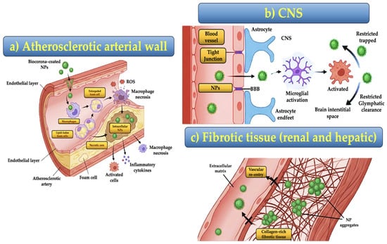
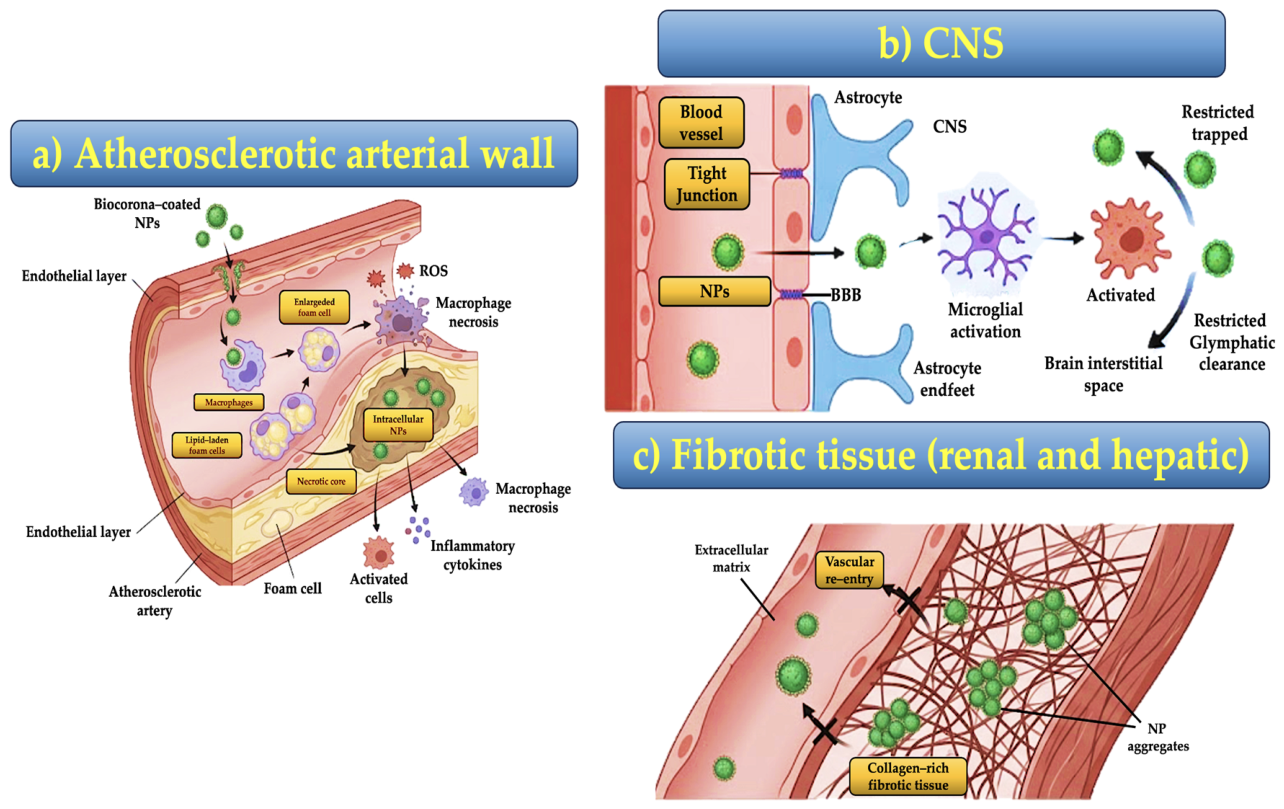

Disclaimer/Publisher’s Note: The statements, opinions and data contained in all publications are solely those of the individual author(s) and contributor(s) and not of MDPI and/or the editor(s). MDPI and/or the editor(s) disclaim responsibility for any injury to people or property resulting from any ideas, methods, instructions or products referred to in the content. |
© 2026 by the authors. Licensee MDPI, Basel, Switzerland. This article is an open access article distributed under the terms and conditions of the Creative Commons Attribution (CC BY) license.