


Abstract
COVID-19 is increasingly recognized as a multisystemic disease with significant neurocognitive consequences. However, its specific impact on spatial memory, a cognitive domain essential for daily navigation and functional independence, remains insufficiently explored. This narrative review provides a critical synthesis of current evidence regarding spatial and visuospatial memory alterations across acute and post-acute phases, and post COVID-19 condition (PCC). Clinical findings, conventional and emerging assessment tools ranging from static tasks to immersive virtual reality environments, as well as potential neurobiological mechanisms, were considered. Results suggested that spatial memory is frequently compromised after COVID-19 disease, with deficits being most pronounced at longer retention intervals and within navigational contexts. Neuroimaging and biomarker data further reveal selective vulnerability in the medial temporal lobe, characterized by hippocampal atrophy, hypoperfusion, and disrupted functional connectivity. Importantly, traditional neuropsychological tools may underestimate these impairments due to limited ecological validity. Therefore, implementing multimodal assessment frameworks that integrate navigational paradigms is essential to enhance diagnostic sensitivity and facilitate the development of targeted rehabilitation strategies for PCC patients.
1. Introduction
The coronavirus disease (COVID-19), an infectious disease caused by the severe acute respiratory syndrome coronavirus 2 (SARS-CoV-2), has been increasingly recognized as a systemic condition with significant neurological and neurocognitive consequences. In terms of temporal framing, COVID-19 symptomatology can be classified into an acute phase (up to 4 weeks from symptom onset), a post-acute phase (from 4 to 12 weeks), and post COVID-19 condition (PCC) when symptoms persist beyond 12 weeks (National Institute for Health and Care Excellence; NICE) [1]. COVID-19 can present either symptomatically or asymptomatically [2]. In the symptomatic manifestation, it can be classified as non-severe, severe, or critical according to respiratory involvement and clinical deterioration [3]. Thus, severe forms are defined by pneumonia, hypoxemia, and signs of respiratory distress, whereas critical illness involves acute respiratory distress syndrome, sepsis, or other conditions requiring life-support measures [3].
Beyond its well-established respiratory manifestations, accumulating evidence indicates that COVID-19 affects the nervous system through a combination of limited direct viral involvement, peripheral nervous system pathways, and immune-mediated mechanisms, thereby contributing to the cognitive and neurological sequelae observed during and after infection [4]. Regarding cognitive functioning, COVID-19 has been associated with a wide range of symptoms, including processing speed, attention, memory, and executive functions [5]. These cognitive symptoms have been reported in COVID-19 patients regardless of the severity of the acute infection, including individuals who were not hospitalized [6,7], and may persist for weeks or months into the post-acute and post-COVID-19 phases, contributing to long-term functional impairment [8].
Previous research highlights memory impairment as a frequently reported cognitive sequela following COVID-19. Both cross-sectional and longitudinal studies have described alterations in memory performance during the acute phase of infection and throughout the post-acute period, with deficits observed across different levels of disease severity [9]. For memory assessment, studies have primarily focused on verbal memory, often assessed using conventional neuropsychological measures such as word-list learning tasks [10]. However, other memory systems have been less explored in COVID-19 patients. Specifically, spatial memory, defined as the ability to represent and recall locations and orientations in the environment, supporting navigation and goal-directed behavior [11], has typically been embedded within broader neuropsychological batteries or assessed through tasks that mainly capture the visual component of visuospatial memory capacity, rather than spatial learning, navigation, or allocentric representations [10]. However, spatial memory is crucial in daily life activities, having its impairment a marked impact on functional independence and safety [12]. Moreover, its assessment is clinically relevant because previous studies have observed that spatial memory tasks show greater sensitivity and specificity for identifying cognitive impairment than conventional neuropsychological assessments [13]. As a result, despite the functional and clinical relevance of spatial memory, the current literature provides limited insight into whether COVID-19 is associated with specific alterations in spatial memory processes and their supporting neural systems.
Spatial memory is closely reflected in navigation and orientation abilities, which depend on the capacity to form, retain, and use internal representations of the surrounding environment [14]. Conceptually, spatial memory should be distinguished from visuospatial processing; whereas visuospatial processing primarily relies on perceptual–visual analysis and mental imagery and does not necessarily require the formation of map-like spatial representations, spatial memory supports navigation and orientation through the formation of internal spatial representations that integrate multimodal sensory information, including visual, vestibular, proprioceptive, and motor cues [15].
Spatial memory encompasses multiple core components, including spatial orientation, route learning, navigation, topographical memory, object–location associations, and cognitive map formation [16]. These components rely on egocentric and allocentric reference frames, spatial representations which support human spatial behavior. Thus, egocentric representations enable to encode locations relative to the observer’s viewpoint (subject-to-object relationships), allowing the generation of body-centered representations, while allocentric representations encode spatial relationships independent of the individual’s position (world-based coordinates and object-to-object relationships), being critical for flexible navigation [14,17].
Considering the environment in which the spatial memory task is conducted, neuropsychological assessment of spatial memory encompasses static environments and navigational contexts [18]. Static or non-navigational spaces correspond to the area within one’s reach and do not require bodily movement, enabling tasks to be completed without changes in position or visual perspective. Paper-and-pencil and computerized tasks are typical examples of spatial memory assessments in such contexts. In contrast, navigational environments extend to spaces within walking distance and involve either physical or simulated movement. Accordingly, spatial memory paradigms may incorporate real or immersive virtual navigation, as well as passive, perspective-based video tours.
Following Chandra et al. [19], spatial memory is critically supported by the hippocampal–entorhinal system, where the integration of place and grid cell activity supports the formation of stable and flexible cognitive maps. This medial temporal lobe (MTL) system operates within a broader distributed network, involving the parahippocampal, retrosplenial, and posterior parietal cortices, which facilitates scene representation and the transformation between egocentric and allocentric reference frames.
Hippocampal impairments after SARS-CoV-2 infection, including altered neurogenesis, degeneration, and volume reduction, have been observed in previous studies [20,21]. Complementarily, dysfunctional activity within the hippocampus and between left and right parahippocampal regions was also documented [21]. These findings highlight the importance of investigating spatial memory in COVID-19 patients, as disruptions in hippocampal and parahippocampal function may underlie the spatial cognitive deficits reported in this population.
Despite the growing body of literature documenting cognitive sequelae following SARS-CoV-2 infection, the specific impact of COVID-19 on spatial memory remains unclear. Existing studies have primarily focused on the verbal or visual memory components, whereas spatial memory tasks have been frequently embedded within broader neuropsychological batteries, yielding heterogeneous and difficult-to-compare findings. Moreover, potential neurobiological mechanisms underlying spatial memory alterations following COVID-19 have rarely been examined in an integrated manner. Therefore, the present narrative review aims to provide a critical synthesis of the current evidence on spatial memory alterations associated with COVID-19, encompassing acute and post-acute stages, as well as PCC. Specifically, this review wants to: (1) summarize and critically evaluate clinical evidence of spatial memory deficits and to examine potential moderating factors; (2) review the neuropsychological tools and emerging approaches employed for spatial memory assessment in this population; and (3) examine potential neurobiological mechanisms that may contribute to spatial memory dysfunction, with particular emphasis on hippocampal and network-level vulnerability. By integrating clinical, neurobiological, and methodological perspectives, this review aims to clarify current knowledge gaps and to outline directions for future research in this underexplored cognitive domain. Additionally, given the limited number of studies directly assessing spatial memory, this review will also consider evidence derived from neuropsychological tasks targeting visuospatial memory processes, as these measures capture cognitive components closely related to spatial memory functioning. However, it is important to acknowledge that these traditional tools (discussed in greater detail in Section 3) are inherently heterogeneous and often combine spatial memory with executive, organizational, and attentional processes. Consequently, impairments identified through these proxy measures should be interpreted with caution, as they may reflect executive inefficiencies rather than primary deficits in spatial mapping.
2. Spatial Memory Alterations After COVID-19
Studies designed to characterize spatial memory across the different phases of the disease remain limited.
2.1. Spatial Memory in Acute and Post-Acute Phases
In the acute and post-acute literature, cognitive assessment has most commonly relied on brief, global cognitive screening instruments, particularly the Montreal Cognitive Assessment and Mini Mental State Examination (MoCA and MMSE, respectively), as well as broader, multi-domain neuropsychological test batteries [22,23]. More specific cognitive functions, such as spatial memory, have received comparatively less attention in these phases. An illustrative example of this sequencing is provided by Bonizzato et al. [24]. In their longitudinal follow-up, early assessments emphasized global cognitive screening; spatial memory was not examined until later follow-up visits, by which time participants were already characterized as having PCC and domain-specific profiling had been implemented.
The uneven emphasis placed on different time points means that the evidence base relevant to spatial memory is disproportionately drawn from persistent presentations. This limits the feasibility of meaningful phase-based comparisons. Few studies have directly addressed this issue. One of the few studies examining visuospatial memory over time is that of Peskar et al. [25], who assessed hospitalized COVID-19 patients 10 days after their last positive polymerase chain reaction (PCR) test and again approximately 2 months later. The study found no significant change in spatial short-term memory between the post-COVID assessment and the 2-month follow-up.
2.2. Spatial Memory in PCC
This imbalance is particularly relevant in light of findings from PCC, where spatial memory, including visuospatial components, has been consistently reported as an affected domain in PCC [26,27,28].
Evidence derived mainly from visuospatial measures further suggests that visuospatial memory is commonly impaired in PCC, alongside deficits reported in other cognitive domains [29,30]. Impairments have been studied at both short and longer delays [31,32,33]. To quantify the extent of these visuospatial-memory difficulties, several studies have benchmarked performance against normative reference values and, less frequently, against control groups. Normative comparisons indicate below-expected performance on individuals with PCC perform below expected levels on delayed visuospatial memory, with proportions ranging from 23% (≤24th percentile) to 39.6% (≥1 SD below the normative mean) [30] and 8% falling in the severe range (≤8th percentile) [28]. Other studies have benchmarked both immediate and delayed visuospatial memory against normative cut-offs. For example, in the study of Herrera et al. [31] PCC sample, 11.4% scored below the impairment threshold at immediate recall and 2.4% met the severe-impairment criterion. Also, at delayed recall, the corresponding proportions were 13.7% and 1.1%, respectively, indicating a slightly higher proportion meeting the impairment threshold at the longer delay. Longitudinal evidence suggests a potentially dynamic profile; for example, Guillén et al. [33] observed improvement in delayed visuospatial recall from the initial assessment to the 6-month follow-up, with progressively fewer participants meeting the impairment criterion and higher mean recall scores across follow-up visits. However, these findings should be interpreted cautiously, as the authors note that repeated administration of the same cognitive battery may have introduced practice effects, potentially inflating apparent longitudinal improvements.
Overall, evidence from case–control comparisons indicates altered visuospatial memory in PCC, with participants showing poorer delayed recall than controls [34], including when contrasted with infection-recovered controls without persistent neurological symptoms [28]. By contrast, Mattioli et al. [29] found no significant case–control difference in long-term visuospatial memory.
Alongside visuospatial measures, spatial memory in PCC has also been assessed using non-navigational spatial memory tests. Similarly, to visuospatial memory, performance on these measures has been reported as impaired at rates comparable to those observed in other cognitive domains in PCC cohorts [24,27]. Impairments rates have been reported at both short and longer delays [26,27]. Bonizzato et al. [24] reported norm-referenced below-cut-off performance in 37.5% of participants on spatial working memory and on immediate spatial-memory measure. In follow-up cohorts, Ferrucci et al. [27] observed that long-term visuospatial memory was among the most frequently impaired domains at both the 5-month and 12-month assessments, with paired comparisons indicating no significant improvement over time. The case–control literature in PCC more consistently points to poorer performance in short-term spatial memory [28,35,36,37], whereas findings for spatial working memory are less consistent and are frequently non-significant [35,36,38].
Navigation-supported spatial memory paradigms remain comparatively under-represented in PCC research. To date, only two studies have examined navigation-supported spatial memory in individuals with PCC, and both operationalized this approach using a virtual reality (VR) object–location memory paradigm [35,39]. In Llana et al. [39], no group differences were observed between PCC and controls at immediate recall. By contrast, Llana et al. [35] reported a consistently poorer behavioral profile in PCC across immediate recall, 20-min delayed recall, and 24-h delayed recall, with the largest group differences emerging at 24 h. The PCC group also required more attempts in the delayed conditions (with no significant difference at immediate recall) and showed longer completion times across trials, again most pronounced at 24 h. Importantly, within the PCC group, performance declined from immediate to delayed recall, mirroring the between-group pattern. So, these findings suggest that PCC-related differences in object–location memory are most evident at longer retention intervals.
Beyond the scarcity of spatial memory research in PCC, a critical synthesis of the evidence reveals that direct cross-study comparisons are further limited by significant differences in sample sizes and research methods. Across the reviewed literature, age ranges vary widely and sex distributions are often imbalanced. This lack of demographic and methodological homogeneity limits the precision of current findings and may hinder the identification of a consistent spatial memory phenotype in PCC, thereby contributing to inconsistencies across cohorts. Additionally, temporal anchoring is often inconsistent, with assessment timing determined using proxies (e.g., hospital discharge, diagnosis, or test positivity) rather than symptom onset. This makes it more difficult to align findings with acute, post-acute, and persistent phases.
Nevertheless, taken together, the available literature suggests that spatial memory—most often operationalized through visuospatial recall measures and non-navigational paradigms—is frequently compromised in PCC, with converging evidence from normative benchmarking and case–control comparisons. Where navigation-supported paradigms have been used, group differences appear more pronounced at longer retention intervals. To provide a clearer structuring of the evidence base, a summary of the key studies examining spatial and visuospatial memory across COVID-19 patients is provided in Table 1.
Table 1.
Overview of empirical evidence on spatial memory alterations across COVID-19 phases.
2.3. Factors Moderating Spatial Memory
As discussed in the previous section, there is comparatively little evidence on spatial and visuospatial memory performance in individuals with PCC. This complicates the identification of factors that may influence such outcomes, particularly given the methodological heterogeneity and mixed findings in the literature. What follows is a synthesis of the available group-comparison evidence on candidate moderators, with a particular focus on assessment of timing, age, and markers of acute-phase illness severity.
Time since infection represents a plausible temporal moderator that may contribute to variability in spatial memory findings, even within post-acute and persistent presentations. However, the lack of consistency in the timing of assessments across studies—variably defined in relation to symptom onset, hospital discharge, or PCR diagnosis—constitutes a significant methodological challenge. This variability limits the comparability of findings across disease phases and may obscure the true trajectory of spatial memory recovery or decline, explaining why some studies report persistent deficits while others do not. Vergori et al. [40] found that individuals assessed more than six months after SARS-CoV-2 infection more frequently met the criteria for impairment on tests of short-term spatial memory and spatial working memory than those evaluated within six months. However, performance on delayed visuospatial recall did not differ significantly according to the timing since the infection. Taken together, these findings highlight the importance of explicitly accounting for months since infection and clearly specify the reference point used (e.g., symptom onset, diagnosis or PCR positivity) when designing and comparing studies of cognitive functioning in PCC.
Similarly, age may influence visuospatial outcomes. In an age-stratified analysis, Herrera et al. [31] reported group differences in longer-delay visuospatial recall, with individuals aged 50–64 performing better than those aged 40–49. The authors observed that this age-related pattern appears to contradict prevailing assumptions. They suggested that this finding may hypothetically align with an autoimmune account, given that ageing is characterized by immunosenescence, potentially resulting in a reduced autoimmune response. However, this remains a preliminary interpretation that warrants caution, as it has not been consistently replicated and may be influenced by other age-related confounding factors. Other studies have not identified age-related differences in spatial memory performance, including both immediate and delayed measures [26]. Likewise, sociodemographic variables such as sex [26,32] and ethnicity [32] showed no statistically significant between-group differences in spatial or visuospatial memory outcomes.
Third, evidence relating to acute-phase illness severity is mixed, which may partly reflect the fact that severity is indexed using multiple, non-equivalent markers across the literature. The reliance on heterogeneous clinical indicators—ranging from non-hospitalized cohorts to patients requiring invasive mechanical ventilation—hinders the ability to draw definitive conclusions. This clinical diversity, alongside the frequent failure to distinguish between direct viral effects and the secondary consequences of severe systemic illness or prolonged hospitalization, contributes significantly to the conflicting results observed in current reports. The only study to report statistically significant differences as a function of acute-phase severity—operationalized in this case by the need for invasive mechanical ventilation—was Lagravinese et al. [41], who found higher visuospatial-memory performance among individuals who had received invasive mechanical ventilation compared with those who had not. In line with this, Basagni et al. [42] reported that delayed visuospatial memory was among the most affected domains in a cohort of PCC patients who had required invasive mechanical ventilation and were assessed on admission to rehabilitation, with 27.2% of participants showing abnormal performance. Also, de Pádua Serafim et al. [43] reported significant between-group differences and a graded pattern in long-term visuospatial-memory difficulty across severity strata, with difficulty rates of 17.9% in the severe group, 3.0% in the moderate group, and 0% in the mild group.
Finally, when clinical severity was indexed using acute-phase hospitalization status and mechanical ventilation [26,31,32,40,44], hyposmia/dysgeusia [26], acute respiratory distress syndrome during hospitalization [26], premorbid psychiatric diagnosis (i.e., psychiatric conditions documented prior to SARS-CoV-2 infection) [32], anosognosia status [45], or cardiovascular comorbidity status [26], the studies did not demonstrate statistically significant between-group differences in spatial or visuospatial memory outcomes.
Overall, the current evidence regarding factors moderating spatial memory remains inconclusive. It is crucial to emphasize that the lack of significant associations between acute-phase severity and spatial memory in some reports may not necessarily indicate the absence of an effect. Instead, it likely reflects the substantial heterogeneity of severity indicators across studies—ranging from hospitalization status to specific clinical complications—as well as limited statistical power in small cohorts and methodological variability in assessment timing. Consequently, current inconsistencies should be viewed as a reflection of a fragmented evidence base rather than a definitive lack of clinical moderation. The interplay between clinical severity, hospitalization-related factors, and inconsistent temporal framing creates a landscape where the true impact of SARS-CoV-2 on the hippocampal-entorhinal system is likely masked by these methodological factors.
3. Neuropsychological Assessment of Spatial Memory in COVID-19
A structured overview of the principal paradigms used to assess visuospatial and spatial memory in COVID-19 patients is presented in Table 2. To address the methodological fragmentation and the high variability in testing protocols identified in the literature, this table provides a comparative framework that facilitates the identification of tools capable of isolating specific spatial memory components from broader executive or visual–perceptual demands. This summary highlights the methodological diversity of current approaches and the differences in ecological validity and sensitivity to large-scale spatial processes.
Table 2.
Main Neuropsychological Measures of Spatial Memory in COVID-19.
As previously mentioned, evidence examining memory outcomes after SARS-CoV-2 infection has frequently relied on neuropsychological measures targeting visuospatial memory rather than spatial memory per se. Comprehensive neuropsychological batteries have commonly included paper-and-pencil tasks such as the delayed copy of the Rey–Osterrieth Complex Figure Test (ROCF), as well as computerized-based tasks, including the Figural Memory Test (FMT). The ROCF requires participants to copy a complex figure and later reproduce it from memory, enabling the assessment of visuospatial abilities alongside memory, attention, planning, and executive functions [46]. Studies involving COVID-19 participants have applied delayed intervals from 3 min to 30 min [42,47]. The FMT is a computerized neuropsychological measure of figural memory that evaluates short- and long-term recall for abstract visual designs through repeated learning trials followed by free recall and recognition conditions, thereby capturing episodic memory for nonverbal visual material [48]. Delgado-Alonso et al. [47] applied a short version of the FMT for the assessment of visuospatial memory in PCC patients.
Beyond visuospatial measures primarily capturing visual–perceptual memory, spatial memory has been assessed using paradigms that differ substantially in ecological validity, ranging from small-scale static tasks to large-scale navigational environments.
Within static or non-navigational contexts, assessment has frequently relied on computerized paradigms designed to index the maintenance and manipulation of spatial information. One of the most common tools is the Corsi Block Tapping Test (CBTT) [28,36]. In the CBTT, participants observe a sequence of blocks that light up one after another and are required to reproduce the sequence either in the same order (forward condition) or in reverse order (backward condition). Sequence length increases progressively, allowing the assessment of both the maintenance and manipulation of spatial information. Another task employed in static environments was the Spatial Working Memory (SWM) task from the Cambridge Neuropsychological Test Automated Battery (CANTAB) [38]. This task provides a comprehensive evaluation of an individual’s ability to retain and strategically manipulate spatial information in working memory. During the task, participants are presented with an array of colored boxes displayed on a screen and must search for hidden tokens by selecting different locations while avoiding boxes that have already yielded a token. A complementary approach to spatial assessment in static contexts is provided by the 10/36 Spatial Recall Test (SPART) [27], which focuses on the learning and delayed recall of spatial configurations rather than on online manipulation processes. In this task, participants study a checkerboard containing a fixed arrangement of checkers and are subsequently required to reproduce the pattern across multiple learning trials, followed by a delayed recall condition [49]. By capturing both acquisition and retention of object–location associations, the SPART offers a measure of spatial memory that extends beyond working memory demands and more closely reflects episodic spatial learning. However, these non-navigational paradigms share a common limitation: they primarily assess small-scale spatial processing and place minimal demands on allocentric representations or the construction of cognitive maps. This is a critical point of concern, as the reliance on such tools may lead to the underestimation of deficits that only emerge when individuals must integrate multimodal environmental cues during dynamic navigation.
Most available studies rely on these non-navigational spatial paradigms. While standardized neuropsychological measures provide reliable and clinically interpretable indices of spatial maintenance and manipulation, their static format typically constrains assessment to small-scale contexts and places limited demands on allocentric representations or the construction of cognitive maps. As a result, these instruments may underdetect difficulties that become apparent when individuals must continuously update their position and integrate multimodal environmental cues during navigation. Accordingly, experimental paradigms have begun to address these limitations. Llana et al. [35,39] employed an immersive VR object–location memory paradigm. The task was specifically designed to assess the acquisition and retention of spatial associations within a navigable environment. In this context, participants navigated a three-dimensional virtual environment via a head-mounted display and were required to encode the precise locations of multiple objects distributed throughout a virtual office. Subsequently, they were asked to recall and reposition the objects in their correct locations, allowing for the quantification of spatial accuracy, the number of attempts required, and completion time. This paradigm allows the assessment of spatial memory at different time points, enabling the evaluation of long-term consolidation of spatial representations. Moreover, VR offers considerable potential for both assessment and rehabilitation of spatial memory in PCC patients. Although studies specifically targeting PCC spatial memory rehabilitation are still scarce, evidence from VR-based neuropsychological interventions demonstrates the capacity of immersive environments to engage multiple cognitive domains, including attention, memory, working memory, planning, visuospatial skills, and problem-solving [50]. These findings suggest that VR paradigms not only provide ecologically valid contexts for evaluating spatial memory but also hold promise as interactive tools for cognitive training and rehabilitation following neurological or infectious challenges.
Taken together, current evidence indicates that spatial memory assessment in COVID-19 spans a continuum of methodological approaches, from traditional paper-and-pencil or computerized neuropsychological tasks to immersive, navigable virtual environments. The critical synthesis presented in Table 1 underscores that, while traditional tools offer clinical interpretability, they often lack the ecological validity and mechanistic precision necessary to isolate hippocampal-dependent deficits from executive or visual–perceptual inefficiencies. Consequently, the field currently lacks a consensus on optimal assessment strategies, and the interpretation of PCC-related spatial memory impairment should remain cautious until more harmonized and ecologically valid protocols are widely adopted. Future research should prioritize multimodal frameworks that integrate traditional neuropsychological testing with immersive navigational paradigms, which offer greater mechanistic precision for assessing long-term spatial consolidation and hold significant promise for targeted cognitive rehabilitation. Such approaches will be essential to improve diagnostic sensitivity and achieve a precise phenotypic characterization of spatial memory dysfunction following SARS-CoV-2 infection.
4. Neural Substrates of Spatial Memory Alterations After COVID-19
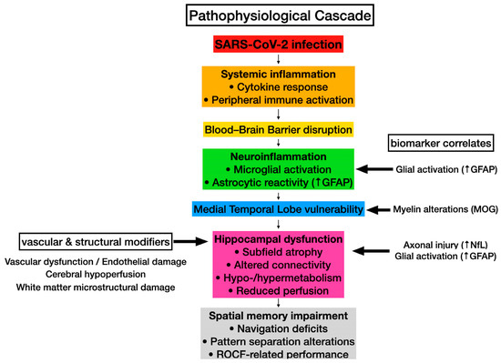
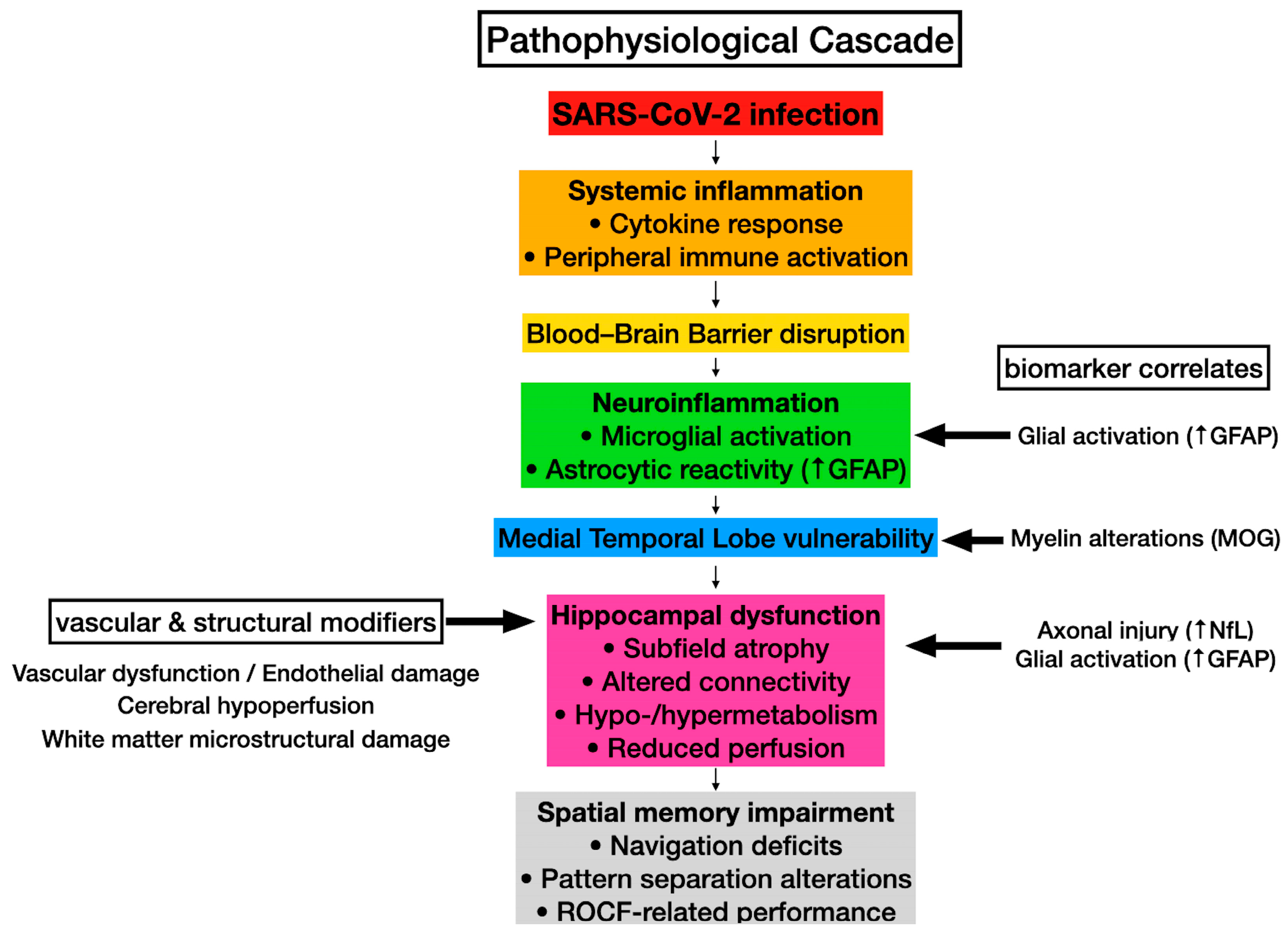
Spatial memory critically depends on the integrity of the MTL, including the hippocampus, the entorhinal cortex, and the parahippocampal cortex. Levels of certain biomarkers, such as neurofilament light chain (NfL), tau, or glial fibrillary acidic protein (GFAP) have been correlated with cognitive decline and brain atrophy [51,52]. Converging neuroimaging and biomarker evidence suggests that these MTL structures may be particularly vulnerable following SARS-CoV-2 infection, as illustrated in the hypothetical integrative model (Figure 1).
Figure 1.
Hypothetical integrated model linking SARS-CoV-2 infection to spatial memory dysfunction. Systemic inflammation and blood–brain barrier disruption may trigger neuroinflammation, increasing medial temporal lobe and hippocampal vulnerability. Biomarker alterations and neuroimaging findings, including hippocampal atrophy, perfusion changes, connectivity alterations (both within the MTL and across large-scale cortical networks), and white matter disruption, support neuronal and glial injury. Vascular dysfunction and hypoperfusion may further modulate these effects, contributing to spatial memory impairment. These relationships, while supported by indirect biomarker and neuroimaging findings, remain to be fully established through direct mechanistic studies.
4.1. Biomarker Studies
Acute SARS-CoV-2 infection triggers systemic inflammation and glial reactivity, leading to blood–brain barrier disruption and neuroinflammatory cascades [53]. These mechanisms provide a plausible pathophysiological basis by which acute-phase biomarker elevations may contribute to subsequent hippocampal and MTL dysfunction underlying spatial memory deficits.
Histopathological and molecular analyses of brain tissue from patients in the acute phase of COVID-19 have provided direct evidence of widespread neuroinflammatory changes. In a post-mortem case series of 43 patients, Matschke et al. [54] performed a comprehensive neuropathological workup using immunohistochemical staining for activated astrocytes (GFAP), activated microglia (HLA-DR, IBA1), and cytotoxic T lymphocytes (CD8). They identified astrogliosis in 86% of the examined brains across all assessed regions, including the olfactory bulb, superior frontal gyrus, putamen, medulla oblongata, and cerebellar hemisphere, with T lymphocyte infiltration observed in 79% of patients. These findings are further elucidated by single-nucleus transcriptomic profiling of 65,309 nuclei from frontal cortex and choroid plexus samples [55]. Although molecular traces of the virus were absent in the brain parenchyma, their analysis revealed that barrier cells of the choroid plexus sense and relay peripheral inflammation into the brain. This process triggers the infiltration of peripheral T cells and the emergence of pathological microglia and astrocyte subpopulations—sharing features with states observed in neurodegenerative diseases—that contribute to cellular distress. Notably, synaptic signaling in upper-layer excitatory neurons, which are critically linked to cognitive function, was preferentially affected, showing molecular overlaps with genetic variants associated with cognition. Collectively, these studies provide a robust cellular-level framework for understanding how acute-phase inflammatory cascades and reactive gliosis may compromise the integrity of the circuits required for spatial memory.
Related to PCC, Wood et al. [37] found elevated plasma levels of NfL and GFAP one year after COVID-19 in previously hospitalized patients, including individuals with new acute neurological or psychiatric complications (NeuroCOVID) and COVID-19 controls without such complications. Importantly, tau levels were increased only in the NeuroCOVID subgroup, suggesting more pronounced neuronal damage in patients with neurological complications. Complementarily, Díez-Cirarda et al. [56] observed elevated plasma GFAP and NfL levels, along with increased MOG and reduced CCL11 levels, in PCC patients assessed 11 months after SARS-CoV-2 infection. Moreover, GFAP and MOG were positively associated with hippocampal volume, while NfL showed an inverse relationship with hippocampal volume.
Collectively, these results strengthen the plausibility that alterations in multiple biomarkers after SARS-CoV-2 infection, throughout the acute and persistent phases, reflect hippocampal injury contributing to cognitive deficits, including spatial memory impairment. Nevertheless, although these biomarker alterations are biologically consistent with hippocampal and MTL dysfunction, existing studies have predominantly examined global cognition rather than domain-specific outcomes [57], and direct associations between biomarker profiles and spatial memory impairment remain unexplored.
4.2. Neuroimaging Studies
Neuroimaging studies provide converging evidence that COVID-19 is associated with structural and functional alterations in MTL regions critically involved in spatial memory. A previous systematic review synthesizing neuroimaging findings after SARS-CoV-2 infection reported frequent but highly heterogeneous alterations in hippocampal and parahippocampal regions across acute, post-acute, and PCC phases [21]. The reviewed studies described both structural and functional abnormalities, including altered hippocampal connectivity, changes in gray matter volume and cortical thickness, abnormal cerebral blood flow, and inconsistent metabolic patterns, with reports of both hypo- and hypermetabolism depending on disease stage and imaging technique [21]. Notably, some studies observed increased hippocampal gray matter volume or metabolic activity shortly after infection, whereas others reported hypometabolism, reduced perfusion, or cortical thinning at later stages, suggesting dynamic and potentially compensatory processes rather than a uniform trajectory of neural injury [21]. Similar heterogeneity was observed in the parahippocampal gyrus, with longitudinal evidence of gray matter thinning and altered metabolic and functional activity across disease phases [21]. Consistent with these findings, a complementary systematic review focusing on memory impairment after COVID-19 highlighted structural and functional alterations not only within the MTL but also in distributed fronto-temporo-parietal regions, including hypometabolic patterns and white matter abnormalities associated with memory complaints [58].
Related to structural neuroimaging studies, Elkoury et al. [59] summarized the Magnetic Resonance Imaging (MRI) studies focused on brain volumetric changes at least two weeks after SARS-CoV-2 infection. They reported evidence of reduced hippocampal volume in several cohorts, although results were heterogeneous across studies and dependent on post-infection timing and clinical characteristics. This highlights the vulnerability of hippocampal and MTL circuits. However, MRI studies in PCC showed mixed results regarding hippocampal and cortical changes and its association with spatial memory performance.
For example, Díez-Cirarda et al. [56] explored hippocampal changes in PCC patients using MRI, and they observed reduced volume across almost all hippocampal subfields compared to healthy subjects. The largest effect sizes were found in subfields of the hippocampal head and the dentate gyrus, regions critically involved in spatial encoding and pattern separation. Volume reductions were more pronounced in previously hospitalized patients, and head hippocampal subfield volumes were associated with cognition, including performance in the ROCF (copy, recall after 3 min, recall after 30 min, and recognition). In parallel, microstructural analyses using neurite orientation dispersion and density imaging (NODDI) revealed increased intracellular volume fraction and orientation dispersion index within the hippocampus, which are indicative of altered gray matter organization. Additionally, hippocampal perfusion measured with arterial spin labeling was reduced and correlated with performance in the ROCF recognition task, further supporting the presence of persistent hippocampal dysfunction at both structural and vascular levels.
Longitudinal evidence further supports the vulnerability of MTL circuits following SARS-CoV-2 infection. Invernizzi et al. [38] observed reduced left hippocampal volume alongside altered functional connectivity in subcortical regions, particularly the amygdala, in adolescents and young adults with mild COVID-19. Moreover, these hippocampal structural alterations were accompanied by poorer spatial working memory performance, suggesting a potential brain–behavior relationship even in non-hospitalized populations. The identification of disrupted connectivity in hubs essential for visual-spatial integration reinforces the notion that COVID-19 affects distributed networks necessary for large-scale navigation rather than inducing isolated deficits. Network-level analyses additionally identified disrupted connectivity in cortical hubs linked to visual processing and memory, including the lingual and intracalcarine cortices, reinforcing the notion that COVID-19 may preferentially affect distributed circuits supporting spatial cognition rather than inducing global brain dysfunction. However, the modest sample size and the possibility that psychosocial stressors contributed to the observed neural changes warrant cautious interpretation.
Functional neuroimaging studies, including resting-state and task-based fMRI investigations, have begun to provide evidence of network-level alterations that may contribute to cognitive dysfunction after COVID-19. Resting-state fMRI studies have identified alterations in large-scale brain networks in individuals with post-COVID cognitive symptoms. For example, altered functional connectivity involving the default mode network (DMN) and temporo-parietal regions has been reported in patients with long-COVID and cognitive impairment [60]. These networks are critically involved in episodic and spatial memory processes, particularly in the integration of spatial representations and contextual information. In addition, studies examining the functional integrity of the DMN have shown that connectivity between key network nodes is associated with neurocognitive performance in individuals reporting persistent cognitive symptoms after mild COVID-19 [61]. Since the DMN and related temporo-parietal networks play a key role in episodic and spatial memory processes, these findings suggest that post-COVID cognitive dysfunction may reflect alterations in distributed functional networks supporting memory rather than only focal structural abnormalities.
Additionally, Pacheco-Jaime et al. [62] assessed structural brain changes with MRI in PCC patients nearly two years after infection. They reported increased cortical thickness in the right superior frontal and rostral middle frontal gyri, but these changes were not correlated with spatial memory performance assessed with the ROCF recall 30 min after learning. Hippocampal volumetric and voxel-based morphometry analyses also showed no significant differences compared to healthy subjects. However, Diffusion Tensor Imaging (DTI) revealed lower fractional anisotropy in the superior longitudinal fasciculus, corpus callosum, and uncinate fasciculus, which correlated with subjective memory failures. These findings suggest subtle microstructural white matter alterations that may indirectly affect spatial memory networks.
Beyond the heterogeneity observed across imaging modalities and disease stages, several methodological and clinical factors may contribute to the variability of reported MTL findings after COVID-19. The review by Rane Levendovszky et al. [63] emphasizes substantial diagnostic heterogeneity, variability in time since infection, and the frequent presence of vascular and cardiovascular comorbidities, all of which may confound neuroimaging results in these patients. Structural MRI studies consistently report temporal and frontal cortical thinning, whereas functional imaging highlights disrupted connectivity involving hippocampal, amygdalar, and olfactory regions [63]. Importantly, potential mediators such as neuroinflammation, endothelial dysfunction, and altered cerebral perfusion remain understudied but may be particularly relevant for spatial memory processes that rely on intact hippocampal–parahippocampal circuits and their vascular support. However, while these neuroimaging findings consistently implicate MTL and cortical regions that are central to spatial memory processing, most studies have relied on global cognitive measures and have rarely combined functional neuroimaging with tasks specifically designed to isolate spatial memory processes. When memory was specifically assessed, studies explored the verbal or visual components. As a result, direct evidence linking neuroimaging alterations to experimentally isolated spatial memory deficits, particularly those assessed with ecologically valid navigation paradigms, remains scarce.
5. Methodological Limitations and Gaps in the Current Literature
Despite the growing interest in cognitive sequelae after SARS-CoV-2 infection, research specifically targeting spatial memory remains methodologically constrained and conceptually heterogeneous. Several interrelated limitations reduce interpretability, cross-study comparability, and mechanistic inference.
A primary limitation concerns the fact that the visual component of visuospatial memory is better characterized and more rigorously measured than its spatial counterpart. Many studies operationalize “spatial memory” through delayed recall of the ROCF or similar small-scale tasks, which predominantly index visual–perceptual encoding and organizational strategies rather than allocentric mapping or navigation-based learning. This reliance on static tasks represents a significant methodological limitation, increasing the risk that observed deficits are attributed to the hippocampal–entorhinal system when they may instead reflect broader network dysfunction. Evidence from neurological populations indicates that ROCF recall performance is strongly influenced by figure organization and executive processing speed and may dissociate from visuospatial perception per se [64]. In a critical synthesis of the evidence, we argue that impairments observed in PCC may reflect strategic or executive inefficiencies originating in frontal circuits rather than a primary disruption of hippocampal-dependent spatial representations themselves (i.e., the ability to form and manipulate cognitive maps). Moreover, few studies explicitly distinguish between egocentric and allocentric reference frames, nor do they clarify whether tasks engage route-based learning, object–location binding, or cognitive map formation. The imprecision in the definition of this construct is not merely a semantic issue; it explains the current inconsistency in findings, as tasks with high executive demand (like the SWM) may yield positive results while tasks requiring pure spatial navigation might show different patterns of impairment. This lack of construct precision complicates theoretical interpretation and limits the ability to build a cohesive model of post-COVID cognitive sequelae. Future research would benefit from explicitly aligning task selection with clearly defined spatial memory components grounded in hippocampal–entorhinal models.
Second, although spatial memory in COVID-19 has been assessed using a range of methodologies, the field remains methodologically fragmented. The predominance of static, paper-and-pencil, or computerized formats creates a ‘blind spot’ in the literature. These instruments provide clinically feasible and normatively interpretable indices of visuospatial performance; however, they predominantly assess small-scale, static spatial processing and place minimal demands on allocentric navigation, path integration, or dynamic spatial updating, core features of everyday spatial behavior. Consequently, the current evidence-base may be systematically underestimating subtle but functional deficits that only emerge in complex, large-scale environments. Conversely, experimental paradigms, particularly VR-based navigational and object–location tasks, offer greater ecological validity and mechanistic precision. These approaches enable the assessment of large-scale navigation, environmental integration, and long-term spatial consolidation. Evidence from immersive VR paradigms in other memory domains further supports the feasibility and construct validity of ecologically embedded tasks [65], demonstrating high levels of presence, tolerability, and significant associations with traditional neuropsychological measures. Such findings reinforce the potential of immersive environments to provide valid and engaging cognitive assessment frameworks. However, despite this methodological promise, the absence of harmonized VR procedures, outcome metrics (e.g., distance error, angular deviation, learning curves), and clearly defined retention intervals further restricts reproducibility and cross-study comparability. At present, no unified framework guides the evaluation of spatial memory following SARS-CoV-2 infection, and assessment strategies remain heterogeneous, hindering precise phenotypic characterization and limiting cumulative knowledge building. Furthermore, it is important to emphasize that evidence from navigational paradigms remains limited and is currently derived from a relatively small number of specialized research groups, indicating the need for broader multi-center validation. The development of integrated and standardized multimodal protocols which combine traditional neuropsychological tools with ecologically valid navigational paradigms will therefore be essential to advance the field and to capture the full complexity of spatial memory alterations in PCC patients.
Third, substantial variability in demographic and clinical characteristics further constrains interpretation. The current literature lacks a structured assessment of methodological quality, and many investigations rely on relatively small, convenience-based samples that are inherently underpowered to detect subtle group differences. This reliance on convenience sampling introduces a significant selection bias, as cohorts may not represent the full clinical spectrum of PCC. Furthermore, the repetitive use of identical test batteries in longitudinal designs introduces potential practice effects, which may bias findings toward apparent recovery and obscure persistent underlying impairments. Additionally, the lack of a standardized approach to classifying acute-phase severity—which ranges from non-hospitalized individuals to those requiring intensive care and mechanical ventilation—further complicates the evidence base. The systematic lack of control for these clinical trajectories, alongside common confounders like fatigue, mood symptoms (including depression and anxiety), or sleep disturbances [66], represents a significant source of bias that must be addressed in future research. This prevents a clear determination of whether spatial memory deficits are a direct consequence of the virus or a secondary effect of severe systemic illness and hospitalization. Moreover, the general lack of premorbid cognitive data makes it difficult to differentiate persistent neurocognitive sequelae from pre-existing conditions or generalized pandemic-related stressors. On the other hand, inflammatory status, vascular risk factors, and pre-existing neurological or psychiatric conditions are variably documented across studies. Without systematic adjustment for these potential confounders, isolating SARS-CoV-2–specific effects on spatial memory remains challenging. Moreover, premorbid cognitive functioning is rarely available, and few studies incorporate matched infection-recovered controls without persistent symptoms. Such comparisons are critical to disentangle persistent neurocognitive sequelae from generalized post-illness effects, hospitalization-related factors, or psychosocial stressors associated with the pandemic context. Additionally, temporal anchoring represents another source of heterogeneity. Assessment timing is often defined relative to hospital discharge, PCR testing, or self-reported diagnosis rather than symptom onset. This inconsistency is a significant methodological limitation, as it complicates phase-based comparisons (acute, post-acute, PCC) and may obscure temporal trajectories of spatial memory recovery or decline. Standardized reporting of infection timeline variables, alongside systematic characterization of clinical modifiers, is therefore essential.
Fourth, longitudinal designs remain scarce, and when implemented, frequently rely on repeated administration of the same static task without incorporating navigation-based measures. Consequently, the trajectory of spatial memory consolidation, persistence, or recovery over extended intervals remains insufficiently characterized. This limitation is particularly relevant given emerging evidence suggesting that group differences may become more pronounced at longer retention intervals [35]. Finally, a critical gap exists between the structural findings reported in Section 4 (e.g., hippocampal subfield atrophy) and behavioral outcomes. Direct brain–behavior correlations linking hippocampal alterations to experimentally isolated spatial memory performance remain infrequent. Without tasks designed to specifically stress hippocampal-dependent allocentric mapping, the clinical relevance of the observed neural changes in post-COVID patients remains speculative.
In summary, the current evidence regarding spatial memory in COVID-19 presents both notable strengths and significant limitations. A primary strength lies in the converging neuroimaging and biomarker data that consistently implicate the medial temporal lobe as a zone of vulnerability. However, this evidence more convincingly suggests a vulnerability of certain visuospatial and spatial memory components, particularly in PCC, rather than a consistent impairment of spatial navigational memory. This is primarily due to several methodological weaknesses, most notably the reliance on visuospatial proxy measures and small-scale, static tasks that lack the sensitivity to isolate hippocampal-dependent deficits. Methodological variability, construct ambiguity, and limited multimodal integration constrain definitive conclusions. Inconsistencies across studies, such as conflicting results on the impact of disease severity or the persistence of deficits, can often be traced to differences in how assessment timing is defined and to the varying cognitive demands of the instruments used. Advancing the field will require larger, longitudinal, and harmonized studies that explicitly operationalize spatial memory constructs, incorporate ecologically valid navigation paradigms, and directly link behavioral outcomes to neural and biological markers. Only through such integrative approaches will it be possible to delineate the true scope, mechanisms, and clinical relevance of spatial memory alterations in post-COVID-19 populations.
6. Conclusions
In synthesis, this review clarifies the impact of COVID-19 on spatial memory by distinguishing established evidence from remaining gaps.
The available evidence more convincingly suggests a selective vulnerability of certain visuospatial and spatial memory components following COVID-19 infection, particularly in individuals with PCC, rather than a consistent impairment of spatial navigational memory. Although the convergence between behavioral impairments in navigational contexts and structural alterations in the MTL, including hippocampal atrophy, hypoperfusion, and disrupted functional connectivity, is notable, this body of literature remains limited, heterogeneous, and largely reliant on visuospatial proxy measures. These alterations appear more pronounced at longer retention intervals and within navigational contexts, where participants show decreased accuracy and increased difficulty in consolidating spatial representations.
However, these findings must be interpreted with caution due to the inherent clinical heterogeneity of the studied populations and the limitations of traditional neuropsychological tools, which often engage spatial memory alongside executive processes. Uncertainty also persists regarding the exact temporal trajectory of these deficits across the acute and post-acute phases, due to inconsistent definitions of assessment timing and the scarcity of longitudinal designs. Furthermore, it remains unclear to what extent these impairments reflect a primary disruption of spatial representations versus a secondary effect of generalized factors such as fatigue, mood symptoms, and systemic inflammation.
To address these methodological gaps, future research should prioritize the implementation of standardized, multimodal assessment protocols that integrate immersive VR paradigms. Such approaches will enhance diagnostic sensitivity and provide a better phenotypic characterization of spatial memory dysfunction. Additionally, there is a critical need for longitudinal studies that link domain-specific spatial behavior with neural biomarkers to provide a foundation for targeted rehabilitation strategies aimed at restoring spatial navigation and functional independence in the post-COVID population.
Author Contributions
Conceptualization, T.L., S.G.-A. and M.M.; methodology, T.L. and S.G.-A.; investigation, T.L., S.G.-A. and M.M.; resources, T.L., S.G.-A. and M.M.; data curation, T.L. and S.G.-A.; writing—original draft preparation, T.L., S.G.-A. and M.M.; writing—review and editing, T.L., S.G.-A. and M.M.; visualization, T.L. and M.M.; supervision, M.M.; project administration, M.M. All authors have read and agreed to the published version of the manuscript.
Funding
This research was funded by the Programa “Severo Ochoa” de la Consejería de Ciencia, Empresas, Formación y Empleo del Principado de Asturias to T.L. [Grant Number: PA-23-BP22–005].
Institutional Review Board Statement
Not applicable.
Informed Consent Statement
Not applicable.
Data Availability Statement
Data sharing is not applicable.
Conflicts of Interest
The authors declare no conflicts of interest.
Abbreviations
The following abbreviations are used in this manuscript:
| COVID-19 | Coronavirus disease |
| SARS-CoV-2 | Severe acute respiratory syndrome coronavirus 2 |
| PCC | Post COVID-19 condition |
| NICE | National Institute for Health and Care Excellence |
| MTL | Medial temporal lobe |
| MoCA | Montreal Cognitive Assessment |
| MMSE | Mini Mental State Examination |
| PCR | Polymerase chain reaction |
| VR | Virtual reality |
| ROCF | Rey–Osterrieth Complex Figure Test |
| FMT | Figural Memory Test |
| CBTT | Corsi Block Tapping Test |
| SWM | Spatial Working Memory |
| CANTAB | Cambridge Neuropsychological Test Automated Battery |
| SPART | 10/36 Spatial Recall Test |
| NfL | Neurofilament light chain |
| GFAP | Glial fibrillary acidic protein |
| MOG | Myelin oligodendrocyte glycoprotein |
| CCL11 | Eotaxin-1 |
| MRI | Magnetic Resonance Imaging |
| NODDI | Neurite orientation dispersion and density |
| DMN | Default Mode Network |
| DTI | Diffusion Tensor Imaging |
References
- National Institute for Health and Care Excellence (NICE); Royal College of General Practitioners; Healthcare Improvement Scotland SIGN. COVID-19 Rapid Guideline: Managing the Long-Term Effects of COVID-19; National Institute for Health and Care Excellence: London, UK, 2020; Available online: www.nice.org.uk/guidance/ng188 (accessed on 27 January 2026).
- Long, B.; Carius, B.M.; Chavez, S.; Liang, S.Y.; Brady, W.J.; Koyfman, A.; Gottlieb, M. Clinical update on COVID-19 for the emergency clinician: Presentation and evaluation. Am. J. Emerg. Med. 2022, 54, 46–57. [Google Scholar] [CrossRef] [Scilit] [PubMed]
- Agarwal, A.; Hunt, B.; Stegemann, M.; Rochwerg, B.; Lamontagne, F.; Siemieniuk, R.A.; Agoritsas, T.; Askie, L.; Lytvyn, L.; Leo, Y.S.; et al. A living WHO guideline on drugs for covid-19. BMJ 2020, 370, m3379. [Google Scholar] [CrossRef] [Scilit]
- Norouzi, M.; Miar, P.; Norouzi, S.; Nikpour, P. Nervous System Involvement in COVID-19: A Review of the Current Knowledge. Mol. Neurobiol. 2021, 58, 3561–3574. [Google Scholar] [CrossRef] [Scilit]
- Aderinto, N.; Olatunji, G.; Kokori, E.; Ogieuhi, I.J.; Yusuf, I.A.; Egbunu, E.; Ukoaka, B.M.; Babalola, A.E.; Adefusi, T.O.; Aboje, J.E.; et al. COVID-19 and cognitive impairment: A review of the emerging evidence. Discov. Ment. Health 2025, 5, 56. [Google Scholar] [CrossRef] [Scilit]
- Kirchberger, I.; Peilstöcker, D.; Warm, T.D.; Linseisen, J.; Hyhlik-Dürr, A.; Meisinger, C.; Goßlau, Y. Subjective and Objective Cognitive Impairments in Non-Hospitalized Persons 9 Months after SARS-CoV-2 Infection. Viruses 2023, 15, 256. [Google Scholar] [CrossRef] [Scilit]
- Tsiaras, Y.; Kitsakis, N.; Papadopoulou, E.; Karanikas, E.; Kourbetis, D.; Aretouli, E. Neuropsychological Profile of Hospitalized Patients Due to COVID-19: Clinical and Inflammatory Correlates. Arch. Clin. Neuropsychol. 2023, 38, 1564–1577. [Google Scholar] [CrossRef] [Scilit]
- Charles James, J.; Schulze, H.; Siems, N.; Prehn, C.; Quast, D.R.; Trampe, N.; Gold, R.; Faissner, S. Neurological post-COVID syndrome is associated with substantial impairment of verbal short-term and working memory. Sci. Rep. 2025, 15, 1695. [Google Scholar] [CrossRef] [Scilit]
- Alt, L.L.; Bortolini, A.D.; Andreis, B.S.; Schmidt, C.A.; Rugeri, G.; Pagliarin, M.C.T.; Kindel, M.E.D.; Costa, M.F.A.; Costa, N.L.; Dariz, P.H.S.; et al. Memory Impairment in Post-COVID-19 Syndrome; What Is the Evidence? J. Mind Med. Sci. 2024, 11, 285–290. [Google Scholar] [CrossRef] [Scilit]
- Llana, T.; Zorzo, C.; Mendez-Lopez, M.; Mendez, M. Memory alterations after COVID-19 infection: A systematic review. Appl. Neuropsychol. Adult 2024, 31, 292–305. [Google Scholar] [CrossRef] [Scilit]
- Madl, T.; Chen, K.; Montaldi, D.; Trappl, R. Computational cognitive models of spatial memory in navigation space: A review. Neural Netw. 2015, 65, 18–43. [Google Scholar] [CrossRef] [Scilit] [PubMed]
- García-Navarra, S.; Llana, T.; Méndez, M. Spatial memory deficits in Parkinson’s disease: Neural mechanisms and assessment. Am. J. Neurodegener. Dis. 2025, 14, 67–81. [Google Scholar] [CrossRef] [Scilit]
- Park, J.H. Can the Virtual Reality-Based Spatial Memory Test Better Discriminate Mild Cognitive Impairment than Neuropsychological Assessment? Int. J. Environ. Res. Public Health 2022, 19, 9950. [Google Scholar] [CrossRef] [Scilit]
- Montana, J.I.; Tuena, C.; Serino, S.; Cipresso, P.; Riva, G. Neurorehabilitation of Spatial Memory Using Virtual Environments: A Systematic Review. J. Clin. Med. 2019, 8, 1516. [Google Scholar] [CrossRef] [Scilit]
- Ekstrom, A.D.; Huffman, D.J.; Starrett, M. Interacting networks of brain regions underlie human spatial navigation: A review and novel synthesis of the literature. J. Neurophysiol. 2017, 118, 3328–3344. [Google Scholar] [CrossRef] [Scilit]
- Fan, C.L.; Sokolowski, H.M.; Rosenbaum, R.S.; Levine, B. What about “space” is important for episodic memory? Wiley Interdiscip. Rev. Cogn. Sci. 2023, 14, e1645. [Google Scholar] [CrossRef] [Scilit] [PubMed]
- Martín-Pozuelo, N.; Carballo-Costa, L.; Solís-García, M.; Giancola, M.; Piccardi, L.; De Las Cuevas-Terán, I.; Robles-García, V. Measuring spatial navigation during locomotion in children: A systematic review. Heliyon 2024, 10, e33817. [Google Scholar] [CrossRef] [Scilit] [PubMed]
- Llana, T.; Mendez, M.; Juan, M.C.; Mendez-Lopez, M. Navigational object-location memory assessment in real and virtual environments: A systematic review. Behav. Brain Res. 2025, 480, 115388. [Google Scholar] [CrossRef] [Scilit] [PubMed]
- Chandra, S.; Sharma, S.; Chaudhuri, R.; Fiete, I. Episodic and associative memory from spatial scaffolds in the hippocampus. Nature 2025, 638, 739–751. [Google Scholar] [CrossRef] [Scilit]
- Douaud, G.; Lee, S.; Alfaro-Almagro, F.; Arthofer, C.; Wang, C.; McCarthy, P.; Lange, F.; Andersson, J.L.R.; Griffanti, L.; Duff, E.; et al. SARS-CoV-2 is associated with changes in brain structure in UK Biobank. Nature 2022, 604, 697–707. [Google Scholar] [CrossRef] [Scilit]
- Zorzo, C.; Solares, L.; Mendez, M.; Mendez-Lopez, M. Hippocampal alterations after SARS-CoV-2 infection: A systematic review. Behav. Brain Res. 2023, 455, 114662. [Google Scholar] [CrossRef] [Scilit]
- Jacot de Alcântara, I.; Nuber-Champier, A.; Voruz, P.; Cionca, A.; Assal, F.; Péron, J.A. Cognitive Deficits in the Acute Phase of COVID-19: A Review and Meta-Analysis. J. Clin. Med. 2023, 12, 762. [Google Scholar] [CrossRef] [Scilit]
- Cipolli, G.C.; Alonso, V.; Yasuda, C.L.; Assumpção, D.; Cachioni, M.; Melo, R.C.; Hinsliff-Smith, K.; Yassuda, M.S. Cognitive impairment in post-acute COVID-19 syndrome: A scoping review. Comprometimento cognitivo na síndrome pós-COVID-19 aguda: Uma revisão de escopo. Arq. Neuro-Psiquiatr. 2023, 81, 1053–1069. [Google Scholar] [CrossRef] [Scilit]
- Bonizzato, S.; Ghiggia, A.; Ferraro, F.; Galante, E. Cognitive, behavioral, and psychological manifestations of COVID-19 in post-acute rehabilitation setting: Preliminary data of an observational study. Neurol. Sci. 2022, 43, 51–58. [Google Scholar] [CrossRef] [Scilit]
- Peskar, M.; Šimunič, B.; Šlosar, L.; Pišot, S.; Teraž, K.; Gasparini, M.; Pišot, R.; Marusic, U. Effects of COVID-19 on cognition and mood after hospitalization and at 2-month follow-up. Front. Psychol. 2023, 14, 1141809. [Google Scholar] [CrossRef] [Scilit] [PubMed]
- Ferrucci, R.; Dini, M.; Groppo, E.; Rosci, C.; Reitano, M.R.; Bai, F.; Poletti, B.; Brugnera, A.; Silani, V.; D’Arminio Monforte, A.; et al. Long-Lasting Cognitive Abnormalities after COVID-19. Brain Sci. 2021, 11, 235. [Google Scholar] [CrossRef] [Scilit] [PubMed]
- Ferrucci, R.; Dini, M.; Rosci, C.; Capozza, A.; Groppo, E.; Reitano, M.R.; Allocco, E.; Poletti, B.; Brugnera, A.; Bai, F.; et al. One-year cognitive follow-up of COVID-19 hospitalized patients. Eur. J. Neurol. 2022, 29, 2006–2014. [Google Scholar] [CrossRef] [Scilit] [PubMed]
- Serrano Del Pueblo, V.M.; Serrano-Heras, G.; Romero Sánchez, C.M.; Landete, P.P.; Rojas-Bartolome, L.; Feria, I.; Morris, R.G.M.; Strange, B.; Mansilla, F.; Zhang, L.; et al. Brain and cognitive changes in patients with long COVID compared with infection-recovered control subjects. Brain 2024, 147, 3611–3623. [Google Scholar] [CrossRef] [Scilit]
- Mattioli, F.; Stampatori, C.; Righetti, F.; Sala, E.; Tomasi, C.; De Palma, G. Neurological and Cognitive Sequelae of COVID-19: A Four Month Follow-Up. J. Neurol. 2021, 268, 4422–4428. [Google Scholar] [CrossRef] [Scilit]
- Serrano-Castro, P.J.; Garzón-Maldonado, F.J.; Casado-Naranjo, I.; Ollero-Ortiz, A.; Mínguez-Castellanos, A.; Iglesias-Espinosa, M.; Baena-Palomino, P.; Sánchez-Sanchez, V.; Sánchez-Pérez, R.M.; Rubi-Callejon, J.; et al. The cognitive and psychiatric subacute impairment in severe Covid-19. Sci. Rep. 2022, 12, 3563. [Google Scholar] [CrossRef] [Scilit]
- Herrera, E.; Pérez-Sánchez, M.D.C.; San Miguel-Abella, R.; Barrenechea, A.; Blanco, C.; Solares, L.; González, L.; Iza, C.; Castro, I.; Nicolás, E.; et al. Cognitive impairment in young adults with post COVID-19 syndrome. Sci. Rep. 2023, 13, 6378. [Google Scholar] [CrossRef] [Scilit]
- Whiteside, D.M.; Basso, M.R.; Naini, S.M.; Porter, J.; Holker, E.; Waldron, E.J.; Melnik, T.E.; Niskanen, N.; Taylor, S.E. Outcomes in post-acute sequelae of COVID-19 (PASC) at 6 months post-infection Part 1: Cognitive functioning. Clin. Neuropsychol. 2022, 36, 806–828. [Google Scholar] [CrossRef] [Scilit] [PubMed]
- Guillén, N.; Pérez-Millan, A.; Falgàs, N.; Lledo-Ibanez, G.N.; Rami, L.; Sarto, J.; Boti, M.A.; Aralos-Perez, C.; Ruiz-Garcia, R.; Narano, L.; et al. Cognitive profile, neuroimaging and fluid biomarkers in post-acute COVID-19 syndrome. Sci. Rep. 2024, 14, 12927. [Google Scholar] [CrossRef] [Scilit] [PubMed]
- Schlenker, L.S.; Hartung, T.J.; Klabunn, P.; Schwichtenberg, K.; Heine, J.; Adam, L.; Franke, C.; Finke, C. Altered putamen connectivity in patients with neurological post-COVID condition. Brain Commun. 2025, 7, fcaf291. [Google Scholar] [CrossRef] [Scilit] [PubMed]
- Llana, T.; Garces-Arilla, S.; Juan, M.C.; Mendez-Lopez, M.; Mendez, M. An immersive virtual reality-based object-location memory task reveals spatial long-term memory alterations in Long-COVID. Behav. Brain Res. 2024, 471, 115127. [Google Scholar] [CrossRef] [Scilit]
- Meyer, P.; Zaiser, A.K. Insights on the neurocognitive mechanisms underlying hippocampus-dependent memory impairment in COVID-19. Sci. Rep. 2025, 15, 20114. [Google Scholar] [CrossRef] [Scilit]
- Wood, G.K.; Sargent, B.F.; Ahmad, Z.U.; Tharmaratnam, K.; Dunai, C.; Egbe, F.N.; Martin, N.H.; Facer, B.; Pendered, S.L.; Rogers, H.C.; et al. Posthospitalization COVID-19 cognitive deficits at 1 year are global and associated with elevated brain injury markers and gray matter volume reduction. Nat. Med. 2025, 31, 245–257. [Google Scholar] [CrossRef] [Scilit]
- Invernizzi, A.; Renzetti, S.; van Thriel, C.; Rechtman, E.; Patrono, A.; Ambrosi, C.; Mascaro, L.; Corbo, D.; Cagna, G.; Gasparotti, R.; et al. COVID-19 related cognitive, structural and functional brain changes among Italian adolescents and young adults: A multimodal longitudinal case-control study. Transl. Psychiatry 2024, 14, 402. [Google Scholar] [CrossRef] [Scilit]
- Llana, T.; Garces-Arilla, S.; Garcia-Navarra, S.; Mendez-Lopez, M.; Juan, M.C.; Mendez, M. Cybersickness and sense of presence as predictors of VR task performance in individuals with and without Post-COVID-19 condition. Virtual Real. 2025, 29, 167. [Google Scholar] [CrossRef] [Scilit]
- Vergori, A.; Del Duca, G.; Borrelli, P.; Brita, A.C.; Pinnetti, C.; Mastrorosa, I.; Camici, M.; Mondi, A.; Mazzotta, V.; Chinello, P.; et al. Cognitive outcomes and psychological symptoms in an Italian cohort with post-acute COVID-19 condition (PACC). Heliyon 2024, 10, e39431. [Google Scholar] [CrossRef] [Scilit]
- Lagravinese, G.; Castellana, G.; Castellana, F.; Genco, M.; Petrelli, R.; Ruccia, M.; Aliani, M.; Carone, M.; Sardone, R.; Battista, P. Cognitive Deficits among Individuals Admitted to a Post-Acute Pneumological Rehabilitation Unit in Southern Italy after COVID-19 Infection. Brain Sci. 2023, 13, 84. [Google Scholar] [CrossRef] [Scilit]
- Basagni, B.; Abbruzzese, L.; Damora, A.; Conforti, J.; Saviola, D.; De Tanti, A.; Podgorska, A.; Biagioni, C.; Bacci, M.; Gambarelli, C.; et al. Cognition in COVID-19 infected patients undergoing invasive ventilation: Results from a multicenter retrospective study. Appl. Neuropsychol. Adult 2025, 32, 492–501. [Google Scholar] [CrossRef] [Scilit]
- de Pádua Serafim, A.; Saffi, F.; Soares, A.R.A.; Morita, A.M.; Medeiros Assed, M.; de Toledo, S.; Rocca, C.C.A.; Duraes, R.S.S. Cognitive performance of post-covid patients in mild, moderate, and severe clinical situations. BMC Psychol. 2024, 12, 236. [Google Scholar] [CrossRef] [Scilit] [PubMed]
- Paz-Rodríguez, F.; Lozano-Tovar, S.; Rodríguez-Agudelo, Y.; Cruz-Narciso, B.; Rodríguez-Rodríguez, M.; García-Santos, A.; López-González, D.; Soto-Moreno, F.J.; González-Navarro, M.; González-Alonso, K.; et al. Assessment of visuospatial functions in post-Covid 19 patients: Beyond the traditional paradigm. Behav. Brain Res. 2024, 471, 115095. [Google Scholar] [CrossRef] [Scilit]
- Voruz, P.; Cionca, A.; Jacot de Alcântara, I.; Nuber-Champier, A.; Allali, G.; Benzakour, L.; Thomasson, M.; Lalive, P.H.; Lövblad, K.O.; Braillard, O.; et al. Functional Connectivity Underlying Cognitive and Psychiatric Symptoms in Post-COVID-19 Syndrome: Is Anosognosia a Key Determinant? Brain Commun. 2022, 4, fcac057. [Google Scholar] [CrossRef] [Scilit]
- Zhang, X.; Lv, L.; Min, G.; Wang, Q.; Zhao, Y.; Li, Y. Overview of the Complex Figure Test and Its Clinical Application in Neuropsychiatric Disorders, Including Copying and Recall. Front. Neurol. 2021, 12, 680474. [Google Scholar] [CrossRef] [Scilit]
- Delgado-Alonso, C.; Valles-Salgado, M.; Delgado-Álvarez, A.; Yus, M.; Gómez-Ruiz, N.; Jorquera, M.; Polidura, C.; Gil, M.J.; Marcos, A.; Matías-Guiu, J.; et al. Cognitive dysfunction associated with COVID-19: A comprehensive neuropsychological study. J. Psychiatr. Res. 2022, 150, 40–46. [Google Scholar] [CrossRef] [Scilit]
- Figural Memory Test (FGT). Available online: https://marketplace.schuhfried.com/en/FGT (accessed on 12 February 2026).
- Gerstenecker, A.; Martin, R.; Marson, D.C.; Bashir, K.; Triebel, K.L. Introducing demographic corrections for the 10/36 Spatial Recall Test. Int. J. Geriatr. Psychiatry 2016, 31, 406–411. [Google Scholar] [CrossRef] [Scilit] [PubMed]
- Pete, K.N.; Varga, J.T.; Geréb, Z.V.; Gőbel, O.; Sik-Lanyi, C.; Müller, V.; Cserjési, R. Immersive virtual reality intervention to enhance cognitive-affective neuropsychological functions in post-COVID-19 condition: Research protocol and a case report. Acta Psychol. 2026, 263, 106217. [Google Scholar] [CrossRef] [Scilit] [PubMed]
- Chang, Y.; Liu, J.; Xu, X.; Sun, S.; Zhang, J.; Zhang, X.; Lu, G.; Xiao, S.; Cao, Y.; Wu, R.; et al. Subcortical tau deposition and plasma glial fibrillary acidic protein as predictors of cognitive decline in mild cognitive impairment and Alzheimer’s disease. Eur. J. Nucl. Med. Mol. Imaging 2025, 52, 1496–1509. [Google Scholar] [CrossRef] [Scilit]
- Shi, Y.; Lu, X.; Zhang, L.; Shu, H.; Gu, L.; Wang, Z.; Gao, L.; Zhu, J.; Zhang, H.; Zhou, D.; et al. Potential Value of Plasma Amyloid-β, Total Tau, and Neurofilament Light for Identification of Early Alzheimer’s Disease. ACS Chem. Neurosci. 2019, 10, 3479–3485. [Google Scholar] [CrossRef] [Scilit]
- Saucier, J.; Comeau, D.; Robichaud, G.A.; Chamard-Witkowski, L. Reactive gliosis and neuroinflammation: Prime suspects in the pathophysiology of post-acute neuroCOVID-19 syndrome. Front. Neurol. 2023, 14, 1221266. [Google Scholar] [CrossRef] [Scilit] [PubMed]
- Matschke, J.; Lütgehetmann, M.; Hagel, C.; Sperhake, J.P.; Schröder, A.S.; Edler, C.; Mushumba, H.; Fitzek, A.; Allweiss, L.; Dandri, M.; et al. Neuropathology of patients with COVID-19 in Germany: A post-mortem case series. Lancet Neurol. 2020, 19, 919–929. [Google Scholar] [CrossRef] [Scilit]
- Yang, A.C.; Kern, F.; Losada, P.M.; Agam, M.R.; Maat, C.A.; Schmartz, G.P.; Fehlmann, T.; Stein, J.A.; Schaum, N.; Lee, D.P.; et al. Dysregulation of brain and choroid plexus cell types in severe COVID-19. Nature 2021, 595, 565–571. [Google Scholar] [CrossRef] [Scilit]
- Díez-Cirarda, M.; Yus-Fuertes, M.; Sanchez-Sanchez, R.; Gonzalez-Rosa, J.J.; Gonzalez-Escamilla, G.; Gil-Martínez, L.; Delgado-Alonso, C.; Gil-Moreno, M.J.; Valles-Salgado, M.; Cano-Cano, F.; et al. Hippocampal subfield abnormalities and biomarkers of pathologic brain changes: From SARS-CoV-2 acute infection to post-COVID syndrome. EBioMedicine 2023, 94, 104711. [Google Scholar] [CrossRef] [Scilit]
- Cysique, L.A.; Jakabek, D.; Bracken, S.G.; Allen-Davidian, Y.; Heng, B.; Chow, S.; Dehhaghi, M.; Staats Pires, A.; Darley, D.R.; Byrne, A.; et al. The kynurenine pathway relates to post-acute COVID-19 objective cognitive impairment and PASC. Ann. Clin. Transl. Neurol. 2023, 10, 1338–1352. [Google Scholar] [CrossRef] [Scilit] [PubMed]
- Shan, D.; Li, S.; Xu, R.; Nie, G.; Xie, Y.; Han, J.; Gao, X.; Zheng, Y.; Xu, Z.; Dai, Z. Post-COVID-19 human memory impairment: A PRISMA-based systematic review of evidence from brain imaging studies. Front. Aging Neurosci. 2022, 14, 1077384. [Google Scholar] [CrossRef] [Scilit]
- Elkoury, E.; Yehia, A.; Caparelli, E.C.; Geda, Y.E.; Ortega, D.; Yamada, N.; Hakhu, S.; Beeman, S.C.; Ross, T.J.; Yang, Y.; et al. Brain Volumetric Changes Post-COVID-19: A Systematic Review. Brain Sci. 2025, 15, 1255. [Google Scholar] [CrossRef] [Scilit]
- Leitner, M.; Pinter, D.; Ropele, S.; Koini, M. Functional connectivity changes in long-Covid patients with and without cognitive impairment. Cortex 2025, 191, 74–89. [Google Scholar] [CrossRef] [Scilit]
- Madden, D.; Stephens, T.M.; Scott, J.; O’Neal Swann, C.; Prather, K.; Hoffmeister, J.; Ding, L.; Dunn, I.F.; Conner, A.K.; Yuan, H. Functional connectivity of default mode network in non-hospitalized patients with post-COVID cognitive complaints. Front. Neurosci. 2025, 19, 1576393. [Google Scholar] [CrossRef] [Scilit]
- Pacheco-Jaime, L.; Garcia-Vicente, C.; Ariza, M.; Cano, N.; Garolera, M.; Carreras-Vidal, L.; Roura, I.; Capdevila-Lacasa, C.; Oltra, J.; Pardo, J.; et al. Structural brain changes in post-COVID condition and its relationship with cognitive impairment. Brain Commun. 2025, 7, fcaf070. [Google Scholar] [CrossRef] [Scilit] [PubMed]
- Rane Levendovszky, S.; Patel, P.; Zhu, C.; Rutman, A.M.; Basha, M.M. Neuroimaging biomarkers of post-acute sequelae of Coronavirus Disease 2019. Br. J. Radiol. 2025, 98, 1165–1175. [Google Scholar] [CrossRef] [Scilit]
- Slapik, M.; Kronemer, S.I.; Morgan, O.; Bloes, R.; Lieberman, S.; Mandel, J.; Rosenthal, L.; Marvel, C. Visuospatial Organization and Recall in Cerebellar Ataxia. Cerebellum 2019, 18, 33–46. [Google Scholar] [CrossRef] [Scilit] [PubMed]
- Corriveau Lecavalier, N.; Ouellet, É.; Boller, B.; Belleville, S. Use of immersive virtual reality to assess episodic memory: A validation study in older adults. Neuropsychol. Rehabil. 2020, 30, 462–480. [Google Scholar] [CrossRef] [Scilit] [PubMed]
- Krishnan, K.; Miller, A.K.; Reiter, K.; Bonner-Jackson, A. Neurocognitive Profiles in Patients with Persisting Cognitive Symptoms Associated With COVID-19. Arch. Clin. Neuropsychol. 2022, 37, 729–737. [Google Scholar] [CrossRef] [Scilit] [PubMed]

Disclaimer/Publisher’s Note: The statements, opinions and data contained in all publications are solely those of the individual author(s) and contributor(s) and not of MDPI and/or the editor(s). MDPI and/or the editor(s) disclaim responsibility for any injury to people or property resulting from any ideas, methods, instructions or products referred to in the content. |
© 2026 by the authors. Licensee MDPI, Basel, Switzerland. This article is an open access article distributed under the terms and conditions of the Creative Commons Attribution (CC BY) license.