


Abstract
Infection with SARS-CoV-2, the virus responsible for COVID-19, is associated with cytokine storm, an excessive immune response. Interleukin-6, a multifunctional cytokine, is involved in the COVID-19 immune response. Functional polymorphisms in the interleukin-6 gene promoter, namely rs1800796, rs1800797, and rs2069845, may contribute to individual susceptibility to or severity of COVID-19. In this study, 106 healthy SARS-CoV-2-negative individuals (controls) and 106 patients with severe COVID-19 (COVID-19 group), confirmed by qPCR or rapid antigen tests, were genotyped using fluorescent probes for polymorphisms. All participants were from southern Brazil. Groups were matched for sex and body mass index, with a median age of 56–57 years. The COVID-19 group exhibited blood biomarker concentrations consistent with severe disease. No significant differences were detected in genotypic or allelic frequencies between groups, and all polymorphisms conformed to the Hardy–Weinberg equilibrium. The control group minor allelic frequencies for rs1800796 (allele C, 11.3%; 95% CI, 7–16), rs1800797 (allele A, 28.3%; 95% CI, 22–34), and rs2069845 (allele G, 36.8%; 95% CI, 30–50) were similar to those of African, American, and European populations. The polymorphisms investigated were not associated with severe COVID-19 in this cohort.
1. Introduction
Severe acute respiratory syndrome coronavirus 2 (SARS-CoV-2) is the etiologic agent of coronavirus disease 2019 (COVID-19), presenting across a broad clinical spectrum, from asymptomatic to severe outcomes such as acute respiratory distress syndrome. Although infection is often asymptomatic or associated with mild to moderate self-limiting symptoms, including fever, dry cough, myalgia, and fatigue [1,2,3,4,5], a subset of patients develops a clinically severe hyperinflammatory state, or cytokine storm, for which acute respiratory distress syndrome is a cardinal feature [1,2,3,4,5,6]. Furthermore, a distinct multisystem inflammatory syndrome has been observed in a subgroup of previously healthy children with acute SARS-CoV-2 infection, differing from the adult cytokine storm [7].
To understand disease progression from asymptomatic to extremely severe is necessary to elucidate the mechanisms underlying cell damage and immune responses. The literature demonstrates that while some infected subjects have mild or no symptoms, in others the disease is severe and even fatal [8]. The severity of COVID-19 is determined by multiple factors, such as diabetes mellitus, arterial hypertension, and obesity; age and comorbidities are also severity predictors [9,10]. Severe outcomes are frequently characterized by hyperinflammation and elevated levels of proinflammatory cytokines (e.g., IFN-α, IFN-γ, and IL-6) and chemokines, namely monocyte chemotactic protein-3 (MCP-1/CCL2) [11,12,13]. Additionally, various studies have reported that high levels of IL-6 are predictive of mortality and associated with respiratory failure, shock, and severe symptoms in hospitalized patients [14,15,16].
Genetic variations may affect innate and adaptive immune responses [17,18,19]. Polymorphisms in genes encoding proinflammatory cytokines may play a role in their production and contribute to the pathogenesis of severe COVID-19. Single-nucleotide polymorphisms (SNPs) in the TNFA, IL6, IL8, IL10, CCL5, and CXCL6 genes have been associated with COVID-19 severity, suggesting that SNPs could help explain COVID-19 outcomes [12,20,21,22,23,24,25,26]. Interleukin-6 (IL-6) was targeted in this study because it is a primary inflammatory agent associated with the response to viral infection. Variability in the IL-6 gene, particularly in the promoter region, may be associated with COVID-19 and/or the severity of viremia.
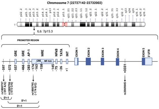
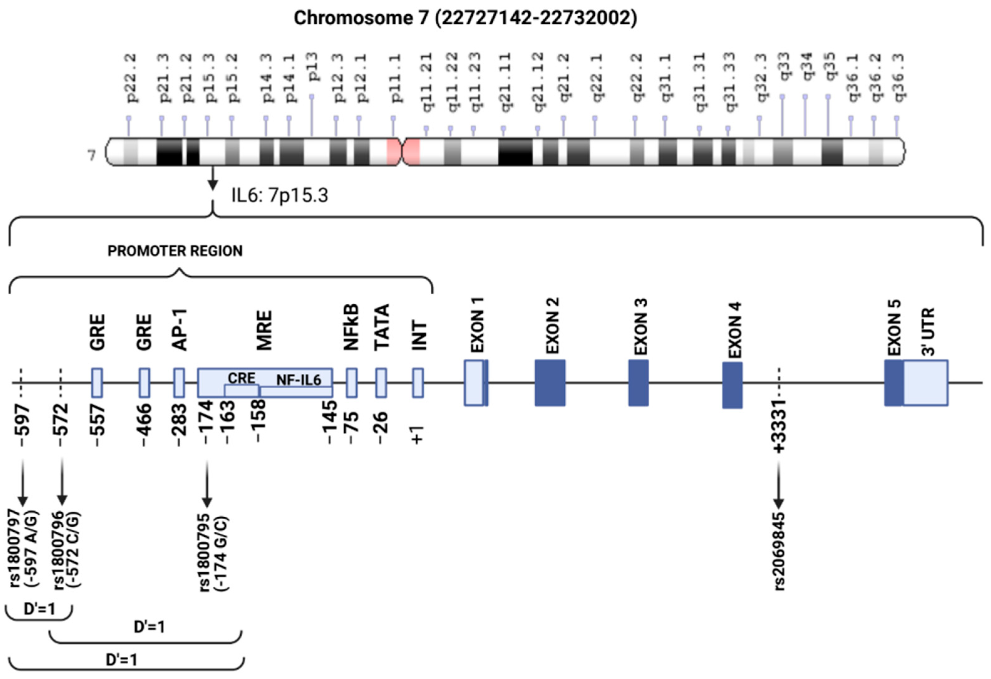
Polymorphic variation affecting IL6 expression could therefore influence individual susceptibility. In humans, the IL6 gene (OMIN 147620) is located on chromosome 7p21 [27] and contains four introns and five exons [27,28]. The IL6 gene alone has 3569 SNPs in coding, non-coding, and regulatory regions [29]. Several common polymorphisms have been identified in the 5′ flanking region of IL6, including rs1800797 (−597 A>G), rs1800796 (−572 C>G), and rs1800795 (−174 G>C) [30,31,32], and rs2069845 at an intronic position. The −597A and −174C alleles show almost complete allelic association in Caucasian subjects [31,32]. In contrast, the −572C allele generally occurs with the −174G [32]. These polymorphisms are positioned near cis-acting regulatory elements involved in controlling IL-6 expression at the transcription level, suggesting that they may influence protein–DNA interactions at these sites [33] (Figure 1).
Figure 1.
Schematic representation of the IL6 gene intron–exon structure and 5′-flanking regions. Arrows indicate the positions of single-nucleotide polymorphisms. Light blue boxes represent known and putative regulatory elements and dark blue boxes exons. The IL6 gene, located on chromosome 7p15.3, comprises 5 exons and 4 introns. Exons, introns, untranslated regions (UTRs), the evaluated polymorphisms (rs1800796 [−572 C/G][−572 C/G], rs1800796 [−597 A/G], and rs2069845 [+3.331 G/A], and rs1800795 [−174G/C]) and the putative transcription factor binding sites as well as the position and orientation of the oligonucleotide pairs used in single-strand conformation polymorphism (SSCP), restriction fragment length polymorphism (RFLP), and sequencing analyses, are depicted above. Binding sites are relative to the main transcription initiation site: TATA box −27 to −24; NF-kB −73 to −64; NF-IL6) −158 to −145; CRE: cAMP responsive element, −163 to −158; NRD: negative regulatory domain, −225 to −164; AP-1 −283 to −277; AnTn replication polymorphism −392 to −373; GRE1 and GRE2: glucocorticoid-responsive elements 1 and 2, −466 to −461 and −557 to −552, respectively. Shaded blocks: coding exons; white blocks: 5′ and 3′ UTRs; +1: first base of the transcription start site; GRE: glucocorticoid-responsive element; AP-1: activator protein-1; MRE: metal-responsive element; GRE: cAMP-responsive element; NF-IL6: nuclear factor for IL-6 expression; NFkB: nuclear factor kappa B; TATA box: promoter sequence; INT: transcription start. D’ values between SNPs are also presented. Adapted from Fishman et al. [30]; Chung et al. [34]; and Yee et al. [35].
The rs1800795 and rs1800796 polymorphisms were linked to COVID-19 severity in a western Sicilian [36], Turkish [37], Mexican [38], and Ukrainian [39] populations. The rs2069845 variant in intron 4 of IL6, which is in moderate linkage disequilibrium with rs1800795 (D’ = 1.0; R2 = 0.63), is associated with overweight/obesity and measures of adiposity in children [40,41,42].
Therefore, this study aimed to evaluate the associations between rs1800796, rs1800797, and rs2069845 polymorphisms located in the promoter or enhancer region of IL6 genes that were chosen based on evidence from the HaploReg [43] and ENCODE [44] databases, indicating functional regulatory activity, with COVID-19 outcomes and serum levels of biochemical biomarkers.
2. Materials and Methods
2.1. Sample
Sampling was performed with patients hospitalized for COVID-19 and control individuals from October 2021 to March 2023. In this case–control study, individuals testing positive for COVID-19 (n = 106) and the asymptomatic controls testing negative for COVID-19 (n = 106) were evaluated. The population under study, residing in Curitiba, Paraná State (southern Brazil), is composed of 65% Euro-Brazilians, 34% Afro-Brazilians, and 1% Asians and Indigenous people, according to the 2022 census, which uses predominantly (>98%) in-person data collection [45].
The inclusion criteria were as follows: individuals of either sex, aged between 18 and 80 years, with clinical features of COVID-19 and a positive COVID-19 test. The COVID-19 patients were admitted to the intensive care unit (ICU) of the University Hospital of the Federal University of Paraná or the Worker’s Hospital of the Paraná State Health Department and classified as severe COVID-19 cases [46]. Exclusion criteria included pregnancy, incomplete clinical records, and individuals related to the participants. The control group was recruited from the University Hospital of the Federal University of Paraná’s blood bank. All controls tested negative for COVID-19 and reported no signs or symptoms of viremia.
The study was approved by the human research ethics committee of the Federal University of Paraná, the University Hospital of the Federal University of Paraná, and the Worker’s Hospital of the Federal University of Paraná (CAAE nos. 43948621.7.3001.0096, 43948621.7.3003.5225, and 43948621.7.0000.0102, respectively). Informed consent was obtained from all participants. All samples were collected from public health institutions providing free care.
2.2. Genotyping
Genomic DNA was isolated by the salting-out method described by Lahiri and Nuerberger [47]. The DNA quality was assessed by the 260/280 nm absorbance ratio; only ratios between 1.6 and 1.8 were accepted. The DNA integrity was also verified via 1% agarose gel electrophoresis stained with ethidium bromide (Êxodo Científica, São Paulo, Brazil). The DNA concentration was quantified using spectrophotometry (UV-Vis BioDrop Duo+, Biochrom, Harvard Bioscience, Holliston, MA, USA) and adjusted to 20 ng/mL. Genotyping for rs1800796 (C__11326893_10), rs1800797 (C___1839695_20), and rs2069845 (C___1839699_10) were performed through real-time polymerase chain reaction (PCR) with fluorescent probes on the TaqMan® system (Applied Biosystems/ThermoFisher, Waltham, MA, USA), employing the 7500 Fast real-time PCR thermocycler (Applied Biosystems/ThermoFisher, MA, USA) following the reagent manufacturer’s instructions.
2.3. Biochemical Biomarkers
Whole blood (EDTAK2 tube), serum (SST tube), and plasma (EDTAK2 and NaF-EDTAK2 tubes) samples were collected from all participants using BD Vacutainer tubes (Becton, Dickinson and Company, Franklin Lakes, NJ, USA). Biomarkers of glycemic, lipid, and nutritional profiles, as well as renal and hepatic function, were quantified using automated analyzers Architect Ci8200 (Abbott Diagnostics, Chicago, IL, USA); or Labmax 400 (Labtest, Minas Gerais, Brazil) with appropriate calibrators, controls, and reagents provided by their manufacturers. The 1,5-anhydroglucitol was determined by enzymatic assay GlycoMark (Diazyme Laboratories, Inc., Gregg Ct., Poway, CA, USA) on the Labmax 400 (Labtest, Lagoa Santa, Minas Gerais, Brazil).
2.4. COVID-19 Detection
Reverse-transcription PCR was performed by using Reagent BIOMOL One-Step/COVID-19 (Instituto de Biologia Molecular do Paraná—IBMP, Paraná, Brazil) in nasopharyngeal swab. This assay includes two viral genes ORF 1ab gene and N-region gene SARS-CoV-2 detection according to the manufacturer’s instructions, a human endogenous gene was used as target for internal control. Serological tests were carried out using lateral flow immunochromatographic assays (LFAs), based on colloidal gold-labeled immunochromatography (GICA) principle and one-step method with results obtained within 15 min. The reagents detect IgM/IgG antibodies for SARS-CoV-2 were COVID-19 IgG/Ig MECO Test (Eco Diagnóstica, Minas Gerais, Brazil), COVID-AG (Instituto de Biologia Molecular do Parana, Paraná—IBMP, Paraná, Brazil), COVID-19 IgG/IgM ECO Teste (Fundação Oswaldo Cruz-Fiocruz, Rio de Janeiro, Brazil) and PANBIO™ COVID-19 (Abbott Diagnostics, IL, USA). All the tests were performed at room temperature according to the manufacturer’s instructions.
2.5. Statistical Analysis
The continuous variables’ normality was verified using the Kolmogorov–Smirnov test. Since these variables were not normally distributed in either group, values were reported as medians and interquartile ranges (25–75%). Comparisons were performed using the Mann–Whitney U test. Categorical variables were analyzed using the Chi-square test, while the continuous variables were compared utilizing the Mann–Whitney U test.
Hardy–Weinberg equilibrium, genotype and allele frequencies, and 95% confidence intervals (95% CI) for minor allele frequencies were obtained using the https://sites.google.com/view/generiskcalc/home/gene-association-calculator (accessed on 22 August 2025). Genotypes were coded as 1 (most frequent; homozygous), 2 (heterozygous), and 3 (least frequent; homozygous).
For comparison, allele frequencies from other populations were considered similar when they fell within the 95% CI. Statistical analyses were conducted using MedCalc software (v. 23.0.2, MedCalc Software Ltd., Ostend, Belgium), Statistica (v. 14, Cloud Software Group, Santa Clara, CA, USA), and Jamovi project (2025) v. 2.7. Statistical significance was defined as p < 0.05.
3. Results
3.1. Study Population
The participant’s characteristics and clinical information are shown in Table 1. Groups were matched by sex and body mass index, with similar median ages (56–57 years).
Table 1.
Anthropometric and laboratory characteristics of the study groups.
The glycated hemoglobin A1c did not differ between groups, with concentrations consistent with the absence of chronic hyperglycemia. The median blood glucose was elevated in the COVID-19 group (reference interval < 100 mg/dL), approximately 20% higher than in the control group (117/96 = 1.22). This group also exhibits a reduction in 1,5-anhydroglucitol. When HbA1c, a marker of chronic hyperglycemia, shows no difference between groups, the increase in blood glucose may be attributed to stress-induced hyperglycemia, a transient increase in blood glucose during acute physiological stress such as COVID-19. All other parameters showed significant differences between groups (p ≤ 0.001).
For renal function markers, urea and creatinine were significantly higher in the COVID-19 group. The median urea concentration exceeded the reference interval (15–45 mg/dL), roughly twofold higher than in the control group, while creatinine remained within the reference range (0.4–1.2 mg/dL). The COVID-19 group had significantly lower lipid parameters compared to the control group, except for triglycerides, which were not significantly different. The HDL-C was notably reduced by 50% in the COVID-19 group.
The median serum albumin in the COVID-19 group was around 20% lower than in the control group and below the reference range (3.5–5.5 g/dL), suggesting hypoalbuminemia. The aminotransferases (aspartate aminotransferase and alanine aminotransferase) associated with liver function were elevated in the COVID-19 group, albeit within the reference range (<40 U/L).
The nonspecific marker of inflammatory processes, median high-sensitivity C-reactive protein, was approximately 40 times higher (65/1.6) in the COVID-19 group, representing the greatest difference among all biomarkers assessed.
3.2. Allelic and Genotypic Frequencies and Linkage Disequilibrium
To determine the relationship between IL6 promoter polymorphisms and susceptibility to COVID-19, three distinct polymorphic regions, rs1800796 (−572 G>C), rs180097 (−597 G>A), and rs2069845, were investigated. The genotypes and allele frequencies’ distribution in the control and severe COVID-19 groups are summarized in Table 2. No significant deviation from the Hardy–Weinberg equilibrium was detected in any group (p > 0.05).
Table 2.
Allelic and genotypic frequencies of polymorphisms in the IL6 gene in the study groups.
The polymorphisms under study were also evaluated using binomial logistic regression with sex and age as confounders, confirming the absence of association. Therefore, no statistically significant differences were observed in allele and genotype distributions between the control and severe COVID-19 groups.
4. Discussion
The COVID-19 pandemic has exposed geographical disparities in infection and mortality rates, driven by public health interventions, healthcare infrastructure, socioeconomic factors, and individual behaviors [48,49]. Additionally, genetic variants may influence individual susceptibility to the disease. Infectious diseases are a major selective pressure [50,51,52], and genes involved in immune responses are among the most numerous and diverse in the human genome [53], supporting the evolutionary advantage of a varied immunological response across pathogens [54].
Genome-wide association studies and candidate gene studies have identified SNPs associated with susceptibility to SARS-CoV-2 infection and severe COVID-19 [17,18,55]. The IL-6 has been implicated in regulating genes and proteins involved in angiogenesis and immune cell recruitment [20], also exerting pleiotropic effects and contributing to various physiological and pathological processes, including inflammation, tumorigenesis, and immune modulation [22].
The IL6 polymorphisms have been associated with viral infection outcomes through their effects on IL-6 production, an early indicator of tissue injury and systemic inflammation. Thus, this study investigated the relationship between IL6 rs1800796, rs1800797, and rs2069845 polymorphisms and COVID-19, as well as serum biomarkers.
The SARS-CoV-2 virus, the agent of COVID-19, affects multiple organs beyond the respiratory system [56]. Our case–control analysis identified severe viremia in the COVID-19 group, underscoring the severity of the ongoing process.
Although respiratory symptoms predominate in COVID-19, metabolic disturbances, including hyperglycemia and new-onset diabetes, have been reported soon after the pandemic’s onset [57,58]. Hyperglycemia was most frequently associated with an increased risk of adverse outcomes (e.g., disease severity, physiological stress, and intubation) [59,60], especially in patients with pre-existing diabetes [61]. Although elevated blood glucose levels are common in critical illness, the prevalence of hyperglycemia and new-onset diabetes among COVID-19 patients appears to be unexpectedly high [62]. The observed hyperglycemia is associated with stress-induced insulin resistance and increased glucose synthesis (Table 1) [63]. The reference-range glycated hemoglobin fraction A1c and 1,5-anhydroglucitol values suggest that no patients with diabetes were present in the study groups [64].
The increase in urea and creatinine concentrations in early COVID-19 has been documented and is considered a severity marker [65,66]. Reduced albumin is reported in many chronic and acute inflammatory processes [67]. In COVID-19, hypoalbuminemia (serum albumin < 3.4 g/dL) has been proposed as a predictor of in-hospital mortality, independent of age, comorbidities, and inflammation [68].
The relationship between COVID-19 and lipid profile is complex [69]. Lower high-density lipoprotein cholesterol concentrations are associated with a higher likelihood of severe COVID-19 [70]. These studies are consistent with the present study findings of the COVID-19 group, which showed a 50% lower median high-density lipoprotein cholesterol than the control group (Table 1).
Markedly increased high-sensitivity C-reactive protein in the COVID-19 group is a severity marker associated with ICU admission, acute kidney injury, and poor outcomes [71]. Given the symptoms displayed, the COVID-19 group had severe viremia. The results revealed that the IL6 rs1800796, rs1800797, and rs2069845 were not associated with an increased risk of COVID-19 (Table 2). Although the rs1800796 (−572 C>G) variant has been related to COVID-19 severity in a western Sicilian [36], Turkish [37], Mexican [38,72], and Ukrainian [39] populations, no correlation was found between rs1800796 and COVID-19 in Iranian individuals [73] and in a systematic review across 23 countries [74]. Similarly, studies in Iranian [73], Moroccan [75], Chinese [76], and Indian [77] populations reported no association between rs1800797 (−597 A>G) and COVID-19. Conversely, Chinese homozygous carriers of the C allele at rs1800796 presented a lower risk of severe symptoms [76], while in American and European populations, it increased the risk of severe disease, likely reflecting differences in immunogenetic backgrounds among populations.
Indeed, rs1800796, highly prevalent in Asia, has been proposed to explain ethnic-specific susceptibility to certain infectious diseases. Individuals of Hmong ancestry (China) harboring rs1800796, rs1524107, and rs2066992 had a greater risk of severe fungal infection than individuals of European ancestry harboring the wild-type haplotype [78]. For hepatitis B virus (HBV) infection that is endemic to Asian countries, the rs1800796 C/C genotype was detrimental to spontaneous HBV clearance in Chinese [79] and increased risk of chronic HBV infection in Malaysians [80]. The IL-6 promoter SNPs rs1800795 and rs1800797 are exclusively polymorphic in Caucasians. The G/G genotype of rs1800795 was protective against pneumococcal pneumonia in white Spanish patients [31,81]. In contrast, the G-G alleles at rs1800795 and rs1800797 were linked to worse chronic HBV in Italians [82] and susceptibility to human papillomavirus HPV-associated cervical cancer in North Indians [83]. Thus, the impact of IL-6 polymorphisms in infectious disease susceptibility varies with both pathogen and ethnicity.
Notably, the two promoter SNPs exhibited striking ethnic disparities in global allele frequencies (Table 3). The rs1800796 variant was most frequent in East Asians (79%), followed by South Asians (39%) and Americans (30%), but rare in Africans (10%) and Europeans (5%). By contrast, rs1800797 was highly polymorphic in Caucasians, albeit nearly monomorphic in Asians and Africans. The rs1800797 and rs2069845 were most and least prevalent, respectively, in Africans (32 and 2%), whereas distribution in other populations was more similar (Table 3). The allele frequency of the Brazilian population (this study) of rs1800796 (11.3%, 95% CI 7–16) was similar only to Africans (10%), and rs1800797 (28.3%, 95% CI 22–34) was not similar to any other population. The rs2069845 (36.8%, 95% CI 30–43) was similar to Africans (32%), Europeans (43%), Americans (29%), and other South Brazilians (Table 3) [42,84].
Table 3.
Comparisons between allele frequencies for the studied polymorphisms in different populations based on Ensembl data.
Considering the ethnic disparities in IL-6 functional SNPs, Chen et al. [76] compared IL-6 expression levels in European, African, and East Asian populations, noting significantly higher levels in Europeans, intermediate levels in Africans, and the lowest in East Asians. Therefore, genetic variability among populations may partially explain differences in COVID-19 course and susceptibility to its complications. Other unexamined variants in linear dichroism could likely influence cytokine levels.
Our data showed that these three SNPs are not associated with COVID-19 in the southern Brazilian population. Despite our promising findings, this study has some limitations that should be acknowledged. First, RNA expression or IL-6 protein levels were not measured to assess the effects of three SNP genotypes on IL-6 expression. Second, the sample size was relatively small, and the patients were selected from only one region of Brazil. Third, additional SNPs in other IL-6 regions were not examined. Fourth, several factors that may affect the validity and generalizability of the results, specifically regarding potential population stratification, lack of adjustment for comorbidities, and the absence of multivariable modeling. Lastly, to confirm these results, more extensive studies across diverse ethnic populations are required to investigate the association between IL-6 genetic polymorphisms and COVID-19 pathogenesis. Further investigations of other SNPs in these and related genes implicated in COVID-19 severity are necessary to develop prevention strategies and precision medical therapies. In summary, in the study population, the COVID-19 group did not differ from healthy controls in the allelic or genotypic frequencies of the studied IL6 variants. Further studies with larger sample sizes are needed to confirm the results presented.
Author Contributions
Conceptualization, R.S.G.d.S., F.G.d.M.R. and G.P.; methodology, N.M.P.S., C.M.G.d.L. and P.R.S.; validation, M.H.M.S. and A.L.d.P.; formal analysis, G.P. and R.S.G.d.S.; investigation, I.R., G.C. and M.H.M.S.; data curation, A.L.d.P.; writing—original draft preparation, F.G.d.M.R. and R.S.G.d.S.; writing—review and editing, G.P. and M.B.N.; project administration, F.G.d.M.R.; funding acquisition, F.G.d.M.R. and G.P. All authors have read and agreed to the published version of the manuscript.
Funding
This study was funded by Araucaria Foundation (grant no. SUS2020131000069), the National Council for Scientific and Technological Development—CNPq (projects nos. 407034/2023-4 and 405828/2025-0), and the Federal University of Paraná.
Institutional Review Board Statement
The human research ethics committee approved this study (CAAE nos. 43948621.7.3001.0096 on 4 June 2021, 43948621.7.3003.5225 on 10 June 2021, and 43948621.7.0000.0102 on 5 May 2021).
Informed Consent Statement
Informed consent for participation was obtained from all subjects involved in the study.
Data Availability Statement
The data supporting the findings of this study are available within the article.
Acknowledgments
The authors kindly acknowledge the Araucaria Foundation, the National Council for Scientific and Technological Development—CNPq, the Coordination for the Improvement of Higher Education Personnel—CAPES and its Institutional Internationalization Program, and the Federal University of Paraná for the research grants and scholarships (Finance Code 001).
Conflicts of Interest
The authors declare that the research was conducted in the absence of any commercial or financial relationships that could be construed as a potential conflict of interest.
Abbreviations
The following abbreviations are used in this manuscript:
| SARS-CoV-2 | Severe acute respiratory syndrome coronavirus 2 |
| COVID-19 | Coronavirus disease 2019 |
| SNPs | Single-nucleotide polymorphisms |
| ICU | Intensive care unit |
| HBV | Hepatitis B virus |
References
- Zhou, F.; Yu, T.; Du, R.; Fan, G.; Liu, Y.; Liu, Z.; Xiang, J.; Wang, Y.; Song, B.; Gu, X.; et al. Clinical course and risk factors for mortality of adult inpatients with COVID-19 in Wuhan, China: A retrospective cohort study. Lancet 2020, 395, 1054–1062. [Google Scholar] [CrossRef] [Scilit] [PubMed]
- Huang, C.; Wang, Y.; Li, X.; Ren, L.; Zhao, J.; Hu, Y.; Zhang, L.; Fan, G.; Xu, J.; Gu, X.; et al. Clinical features of patients infected with 2019 novel coronavirus in Wuhan, China. Lancet 2020, 395, 497–506. [Google Scholar] [CrossRef] [Scilit]
- Wu, C.; Chen, X.; Cai, Y.; Xia, J.; Zhou, X.; Xu, S.; Huang, H.; Zhang, L.; Zhou, X.; Du, C.; et al. Risk Factors Associated with Acute Respiratory Distress Syndrome and Death in Patients with Coronavirus Disease 2019 Pneumonia in Wuhan, China. JAMA Intern. Med. 2020, 180, 934–943. [Google Scholar] [CrossRef] [Scilit]
- Wu, C.; Chen, X.; Cai, Y.; Xia, J.; Zhou, X.; Xu, S.; Huang, H.; Zhang, L.; Zhou, X.; Du, C.; et al. Clinical and immunological features of severe and moderate coronavirus disease 2019. J. Clin. Investig. 2020, 130, 2620–2629. [Google Scholar] [CrossRef] [Scilit]
- Wang, D.; Hu, B.; Hu, C.; Zhu, F.; Liu, X.; Zhang, J.; Wang, B.; Xiang, H.; Cheng, Z.; Xiong, Y.; et al. Clinical Characteristics of 138 Hospitalized Patients with 2019 Novel Coronavirus-Infected Pneumonia in Wuhan, China. JAMA 2020, 323, 1061–1069. [Google Scholar] [CrossRef] [Scilit]
- Ruan, Q.; Yang, K.; Wang, W.; Jiang, L.; Song, J. Clinical predictors of mortality due to COVID-19 based on an analysis of data of 150 patients from Wuhan, China. Intensive Care Med. 2020, 46, 846–848. [Google Scholar] [CrossRef] [Scilit]
- Riphagen, S.; Gomez, X.; Gonzalez-Martinez, C.; Wilkinson, N.; Theocharis, P. Hyperinflammatory shock in children during COVID-19 pandemic. Lancet 2020, 395, 1607–1608. [Google Scholar] [CrossRef] [Scilit]
- Yildirim, Z.; Sahin, O.S.; Yazar, S.; Bozok Cetintas, V. Genetic and epigenetic factors associated with increased severity of Covid-19. Cell Biol. Int. 2021, 45, 1158–1174. [Google Scholar] [CrossRef] [Scilit]
- Fang, L.; Karakiulakis, G.; Roth, M. Are patients with hypertension and diabetes mellitus at increased risk for COVID-19 infection? Lancet Respir. Med. 2020, 8, e21. [Google Scholar] [CrossRef] [Scilit] [PubMed]
- Hamet, P.; Pausova, Z.; Attaoua, R.; Hishmih, C.; Haloui, M.; Shin, J.; Paus, T.; Abrahamowicz, M.; Gaudet, D.; Santucci, L.; et al. SARS-CoV-2 Receptor ACE2 Gene Is Associated with Hypertension and Severity of COVID 19: Interaction with Sex, Obesity, and Smoking. Am. J. Hypertens. 2021, 34, 367–376. [Google Scholar] [CrossRef] [Scilit] [PubMed]
- Ghazavi, A.; Ganji, A.; Keshavarzian, N.; Rabiemajd, S.; Mosayebi, G. Cytokine profile and disease severity in patients with COVID-19. Cytokine 2021, 137, 155323. [Google Scholar] [CrossRef] [Scilit]
- Yessenbayeva, A.; Apsalikov, B.; Massabayeva, M.; Kazymov, M.; Shakhanova, A.; Mussazhanova, Z.; Kadyrova, I.; Aukenov, N.; Shaimardanov, N. Biomarkers of immunothrombosis and polymorphisms of IL2, IL6, and IL10 genes as predictors of the severity of COVID-19 in a Kazakh population. PLoS ONE 2023, 18, e0288139. [Google Scholar] [CrossRef] [Scilit] [PubMed]
- Zanza, C.; Romenskaya, T.; Manetti, A.C.; Franceschi, F.; La Russa, R.; Bertozzi, G.; Maiese, A.; Savioli, G.; Volonnino, G.; Longhitano, Y. Cytokine Storm in COVID-19: Immunopathogenesis and Therapy. Medicina 2022, 58, 144. [Google Scholar] [CrossRef] [Scilit] [PubMed]
- Aziz, M.; Fatima, R.; Assaly, R. Elevated interleukin-6 and severe COVID-19: A meta-analysis. J. Med. Virol. 2020, 92, 2283–2285. [Google Scholar] [CrossRef] [Scilit]
- Galván-Román, J.M.; Rodríguez-García, S.C.; Roy-Vallejo, E.; Marcos-Jiménez, A.; Sánchez-Alonso, S.; Fernández-Díaz, C.; Alcaraz-Serna, A.; Mateu-Albero, T.; Rodríguez-Cortes, P.; Sánchez-Cerrillo, I.; et al. IL-6 serum levels predict severity and response to tocilizumab in COVID-19: An observational study. J. Allergy Clin. Immunol. 2021, 147, 72–80.e8. [Google Scholar] [CrossRef] [Scilit] [PubMed]
- Yin, J.-X.; Agbana, Y.L.; Sun, Z.-S.; Fei, S.-W.; Zhao, H.-Q.; Zhou, X.-N.; Chen, J.-H.; Kassegne, K. Increased interleukin-6 is associated with long COVID-19: A systematic review and meta-analysis. Infect. Dis. Poverty 2023, 12, 43. [Google Scholar] [CrossRef] [Scilit]
- Fricke-Galindo, I.; Falfán-Valencia, R. Genetics Insight for COVID-19 Susceptibility and Severity: A Review. Front. Immunol. 2021, 12, 622176. [Google Scholar] [CrossRef] [Scilit]
- Chakravarty, S. COVID-19: The Effect of Host Genetic Variations on Host-Virus Interactions. J. Proteome Res. 2021, 20, 139–153. [Google Scholar] [CrossRef] [Scilit]
- Alkhalil, E.; Jarad, B.; Saleh, T.; Aldarwish, W.; Joujeh, D. BioNTech/Pfizer (BNT162b2) COVID-19 mRNA vaccine: Manufacturing, immunogenicity, efficacy and safety. Prospect. Pharm. Sci. 2025, 23, 31–46. [Google Scholar] [CrossRef] [Scilit]
- Santa Cruz, A.; Mendes-Frias, A.; Oliveira, A.I.; Dias, L.; Matos, A.R.; Carvalho, A.; Capela, C.; Pedrosa, J.; Castro, A.G.; Silvestre, R. Interleukin-6 Is a Biomarker for the Development of Fatal Severe Acute Respiratory Syndrome Coronavirus 2 Pneumonia. Front. Immunol. 2021, 12, 613422. [Google Scholar] [CrossRef] [Scilit]
- Santiago, A.M.; da Silva Graça Amoras, E.; Queiroz, M.A.F.; da Silva Conde, S.R.S.; Cayres-Vallinoto, I.M.V.; Ishak, R.; Vallinoto, A.C.R. TNFA-308G>A and IL10-1082A>G polymorphisms seem to be predictive biomarkers of chronic HCV infection. BMC Infect. Dis. 2021, 21, 1133. [Google Scholar] [CrossRef] [Scilit]
- Song, J.; Park, D.W.; Moon, S.; Cho, H.J.; Park, J.H.; Seok, H.; Choi, W.S. Diagnostic and prognostic value of interleukin-6, pentraxin 3, and procalcitonin levels among sepsis and septic shock patients: A prospective controlled study according to the Sepsis-3 definitions. BMC Infect. Dis. 2019, 19, 968. [Google Scholar] [CrossRef] [Scilit] [PubMed]
- Grębowski, R.; Saluk, J.; Bijak, M.; Szemraj, J.; Wigner, P. Variability, Expression, and Methylation of IL-6 and IL-8 Genes in Bladder Cancer Pathophysiology. Int. J. Mol. Sci. 2023, 24, 6266. [Google Scholar] [CrossRef] [Scilit]
- Lu, L.; Zhang, H.; Dauphars, D.J.; He, Y.W. A Potential Role of Interleukin 10 in COVID-19 Pathogenesis. Trends Immunol. 2021, 42, 3–5. [Google Scholar] [CrossRef] [Scilit]
- Korbecki, J.; Kojder, K.; Kapczuk, P.; Kupnicka, P.; Gawrońska-Szklarz, B.; Gutowska, I.; Chlubek, D.; Baranowska-Bosiacka, I. The Effect of Hypoxia on the Expression of CXC Chemokines and CXC Chemokine Receptors—A Review of Literature. Int. J. Mol. Sci. 2021, 22, 843. [Google Scholar] [CrossRef] [Scilit]
- Kouhpayeh, H.R.; Taheri, M.; Baziboroon, M.; Naderi, M.; Bahari, G.; Hashemi, M. CCL5 rs2107538 Polymorphism Increased the Risk of Tuberculosis in a Sample of Iranian Population. Prague Med. Rep. 2016, 117, 90–97. [Google Scholar] [CrossRef] [Scilit] [PubMed]
- Sehgal, P.B.; Zilberstein, A.; Ruggieri, R.M.; May, L.T.; Ferguson-Smith, A.; Slate, D.L.; Revel, M.; Ruddle, F.H. Human chromosome 7 carries the beta 2 interferon gene. Proc. Natl. Acad. Sci. USA 1986, 83, 5219–5222. [Google Scholar] [CrossRef] [Scilit]
- Yasukawa, K.; Hirano, T.; Watanabe, Y.; Muratani, K.; Matsuda, T.; Nakai, S.; Kishimoto, T. Structure and expression of human B cell stimulatory factor-2 (BSF-2/IL-6) gene. EMBO J. 1987, 6, 2939–2945. [Google Scholar] [CrossRef] [Scilit]
- Kushwah, A.S.; Banerjee, M. Role of Interleukin 6 (IL-6) and Genetic Variants in Complex Diseases. In Advances in Health and Disease; Nova Science Publishers, Inc.: Hauppauge, NY, USA, 2020; pp. 1–31. [Google Scholar]
- Fishman, D.; Faulds, G.; Jeffery, R.; Mohamed-Ali, V.; Yudkin, J.S.; Humphries, S.; Woo, P. The effect of novel polymorphisms in the interleukin-6 (IL-6) gene on IL-6 transcription and plasma IL-6 levels, and an association with systemic-onset juvenile chronic arthritis. J. Clin. Investig. 1998, 102, 1369–1376. [Google Scholar] [CrossRef] [Scilit] [PubMed]
- Terry, C.F.; Loukaci, V.; Green, F.R. Cooperative influence of genetic polymorphisms on interleukin 6 transcriptional regulation. J. Biol. Chem. 2000, 275, 18138–18144. [Google Scholar] [CrossRef] [Scilit]
- Brull, D.; Montgomery, H.; Sanders, J.; Dhamrait, S.; Luong, L.; Rumley, A.; Lowe, G.; Humphries, S. Interleukin-6 gene −174g>c and−572g>c promoter polymorphisms are strong predictors of plasma interleukin-6 levels after coronary artery bypass surgery. Arterioscler. Thromb. Vasc. Biol. 2001, 21, 1458–1463. [Google Scholar] [CrossRef] [Scilit]
- Tanabe, O.; Akira, S.; Kamiya, T.; Wong, G.G.; Hirano, T.; Kishimoto, T. Genomic structure of the murine IL-6 gene. High degree conservation of potential regulatory sequences between mouse and human. J. Immunol. 1988, 141, 3875–3881. [Google Scholar] [CrossRef] [Scilit]
- Chung, H.W.; Seo, J.-S.; Hur, S.E.; Kim, H.L.; Kim, J.Y.; Jung, J.H.; Kim, L.H.; Park, B.L.; Shin, H.D. Association of interleukin-6 promoter variant with bone mineral density in pre-menopausal women. J. Hum. Genet. 2003, 48, 243–248. [Google Scholar] [CrossRef] [Scilit]
- Yee, L.J.; Im, K.; Borg, B.; Yang, H.; Liang, T.J. Interleukin-6 haplotypes and the response to therapy of chronic hepatitis C virus infection. Genes Immun. 2009, 10, 365–372. [Google Scholar] [CrossRef] [Scilit]
- Giannitrapani, L.; Augello, G.; Mirarchi, L.; Amodeo, S.; Veronese, N.; Sasso, B.L.; Giglio, R.V.; Licata, A.; Barbagallo, M.; Ciaccio, M.; et al. Outcome predictors in SARS-CoV-2 disease (COVID-19): The prominent role of IL-6 levels and an IL-6 gene polymorphism in a western Sicilian population. J. Infect. 2022, 85, 174–211. [Google Scholar] [CrossRef] [Scilit] [PubMed]
- Cekin, N.; Akin, S.; Pinarbasi, E.; Doğan, O.H. Impact of IL-6 rs1800795 and rs1800796 polymorphisms on clinical outcomes of COVID-19: A study on severity of disease in Turkish population. Mamm. Genome 2025, 36, 213–229. [Google Scholar] [CrossRef] [Scilit]
- Torres-Poveda, K.; Bahena-Román, M.; Contreras-Ochoa, C.O.; Lagunas-Martínez, A.; Bermúdez-Morales, V.H.; Pando-Robles, V.; Ortiz-Flores, E.; Cortés-Pedroza, F.; Santana-Román, M.E.; Martínez-Campos, C.; et al. High nasopharyngeal and serum IL-6 levels and the −573G > C polymorphism (rs1800796) are linked with the risk of severe COVID-19 in a Mexican population: A case–control study. BMC Infect. Dis. 2025, 25, 315. [Google Scholar] [CrossRef] [Scilit]
- Fishchuk, L.; Rossokha, Z.; Pokhylko, V.; Cherniavska, Y.; Tsvirenko, S.; Kovtun, S.; Medvedieva, N.; Vershyhora, V.; Gorovenko, N. Modifying effects of TNF-alpha, IL-6 and VDR genes on the development risk and the course of COVID-19. Pilot study. Drug Metab. Pers. Ther. 2021, 37, 133–139. [Google Scholar] [CrossRef] [Scilit] [PubMed]
- Tabassum, R.; Mahendran, Y.; Dwivedi, O.P.; Chauhan, G.; Ghosh, S.; Marwaha, R.K.; Tandon, N.; Bharadwaj, D. Common variants of IL6, LEPR, and PBEF1 are associated with obesity in Indian children. Diabetes 2012, 61, 626–631. [Google Scholar] [CrossRef] [Scilit] [PubMed]
- Joffe, Y.T.; van der Merwe, L.; Evans, J.; Collins, M.; Lambert, E.V.; September, A.V.; Goedecke, J.H. Interleukin-6 gene polymorphisms, dietary fat intake, obesity and serum lipid concentrations in black and white South African women. Nutrients 2014, 6, 2436–2465. [Google Scholar] [CrossRef] [Scilit]
- Todendi, P.F.; Klinger, E.I.; Ferreira, M.B.; Reuter, C.P.; Burgos, M.S.; Possuelo, L.G.; Valim, A.R. Association of IL-6 and CRP gene polymorphisms with obesity and metabolic disorders in children and adolescents. An. Acad. Bras. Cienc. 2015, 87, 915–924. [Google Scholar] [CrossRef] [Scilit] [PubMed]
- Ward, L.D.; Kellis, M. HaploReg: A resource for exploring chromatin states, conservation, and regulatory motif alterations within sets of genetically linked variants. Nucleic Acids Res. 2011, 40, D930–D934. [Google Scholar] [CrossRef] [Scilit] [PubMed]
- Dunham, I.; Kundaje, A.; Aldred, S.F.; Collins, P.J.; Davis, C.A.; Doyle, F.; Epstein, C.B.; Frietze, S.; Harrow, J.; Kaul, R.; et al. An integrated encyclopedia of DNA elements in the human genome. Nature 2012, 489, 57–74. [Google Scholar] [CrossRef] [Scilit]
- IBGE. Cidades e Estados: Curitiba. Instituto Brasileiro de Geografia e Estatística. 2022. Available online: https://www.ibge.gov.br/cidades-e-estados/pr/curitiba.html (accessed on 11 November 2025).
- Iser, B.P.M.; Sliva, I.; Raymundo, V.T.; Poleto, M.B.; Schuelter-Trevisol, F.; Bobinski, F. Definição de caso suspeito da COVID-19: Uma revisão narrativa dos sinais e sintomas mais frequentes entre os casos confirmados. Epidemiol. Serviços Saúde 2020, 29, e2020233. [Google Scholar] [CrossRef] [Scilit]
- Lahiri, D.K.; Nurnberger, J.I., Jr. A rapid non-enzymatic method for the preparation of HMW DNA from blood for RFLP studies. Nucleic Acids Res. 1991, 19, 5444. [Google Scholar] [CrossRef] [Scilit]
- Bambra, C.; Riordan, R.; Ford, J.; Matthews, F. The COVID-19 pandemic and health inequalities. J. Epidemiol. Community Health 2020, 74, 964. [Google Scholar] [CrossRef] [Scilit]
- Shadmi, E.; Chen, Y.; Dourado, I.; Faran-Perach, I.; Furler, J.; Hangoma, P.; Hanvoravongchai, P.; Obando, C.; Petrosyan, V.; Rao, K.D.; et al. Health equity and COVID-19: Global perspectives. Int. J. Equity Health 2020, 19, 104. [Google Scholar] [CrossRef] [Scilit]
- Frodsham, A.J.; Hill, A.V. Genetics of infectious diseases. Hum. Mol. Genet. 2004, 13, R187–R194. [Google Scholar] [CrossRef] [Scilit]
- Walsh, E.C.; Sabeti, P.; Hutcheson, H.B.; Fry, B.; Schaffner, S.F.; de Bakker, P.I.; Varilly, P.; Palma, A.A.; Roy, J.; Cooper, R.; et al. Searching for signals of evolutionary selection in 168 genes related to immune function. Hum. Genet. 2006, 119, 92–102. [Google Scholar] [CrossRef] [Scilit]
- Tishkoff, S.A.; Dietzsch, E.; Speed, W.; Pakstis, A.J.; Kidd, J.R.; Cheung, K.; Bonné-Tamir, B.; Santachiara-Benerecetti, A.S.; Moral, P.; Krings, M. Global patterns of linkage disequilibrium at the CD4 locus and modern human origins. Science 1996, 271, 1380–1387. [Google Scholar] [CrossRef] [Scilit] [PubMed]
- Murphy, P.M. Molecular mimicry and the generation of host defense protein diversity. Cell 1993, 72, 823–826. [Google Scholar] [CrossRef] [Scilit]
- Wills, C. Maintenance of multiallelic polymorphism at the MHC region. Immunol. Rev. 1991, 124, 165–220. [Google Scholar] [CrossRef] [Scilit]
- Choudhri, Y.; Maslove, D.M.; Rauh, M.J. COVID-19 and the Genetics of Inflammation. Crit. Care Med. 2023, 51, 817–825. [Google Scholar] [CrossRef] [Scilit]
- Prado, A.L.d.; Cobre, A.d.F.; Volanski, W.; Signorini, L.; Valdameri, G.; Moure, V.R.; Alves, A.d.S.C.; Rego, F.G.d.M.; Picheth, G. Machine Learning Models to Discriminate COVID-19 Severity with Biomarkers Available in Brazilian Public Health. COVID 2025, 5, 167. [Google Scholar] [CrossRef] [Scilit]
- Guo, W.; Li, M.; Dong, Y.; Zhou, H.; Zhang, Z.; Tian, C.; Qin, R.; Wang, H.; Shen, Y.; Du, K.; et al. Diabetes is a risk factor for the progression and prognosis of COVID-19. Diabetes/Metab. Res. Rev. 2020, 36, e3319. [Google Scholar] [CrossRef] [Scilit] [PubMed]
- Chen, M.; Zhu, B.; Chen, D.; Hu, X.; Xu, X.; Shen, W.J.; Hu, C.; Li, J.; Qu, S. COVID-19 May Increase the Risk of Insulin Resistance in Adult Patients Without Diabetes: A 6-Month Prospective Study. Endocr. Pract. 2021, 27, 834–841. [Google Scholar] [CrossRef] [Scilit] [PubMed]
- Khunti, K.; Del Prato, S.; Mathieu, C.; Kahn, S.E.; Gabbay, R.A.; Buse, J.B. COVID-19, Hyperglycemia, and New-Onset Diabetes. Diabetes Care 2021, 44, 2645–2655. [Google Scholar] [CrossRef] [Scilit]
- Rubino, F.; Amiel, S.A.; Zimmet, P.; Alberti, G.; Bornstein, S.; Eckel, R.H.; Mingrone, G.; Boehm, B.; Cooper, M.E.; Chai, Z.; et al. New-Onset Diabetes in Covid-19. N. Engl. J. Med. 2020, 383, 789–790. [Google Scholar] [CrossRef] [PubMed]
- Schlesinger, S.; Neuenschwander, M.; Lang, A.; Pafili, K.; Kuss, O.; Herder, C.; Roden, M. Risk phenotypes of diabetes and association with COVID-19 severity and death: A living systematic review and meta-analysis. Diabetologia 2021, 64, 1480–1491. [Google Scholar] [CrossRef] [Scilit]
- Langouche, L.; Van den Berghe, G.; Gunst, J. Hyperglycemia and insulin resistance in COVID-19 versus non-COVID critical illness: Are they really different? Crit. Care 2021, 25, 437. [Google Scholar] [CrossRef] [Scilit]
- Zahedi, M.; Kordrostami, S.; Kalantarhormozi, M.; Bagheri, M. A Review of Hyperglycemia in COVID-19. Cureus 2023, 15, e37487. [Google Scholar] [CrossRef] [Scilit]
- ADA. 2. Diagnosis and Classification of Diabetes: Standards of Care in Diabetes—2025. Diabetes Care 2025, 48, s27–s49.
- Mahmoudi, H.; Alikhani, M.Y.; Mofrad Taheri, N.; Behzadi, A. Blood Urea Nitrogen and Creatinine Clinical Prognosis in Patients with Severe Coronavirus Disease 19. Avicenna J. Clin. Microbiol. Infect. 2025, 12, 29–33. [Google Scholar] [CrossRef] [Scilit]
- Diao, B.; Wang, C.; Wang, R.; Feng, Z.; Tan, Y.; Wang, H.; Wang, C.; Liu, L.; Liu, Y.; Liu, Y.; et al. Human Kidney is a Target for Novel Severe Acute Respiratory Syndrome Coronavirus 2 (SARS-CoV-2) Infection. Nat. Commun. 2021, 12, 2506. [Google Scholar] [CrossRef] [Scilit]
- Goldwasser, P.; Feldman, J. Association of serum albumin and mortality risk. J. Clin. Epidemiol. 1997, 50, 693–703. [Google Scholar] [PubMed]
- Viana-Llamas, M.C.; Arroyo-Espliguero, R.; Silva-Obregón, J.A.; Uribe-Heredia, G.; Núñez-Gil, I.; García-Magallón, B.; Torán-Martínez, C.G.; Castillo-Sandoval, A.; Díaz-Caraballo, E.; Rodríguez-Guinea, I.; et al. Hypoalbuminemia on admission in COVID-19 infection: An early predictor of mortality and adverse events. A retrospective observational study. Med. Clin. 2021, 156, 428–436. [Google Scholar] [CrossRef] [Scilit] [PubMed]
- Ochoa-Ramírez, L.A.; De la Herrán Arita, A.K.; Sanchez-Zazueta, J.G.; Ríos-Burgueño, E.; Murillo-Llanes, J.; De Jesús-González, L.A.; Farfan-Morales, C.N.; Cordero-Rivera, C.D.; Del Ángel, R.M.; Romero-Utrilla, A.; et al. Association between lipid profile and clinical outcomes in COVID-19 patients. Sci. Rep. 2024, 14, 12139. [Google Scholar] [CrossRef] [Scilit]
- Wang, G.; Zhang, Q.; Zhao, X.; Dong, H.; Wu, C.; Wu, F.; Yu, B.; Lv, J.; Zhang, S.; Wu, G.; et al. Low high-density lipoprotein level is correlated with the severity of COVID-19 patients: An observational study. Lipids Health Dis. 2020, 19, 204. [Google Scholar]
- am-Arunthai, K.; Chamnanchanunt, S.; Thungthong, P.; Chinapha, A.; Nakhahes, C.; Suwanban, T.; Umemura, T. COVID-19 with high-sensitivity CRP associated with worse dynamic clinical parameters and outcomes. Front. Med. 2024, 11, 1346646. [Google Scholar] [CrossRef] [Scilit]
- Martínez-Gómez, L.E.; Oropeza-Vélez, C.I.; Almonte-Becerril, M.; Chavez-Galan, L.; Martinez-Armenta, C.; Vidal-Vázquez, R.P.; Ramírez-Hinojosa, J.P.; Vázquez-Cárdenas, P.; Gómez-Martín, D.; Vargas-Alarcón, G.; et al. Association of proinflammatory and anti-inflammatory cytokine polymorphisms with COVID-19 severity in unvaccinated patients. Front. Immunol. 2025, 16, 1641285. [Google Scholar] [CrossRef] [Scilit]
- Falahi, S.; Zamanian, M.H.; Feizollahi, P.; Rezaiemanesh, A.; Salari, F.; Mahmoudi, Z.; Gorgin Karaji, A. Evaluation of the relationship between IL-6 gene single nucleotide polymorphisms and the severity of COVID-19 in an Iranian population. Cytokine 2022, 154, 155889. [Google Scholar] [CrossRef] [Scilit]
- Karcioglu Batur, L.; Hekim, N. Correlation between interleukin gene polymorphisms and current prevalence and mortality rates due to novel coronavirus disease 2019 (COVID-2019) in 23 countries. J. Med. Virol. 2021, 93, 5853–5863. [Google Scholar] [CrossRef] [Scilit] [PubMed]
- Noureddine, R.; Baba, H.; Aqillouch, S.; Abounouh, K.; Laazaazia, O.; Elmessaoudi-Idrissi, M.; Bahmani, F.Z.; Tanouti, I.A.; Ouladlahsen, A.; Sarih, M.; et al. The Interleukin-6 gene variants may protect against SARS-CoV-2 infection and the severity of COVID-19: A case-control study in a Moroccan population. BMC Med. Genom. 2024, 17, 139. [Google Scholar] [CrossRef] [Scilit]
- Chen, T.; Lin, Y.X.; Zha, Y.; Sun, Y.; Tian, J.; Yang, Z.; Lin, S.W.; Yu, F.; Chen, Z.S.; Kuang, B.H.; et al. A Low-Producing Haplotype of Interleukin-6 Disrupting CTCF Binding Is Protective against Severe COVID-19. mBio 2021, 12, e0137221. [Google Scholar] [CrossRef] [Scilit]
- Verma, S.; Verma, S.; Khan, F.H.; Siddiqi, Z.; Raza, S.T.; Abbas, M.; Mahdi, F. Genetic polymorphisms of IL6 gene −174G > C and −597G > A are associated with the risk of COVID-19 severity. Int. J. Immunogenet. 2023, 50, 5–11. [Google Scholar] [CrossRef] [Scilit] [PubMed]
- Merkhofer Richard, M.; O’Neill Mary, B.; Xiong, D.; Hernandez-Santos, N.; Dobson, H.; Fites, J.S.; Shockey Abigail, C.; Wuethrich, M.; Pepperell Caitlin, S.; Klein Bruce, S. Investigation of Genetic Susceptibility to Blastomycosis Reveals Interleukin-6 as a Potential Susceptibility Locus. mBio 2019, 10, e01224-19. [Google Scholar] [CrossRef] [Scilit] [PubMed]
- El-Maadawy, E.A.; Talaat, R.M.; Ahmed, M.M.; El-Shenawy, S.Z. Interleukin-6 promotor gene polymorphisms and susceptibility to chronic hepatitis B virus in Egyptians. Hum. Immunol. 2019, 80, 208–214. [Google Scholar] [CrossRef] [Scilit]
- Riazalhosseini, B.; Mohamed, Z.; Apalasamy, Y.D.; Shafie, N.S.; Mohamed, R. Interleukin-6 gene variants are associated with reduced risk of chronicity in hepatitis B virus infection in a Malaysian population. Biomed. Rep. 2018, 9, 213–220. [Google Scholar] [CrossRef] [Scilit]
- Martín-Loeches, I.; Solé-Violán, J.; Rodríguez de Castro, F.; García-Laorden, M.I.; Borderías, L.; Blanquer, J.; Rajas, O.; Briones, M.L.; Aspa, J.; Herrera-Ramos, E.; et al. Variants at the promoter of the interleukin-6 gene are associated with severity and outcome of pneumococcal community-acquired pneumonia. Intensive Care Med. 2012, 38, 256–262. [Google Scholar] [CrossRef] [Scilit]
- Cussigh, A.; Falleti, E.; Fabris, C.; Bitetto, D.; Cmet, S.; Fontanini, E.; Bignulin, S.; Fornasiere, E.; Fumolo, E.; Minisini, R.; et al. Interleukin 6 promoter polymorphisms influence the outcome of chronic hepatitis C. Immunogenetics 2011, 63, 33–41. [Google Scholar] [CrossRef] [Scilit]
- Gupta, M.K.; Singh, R.; Banerjee, M. Cytokine gene polymorphisms and their association with cervical cancer: A North Indian study. Egypt. J. Med. Hum. Genet. 2016, 17, 155–163. [Google Scholar] [CrossRef] [Scilit]
- Feira, M.F.; Sbruzzi, R.C.; Maciel-Fiuza, M.F.; Griebeler, V.C.; Gregianini, T.S.; Martins, L.G.; Cadore, N.A.; Chies, J.A.B.; Kowalski, T.W.; Vianna, F.S.L. Association Between Genetic Variants in TNF, IL6, and IL1B Genes and Severity of COVID-19: A Cross-Sectional Study of Patients from Southern Brazil. Diagnostics 2025, 15, 1403. [Google Scholar] [CrossRef] [Scilit] [PubMed]
- Ensembl. Ensembl Genome Browser 115. 2025. Available online: https://www.ensembl.org/index.html (accessed on 23 February 2026).

Disclaimer/Publisher’s Note: The statements, opinions and data contained in all publications are solely those of the individual author(s) and contributor(s) and not of MDPI and/or the editor(s). MDPI and/or the editor(s) disclaim responsibility for any injury to people or property resulting from any ideas, methods, instructions or products referred to in the content. |
© 2026 by the authors. Licensee MDPI, Basel, Switzerland. This article is an open access article distributed under the terms and conditions of the Creative Commons Attribution (CC BY) license.