


Abstract
Graphitic carbon nitrides (g-C3N4) are well-known fluorescent nanosheets that are photoactive under the UV–visible light range and could generate reactive oxygen species (ROS) upon appropriate light exposure. Therefore, these materials are generally favored in diagnostic applications for bioimaging and light-activated treatments simultaneously, e.g., theranostic applications in cancer treatments. Here, the natural polymer, hyaluronic acid (HA), was physically crosslinked with trivalent metal ions such as Gd(III) or Fe(III) ions in the presence of boron (B)- or sulfur (S)-doped graphitic carbon nitride (g-C3N4) nanosheets to attain spherical light-sensitive g-C3N4@HA-M(III) (M: G(III) or Fe(III) ions) composite microparticles. The g-C3N4@HA-M(III) particles were in the 0.5–20 μm size range, which is injectable for possible intravenous administration. No significant toxicity was determined for g-C3N4@HA-Gd(III) particles up to 500 μg/mL concentration on L929 fibroblast cells; for example, g-C3N4@HA-Fe(III) particles could be used in vivo applications safely up to 100 μg/mL concentration with no toxicity. The g-C3N4-based materials exhibited strong fluorescence at λex 380 nm, and S-doped g-C3N4@HA-Gd(III) particles provided the highest emission intensity for possible cell imaging applications as a diagnostic tool material. Especially, S-doped g-C3N4@HA-M(III) particles delivered photoinduced anticancer activity on SKMEL 30 skin cancer cells after 30 min of UV-A treatment at 6.88 mW/cm2 irradiance and 12.38 J/cm fluence via the reactive oxygen species (ROS) production capability. In addition to the targeting ability of HA-M(III) particles, the photoinduced anticancer activity of g-C3N4@HA-M(III) particles, e.g., on SKMEL 30 melanoma cells, offer great alternatives to toxic chemo- or radiotherapy. Furthermore, HA-Gd(III)-based particles show the highest signal intensity with better proton relaxation times and the highest proton longitudinal relaxivity. Overall, HA-Gd/Fe(III) particles with heteroatom-doped g-C3N4 revealed excellent assets with enhanced MRI capabilities in addition to specific targeted cancer treatments and photoinduced therapy for multifaceted theranostic applications.
1. Introduction
Despite major advances in diagnostic and therapeutic strategies, cancer remains one of the world’s leading killers [1]. Conventional chemotherapy and radiotherapy are often poor in selectivity, systemic toxicity, and lack of therapeutic efficacy, which has prompted the development of multifunctional nanoparticles for targeted cancer diagnosis and treatment [2]. In recent years, radiotherapy systems that integrate imaging and treatment into a single platform have attracted considerable attention for their ability to diagnose, monitor, and treat tumors with greater accuracy and lower adverse effects [3,4].
Among the various photoactive nanomaterials, graphite carbon nitride (g-C3N4) has been identified as a promising metal-free semiconductor with a unique electronic structure, high physicochemical stability, ease of synthesis, and photolytic activity [5]. The layered conjugated structure of g-C-C-N-N has the potential to transfer and emit light efficiently and is therefore highly suitable for biomedical imaging applications. In addition, under UV–visible light, g-C3N4 can induce the formation of reactive oxygen species (ROS), including singlet oxygen and superoxide radicals, which can induce the death of cancer cells by photodynamic therapy mechanisms [6,7]. Furthermore, g-C3N4 could be used as a biocompatible UV-shielding pigment [8].
Although pristine g-C3N4 has excellent optical and photocatalytic properties, its practical biomedical applications are often hampered by the rapid recombination of electrons and its relatively low quantum efficiency. Heteroatom doping strategies have been extensively studied to overcome these limitations. In particular, doping with boron (B) and sulfur (S) has been shown to effectively modify the electronic band structure, increase the absorption of visible light, increase the charge separation efficiency, and increase the ability to generate ROS [9]. In particular, sulfur-doped g-C3N4 has enhanced fluorescence intensity and photocatalytic activity by creating additional electronic states and defect-mediated charge transfer pathways [10].
In biomedical applications, the incorporation of photoactive nanomaterials into biocompatible polymeric matrices is highly advantageous for improving colloidal stability, injectability, and tumor-targeting performance. Hyaluronic acid (HA), a natural polysaccharide, has been widely employed in drug delivery and tissue engineering due to its excellent biocompatibility and biodegradability [11], and cancer cell targetability via its affinity toward CD44 receptors over-expressed in many cancer cells [12]. Furthermore, HA-based formulations such as microparticles can provide effective encapsulation and controlled delivery of functional nanomaterials while simultaneously enhancing cellular uptake and tumor accumulation. It is a native component of the mammalian extracellular matrix rather than merely an exogenous biomaterial [13,14]. HA is the only non-sulfated glycosaminoglycan and exists as a free linear polymer with no covalent binding to the parent protein [15]. HA has exceptional water-imbibing ability, possessing viscoelastic and space-filling properties that enable it to create a highly hydrated microenvironment that supports lubrication, nutrient transfer, and mechanical buffering [16]. Also, HA can actively regulate cell migration, proliferation, inflammation, and tissue remodeling through specific receptors such as CD44 and receptor for hyaluronan-mediated motility (RHAMM) [17,18].
In addition to phototherapeutic functions, magnetic resonance imaging (MRI) capability is another desirable feature for theranostic systems. Paramagnetic metal ions such as Gd(III) and Fe(III) are commonly utilized as MRI contrast agents due to their ability to alter proton relaxation behavior and improve imaging contrast [19]. Particularly, Gd(III)-containing systems exhibit high longitudinal relaxivity and excellent signal enhancement in T1-weighted MRI applications [20]. The integration of MRI-active metal ions with photoresponsive g-C3N4-based microgels therefore offers a multifunctional platform capable of simultaneous imaging and photoinduced cancer therapy.
In our previous study, HA-Gd(III) and HA-Fe(III) particles were synthesized in the presence of fluorescence carbon dots for bioimaging application and photodynamic therapy [11]. In this study, injectable spherical g-C3N4@HA-M(III) composite microparticles (M(III): Gd(III) or Fe(III) ions) incorporating B- or S-doped g-C3N4 nanosheets were successfully fabricated through physical crosslinking of HA with Gd(III) or Fe(III) ions. Although HA-based carriers, g-C3N4-mediated photodynamic therapy [21], and Gd(III)/Fe(III)-based MRI contrast agents have each been extensively investigated [11], studies integrating these components into a single heteroatom-doped HA-based microparticle for simultaneous fluorescence imaging, MRI, and photodynamic therapy remain incomplete. The novelty of the present work lies in the rational integration of these functionalities into one multifunctional theranostic platform while maintaining favorable biocompatibility and enhanced imaging and therapeutic performance. The obtained particles exhibited excellent fluorescence properties upon UV-A excitation, favorable biocompatibility, and enhanced MRI performance. In particular, the S-doped g-C3N4@HA-Gd/Fe(III) particle composite demonstrated remarkable photoinduced anticancer activity against SKMEL 30 melanoma cells because of its superior ROS generation capability. The multifunctional microparticles developed therefore represent a promising theranostic platform for targeted cancer imaging and photodynamic therapy applications.
2. Materials and Methods
2.1. Materials
In the preparation of g-C3N4-embedded HA-Metal(III) microparticles, sodium hyaluronate (HA, 1.5–2.2 MDa, 95%, Acros Organics, Segrate, Italy), gadolinium(III) chloride hexahydrate (Aldrich, 99%, St. Louis, MO, USA), Fe(III) chloride anhydrous (Fluka, 97%, Neu-Ulm, Germany), dicyandiamide (Aldrich, 99%, Trostberg, Germany), boric acid (99.5%, Sigma-Aldrich, Milwaukee, WI, USA), sulfur (Reagent grade, Sigma Aldrich, St. Louis, MO, USA), sodium bis(2-ethylhexyl) sulfosuccinate (AOT, 96%, Acros Organics, Geel, Belgium), and 2,2,4-trimethylpentane (isooctane, ≥99.5%, Isolab, Eschau, Germany) were used as received. L929 fibroblast cells (Mouse C3, a connective tissue, HUKUK No: 92123004) and SK-MEL 30 melanoma cells (human melanoma, An1, HUKUK No: 03010901) were obtained from the Culture Collection of Animal Cells (SAP Institute, Ankara, Turkey). In the cytotoxicicty test, Dulbecco’s Modified Eagle’s Medium (DMEM, with 4.5 g/L glucose, 3.7 g/L sodium pyruvate, and L-Glutamine 0.5 g/mL; Pan-biotech GmbH, Aidenbach, Germany), RPMI-1640 (2 mM L-glutamine, 1 mM sodium pyruvate, 4.5 g/L glucose, 10 mM HEPES, and 1.5 g/L NaCO3; Pan-biotech GmbH, Aidenbach, Germany), fetal bovine serum (FBS; heat-inactivated; Pan-biotech GmbH, Aidenbach, Germany), penicillin/streptomycin (10,000 U/ mL penicillin and 10 mg/mL streptomycin; Pan-biotech GmbH, Germany), trypsin (0.25%, EDTA 0.02% in PBS; Pan-biotech GmbH, Aidenbach, Germany), trypan blue solution (0.5%, Biological Industries, Haifa, Israel), 3-(4,5-dimethylthiazol-2-yl)-2,5- diphenyltetrazolium bromide (MTT) agent (neoFroxx GmbH, Einhausen, Germany), and dimethyl sulfoxide (DMSO, 99.9%, Carlo-Erba GmbH, Emmendingen, Germany) were used as received. Furthermore, acetone (99%, BRK, Ankara, Turkey), ethanol (99%, Carlo-Erba GmbH, Emmendingen, Germany), and DI water (Millipore-Direct Q UV3 at 18.2 M.Ω.cm, Molsheim, France) were utilized.
2.2. Synthesis of Graphitic Carbon Nitride
Graphitic-C3N4 (g-C3N4) nanosheets were prepared with a method described in the literature, specifically through the thermal polymerization of dicyandiamide [22]. In brief, to synthesize g-C3N4-based materials, 10 g of dicyandiamide were transferred into a ceramic crucible and placed in a furnace. In the synthesis, boric acid was incorporated into dicyandiamide at a molar ratio of 50%. For the preparation of sulfur-doped g-C3N4, Sg-C3N4, sulfur was similarly added at a molar ratio of 50%. Both compounds were mechanically mixed for 5 min to ensure homogeneous mixing. The crucibles were subsequently heated to 550 °C at a rate of 3 °C/min and maintained in the furnace for 4 h. The synthesized yellow g-C3N4 nanosheets were dispersed in 500 mL of distilled water for washing, vigorously stirred at 300 rpm for one hour, and underwent a 20 min sonication process. The g-C3N4 nanosheets were synthesized using a repetitive mixing–sonication procedure performed five times. The suspension was centrifuged at 10,000 rpm for 5 min to induce precipitation, followed by drying via the freeze-drying technique.
2.3. Synthesis of g-C3N4-Embedded HA-M(III) Microparticles
To prepare g-C3N4-doped HA-Gd(III) and HA-Fe(III) particles, a 15 mg/mL concentration of HA solution was dissolved in 10 mL of a 0.1 M NaOH aqueous solution. In a separate vial, a g-C3N4 aqueous suspension was prepared at a concentration of 5 mg/mL and sonicated for 30 min to separate the g-C3N4 nanosheets. Then, 1 mL of g-C3N4 suspension was added to the prepared 10 mL HA solution and mixed for 1 h at 300 rpm. Next, 1.1 mL of the g-C3N4-HA suspension was added to 30 mL of a 0.2 M AOT–isooctane emulsion medium and mixed at 1000 rpm for 10 min. As a crosslinker, a GdCl3 solution at a concentration of 4.45 mg/mL or an FeCl3 solution at a concentration of 1.94 mg/mL was prepared in a 0.1 M HCl solution. Then, based on the repeating unit of HA, 20 µL of GdCl3 solution at 4.45 mg/mL or FeCl3 solution at 1.94 mg/mL concentration prepared in a 0.1 M HCl solution, with a molar concentration of 0.66%, was added as a cross-linker to the AOT-isooctane emulsion medium and subjected to a reaction at room temperature for 1 h. Physically cross-linked g-C3N4-doped HA-Gd(III) and HA-Fe(III) particles were precipitated in a large amount of acetone, and then washed twice with acetone, twice with an acetone: water (v/v, 80:20) mixture, and once with acetone using a centrifuge at 10,000 rpm for 5 min. The particles were then dried in an oven at 50 °C and stored in closed containers.
2.4. Characterization of g-C3N4-Based Materials and g-C3N4@HA-M(III) Particles
The prepared g-C3N4 structures were visualized using transmission electron microscopy (TEM, JEOL JEM-ARM 200F Cold FEG instrument, Tokyo, Japan). The scanning electron microscope (SEM) images of the prepared HA-Gd/Fe(III) particles and g-C3N4@HA-Gd/Fe(III) particles were taken at a voltage of 10–30 kV using SEM (Hitachi Ultra High-Resolution Analytical, FE-SEM SU-70, Tokyo, Japan). Before imaging, the materials were placed on the SEM substrate and coated with Au/Pd for 10 s.
The functional structure of the g-C3N4@HA-Metal(III)-based particles and the changes in their chemical composition were determined using Fourier transform infrared radiation (FT-IR, Nicolet iS10, Thermo, White Bear Lake, MN, USA) spectroscopy with a resolution of 4 cm−1 in the spectral range of 4000–500 cm−1 using the ATR technique.
X-ray photoemission spectroscopy (XPS, ThermoFisher Scientific K-Alpha, Waltham, MA, USA) measurements were performed to determine the chemical structure and phase of g-C3N4 within the particle matrix. XPS measurements were determined using a system with a 400 μm spot size and 180° double-focusing hemispherical analysis, utilizing Aluminum (Al) K-α characteristic radiation (λ = 1.487 Angstrom). The scan spectra were recorded in the range of 0–1350 eV using a 1 eV energy step and 15 scans. For high-resolution scans of individual peaks, a finer energy step of 0.1 eV was used, and 10 scans were collected. Peak identification and theoretical fitting were performed using the built-in Software, Advantage v5.x: that comes with the XPS system.
The optical properties of the g-C3N4@HA-M(III) particles prepared for bioimaging purposes were obtained using a fluorescence spectrometer (FLSP920, Edinburgh Instruments, Livingston, UK) equipped with a 450 W Xe900 Xenon lamp as the excitation source and a single-photon counter/photomultiplier tube as the detector. Measurements were conducted at room temperature using a particle suspension diluted with pure water to enhance transparency. Emission spectra were recorded in 2 nm steps and with a 5 nm scanning slit between 300 and 700 nm.
The time-dependent photoluminescence decay curves of g-C3N4@HA-Gd/Fe(III), Bg-C3N4@HA-Gd/Fe(III), and Sg-C3N4@HA-Gd/Fe(III) particles were obtained using a fluorescence spectrometer (FLSP920, Edinburgh Instruments, Livingston, Scotland) by recording the photoluminescence (PL) signal on a nanosecond scale after the samples were excited with nanosecond pulses.
For MRI experiments, a 0.5 T Small Animal MRI scanner (Suzhou Niumag Analytical Instruments, Suzhou, China) was used to obtain T1-weighted cross-section MR images of a phantom including the aqueous suspensions of microgels in equal concentrations. Images were obtained using an IR pulse sequence with parameters TR = 3000 ms, TE = 20 ms, and TI = 20 ms.
2.5. Blood Compatibility of g-C3N4-Based Materials and g-C3N4@HA-M(III) Particles
The blood compatibility of the synthesized g-C3N4@HA-Gd(III)- and g-C3N4@HA-Fe(III)-based composite particles was assessed by means of hemolysis and blood clotting assays following established protocols described in the literature. The study was conducted in accordance with the Declaration of Helsinki, and the protocol was approved by the Human Research Ethics Committee of Çanakkale Onsekiz Mart University (protocol code: 2011-KAEK-27) in September 2022.
To assess the hemolytic influence of g-C3N4@HA-M (III)-based composite particles on erythrocytes, suspensions of the particles at varying concentrations, in the 50–1000 µg/mL range, were prepared in 10 mL of 0.9% NaCl solution (physiological saline, PS) and incubated in a shaking water bath at 37 °C for approximately 10 min. Blood obtained from healthy individuals was placed in tubes containing anticoagulant and carefully mixed. Two milliliters of blood were diluted with 2.5 mL of PS. Subsequently, 0.2 mL of diluted blood was gradually introduced into 10 mL of PS containing particles and incubated in a shaking water bath at 37 °C for one hour. For the negative control, 0.2 mL of diluted blood was added to 10 mL of PS, while for the positive control, 0.2 mL of diluted blood was added to 10 mL of DI water. After the incubation, the blood sample was centrifuged at 100 g for 5 min, and the absorbance of the supernatant was measured at a wavelength of 542 nm using a UV–vis spectrophotometer (T60+UV/Vis spectrometer, PG Instrument, Leicestershire, UK). The absorbance value of the suspension indicates the quantity of hemoglobin released from the hemolysis of erythrocyte cells, and the percentage of hemolysis of the particles was calculated using Equation (1).
Hemolysis ratio% = (Aparticles − Aneg)/(Apos − Aneg) × 100
Aparticles is the absorbance value of the particle-containing blood suspension, while Aneg and Apos are the absorbance values of the negative and positive controls, respectively. The test was performed in at least three replicates, and the standard deviations of the hemolysis ratio% were given.
In the blood clotting analysis, 100 µL of g-C3N4@HA-M(III)-based composite particle suspensions at varying concentrations, e.g., from 50 to 1000 μg/mL prepared in the PS solution, were transferred into flat-bottom centrifuge tubes. The tubes with the particles were maintained in a shaking water bath at 37 °C for 10 min. Then, 0.81 mL of blood and 0.064 mL of 0.2 M CaCl2 solution were mixed. Next, 270 µL of this mixture was placed onto the particle suspension. The tubes were incubated in a shaking water bath at 37 °C for 10 min, followed by the gradual addition of 10 mL of distilled water at 37 °C, and were subsequently centrifuged at 100 g for 30 s. The supernatant from the tube was taken and diluted with 40 mL of distilled water at 37 °C. This mixture was then incubated in a shaking water bath at 37 °C for 1 h more. For the negative control, 250 µL of blood was added to 50 mL of distilled water at 37 °C and incubated under the same conditions for 1 h. At the end of the incubation, the absorbance of the suspension was measured at a wavelength of 542 nm with a UV–vis spectrophotometer (T60+UV/Vis spectrometer, PG Instrument, Leicestershire, UK). The blood clotting index % was determined using the formula presented in Equation (2).
Blood clotting index % = (Aparticle+blood/Ablood) × 100
Aparticles+blood is the absorbance value of the blood suspension in contact with the sample, while Ablood is the absorbance value of the negative control. The test was performed with at least three replicates and given with standard deviations.
2.6. Cytotoxicity of g-C3N4@HA-M(III)-Based Particles
The cytotoxic effects of bare HA-M(III) particles and g-C3N4@HA-M(III) composite particles at different doses were examined on L929 fibroblast cells using MTT analysis. DMEM, supplemented with 10% FBS and 1% antibiotics, served as the growth medium for fibroblasts. Briefly, 100 µL of a cell suspension at a concentration of 1 × 10⁵ cells/mL was added to each well of a 96-well plate, which was subsequently incubated at 37 °C in a humidified 5% CO2 environment for 24 h. The particle suspension was prepared in growth media at concentrations ranging from 100 to 1000 µg/mL, and 100 µL of this suspension was applied to the adherent fibroblast cells. A growth medium devoid of any particles was utilized as a negative control. The plate was incubated for 24 h. The supernatant was subsequently discarded from the well, and the cells were washed twice with phosphate buffer solution. The cells were treated with 100 µL of 0.5 mg/mL MTT solution for 2 h in the dark. Following that, the resulting formazan crystals were dissolved in 200 µL of DMSO, and the absorbance was measured using a plate reader (Heales, MB-530, Shenzhen Huisong Technology Development Co., Ltd., Shenzhen, China) at 570 nm. All analyses were performed three times, and the results were provided with standard deviations.
Images of the fibroblast cells were captured using light and fluorescence microscopy after exposure to a 500 µg/mL concentration of the g-C3N4@HA-M(III) composite particles. Before taking the image, the particle dispersion was removed, and the adhered cells were rinsed twice with PBS. Fluorescence images were acquired using a fluorescence microscope (Axioscope, Carl Zeiss, Jena, Germany) with a DAPI filter at 365 nm, without any agent, after a 24 h incubation period.
2.7. Photosensitive Anticancer Activity of g-C3N4@HA-M(III)-Based Particles
The cytotoxicity of bare HA-M(III) particles and g-C3N4@HA-M(III) composite particles against SK-MEL 30 cancer cells was examined by MTT assay and compared with their UV-A treated one after 30 min of exposure, with a control group that did not receive UV-A exposure. RPMI, supplemented with 10% FBS and 1% antibiotics, served as the growth medium for melanoma cancer cells. In brief, 100 µL of a cell suspension with a concentration of 5 × 10⁴ cells/mL was transferred into each well of a 96-well plate, which was then incubated for 24 h at 37 °C in a humidified atmosphere containing 5% CO2. The particle suspension was prepared in growth media at a concentration of 500 µg/mL, and 100 µL of this suspension was administered to the adherent cells. A particle-free growth medium was employed as a negative control. Two identical plates were prepared. One plate was exposed to a UV-A light source (315–400 nm, Ultra Vitalux, Osram GmbH, Munich, Germany) for 30 min, with an irradiance of 6.88 mW/cm2 and a fluence of 12.38 J/cm; the other plate was incubated for 30 min without UV-A light exposure. Subsequently, these two plates were incubated for 24 h, and the vitality of the cancer cells was assessed using the procedure outlined in Section 2.6, employing an MTT assay. All analyses were performed three times, and the results were provided with standard deviations.
Statistical analysis of cytotoxicity was conducted using GraphPad Prism 11 software with one-way ANOVA and Dunnett’s multiple-comparison test. Statistical analysis was denoted as * p < 0.05 and ** p < 0.001 in comparison to the negative control.
3. Results and Discussion
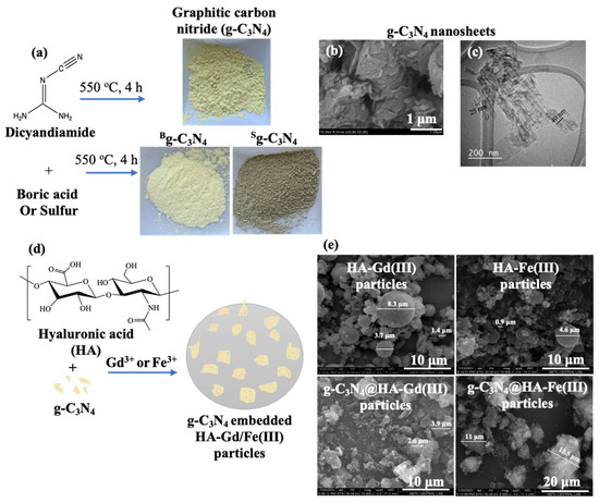
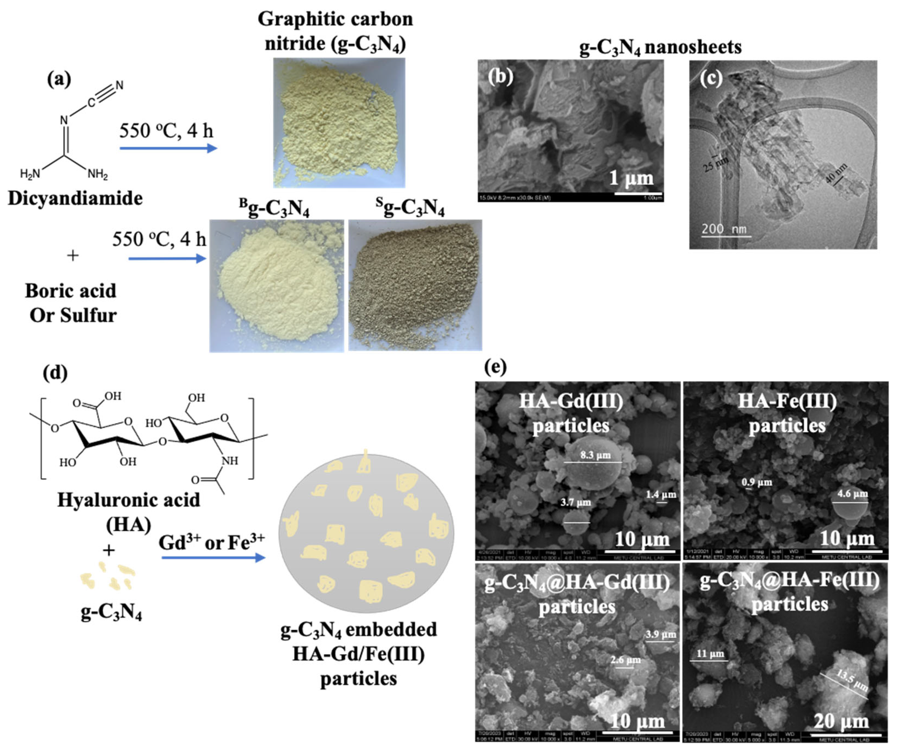
Two-dimensional g-C3N4-based structures were easily synthesized using dicyandiamide precursors via thermolysis and condensation processes at 550 °C for 4 h; the schematic representation of g-C3N4-based structures is illustrated in Figure 1a. The two-dimensional g-C3N4 nanosheets, comprising heptazine blocks, were also synthesized with the independent addition of boric acid or sulfur to obtain boron-doped and sulfur-doped g-C3N4 nanosheets, which are Bg-C3N4 and Sg-C3N4, respectively. The yellow color of g-C3N4 transformed to a lighter yellow in the Bg-C3N4 and to greenish in the Sg-C3N4 upon their B and S doping. The SEM and TEM images of the g-C3N4 are visualized in Figure 1b,c. The g-C3N4 structures are formed by the combination of overlapping multilayered irregular nanosheets having a size range of about 32 ± 8 nm.
Figure 1.
(a) The use of dicyandiamide for graphitic carbon nitride synthesis (g-C3N4) and the use of boric acid and sulfur for the preparation of heteroatom-doped g-C3N4 as Bg-C3N4 and S g-C3N4 nanosheets with corresponding digital camera, (b) scanning electron microscope (SEM), and (c) transmission electron microscope (TEM) images, and (d) the schematic representation of g-C3N4-embedded HA-Gd/Fe(III) (g-C3N4@HA-Gd/Fe(III)) particle preparation and (e) their corresponding SEM images.
The schematic representation of g-C3N4-embedded HA-Gd(III) and HA-Fe(III) particle preparation is shown in Figure 1d. Firstly, g-C3N4-based materials were suspended in a linear HA solution. A linear HA and g-C3N4 mixture dispersed in a microemulsion environment was then physically cross-linked with Gd(III) or Fe(III) ions in a short period, e.g., 1 h of mixing, to acquire g-C3N4-embedded HA-Gd/Fe(III) particles. The cross-linking occurs between the -COO- groups of the HA molecule and the trivalent metal ions, Gd(III) or Fe(III). The SEM images of the HA-Gd/Fe(III) and g-C3N4@HA-Gd/Fe(III) particles are shown in Figure 1e. The HA-Gd/Fe(III) particles exhibit a spherical morphology with sizes ranging from 0.5 to 10 μm. As seen in the SEM images, HA-Gd/Fe(III) particles are not monodispersed and are generally in sub-micron size range. The maximum size of HA-Gd/Fe(III) particles increased slightly from 8.3 μm to 13.5 μm for the composite form with g-C3N4. The g-C3N4@HA-Gd/Fe(III) particles designed for biomolecular imaging applications are injectable materials with a micrometer size distribution > 15 μm, and these particle suspensions can easily be injected with a syringe needle having a size distribution ranging from 340 to 290 μm [23]. The aggregated g-C3N4 structures are evidently seen on the surface of the g-C3N4@HA-Gd/Fe(III) particles, resulting in a composite structure.
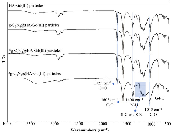
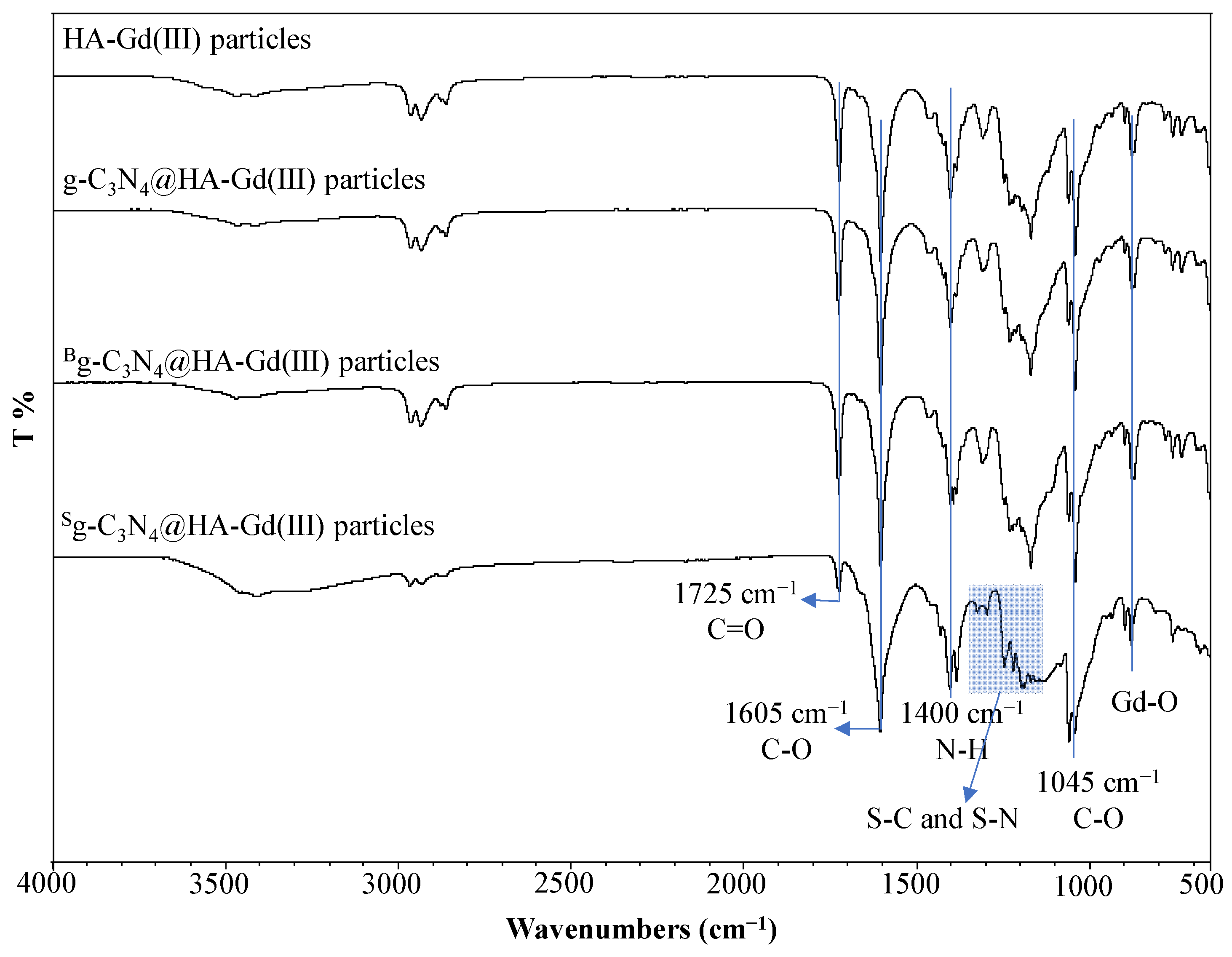
Figure 2 reveals the chemical composition of the bare HA-Gd(III) particles and g-C3N4@HA-Gd(III) composites as analyzed by FT-IR spectroscopy. No notable change was detected in the FT-IR spectra of HA-Gd(III) and HA-Fe(III) particles [24]; however, changes following the incorporation of g-C3N4 were seen for g-C3N4@HA-Gd(III) composites.
Figure 2.
FT-IR spectra of HA-Gd(III), g-C3N4@HA-Gd(III), Bg-C3N4@HA-Gd(III), and Sg-C3N4@HA-Gd(III) particles.
The specific bands for HA-Gd(III) particles are the stretching vibration at 1725 cm−1, originating from the carboxylic acids in the structure, while the stretching vibrations at 1605, 1400, and 1045 cm−1 are attributed to -C-O and -N-H groups in the amide groups of the HA molecule and the -C-O groups in the ether bond, respectively. The metal ion-O band was observed at 550 cm−1 stretching vibration of the chelation of the Gd(III) metal ion with the carboxyl group present in HA. This band, which represents the Gd(III)-O groups in the structure, indicates that the HA-Gd(III) particles were successfully prepared. The FT-IR spectra of g-C3N4@HA-Gd(III) composite particles were identical to those of bare HA-Gd(III) particles, leading to the conclusion that several slight variations in stretching frequencies were detected in the g-C3N4@HA-Gd(III) composite particles; e.g., the vibration in the 1200–1300 cm−1 region can be attributed to the C-N and C=N stretching vibrations of g-C3N4. It was also observed that among the g-C3N4-embedded HA-Gd(III) particles, only the Sg-C3N4 @HA-Gd(III) particles possessed many distinct bands within the 1100–1400 cm−1 range, which can be attributed to the S-C and S-N stretching vibrations in the Sg-C3N4 nanostructure.
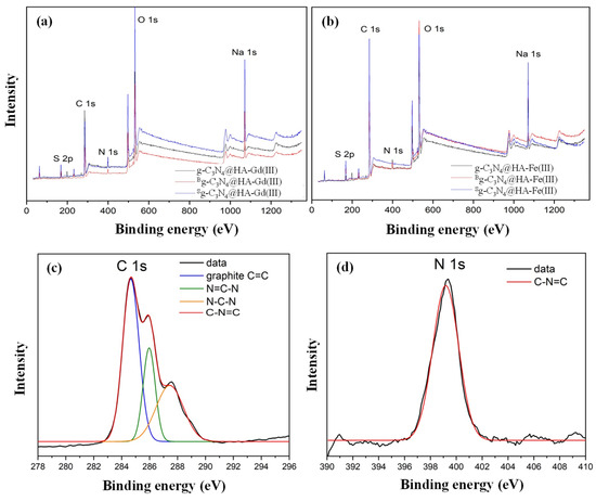
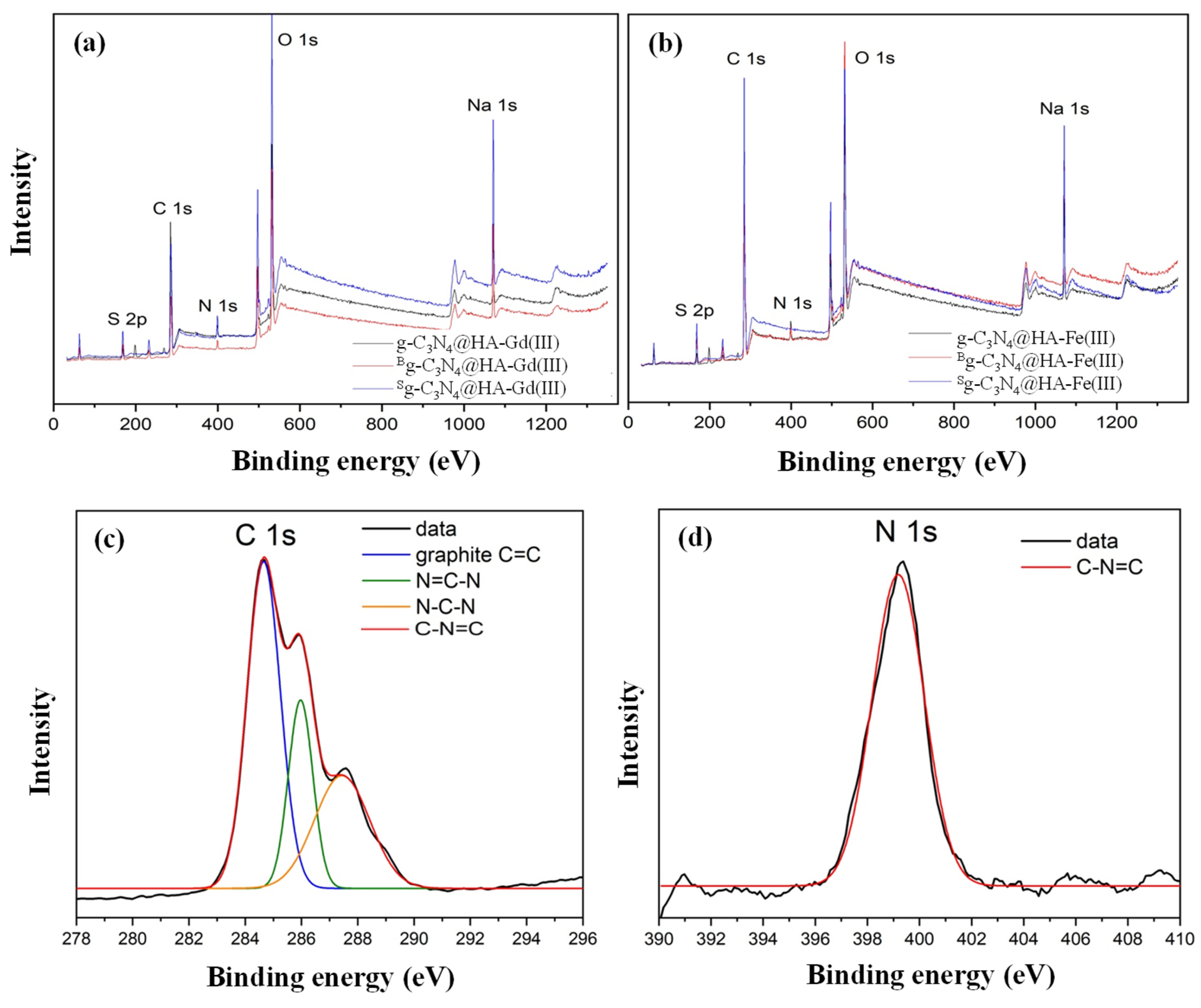
Figure 3 shows the full XPS spectra of the g-C3N4@HA-Gd(III) and g-C3N4@HA-Fe(III) particles, along with theoretical fits of the high-resolution C 1s and N 1s spectra and their decomposition into individual contributions from different atom species.
Figure 3.
(a) XPS spectra of g-C3N4@HA-Gd(III), Bg-C3N4 @HA-Gd(III), and Sg-C3N4@HA-Gd(III); (b) g-C3N4@HA-Fe(III), Bg-C3N4 @HA-Fe(III), and Sg-C3N4@HA-Fe(III) particles, and high-resolution XPS spectra of the g-C3N4 @HA-Gd(III) particles (c) C 1s and (d) N 1s.
The high-resolution XPS spectra for the C 1s peak can be deconvoluted into three peaks, where the most intense peak observed at binding energy 284.5 eV originates from the graphitic phase in the aromatic ring. The other two components located at 286–288 eV may be associated with C atoms in N=C-N and N-C-N groups, respectively. Consistently, the single peak observed for N 1s at 399.5 eV can be assigned to sp2 hybridized N atoms in triazine rings.
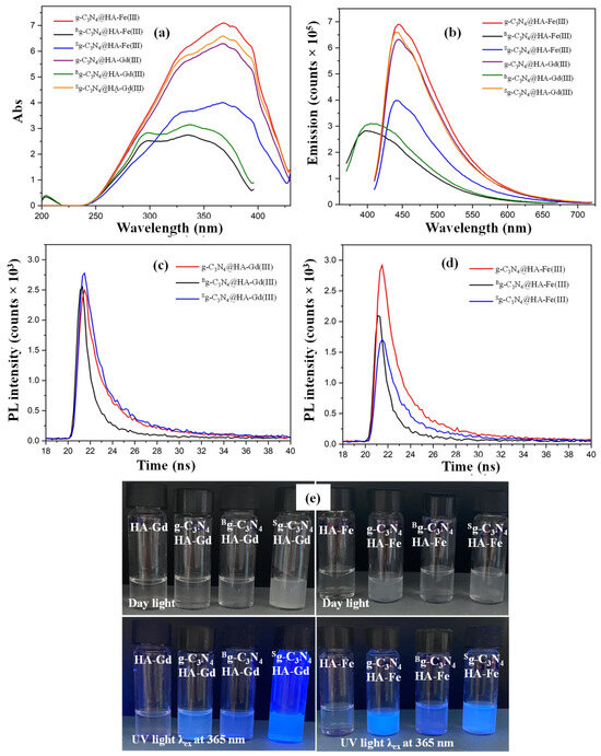
Optical properties of the g-C3N4 @HA-Gd/Fe(III)-based materials were investigated by UV–vis and fluorescence spectroscopy, and the corresponding results are shown in Figure 4a and Figure 4b, respectively. HA-M(III) particles embedded with g-C3N4 exhibit wide absorption spectra in the UV range, reaching a maximum value at a wavelength of λ = 396 nm for undoped and S-doped g-C3N4@HA-Gd/Fe(III) particles, and λ = 380 nm for B-doped g-C3N4@HA-Gd/Fe(III) particles.
Figure 4.
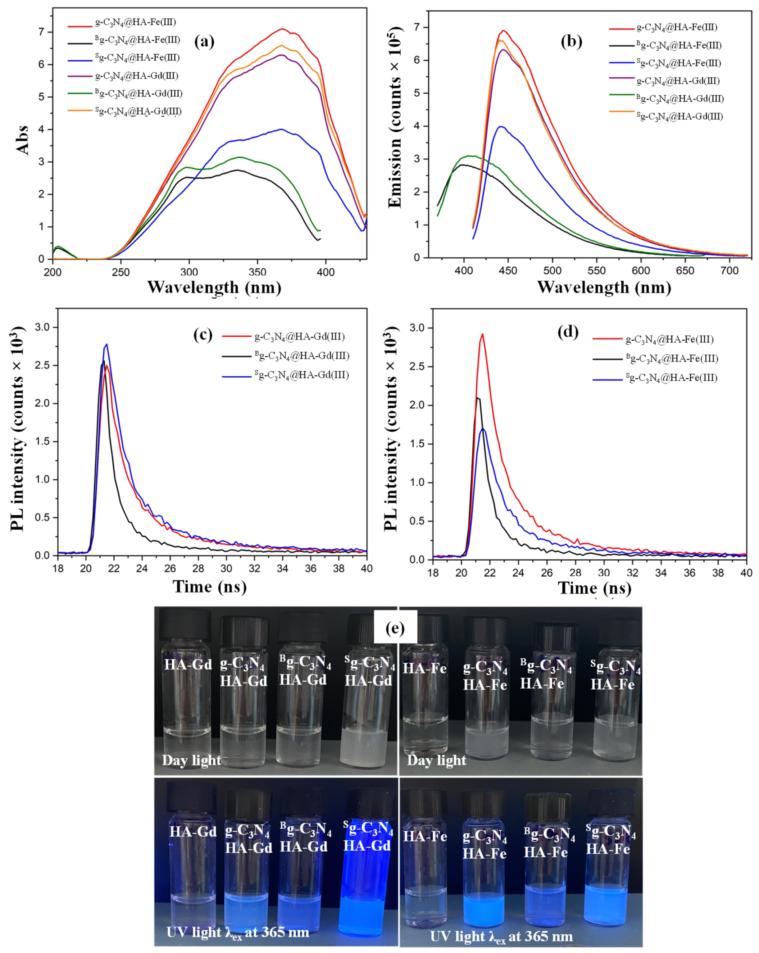
(a) UV–vis spectra and (b) fluorescence spectra of the HA-Gd/Fe(III), g-C3N4@HA-Gd/Fe(III), Bg-C3N4@HA-Gd/Fe(III), and Sg-C3N4@HA-Gd/Fe(III) particles; time decay of photoluminescence (PL) emission in nanoseconds scale recorded for (c) g-C3N4@HA-Gd(III), Bg-C3N4 @HA-Gd(III), and Sg-C3N4@HA-Gd(III), (d) g-C3N4@HA-Fe(III), Bg-C3N4 @HA-Fe(III), and Sg-C3N4@HA-Fe(III) particles, and (e) their digital camera images at daylight and UV light λex at 365 nm, in equal concentrations.
From the emission spectra given in Figure 4b, it can be concluded that the g-C3N4@HA-Gd/Fe(III) and Sg-C3N4@HA-Gd/Fe(III) particles do not cause significant changes in the forbidden energy band gap, while the Bg-C3N4@HA-Gd/Fe(III) particles increase the band gap due to the shift in the emission peak to lower wavelengths. This difference stems from the doping atoms’ atomic sizes, electronic structures, and the location where they are integrated into the host lattice. In particular, B atoms easily substitute into the interstitial or substitutional positions of the g-C3N4 framework due to their compatible atomic size, hence directly altering the electronic band structure. On the other hand, S has a much larger atomic radius than C and N, and it typically sits on the surface, edges, or interlayers of the g-C3N4 sheets instead of entering the core lattice. Since it does not modify the core conjugated aromatic system, the fundamental energy gap between the valence and conduction bands remains unchanged [24].
Additionally, the optical activity (absorption/emission) of Sg-C3N4@HA-Gd(III) particles is higher compared to g-C3N4@HA-Gd(III) particles. The fundamental reason S doping outperforms B doping in photoluminescence lies in the electron-hole recombination pathways. S atoms possess unshared lone pairs of electrons (n-electrons). As S is doped into a carbon matrix, it forms abundant surface states like C=S or S=O groups. These surface states act as highly effective energy traps that capture excited electrons and force them to undergo radiative recombination (n ⟶ π*), generating strong fluorescence intensity [25]. On the other hand, B has only three valence electrons and doping with B creates an electron-deficient matrix with a high density of localized structural holes, which function as non-radiative recombination centers in optics. In other words, excited electrons falling into boron defects lose their energy as thermal vibration (heat) rather than light, significantly quenching the fluorescence intensity [26].
By performing a theoretical fitting of the photoluminescence decay curves recorded for the g-C3N4@HA-Gd/Fe(III)-based particles (Figure 4c,d) using biexponential functions, decay times of τ1 = 1.38 ns and τ2 = 9.16 ns for the g-C3N4@HA-Gd(III) particle, and τ1 = 1.02 ns and τ2 = 6.03 ns for the g-C3N4@HA-Fe(III) particle were obtained, respectively. These decay times become shorter for B-doped and longer for S-doped counterparts, which is expected since doping creates impurity states (levels) in the band gap at which the photogenerated charge carriers (electrons and holes) localize and jump back to the corresponding bands rather than undergoing recombination; this increases the photoinduced redox performance of doped g-C3N4. As mentioned above, in the case of Boron doping, the excited electrons drop down almost instantly through fast, non-radiative thermal transitions. This rapid, non-emitting pathway cuts the excited-state population short, leaving a very brief, low-efficiency optical lifetime. On the contrary, in the case of S doping, the created rich surface states behave like a reservoir, which “store” the exciton energy and release it slowly via radiative decay, leading to measured lifetimes that are nanoseconds longer [27].
Figure 4e displays photographs of the particle suspensions prepared at a concentration of 1 mg/mL under daylight and UV light at an excitation wavelength of 365 nm. It is clearly seen that the g-C3N4 @HA-Gd/Fe(III)-based composite particle suspensions appear colorless and with a near-transparent cloudiness in daylight, whereas they emit blue light under UV light at a 365 nm excitation wavelength. As a control, images of bare HA-Gd/Fe(III) particles did not emit any blue light under UV light exposure due to the absence of g-C3N4. The results indicate that the fluorescence property originates from the g-C3N4-based materials within the g-C3N4 @HA-Gd/Fe(III)-based composite particles, demonstrating that the composite structure was successfully synthesized.
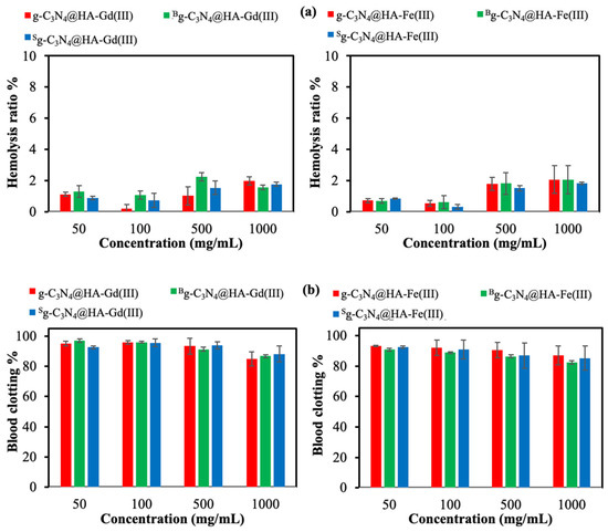
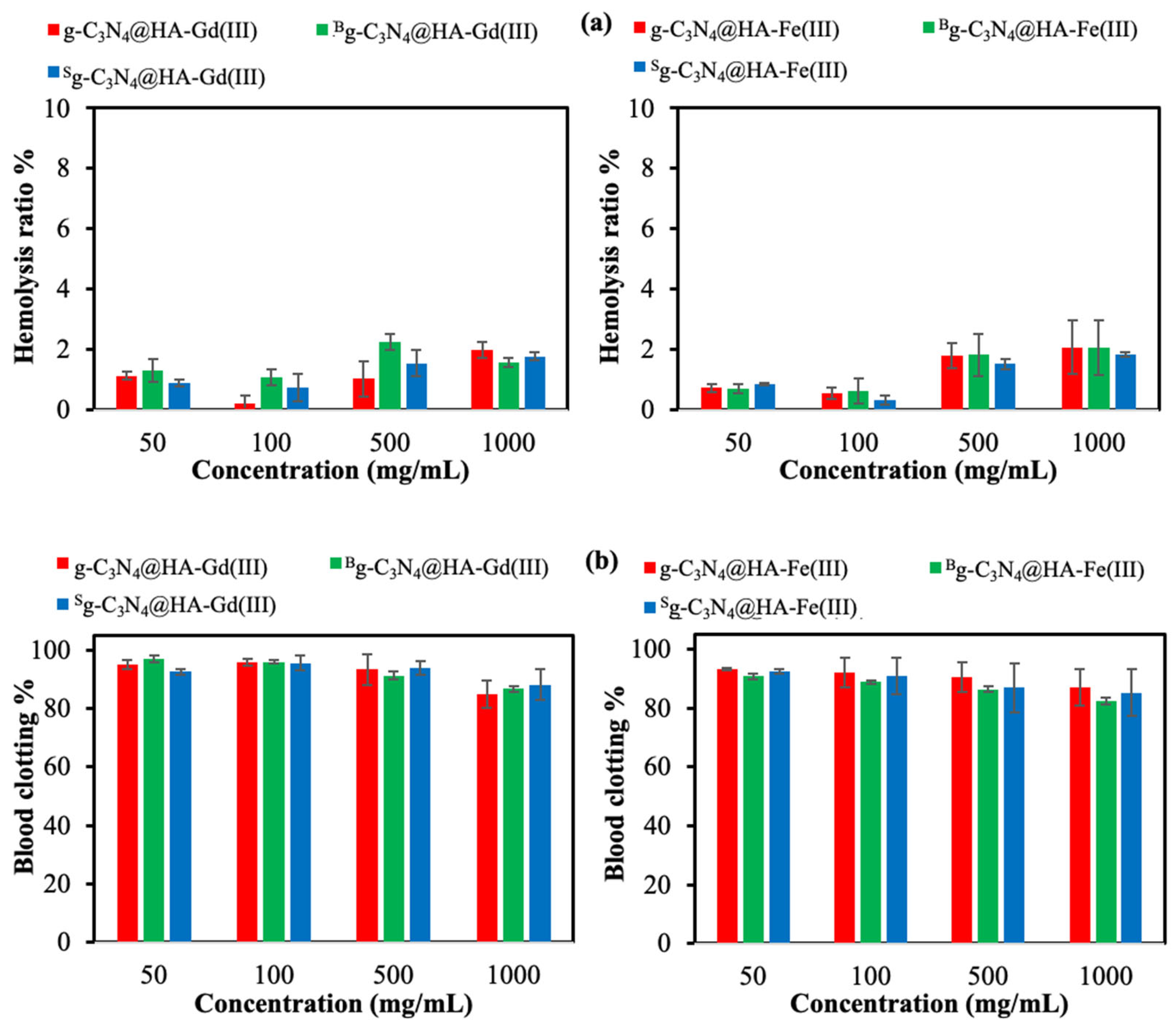
The determination of blood compatibility of g-C3N4@HA-Gd/Fe(III) particles, designed for bioimaging, is an important criterion for evaluating their potential for in vivo applications. In MRI contrast enhancers or targeted cancer therapies, materials such as g-C3N4@HA-Gd/Fe(III) particles are required to be hemocompatible upon intravenous administration. The hemolysis assay is a standard procedure to demonstrate material-induced damage to erythrocytes, with the hemolysis ratio required to remain below 5% during interaction with blood cells. The results of the hemolysis ratio% of the g-C3N4@HA-Gd/Fe(III)-based particles at different concentrations from 50 to 1000 μg/mL are summarized in Figure 5a. The hemolysis ratio% of all g-C3N4@HA-Gd/Fe(III)-based particles was determined to be <2% at a concentration of 100 μg/mL, indicating their non-hemolytic nature. Nevertheless, their hemolytic ratio % was increased to approximately 2% at a 1000 μg/mL concentration. These results show that g-C3N4@HA-Gd/Fe(III)-based particles can be safely applied up to 1000 μg/mL with a hemolysis rate of less than 5%. The effect of the materials on blood coagulation, another blood compatibility test, is shown in Figure 5b. A high blood clotting index indicates excellent blood compatibility, demonstrating no impact on the blood coagulation mechanism. The blood clotting % value of g-C3N4@HA-Gd(III) particles exceeds 85%, even at 1000 μg/mL concentration. In addition, the g-C3N4@HA-Fe(III) particles exhibited no significant impact on the blood coagulation mechanism at concentrations up to 500 μg/mL, but they exerted a minor influence at 1000 μg/mL, resulting in a blood clotting index % value of 82 ± 1. Our previous research indicated that bare HA-Gd(III) and HA-Fe(III) particles at a concentration of 1000 μg/mL exhibited blood coagulation indices of 88 ± 4 and 85 ± 7, respectively [28].
Figure 5.
(a) Hemolysis and (b) blood clotting index of HA-Gd/Fe(III)-based particles.
These results suggest that the g-C3N4 in the particle slightly affects the blood coagulation mechanism. Both blood compatibility tests show that g-C3N4@HA-Gd/Fe(III)-based particles up to a concentration of 1000 µg/mL can be safely used in applications interacting with blood, such as MRI contrast agents, without causing any adverse effects.
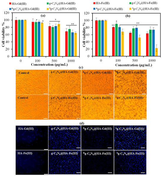
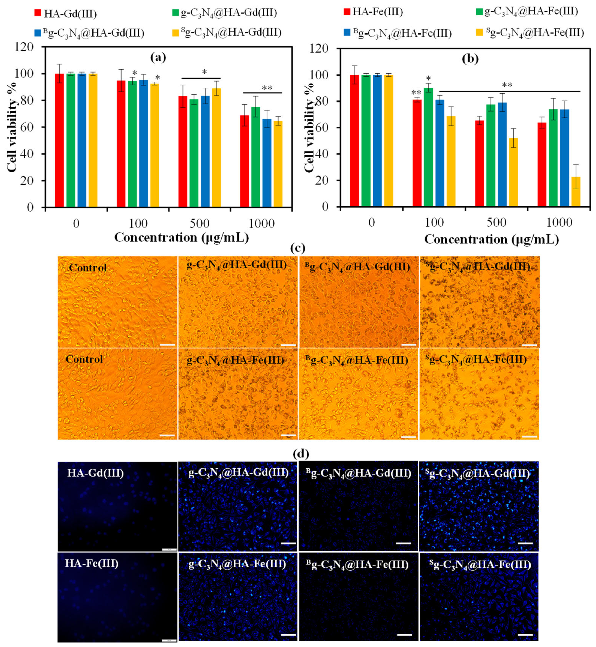
Cytotoxicity of g-C3N4@HA-Gd/Fe(III)-based composite particles was investigated on L929 fibroblast cells for a 24 h incubation time for different concentrations of the particles, and results are given in Figure 6. HA-Gd(III) particles and g-C3N4@HA-Gd(III) composite particles show almost the same toxic effect without any significant difference. No toxic effect was observed at a concentration of 100 µg/mL of g-C3N4@HA-Gd(III)-based particles, with cell viability >92%, as shown in Figure 6a. The cell viability of the g-C3N4@HA-Gd(III)-based particles slightly decreased to 83% and 70% at concentrations of 500 and 1000 μg/mL, respectively. Concentration-dependent toxicity of g-C3N4@HA-Fe(III)-based composite particles is also illustrated in Figure 6b, where no significant toxicity was detected at 100 mg/mL for both the HA-Fe(III) and g-C3N4@HA-Fe(III) particles. Notably, Sg-C3N4@HA-Fe(III) particles exhibited a significant toxic effect, with cell viability recorded at 52 ± 7% and 23 ± 9% at concentrations of 500 and 1000 μg/mL, respectively. Apart from Sg-C3N4@HA-Fe(III) particles, other g-C3N4@HA-Fe(III)-based composite particles demonstrated safe use at a concentration of 500 μg/mL, resulting in approximately 79 ± 6% cell viability. These results indicate that g-C3N4@HA-Gd(III)-based particles are more biocompatible than their g-C3N4@HA-Fe(III)-based particles, with Sg-C3N4@HA-Fe(III) particles identified as the most toxic to fibroblasts at high concentrations.
Figure 6.
Cytotoxicity of L929 fibroblast cells treated with (a) HA-Gd(III)- and (b) HA-Fe(III)-based particles. Statistical analysis is expressed as * p < 0.05 and ** p < 0.001, which compare with the control group. (c) Light microscope and (d) fluorescence microscope images of the (a) fibroblast cells in the presence of 500 μg/mL concentration of g-C3N4@HA-Gd/Fe(III)-based particles. Fluorescence images were taken at a 24 h incubation time under a DAPI filter at 365 nm without any agent. The scale bar is 50 μm.
Figure 6c presents a light microscope image of fibroblast cells treated with 500 μg/mL of g-C3N4@HA-Gd/Fe(III)-based composite particles. These findings support the observed percentage of fibroblast cell viability, suggesting that fibroblasts interacting with g-C3N4- and Bg-C3N4-embedded HA-Gd/Fe(III) particles are healthier compared to the cells interacting with Sg-C3N4@HA-Gd/Fe(III) particles. The g-C3N4-based HA-Gd/Fe(III) particles exhibit fluorescence at an excitation wavelength of 360–380 nm due to the presence of fluorescent g-C3N4 in their matrices, as seen in Figure 4. The cell labeling property of the g-C3N4@HA-Gd/Fe(III) composite particles was investigated by fluorescence microscopy under a DAPI filter at λex 365 nm wavelength without using any fluorescence agent. The fibroblast cells were incubated with the HA-Gd/Fe(III)-based particles for 24 h. Then, the particle suspension was taken from the well, and the attached cells inside the well were washed with PBS three times to remove the non-interacted HA-Gd/Fe(III)-based particles. Fluorescence microscope images of the L929 fibroblast cells treated with HA-Gd/Fe(III) particles and their g-C3N4-containing composite forms are visualized and presented in Figure 6d. Only HA-Gd/Fe(III) particles did not give fluorescence emission. In the composite forms of HA-Gd/Fe(III) particles, the g-C3N4- and Sg-C3N4-containing particles exhibited brighter blue emission than Bg-C3N4-embedded HA-Gd/Fe(III) particles because fluorescence intensity was significantly lower in Bg-C3N4@HA-Gd/Fe(III) particles. These results indicated that g-C3N4- and Sg-C3N4-embedded HA-Gd/Fe(III) particles could be used as a fluorescent probe in bioimaging applications.
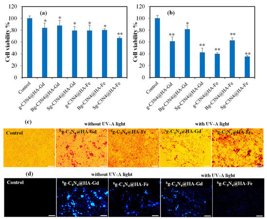
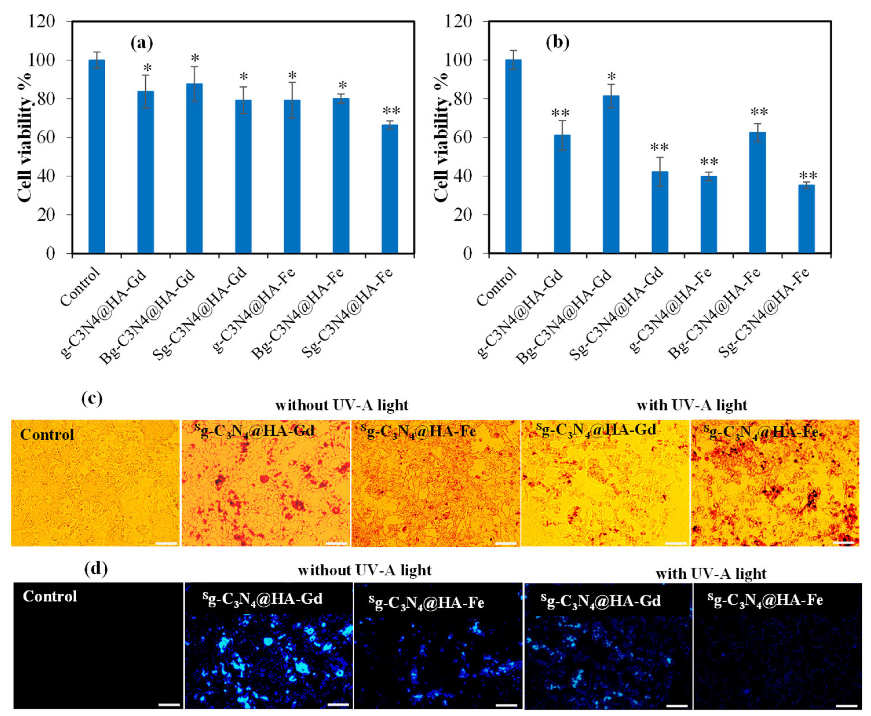
Light-induced anticancer activity of the prepared g-C3N4@HA-Gd/Fe(III)-based particles was also determined on melanoma cells treated with UV-A exposure. The g-C3N4@HA-Gd/Fe(III) particles at a concentration of 500 µg/mL, which do not show significant cytotoxic effects on healthy L929 fibroblast cells, were investigated for their anticancer effects on melanoma cancer cells. The cell viability % of the SKMEL skin cancer cells in the presence of 500 μg/mL of g-C3N4@HA-Gd/Fe(III)-based particles without any UV-A treatment and after UV-A exposure for 30 min are shown in Figure 7a and Figure 7b, respectively.
Figure 7.
The viability of SKMEL30 skin cancer cells cultured with 500 μg/mL HA-Gd(III)- and HA-Fe(III)-based particles (a) without UV-A light exposure and (b) following 30 min of UV-A light exposure. Statistical analyses are reported as * p < 0.05 and ** p < 0.001 in comparison to the control group. (c) Light microscope and (d) fluorescence microscope images of the skin cancer cells in the presence of 500 μg/mL concentration of sg-C3N4@HA-Gd/Fe(III)-based particles without UV-A light exposure and following 30 min of UV-A light exposure. Fluorescence images were taken under a DAPI filter at 365 nm without any agent at a 24 h incubation time. The scale bar is 50 μm.
The proliferation of melanoma cells, without light treatment, was quantified as 84 ± 8%, 88 ± 9%, 79 ± 7%, 79 ± 9%, 80 ± 2%, and 66 ± 7% for g-C3N4@HA-Gd(III), Bg-C3N4@HA-Gd(III), Sg-C3N4@HA-Gd(III), g-C3N4@HA-Fe(III), Bg-C3N4@HA-Fe(III), and Sg-C3N4@HA-Fe(III) composite particles, respectively. The composite particles exhibited a cytotoxic effect on the cancer cells, resulting in over 80% cell viability, apart from the Sg-C3N4@HA-Fe(III) particles. Following 30 min of UV-A exposure, the viability of malignant cells was reduced to 61 ± 8%, 81 ± 6%, 42 ± 8%, 40 ± 2%, 62 ± 5%, and 35 ± 2% in the presence of g-C3N4@HA-Gd(III), Bg-C3N4@HA-Gd(III), Sg-C3N4@HA-Gd(III), g-C3N4@HA-Fe(III), Bg-C3N4@HA-Fe(III), and Sg-C3N4@HA-Fe(III) composite particles, respectively. As reported by our previous study, the viability of SKMEL 30 cells exposed to 30 min UV-A light as a control was determined to be 97 ± 6%, in contrast to the control group that did not receive UV-A treatment. The findings indicated that 30 min exposure to UV-A radiation does not significantly affect the viability of melanoma cells [11]. The light microscope and fluorescence microscope images of the melanoma cells in the presence of Sg-C3N4@HA-Gd(III) and Sg-C3N4@HA-Fe(III) composite particles without UV-A exposure and 30 min of UV-A exposure are visualized and illustrated in Figure 7c,d. As seen in the light microscope images, the viability of the cancer cells treated with the particles was healthy without UV-A exposure, but the number of the cells were significantly decreased after UV-A contact. The fluorescence images of cancer cells also supported these results. Fluorescence intensity of the particles on the cells is brighter in no light-treated particles. However, the lesser blue fluorescence image was observed for Sg-C3N4@HA-Gd(III) particles by UV-A treatment. These results indicated that cancer cells were affected of the UV-A treatment in the presence of Sg-C3N4@HA-Gd(III) and Sg-C3N4@HA-Fe(III) composite particles. Light-induced anticancer activity of the g-C3N4 was reported in the literature [29]. N-rich g-C3N4 nanostructures exhibit strong light absorption, generating superoxide anions and singlet oxygen, recognized as reactive oxygen species (ROS), upon light exposure. This process induces oxidative stress, activating the apoptosis pathway and resulting in cell death in malignant cells [29,30]. Our findings revealed that B-doped g-C3N4 had no notable impact on photosensitive anticancer activity, whereas S-doped g-C3N4 particles elicited photodynamic inhibition of cancer cells when exposed to UV-A radiation. This phenomenon may arise from the better fluorescent property of S-doped g-C3N4 relative to B-doped g-C3N4, resulting in increased production of reactive oxygen species (ROS) upon excitation with UV-A radiation.
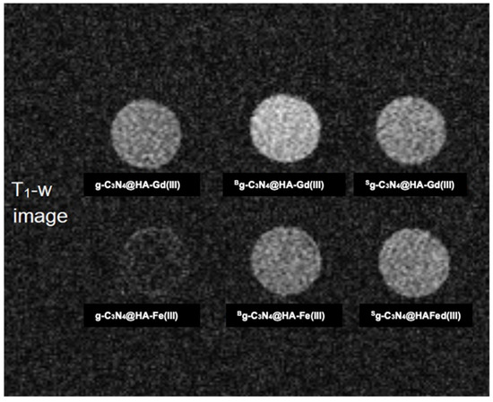
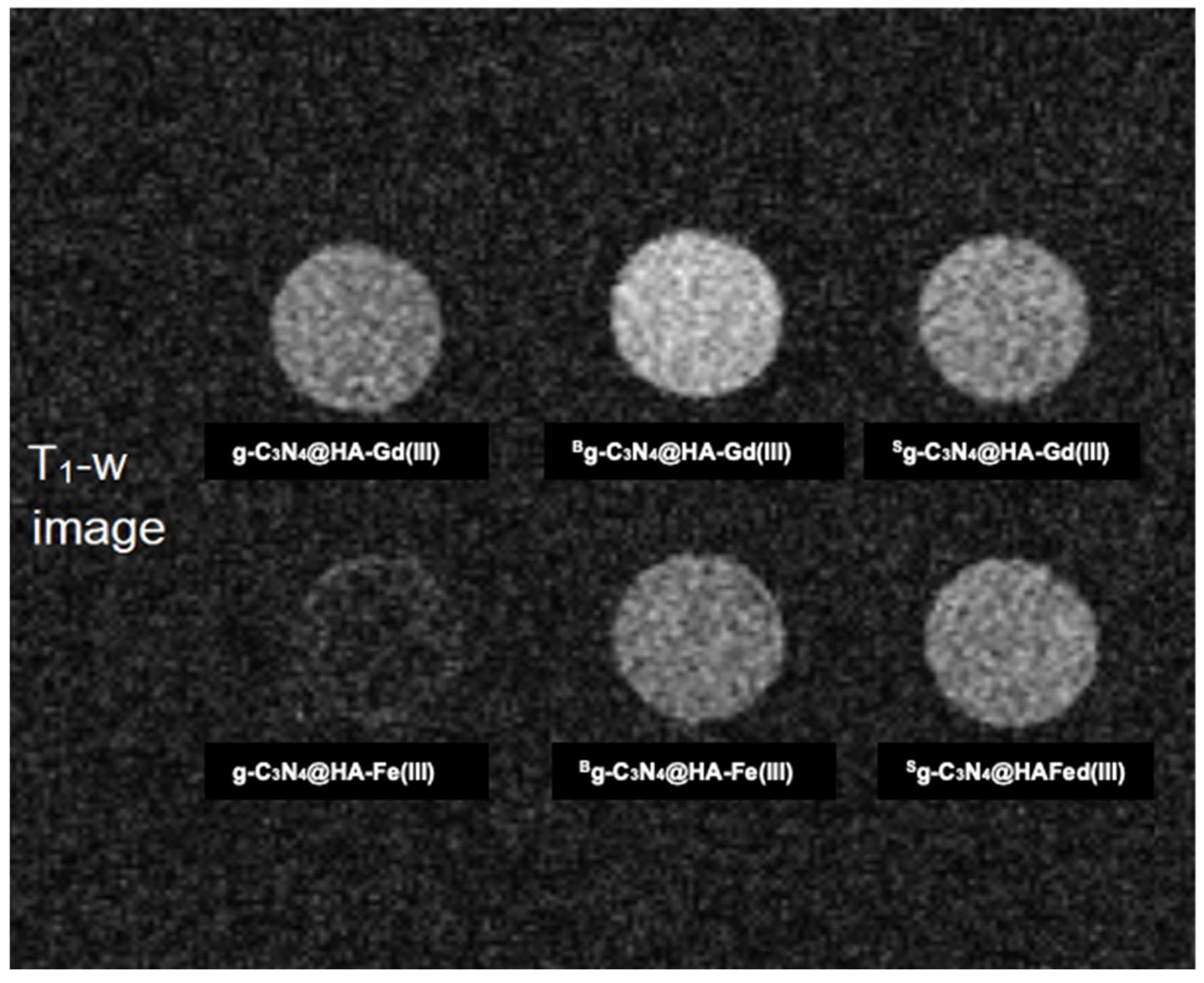
The MRI-enhancing capabilities of g-C3N4@HA-Gd(III)/Fe(III) particles were also investigated, and the corresponding results were shown in Figure 8. Heteroatom-doped g-C3N4@HA-Gd/Fe(III) particles have significantly improved contrast enhancement capability in MRI.
Figure 8.
T1-weighted (T1-W) MRI images of B- and S-atom-doped and undoped g-C3N4@HA-Gd(III)/Fe(III) microgels obtained on a 0.5 T MRI scanner.
In Figure 8, g-C3N4@HA-Gd(III) particles show brighter contrast as compared to g-C3N4@HA-Fe(III) particles since the Gd(III) ion has a higher magnetic moment (≈8 µB, where µB is Bohr magneton) than the Fe(III) ion (~6 µB). Among the Gd-containing samples, B-doped g-C3N4@HA-Gd shows the highest proton relaxivity, corresponding to higher MRI contrast enhancement efficiency. This can be related to electron deficiencies (holes) which act as paramagnetic centers in the gel matrix. As mentioned before, B has one less valence electron than C; hence, doping with B induces paramagnetic centers whose contribution to MRI contrast enhancement is, nevertheless, smaller than Gd(III) paramagnetic ions, which have seven unpaired electrons. Therefore, the heteroatom doping of g-C3N4@HA-Gd(III)/Fe(III) particles can be effectively used as multifunctional diagnostic and theranostic materials. Regarding the diagnostic functions, it should be noted that MRI contrast enhancement and fluorescence emission are independent processes, so two functions can be utilized individually or simultaneously from these prepared materials. This needs, however, either special imaging setups, i.e., integrating irradiating light source and detectors into an MRI machine similar to PET-MRI scanners or scanning the microgel injected subject sequentially in two separate machines and superimposing two images via postprocessing. The latter procedure is much more common since building a single-bore hybrid machine is highly complex, because the strong magnetic field of an MRI destroys traditional optical electronic components, requiring advanced fiber-optics or non-magnetic sensors [31,32].
Unlike previous studies that mainly focused on individual imaging or therapeutic functions, the present work integrates fluorescence imaging, MRI contrast enhancement, and photodynamic anticancer activity and targetability within a single HA-based composite microparticle platform. The combination of heteroatom-doped g-C3N4 with Gd(III)/Fe(III) ions crosslinked with HA provides a multifunctional theranostic system with favorable blood compatibility, enhanced imaging performance, and efficient light-triggered anticancer activity with cancerous cell targetability, thereby broadening the potential of HA-based hybrid materials for image-guided cancer therapy.
4. Conclusions
Light-sensitive g-C3N4 and its heteroatom-doped forms, namely Bg-C3N4 and Sg-C3N4 nanosheets, were incorporated within ionically crosslinked HA-Gd/Fe(III) particles for diagnostic and theranostic utilization potential. The g-C3N4@HA-Gd/Fe(III)-based composite microparticles were designed at injectable dimensions that are suitable for intravenous applications. It was established that g-C3N4 @HA-Gd/Fe(III)-based particles at a concentration of 1000 μg/mL had no significant effect on the blood clotting mechanism, with a blood clotting index % exceeding 82%. The g-C3N4@HA-Fe(III)-based particles exhibit higher toxicity than g-C3N4@HA-Gd(III)-based particles; however, all these particles can be safely utilized in biomedical applications due to their excellent biocompatibility with healthy cells, such as fibroblasts, at a concentration of 100 µg/mL. The Sg-C3N4@HA-Gd(III) composite particles, at a concentration of 500 µg/mL, display biocompatibility with L929 fibroblast cells; however, significant toxicity toward SKMEL 30 skin cancer cells was attained when exposed to UV-A light for 30 min. Among all forms of g-C3N4 @HA-M(III) particles, Sg-C3N4@HA-Gd(III) microparticles appeared as promising potentials for diagnostic and photodynamic theranostic applications. Particularly, g-C3N4@HA-Gd/Fe(III)-based composite particles can serve as diagnostic materials for multimodal imaging, including fluorescence bioimaging, due to their g-C3N4 composition, whereas MRI signals depend on the incorporation of Gd/Fe(III) ions inside the HA matrix, which is also further enhanced by the heteroatom doping such as B and S. Therefore, g-C3N4@HA-Gd(III)/Fe(III) particles with B or S doping attain additional functionality and offer an intriguing biomedical avenue as multifunctional diagnostic and theranostic materials.
Author Contributions
Conceptualization, N.S.; methodology, S.S.S., M.S., E.U., and N.S.; validation, S.S.S., M.S., and E.U.; formal analysis, S.S.S., M.S., E.U., and N.S.; investigation, S.S.S., M.S., E.U., and N.S.; resources, N.S.; writing—original draft preparation, S.S.S., M.S., and E.U.; writing—review and editing, N.S.; visualization, N.S.; supervision, N.S.; project administration, N.S.; and funding acquisition, N.S. All authors have read and agreed to the published version of the manuscript.
Funding
This research is supported by the Ministry of Health of The Republic of Türkiye (TUSEB-33379).
Institutional Review Board Statement
The study was conducted in accordance with the Declaration of Helsinki, and the protocol was approved by the Human Research Ethics Committee of Çanakkale Onsekiz Mart University (protocol code: 2011-KAEK-27) in September 2022.
Informed Consent Statement
Informed consent was obtained from all subjects involved in the study.
Data Availability Statement
The original contributions presented in this study are included in the article. Further inquiries can be directed to the corresponding author.
Acknowledgments
All individuals who volunteer to donate blood are greatly appreciated.
Conflicts of Interest
The authors declare no conflicts of interest.
References
- Kaur, I.; Doja, M.N.; Ahmad, T. Data mining and machine learning in cancer survival research: An overview and future recommendations. J. Biomed. Inform. 2022, 128, 104026. [Google Scholar] [CrossRef] [Scilit]
- Wu, J.; Zhang, L. Targeting the tumor microenvironment for sensitization of colorectal cancer to chemotherapy and radiotherapy. Pathol. Res. Pract. 2026, 283, 156480. [Google Scholar] [CrossRef] [Scilit]
- Chen, H.; Zhang, W.; Zhu, G.; Xie, J.; Chen, X. Rethinking cancer nanotheranostics. Nat. Rev. Mater. 2017, 2, 17024. [Google Scholar] [CrossRef] [Scilit]
- Kelkar, S.S.; Reineke, T.M. Theranostics: Combining Imaging and Therapy. Bioconjug. Chem. 2011, 22, 1879–1903. [Google Scholar] [CrossRef] [Scilit]
- Wang, Y.; Wang, X.; Antonietti, M. Polymeric Graphitic Carbon Nitride as a Heterogeneous Organocatalyst: From Photochemistry to Multipurpose Catalysis to Sustainable Chemistry. Angew. Chem. Int. Ed. 2012, 51, 68–89. [Google Scholar] [CrossRef] [Scilit]
- Ge, L.; Han, C.; Liu, J. In situ synthesis and enhanced visible light photocatalytic activities of novel PANI–g-C3N4 composite photocatalysts. J. Mater. Chem. 2012, 22, 11843. [Google Scholar] [CrossRef] [Scilit]
- Zheng, Y.; Lin, L.; Wang, B.; Wang, X. Graphitic Carbon Nitride Polymers toward Sustainable Photoredox Catalysis. Angew. Chem. Int. Ed. 2015, 54, 12868–12884. [Google Scholar] [CrossRef] [Scilit]
- Sakamoto, M.; Nakata, A.; Shimizu, M.; Tagashira, A.; Hirazawa, H.; Imai, Y. Microstructure-Controlled g-C3N4: From Photocatalyst to Potential UV-Shielding Pigment with Enhanced Skin Feel. Micro 2026, 6, 37. [Google Scholar] [CrossRef] [Scilit]
- Ong, W.-J.; Tan, L.-L.; Ng, Y.H.; Yong, S.-T.; Chai, S.-P. Graphitic Carbon Nitride (g-C3N4)-Based Photocatalysts for Artificial Photosynthesis and Environmental Remediation: Are We a Step Closer to Achieving Sustainability? Chem. Rev. 2016, 116, 7159–7329. [Google Scholar] [CrossRef] [Scilit]
- Zhang, J.; Chen, Y.; Wang, X. Two-dimensional covalent carbon nitride nanosheets: Synthesis, functionalization, and applications. Energy Environ. Sci. 2015, 8, 3092–3108. [Google Scholar] [CrossRef] [Scilit]
- Suner, S.S.; Sahiner, M.; Umut, E.; Sahiner, N. Gd(III) and Fe(III) ion crosslinked hyaluronic acid microgels composites embedding hetero atom doped carbon quantum dots render photodynamic therapy with improved bioimaging capability. J. Photochem. Photobiol. B Biol. 2026, 274, 113342. [Google Scholar] [CrossRef] [Scilit]
- Highley, C.B.; Prestwich, G.D.; Burdick, J.A. Recent advances in hyaluronic acid hydrogels for biomedical applications. Curr. Opin. Biotechnol. 2016, 40, 35–40. [Google Scholar] [CrossRef] [Scilit]
- Stępień, A.; Juszczak, L.; Koronowicz, A.; Such, A.; Kowalski, G.; Synkiewicz-Musialska, B.; Zachariasz, P.; Jamróz, E. Effect of Hyaluronic Acid Content on Functional Properties, Antioxidant Activity, and In Vitro Digestion of Food-Grade Furcellaran Hydrogels and Emulgels. Materials 2025, 18, 5581. [Google Scholar] [CrossRef] [Scilit]
- Iaconisi, G.N.; Lunetti, P.; Gallo, N.; Cappello, A.R.; Fiermonte, G.; Dolce, V.; Capobianco, L. Hyaluronic Acid: A Powerful Biomolecule with Wide-Ranging Applications—A Comprehensive Review. Int. J. Mol. Sci. 2023, 24, 10296. [Google Scholar] [CrossRef] [Scilit]
- Zanchetta, C.; Scandolera, A.; Reynaud, R. Hyaluronic Acid in Topical Applications: The Various Forms and Biological Effects of a Hero Molecule in the Cosmetics Industry. Biomolecules 2025, 15, 1656. [Google Scholar] [CrossRef] [Scilit]
- Palenčárová, K.; Köszagová, R.; Nahálka, J. Hyaluronic Acid and Its Synthases—Current Knowledge. Int. J. Mol. Sci. 2025, 26, 7028. [Google Scholar] [CrossRef] [Scilit]
- Murakami, T.; Otsuki, S.; Okamoto, Y.; Nakagawa, K.; Wakama, H.; Okuno, N.; Neo, M. Hyaluronic acid promotes proliferation and migration of human meniscus cells via a CD44-dependent mechanism. Connect. Tissue Res. 2019, 60, 117–127. [Google Scholar] [CrossRef] [Scilit]
- Lee, S.Y.; Kang, M.S.; Jeong, W.Y.; Han, D.W.; Kim, K.S. Hyaluronic acid-based theranostic nanomedicines for targeted cancer therapy. Cancers 2020, 12, 940. [Google Scholar] [CrossRef] [Scilit]
- Caravan, P.; Ellison, J.J.; McMurry, T.J.; Lauffer, R.B. Gadolinium(III) Chelates as MRI Contrast Agents: Structure, Dynamics, and Applications. Chem. Rev. 1999, 99, 2293–2352. [Google Scholar] [CrossRef] [Scilit]
- Wang, L.; Ai, Q.; Chen, W.; Cai, Z.; Yu, Z.; Abrigo, J.; So, T.Y. Effect of gadolinium-based contrast media on intravoxel incoherent motion (IVIM) MRI parameters in brain imaging. Phys. Med. 2025, 139, 105172. [Google Scholar] [CrossRef] [Scilit]
- Demirci, S.; Suner, S.S.; Neli, O.U.; Koca, A.; Sahiner, N. B, P, and S heteroatom doped, bio- and hemo-compatible 2D graphitic-carbon nitride (g-C3N4) with antioxidant, light-induced antibacterial, and bioimaging endeavors. Nanotechnology 2024, 35, 025101. [Google Scholar] [CrossRef] [Scilit]
- Kong, X.; Liu, X.; Zheng, Y.; Chu, P.K.; Zhang, Y.; Wu, S. Graphitic carbon nitride-based materials for photocatalytic antibacterial application. Mater. Sci. Eng. R Rep. 2021, 145, 100610. [Google Scholar] [CrossRef] [Scilit]
- Schwartz, S.; Hassman, D.; Shelmet, J.; Sievers, R.; Weinstein, R.; Liang, J.; Lyness, W. A multicenter, open-label, randomized, two-period crossovertrial comparing glycemic control, satisfaction, and preference achieved with a 31 gauge × 6 mm needle versus a 29 gauge × 12.7 mm needle in obese patients with diabetes mellitus. Clin. Ther. 2004, 26, 1663–1678. [Google Scholar] [CrossRef] [Scilit]
- Pan, H.; Zhang, H.; Liu, H.; Chen, L. Interstitial boron doping effects on the electronic and magnetic properties of graphitic carbon nitride materials. Solid State Commun. 2015, 203, 35–40. [Google Scholar] [CrossRef] [Scilit]
- Somaraj, G.; Mathew, S.; Kannankara Abraham, T.; Ambady, K.G.; Mohan, C.; Mathew, B. Nitrogen and Sulfur Co-Doped Carbon Quantum Dots for Sensing Applications: A Review. Chemistryselect 2022, 7, e202200473. [Google Scholar] [CrossRef] [Scilit]
- Zhou, Y.; He, X.; Li, M. Roles of doping in enhancing the performance of graphene/graphene-like semiconductors. AIP Adv. 2025, 15, 010701. [Google Scholar] [CrossRef] [Scilit]
- Joseph, J.; Anappara, A.A. Comparative photoluminescence study of nitrogen-doped carbon dots co-doped with boron and sulphur. Luminescence 2022, 37, 1475–1481. [Google Scholar] [CrossRef] [Scilit]
- Sahiner, N.; Umut, E.; Suner, S.S.; Sahiner, M.; Culha, M.; Ayyala, R.S. Hyaluronic acid (HA)-Gd(III) and HA-Fe(III) microgels as MRI contrast enhancing agents. Carbohydr. Polym. 2022, 277, 118873. [Google Scholar] [CrossRef] [Scilit]
- Su, N.; Uk, S.; Rethinasabapathy, M.; Zoo, E.; Cho, H.; Yeong, S.; Rak, S.; Kim, Y.; Hong, W.G.; Krishnan, G.; et al. Materials Science & Engineering C Visible-light-driven dynamic cancer therapy and imaging using graphitic carbon nitride nanoparticles. Mater. Sci. Eng. C 2018, 90, 531–538. [Google Scholar] [CrossRef] [Scilit]
- Carabineiro, C.; Nawaz, A.; Babar, M. Graphitic carbon nitride as an efficient carrier for anti-cancer drug systems: A review. Next Nanotechnol. 2024, 6, 100074. [Google Scholar] [CrossRef] [Scilit]
- Davis, S.C.; Pogue, B.W.; Springett, R.; Leussler, C.; Mazurkewitz, P.M.; Tuttle, S.B.; Gibbs-Strauss, S.L.; Jiang, S.; Dehghani, H.; Dehghani, H.; et al. Magnetic resonance-coupled fluorescence tomography scanner for molecular imaging of tissue. Rev. Sci. Instrum. 2008, 79, 064302. [Google Scholar] [CrossRef] [Scilit]
- Stuker, F.; Baltes, C.; Dikaiou, K.; Vats, D.; Carrara, L.; Charbon, E.; Ripoll, J.; Rudin, M. Hybrid Small Animal Imaging System Combining Magnetic Resonance Imaging with Fluorescence Tomography Using Single Photon Avalanche Diode Detectors. IEEE Trans. Med. Imaging 2011, 30, 1265–1273. [Google Scholar] [CrossRef] [Scilit]
Disclaimer/Publisher’s Note: The statements, opinions and data contained in all publications are solely those of the individual author(s) and contributor(s) and not of MDPI and/or the editor(s). MDPI and/or the editor(s) disclaim responsibility for any injury to people or property resulting from any ideas, methods, instructions or products referred to in the content. |
© 2026 by the authors. Licensee MDPI, Basel, Switzerland. This article is an open access article distributed under the terms and conditions of the Creative Commons Attribution (CC BY) license.