


Abstract
Biofilm-associated infections represent a critical challenge in modern medicine, accounting for approximately 80% of all microbial infections and demonstrating up to 1000-fold higher antimicrobial resistance compared to planktonic bacteria. The extraordinary recalcitrance of biofilms stems from a complex interplay of physical barriers (extracellular polymeric substance matrix), chemical gradients (pH and oxygen heterogeneity), and biological defenses (persister cells and horizontal gene transfer), rendering conventional antibiotics largely ineffective. This comprehensive review highlights the transformative potential of antimicrobial nanoformulations in overcoming these formidable barriers through strategic design principles and diverse mechanisms of action. Evidence demonstrates that rationally engineered nanocarriers achieve improvements in bacterial killing, biofilm biomass reduction, and colony-forming unit reductions compared to free antibiotics. Advanced stimuli-responsive systems exploiting biofilm-specific triggers (acidic pH, bacterial enzymes, elevated ATP) and externally applied stimuli (near-infrared photothermal therapy, ultrasound sonodynamic therapy) enable on-demand therapeutic activation with unprecedented precision, achieving >99.999% bacterial elimination and near-complete biofilm eradication. Despite these remarkable advances, clinical translation remains hindered by challenges in scalability, comprehensive safety evaluation, and regulatory pathway navigation. This review establishes a consolidated evidence base for the design of next-generation antimicrobial nanoformulations, highlights their potential to address biofilm-associated infections, and identifies key knowledge gaps and translation barriers that must be addressed to realize their therapeutic promise.
1. Introduction to Biofilm Barriers: Physiological and Structural Hurdles to Conventional Therapeutics
Biofilm-associated infections represent one of the most formidable challenges in contemporary clinical microbiology, accounting for approximately 80% of all microbial infections and over 60% of nosocomial infections [1]. These infections impose a staggering clinical and economic burden, with over 500,000 biofilm-related implant infections occurring annually in the United States alone, and prosthetic joint infections projected to incur revision surgery costs exceeding USD 1.62 billion by 2030 [2]. The mortality rates associated with biofilm infections are alarmingly high, exemplified by Candida biofilm-related bloodstream infections demonstrating a mortality rate of 70.0% (95% CI: 52.8 to 84.8%) compared to 37.9% (95% CI: 26.2 to 50.2%) for all Candida-related bloodstream infections [3]. Infective endocarditis, frequently involving biofilm formation on cardiac valves, exhibits acute mortality rates of 20 to 25% and 1-year mortality rates reaching 40%, with almost 50% of cases requiring heart valve surgery due to insufficient antibiotic efficacy [4]. The recalcitrance of biofilm infections is further underscored by treatment failure rates, with more than 80% of patients experiencing recurrence of Staphylococcus aureus infections after antibiotic cessation [5] and Mycobacterium avium-intracellulare complex pulmonary disease patients showing 8.3 to 48% recurrence rates [6]. The intrinsic resistance of biofilms to conventional antimicrobials is profound, with bacteria and fungi able to resist up to 1000-fold concentrations of antibiotics relative to their planktonic counterparts [7,8,9]. This extraordinary resistance stems from a complex interplay of physical, chemical, and biological barriers that collectively impede drug penetration, inactivate antimicrobial agents, and induce phenotypic tolerance, necessitating the development of innovative therapeutic strategies such as antimicrobial nanoformulations.
While numerous reviews have addressed antimicrobial nanoformulations and biofilm infections independently, this review distinguishes itself through several unique contributions. First, the comprehensive integration of quantitative design principles (specifically, the establishment of evidence-based design thresholds (size: 50 to 130 nm; zeta potential: +40 to +47 mV; aspect ratios for anisotropic particles)) provides actionable engineering guidelines rarely compiled in a single source. Second, the systematic comparative analysis across all four major stimuli-responsive modalities (pH, enzyme, ATP, and external triggers) with quantitative efficacy benchmarks offers a holistic framework that is absent from prior reviews. Third, the critical assessment of clinical translation barriers, including scalability, safety evaluation, and regulatory navigation, contextualizes laboratory findings within the practical challenges of therapeutic development. Fourth, the integration of mechanistic diversity spanning contact-mediated disruption, enzymatic matrix degradation, photothermal/photodynamic therapy, quorum-sensing inhibition, and synergistic drug delivery enables researchers to evaluate complementary strategies rather than isolated approaches. Fifth, the tabular comparative analysis presented in Table 1 explicitly delineates how this review advances beyond the existing literature, addressing gaps in size-performance correlations, surface charge optimization, and multi-stimulus integration. This evidence-based and translation-focused approach positions this review as both a comprehensive resource and a practical guide for designing next-generation antimicrobial nanoformulations.
Table 1.
Comparative analysis of present review with previous reviews.
1.1. Physical Barriers: The EPS Matrix
The EPS matrix constitutes the primary physical barrier to antimicrobial penetration in biofilms, comprising 50% to 90% of the total biofilm dry mass and forming a highly structured, three-dimensional scaffold that encapsulates bacterial communities [9,20]. This complex matrix is composed of a heterogeneous mixture of exopolysaccharides, proteins, extracellular DNA (eDNA), lipids, and nucleic acids, with the composition varying significantly among bacterial species [7,21]. In S. aureus biofilms, the EPS matrix comprises nucleic acid (eDNA), proteins, lipids, and polysaccharides, with polysaccharide intercellular adhesin (PIA), a polyGlcNAc polymer, serving as a key structural component [22]. The partial deacetylation of PIA produces positively charged molecules that facilitate electrostatic interactions with negatively charged bacterial cell surfaces and other anionic biofilm components such as teichoic acid, thereby enhancing structural cohesion [22,23]. Pseudomonas aeruginosa biofilms produce species-specific exopolysaccharides including Psl, Pel, and alginate, which contribute to resistance against colistin, polymyxin B, tobramycin, and ciprofloxacin, with biofilms lacking Pel demonstrating increased susceptibility to tobramycin and gentamicin [24].
The EPS matrix functions as a semi-permeable barrier that restricts antimicrobial diffusion through multiple mechanisms, including charge-based interactions, physical sequestration, and enzymatic degradation [8,20]. The negatively charged nature of the EPS matrix significantly hinders the entry of positively charged antibiotics such as aminoglycosides, with studies demonstrating that the matrix sequestered the positively charged antibiotic tobramycin, while the neutral ciprofloxacin was able to penetrate more effectively [9,25]. eDNA plays a critical structural role, forming grid-like structures and filamentous networks that act as intercellular links and contribute to the viscoelasticity of the biofilm [20,22]. The presence of exogenous DNA in P. aeruginosa biofilms has been shown to increase resistance by 3-fold for tobramycin and 2-fold for gentamicin, while DNase I pre-treatment of S. aureus biofilms led to a 6-log reduction in viability when exposed to chlorhexidine gluconate [7,24]. The EPS matrix also accumulates antibiotic-degrading enzymes, exemplified by Klebsiella pneumoniae biofilms producing β-lactamase that degrades ampicillin, thereby creating localized zones of antibiotic inactivation [24]. The mechanical properties of the EPS matrix further contribute to its protective function, with the Young’s Modulus of P. aeruginosa biofilms increasing from 10 Pa in early development to 25 Pa when mature and elastic modulus values ranging from 0.8 to 100 Pa depending on growth conditions [9]. These structural and compositional features collectively render the EPS matrix (Table 2) a formidable barrier that reduces antimicrobial diffusion, adsorbs antimicrobial agents, and provides mechanical resistance to immune-cell-mediated clearance [20,23].
Table 2.
EPS components: Composition, structural role, and contribution to antibiotic resistance.
1.2. Chemical Barriers: Microenvironments, pH and Oxygen Gradients
Biofilms establish complex chemical microenvironments characterized by steep pH and oxygen gradients that profoundly influence antimicrobial efficacy and bacterial metabolism [9,24]. The pH within pathogenic biofilms typically ranges from 5 to 9, which is significantly lower than physiological pH levels, creating acidic microenvironments in deeper biofilm layers due to the accumulation of metabolic byproducts such as lactic acid and other organic acids [26]. These pH variations have direct implications for antibiotic activity, as many antimicrobial agents exhibit pH-dependent efficacy; for instance, vancomycin-loaded quatsomes demonstrated a reduced minimum inhibitory concentration (MIC) of 0.97 µg/mL at pH 6.0 compared to 3.90 µg/mL at pH 7.4, illustrating the impact of acidic conditions on drug performance [22]. The pH gradients also influence biofilm structural properties, with Group B Streptococcus biofilms forming preferentially at low pH and exhibiting Young’s Modulus values ranging from 2 to 100 kPa as the pH increased [9].
Oxygen gradients within biofilms create distinct metabolic zones, with aerobic regions near the biofilm surface transitioning to anaerobic conditions in deeper layers, resulting in heterogeneous bacterial populations with varying metabolic activities [7,24]. These oxygen-limited zones harbor slow-growing or dormant bacterial cells that exhibit reduced metabolic activity, rendering them inherently less susceptible to antibiotics that target active cellular processes such as cell wall synthesis, protein synthesis, or DNA replication [9,22]. The deeper regions of biofilms, characterized by insufficient nutrients and oxygen, typically contain persister cells in a dormant or quiescent state that tolerate antibiotics by diverging from active growth [22]. The establishment of these chemical gradients is facilitated by the EPS matrix, which restricts the diffusion of oxygen and nutrients while allowing the accumulation of metabolic waste products, thereby creating spatially distinct microenvironments within a single biofilm structure [26]. The presence of anaerobic regions and significant pH changes within biofilms contributes to the development of phenotypic heterogeneity, with bacterial subpopulations exhibiting specialized metabolic states that collectively enhance biofilm resilience to antimicrobial challenge [7]. These chemical barriers (Table 3), in concert with physical and biological mechanisms, create a hostile environment for conventional antibiotics, necessitating therapeutic approaches capable of penetrating and functioning effectively across diverse microenvironmental conditions.
Table 3.
Biofilm microenvironmental gradients: Gradient type, range/magnitude, and effect on antibiotic efficacy.
1.3. Biological Barriers: Persister Cells and Horizontal Gene Transfer
Persister cells represent a critical biological barrier to antimicrobial eradication, constituting a phenotypically distinct subpopulation of dormant or slow-growing bacteria that exhibit extraordinary multidrug tolerance without genetic modification [7,26]. These cells comprise approximately 1% of the total biofilm population, with the deeper zones of biofilms harboring the highest concentrations of persisters due to nutrient and oxygen limitation [7,22]. Persister cells are formed through multiple mechanisms, including stochastic induction via HipA-mediated activation of mRNA endonucleases through (p)ppGpp signaling, as well as mutations in genes encoding tRNA synthetase, ribose-phosphate diphosphatekinase, and toxin–antitoxin systems that induce cell dormancy and tolerance [23,24]. The metabolic inactivity of persister cells renders them refractory to antibiotics that target active cellular processes, allowing them to survive antibiotic treatment and subsequently reactivate to repopulate the biofilm once the antimicrobial pressure is removed [9,25]. This phenomenon is believed to be a major contributor to infection recurrence, with persisters representing a minor subpopulation (<1%) of biofilm communities that are critical for chronic infections [27].
Horizontal gene transfer (HGT) within biofilms serves as a potent mechanism for the dissemination of antimicrobial resistance genes, facilitated by the high cell density, spatial proximity, and presence of eDNA that characterize biofilm communities [23,24]. The frequency of HGT is dramatically enhanced in biofilms compared to planktonic cultures, with the conjugal transfer frequency of multidrug resistance plasmids in S. aureus biofilms being 10,000 times greater than in planktonic cultures [23,24]. Antibiotic exposure further accelerates HGT, with tetracycline and cephradine promoting the transmission of plasmid pB10 among P. aeruginosa or Escherichia coli biofilm biomass at 2 to 5 times faster rates than without antibiotic treatment [26]. eDNA within the biofilm matrix serves as both a structural component and a vehicle for HGT through transformation, enabling the acquisition of resistance genes by competent cells [23,24]. The spatial structure and heterogeneity of biofilms promote genetic diversity, with multidrug resistance genes accumulating on resistance plasmids or transposons that can be transferred via conjugation, transformation, and transduction [20,21]. The mutation frequency is also elevated in biofilms, with the ciprofloxacin-resistant mutant mutation frequency being approximately 2-log higher in P. aeruginosa biofilm cells compared to planktonic cells [24]. The higher bacterial cell density in biofilms contributes to the transfer of tolerance genes between bacterial cells, promoting the emergence of subpopulations with specialized resistance mechanisms including antibiotic-degrading enzymes, low-affinity antibiotic targets, and overexpression of efflux pumps [9,23]. These biological barriers—persister cells and HGT—collectively ensure that biofilms not only tolerate antimicrobial challenge but also evolve enhanced resistance over time, perpetuating chronic infections and treatment failures.
2. Literature Search and Review Methodology
This review was conducted as a comprehensive narrative synthesis of the published literature on antimicrobial nanoformulations for biofilm-associated infections. The following search strategy and selection criteria were employed.
2.1. Search Strategy
A systematic literature search was performed using the following electronic databases: PubMed, Scopus, Web of Science, and Google Scholar. The search was conducted from January 2020 to May 2026 to capture recent advancements while maintaining historical context. The following search terms and Boolean operators were used: (‘antimicrobial nanoformulations’ OR ‘nanoparticles’ OR ‘nanocarriers’ OR ‘nanomedicine’) AND (‘biofilm’ OR ‘biofilm-associated infections’ OR ‘extracellular polymeric substance’ OR ‘EPS’) AND (‘design principles’ OR ‘size’ OR ‘surface charge’ OR ‘morphology’ OR ‘targeting’ OR ‘stimuli-responsive’ OR ‘photothermal’ OR ‘photodynamic’ OR ‘quorum sensing’). Additional relevant articles were identified through manual screening of reference lists of included studies and key review articles.
2.2. Inclusion and Exclusion Criteria
Studies were included if they: (i) were primary research articles, reviews, or meta-analyses published in peer-reviewed English-language journals; (ii) evaluated antimicrobial nanoformulations against biofilm-forming bacteria; (iii) reported quantitative data on nanoparticle physicochemical properties (size, charge, morphology), antimicrobial efficacy (MIC, MBIC, MBEC), or mechanisms of action; and (iv) addressed biofilm-associated infections in clinically relevant contexts. Studies were excluded if they: (i) were non-English publications, conference abstracts, or book chapters; (ii) focused exclusively on planktonic bacteria without biofilm relevance; (iii) lacked sufficient methodological detail for data extraction; or (iv) were duplicate publications.
2.3. Study Selection and Data Extraction
Titles and abstracts were screened against the inclusion criteria. Full texts of potentially relevant studies were retrieved and assessed for eligibility. Data were extracted using a standardized form capturing the following: nanoparticle type and physicochemical properties, bacterial species and biofilm model, experimental conditions, key efficacy outcomes (MIC, MBIC, MBEC, biofilm reduction percentage, log reduction), mechanisms of action, and reported limitations. Given the heterogeneity of study designs, nanoparticle formulations, and experimental conditions, a narrative synthesis approach was adopted to organize and integrate findings across studies.
2.4. Quality Assessment and Limitations
The quality of included studies was assessed based on: (i) the clarity of nanoparticle characterization (size, charge, morphology); (ii) the reproducibility of experimental methods; (iii) the use of appropriate controls; and (iv) statistical analyses. Studies with incomplete characterization or unclear methodology were interpreted with appropriate caution. We acknowledge that the heterogeneity of experimental conditions (bacterial species, biofilm models, assay conditions) limits direct quantitative comparison across studies; therefore, we present ranges of reported values and highlight consensus findings where they emerge. This review is designed as a comprehensive narrative synthesis rather than a systematic review or meta-analysis, and the conclusions presented are based on the preponderance of evidence from the included studies.
3. Clinical Impact: Reduced Drug Penetration, Tolerance, and the Case for Nanoformulations
The convergence of physical, chemical, and biological barriers in biofilms results in profound reductions in antimicrobial efficacy, manifesting as dramatic increases in MICs and minimum biofilm eradication concentrations (MBECs) compared to planktonic bacteria [8,24]. Biofilmed microbes tolerate 10- to 1000-fold higher antibiotic concentrations than their planktonic counterparts, with some studies reporting that bacteria and fungi can resist up to 1000-fold concentrations of organic small-molecule antibiotics or antimicrobial heavy metals relative to their planktonic form [7,24]. For Candida biofilm-related bloodstream infections, antifungal resistance rates for biofilm-forming strains reached 70.5% for fluconazole, 67.9% for voriconazole, and 72.8% for caspofungin [3]. In chronic rhinosinusitis patients, 76.7% of bacterial strains formed biofilms in vitro, with planktonic isolates showing high resistance to amoxicillin (82.6%) and clarithromycin (39.1%), while biofilm forms exhibited even higher resistance levels [28]. The concentrations needed to eradicate mycobacteria in biofilms can be 100 to 100,000 times higher than the MICs of planktonic forms, illustrating the extreme recalcitrance of biofilm-associated infections [6].
The clinical consequences of reduced drug penetration and tolerance are severe, encompassing prolonged hospital stays, increased healthcare costs, elevated mortality rates, and high rates of treatment failure and recurrence [1,2]. Biofilm-mediated infections prolong hospital stays due to persistent infections, with biofilm-producing phenotypes of Acinetobacter baumannii affecting patients in intensive care units and burn units, leading to ventilator-associated pneumonia, bloodstream infection, urinary-tract infection, and orthopedic implant infections [1]. Urinary tract infections, which account for approximately 40% of all bacterial infections and require around 15% of all antibiotic prescriptions, are frequently associated with biofilm formation on urinary catheters [29]. Percutaneous osseointegrated implants are susceptible to biofilm formation, with soft-tissue infection rates soaring as high as 75%, representing a recurring infection type that often requires repeated antibiotic therapy [30]. The economic burden is substantial, with prosthetic joint infections alone projected to incur revision surgery costs exceeding USD 500 million per year, rising to USD 1.62 billion by 2030 [2].
Conventional antibiotics fail to adequately address biofilm infections due to their inability to penetrate the EPS matrix, their reduced activity in acidic and anaerobic microenvironments, and their ineffectiveness against dormant persister cells [9,20]. The physical barrier imposed by the EPS matrix reduces antimicrobial diffusion, with the negatively charged matrix hindering the entry of positively charged antibiotics and sequestering them before they reach bacterial cells [9,25]. Chemical gradients within biofilms alter antibiotic activity, with pH-dependent efficacy reducing drug performance in acidic microenvironments [22]. Persister cells, which are metabolically inactive, evade antibiotics that target active cellular processes, allowing them to survive treatment and reactivate once the antimicrobial pressure is removed [9,27]. The enhanced HGT within biofilms accelerates the acquisition and dissemination of resistance genes, further compromising antibiotic efficacy over time [23,24].
These multifaceted challenges necessitate the development of innovative therapeutic strategies capable of overcoming the physical, chemical, and biological barriers inherent to biofilms. Antimicrobial nanoformulations represent a promising intervention, offering unique advantages including enhanced penetration through the EPS matrix due to their nanoscale dimensions, sustained drug release to maintain therapeutic concentrations over extended periods, targeted delivery to biofilm sites, and the ability to disrupt biofilm structure through physical and chemical mechanisms [9,22]. Nanoformulations have demonstrated significant improvements in antibiofilm efficacy, with examples including silver nanoparticles inhibiting K. pneumoniae biofilm by 64% and 86% at 100 µg/mL, ciprofloxacin-loaded niosomes showing a 4- to 5-fold increase in antibacterial potency compared to the free drug against S. aureus strains, and cefuroxime-loaded solid lipid nanoparticles resulting in a 2-fold reduction of the minimum biofilm inhibitory concentration (MBIC; 40 µg/mL versus 80 µg/mL for the free drug) [21,22]. The convergence of nanotechnology and antimicrobial therapy offers a rational approach to address the biofilm barrier, providing a foundation for the development of next-generation therapeutics capable of eradicating biofilm-associated infections and mitigating the clinical and economic burdens they impose. Specific examples of this resistance amplification are abundant across clinically relevant pathogens and antibiotics (Table 4).
Table 4.
MIC fold increase in biofilm vs. planktonic bacteria: Organism, antibiotic class, and fold increase.
4. Strategic Design Principles for Anti-Biofilm Nanocarriers
The rational design of antimicrobial nanocarriers requires the precise control over multiple physicochemical parameters that govern nanoparticle–biofilm interactions. As detailed in Section 1, biofilms present formidable barriers including the EPS matrix, pH gradients, oxygen gradients, and persister cells. Strategic manipulation of the nanocarrier size, surface charge, morphology, polymer coating, and targeting ligands can dramatically enhance biofilm penetration and antimicrobial efficacy. For instance, pH-responsive protein-polycation nanocarriers that shrink from 550 nm to approximately 48 nm under acidic conditions achieve 70 to 80% biofilm biomass elimination [31], while cationic poly-L-lysine (PLL)-coated nanoparticles demonstrate ~4-fold higher bacterial uptake compared to uncoated particles [32]. Antibody-conjugated nanocarriers exhibit 2- to 5-fold reductions in MBEC values compared to free antibiotics [33]. This section examines the quantitative relationships between nanocarrier design parameters and antibiofilm performance, providing evidence-based guidelines for engineering next-generation antimicrobial nanoformulations.
4.1. Size Effects on Biofilm Penetration and Cellular Uptake
Nanoparticle size critically determines diffusion through the EPS matrix. Liposomal nanocarriers (50 to 500 nm) demonstrate effective biofilm penetration, with deformable liposomes showing superior penetration into deeper layers [34]. Nanoparticles smaller than 100 nm exhibit a superior ability to overcome the steric barrier of the EPS [35]. Studies on Burkholderia multivorans and P. aeruginosa biofilms reveal that the size cutoff for nanoparticles capable of penetrating dense biofilm clusters is approximately 130 nm among tested diameters ranging from 40 to 550 nm [32]. pH-responsive nanocarriers that shrink from 550 nm to 48 nm under acidic biofilm microenvironments eliminate over 95% of intracellular bacteria [31].
Enzyme-functionalized mesoporous silica nanoparticles (MSNs; ~36 nm diameter) demonstrate 7.5-fold decreases in the MIC and 5-fold decreases in the MBIC for methicillin-resistant S. aureus (MRSA) compared to free lysostaphin [36]. Silver nanoparticles smaller than 10 nm alter bacterial cell permeability, with 8.3 nm particles affecting P. aeruginosa biofilms and 20 µg/mL producing 67% inhibition on sensitive biofilms and 56% on resistant strains [37]. Smaller spherical silver nanoparticles (9.3 ± 3.5 nm) exhibit MBIC values of 12.5 µg/mL for Gram-positive and 25 µg/mL for Gram-negative bacteria, while larger particles (64.0 ± 10.2 nm) require 50 µg/mL for equivalent biofilm inhibition [38]. Table 5 presents the size-dependent biofilm penetration data.
Table 5.
Size-dependent biofilm penetration and antimicrobial efficacy.
4.2. Surface Charge Influence on Biofilm Interaction and Bacterial Uptake
Surface charge governs the electrostatic interactions with the negatively charged EPS matrix and bacterial membranes. Cationic nanoparticles demonstrate superior biofilm penetration and bacterial uptake compared to anionic or neutral counterparts. PLL-coated nanoparticles exhibit zeta potentials of +40 to +47 mV compared to −52 to −56 mV for uncoated particles, resulting in ~4-fold higher bacterial uptake [32]. PLL-coated rifampicin-loaded nanoparticles maintain effective antibiotic concentrations after washing, while negatively charged particles fail to retain antibiotics [32]. Cationic liposomes demonstrate a 3-log CFU reduction against P. aeruginosa biofilms [34]. Gold nanorods coated with poly(diallyldimethylammonium chloride) (PDADMAC)/alginate (cationic, +40 to +47 mV) eradicate 86% of MRSA biofilms and 93% of methicillin-sensitive S. aureus (MSSA) biofilms at 0.15 nM, with MBEC50 values of 0.029 nM for MRSA and 0.032 nM for MSSA [41]. PLL-coated nanoparticles show slowed migration and improved retention in biofilms compared to negatively charged particles, with hydrodynamic diameter increases of 10 to 20% [32]. Table 6 presents the surface charge effects.
Table 6.
Surface charge effects on biofilm interaction and bacterial uptake.
4.3. Shape and Morphology Effects on Biofilm Penetration
Anisotropic particles demonstrate superior biofilm eradication through enhanced torque-driven penetration and mechanical disruption. Gold nanorods (length 55.71 ± 1.15 nm, width 23.70 ± 1.13 nm, aspect ratio 2.35) achieve MBEC50 values of 0.029 nM for MRSA and 0.032 nM for MSSA, eradicating 86% and 93% of biofilms, respectively [41]. Mechano-bactericidal anisotropic calcium carbonate particles show superior performance against E. coli K12 (227 ± 32.6% and 215 ± 33% for urchin and stick shapes) and S. aureus ATCC 209P (210 ± 54.7% and 202 ± 55.5%) compared to spherical particles [42]. Silver nanoparticles exhibit shape-dependent effects (spherical > disc > triangular), with spherical morphology demonstrating the highest efficacy [37]. Table 7 presents the shape-dependent data.
Table 7.
Shape and morphology effects on biofilm eradication.
4.4. Stealth Properties via Polymer Coating
Polymer surface modification, particularly polyethylene glycol (PEG)ylation and coating with cationic polymers, enhances the nanocarrier circulation time, biofilm penetration, and antimicrobial efficacy through stealth properties and charge modulation. PEGylated liposomes demonstrate a 2-fold MIC reduction for nafcillin compared to unmodified liposomal nafcillin, with an 8-fold enhancement in anti-biofilm activity versus bare drug [34]. PEGylated liposomal doxorubicin exhibits a prolonged circulation time, while PEGylated nanoparticles show increased mobility in bacterial biofilms compared to cystic fibrosis sputum [34,35]. Chitosan-modified polymyxin B-loaded liposomes combined with ultrasound microbubbles achieve almost complete elimination of A. baumannii biofilms, and chitosan-modified liposomes with gentamicin destroy Listeria monocytogenes biofilms [34]. PLL coating (16 kDa molecular weight) increases the nanoparticle hydrodynamic diameter by 10 to 20% (e.g., 156 ± 1 nm for PLL-coated versus 122 ± 1 nm for uncoated), shifts the zeta potential from −52 to −56 mV (uncoated) to +40 to +47 mV (coated), and produces an almost 4-fold higher bacterial uptake at low nanoparticle-to-bacteria ratios [32]. PLL-coated rifampicin nanoparticles maintain effective antibiotic concentrations after washing, achieving biofilm biomass reduction comparable to non-washed rifampicin, while negatively charged particles fail to retain antibiotics [32]. PLL-coated nanoparticles exhibit slowed migration and improved retention in biofilms compared to their negatively charged counterparts [32]. PDADMAC and alginate coating on gold nanorods produces cationic particles that eradicate 86% of MRSA biofilms and 93% of MSSA biofilms at 0.15 nM, with MBEC50 values of 0.029 nM for MRSA and 0.032 nM for MSSA, which are significantly lower than those of triclosan (10,784 nM for MRSA, 5967 nM for MSSA) [41]. The electrostatic interaction between PDADMAC-coated nanorods and negatively charged EPS disrupts biofilm matrix integrity [41]. Polyamine-decorated MSNs exhibit 100-fold higher antimicrobial power than free polyamines against L. monocytogenes through cell membrane disruption [44]. Polycationic dendrimer (G3)-functionalized MSNs achieve 100% antibiofilm efficacy against Gram-negative E. coli [44].
4.5. Targeting Ligands for Biofilm Matrix and Bacterial Adhesin Binding
Active targeting strategies employing antibodies, lectins, peptides, and aptamers significantly enhance nanocarrier specificity for biofilm components and bacterial surface adhesins, improving localized drug delivery and antimicrobial efficacy. Antibody-conjugated nanocarriers demonstrate 2- to 5-fold reductions in MBEC values compared to free-form antibiotics, with targeted nano-levaquin (NP-LEV@Staph) achieving 100-fold bacterial count reduction compared to untreated controls in short-time killing assays [33]. Anti-staphylococcal antibody conjugation yields approximately 35 µg of antibodies per 1 mg of nano-antibiotics, inducing significant bacterial aggregation compared to untargeted counterparts [33]. Antibody-conjugated gold nanoparticles show a 7-fold increase in binding to MRSA biofilm versus non-conjugated nanoparticles, while antibody-modified gold–silver nanoparticles demonstrate an 11-fold enhancement in MRSA targeting in vitro [45]. Antibody-conjugated solid lipid nanoparticles (C17-SLNs) exhibit greater efficacy against MRSA than unconjugated or IgG-conjugated formulations [45].
Lectin-targeted liposomes functionalized with LecA- and LecB-binding phospholipids achieve IC50 values of 1.4 ± 0.2 µM for LecA-TL (1%) and 1.6 ± 0.3 µM for LecA-TL (15%), with LecB-TL demonstrating IC50 values of 0.14 ± 0.10 µM (1%) and 0.03 ± 0.02 µM (15%), representing an over 15-fold increase in relative potency for LecA and a nearly 20-fold increase for LecB compared to free monomeric ligands [46]. Concanavalin A (ConA) lectin-functionalized MSNs achieve 75% biofilm targeting, 65% matrix biofilm reduction, and approximately 100% synergistic antibiofilm effects [44]. Galactosylated resveratrol-loaded cationic liposomes demonstrate good anti-biofilm activity against preformed MRSA biofilms, while usnic acid-loaded glucosylated cationic liposomes exhibit strong anti-Staphylococcus epidermidis biofilm activity, particularly below the MICs [34]. Aptamer-functionalized nanocarriers show remarkable targeting specificity, with SA20hp aptamer-decorated MSNs demonstrating 15-fold higher antimicrobial efficacy against S. aureus compared to S. epidermidis [44].
Zeolitic imidazolate framework-9 (ZIF-8)-based nanocomposites have also been functionalized with biopolymers and metal dopants for targeted antimicrobial applications [47]. For instance, gold/ZIF-8 and cellulose nanocrystal (CNC)/ZIF-8 nanocomposites demonstrated enhanced targeting and disruption of Streptococcus mutans biofilms through multiple mechanisms, including EPS degradation, glucan synthesis inhibition, and acid production suppression. The incorporation of gold nanoparticles or CNCs into the ZIF-8 framework improved the stability and enabled controlled release of antibacterial agents directly at the biofilm site.
DNA aptamer-functionalized gold nanorods inactivate over 95% of bacterial cells [45]. Aptamer-functionalized poly(lactic-co-glycolic acid) (PLGA) nanoparticles loaded with teicoplanin achieve a 64-fold decrease in MIC versus free teicoplanin [45]. Peptide-functionalized nanocarriers, including LL-37 and FB11 antibody-conjugated MSNs, demonstrate a 5-fold higher intensity in target Francisella tularensis versus non-target Francisella novicida [44]. IDR-1018 peptide combined with tobramycin in cationic liposomes exhibits significant antibiofilm effects, while Alpep7 peptide in cationic liposomes shows higher anti-biofilm activity than unencapsulated peptide [34]. Anti-α-hemolysin antibody-conjugated liposomes with isosorbide mononitrate demonstrate the highest inhibitory effect at 45 mg/mL, outperforming untargeted liposomes and bare drug [34]. Table 8 presents examples of targeting ligand strategies for enhanced biofilm eradication.
Table 8.
Targeting ligand strategies for enhanced biofilm eradication.
4.6. Interaction with Biofilm EPS Matrix
The EPS matrix, composed of polysaccharides, proteins, eDNA, and lipids, constitutes the primary physical and chemical barrier limiting nanocarrier penetration and antimicrobial efficacy in biofilms. Strategic nanocarrier design enables EPS degradation, matrix disruption, and enhanced penetration through enzyme functionalization, electrostatic interactions, and size optimization. Enzyme-functionalized MSNs achieve near-complete dispersal of MRSA biofilm EPS matrix, with combination enzyme–MSN treatment producing 50% dispersal of 24 h MSSA biofilms and a 25% reduction in 48 h MSSA biofilms [36]. Lysostaphin-functionalized MSNs demonstrate a 7.5-fold MIC decrease for MRSA and a 5-fold decrease for MSSA, with MBIC values decreasing 3.75-fold for MSSA [36]. Free enzyme combinations leave over 30% of bacterial biomass remaining, highlighting the superiority of nanoparticle-mediated enzyme delivery [36]. Cationic PDADMAC/alginate-coated gold nanorods disrupt biofilm matrix integrity through electrostatic interactions with the negatively charged EPS, causing loss of bacterial and structural components, with confocal laser scanning microscopy studies revealing a significant reduction in viable biofilm bacteria, biofilm mass, and disruption of 3D biofilm structure [41]. Silver nanoparticles reduce the hydrophobicity index by 76% in Aeromonas hydrophila and 88% in Enterococcus faecalis, inhibit exoprotease activity by 53.3% and rhamnolipid production by 60.0% in P. aeruginosa, and reduce EPS formation from 92%, 86%, and 90% to 65%, 60%, and 69% in P. aeruginosa, S. aureus, and E. coli, respectively [48].
Zinc oxide nanoparticles inhibit EPS production and cell surface hydrophobicity at sub-MICs [48]. PLGA nanoparticles enhance xylitol penetration into the EPS, disrupting S. aureus biofilm, while cumin oil-containing nanoemulsions reduce EPS production [49]. Garlic extract-loaded nanoparticles reduce biofilm thickness from 9.6 µm (control) to 2.3 µm and decrease bacterial viability to 80% [49]. Mycogenic silver nanoparticles (average size <20 nm) disrupt the EPS at concentrations as low as 0.078 µg/mL, inhibiting biofilm formation by 70% for ATCC 25922 and 60% for ESBL-producing E. coli at 2.5 µg/mL [40]. Titanium dioxide nanoparticles (58 to 117 nm) achieve 88.05% biofilm inhibition at 1/4 × MIC (42.5 µg/mL) after 48 h against V. cholerae, with MIC demonstrating 87% inhibition at 160 µg/mL [39]. Polydisperse silver nanoparticles demonstrate synergistic action with biologically active extract compounds for biofilm cells (MBIC80 20 mg/L), while isolated nanoparticles suspended in water show higher activity against suspension cells (MIC 20 mg/L) [50]. Nanoparticles smaller than 100 nm exhibit a superior ability to overcome the steric barrier of mucus and EPS, with PEGylated nanoparticles showing increased mobility in bacterial biofilms [35]. Dispersin B, DNase I, and α-amylase degrade exopolysaccharides, eDNA, and biofilm matrix, respectively, with chitosan/silver/titanium dioxide nanocomposites enhancing oxidative stress and membrane permeability at 0.005 and 0.003 wt.% concentrations [20].
In summary, strategic manipulation of the nanocarrier physicochemical properties—including size optimization (50 to 130 nm for optimal penetration), positive surface charge (zeta potential +40 to +47 mV), anisotropic morphologies (rods, urchins, sticks), polymer coating (PEG, PLL, chitosan, PDADMAC), active targeting ligands (antibodies, lectins, aptamers, peptides), and EPS-degrading functionalization—enables dramatic enhancements in biofilm penetration and antimicrobial efficacy. Quantitative evidence demonstrates that rationally designed nanocarriers achieve 2- to 100-fold improvements in bacterial killing, 50 to 95% biofilm biomass reduction, 3- to 8-log CFU reductions, and MBEC values 5- to 64-fold lower than free antibiotics or untargeted formulations. The integration of multiple design principles, such as combining cationic polymer coating with antibody targeting, or enzyme functionalization with size optimization, produces synergistic antibiofilm effects exceeding individual strategies. These evidence-based design principles provide a quantitative framework for engineering next-generation antimicrobial nanoformulations capable of overcoming the formidable barriers presented by biofilm-associated infections.
5. Mechanisms of Action: Eradication and Disruption by Nanoformulations
Antimicrobial nanoformulations employ diverse mechanisms to eradicate and disrupt biofilm-associated infections, ranging from direct physical-contact-mediated membrane disruption to sophisticated biochemical interventions targeting quorum-sensing networks. This section systematically examines how engineered nanoparticles achieve biofilm eradication through six principal mechanisms: ROS generation and oxidative stress, membrane disruption and permeabilization, enzymatic matrix degradation, QS inhibition, synergistic drug delivery, and photodynamic/photothermal antimicrobial activity.
5.1. ROS Generation and Oxidative Stress
ROS generation represents a fundamental mechanism by which metallic nanoparticles induce lethal damage to bacterial cells and biofilm structures. Silver, copper, zinc, and their oxide nanoparticles have demonstrated potent ROS-mediated antibiofilm activity across multiple pathogenic species, with efficacy strongly correlated to nanoparticle concentration, surface charge, and particle size.
Silver nanoparticles achieve biofilm disruption primarily through membrane adhesion followed by intracellular ROS generation and oxidative stress induction [51,52,53,54]. Near-complete biofilm eradication was achieved at a silver nanoparticle concentration of 200 ppm against mastitis-associated pathogens, with substantially lower reductions observed at 50 and 100 ppm concentrations [55]. Janus silver nanoparticles demonstrated antibiofilm activity at 31.25 µM against S. aureus biofilms, enhancing membrane permeability and inducing oxidative damage [54].
Copper-containing nanoarchitectures employ Fenton-like catalytic mechanisms to generate ROS in situ, producing sustained oxidative stress that disrupts both planktonic and biofilm-associated bacteria [56]. Silver–copper bimetallic complexes achieved nearly 100% biofilm reduction at 200 ppm for each metal component when tested against multiple mastitis pathogen species, demonstrating synergistic enhancement over single-metal formulations [55]. The copper oxide shell catalyzes Fenton-like reactions that continuously generate hydroxyl radicals and superoxide anions, overwhelming bacterial antioxidant defenses and causing irreversible membrane damage [56].
Zinc and zinc oxide nanoparticles induce ROS-mediated oxidative stress coupled with membrane disruption, achieving species-dependent biofilm inhibition [57,58]. Biogenic zinc nanoparticles inhibited biofilm formation by approximately 50% for E. coli and up to 80% for S. aureus in comparative assays [59]. Lignin-encapsulated zinc oxide nanohybrids demonstrated enhanced ROS generation capacity, with the organic coating modulating release kinetics and prolonging antimicrobial activity against oral biofilm bacteria [57,60].
In a recent study [47] investigating ZIF-8-derived nanocomposites for dental applications, CNC/ZIF-8 (cellulose nanocrystals functionalized with ZIF-8) demonstrated potent antibiofilm activity against cariogenic S. mutans, achieving 68% biofilm reduction and over 51% inhibition of EPS formation at sub-MICs. The nanocomposite’s mechanism involved pH-responsive Zn2+ release in acidic biofilm microenvironments, ROS generation, and disruption of glucosyltransferase activity, which is essential for water-insoluble glucan synthesis. When applied as a coating on acrylic dental surfaces, CNC/ZIF-8 reduced S. mutans biofilm formation by 95%, demonstrating its potential for dental implant surface modification.
Zinc oxide nanostructures enhanced antimicrobial photodynamic inactivation against A. baumannii biofilms through combined ROS generation and membrane permeabilization mechanisms [58]. Table 9 presents examples of contact-mediated disruption by metallic nanoparticles.
Table 9.
Contact-mediated disruption by metallic nanoparticles.
5.2. Membrane Disruption and Permeabilization
Cationic nanoformulations exert intrinsic antimicrobial activity through electrostatic interaction with negatively charged bacterial membranes, leading to membrane disruption, increased permeability, and bacterial cell death. Chitosan-based nanoparticles represent the most extensively studied cationic platform, demonstrating broad-spectrum antibiofilm activity with MICs ranging from sub-µg/mL to several hundred µg/mL depending on formulation, target organism, and degree of chitosan modification.
Chitosan nanoparticles achieved MIC values of 8 µg/mL against Streptococcus salivarius, S. mutans, and E. faecalis, with a slightly higher MIC of 16 µg/mL required for Streptococcus sanguinis [61]. Ultra-small chitosan nanoparticles (20 to 30 nm diameter) demonstrated exceptional potency against clinical S. mutans isolates, with an MIC range of 0.625 to 2.5 µg/mL, MBC range of 1.25 to 5 µg/mL, and biofilm inhibition observed at 0.75 µg/mL [62]. Colloidal selenium–chitosan nanoparticles exhibited an MIC of 0.068 mg/mL (68 µg/mL) against S. mutans, 0.137 mg/mL (137 µg/mL) against Lactobacillus acidophilus, and 0.274 mg/mL (274 µg/mL) against Candida albicans, with corresponding MBC and kill-time kinetics documented [63]. Higher MIC values were reported for certain formulations, including complete growth inhibition above 0.6 mg/mL (600 µg/mL) for E. coli and S. aureus [64] and an MIC of 550.0 µg/mL with an MBC of 275.0 µg/mL against S. mutans for chitosan nanoparticles with a 75.8 nm diameter and a zeta potential of +46.2 mV [65].
Membrane disruption mechanisms were demonstrated through multiple complementary assays, though quantitative membrane potential changes and ion leakage percentages were not consistently reported. Tyrosol-functionalized chitosan–gold nanoparticles induced increased propidium iodide and ROS staining in C. albicans and Candida glabrata, with field-emission scanning electron microscopy revealing membrane compromise and morphological damage (Figure 1) [66]. Chitosan nanoparticles loaded with Ocimum basilicum essential oil caused cell membrane damage and leakage in bacterial cells [67]. Octominin-encapsulated chitosan nanoparticles (372.80 ± 2.31 nm size, +51.23 ± 0.38 mV zeta potential) demonstrated increased membrane permeability and ROS generation against C. albicans and A. baumannii compared to free peptide [68]. Hydrophobically modified chitosan nanoparticles (140.3 to 166.6 nm size, +10.5 to +14.4 mV zeta potential) enhanced the interaction with membrane phospholipids of P. aeruginosa, inducing permeabilization through biophysical mechanisms [69].
Figure 1.
The effect of Chi-TY-AuNPs on C. albicans and C. glabrata is presented, detailing their biofilm eradication (A) and inhibition (B) properties. Additionally, micrographs (C) reveal the morphological damage inflicted by the nanoparticles; images (i,iii) show untreated biofilms, while images (ii,iv) show the disrupted biofilms following treatment for each species. Reproduced from [66].
Biofilm eradication outcomes varied substantially by formulation and target organism. Tyrosol-functionalized chitosan–gold nanoparticles achieved 100% eradication of mature C. albicans and C. glabrata biofilms [66]. Charge-reversal DA-AZI nanoparticles that permeabilize membranes and release azithromycin in situ produced a greater than 99.999% reduction in P. aeruginosa colony counts, representing complete biofilm eradication [70]. Hydrophobic chitosan nanoparticles achieved 46 to 53% biomass reduction and 7 to 25% viable-cell reduction within P. aeruginosa biofilms, with additional reductions of 0.078 to 2.0 log CFU/cm2 and 40 to 60% lowered swarming motility [69]. The ultra-small chitosan nanoparticles inhibited biofilm formation at 0.75 µg/mL against clinical S. mutans isolates [62].
Nanoparticle physical properties strongly influenced antimicrobial efficacy, with smaller particles and higher positive zeta potentials generally correlating with enhanced antibacterial activity. Chitosan nanoparticles with 20.3 ± 3.2 nm size and +55.4 ± 2.5 mV zeta potential demonstrated potent activity against bovine mastitis S. aureus isolates [71]. Phyto-fabricated chitosan, copper oxide, and chitosan–copper oxide nanoparticles exhibited crystallite sizes of 33.54 nm (chitosan), 20.13 nm (copper oxide), and 24.14 nm (chitosan–copper oxide composite) against A. baumannii isolates [72].
5.3. Matrix Degradation
Enzymatic degradation of biofilm extracellular matrix represents a targeted strategy to dismantle the protective scaffold that shields embedded bacteria from antimicrobial agents. Nanoparticle-functionalized enzymes—particularly DNase I, dispersin B, proteinase K, and alginate lyase—achieve enhanced matrix penetration and sustained enzymatic activity compared to free enzymes, resulting in substantial biofilm biomass reduction and increased antibiotic susceptibility.
DNase I-functionalized chitosan nanoparticles demonstrated superior biofilm eradication capacity through targeted degradation of eDNA, a critical structural component of bacterial biofilms. The optimized formulation exhibited a mean particle size of 427.0 ± 15.1 nm, polydispersity index of 0.114 ± 0.034, and zeta potential of +52.5 ± 0.2 mV, with a nisin encapsulation efficiency of 46.5 ± 3.6% and DNase conjugation rate of 70.4 ± 0.2% [73]. This DNase–chitosan–nisin (DNase-CS-N) formulation produced a 3 log CFU/cm2 reduction in L. monocytogenes biofilm cells, significantly outperforming chitosan nanoparticles alone (1.4 log reduction), chitosan–nisin without DNase (1.8 log reduction), and chitosan–nisin combined with free DNase I (2.2 log reduction) [73]. At sub-inhibitory concentrations (1/2 MIC), DNase-CS-N decreased L. monocytogenes biofilm cell numbers by approximately 99%, with a 99.5% reduction at the MIC [74]. High-dose treatment at 4× MIC caused a 3 to 4 log reduction in preformed L. monocytogenes biofilms, demonstrating efficacy against mature biofilm structures [74].
In polymicrobial wound biofilm models, DNase I-functionalized nanoparticles combined with antibiotics reduced P. aeruginosa viability to 0.017% and S. aureus viability to 7.7%, representing dramatic enhancement of antibiotic efficacy through matrix disruption [75]. The DNase-mediated eDNA degradation disrupted biofilm structural integrity, facilitating antibiotic penetration to deeply embedded bacterial cells and persister populations [75]. Dispersin B immobilized on magnetic Fe3O4@SiO2 nanoparticles achieved removal of biofilms formed by S. aureus and other medical-source bacteria through targeted degradation of poly-N-acetylglucosamine (PNAG), a key exopolysaccharide component [76]. The magnetic nanoparticle platform enabled enzyme recovery and reuse while maintaining catalytic activity against mature biofilm matrices [76]. Proteinase K functionalized on silver nanoparticles showed an insignificant antibiofilm effect relative to DNase I in comparative wound biofilm studies, suggesting that protein degradation alone provides limited benefit when eDNA remains structurally intact [75]. This finding underscores the critical importance of eDNA as a biofilm structural component and the superior efficacy of DNase-based matrix degradation strategies [75].
5.4. QS Inhibition
QS inhibition represents a sophisticated anti-virulence strategy that disrupts bacterial cell-to-cell communication networks, attenuating biofilm formation, virulence factor production, and coordinated resistance mechanisms without imposing direct bactericidal selection pressure. Nanoformulations incorporating QS inhibitors or possessing intrinsic anti-QS activity achieve dramatic reductions in biofilm formation and virulence phenotypes across multiple pathogenic species.
Gentamicin–acylase hybrid nanospheres achieved 97 ± 1.8% attenuation of QS-regulated virulence factor production in P. aeruginosa, representing near-complete suppression of QS-dependent pathogenicity [77]. The dual-function formulation combined bactericidal gentamicin activity with acylase-mediated degradation of N-acyl homoserine lactone signaling molecules, simultaneously killing bacteria and disrupting intercellular communication. Gene expression analysis revealed the suppression of genes associated with biofilm formation, quorum sensing, motility, and virulence, indicating comprehensive disruption of QS-regulated pathways [77].
Phyto-synthesized silver nanoparticles demonstrated potent QS inhibition across multiple reporter strains and pathogenic species. The formulation produced greater than 80% inhibition of QS-mediated violacein production in Chromobacterium violaceum 12472 and up to 90% inhibition of QS-mediated prodigiosin in Serratia marcescens MTCC 97 [78]. Against P. aeruginosa PAO1, phyto-synthesized silver nanoparticles caused dose-dependent decreases in multiple virulence traits including protease, elastase, and pyocyanin production. Biofilm formation was reduced by 87.39% for C. violaceum 12472, 81.54% for S. marcescens MTCC 97, and 71.34% for P. aeruginosa PAO1 [78].
Silver nanoparticles derived from Lactobacillus rhamnosus achieved greater than 70% inhibition of QS markers (violacein and prodigiosin) in C. violaceum and S. marcescens, with dose-dependent decreases in P. aeruginosa virulence outputs [79]. Biofilm formation was reduced by 72.56% for C. violaceum, 61.70% for S. marcescens, and 64.66% for P. aeruginosa at the highest sub-MICs tested. The probiotic-derived synthesis route produced nanoparticles with enhanced biocompatibility and reduced cytotoxicity compared to chemically synthesized silver nanoparticles [79].
Cerium oxide nanocomposites (cerium oxide/polycarbonate surfaces) reduced pyocyanin production by up to 55% in P. aeruginosa PA14, with biofilm formation reduced by up to 85% on the nanocomposite surface [80]. The cerium oxide component provided dual functionality through ROS scavenging (reducing oxidative stress that triggers QS activation) and direct interference with QS signaling pathways. This surface-immobilized approach has potential applications in medical device coatings that prevent biofilm formation through continuous QS disruption [80].
Biogenic silver nanoparticles exhibited strain-dependent effects on P. aeruginosa QS systems, reducing elastase and rhamnolipid production in PAO1 (statistically significant, p < 0.05) but showing opposite effects in PA14 [81]. Gene expression analysis revealed increased expression of lasI, lasR, rhlI, rhlR, pqsA, and mvfR in both the PAO1 and PA14 strains, indicating QS gene induction rather than suppression in these specific experimental conditions. This strain-dependent variability underscores the complexity of QS networks and the importance of comprehensive strain testing in anti-QS nanoformulation development [81].
Biogenic gold nanocrystals (approximately 20 nm diameter) strongly suppressed elastase, protease, pyocyanin, alginate, and biofilm formation in P. aeruginosa PAO1, with quantitative real-time PCR demonstrating notable suppression of quorum-sensing, biofilm-forming, and virulence-regulating gene expression [82]. Iron oxide nanoparticles with silver-modified surfaces significantly reduced LasI and LasR expression in clinical P. aeruginosa isolates, disrupting the las QS system that regulates elastase, protease, and exotoxin A production [83].
5.5. Synergistic Drug Delivery
Nanoformulation-mediated synergistic drug delivery represents a powerful strategy to overcome bacterial resistance mechanisms, including efflux pumps, biofilm-associated tolerance, and persister cell populations. Co-delivery of antimicrobial agents via nanocarriers achieves dramatic reductions in MICs, MBICs, and MBECs, with fractional inhibitory concentration indices (FICIs) indicating strong synergistic interactions.
Thymol-loaded PLGA nanoparticles combined with streptomycin demonstrated exceptional synergy against K. pneumoniae biofilms. Thymol-loaded PLGA nanoparticles alone exhibited MIC values of 1 to 8 µg/mL, MBIC values of 16 to 64 µg/mL, and MBEC values of 32 to 128 µg/mL [84]. When combined with streptomycin, the formulation produced up to a 128-fold reduction in the streptomycin MBIC and MBEC values, with FICI values ranging from 0.13 to 0.28, indicating strong synergy [84]. The dramatic fold-reduction in antibiotic requirements suggests that thymol disrupts multiple resistance mechanisms simultaneously, including membrane integrity and efflux pump function [84].
Graphene oxide and zinc oxide nanocomposites combined with tetracycline achieved an approximately 16- to 64-fold reduction in the tetracycline MIC against the multidrug-resistant P. aeruginosa strain PS-2 [85]. The nanocomposite panel, including graphene oxide, graphene oxide/zinc oxide, graphene oxide–chitosan, graphene oxide–chitosan/zinc oxide, and zinc oxide nanoparticles, demonstrated synergistic enhancement through multiple mechanisms, with ethidium bromide synergy studies indicating interference with efflux pump function [85]. The graphene oxide component disrupted membrane integrity, while the zinc oxide generated reactive oxygen species, creating a multi-pronged attack that overwhelmed the bacterial resistance mechanisms [85].
Silver nanoparticles combined with lincomycin showed additive to synergistic effects against Gram-positive pathogens. Silver nanoparticles alone exhibited MIC and MBC values of 100 µg/mL against both Bacillus cereus and Proteus mirabilis [86]. The combination formulation achieved MICs of 100 + 12.5 µg/mL for B. cereus and 50 + 12.5 µg/mL for P. mirabilis, representing 2-fold and 4-fold reductions in silver nanoparticles requirements, respectively [86]. This synergy likely results from silver-mediated membrane permeabilization enhancing lincomycin penetration to intracellular ribosomal targets [86].
Chitosan and silica nanoparticle combinations delivering tetracycline and chlorpromazine achieved an 83.02 ± 14.35% reduction in intracellular pathogen load against multidrug-resistant Salmonella enterica serovar Typhimurium [87]. The dual-nanocarrier system targeted multiple resistance mechanisms simultaneously: the chitosan nanoparticles delivered tetracycline to overcome ribosomal protection, while the silica nanoparticles delivered chlorpromazine to inhibit efflux pump activity [87]. The combination demonstrated superior efficacy against intracellular persister populations that typically evade conventional antibiotic therapy [87].
The FICI serves as a quantitative metric for synergy assessment, with FICI ≤ 0.5 indicating synergy, 0.5 < FICI ≤ 1.0 indicating additivity, 1.0 < FICI ≤ 4.0 indicating indifference, and FICI > 4.0 indicating antagonism. The thymol–streptomycin combination achieved FICI values of 0.13 to 0.28, well below the 0.5 threshold, confirming the strong synergistic interaction [84]. These low FICI values indicate that the combination achieves antimicrobial effects at concentrations substantially lower than would be predicted from additive effects alone [84]. Table 10 presents examples of synergistic nanoformulation combinations overcoming resistance mechanisms.
Table 10.
Synergistic nanoformulation combinations overcoming resistance mechanisms.
5.6. Photodynamic and Photothermal Antimicrobial Activity
Photodynamic therapy (PDT) and PTT represent light-activated antimicrobial strategies that generate ROS and localized hyperthermia, respectively, to eradicate biofilm-associated bacteria. Nanoformulations incorporating photosensitizers, photothermal agents, or dual-function materials achieve rapid bacterial killing with minimal off-target effects, demonstrating particular promise against multidrug-resistant pathogens and deep-tissue biofilm infections.
Gold–silver-Prussian blue core–shell ‘nanojujubes’ (GSP nanojujubes) achieved greater than 99.9% antibacterial efficiency within 5 min of light activation, with effective elimination of complex S. aureus biofilms (Figure 2) [88]. The multi-component architecture enabled simultaneous photodynamic ROS generation and photothermal heating, creating synergistic bactericidal effects that rapidly disrupted biofilm structure and killed embedded bacteria [88].
Figure 2.
The bactericidal efficacy of the nanojujubes was assessed through colony-count assays, both with and without NIR irradiation. Photographs of S. aureus cultures on agar plates (A) and their corresponding statistics (B) confirm activity at 80 μg/mL, while panels (C,D) demonstrate the concentration-dependent effect of GSPv nanojujubes. Mechanistic investigations revealed that treatment induced severe structural damage, as visualized by SEM (E), and compromised cell membranes, evidenced by increased LDH release (F). This antibacterial action was associated with oxidative damage, confirmed by DCFH-DA fluorescence (G), and the formulation also effectively disrupted established biofilms, as shown by crystal violet absorbance at 590 nm (H). Error bars indicate means ± standard deviations (n = 3 biologically independent samples). Statistical significance was analyzed by one-way ANOVA with Turkey test: * p < 0.05, ** p < 0.01 and *** p < 0.001. Reproduced from [88].
NIR-II photosensitizer nanoparticles (PNIR-II) demonstrated exceptional tissue-penetration capability, retaining approximately 50% PDT efficacy after passage through a 2.6 cm tissue barrier when activated with 1064 nm irradiation [89]. This degradable polymer-assembled formulation incorporated a glutathione-triggered nitric oxide donor, achieving complete eradication of MRSA biofilms in deep-tissue infection models [89]. The NIR-II wavelength range (1000 to 1700 nm) offers superior tissue penetration compared to conventional visible-light or NIR-I photosensitizers, enabling treatment of deep-seated biofilm infections previously inaccessible to phototherapy [89].
Oxygen-loaded liposomal chlorin e6 formulations addressed the hypoxic microenvironment characteristic of mature biofilms, which typically limits PDT efficacy. The cationic liposomes co-loaded with perfluorohexane (oxygen carrier), chlorin e6 photosensitizer, and silver nanoparticles produced singlet oxygen detected by the 9,10-anthracenediyl-bis(methylene)dimalonic acid (ABDA) assay, promoting synergistic PDT and silver ion antimicrobial activity with strong in vivo sterilization of subcutaneous abscesses [90]. The perfluorohexane component released molecular oxygen within the biofilm microenvironment, sustaining singlet oxygen generation even under hypoxic conditions [90]. Metalated asymmetrical porphyrin–silver/nanodiamond hybrids exhibited high singlet oxygen quantum yield relative to unmetalated porphyrin cores, enhancing photodynamic antimicrobial efficacy [91]. The silver and nanodiamond components provided additional antimicrobial mechanisms through contact-mediated disruption and enhanced cellular uptake, creating multi-modal bactericidal activity [91].
Photothermal conversion efficiency represents a critical performance metric for PTT nanoformulations. Cu3SnS4 nanoflakes demonstrated a photothermal conversion efficiency of 55.7%, combining photocatalytic ROS generation with NIR-induced hyperthermia to eradicate E. coli and MRSA in vitro and in vivo [92]. The dual ROS/heat mechanism overwhelmed bacterial stress-response systems, achieving complete biofilm eradication [92]. Vanadium dioxide nanozyme nanoparticles exhibited a photothermal conversion rate up to 36.9% when combined with 808 nm laser irradiation at 1 W/cm2, providing PTT-augmented antibacterial therapy with additional peroxidase-like catalytic activity [93].
Poly(N-phenylglycine) PEGylated nanoparticles activated with 810 nm NIR light for 10 min destroyed E. coli and S. aureus biofilms through localized hyperthermia [94]. Polydopamine–phycocyanin nanoparticles (PDA@PC, approximately 146 nm diameter) generated heat and ROS under NIR irradiation, achieving greater than 50% inhibition of S. aureus and P. aeruginosa biofilms [95]. Mesoporous silica-coated gold nanorods loaded with tetrazolyl phthalocyanine functioned as NIR light-activated nano-switches, producing simultaneous ROS generation and temperature enhancement for effective inactivation of antibiotic-resistant E. coli biofilms [96]. Table 11 presents examples of photodynamic and photothermal nanoformulations for biofilm eradication.
Table 11.
Photodynamic and photothermal nanoformulations for biofilm eradication.
6. Advanced Stimuli-Responsive Nanoformulations for On-Demand Action
Stimuli-responsive nanoformulations represent a paradigm shift in antimicrobial therapy against biofilm-associated infections, offering spatiotemporal control over drug release and therapeutic activation. These intelligent nanosystems respond to specific biochemical or physical triggers present in the biofilm microenvironment or externally applied stimuli, enabling on-demand therapeutic action while minimizing off-target toxicity to healthy tissues. The biofilm microenvironment is characterized by distinct physicochemical features including acidic pH (5.0 to 6.5), elevated enzyme concentrations (hyaluronidase, lipase, gelatinase), and high adenosine triphosphate (ATP) levels, which can be exploited as endogenous triggers. Complementarily, exogenous triggers such as NIR light and ultrasound provide non-invasive, externally controlled activation mechanisms. This section comprehensively examines both endogenous and exogenous stimuli-responsive nanoformulations, emphasizing quantitative efficacy data, mechanistic insights, and their potential to overcome the recalcitrance of biofilm-associated infections.
6.1. pH-Responsive Nanoformulations
The acidic microenvironment of bacterial biofilms, resulting from bacterial metabolic byproducts and hypoxic conditions, serves as a reliable endogenous trigger for pH-responsive nanoformulations. These systems typically incorporate pH-sensitive chemical bonds (acylhydrazone, Schiff base, boronate ester) or pH-dependent charge-switching polymers that undergo structural transformations at acidic pH, facilitating enhanced biofilm penetration and controlled drug release.
pH-responsive hollow MSNs functionalized with glutaraldehyde and polyethyleneimine (AHMSN@GA@PEI@Cur) demonstrated remarkable pH-dependent curcumin release profiles against S. aureus biofilms, with release rates escalating from 16.39% at physiological pH 7.4 to 87.88% at pH 5.0 within 24 h, culminating in 98.20% bacterial reduction and viable bacterial concentrations of 5.3 × 10³ CFU/mL following photodynamic activation (Figure 3) [97]. Surface-charge switchable electrostatic complexation systems (pHSM/LZD@HA) incorporating poly(β-amino esters)-methoxy poly(ethylene glycol) exhibited rapid surface charge reversal within 0.5 h under acidic conditions, promoting bacterial binding and biofilm penetration for the efficient elimination of MRSA infections [98]. pH-responsive swelling micelles loaded with ciprofloxacin demonstrated superior efficacy in eliminating MRSA biofilm bacteria compared to free antibiotic, with blank pH-responsive micelles alone showing comparable or greater reduction in bacterial burden, highlighting the intrinsic antibiofilm properties of the nanocarrier architecture [99].
Figure 3.
pH-triggered curcumin release from nanocomposites for biofilm eradication. At low pH (6.5), the carrier degrades to release curcumin, which, upon irradiation, produces oxidative damage leading to S. aureus cell death. Reproduced from [97].
Polymeric micelles with pH-sensitive acylhydrazone bonds (AZM-SCSMs) composed of PLA-PEI-hyd-mPEG achieved targeted delivery of azithromycin to P. aeruginosa biofilms, with bond cleavage in the acidic biofilm microenvironment triggering charge switching and enhanced penetration, resulting in reduced bacterial burden in abscess-bearing murine models [100]. Niosomes modified with a pH-responsive mPEG-OA coating enhanced vancomycin antibacterial and anti-biofilm activity against MRSA, demonstrating significant CFU reduction compared to untreated controls [101]. pH-triggered size-transformable chimeric peptide nanoassemblies underwent morphological transformation from nanofibers at pH 7.4 to nanoparticles at pH 5.0, enabling enhanced penetration into bacterial biofilms and membrane cleavage of drug-resistant bacteria in both mouse and piglet infection models [102].
Poly-L-lysine modified metal–organic framework (MOF) nanoparticles (ZIF/PLL-CIP/CUR) exhibited pH/ROS dual-responsive ciprofloxacin release, with 70.9% drug release at pH 5.5 in the presence of hydrogen peroxide, achieving a 98.81% healing rate in MRSA-infected mice and demonstrating charge conversion from −4.6 mV to +2.6 mV upon pH transition [103]. Biofilm microenvironment-responsive polymeric carbon monoxide-releasing micelles utilizing pH-cleavable boronate ester bonds enhanced amikacin efficacy through synergistic carbon monoxide release under acidic biofilm conditions [104]. Table 12 presents examples of pH-responsive nanoformulations for biofilm eradication.
Table 12.
pH-responsive nanoformulations for biofilm eradication.
6.2. Enzyme-Responsive Nanoformulations
Bacterial biofilms secrete elevated concentrations of specific enzymes including hyaluronidase, lipase, and gelatinase to facilitate nutrient acquisition and matrix remodeling. Enzyme-responsive nanoformulations exploit these elevated enzyme levels as biofilm-specific triggers, incorporating enzyme-cleavable linkers or substrates that undergo degradation exclusively in the presence of bacterial enzymes, ensuring spatially confined drug release.
6.2.1. Hyaluronidase-Responsive Nanoformulations
Hyaluronidase-responsive nanoparticles coated with hyaluronic acid demonstrated bacteria-specific degradation in the biofilm microenvironment. Biopolymer-coated gelatin nanoparticles with hyaluronic acid and chitosan shells exhibited multi-stimuli-responsive degradation mediated by hyaluronidase and gelatinase, regulated by chitosan protonation in acidic conditions, achieving excellent biofilm eradication efficacy against Vibrio vulnificus that was not observed with free drug at equivalent concentrations [105]. Dual pH/hyaluronidase-responsive surface-charge switchable systems (pHSM/LZD@HA) demonstrated synergistic activation, with hyaluronidase accelerating surface charge transformation and promoting bacterial binding [98]. Bioorthogonal nanocatalysts activated by the hyaluronidase secreted by bacteria and the acidic pH of biofilms provided on-demand catalytic activation against biofilm-associated infections [106].
Hyaluronidase-responsive nanoparticles with hyaluronic acid capping achieved over 85% dispersion of MRSA biofilm with significant bacterial killing, while rhamnolipid-modified nanoparticles removed 90.4% of bacterial biofilms and reduced Helicobacter pylori to nearly 0.6 log10 [107]. Enzyme-responsive nanoparticles enhanced the ability of endolysins to eradicate S. aureus biofilms, with alkaline phosphatase from biofilms triggering peptidoglycan hydrolase delivery, resulting in dramatic reductions in the MIC and MBC [108].
6.2.2. Lipase-Responsive Nanoformulations
Bacterial lipases, particularly abundant in Staphylococcus and Pseudomonas biofilms, serve as specific triggers for lipase-responsive nanoformulations. Bacterial lipase-responsive polydopamine nanoparticles (mP-Rb@CIP) demonstrated dual diagnostic and therapeutic functions, with lipase hydrolyzing ester bonds to restore fluorescence for detection, while NIR illumination provided photothermal-triggered drug release for efficacious treatment of biofilm-infected wounds in vivo and in vitro [101]. Lipase and pH-responsive diblock copolymers featuring fluorocarbon and carboxyl betaine (CIP@FCBMs) self-assembled into micelles encapsulating ciprofloxacin, exhibiting enhanced efficacy and powerful antimicrobial and antibiofilm activities against MRSA in vitro and in vivo [109].
Charge-switchable nanoparticles (DA-AZI NPs) responsive to acidic conditions for charge reversal and lipase for dissociation achieved enhanced eradication activity against P. aeruginosa biofilms with a >99.999% decrease in bacterial colonies [70]. pH- and lipase-responsive nanocarrier-mediated dual drug delivery systems demonstrated CFU-based bacterial killing and drug release under lipase stimulation from periodontal pathogens in diabetic rat periodontitis models [110]. MOF/polypeptide hybrid nanocomposites with pH/lipase dual-responsive properties achieved 99.4% ciprofloxacin and 76.0% methylene blue release at pH 5.5 in the presence of lipase, demonstrating efficacy against both planktonic and biofilm phenotypes with a significant CFU reduction [103].
Lipid polymer hybrid nanoparticles with pH-responsive sustained release reduced MRSA by 75% in less than 12 h, representing a 16-fold higher antibacterial effect, while vancomycin-loaded solid lipid nanoparticles with lipase enzyme-responsive properties caused doubly higher growth inhibition of MRSA biofilm for 5 days and a 3.44-fold reduction of bacteria in skin-infected mice [111].
A recent study has demonstrated the versatility of ZIF-8 as a platform for enzyme-responsive antimicrobial delivery. Copper-doped ZIF-8 nanoparticles coated with hyaluronic acid (Cu-ZIF-8@HA) exhibited potent antibacterial activity against Salmonella typhimurium and E. coli O157:H7 on stainless steel surfaces, achieving >6-log reductions at twice the minimum bactericidal concentration. The hyaluronic acid coating improved the colloidal stability (zeta potential shift from +22.1 to −18.7 mV) and biocompatibility (>70% cell viability at ≤1000 µg/mL), while copper doping enhanced redox activity and ROS generation.
6.3. ATP-Responsive Nanoformulations
The ATP concentrations in bacterial biofilms are significantly elevated (millimolar range) compared to extracellular environments, providing a biofilm-specific trigger for ATP-responsive nanoformulations. These systems typically incorporate ATP-binding aptamers or ATP-sensitive frameworks that undergo conformational changes or structural disassembly upon ATP binding, enabling precise localization and drug release within biofilm microenvironments.
Nanocontainers with successive pH- and ATP-responsive properties, composed of ATP-responsive ZIF-90 cores encapsulated in pH-responsive amorphous calcium carbonate/poly(acrylic acid) shells, achieved precise localization and simultaneous bacterial eradication of biofilms through sequential activation mechanisms [112]. The dual-responsive design enabled initial pH-triggered shell degradation for biofilm penetration, followed by ATP-triggered core disassembly for localized drug release at sites of high bacterial metabolic activity. Lipid nanoparticles with Lewis pair-mediated targeting demonstrated multiple stimuli-responsive delivery, including ATP-triggered antibiotic release through Lewis acid interactions, effectively eradicating both Gram-positive and Gram-negative bacteria in vitro and in vivo [113].
The integration of ATP-responsive mechanisms with other endogenous triggers (pH, enzymes) enables sequential activation strategies that enhance specificity and minimize premature drug release [113]. ATP-responsive systems exploit the fundamental metabolic differences between biofilm and planktonic bacteria, with biofilm-associated bacteria exhibiting elevated ATP production due to increased metabolic activity within the protected biofilm matrix. This metabolic signature provides a reliable biomarker for targeted therapeutic intervention.
6.4. NIR Light-Triggered PTT
NIR light-triggered PTT offers non-invasive, externally controlled activation of antimicrobial nanoagents with deep tissue penetration (NIR-I: 700 to 950 nm; NIR-II: 1000 to 1350 nm). Photothermal nanoagents convert NIR light into localized hyperthermia, inducing bacterial membrane disruption, protein denaturation, and biofilm matrix degradation while minimizing damage to surrounding healthy tissues through precise spatial and temporal control.
Gold-in-gold cage nanoparticles (PTNPs) demonstrated an exceptional photothermal conversion efficiency of 77% under 808 nm NIR irradiation, achieving temperature increases of 50 °C above control and delivering a >5-log reduction (99.99%) of S. mutans and >6-log reduction (99.99%) of S. aureus biofilms, representing over 100-fold greater efficacy than chlorhexidine’s ~3-log reduction [114]. NIR-II-responsive rhodium single-atom nanozymes harnessed synergistic photothermal–catalytic therapy for the eradication of multidrug-resistant MRSA biofilms in deep tissues, with enhanced peroxidase and oxidase-mimetic activities under NIR-II irradiation modulating inflammation signaling and the immune microenvironment [115].
Oregano essential oil (OEO)-loaded ZIF-8 nanocomposites functionalized with hyaluronic acid and doped with silver or iron (OEO-Ag-ZIF-8-HA and OEO-Fe-ZIF-8-HA) represent an emerging class of multifunctional antimicrobial systems [116]. The silver-doped formulation exhibited the lowest MIC and MBC values (125/375 µg/mL) against Listeria monocytogenes, with the ZIF-8 framework providing sustained metal-ion release and the hyaluronic acid coating enhancing biocompatibility. On food-contact latex surfaces, OEO–silver–ZIF-8–hyaluronic acid achieved >5-log reductions, while on baby arugula leaves, it produced 3.39-log CFU/g reductions within 15 min, outperforming 200 ppm chlorine treatment. The system’s antibacterial efficacy arises from the synergistic actions of silver ions, zinc ion release, and phenolic compounds from OEO, which collectively disrupt bacterial membranes and induce oxidative stress.
NIR-light-triggered nitric oxide nanocomposites (Ag2S@ZIF-90/Arg/ICG) utilizing 808 nm NIR simultaneously activated antibacterial PTT and PDT, generating heat and ROS that triggered nitric oxide release, achieving a 4-log reduction in vitro and a nearly 3-log CFU reduction of Porphyromonas gingivalis in vivo, with gingival site temperatures rapidly rising to 44.3 °C (Figure 4) [117]. NIR-II excitation nanoplatforms (BTFB@Fe@Van) for photothermal/chemodynamic/antibiotic synergistic therapy demonstrated outstanding hyperthermia under 1064 nm laser with accelerated vancomycin release and hydroxyl radical generation by Fe2+ ions in oxidative biofilm microenvironments [118].
Figure 4.
Mechanistic illustration of the Ag2S@ZIF-90/Arg/ICG nanocomposite for periodontal biofilm therapy. (1) Schematic representation of the nanocomposite structure comprising Ag2S quantum dots (photothermal agent), ZIF-90 framework (pH-responsive carrier), L-arginine (nitric oxide donor), and indocyanine green (ICG, photosensitizer). (2) Upon 808 nm NIR irradiation at 1.0 W/cm2 for 10 min, the nanocomposite generates localized hyperthermia (PTT) and ROS (PDT). The heat and ROS trigger the release of nitric oxide (NO) from L-arginine, creating a triple-action synergistic therapeutic effect. (3) Temperature rise kinetics showing a rapid increase to 44.3 °C at the gingival site upon NIR irradiation. (4) Quantitative biofilm eradication outcomes demonstrating 4-log reduction in vitro and nearly 3-log CFU reduction of P gingivalis in vivo in a periodontal infection model. The combination of PTT, PDT, and NO-mediated antimicrobial activity disrupts the biofilm matrix, eradicates embedded bacteria, and alleviates periodontal inflammation. Reproduced from [117].
Biofilm microenvironment-activated multimodal therapy nanoplatforms (DM/Cu2+-CuS) achieved sterilization rates of 99.3% against S. aureus and 99.6% against E. coli in vitro under 808 nm laser irradiation, with cooper sulfide exhibiting peroxidase-like activity enhanced by glutathione-mediated reduction generating abundant hydroxyl radicals and releasing 793.5 μM hydrogen sulfide within 72 h [119]. Photothermally responsive magnetic nanoparticles (Fe3O4-PDA) loaded with nitric oxide donors demonstrated synergistic photothermal and nitric oxide gas therapies against S. aureus biofilms, with magnetic field-enhanced penetration and accumulation accelerating wound healing in murine infection models [120].
Copper sulfide/curcumin hybrid materials utilizing photo–sono-responsive ability killed 99.56% of S. aureus and 99.48% of E. coli under 808 nm NIR irradiation and ultrasound for 15 min through synergy of ROS, hyperthermia, released Cu2+, and curcumin’s antimicrobial properties [121]. Gold–silver alloy nanoparticles with visible light irradiation (2.02 W/cm2) achieved 100% biofilm reduction for some Streptococcus dysgalactiae strains and 89 to 91% inhibition of de novo biofilm formation, with downregulation of biofilm-associated genes (brpA-like: ~0.45-fold; fbpA: ~0.2- to 0.4-fold) [122].
6.5. Ultrasound-Triggered Sonodynamic Therapy
Ultrasound-triggered sonodynamic therapy (SDT) harnesses acoustic cavitation and ROS generation to disrupt biofilm architecture and eradicate embedded bacteria. Ultrasound waves induce mechanical vibrations, cavitation effects, and sonosensitizer activation, generating localized ROS that overcome the biofilm-associated antimicrobial resistance while enhancing drug penetration through physical disruption of the EPS.
IR780-based PLGA nanoparticles as sonosensitizers demonstrated that 10.0 mg/mL IR780@PLGA with 2.0 W/cm2 ultrasound irradiation almost completely obliterated MRSA bacteria, with the SDT group showing the best ability to destroy the bacterial biofilm and a 3-fold increase in the fluorescence intensity of the singlet oxygen sensor green solution with prolonged irradiation (210 s, 2 W/cm2) [123]. Ultrasound-responsive multifunctional nanodroplets for synergistic sonodynamic/gas therapy demonstrated a strong CFU reduction and biofilm disruption through an ultrasound-triggered phase transition and ROS generation (Figure 5) [124].
Figure 5.
Design and working principle of nanodroplets for biofilm eradication. (a) The nanoplatform is assembled from lipid components (DMPC, DOTAP, DSPE-PEG) loaded with Ce6, BNN6, and PFP. (b) Under ultrasound irradiation, the nanodroplets produce cavitational forces that break down the biofilm matrix while simultaneously generating ROS and nitric oxide. The synergistic interplay of oxidative/nitrosative stress and mechanical disruption efficiently kills S. aureus within biofilms on titanium implants. Reproduced from [124].
Ultrasound-activatable phase-shift nanoparticles (PLGA-PFP-meropenem) with perfluoropentane cores produced microbubbles upon ultrasound exposure, inducing cavitation effects that disrupted EPS components and released meropenem to kill P. aeruginosa directly, eradicating biofilms effectively in vitro and exhibiting potent anti-infective activity in vivo [125]. Ultrasound-launched targeted nanoparticles enhanced antibacterial SDT through the rapid liquid–gas phase transition of perfluoropentane causing cavitation effects to destroy biofilm EPS, with ciprofloxacin inducing additional ROS production under ultrasound for effective eradication of P. aeruginosa biofilms [126].
Curcumin–nisin-based poly(L-lactic acid) nanoparticle (CurNisNp)-mediated antimicrobial photo–sonodynamic therapy achieved 93.6% biofilm reduction against A. baumannii, significantly higher than silver sulfadiazine (75.3%), reducing bacterial counts by 4.72 ± 0.15 log10 CFU/mL on day 15, with ROS production increasing 13.7-fold (ultrasound) and 15.2-fold (LED) [127]. Gold–titanium dioxide heterojunction for enhanced SDT demonstrated excellent antibacterial ability in vivo with a 3-log CFU reduction for peri-implant infection treatment [128].
Synergistic PTT-SDT using temperature-sensitive nanoplatforms demonstrated CFU reductions and MIC improvements through combined NIR-triggered heating and ultrasound-activated ROS generation for antibacterial action and immune reprogramming in chronic osteomyelitis [129]. Methylene blue and silver nanoparticle-mediated PTT combined with sonodynamic activation achieved the complete absence of detectable bacterial colonies against E. faecalis biofilms when methylene blue was coupled with silver nanoparticles under 660 nm diode laser (250 mW, 180 s) [130].
In summary, advanced stimuli-responsive nanoformulations represent a transformative approach to combating biofilm-associated infections through intelligent, on-demand therapeutic activation. Endogenous triggers—pH gradients, bacterial enzymes (hyaluronidase, lipase), and elevated ATP concentrations—enable autonomous, biofilm-specific drug release with minimal off-target effects, as evidenced by pH-responsive systems achieving up to 98.20% bacterial reduction and enzyme-responsive platforms demonstrating >99.999% bacterial colony decreases. Exogenous triggers—NIR PTT and ultrasound SDT—provide non-invasive, externally controlled activation with deep tissue penetration, delivering log-scale bacterial reductions (>6-log for NIR-PTT, 3- to 4-log for ultrasound-SDT) and biofilm eradication rates exceeding 90%. The integration of multiple stimuli-responsive mechanisms (dual pH/enzyme, sequential pH/ATP, combined PTT/SDT) enhances therapeutic precision and efficacy, addressing the multifaceted challenges of biofilm recalcitrance including limited drug penetration, metabolic heterogeneity, and antimicrobial resistance. Future directions should focus on clinical translation, the optimization of trigger sensitivity, and the development of multifunctional platforms that combine diagnostic capabilities with therapeutic interventions for personalized antimicrobial therapy.
7. Current Limitations and Challenges
Antimicrobial nanoformulations present a promising frontier in combating biofilm-associated infections that are resistant to conventional antibiotic treatments. These nanoformulations offer enhanced drug delivery, stability, and targeted action against biofilms. However, their clinical application faces several significant limitations that must be systematically addressed before widespread therapeutic adoption can be realized.
- Clinical translation barriers: Despite encouraging in vitro and preclinical results, the translation of antimicrobial nanoformulations to clinical practice remains limited. Most studies focus on biofilm disruption and bacterial killing in controlled laboratory settings but lack assessment of clinical therapeutic outcomes such as wound healing acceleration, infection recurrence prevention, or cavity prevention in dental applications. Conducting longitudinal in vivo studies that evaluate the impact of nanoformulations on clinical endpoints such as tissue regeneration, wound closure rates, and long-term infection clearance is essential for demonstrating therapeutic value and gaining clinical acceptance [131]. Furthermore, the gap between in vitro efficacy and clinically meaningful benefit remains substantial, as laboratory biofilm models often fail to capture the complexity of human infections, including host immune responses, dynamic wound environments, and polymicrobial interactions.
- Scalability and manufacturing challenges: The transition from laboratory-scale synthesis to commercial manufacturing presents formidable obstacles. Batch-to-batch variability in nanoparticle synthesis (including differences in particle size, surface charge, drug loading efficiency, and polydispersity) remains a persistent challenge that complicates quality control and regulatory approval [131,132]. Good Manufacturing Practice compliance requirements for nanomedicines are rigorous and costly, particularly given the hybrid nature of many nanoformulations that combine chemical, biological, and mechanical elements. Sterilization procedures represent another critical hurdle, as commonly used methods (autoclaving, filtration, gamma irradiation) can alter the nanoparticle’s physicochemical properties, release kinetics, or colloidal stability or inadvertently increase toxicity. Storage stability is equally problematic: lipid-based systems such as liposomes are prone to leakage and degradation over time, while polymeric carriers may exhibit better shelf stability but require optimized lyophilization protocols to prevent aggregation. Process intensification tools, including advanced manufacturing technologies such as microfluidic platforms and continuous production systems, offer potential solutions but require significant investment and validation [131].
- Safety, biocompatibility, and toxicity concerns: The properties that make nanoparticles effective antimicrobial agents (particularly their ability to generate ROS, disrupt membranes, and release toxic ions) are also primary mechanisms of toxicity to mammalian cells. This creates a narrow therapeutic window that researchers must carefully navigate. Comprehensive safety evaluation must address multiple parameters: (i) Cytotoxicity and hemocompatibility—Metallic nanoparticles, particularly silver- and copper-based formulations, have demonstrated concentration-dependent cytotoxicity in mammalian cell lines. The minimum cytotoxic concentration of silver nanoparticles for eukaryotic cells is consistently >5 µg/mL, generally exceeding bactericidal doses, supporting the feasibility of a safe therapeutic window with thoughtful formulation [129,133]. However, systematic evaluation across different cell types and exposure durations remains limited. (ii) Immunogenicity and inflammatory responses—Nanoparticles can activate innate immune pathways, with cationic and lipid-based formulations shown to activate Toll-like receptor 2 and NLRP3 inflammasome signaling, resulting in pro-inflammatory cytokine release. Surface characteristics such as topography and charge can influence immune cell function and macrophage differentiation, potentially affecting biomaterial integration [133,134]. (iii) Biodistribution, clearance, and accumulation—Long-term biodistribution studies have revealed that nanoparticles can accumulate in organs such as the liver, spleen, and kidneys, with radiolabeled particles showing prolonged retention. The protein corona formed upon exposure to biological fluids dictates cellular uptake, organ distribution, and immunogenicity, making its characterization critical for clinical development [134]. (iv) Degradation products and long-term exposure—The fate of nanoparticle degradation products and their potential toxicity over extended periods remain poorly understood. While biodegradable polymers such as PLGA and chitosan are generally considered safe, their degradation byproducts may accumulate or cause local irritation if degradation is slow or incomplete [129,133]. (v) Microbiome disruption—The broad-spectrum antimicrobial activity of nanoparticles raises concerns about the disturbance of the commensal microbiota, with potential downstream consequences including dysbiosis and secondary infections [131,134].
- Standardization and regulatory challenges: The regulatory landscape for nanomedicines remains fragmented and evolving. The hybrid nature of nanomedicines (often combining chemical, biological, and mechanical elements) means that they do not fit neatly into traditional regulatory classifications of drugs, biologics, or medical devices. This creates ambiguity in the approval process and significant challenges for developers [131]. Key issues include: (i) Lack of standardized characterization protocols—Even small changes in synthesis parameters can lead to significant variations in nanoparticle physicochemical properties, affecting safety and efficacy. Regulatory bodies require predictable and replicable data to assess health risks and benefits, which the current state of nano-manufacturing struggles to provide. (ii) Absence of harmonized international guidelines—Different regulatory agencies have varying requirements and definitions for nanomaterials, complicating global development and approval strategies. (iii) Limited guidance for novel formulation types—Emerging platforms such as stimuli-responsive systems, metal–organic frameworks, and biomimetic nanoparticles lack specific regulatory pathways, requiring case-by-case evaluation that delays clinical translation.
- Biofilm heterogeneity and model limitations: Current research predominantly focuses on single-species biofilms grown under idealized laboratory conditions, neglecting the complexity of polymicrobial biofilms that characterize most clinical infections. Polymicrobial biofilms exhibit enhanced resistance through synergistic interactions, metabolic cooperation, and shared protective mechanisms that are poorly captured by monoculture models [135,136]. Furthermore, in vitro biofilm models (including microtiter plate assays, flow cells, and Calgary Biofilm Devices) fail to replicate the dynamic environment of actual infections, including fluid shear, nutrient gradients, host immune responses, and the presence of extracellular matrix components from host tissues [131]. Animal models, while more physiologically relevant, often use acute infection protocols that do not reflect the chronic nature of biofilm-associated infections in humans and may not accurately predict human responses due to species-specific differences in immune function and tissue architecture. The lack of standardized, clinically relevant biofilm models hinders the translation of promising laboratory findings to effective clinical therapies.
8. Future Perspectives and Emerging Directions
Despite the significant challenges outlined above, antimicrobial nanoformulations hold immense promise for transforming the treatment of biofilm-associated infections. Future research and development efforts should focus on the following strategic directions.
- Multifunctional and theranostic platforms: The next generation of antimicrobial nanoformulations will increasingly integrate multiple functionalities within a single platform. Theranostic nanoparticles (combining diagnostic imaging capabilities with therapeutic functions) enable real-time monitoring of biofilm disruption and treatment efficacy, providing valuable feedback for clinicians and allowing the optimization of therapeutic strategies [137]. Multifunctional platforms capable of co-delivering antimicrobial agents and bioactive molecules (such as growth factors, anti-inflammatory agents, or tissue-regenerative factors) could simultaneously treat infection and promote healing, addressing both the pathogenic and reparative aspects of chronic wound- and implant-associated infections. The integration of targeting ligands with stimuli-responsive release mechanisms offers the potential for highly specific, on-demand therapeutic activation that minimizes off-target effects and enhances localized drug delivery.
- Clinical trial design and translational pathways: To accelerate clinical translation, standardized clinical endpoints specifically designed for biofilm-associated infections must be developed. Current trial endpoints often focus on planktonic bacterial clearance, which may not reflect therapeutic success in biofilm-related conditions. Future trial designs should incorporate endpoints such as (i) biofilm biomass reduction measured by advanced imaging techniques, (ii) the prevention of infection recurrence over extended follow-up periods, (iii) wound healing acceleration and tissue regeneration metrics, and (iv) patient-reported outcomes and quality of life measures. Adaptive trial designs that allow for real-time modification based on emerging data could accelerate the evaluation of promising nanoformulations [131]. Early engagement with regulatory bodies through structured dialogs and qualification programs can help define appropriate development pathways and reduce uncertainty in the approval process.
- Personalized and precision nanoformulations: Advances in diagnostics, genomics, and biomarker identification are paving the way for personalized antimicrobial nanoformulations tailored to individual patient characteristics. Biomarker-guided therapy selection (using molecular signatures of infection or host response to predict treatment efficacy) could optimize patient outcomes and reduce unnecessary exposure to antimicrobial agents [132].
- Regulatory harmonization and standardization: The development of harmonized regulatory frameworks specific to nanomedicines is essential for streamlining clinical translation. Key priorities include the following: (i) Standardized characterization protocols—Establishing consensus guidelines for nanoparticle physicochemical characterization, including size, surface charge, drug loading, release kinetics, and stability testing, would facilitate comparability across studies and support regulatory review. (ii) Harmonized international guidelines—Collaborative efforts among regulatory agencies to develop consistent requirements for nanomedicine approval would reduce duplication of effort and accelerate global development. (iii) Nanomedicine-specific guidance—Clear regulatory pathways for emerging formulation types (including stimuli-responsive systems, metal–organic frameworks, and biomimetic nanoparticles) would provide developers with predictable development and approval processes. The establishment of reference materials and standard operating procedures for key nanoformulation types would support both research reproducibility and regulatory evaluation.
9. Conclusions
The literature indicates that antimicrobial nanoformulations represent a transformative advance in addressing biofilm-associated infections such as dental plaque, chronic wounds, and catheter-related infections. These infections are notoriously difficult to treat due to the protective EPS matrix that impedes antibiotic penetration and fosters bacterial resistance. Nanotechnology-enabled approaches overcome these challenges by enhancing targeted delivery, biofilm matrix disruption, and controlled release of antimicrobial agents, thereby improving therapeutic efficacy beyond conventional treatments.
A key insight is that the design and physicochemical properties of nanoformulations, including size, shape, surface charge, and functionalization, critically influence their ability to penetrate biofilms and interact with bacterial communities. Strategies integrating the enzymatic degradation of EPS, PTT or PDT with QS inhibition further enhance biofilm eradication, highlighting the advantage of combinational and multitargeted approaches. Comparative studies reveal that metallic nanoparticles often provide robust intrinsic antimicrobial activity and biofilm matrix disruption but face challenges related to cytotoxicity and stability. Polymeric and liposomal carriers excel in biocompatibility and controlled drug delivery, facilitating sustained antimicrobial release and reduced systemic toxicity. Emerging theranostic platforms enable simultaneous biofilm imaging and treatment, supporting precise and safe clinical interventions. Moreover, nanocarriers functionalized with targeting ligands or responsive to bacterial biomarkers improve selectivity and minimize collateral damage to host tissues.
In vitro and in vivo models consistently demonstrate superior efficacy of nanoformulations in penetrating mature biofilms, reducing bacterial load, and accelerating wound healing compared to free antibiotics. Importantly, several studies underscore the potential of these nanoformulations to combat multidrug-resistant strains and prevent reinfection by disrupting biofilm integrity and QS pathways. Nonetheless, translation to clinical application remains hindered by limited long-term safety data, scalability issues, and regulatory challenges.
In conclusion, the current research indicates that antimicrobial nanoformulations offer promising, multifaceted approaches to biofilm-associated infections by enhancing drug penetration, disrupting EPS matrices, and synergizing antimicrobial mechanisms. The compiled evidence suggests that strategic manipulation of nanocarrier physicochemical properties can significantly improve antibiofilm efficacy compared to conventional antibiotics. However, the translation of these laboratory findings to clinical practice faces substantial barriers, including limited long-term safety data, scalability issues, and regulatory challenges. Future research must prioritize optimizing biocompatibility, developing standardized characterization protocols, understanding nano–bio interactions in complex infection microenvironments, and advancing clinical validation to assess whether the promising in vitro and preclinical results can be translated into meaningful patient benefits.
Author Contributions
P.K.A.: Conceptualization, writing—original draft preparation, writing—review and editing; R.G.: Conceptualization, writing—original draft preparation, writing—review and editing; S.D.: Conceptualization, writing—original draft preparation, writing—review and editing; A.V.: writing—original draft preparation, writing—review and editing, supervision. All authors have read and agreed to the published version of the manuscript.
Funding
This research received no external funding.
Institutional Review Board Statement
Not applicable.
Informed Consent Statement
Not applicable.
Data Availability Statement
No new data were created or analyzed in this study.
Acknowledgments
During the preparation of this work, the authors used Grammarly Pro in order to improve the readability and sentence structure. After using this tool/service, the authors reviewed and edited the content as needed and take full responsibility for the content of the publication.
Conflicts of Interest
Author Praveen Kumar Annagowni was employed by the company of CorePharma LLC. The remaining authors declare that the research was conducted in the absence of any commercial or financial relationships that could be construed as a potential conflict of interest.
Abbreviations
The following abbreviations are used in this manuscript:
| EPS | Extracellular polymeric substance |
| eDNA | Extracellular DNA |
| PIA | Polysaccharide intercellular adhesin |
| MIC | Minimum inhibitory concentration |
| HGT | Horizontal gene transfer |
| MBEC | Minimum biofilm eradication concentration |
| MBIC | Minimum biofilm inhibitory concentration |
| PLL | Poly-L-lysine |
| CFUs | Colony-forming units |
| MBC | Minimum bactericidal concentration |
| MSNs | Mesoporous silica nanoparticles |
| MRSA | Methicillin-resistant Staphylococcus aureus |
| ESBL | Extended-spectrum beta-lactamase |
| PDADMAC | Poly(diallyldimethylammonium chloride |
| MSSA | Methicillin-sensitive Staphylococcus aureus |
| MFC | Minimum fungicidal concentration |
| PEG | Polyethylene glycol |
| CNCs | Cellulose nanocrystals |
| SLNs | Solid lipid nanoparticles |
| ConA | Concanavalin A |
| PLGA | Poly(lactic-co-glycolic acid) |
| QS | Quorum sensing |
| ROS | Reactive oxygen species |
| DNAase-CS-N | DNase-chitosan-nisin |
| PNAG | Poly-N-acetylglucosamine |
| FICIs | Fractional inhibitory concentration indices |
| PDT | Photodynamic therapy |
| PTT | Photothermal therapy |
| ABDA | Anthracenediyl-bis(methylene)dimalonic acid |
| NIR | Near-infrared |
| ATP | Adenosine triphosphate |
| MOF | Metal–organic framework |
| ZIF | Zeolitic imidazolate framework |
| SDT | Sonodynamic therapy |
| OEO | Oregano essential oil |
References
- Devanga Ragupathi, N.K.; Veeraraghavan, B.; Karunakaran, E.; Monk, P.N. Editorial: Biofilm-Mediated Nosocomial Infections and Its Association with Antimicrobial Resistance: Detection, Prevention, and Management. Front. Med. 2022, 9, 987011. [Google Scholar] [CrossRef] [Scilit]
- Scalia, A.C.; Najmi, Z. Targeting Bacterial Biofilms on Medical Implants: Current and Emerging Approaches. Antibiotics 2025, 14, 802. [Google Scholar] [CrossRef] [Scilit]
- Atiencia-Carrera, M.B.; Cabezas-Mera, F.S.; Tejera, E.; Machado, A. Prevalence of Biofilms in Candida spp. Bloodstream Infections: A Meta-Analysis. PLoS ONE 2022, 17, e0263522. [Google Scholar] [CrossRef] [Scilit]
- Lerche, C.J.; Schwartz, F.; Theut, M.; Fosbøl, E.L.; Iversen, K.; Bundgaard, H.; Høiby, N.; Moser, C. Anti-Biofilm Approach in Infective Endocarditis Exposes New Treatment Strategies for Improved Outcome. Front. Cell Dev. Biol. 2021, 9, 643335. [Google Scholar] [CrossRef] [Scilit]
- Tuon, F.F.; Suss, P.H.; Telles, J.P.; Dantas, L.R.; Borges, N.H.; Ribeiro, V.S.T. Antimicrobial Treatment of Staphylococcus aureus Biofilms. Antibiotics 2023, 12, 87. [Google Scholar] [CrossRef] [Scilit]
- Portell-Buj, E.; González-Criollo, C.; López-Gavín, A.; Fernández-Pittol, M.; Busquets, M.A.; Estelrich, J.; Garrigó, M.; Rubio, M.; Tudó, G.; Gonzalez-Martin, J. Activity of Antibiotics and Potential Antibiofilm Agents against Biofilm-Producing Mycobacterium avium-intracellulare Complex Causing Chronic Pulmonary Infections. Antibiotics 2022, 11, 589. [Google Scholar] [CrossRef] [Scilit]
- Blackman, L.D.; Qu, Y.; Cass, P.; Locock, K.E.S. Approaches for the Inhibition and Elimination of Microbial Biofilms Using Macromolecular Agents. Chem. Soc. Rev. 2021, 50, 1587–1616. [Google Scholar] [CrossRef] [Scilit]
- Rogers, J.V.; Hall, V.L.; McOsker, C.C. Crumbling the Castle: Targeting DNABII Proteins for Collapsing Bacterial Biofilms as a Therapeutic Approach to Treat Disease and Combat Antimicrobial Resistance. Antibiotics 2022, 11, 104. [Google Scholar] [CrossRef] [Scilit]
- Crivello, G.; Fracchia, L.; Ciardelli, G.; Boffito, M.; Mattu, C. In Vitro Models of Bacterial Biofilms: Innovative Tools to Improve Understanding and Treatment of Infections. Nanomaterials 2023, 13, 904. [Google Scholar] [CrossRef] [Scilit]
- Kumar, L.; Bisen, M.; Harjai, K.; Chhibber, S.; Azizov, S.; Lalhlenmawia, H.; Kumar, D. Advances in Nanotechnology for Biofilm Inhibition. ACS Omega 2023, 8, 21391–21409. [Google Scholar] [CrossRef] [Scilit]
- Ferraz, M.P. Advanced Nanotechnological Approaches for Biofilm Prevention and Control. Appl. Sci. 2024, 14, 8137. [Google Scholar] [CrossRef] [Scilit]
- Xie, Y.; Liu, H.; Teng, Z.; Ma, J.; Liu, G. Nanomaterial-Enabled Anti-Biofilm Strategies: New Opportunities for Treatment of Bacterial Infections. Nanoscale 2025, 17, 5605–5628. [Google Scholar] [CrossRef] [Scilit]
- Mohanta, Y.K.; Chakrabartty, I.; Mishra, A.K.; Chopra, H.; Mahanta, S.; Avula, S.K.; Patowary, K.; Ahmed, R.; Mishra, B.; Mohanta, T.K.; et al. Nanotechnology in Combating Biofilm: A Smart and Promising Therapeutic Strategy. Front. Microbiol. 2022, 13, 1028086. [Google Scholar] [CrossRef] [Scilit]
- Pechnikova, N.A.; Poimenidou, M.; Patsidou, C.; Zafeiriou-Chatziefraimidou, M.; Iliadis, I.; Yaremenko, A.V. Engineered Nanoparticles for Combating Drug-Resistant Bacteria and Bacterial Biofilms. ACS Nano Med. 2026, 1, 492–546. [Google Scholar] [CrossRef] [Scilit]
- Sedighi, O.; Bednarke, B.; Sherriff, H.; Doiron, A.L. Nanoparticle-Based Strategies for Managing Biofilm Infections in Wounds: A Comprehensive Review. ACS Omega 2024, 9, 27853–27871. [Google Scholar] [CrossRef] [Scilit]
- Mbaraka, A.; Meena, R.R.; Menghani, E.; Verma, N. Targeting Biofilm-Driven Antibiotic Resistance: Emerging Mechanisms and next-Generation Therapeutic Interventions. Front. Microbiol. 2026, 17, 1823476. [Google Scholar] [CrossRef] [Scilit]
- Mouzakis, A.; Panagopoulos, P.; Papazoglou, D.; Petrakis, V. A Comprehensive Review of Nanoparticles in the Fight Against Antimicrobial Resistance. Pathogens 2025, 14, 1090. [Google Scholar] [CrossRef] [Scilit]
- Farah, H.; Kadhim-Abosaoda, M.; Mohaisen-Mousa, H.; Renuka Jyothi, S.; Priyadarshini-Nayak, P.; Bethanney Janney, J.; Singh, G.; Singh-Chauhan, A.; Kumar-Mishra, M. Nanomedicine Strategies Against Biofilm-Associated Infections: Advances, Challenges, and Translational Barriers. Microbiologyopen 2026, 15, e70210. [Google Scholar] [CrossRef] [Scilit]
- Damyanova, T.; Dimitrova, P.D.; Borisova, D.; Topouzova-Hristova, T.; Haladjova, E.; Paunova-Krasteva, T. An Overview of Biofilm-Associated Infections and the Role of Phytochemicals and Nanomaterials in Their Control and Prevention. Pharmaceutics 2024, 16, 162. [Google Scholar] [CrossRef] [Scilit]
- Mishra, S.; Gupta, A.; Upadhye, V.; Singh, S.C.; Sinha, R.P.; Häder, D.-P. Therapeutic Strategies against Biofilm Infections. Life 2023, 13, 172. [Google Scholar] [CrossRef] [Scilit]
- Nadar, S.; Khan, T.; Patching, S.G.; Omri, A. Development of Antibiofilm Therapeutics Strategies to Overcome Antimicrobial Drug Resistance. Microorganisms 2022, 10, 303. [Google Scholar] [CrossRef] [Scilit]
- Arunachalam, K.; Pandurangan, P.; Shi, C.; Lagoa, R. Regulation of Staphylococcus aureus Virulence and Application of Nanotherapeutics to Eradicate S. aureus Infection. Pharmaceutics 2023, 15, 310. [Google Scholar] [CrossRef] [Scilit]
- Lu, Y.; Cai, W.; Ren, Z.; Han, P. The Role of Staphylococcal Biofilm on the Surface of Implants in Orthopedic Infection. Microorganisms 2022, 10, 1909. [Google Scholar] [CrossRef] [Scilit]
- Wang, H.; Xiong, C.; Yu, Z.; Zhang, J.; Huang, Y.; Zhou, X. Research Progress on Antibacterial Coatings for Preventing Implant-Related Infection in Fractures: A Literature Review. Coatings 2022, 12, 1921. [Google Scholar] [CrossRef] [Scilit]
- An, A.Y.; Choi, K.-Y.G.; Baghela, A.S.; Hancock, R.E.W. An Overview of Biological and Computational Methods for Designing Mechanism-Informed Anti-Biofilm Agents. Front. Microbiol. 2021, 12, 640787. [Google Scholar] [CrossRef] [Scilit]
- Jana, S.; Chakraborty, T. Contribution of Biofilm Activity in Development of Antibiotic Resistance—A Global Threat. Int. J. Pharm. Sci. Rev. Res. 2023, 80, 47–59. [Google Scholar] [CrossRef] [Scilit]
- Dostert, M.; Trimble, M.J.; Hancock, R.E.W. Antibiofilm Peptides: Overcoming Biofilm-Related Treatment Failure. RSC Adv. 2021, 11, 2718–2728. [Google Scholar] [CrossRef] [Scilit]
- Sabino, H.A.C.; Valera, F.C.P.; Santos, D.V.; Fantucci, M.Z.; Titoneli, C.C.; Martinez, R.; Anselmo-Lima, W.T.; Tamashiro, E. Biofilm and Planktonic Antibiotic Resistance in Patients with Acute Exacerbation of Chronic Rhinosinusitis. Front. Cell. Infect. Microbiol. 2022, 11, 813076. [Google Scholar] [CrossRef] [Scilit]
- Mancuso, G.; Trinchera, M.; Midiri, A.; Zummo, S.; Vitale, G.; Biondo, C. Novel Antimicrobial Approaches to Combat Bacterial Biofilms Associated with Urinary Tract Infections. Antibiotics 2024, 13, 154. [Google Scholar] [CrossRef] [Scilit]
- Ong, J.; Godfrey, R.; Nazarian, A.; Tam, J.; Isaacson, B.M.; Pasquina, P.F.; Williams, D.L. Comparison of Staphylococcus aureus Tolerance between Antimicrobial Blue Light, Levofloxacin, and Rifampin. Front. Microbiol. 2023, 14, 1158558. [Google Scholar] [CrossRef] [Scilit]
- Amier, Y.; Ji, W.; Xun, Y.; Yu, X.; Zhu, Z.; Rao, J. PH-Responsive Protein-Polycation Nanocarriers for Efficient Eradication of Bacterial Biofilms and Intracellular Bacteria. Macromol. Rapid Commun. 2025, 46, 2400809. [Google Scholar] [CrossRef] [Scilit]
- Da Costa, D.; Exbrayat-Héritier, C.; Rambaud, B.; Megy, S.; Terreux, R.; Verrier, B.; Primard, C. Surface Charge Modulation of Rifampicin-Loaded PLA Nanoparticles to Improve Antibiotic Delivery in Staphylococcus aureus Biofilms. J. Nanobiotechnol. 2021, 19, 12. [Google Scholar] [CrossRef] [Scilit]
- Le, H.; Dé, E.; Le Cerf, D.; Karakasyan, C. Using Targeted Nano-Antibiotics to Improve Antibiotic Efficacy against Staphylococcus aureus Infections. Antibiotics 2023, 12, 1066. [Google Scholar] [CrossRef] [Scilit]
- Makhlouf, Z.; Ali, A.A.; Al-Sayah, M.H. Liposomes-Based Drug Delivery Systems of Anti-Biofilm Agents to Combat Bacterial Biofilm Formation. Antibiotics 2023, 12, 875. [Google Scholar] [CrossRef] [Scilit]
- Huang, Z.; Kłodzińska, S.N.; Wan, F.; Nielsen, H.M. Nanoparticle-Mediated Pulmonary Drug Delivery: State of the Art towards Efficient Treatment of Recalcitrant Respiratory Tract Bacterial Infections. Drug Deliv. Transl. Res. 2021, 11, 1634–1654. [Google Scholar] [CrossRef] [Scilit]
- Devlin, H.; Fulaz, S.; Hiebner, D.W.; O’Gara, J.P.; Casey, E. Enzyme-Functionalized Mesoporous Silica Nanoparticles to Target Staphylococcus aureus and Disperse Biofilms. Int. J. Nanomed. 2021, 16, 1929–1942. [Google Scholar] [CrossRef] [Scilit]
- More, P.R.; Pandit, S.; Filippis, A.D.; Franci, G.; Mijakovic, I.; Galdiero, M. Silver Nanoparticles: Bactericidal and Mechanistic Approach against Drug Resistant Pathogens. Microorganisms 2023, 11, 369. [Google Scholar] [CrossRef] [Scilit]
- dos Santos, E.M.P.; Martins, C.C.B.; de Oliveira Santos, J.V.; da Silva, W.R.C.; Silva, S.B.C.; Pelagio-Flores, M.A.; Galembeck, A.; Cavalcanti, I.M.F. Silver Nanoparticles–Chitosan Composites Activity against Resistant Bacteria: Tolerance and Biofilm Inhibition. J. Nanoparticle Res. 2021, 23, 196. [Google Scholar] [CrossRef] [Scilit]
- Narayanan, M.; Srinivasan, S.; Gnanasekaran, C.; Ramachandran, G.; Chelliah, C.K.; Rajivgandhi, G.; Maruthupandy, M.; Quero, F.; Li, W.-J.; Hayder, G.; et al. Synthesis and Characterization of Marine Seagrass (Cymodocea serrulata) Mediated Titanium Dioxide Nanoparticles for Antibacterial, Antibiofilm and Antioxidant Properties. Microb. Pathog. 2024, 189, 106595. [Google Scholar] [CrossRef] [Scilit]
- Akther, T.; Ranjani, S.; Hemalatha, S. Nanoparticles Engineered from Endophytic Fungi (Botryosphaeria rhodina) against ESBL-Producing Pathogenic Multidrug-Resistant E. coli. Environ. Sci. Eur. 2021, 33, 83. [Google Scholar] [CrossRef] [Scilit]
- Manimaran, M.; Teo, Y.Y.; Kah, J.C.Y.; Beishenaliev, A.; Loke, Y.L.; Foo, Y.Y.; Ng, S.-F.; Chee, C.F.; Chin, S.P.; Faruqu, F.N.; et al. PDADMAC/Alginate-Coated Gold Nanorod For Eradication of Staphylococcus aureus Biofilms. Int. J. Nanomed. 2024, 19, 3697–3714. [Google Scholar] [CrossRef] [Scilit]
- Protasiuk, L.E.; Serov, N.S.; Lokteva, A.V.; Kladko, D.V.; Koshel, E.I.; Vinogradov, V.V. Mechano-Bactericidal Anisotropic Particles for Oral Biofilm Treatment. J. Mater. Chem. B 2022, 10, 4867–4877. [Google Scholar] [CrossRef] [Scilit]
- Almotairy, A.R.Z.; Elwakil, B.H.; El-Khatib, M.; Eldrieny, A.M. Chemically Engineered Nano Selective Silver Shapes: Novel Synthesis and Their Potential Activity as Anti-Candida Agents. Colloids Surf. A Physicochem. Eng. Asp. 2024, 688, 133538. [Google Scholar] [CrossRef] [Scilit]
- Vallet-Regí, M.; Schüth, F.; Lozano, D.; Colilla, M.; Manzano, M. Engineering Mesoporous Silica Nanoparticles for Drug Delivery: Where Are We after Two Decades? Chem. Soc. Rev. 2022, 51, 5365–5451. [Google Scholar] [CrossRef] [Scilit]
- Andrade, S.; Ramalho, M.J.; Santos, S.B.; Melo, L.D.R.; Santos, R.S.; Guimarães, N.; Azevedo, N.F.; Loureiro, J.A.; Pereira, M.C. Fighting Methicillin-Resistant Staphylococcus aureus with Targeted Nanoparticles. Int. J. Mol. Sci. 2023, 24, 9030. [Google Scholar] [CrossRef] [Scilit]
- Metelkina, O.; Huck, B.; O’Connor, J.S.; Koch, M.; Manz, A.; Lehr, C.-M.; Titz, A. Targeting Extracellular Lectins of Pseudomonas aeruginosa with Glycomimetic Liposomes. J. Mater. Chem. B 2022, 10, 537–548. [Google Scholar] [CrossRef] [Scilit]
- Naseem, S.; Khan, M.; Mujahid, S.; Kumari, T.; Ali, S.H.; Khan, A.U. A Novel CNC-ZIF-8 Nanocomposite Exhibits Potent Antibiofilm Activity against Cariogenic Streptococcus mutans. Microb. Pathog. 2026, 214, 108401. [Google Scholar] [CrossRef] [Scilit]
- Das, P.; Ghosh, S.; Nayak, B. Phyto-Fabricated Nanoparticles and Their Anti-Biofilm Activity: Progress and Current Status. Front. Nanotechnol. 2021, 3, 739286. [Google Scholar] [CrossRef] [Scilit]
- Arunachalam, K.; Krishnan, G.P.; Sethuraman, S.; Abraham, S.V.P.I.; Krishnan, S.T.; Venkateswar, A.; Arunkumar, J.; Shi, C.; MubarakAli, D. Exploring Possible Ways to Enhance the Potential and Use of Natural Products through Nanotechnology in the Battle against Biofilms of Foodborne Bacterial Pathogens. Pathogens 2023, 12, 270. [Google Scholar] [CrossRef] [Scilit]
- Miškovská, A.; Rabochová, M.; Michailidu, J.; Masák, J.; Čejková, A.; Lorinčík, J.; Maťátková, O. Antibiofilm Activity of Silver Nanoparticles Biosynthesized Using Viticultural Waste. PLoS ONE 2022, 17, e0272844. [Google Scholar] [CrossRef] [Scilit]
- Wang, L.; Liu, L.; Zhou, X. Novel Mercaptide-Coupled Silver Nanoparticles AgDAMP Against Salmonella Through Membrane Disruption, Biofilm Obstruction, and Protein Expression Alteration. Food Bioprocess Technol. 2024, 17, 2304–2325. [Google Scholar] [CrossRef] [Scilit]
- de Lacerda Coriolano, D.; de Souza, J.B.; Bueno, E.V.; de Fátima Ramos dos Santos Medeiros, S.M.; Cavalcanti, I.D.L.; Cavalcanti, I.M.F. Antibacterial and Antibiofilm Potential of Silver Nanoparticles against Antibiotic-Sensitive and Multidrug-Resistant Pseudomonas aeruginosa Strains. Braz. J. Microbiol. 2021, 52, 267–278. [Google Scholar] [CrossRef] [Scilit]
- Tripathi, N.; Goshisht, M.K. Recent Advances and Mechanistic Insights into Antibacterial Activity, Antibiofilm Activity, and Cytotoxicity of Silver Nanoparticles. ACS Appl. Bio Mater. 2022, 5, 1391–1463. [Google Scholar] [CrossRef] [Scilit]
- Subramaniyan, S.B.; Megarajan, S.; Dharshini, K.S.; Veerappan, A. Artocarpus Integrifolia Seed Lectin Enhances Membrane Damage, Oxidative Stress and Biofilm Inhibition Activity of Silver Nanoparticles against Staphylococcus aureus. Colloids Surf. A Physicochem. Eng. Asp. 2021, 624, 126842. [Google Scholar] [CrossRef] [Scilit]
- Lange, A.; Grzenia, A.; Wierzbicki, M.; Strojny-Cieslak, B.; Kalińska, A.; Gołębiewski, M.; Radzikowski, D.; Sawosz, E.; Jaworski, S. Silver and Copper Nanoparticles Inhibit Biofilm Formation by Mastitis Pathogens. Animals 2021, 11, 1884. [Google Scholar] [CrossRef] [Scilit]
- Gaware, M.G.; Goswami, S.; Sahai, S.; Chate, G.P.; Banerjee, T.; Biswas, S.; Wavhale, R.D.; Banerjee, S.S. A Biofilm-Penetrating Nanozyme Robot for Drug-Free Inactivation of Drug-Resistant Bacteria. J. Mater. Chem. B 2026, 14, 3885–3898. [Google Scholar] [CrossRef] [Scilit]
- Samal, P.; Satpathy, S.; Rath, G.; Sabut, T.; Sahoo, A.P.; Arakha, M. Lignin-Encapsulated ZnO Nano-Hybrids Exhibit Enhanced Antibacterial and Photocatalytic Activity via ROS-Mediated Oxidative Stress and Bacterial Membrane Disruption. ACS Omega 2026, 11, 6133–6146. [Google Scholar] [CrossRef] [Scilit]
- Fadishehei, R.; Fekrirad, Z.; Hajnorouzi, A.; Darabpour, E.; Mosavi Gargari, S.L. Enhanced Photodynamic Disruption of Multidrug-Resistant Acinetobacter baumannii Biofilm Using Silver and Zinc Oxide Nanostructures. Biochem. Biophys. Rep. 2025, 44, 102324. [Google Scholar] [CrossRef] [Scilit]
- Lopez Venditti, E.D.; Crespo Andrada, K.F.; Bustos, P.S.; Maldonado Torales, M.; Manrrique Hughes, I.; Paraje, M.G.; Guiñazú, N. Antibacterial, Antifungal, and Antibiofilm Activities of Biogenic Zinc Nanoparticles against Pathogenic Microorganisms. Front. Cell. Infect. Microbiol. 2025, 15, 1545119. [Google Scholar] [CrossRef] [Scilit]
- Emram, R.; Sionov, R.V.; Gutkin, V.; Wilensky, A.; Steinberg, D.; Assad, R. Mechanism of Action of Zinc Oxide Nanoparticles as an Antibacterial Agent Against Streptococcus mutans. Biomolecules 2025, 15, 1660. [Google Scholar] [CrossRef] [Scilit]
- Alhazmi, A.S.; Syame, S.M.; Mohamed, W.S.; Hakim, A.S. Incorporation of Plant Extracted Hydroxyapatite and Chitosan Nanoparticles on the Surface of Orthodontic Micro-Implants: An In-Vitro Antibacterial Study. Microorganisms 2022, 10, 581. [Google Scholar] [CrossRef] [Scilit]
- Valian, A.; Goudarzi, H.; Nasiri, M.J.; Roshanaei, A.; Sadeghi Mahounak, F. Antibacterial and Anti-Biofilm Effects of Chitosan Nanoparticles on Streptococcus mutans Isolates. J. Iran. Med. Counc. 2023, 6, 292–298. [Google Scholar] [CrossRef] [Scilit]
- Darroudi, M.; Rangrazi, A.; Ghazvini, K.; Bagheri, H.; Boruziniat, A. Antimicrobial Activity of Colloidal Selenium Nanoparticles in Chitosan Solution against Streptococcus mutans, Lactobacillus acidophilus, and Candida albicans. Pesqui. Bras. Odontopediatria Clin. Integr. 2021, 21, e0121. [Google Scholar] [CrossRef] [Scilit]
- Xing, Y.; Wang, X.; Guo, X.; Yang, P.; Yu, J.; Shui, Y.; Chen, C.; Li, X.; Xu, Q.; Xu, L.; et al. Comparison of Antimicrobial Activity of Chitosan Nanoparticles against Bacteria and Fungi. Coatings 2021, 11, 769. [Google Scholar] [CrossRef] [Scilit]
- Costa, B.P.; Reis, R.M.; de Castro Bernardino, K.; Aguiar, R.T.; Carlo, H.L.; dos Santos, R.L.; de Carvalho, F.G. Synthesis and Characterization of Chitosan Nanoparticles and Their Inhibitory Effects against Streptococcus mutans: Role of Concentration and Exposure Time. Odontology 2025. [Google Scholar] [CrossRef] [Scilit]
- Yadav, T.C.; Gupta, P.; Saini, S.; Mohiyuddin, S.; Pruthi, V.; Prasad, R. Plausible Mechanistic Insights in Biofilm Eradication Potential against Candida spp. Using In Situ-Synthesized Tyrosol-Functionalized Chitosan Gold Nanoparticles as a Versatile Antifouling Coating on Implant Surfaces. ACS Omega 2022, 7, 8350–8363. [Google Scholar] [CrossRef] [Scilit]
- Cai, M.; Wang, Y.; Wang, R.; Li, M.; Zhang, W.; Yu, J.; Hua, R. Antibacterial and Antibiofilm Activities of Chitosan Nanoparticles Loaded with Ocimum basilicum L. Essential Oil. Int. J. Biol. Macromol. 2022, 202, 122–129. [Google Scholar] [CrossRef] [Scilit]
- Jayathilaka, E.H.T.T.; Nikapitiya, C.; De Zoysa, M.; Whang, I. Antimicrobial Peptide Octominin-Encapsulated Chitosan Nanoparticles Enhanced Antifungal and Antibacterial Activities. Int. J. Mol. Sci. 2022, 23, 15882. [Google Scholar] [CrossRef] [Scilit]
- Bernal-Mercado, A.T.; Juarez, J.; Valdez, M.A.; Ayala-Zavala, J.F.; Del-Toro-Sánchez, C.L.; Encinas-Basurto, D. Hydrophobic Chitosan Nanoparticles Loaded with Carvacrol against Pseudomonas aeruginosa Biofilms. Molecules 2022, 27, 699. [Google Scholar] [CrossRef] [Scilit]
- Li, P.; Pan, J.; Dong, Y.; Sun, Y.; Wang, Y.; Liao, K.; Chen, Y.; Deng, X.; Yu, S.; Hu, H. Microenvironment Responsive Charge-Switchable Nanoparticles Act on Biofilm Eradication and Virulence Inhibition for Chronic Lung Infection Treatment. J. Control. Release 2024, 365, 219–235. [Google Scholar] [CrossRef] [Scilit]
- Godoy, C.A.; Balic, I.; Moreno, A.A.; Diaz, O.; Arenas Colarte, C.; Bruna Larenas, T.; Gamboa, A.; Caro Fuentes, N. Antimicrobial and Antibiofilm Activity of Chitosan Nanoparticles Against Staphylococcus aureus Strains Isolated from Bovine Mastitis Milk. Pharmaceutics 2025, 17, 186. [Google Scholar] [CrossRef] [Scilit]
- Sarfraz, M.H.; Zubair, M.; Aslam, B.; Ashraf, A.; Siddique, M.H.; Hayat, S.; Cruz, J.N.; Muzammil, S.; Khurshid, M.; Sarfraz, M.F.; et al. Comparative Analysis of Phyto-Fabricated Chitosan, Copper Oxide, and Chitosan-Based CuO Nanoparticles: Antibacterial Potential against Acinetobacter baumannii Isolates and Anticancer Activity against HepG2 Cell Lines. Front. Microbiol. 2023, 14, 1188743. [Google Scholar] [CrossRef] [Scilit]
- Hu, X.; Du, X.; Li, M.; Sun, J.; Li, X.; Pang, X.; Lu, Y. Preparation and Characterization of Nisin-loaded Chitosan Nanoparticles Functionalized with DNase I for the Removal of Listeria monocytogenes Biofilms. J. Food Sci. 2024, 89, 2305–2315. [Google Scholar] [CrossRef] [Scilit]
- Pang, X.; Du, X.; Hu, X.; Feng, Z.; Sun, J.; Li, X.; Lu, Y. Inhibitory Effect of DNase–Chitosan–Nisin Nanoparticles on Cell Viability, Motility, and Spatial Structures of Listeria monocytogenes Biofilms. Foods 2024, 13, 3544. [Google Scholar] [CrossRef] [Scilit]
- Rubio-Canalejas, A.; Baelo, A.; Herbera, S.; Blanco-Cabra, N.; Vukomanovic, M.; Torrents, E. 3D Spatial Organization and Improved Antibiotic Treatment of a Pseudomonas aeruginosa–Staphylococcus aureus Wound Biofilm by Nanoparticle Enzyme Delivery. Front. Microbiol. 2022, 13, 959156. [Google Scholar] [CrossRef] [Scilit]
- Liu, Z.; Zhao, Z.; Zeng, K.; Xia, Y.; Xu, W.; Wang, R.; Guo, J.; Xie, H. Functional Immobilization of a Biofilm-Releasing Glycoside Hydrolase Dispersin B on Magnetic Nanoparticles. Appl. Biochem. Biotechnol. 2022, 194, 737–747. [Google Scholar] [CrossRef] [Scilit]
- Ivanova, K.; Ivanova, A.; Hoyo, J.; Pérez-Rafael, S.; Tzanov, T. Nano-Formulation Endows Quorum Quenching Enzyme-Antibiotic Hybrids with Improved Antibacterial and Antibiofilm Activities against Pseudomonas aeruginosa. Int. J. Mol. Sci. 2022, 23, 7632. [Google Scholar] [CrossRef] [Scilit]
- Qais, F.A.; Ahmad, I.; Altaf, M.; Manoharadas, S.; Al-Rayes, B.F.; Ali Abuhasil, M.S.; Almaroai, Y.A. Biofabricated Silver Nanoparticles Exhibit Broad-Spectrum Antibiofilm and Antiquorum Sensing Activity against Gram-Negative Bacteria. RSC Adv. 2021, 11, 13700–13710. [Google Scholar] [CrossRef] [Scilit]
- Awadelkareem, A.M.; Siddiqui, A.J.; Noumi, E.; Ashraf, S.A.; Hadi, S.; Snoussi, M.; Badraoui, R.; Bardakci, F.; Ashraf, M.S.; Danciu, C.; et al. Biosynthesized Silver Nanoparticles Derived from Probiotic Lactobacillus rhamnosus (AgNPs-LR) Targeting Biofilm Formation and Quorum Sensing-Mediated Virulence Factors. Antibiotics 2023, 12, 986. [Google Scholar] [CrossRef] [Scilit]
- Sarif, M.; Jegel, O.; Gazanis, A.; Hartmann, J.; Plana-Ruiz, S.; Hilgert, J.; Frerichs, H.; Viel, M.; Panthöfer, M.; Kolb, U.; et al. High-Throughput Synthesis of CeO2 Nanoparticles for Transparent Nanocomposites Repelling Pseudomonas aeruginosa Biofilms. Sci. Rep. 2022, 12, 3935. [Google Scholar] [CrossRef] [Scilit]
- Saeki, E.K.; Martins, H.M.; Camargo, L.C.d.; Anversa, L.; Tavares, E.R.; Yamada-Ogatta, S.F.; Lioni, L.M.Y.; Kobayashi, R.K.T.; Nakazato, G. Effect of Biogenic Silver Nanoparticles on the Quorum-Sensing System of Pseudomonas aeruginosa PAO1 and PA14. Microorganisms 2022, 10, 1755. [Google Scholar] [CrossRef] [Scilit]
- Kumar, S.; Paliya, B.S.; Singh, B.N.; Agrawal, S. Biogenic Gold Nanocrystals Knock Down Pseudomonas aeruginosa Virulence via Quorum-Sensing and Antibiofilm Potential. Nanomaterials 2025, 15, 1648. [Google Scholar] [CrossRef] [Scilit]
- Zamirian, M.A.; Kariz, S.Z. Expression of LasR and LasI Genes in Quorum Sensing System under the Influence of Iron Oxide Nanoparticles Modified with Silver Surface Structure in the Clinical Strain of Pseudomonas aeruginosa. Eur. J. Biol. Med. Sci. Res. 2022, 10, 57–73. [Google Scholar] [CrossRef] [Scilit]
- Ndezo Bisso, B.; Tokam Kuaté, C.R.; Boulens, N.; Allémann, E.; Delie, F.; Dzoyem, J.P. Antibiofilm Synergistic Activity of Streptomycin in Combination with Thymol-Loaded Poly (Lactic-Co-Glycolic Acid) Nanoparticles against Klebsiella pneumoniae Isolates. Evid.-Based Complement. Altern. Med. 2022, 2022, 936165. [Google Scholar] [CrossRef] [Scilit]
- Pandey, P.; Sahoo, R.; Singh, K.; Pati, S.; Mathew, J.; Pandey, A.C.; Kant, R.; Han, I.; Choi, E.-H.; Dwivedi, G.R.; et al. Drug Resistance Reversal Potential of Nanoparticles/Nanocomposites via Antibiotic’s Potentiation in Multi Drug Resistant P. aeruginosa. Nanomaterials 2021, 12, 117. [Google Scholar] [CrossRef] [Scilit]
- Abdul-Jabbar, A.M.; Hussian, N.N.; Mohammed, H.A.; Aljarbou, A.; Akhtar, N.; Khan, R.A. Combined Anti-Bacterial Actions of Lincomycin and Freshly Prepared Silver Nanoparticles: Overcoming the Resistance to Antibiotics and Enhancement of the Bioactivity. Antibiotics 2022, 11, 1791. [Google Scholar] [CrossRef] [Scilit]
- Brar, A.; Majumder, S.; Navarro, M.Z.; Benoit-Biancamano, M.-O.; Ronholm, J.; George, S. Nanoparticle-Enabled Combination Therapy Showed Superior Activity against Multi-Drug Resistant Bacterial Pathogens in Comparison to Free Drugs. Nanomaterials 2022, 12, 2179. [Google Scholar] [CrossRef] [Scilit]
- Zhang, X.; Xiong, Y.; Cai, S.; Wu, T.; Lian, Z.; Wang, C.; Zhang, W.; Yang, R. Versatile Gold-Silver-PB Nanojujubes for Multi-Modal Detection and Photo-Responsive Elimination against Bacteria. Front. Chem. 2023, 11, 1211523. [Google Scholar] [CrossRef] [Scilit]
- Bu, F.; Kang, X.; Tang, D.; Liu, F.; Chen, L.; Zhang, P.; Feng, W.; Yu, Y.; Li, G.; Xiao, H.; et al. Enhancing Near-Infrared II Photodynamic Therapy with Nitric Oxide for Eradicating Multidrug-Resistant Biofilms in Deep Tissues. Bioact. Mater. 2024, 33, 341–354. [Google Scholar] [CrossRef] [Scilit]
- Liang, H.; Morken, J.P. Stereospecific Transformations of Alkylboronic Esters Enabled by Direct Boron-to-Zinc Transmetalation. J. Am. Chem. Soc. 2023, 145, 9976–9981. [Google Scholar] [CrossRef] [Scilit]
- Openda, Y.I.; Ngoy, B.P.; Nyokong, T. Photodynamic Antimicrobial Action of Asymmetrical Porphyrins Functionalized Silver-Detonation Nanodiamonds Nanoplatforms for the Suppression of Staphylococcus aureus Planktonic Cells and Biofilms. Front. Chem. 2021, 9, 628316. [Google Scholar] [CrossRef] [Scilit]
- Yang, Y.; Wang, C.; Wang, N.; Li, J.; Zhu, Y.; Zai, J.; Fu, J.; Hao, Y. Photogenerated Reactive Oxygen Species and Hyperthermia by Cu3SnS4 Nanoflakes for Advanced Photocatalytic and Photothermal Antibacterial Therapy. J. Nanobiotechnol. 2022, 20, 195. [Google Scholar] [CrossRef] [Scilit]
- Fu, W.; Huang, T.; Wang, Y.-X.; Zhao, J.; Shen, J.; Yang, Q. Simple-Synthesis VO2 Nanoparticles as Robust Nanozymes for Synergistic Antibacterial Therapy and Abscess Repair. J. Nanobiotechnol. 2025, 23, 729. [Google Scholar] [CrossRef] [Scilit]
- Ghayyem, S.; Barras, A.; Faridbod, F.; Szunerits, S.; Boukherroub, R. Effective PDT/PTT Dual-Modal Phototherapeutic Killing of Bacteria by Using Poly(N-Phenylglycine) Nanoparticles. Microchim. Acta 2022, 189, 150. [Google Scholar] [CrossRef] [Scilit]
- Fakouri, A.; Mahmoudifard, M.; Ghorbani, N.; Shafiei, M.; Noghabi, K.A. Natural-Synthetic Hybrid Nanoparticles: Polydopamine-Phycocyanin Composites for Enhanced Antibacterial Phototherapy against Bacterial Pathogens. J. Photochem. Photobiol. B Biol. 2025, 270, 113222. [Google Scholar] [CrossRef] [Scilit]
- Ye, Q.; Xiao, S.; Lin, T.; Jiang, Y.; Peng, Y.; Huang, Y. Mesoporous Silica-Coated Gold Nanorods Loaded with Tetrazolyl Phthalocyanine as NIR Light-Activated Nano-Switches for Synergistic Photothermal and Photodynamic Inactivation of Antibiotic-Resistant Escherichia coli. Mater. Adv. 2021, 2, 1695–1705. [Google Scholar] [CrossRef] [Scilit]
- Zhao, N.; Cai, R.; Zhang, Y.; Wang, X.; Zhou, N. A PH-Gated Functionalized Hollow Mesoporous Silica Delivery System for Photodynamic Sterilization in Staphylococcus aureus Biofilm. Materials 2022, 15, 2815. [Google Scholar] [CrossRef] [Scilit]
- Li, W.; Fan, Q.; Cong, W.; Wang, L.; Li, X.; Li, W.; Hu, S.; Chen, X.; Hong, W. PH/Hyal-Responsive Surface-Charge Switchable Electrostatic Complexation for Efficient Elimination of MRSA Infection. Mol. Pharm. 2023, 20, 3683–3692. [Google Scholar] [CrossRef] [Scilit]
- Deusenbery, C.; Gomez Casas, C.; Shukla, A. PH-Responsive Swelling Micelles for the Treatment of Methicillin-Resistant Staphylococcus aureus Biofilms. ACS Appl. Polym. Mater. 2023, 5, 7400–7410. [Google Scholar] [CrossRef] [Scilit]
- Chen, X.; Guo, R.; Wang, C.; Li, K.; Jiang, X.; He, H.; Hong, W. On-Demand PH-Sensitive Surface Charge-Switchable Polymeric Micelles for Targeting Pseudomonas aeruginosa Biofilms Development. J. Nanobiotechnol. 2021, 19, 99. [Google Scholar] [CrossRef] [Scilit]
- Jiang, H.; Huang, X.; Li, H.; Ren, F.; Li, D.; Liu, Y.; Tong, Y.; Ran, P. Bacterial Lipase-Responsive Polydopamine Nanoparticles for Detection and Synergistic Therapy of Wound Biofilms Infection. Int. J. Biol. Macromol. 2024, 270, 132350. [Google Scholar] [CrossRef] [Scilit]
- Tan, P.; Wu, C.; Tang, Q.; Wang, T.; Zhou, C.; Ding, Y.; Fu, H.; Xu, S.; Feng, Y.; Zhang, Y.; et al. PH-Triggered Size-Transformable and Bioactivity-Switchable Self-Assembling Chimeric Peptide Nanoassemblies for Combating Drug-Resistant Bacteria and Biofilms. Adv. Mater. 2023, 35, 2210766. [Google Scholar] [CrossRef] [Scilit]
- Xiang, Y.; Tang, D.; Yan, L.; Deng, L.; Wang, X.; Liu, X.; Zhou, Q. Poly-l-Lysine Modified MOF Nanoparticles with PH/ROS Sensitive CIP Release and CUR Triggered Photodynamic Therapy against Drug-Resistant Bacterial Infection. Int. J. Biol. Macromol. 2024, 266, 131330. [Google Scholar] [CrossRef] [Scilit]
- Zhou, Q.; Wang, T.; Li, K.; Zhang, S.; Wang, K.; Hong, W.; Liu, R.; Li, P. Biofilm Microenvironment-Responsive Polymeric CO Releasing Micelles for Enhanced Amikacin Efficacy. J. Control. Release 2023, 357, 561–571. [Google Scholar] [CrossRef] [Scilit]
- Wang, Y.; Shukla, A. Bacteria-Responsive Biopolymer-Coated Nanoparticles for Biofilm Penetration and Eradication. Biomater. Sci. 2022, 10, 2831–2843. [Google Scholar] [CrossRef] [Scilit]
- Sun, Y.; Zhao, H.; Pu, F.; Liu, H.; Chen, L.; Ren, J.; Qu, X. On-Demand Activatable and Integrated Bioorthogonal Nanocatalyst against Biofilm-Associated Infections. Adv. Healthc. Mater. 2024, 13, 2400899. [Google Scholar] [CrossRef] [Scilit]
- Li, X.; Lin, S.; Wang, Y.; Chen, Y.; Zhang, W.; Shu, G.; Li, H.; Xu, F.; Lin, J.; Peng, G.; et al. Application of Biofilm Dispersion-Based Nanoparticles in Cutting off Reinfection. Appl. Microbiol. Biotechnol. 2024, 108, 386. [Google Scholar] [CrossRef] [Scilit]
- Blanco Massani, M.; To, D.; Meile, S.; Schmelcher, M.; Gintsburg, D.; Coraça-Huber, D.C.; Seybold, A.; Loessner, M.; Bernkop-Schnürch, A. Enzyme-Responsive Nanoparticles: Enhancing the Ability of Endolysins to Eradicate Staphylococcus aureus Biofilm. J. Mater. Chem. B 2024, 12, 9199–9205. [Google Scholar] [CrossRef] [Scilit]
- Xiao, J.; Yin, M.; Yang, M.; Ren, J.; Liu, C.; Lian, J.; Lu, X.; Jiang, Y.; Yao, Y.; Luo, J. Lipase and PH-Responsive Diblock Copolymers Featuring Fluorocarbon and Carboxyl Betaine for Methicillin-Resistant Staphylococcus aureus Infections. J. Control. Release 2024, 369, 39–52. [Google Scholar] [CrossRef] [Scilit]
- Wang, L.; Li, Y.; Ren, M.; Wang, X.; Li, L.; Liu, F.; Lan, Y.; Yang, S.; Song, J. PH and Lipase-Responsive Nanocarrier-Mediated Dual Drug Delivery System to Treat Periodontitis in Diabetic Rats. Bioact. Mater. 2022, 18, 254–266. [Google Scholar] [CrossRef] [Scilit]
- Jampilek, J.; Kralova, K. Advances in Nanostructures for Antimicrobial Therapy. Materials 2022, 15, 2388. [Google Scholar] [CrossRef] [Scilit]
- Wang, Y.; Zhang, D.; Sun, Y.; Zeng, Y.; Qi, P. Precise Localization and Simultaneous Bacterial Eradication of Biofilms Based on Nanocontainers with Successive Responsive Property toward PH and ATP. ACS Appl. Mater. Interfaces 2023, 15, 8424–8435. [Google Scholar] [CrossRef] [Scilit]
- Yan, X.; Hua, L.; Wan, H.; Liu, Y.; Jin, T.; Ge, Y.; Hamblin, M.R.; Karimi, M.; Zhao, X.; Hu, Y.; et al. A Lipid with Lewis Pair-Mediated Targeting and Multiple Stimuli-Responsive Delivery of Antibiotics for Bacterial Infections. Adv. Sci. 2025, 12, e07407. [Google Scholar] [CrossRef] [Scilit]
- Hajfathalian, M.; de Vries, C.R.; Hsu, J.C.; Amirshaghaghi, A.; Dong, Y.C.; Ren, Z.; Liu, Y.; Huang, Y.; Li, Y.; Knight, S.A.B.; et al. Theranostic Gold-in-Gold Cage Nanoparticles Enable Photothermal Ablation and Photoacoustic Imaging in Biofilm-Associated Infection Models. J. Clin. Investig. 2023, 133, e168485. [Google Scholar] [CrossRef] [Scilit]
- Mao, B.; Huang, B.; Li, M.; Zhang, W.; Chang, Q.; He, X.; Xu, S. Harnessing NIR-II-Responsive Rh Single-Atom Nanozymes for Photothermal-Catalytic Immunomodulation and Eradication of Drug-Resistant Biofilms in Deep Tissues. Mater. Today Bio 2026, 37, 102910. [Google Scholar] [CrossRef] [Scilit]
- Nguyen, H.L.; Moreira, R.G.; Castell-Perez, M.E. Multifunctional OEO-ZIF-8-HA Nanoparticles for Antibacterial Control on Latex Surfaces and Baby Arugula (Eruca sativa) Leaves. J. Food Sci. 2026, 91, e70896. [Google Scholar] [CrossRef] [Scilit]
- Wu, X.; Qi, M.; Liu, C.; Yang, Q.; Li, S.; Shi, F.; Sun, X.; Wang, L.; Li, C.; Dong, B. Near-infrared Light-Triggered Nitric Oxide Nanocomposites for Photodynamic/Photothermal Complementary Therapy against Periodontal Biofilm in an Animal Model. Theranostics 2023, 13, 2350–2367. [Google Scholar] [CrossRef] [Scilit]
- Wang, X.; Zhang, C.; He, L.; Li, M.; Chen, P.; Yang, W.; Sun, P.; Li, D.; Zhang, Y. Near Infrared II Excitation Nanoplatform for Photothermal/Chemodynamic/Antibiotic Synergistic Therapy Combating Bacterial Biofilm Infections. J. Nanobiotechnol. 2023, 21, 446. [Google Scholar] [CrossRef] [Scilit]
- Li, L.; Xie, Y.; Wang, J.; Sun, Q.; Gao, M.; Li, C. Biofilm Microenvironment-Activated Multimodal Therapy Nanoplatform for Effective Anti-Bacterial Treatment and Wound Healing. Acta Biomater. 2024, 183, 221–234. [Google Scholar] [CrossRef] [Scilit]
- Yuan, X.; Zhao, X.; Xia, R.; Xie, Z.; Lin, Y.; Su, Z. Photothermally Responsive Magnetic Nanoparticles for Nitric Oxide Release to Combat Staphylococcus aureus Biofilms. ACS Appl. Nano Mater. 2022, 5, 18799–18810. [Google Scholar] [CrossRef] [Scilit]
- Liu, H.; Li, J.; Liu, X.; Li, Z.; Zhang, Y.; Liang, Y.; Zheng, Y.; Zhu, S.; Cui, Z.; Wu, S. Photo-Sono Interfacial Engineering Exciting the Intrinsic Property of Herbal Nanomedicine for Rapid Broad-Spectrum Bacteria Killing. ACS Nano 2021, 15, 18505–18519. [Google Scholar] [CrossRef] [Scilit]
- Alves-Barroco, C.; Rivas-García, L.; Fernandes, A.R.; Baptista, P.V. Light Triggered Enhancement of Antibiotic Efficacy in Biofilm Elimination Mediated by Gold-Silver Alloy Nanoparticles. Front. Microbiol. 2022, 13, 841124. [Google Scholar] [CrossRef] [Scilit]
- Huang, B.; Wang, L.; Tang, K.; Chen, S.; Xu, Y.; Liao, H.; Niu, C. IR780 Based Sonotherapeutic Nanoparticles to Combat Multidrug-Resistant Bacterial Infections. Front. Chem. 2022, 10, 840598. [Google Scholar] [CrossRef] [Scilit]
- Lu, L.; Qiu, S.; Li, W.; Gong, H.; Zhang, Q.; Yuwen, L.; Yang, D.; Yin, Z.; Wang, L. Ultrasound-Responsive Multifunctional Nanodroplets for Enhanced Biofilm Penetration and Synergistic Sonodynamic/Gas Therapy of Bacterial Implant Infections. Nano Res. 2025, 18, 94908166. [Google Scholar] [CrossRef] [Scilit]
- Xin, L.; Zhang, C.; Chen, J.; Jiang, Y.; Liu, Y.; Jin, P.; Wang, X.; Wang, G.; Huang, P. Ultrasound-Activatable Phase-Shift Nanoparticle as a Targeting Antibacterial Agent for Efficient Eradication of Pseudomonas aeruginosa Biofilms. ACS Appl. Mater. Interfaces 2022, 14, 47420–47431. [Google Scholar] [CrossRef] [Scilit]
- Xin, L.; Shen, J.; Huang, Z.; Chen, J.; Lin, T.; Chen, H.; Shi, L.; Wang, G.; Huang, P. Ultrasound-Launched Targeted Nanoparticle Enhances Antibacterial Sonodynamic Therapy for Effective Eradication of Pseudomonas aeruginosa Biofilm. BIO Integr. 2024, 5, 994. [Google Scholar] [CrossRef] [Scilit]
- Pourhajibagher, M.; Pourakbari, B.; Bahador, A. Contribution of Antimicrobial Photo-Sonodynamic Therapy in Wound Healing: An in Vivo Effect of Curcumin-Nisin-Based Poly (L-Lactic Acid) Nanoparticle on Acinetobacter baumannii Biofilms. BMC Microbiol. 2022, 22, 28. [Google Scholar] [CrossRef] [Scilit]
- Zhang, X.; Wang, Z.; Liu, Q.; Hu, X.; Mei, J.; Xu, D.; Zhou, J.; Zhang, X.; Li, Q.; Chen, H.; et al. Targeted Modulation and Enhancement of Macrophages via Sonodynamic Therapy-Driven Cupferroptosis-like Stress for Implant-Associated Biofilm Infections. Nano Today 2024, 54, 102092. [Google Scholar] [CrossRef] [Scilit]
- Song, Y.; Li, H.; Yuan, Y.; Zhang, D.; Wang, Z.; Qi, B.; Jiang, P.; Yu, A. Synergistic Photothermal-Sonodynamic Therapy for Antibacterial and Immune Reprogramming in Chronic Osteomyelitis. J. Control. Release 2025, 381, 113612. [Google Scholar] [CrossRef] [Scilit]
- Fouad, E.M.; Tawfiq, H.; Elhady, S.A.; Saafan, A.M. Synergistic Antibiofilm Activity of Methylene Blue and Silver Nanoparticle-Mediated Photothermal Therapy against Enterococcus faecalis Biofilm. Sci. Rep. 2025, 15, 43740. [Google Scholar] [CrossRef] [Scilit]
- Hajfathalian, M.; de Vries, C.R.; Hsu, J.C.; Amirshaghaghi, A.; Dong, Y.C.; Ren, Z.; Liu, Y.; Huang, Y.; Li, Y.; Knight, S.; et al. Theranostic Gold in a Gold Cage Nanoparticle for Photothermal Ablation and Photoacoustic Imaging of Skin and Oral Infections. bioRxiv 2023. [Google Scholar] [CrossRef] [Scilit]
- Zubair, M.; Ayomide, B.E.; Manzoor, M.; Najam, S.; Soomro, M.; Binyameen, M. Pharmaceutical Formulations of Nanotechnology-Based Drug Delivery Systems for Antimicrobial Applications: A Review. Indus J. Biosci. Res. 2025, 3, 473–478. [Google Scholar] [CrossRef] [Scilit]
- Zou, Y.; Liang, C.; Tang, Z.; Huang, Y.; Liang, Q.; Chen, L.; Chen, J.; Zhu, X. Biofilm-Responsive Nano-Antibiotics for Degradation of Extracellular Polymeric Substance Matrix and Reduction of Pathogenicity against Drug-Resistant Bacterial Infections. ACS Nano 2025, 19, 29702–29716. [Google Scholar] [CrossRef] [Scilit]
- Lv, X.; Wang, L.; Mei, A.; Xu, Y.; Ruan, X.; Wang, W.; Shao, J.; Yang, D.; Dong, X. Recent Nanotechnologies to Overcome the Bacterial Biofilm Matrix Barriers. Small 2023, 19, 2206220. [Google Scholar] [CrossRef] [Scilit]
- Kumarage, P.M.; Gul, T.; Green, N.J.; Wardell, S.J.T.; Ali, M.A.; Choudhury, M.; Pletzer, D. Recent Advances in Gold and Zinc Oxide Nanoparticles: Antibiofilm Action, Mechanisms beyond ROS Generation, and in Vivo Efficacy. Microbiol. Res. 2026, 306, 128441. [Google Scholar] [CrossRef] [Scilit]
- Yu, J.; Li, C.; Zhang, W.; Li, Y.; Miao, W.; Huang, H. Photodynamic Black Phosphorus Nanosheets Functionalized with Polymyxin B for Targeted Ablation of Drug-Resistant Mixed-Species Biofilms. J. Control. Release 2024, 372, 795–809. [Google Scholar] [CrossRef] [Scilit]
- Landa, G.; Mendoza, G.; Irusta, S.; Arruebo, M. Bacteria-Responsive Nanostructured Drug Delivery Systems for Targeted Antimicrobial Therapy. Adv. Mater. 2025, e10355. [Google Scholar] [CrossRef] [Scilit]
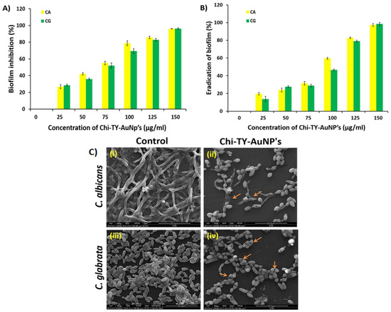
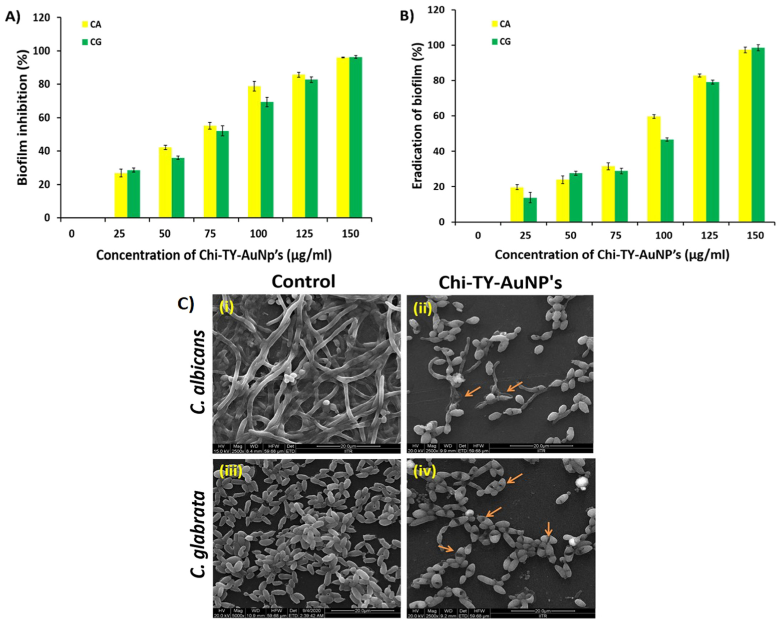
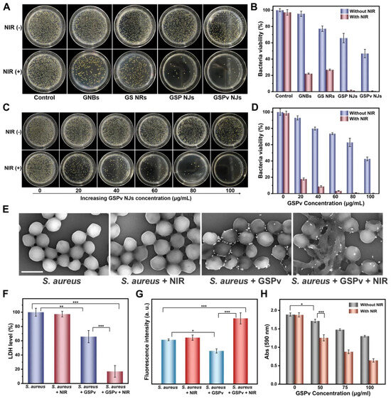
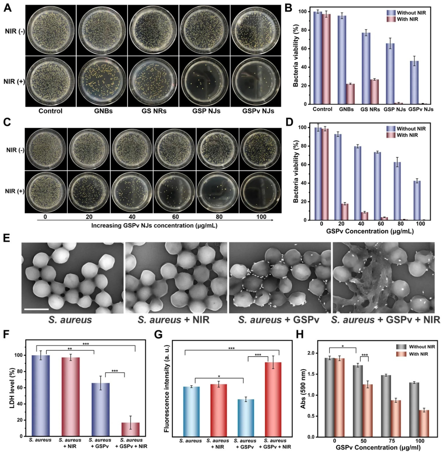
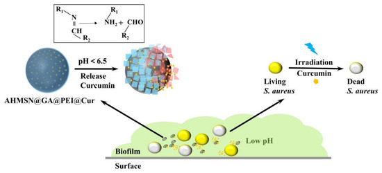
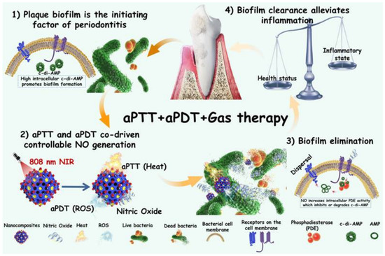
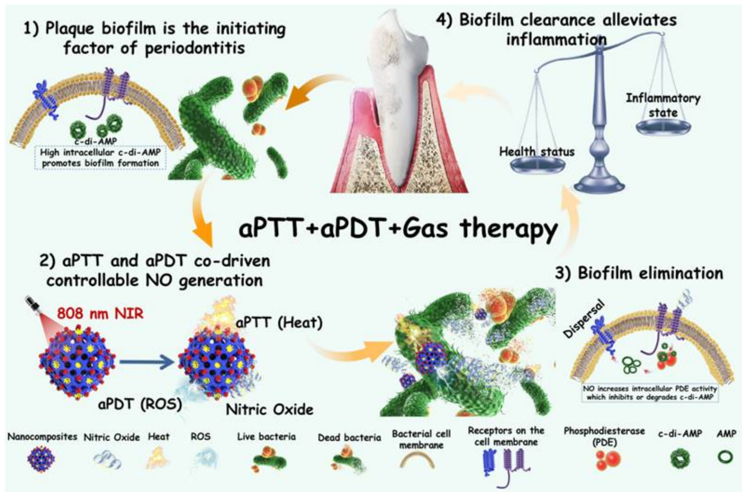
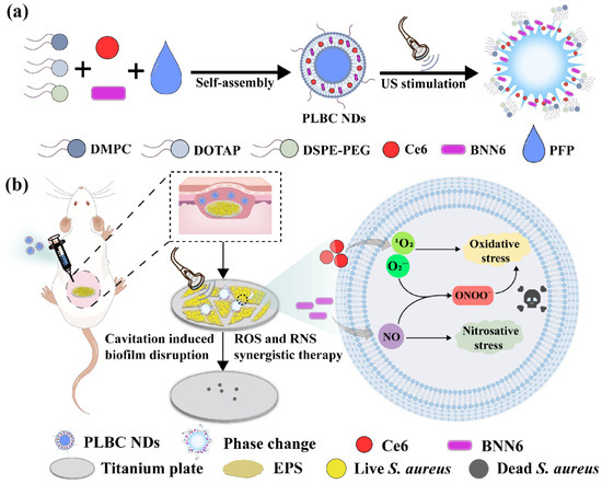
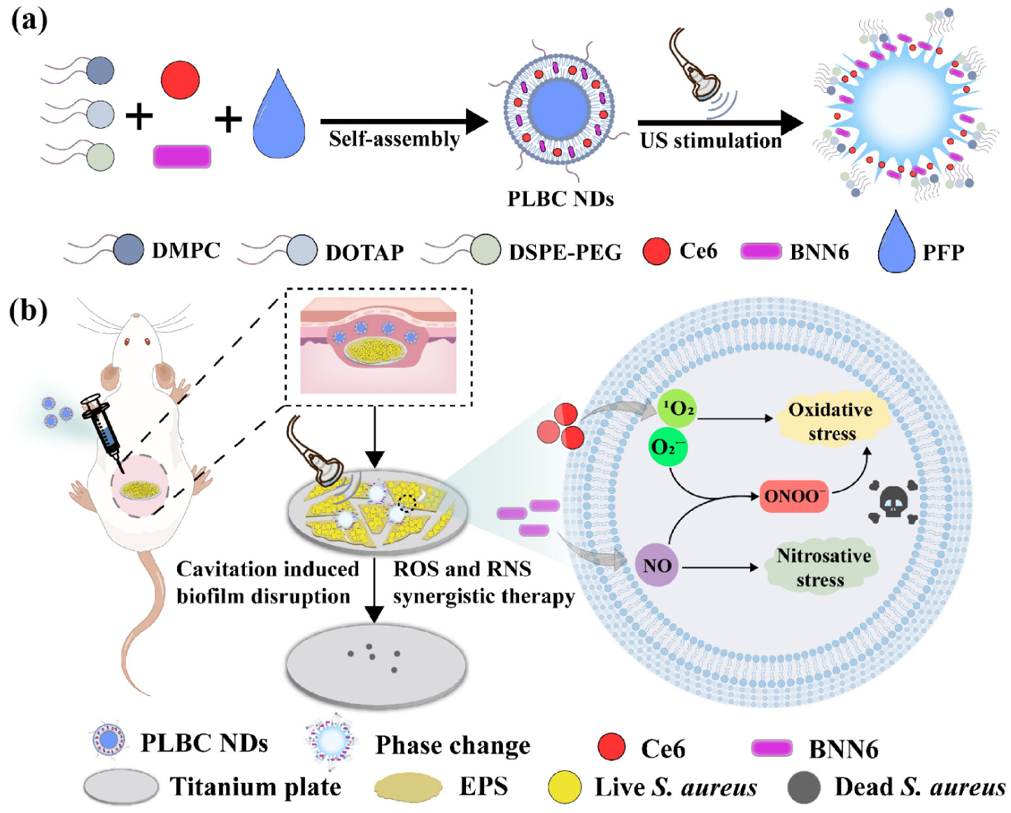

Disclaimer/Publisher’s Note: The statements, opinions and data contained in all publications are solely those of the individual author(s) and contributor(s) and not of MDPI and/or the editor(s). MDPI and/or the editor(s) disclaim responsibility for any injury to people or property resulting from any ideas, methods, instructions or products referred to in the content. |
© 2026 by the authors. Licensee MDPI, Basel, Switzerland. This article is an open access article distributed under the terms and conditions of the Creative Commons Attribution (CC BY) license.