


Abstract
Herpes simplex virus types 1 and 2 (HSV-1 and HSV-2) cause recurrent skin infections that are difficult to treat because of the limited solubility and permeability of acyclovir (ACV). This study developed electrospun polymeric membranes based on polycaprolactone (PCL), poly(lactic acid) (PLA), and cellulose acetate (CA) as controlled ACV delivery systems using uniaxial and coaxial fiber architectures. In the coaxial configuration, ACV-loaded PCL was used as the core and a PLA/CA blend as the shell. Continuous, randomly oriented, bead-free fibers with diameters ranging from 0.68 ± 0.32 µm to 1.45 ± 0.57 µm were obtained. Spectroscopic and thermal analyses confirmed successful drug incorporation, polymer compatibility, and good thermal stability. Coaxial membranes exhibited improved mechanical properties compared with uniaxial systems. Drug release studies showed a prolonged, pH-dependent profile, with greater ACV release at pH 7.3 than at pH 5.5, indicating the effective modulation of drug diffusion by the shell layer. Release kinetics were mainly governed by diffusion and anomalous transport mechanisms. All membranes maintained a cell viability above 80%, demonstrating good in vitro cytocompatibility. These findings support the potential of coaxial electrospun membranes for controlled antiviral drug delivery in skin applications.
1. Introduction
Human herpes simplex viruses, HSV-1 and HSV-2, affect individuals of all ages, with HSV-1 being more prevalent than HSV-2. Both represent a significant public health concern as the virus causes skin lesions leading to severe complications in immunocompromised patients. Acyclovir (ACV), a 2′-deoxyguanosine nucleoside analog that inhibits viral DNA polymerase, is widely used to treat these infections [1,2]. However, its topical administration is limited by low aqueous solubility and poor membrane permeability (0.12 × 10⁻⁶ to 2 × 10⁻⁶ cm/s), and red therapeutic efficacy. Developing alternative drug delivery systems is essential to enhance bioavailability and prevent treatment failure [3,4].
Electrospun polymeric fibers have emerged as promising platforms for drug delivery due to their high surface area, tunable porosity, and ability to efficiently encapsulate bioactive compounds. In particular, electrospinning enables the fabrication of fibers in uniaxial or coaxial configurations, offering the possibility to tailor drug-release profiles. Coaxial electrospinning allows for the formation of core–shell structures, where the drug can be selectively loaded into the core while the shell regulates diffusion, thereby improving drug stability and achieving sustained release behavior [5,6,7].
Aliphatic polyesters such as poly(ε-caprolactone) (PCL) and poly(lactic acid) (PLA) are widely used in biomedical applications due to their biocompatibility and biodegradability. PCL is characterized by its hydrophobic nature and slow degradation rate, which favor prolonged drug retention, whereas PLA exhibits higher stiffness and faster degradation, contributing to improved structural integrity and controlled-release behavior. Thus, the combination of both polymers provides complementary properties not only in mechanical performance, but also in degradation kinetics and drug-release modulation [8,9]. In addition, cellulose acetate (CA), a semi-synthetic polymer derived from cellulose, is commonly employed to enhance surface hydrophilicity and biocompatibility influencing drug diffusion and release profiles [10].
Several studies have explored electrospun systems for ACV delivery using individual polymers. For instance, PCL-based fibers have demonstrated sustained drug release and improved performance compared to commercial formulations [4]. PLA-based systems have shown promising antiviral activity and biocompatibility [11]. Similarly, CA has been used as a matrix for controlled drug incorporation into multilayer fiber systems with sustained-release behavior [12]. However, most of these studies focused on single-polymer or monoaxial configurations.
Although coaxial electrospinning has been extensively investigated for controlled drug delivery, reports combining PCL, PLA, and CA in a single coaxial architecture remain limited [13,14]. In particular, the use of a PLA/CA shell combined with a PCL core for ACV delivery has been scarcely explored, and systematic comparisons between uniaxial and coaxial systems are still lacking [15]. In this context, the rational combination of these polymers offers an opportunity to exploit their complementary physicochemical properties to modulate drug-release behavior and enhance scaffold performance.
Furthermore, while topical antiviral systems are traditionally evaluated as cutaneous patches to manage skin lesions, recent advances in electrospun and solution blow-spun matrices have significantly expanded their applications for ocular drug delivery. Localized polymeric scaffolds and inserts have been successfully developed to encapsulate ACV, providing sustained drug release directly to ocular epithelial tissues and offering promising platforms for the control of corneal herpes simplex virus infections [16,17]. Considering this, exploring the performance of these formulations in human epithelial models, such as the ARPE-19 cell line, provides a valuable means of evaluating the baseline cytocompatibility and versatility of the developed matrices for biomedical applications involving epithelial barriers [16].
Therefore, the aim of this work is to develop electrospun fiber systems based on PCL, PLA, and CA using both uniaxial and coaxial configurations. In the coaxial system, PCL is used as the drug-loaded core, while PLA and CA form the external shell to regulate diffusion and improve mechanical and biological properties. This approach aims to achieve controlled and tunable ACV release profiles while providing a comparative analysis of how polymer arrangement influences the physicochemical characteristics, drug-release kinetics, and biological performance of antiviral scaffolds.
2. Materials and Methods
2.1. Materials
PLA (MW = 145,000 g/mol) was purchased from NatureWorks, Plymouth, MN, USA; PCL (Mn: 80,000) from Sigma-Aldrich; and CA powder with 39.7 wt% acetyl content and an average Mn of 50,000 (Sigma-Aldrich, Toluca, Mexico). ACV was obtained from Sigma-Aldrich (PHR1254); fluorescein isothiocyanate (FITC), Rhodamine B, deuterated chloroform (CDCl3, 99.8 atom % D), and dimethyl sulfoxide-d6 (DMSO-d6, 99.9 atom % D) were purchased from Sigma-Aldrich (St. Louis, MO, USA). All organic solvents used were of analytical grade: ethanol, acetone, chloroform, methanol, and dimethyl sulfoxide (DMSO), purchased from Fermont Chemicals (Monterrey, Mexico). Water was purified using a MilliQ system (Millipore, Bedford, MA, USA). Dulbecco’s modified Eagle’s medium with high glucose, L-glutamine solution (200 mM), L-arginine monohydrochloride, L-asparagine, sodium pyruvate solution (100 mM), penicillin–streptomycin solution, trypsin-EDTA solution (0.25%), and MTT [3-(4,5-dimethylthiazol-2-yl)-2,5-diphenyltetrazolium] were purchased from Sigma-Aldrich. Fetal bovine serum (FBS) was obtained from Gibco Life Technologies (Grand Island, NY, USA). Phosphate-buffered saline (PBS) was prepared using sodium chloride (NaCl, ≥99%), potassium chloride (KCl, ≥99%), disodium hydrogen phosphate (Na2HPO4, ≥99%), and potassium dihydrogen phosphate (KH2PO4, ≥99%), all obtained from Sigma-Aldrich (St. Louis, MO, USA). The pH of the buffer solutions was adjusted to 5.5 and 7.3 using hydrochloric acid (HCl, 37%, analytical grade) and sodium hydroxide (NaOH, ≥98%), also supplied by Sigma-Aldrich. All aqueous solutions were prepared using deionized water (Milli-Q grade).
2.2. Membrane Preparation
Different solvents were selected according to the polymer used. PCL was dissolved in a chloroform/methanol (1:1, v/v) mixture, whereas PLA and CA were dissolved in acetone. Uniaxial polymer solutions were prepared at concentrations ranging from 8 to 12% (w/v). A PLA/CA blend (70/30, w/w) was prepared to serve as the shell solution in the coaxial configuration, while the PCL solution was used as the core phase. ACV was incorporated into the PCL solution at concentrations ranging from 1 to 4% (w/w) relative to the polymer mass. All solutions were magnetically stirred for 24 h at room temperature to ensure complete dissolution and homogeneity.
Electrospinning was performed using a syringe pump (KDS Scientific, Holliston, MA, USA) and a high-voltage power supply (Spellman High Voltage Electronics Corporation, model CZE 1000R, Hauppauge, NY, USA). Polymer solutions were loaded into 10 mL syringes fitted with 18–22 G needles. For coaxial electrospinning, a concentric needle assembly consisting of a 17 G outer needle and a 22 G inner needle was used. During preliminary experiments, the applied voltage (12–18 kV), flow rate (0.2–4.0 mL·h−1), and tip-to-collector distance (10–16 cm) were varied to identify conditions that provided stable jet formation and bead-free fibers, following criteria previously reported by Rodríguez-Félix et al., 2016 [18].
The final electrospinning conditions selected for each formulation are summarized in Table 1. Uniaxial (U) membranes were prepared from PCL (12% w/v), PLA (10% w/v), CA (8% w/v), or a PLA/CA blend (70/30, 10% w/v). PCL-based membranes were electrospun at 15 kV using a tip-to-collector distance of 12 cm and a flow rate of 2.5 mL·h−1, whereas slight adjustments were required for PLA and CA formulations to maintain jet stability and homogeneous fiber formation. Coaxial (C) membranes were fabricated using a PLA/CA (70/30, 10% w/v) shell and a PCL (12% w/v) core. ACV (0–4% w/w) was incorporated into the core solution, and coaxial electrospinning was performed at 15–17 kV, a tip-to-collector distance of 12–15 cm, and a total flow rate of 1.5–2.5 mL·h−1.
Table 1.
Composition and electrospinning parameters of the prepared membranes.
2.3. Morphological Analysis
Polymeric membrane samples obtained through electrospinning were morphologically characterized using a scanning electron microscope (SEM, JSM-5410LV JEOL Ltd., Akishima, Tokyo, Japan) operated at an accelerating voltage of 20 kV. Prior to imaging, the samples were sputter-coated with a thin layer of gold to improve surface conductivity. SEM micrographs were acquired at magnifications of 1500× and 5000×. Fiber diameters were determined from the 5000× SEM micrographs using ImageJ software (version 1.53a, National Institutes of Health, Bethesda, MD, USA). For each formulation, at least four representative micrographs from different regions of the membranes were analyzed, and at least 200 randomly selected fibers were measured to determine the mean fiber diameter and the fiber diameter distribution. Results are presented as mean fiber diameter ± standard deviation (SD).
2.4. Confocal Laser Scanning Microscopy (CLSM) Characterization
The coaxial architecture of the electrospun membranes was evaluated by confocal laser scanning microscopy (CLSM) using a Nikon Eclipse confocal microscope with a C2 confocal system (Nikon, Tokyo, Japan). To generate optical contrast between the concentric compartments, polymer solutions were selectively mixed with fluorescent dyes prior to electrospinning while maintaining the same electrospinning parameters used to fabricate the coaxial membranes described above. Specifically, the PCL core solution was mixed with fluorescein isothiocyanate (FITC), whereas the PLA/CA shell solution was mixed with Rhodamine B. Both fluorophores were incorporated at 0.05 wt% relative to the corresponding polymer content of each spinning solution.
Electrospun membranes were directly mounted onto glass slides and imaged using a 60× water-immersion objective with a 1.5× optical zoom, with distilled water as the immersion medium. Fluorescence images were acquired in sequential scanning mode to prevent spectral overlap and cross-talk between fluorophores. The FITC-containing PCL core was excited using a 488 nm solid-state laser, whereas the Rhodamine B-containing PLA/CA shell was excited using a 561 nm solid-state laser. Detector gain and offset were adjusted independently for each channel to optimize signal detection and clearly distinguish the core and shell compartments. Images were acquired at a resolution of 1024 × 1024 pixels with a pixel dwell time of 2.4 µs, while the confocal pinhole was maintained at 5 Airy units to provide controlled optical sectioning. A CLSM analysis was performed exclusively to verify the concentric core–shell architecture of the electrospun coaxial fibers.
Three-dimensional visualization of the fibers was achieved by Z-stack acquisition from the bottom to the top of the sample over a total depth of 4.25 µm. Consecutive optical sections were collected at 0.1 µm intervals, allowing for reconstruction of the coaxial structure along the Z-axis. Representative images from different focal planes were selected from the Z-stack to assess the continuity and spatial distribution of the core within the surrounding shell. A transmitted-light channel was simultaneously acquired to provide a morphological reference. Final image processing, fluorescence channel merging, and orthogonal reconstructions were performed using NIS-Elements software (version 4.30.02, Nikon, Tokyo, Japan).
2.5. Fourier Transform Infrared Spectroscopy (FTIR)
To confirm the presence of all components in the system, a spectroscopic evaluation was conducted using a PerkinElmer Frontier FTIR spectrometer (Waltham, MA, USA). Spectra were obtained using the attenuated total reflectance (ATR) technique. Membrane samples were analyzed in the range of 4000 to 400 cm−1.
2.6. Proton Nuclear Magnetic Resonance (1H NMR) and Quantification of ACV Loading and Encapsulation Efficiency
The presence of ACV within the electrospun fibers, including both uniaxial and coaxial structures, was confirmed by proton nuclear magnetic resonance (1H NMR) spectroscopy. Approximately 20 mg of each sample was weighed and dissolved in 500 µL of a 1:1 mixture of deuterated chloroform (CDCl3) and deuterated dimethyl sulfoxide (DMSO-d6) to ensure complete solubilization of both the polymeric matrix (PCL, PLA, and CA) and the ACV. The resulting solutions were transferred into 5 mm NMR tubes, and spectra were recorded at 40 °C using a Bruker AVANCE 400 MHz spectrometer (Billerica, MA, USA). Chemical shifts (δ) were reported in parts per million (ppm) relative to residual solvent peaks. 1H NMR spectra were acquired with 64 scans and a relaxation delay of 2 s to improve the signal-to-noise ratio and ensure reliable integration of the ACV resonances.
A quantitative analysis was carried out using a relative qNMR approach according to the methodology described by Holzgrabe [19]. The characteristic ACV signal at 7.81 ppm, corresponding to one proton, was selected as the reference signal. For the polymer components, the signals at 4.01 ppm (PCL, CH2 adjacent to oxygen, 2H), 5.16 ppm (PLA, CH, 1H), and 2.10 ppm (CA, CH3 of acetate groups, 3H) were selected due to the absence of spectral overlap.
The actual drug content was estimated from the relative integrals and the molecular weights of ACV and the repeating units of the polymers according to Equation (1):
where represents the integral area of the selected resonance.
Encapsulation efficiency (EE %) was calculated using Equation (2):
2.7. Wettability Characteristics by Contact Angle Analysis
Contact angles of both uniaxial and coaxial membranes were measured at room temperature using a ChemInstruments CAM-PLUS apparatus (ChemInstruments, Fairfield, OH, USA). Deionized water was used as the testing liquid, and at least ten measurements were taken for each sample to obtain an average value.
2.8. Stress−Strain Mechanical Test
Tensile tests were performed using an Electroforce 5110 testing machine equipped with a 200 N load cell (TA Instruments, Eden Prairie, MN, USA). Rectangular strips were cut at 5 mm in width and 20 mm in length. The film thickness was measured using a Mitutoyo micrometer (Mitutoyo Corporation, Kawasaki, Japan), with the thickness maintained within the range of 0.01–0.1 mm. A constant displacement rate of 0.1 mm/s was applied, with a 4 mm distance between the gripping surfaces. The samples were conditioned to room temperature and humidity before and during testing. An average of 10 specimens was tested for each condition. Test data were processed with WinTest 7 software, and results were plotted using Origin 7.
2.9. Thermogravimetric Analysis (TGA)
Thermogravimetric analysis (TGA) was performed to assess the thermal stability of electrospun membrane samples using an SDT 2960 Simultaneous DTA-TGA (TA Instruments, New Castle, DE, USA). Approximately 20 mg of each sample was placed in an aluminum sample holder and heated from 20 °C to 800 °C at a rate of 10 °C/min under a nitrogen atmosphere. Thermal degradation profiles of pure polymers were used to identify the characteristic degradation regions of each component.
2.10. Differential Scanning Calorimetry (DSC)
Differential scanning calorimetry (DSC) measurements were carried out using an SDT 2960 Simultaneous DTA-TGA (TA Instruments). Approximately 5 mg of each sample was placed in aluminum pans and analyzed under a nitrogen atmosphere. The temperature program was performed from −80 °C to 310 °C at a heating rate of 5 °C/min. The melting enthalpy () associated with the PCL phase was used to estimate the apparent degree of crystallinity of the electrospun membranes according to:
where is the melting enthalpy obtained from the DSC thermograms, and 148 J/g was used as the melting enthalpy of 100% crystalline PCL according to previously reported values [20].
2.11. Drug Release Assay
To determine the ACV release profile from electrospun polymer membranes, weighed membrane samples were placed in phosphate-buffered saline (PBS 1X; 137 mM NaCl, 2.7 mM KCl, 10 mM phosphate buffer, and 1.8 mM KH2PO4) as the release medium at different pH values (5.5 and 7.3) to evaluate the pH-responsive behavior of the scaffolds under distinct physiological environments. A pH value of 5.5 was selected to mimic the physiological acidic mantle of healthy human skin surfaces, whereas a pH of 7.3 simulates the baseline conditions of human ocular tear fluid, also closely matching the neutral microenvironment characteristic of damaged, inflamed, or deeper herpetic cutaneous lesions with a compromised epidermal barrier [21,22]. The temperature was maintained at 37 °C under agitation at 100 rpm using an orbital shaker (Thermo Scientific MaxQ 4000, Thermo Fisher Scientific Inc., Waltham, MA, USA). Aliquots of 3 mL of the release medium were collected at predetermined time intervals throughout the release process until the release equilibrium was reached. Acyclovir concentration was monitored by measuring the absorbance of each aliquot at wavelengths between 230 and 270 nm using an Agilent 8453 UV–Vis spectrometer equipped with a diode array detector (Agilent Technologies, Inc., Santa Clara, CA, USA). Absorbance values were interpolated using a previously prepared calibration curve for the drug to determine the amount of ACV released. Aliquots were returned to the release medium, ensuring that the medium’s volume remained constant. All experiments were conducted in triplicate [23].
Three mathematical models were used to fit the experimental data obtained from release studies, with the goal of determining the release mechanisms of ACV. The equations for the different models are provided below:
2.11.1. Zero-Order Model
2.11.2. First-Order Model
2.11.3. Higuchi Model
2.11.4. Korsmeyer-Peppas Model
2.12. Cell Culture
To perform cell viability assays, the normal human cell line ARPE-19 (retinal pigment epithelial cell line) was used. Although the developed membranes have strong potential for cutaneous applications, as evidenced by their mechanical properties and release behavior at the skin surface, ARPE-19 cells provide a well-established, non-tumorigenic, and highly sensitive human epithelial model to screen the safety of biomaterials. This cell line maintains a stable epithelial phenotype and exhibits high susceptibility to external chemical insults, making it an excellent proxy for the preliminary screening of human epithelial cell barriers. Furthermore, the selection of ARPE-19 cells aligns with established tissue-engineering literature, in which they are routinely used as a standard human epithelial model to evaluate the baseline cytocompatibility, adhesion, and proliferation profiles of electrospun matrices based on PCL and PLA configurations [28,29].
Additionally, the use of this cell line is highly relevant for the evaluation of the scaffold’s suitability for ocular antiviral applications, aligning with recent literature where electrospun matrices loaded with ACV and other co-drugs are explicitly designed for ocular drug delivery systems [16,17]. Cells were propagated as monolayer cultures in 25 cm2 culture flasks using Dulbecco’s modified Eagle medium (DMEM) supplemented with 5% fetal bovine serum (FBS) and 1% penicillin/streptomycin solution. Cultures were maintained at 37 °C in a humidified incubator with 5% CO2 and 95% relative humidity.
2.13. Cytotoxicity Assay
Cytotoxicity was evaluated using the MTT colorimetric assay, a quantitative, sensitive method for measuring cell viability and proliferation, enabling the determination of whether a compound exhibits cytotoxic effects. This method is based on the ability of mitochondrial enzymes (succinate dehydrogenase) in viable cells to reduce the yellow, water-soluble MTT substrate into an insoluble blue-violet formazan product. The amount of formazan produced is directly proportional to the number of viable cells in the culture.
To determine cell viability, electrospun membranes were cut into 5 mm-diameter circular samples weighing 4 mg and placed into wells of 96-well cell culture plates. Membranes were sterilized by adding 200 µL of 70% ethanol exposed to UV irradiation for 30 min on each side. After sterilization, a suspension containing 5 × 10³ cells in 200 µL of DMEM supplemented with 5% FBS was seeded into each well containing the membranes. As a positive control for cytotoxicity, 20 µL of DMSO was added.
Cells were incubated for 24 h at 37 °C in a 5% CO2 atmosphere. Morphological changes or signs of apoptosis were evaluated using an inverted optical microscope (Nikon Ti/Ti C2+, Nikon Corporation, Tokyo, Japan). Subsequently, the culture medium was removed, and cells were washed with 1× PBS to eliminate residual compounds. Fresh medium was then added along with an MTT solution (5 mg/mL), and plates were incubated for 4 h at 37 °C in a 5% CO2 atmosphere. After incubation, the MTT-containing medium was removed, and 100 µL of DMSO was added to dissolve formazan crystals. Once dissolved, absorbance was measured at 630 nm using an ELISA plate reader (Multiskan EX, Thermo Scientific, Waltham, MA, USA). Cell viability percentage was calculated relative to the negative control (untreated cells), considered as 100% cell viability. All assays were performed in quadruplicate and repeated in three independent experiments.
2.14. Statistical Analysis
Statistical analyses were performed using OriginPro 8.0 (OriginLab Corporation, Northampton, MA, USA) and GraphPad Prism 5.0 (GraphPad Software Inc., San Diego, CA, USA). All experiments were performed in triplicate, and the results are expressed as the mean ± standard deviation (SD). Data normality was assessed using the Kolmogorov–Smirnov and Shapiro–Wilk tests. Normally distributed datasets, including cytotoxicity and mechanical property assays, were analyzed using one-way analysis of variance (ANOVA), followed by Tukey’s post hoc test to perform multiple comparisons. Non-normally distributed datasets, including fiber diameter and water contact angle measurements, were analyzed using the non-parametric Kruskal–Wallis test followed by Dunn’s multiple comparisons test. Differences were considered statistically significant at p < 0.05.
3. Results and Discussion
3.1. Morphological Analysis by Scanning Electron Microscopy (SEM)
Once electrospun membranes were obtained, samples were prepared for SEM imaging (Figure 1). In these micrographs, randomly oriented fibers with micrometer-scale diameters were observed. To complement this analysis and confirm the structural homogeneity across larger areas, representative lower-magnification SEM micrographs (1500×) obtained from different regions of the samples are presented in Figure S1. Fiber diameter measurements were performed using ImageJ software; more than 200 fibers were measured for each sample, and the resulting averages and standard deviations are presented in the frequency histograms shown in Figure 1. Additional SEM images of PLA, CA, and PLA/CA, along with their corresponding diameter histograms, are presented in Figure S2. The average diameters of PCL and UNI 1% were 1.22 ± 0.61 µm and 1.45 ± 0.57 µm, respectively, the largest among the evaluated samples. This behavior may be associated with the combined effects of polymer concentration, solution viscosity, and electrohydrodynamic stretching during electrospinning, known to influence the morphology and diameter of electrospun PCL fibers [30].
Figure 1.
SEM micrographs and the corresponding fiber diameter distributions of electrospun membranes: (a) PCL, (b) UNI 1%, (c) COA 0%, (d) COA 1%, (e) COA 2%, and (f) COA 4%. The red line represents the Gaussian fit of the fiber diameter distribution. These results reveal the formation of defect-free microfibrous structures effectively mimicking the extracellular matrix architecture.
PLA fibers exhibited an average diameter of 0.68 ± 0.32 µm, corresponding to the smallest fibers among the evaluated formulations. These values are within the range commonly reported for electrospun PLA fibers prepared under comparable processing conditions [31]. The average diameter of the PLA/CA fibers was 0.88 ± 0.27 µm. This increase suggests that CA incorporation modified the solution’s electrospinning behavior compared to neat PLA fibers. Although CA may increase solution viscosity, the final fiber diameter is governed by multiple factors, including solution conductivity, surface tension, and intermolecular interactions within the polymer solution. These parameters affect the electrohydrodynamic stretching of the jet during electrospinning and consequently influence fiber morphology and diameter [32]. Therefore, the observed change in diameter likely results from the combined influence of these parameters rather than viscosity alone.
It is worth noting that the diameters obtained for all formulations fell within the micrometer range typically reported for electrospun scaffolds. Although these values are larger than those of native collagen and elastin fibers, they remain within the dimensional range commonly employed in electrospun matrices designed to mimic extracellular matrix architecture [33]. Fiber diameters in the submicrometer-to-micrometer range are considered suitable for skin tissue engineering because they provide a high surface area for cell attachment while maintaining the scaffold’s structural integrity [34,35]. For the electrospun membranes with a coaxial arrangement, the conditions shown in Table 1 were selected. The electrospinning parameters for the coaxial formulations (COA 0%, COA 1%, COA 2%, and COA 4%) were kept constant at 17 kV, a working distance of 12 cm, and a flow rate of 1.5 mL·h−1. These conditions had been previously optimized for the coaxial setup. The concentrations and ratios of the polymer solutions were also based on those used in the uniaxial configuration. As with the uniaxial fibers, SEM micrographs were obtained for each coaxial sample (Figure 1), and fiber diameters were measured to generate the corresponding frequency histograms.
Regarding morphology, all coaxial samples exhibited randomly oriented, bead-free fibers with predominantly cylindrical morphology and slight waviness. This morphology is advantageous because it provides a high surface area and resembles the fibrous structure of the extracellular matrix [34]. In the coaxial system, the shell consisted of PLA/CA, while the core was composed of PCL containing different concentrations of ACV (0–4% relative to polymer weight). The resulting fiber diameters were 0.88 ± 0.27 µm, 0.76 ± 0.24 µm, 0.92 ± 0.22 µm, and 1.00 ± 0.32 µm for COA 0%, COA 1%, COA 2%, and COA 4%, respectively. Statistical analysis using the Kruskal–Wallis test followed by Dunn’s multiple comparisons test revealed no statistically significant differences among the coaxial formulations (p > 0.05), indicating that drug concentration did not significantly affect fiber diameter.
Nevertheless, a slight tendency toward increasing diameters at higher drug concentrations (≥2%) can be observed, despite the initial decrease observed for COA 1%. This behavior may be associated with changes in solution properties, including viscosity and intermolecular interactions induced by the presence of the drug. Additionally, the coaxial fibers exhibited smaller diameters than the uniaxial ACV-loaded fibers (1.45 ± 0.57 µm). This behavior may be related to differences in jet stretching and solvent evaporation dynamics arising from the core–shell configuration, which can alter electrohydrodynamic behavior during fiber formation and consequently affect fiber diameter [32]. To further evaluate the specific surface topography of the fibers, high-magnification SEM micrographs were analyzed (Figure S3). The neat polymers (PLA, PCL, and CA) exhibited a smooth, uniform, and continuous surface with no apparent roughness. In contrast, the uniaxial formulation (UNI 1%) exhibited a distinctive morphology with a highly corrugated, wrinkled surface texture. Conversely, the coaxial formulations (COA 0% and COA 1%) retained a notably more cylindrical and regular geometry. Even with the incorporation of the drug in COA 1%, the fibers maintained a uniform, continuous outer surface, displaying only minor surface texture compared to the highly irregular surface observed in the UNI 1% sample.
3.2. Structural Validation of Coaxial Fibers by CLSM
The structural organization of the electrospun coaxial membranes was evaluated by dual-channel confocal laser scanning microscopy (CLSM). As shown in Figure 2a, the FITC-containing PCL phase (green) formed a continuous inner filament, whereas the Rhodamine B-containing PLA/CA phase (red) defined a homogeneous external shell. The merged fluorescence image, together with the transmitted-light image, clearly demonstrated a concentric core–shell arrangement, confirming that the PCL core remained continuously confined within the PLA/CA shell along the fiber axis, with no evidence of phase separation or structural discontinuities. Notably, the overlay channel exhibited a distinct yellow coloration, resulting from the optical superposition of the green (core) and red (shell) channels in the two-dimensional projection, thereby confirming their precise spatial coincidence.
Figure 2.
Confocal laser scanning microscopy (CLSM) characterization of electrospun coaxial membranes. (a) Individual fluorescence channels and merged image of a representative coaxial fiber showing the transmitted light image (COA fiber), the FITC-containing PCL—core (green), the Rhodamine B-containing PLA/CA—shell (red), and the merged image (yellow), confirming the concentric core–shell architecture. (b) Representative optical sections selected from a three-dimensional confocal Z-stack (total depth: 4.25 µm, step size: 0.1 µm), corresponding to the lower, central, and upper focal planes of the electrospun fibers.
To further verify the three-dimensional architecture, optical serial sectioning (Z-stack) was performed at different focal planes throughout the fiber (Figure 2b). The fluorescence associated with the PCL core was predominantly observed at the central focal plane, whereas the lower and upper optical sections were mainly characterized by the fluorescence from the PLA/CA shell. This gradual variation in fluorescence intensity across consecutive optical sections confirms that the core is completely surrounded by the shell throughout the fiber thickness, providing strong evidence of a true coaxial morphology rather than a side-by-side or Janus-like configuration.
The structural features observed in the present study are in good agreement with previous reports using selective fluorescent dyes and CLSM to validate electrospun coaxial fibers. Wu et al. [36] demonstrated the successful visualization of PCL/gelatin core–shell nanofibers by incorporating FITC and Rhodamine B into the core and shell solutions, respectively, confirming continuous encapsulation of the inner compartment. Similarly, Romano et al. [37] reported that dual-channel CLSM enables direct discrimination between compartmentalized core–shell fibers and non-coaxial morphologies through selective fluorescence imaging. Likewise, Zhu et al. [38] used confocal fluorescence imaging combined with Z-stack reconstruction to verify the continuous three-dimensional organization of aqueous-based coaxial silk elastin nanofibers. The close agreement between these studies and the present observations confirms that the electrospinning conditions used successfully produced well-defined coaxial fibers with a continuous core completely enclosed by the surrounding shell.
3.3. Fourier Transform Infrared Spectroscopy (FTIR) Analysis
Figure 3 shows the FTIR spectra of the raw polymers and electrospun fiber systems. From bottom to top, the spectra correspond to pure PCL, ACV, uniaxial fibers containing 1% ACV (UNI 1%), pure PLA, pure CA, and the coaxial fibers COA 0% and COA 4%. This arrangement enables a direct comparison of the characteristic functional groups and provides insight into interactions occurring after fiber formation and drug incorporation. The spectrum of PCL exhibited its characteristic absorption bands, including CH2 asymmetric and symmetric stretching vibrations at ~2940 and ~2865 cm−1, respectively, a strong carbonyl (C=O) stretching band at ~1720–1730 cm−1, and C–O–C stretching vibrations in the 1240–1160 cm−1 region, consistent with the expected structure of semicrystalline aliphatic polyesters [39,40].
Figure 3.
ATR-FTIR spectra of individual polymers, pure ACV, UNI 1% fibers, and COA fibers containing different ACV concentrations (COA-0%, COA-1%, COA-2%, and COA-4%). Spectra were acquired using the attenuated total reflectance (ATR) technique in the spectral range of 4000–400 cm−1. The spectra confirm the chemical compatibility of the components and the preservation of functional groups after electrospinning.
The ACV spectrum displayed a broad band in the 3500–3200 cm−1 region associated with N–H and O–H stretching vibrations, along with bands between ~1700–1600 cm−1 corresponding to C=O and C=N groups of the purine ring. Additional signals in the fingerprint region (1500–1000 cm−1) were attributed to C–N and C–O vibrations, according to standard FTIR band assignments [40]. For the UNI 1% fibers, the spectrum was dominated by the characteristic PCL bands, particularly the intense carbonyl signal and C–O–C vibrations. A slight broadening in the 3500–3000 cm−1 region was observed compared to neat PCL. This feature may be associated with overlapping contributions from O–H and/or N–H stretching vibrations or hydrogen-bonding-related interactions; however, FTIR analysis alone does not allow a definitive assignment. The absence of clearly distinguishable ACV peaks can be attributed to its low concentration and the inherent limitations of ATR-FTIR as a surface-sensitive technique. In this context, the drug-related signals are likely masked by the dominant polymer matrix. Similar limitations in detecting drug-related signals in electrospun systems have been reported at low drug loadings, where polymer bands dominate the spectra [4].
The PLA spectrum showed a strong carbonyl stretching band at ~1750–1760 cm−1, CH3 stretching near ~2995–2945 cm−1, and C–O stretching bands in the 1180–1080 cm−1 region, in agreement with previous studies [41]. The CA spectrum showed a broad O–H stretching band between ~3600–3200 cm−1, C–H stretching near ~2900 cm−1, and a strong carbonyl band at ~1740–1750 cm−1 corresponding to acetyl groups. Additional bands in the 1200–1000 cm−1 region were associated with C–O–C and C–O stretching of the polysaccharide backbone, consistent with previously reported CA spectra [42,43].
In the coaxial fibers without drug (COA 0%), the spectrum contained contributions from all three polymers (PCL core and PLA/CA shell). The carbonyl region (~1730–1750 cm−1) showed overlapping bands with slight broadening, suggesting possible interactions between polymer phases. Similar spectral changes associated with polymer–polymer and polymer–drug interactions have been reported in electrospun systems [44,45]. For the COA 4% fibers, the spectral profile remained very similar to that of COA 0%, indicating that ACV incorporation does not significantly alter the chemical structure of the system. As in the UNI 1% fibers, bands observed in the 3500–3000 cm−1 region are more likely associated with polymeric contributions rather than ACV. The lack of detectable ACV signals may be explained by a combination of factors, including (i) low drug loading and (ii) possible intermolecular interactions. Additionally, it is important to consider that ATR-FTIR primarily probes the sample surface; therefore, in a coaxial system, the signal is expected to be dominated by the outer PLA/CA shell. In this sense, it is plausible that ACV, if preferentially located within the PCL core, remains below the technique’s detection limit.
3.4. 1H NMR Analysis and Quantification of ACV Loading
To confirm the presence of ACV in the fibers and estimate the actual drug loading, 1H NMR measurements were performed. Figure 4 shows the 1H NMR spectra of pure ACV, PCL, COA 0%, COA 1%, COA 2%, and COA 4% after dissolution in a CDCl3/DMSO-d6 mixture. Spectrum A corresponds to pure ACV, spectrum B to PCL, spectrum C to COA 0%, and spectra D, E, and F to the COA formulations containing 1, 2, and 4 wt% ACV, respectively.
Figure 4.
1H NMR spectra of pure ACV (A), UNI 1% (B), COA 0% (C), COA 1% (D), COA 2% (E), and COA 4% (F) fiber samples (dissolved in a CDCl3/DMSO d6 mixture and heated to 40 °C to obtain a homogeneous solution). The characteristic ACV signals are marked with asterisks (*) in the spectra of the UNI 1%, COA 1%, COA 2%, and COA 4% fibers. Signals corresponding to the constituent polymers PCL (1), PLA (2), and CA (3) are also highlighted. These results confirm the chemical integrity of acyclovir within the matrices and provide a reliable basis for quantifying drug loading.
The spectrum of pure ACV exhibited characteristic proton resonances, including a signal at δ ≈ 10.5 ppm assigned to the N–H proton of the guanine moiety, signals in the δ ≈ 7.8–8.2 ppm region associated with protons of the purine ring, and resonances between δ ≈ 5.0 and 6.5 ppm corresponding to protons of the side chain. In the spectra of the ACV-loaded fibers, the signals marked with red asterisks coincide with those observed in the spectrum of pure ACV, confirming the presence of the drug and preservation of its characteristic proton resonances after electrospinning. In contrast, these signals were absent in the drug-free control (COA 0%), supporting the specificity of the assignments and confirming the successful incorporation of ACV into electrospun fibers. The signal at δ ≈ 7.26 ppm corresponds to residual CDCl3 and was not considered for peak assignment.
Additionally, all fiber spectra displayed signals corresponding to constituent polymers, labeled as (1), (2), and (3). The signals marked as (1), attributed to PCL, were observed at δ ≈ 4.0–4.2 ppm (–O–CH2–, ester linkage), δ ≈ 2.2–2.4 ppm (–CO–CH2–), and δ ≈ 1.3–1.7 ppm (aliphatic –CH2– groups), in agreement with previous reports for PCL [46]. The signals labeled as (2), corresponding to PLA, appeared at δ ≈ 5.1–5.3 ppm (methine–H–djacent to the ester group) and δ ≈ 1.4–1.6 ppm (methyl–CH3 group), while the signals marked as (3), assigned to CA, were identified at δ ≈ 3.5–5.0 ppm (ring protons of the anhydroglucose unit) and δ ≈ 1.9–2.1 ppm (acetyl methyl groups), in agreement with previously reported spectra [46,47].
The overlap of signals in the 0–5 ppm region reflects the complexity of the multicomponent polymeric system. Nevertheless, the characteristic resonances attributed to ACV remained clearly distinguishable in the spectra of COA 1%, COA 2%, and COA 4%, while they were absent in the drug-free control. Such spectral overlap was commonly observed in multicomponent polymeric systems analyzed by [40]. Unlike FTIR analysis, which did not reveal clearly distinguishable drug-specific bands, 1H NMR provided direct evidence of ACV incorporation within both uniaxial and coaxial membranes.
Beyond qualitative identification, the 1H NMR spectra were also used to quantify the actual ACV loading in the electrospun fibers using the relative qNMR methodology proposed by Holzgrabe [19]. The signal at δ 7.81 ppm, corresponding to one ACV proton, was selected as the reference resonance and normalized to an integral value of 1. Characteristic signals from PCL (δ 4.01 ppm, 2H), PLA (δ 5.16 ppm, 1H), and CA (δ 2.10 ppm, 3H) were used as internal references. The actual ACV content was calculated from the relative integrals and the molecular weights of ACV and the corresponding polymer repeating units. These results are summarized in Table 2.
Table 2.
Relative 1H NMR integrals used for ACV quantification, experimentally determined drug loading, and encapsulation efficiency of the UNI and COA electrospun fibers.
The calculated ACV loadings showed excellent agreement with the theoretical values used during formulation. Encapsulation efficiencies (EE%) of 94–102% were achieved, indicating high drug incorporation efficiency and minimal ACV loss during electrospinning. These results confirm that the preparation method effectively retained the antiviral agent within the fiber matrix.
3.5. Mechanical Properties
The mechanical properties of electrospun membranes are summarized in Table 3. Significant differences were observed among the evaluated formulations, indicating that both polymer composition and fiber architecture influenced the materials’ mechanical response. Among the neat polymers, PCL exhibited the highest elongation at break (276.7 ± 40.8%), whereas PLA showed a considerably lower value (66.8 ± 6.9%). This behavior reflects the greater flexibility of PCL and the higher rigidity of PLA, associated with its glassy nature at room temperature. Similar differences have been widely reported for electrospun polyester systems, where PCL behaves as a ductile polymer while PLA displays a more brittle mechanical response. Consequently, the fabrication of composite or blended electrospun mats is frequently evaluated to balance flexibility and mechanical integrity, thereby rendering the materials suitable for practical manipulation and clinical deployment as functional biomedical platforms and wound dressings [48,49,50].
Table 3.
Mechanical properties of the prepared membranes.
The incorporation of CA into PLA resulted in an increase in elastic modulus from 4.4 ± 1.5 MPa to 14.9 ± 8.9 MPa, accompanied by a moderate increase in elongation at break. Although these differences were not statistically significant, the observed trend suggests that incorporating CA modified the mechanical response of the PLA matrix. The underlying mechanism cannot be established from the present results, although similar effects have been reported for CA-containing polymer systems [51]. A more pronounced effect was observed in the coaxial membranes. The elastic modulus increased from 11.6 ± 3.3 MPa for PCL to 45.1 ± 14.1 MPa for COA 0%, demonstrating that the core–shell architecture substantially modified the mechanical response of the fibers. At the same time, elongation at break decreased from 276.7 ± 40.8% for PCL to 28.6 ± 3.8% for COA 0%, indicating a transition toward a stiffer and less extensible material. This behavior is likely associated with the presence of the PLA/CA shell, whose greater rigidity limits the extensibility imparted by the PCL core. Similar increases in stiffness have been reported in PLA/PCL multilayer and composite systems [52].
Drug incorporation produced distinct effects depending on fiber architecture. In uniaxial membranes, ACV loading caused moderate increases in tensile strength and elastic modulus, from 6.41 ± 1.44 MPa to 9.15 ± 4.78 MPa and from 11.6 ± 3.3 MPa to 14.3 ± 6.0 MPa, respectively. However, no statistically significant differences were detected relative to neat PCL. Interestingly, this behavior differs from that reported by Costa et al. (2019) [4], who observed a reduction in Young’s modulus and tensile strength after the incorporation of ACV and omega-3 into electrospun PCL fibers. These differences may be related to the simultaneous presence of omega-3 as well as differences in formulation composition and electrospinning conditions.
In contrast, the incorporation of ACV exhibited a marked effect on coaxial membranes. As the drug concentration increased, the elastic modulus also increased, reaching 111.8 ± 19.1 MPa for COA 4%, which was significantly higher than the value obtained for COA 0% (45.1 ± 14.1 MPa). A similar trend was observed for tensile strength, increasing from 3.66 ± 1.11 MPa to 8.48 ± 2.63 MPa. These results indicate that ACV incorporation was associated with a progressive increase in membrane stiffness with increasing drug loading. This behavior may be related to changes in the structural organization of the core–shell fibers induced by drug incorporation. However, additional structural analyses would be required to confirm the specific molecular mechanisms responsible for the increased stiffness. Despite these differences, all formulations maintained their structural integrity during sample handling and mechanical testing. The observed combination of tensile strength and flexibility may be advantageous for wound-healing applications, where membranes are subject to manipulation during placement and use [48].
3.6. Wettability Analysis
The wettability of electrospun membranes was evaluated by static water contact-angle measurements using the sessile drop method. The average contact-angle values are summarized in Table 4. All formulations exhibited contact angles between 100° and 107°, indicating predominantly hydrophobic surfaces. Surface wettability is an important parameter in biomaterial design because it influences protein adsorption and subsequent cell–material interactions, thereby affecting cellular responses and tissue integration [5]. Among the evaluated formulations, neat PCL exhibited a contact angle of 100.8 ± 4.9°, falling within the range reported for electrospun PCL membranes in the literature [53]. The PLA/CA blend presented a higher contact angle (107.4 ± 2.8°), consistent with previous studies describing the hydrophobic character of electrospun PLA- and CA-based materials [7,54]. These results suggest that the surface properties of the blend were largely governed by the PLA component. Statistical analysis using the Kruskal–Wallis test revealed significant differences among the experimental groups (p = 0.0008). Dunn’s multiple comparisons test indicated that neat PCL exhibited a significantly lower contact angle than the PLA/CA membrane (p < 0.05) and the COA 2% membrane (p < 0.01), whereas no statistically significant differences were observed among the remaining formulations.
Table 4.
Contact angles of electrospun polymeric membranes.
The incorporation of ACV into uniaxial PCL fibers resulted in a slight increase in contact angle, from 100.8 ± 4.9° for neat PCL to 103.2 ± 3.1° for UNI 1%. Considering the variability in the measurements, this difference was relatively small and indicates that drug incorporation had a limited influence on the membranes’ surface wettability. In the coaxial membranes, the contact angle values remained relatively stable across drug concentrations, ranging from 104.0 ± 0.2° to 106.0 ± 7.7°. Although COA 0% exhibited a numerically higher contact angle than neat PCL, this difference was not statistically significant (p > 0.05). The increase may be attributed to the presence of the PLA/CA shell. PLA is known to exhibit hydrophobic behavior in electrospun membranes, typically presenting contact angle values above 100°, whereas CA contributes to moderate hydrophobicity depending on its degree of substitution and fiber morphology [8,10,31]. Therefore, the outer PLA/CA layer is expected to dominate the interfacial properties of the coaxial fibers. Although small fluctuations were observed among COA 1%, COA 2%, and COA 4%, no clear relationship between ACV concentration and contact angle was identified. These results suggest that the core–shell architecture effectively minimized the drug’s influence on surface wettability. Since ACV was incorporated within the PCL core, whereas the external surface was primarily composed of the PLA/CA shell, the measured contact angle was largely governed by the shell composition rather than by the drug-loaded core. Similar wettability behavior has been reported in multilayer and combined PLA/PCL systems, where the outer layer predominantly determines the material’s surface characteristics [52].
From a biomedical perspective, although moderate hydrophilicity is generally preferred for cell attachment, the high contact angles observed are typical of electrospun meshes due to surface roughness trapping air pockets (the Cassie–Baxter state) [55]. The literature confirms that such hydrophobic behavior in electrospun PLA/PCL matrices does not impede cell attachment or spreading, as protein adsorption from the culture medium dynamically transitions the interface to support high cell compatibility [56,57].
3.7. Thermal Properties of Membranes
Thermogravimetric analysis was performed to evaluate the thermal stability of the electrospun membranes prepared in uniaxial and coaxial configurations. Figure 5a shows the TGA thermograms of the uniaxial samples (CA, PLA, PCL, and UNI 1%) and the coaxial samples (COA 0% and COA 4%). The thermograms revealed characteristic degradation events for each polymer. PLA exhibited a maximum degradation rate temperature (Tmax) at approximately 360 °C, CA at 371 °C, and PCL at 422 °C, indicating that PCL was the most thermally stable material among those evaluated. The higher thermal stability of PCL compared with PLA and CA is consistent with previous reports describing the thermal behavior of these biodegradable polymers [39,58].
Figure 5.
TGA and DSC analysis of the prepared membranes: (a) TGA thermograms of CA, PLA, PCL, UNI 1%, COA 1%, and COA 4%; (b) DSC thermograms of PCL, COA 0%, COA 1%, COA 2%, and COA 4%. The profiles highlight the thermal stability and molecular reorganization of the polymers, factors directly influencing drug transport.
In contrast, coaxial systems (COA 0% and COA 4%) exhibited two main stages of degradation. The first stage, with a Tmax around 339 °C, was attributed to degradation of the PLA/CA shell, whereas the second stage, with a Tmax near 415 °C, corresponded to degradation of the PCL core. Similar multi-step degradation profiles have been reported for electrospun systems composed of multiple polymeric phases, where each component degrades within its characteristic temperature range [11,58]. The thermograms of PCL and UNI 1% showed only a minimal difference in Tmax, decreasing from 422 °C for neat PCL to 421 °C after the incorporation of 1% ACV. This small variation suggests that incorporating ACV did not substantially affect the thermal stability of the polymer matrix and that the electrospinning process preserved the material’s thermal resistance.
Thermal transitions obtained from DSC analysis provided information about the structural organization of the polymers, including melting temperature (Tm), glass-transition temperature (Tg), and crystallinity. These parameters are particularly relevant in biodegradable polymers because they influence chain mobility, degradation behavior, and drug transport. PCL and PLA are semicrystalline polymers, whereas CA is predominantly amorphous. In semicrystalline systems, crystalline domains possess a highly ordered structure and generally exhibit lower molecular mobility than amorphous regions. Previous studies have demonstrated that electrospinning can modify polymer crystallinity due to rapid solvent evaporation and molecular stretching during jet formation; however, the magnitude and direction of these changes depend strongly on polymer composition, molecular weight, solvent system, and processing conditions [58,59].
Figures S4–S6 present the DSC thermograms of neat polymers before and after electrospinning. All materials exhibited reduced crystallinity after processing, with PLA showing the most pronounced decrease, as evidenced by a less well-defined melting peak. As observed in Figure S4, the DSC profile of the neat PLA pellet exhibited a well-defined endothermic melting peak (≈170 °C), characteristic of its semi-crystalline nature. However, after electrospinning, the thermogram of electrospun PLA fibers showed a significant broadening and a decrease in the enthalpy of fusion (), while the glass transition temperature (Tg) signal became less well resolved. Similar behavior has been reported previously for electrospun PLA systems and has been attributed to modifications in molecular packing induced by rapid solvent evaporation during fiber formation [31]. The DSC thermogram of the neat CA pellet exhibited a characteristic thermal transition near 231 °C. After electrospinning (Figure S5), this transition shifted slightly to approximately 228 °C and became broader with lower intensity, indicating reduced molecular ordering after processing. Similar thermal behavior has been reported for electrospun CA fibers [11,58]. As shown in Figure S6, the crystallinity of electrospun PCL fibers obtained in this study (~45%) fell within the range commonly reported for electrospun PCL, where crystallinity values typically vary between approximately 30% and 60% depending on processing conditions. Factors such as solvent evaporation rate, molecular orientation, fiber alignment, and post-processing treatments can significantly influence crystalline organization. In many cases, electrospinning does not suppress PCL crystallization and may even promote molecular alignment during jet stretching, leading to crystallinity values comparable to those of bulk materials [59].
Drug incorporation also influenced the structural organization of the fibers. Supplementary Figure S7 compares the thermal behavior of PCL and UNI 1%, showing a reduction in crystallinity after the incorporation of ACV. The DSC thermogram of neat ACV (Supplementary Figure S8) exhibited a characteristic endothermic transition at approximately 256 °C [60]. In contrast, this transition was not observed in electrospun membranes, suggesting that ACV was molecularly dispersed or partially amorphized within the polymeric matrix during electrospinning. This behavior may be beneficial for drug delivery systems by improving the apparent solubility and dissolution of the encapsulated drug [61].
Finally, as shown in Figure 5b, the crystallinity values calculated according to Equation (3) indicate that the coaxial membranes containing ACV exhibited slightly lower crystallinity than the corresponding drug-free system. Reduced crystallinity may facilitate drug diffusion by increasing chain mobility and free volume within the polymer matrix, thereby promoting drug dispersion and transport through the material. Such behavior has been widely discussed in polymeric drug-delivery systems, where the balance between crystalline and amorphous domains plays a key role in controlling the release kinetics [62,63].
3.8. Acyclovir Release Behavior
The release parameters summarized in Table 5 are consistent with the profiles shown in Figure 6 and reveal a strong dependence on both pH and fiber architecture. Pure acyclovir (ACV) exhibited rapid dissolution under the same experimental conditions, reaching near-complete dissolution within the first few minutes, reflecting the absence of a polymeric matrix to control drug diffusion. At pH 5.5, the uniaxial system (UNI 1%) exhibited the highest cumulative release (23.87 ± 1.52%), whereas all coaxial systems showed lower values, ranging from 14.20 ± 2.06% for COA 1% to 18.50 ± 0.90% and 19.63 ± 1.02% for COA 2% and COA 4%, respectively. This behavior suggests that under mildly acidic conditions, the PLA/CA shell acts as an effective diffusional barrier restricting drug transport from the PCL core, as previously reported for core–shell electrospun systems where the outer layer controls solvent penetration and diffusion pathways [64,65]. In addition, the reduced release observed under acidic conditions may be associated with limited hydration of the PLA/CA matrix and with hydrogen-bonding and dipole–dipole interactions between ACV and the functional groups of polymers, further restricting drug mobility within the fibrous network.
Table 5.
Summary of the maximum cumulative release of ACV-loaded electrospun fibers at pH 5.5 and pH 7.3. Values are expressed as the mean ± SD, obtained at 48 h.
Figure 6.
Cumulative release profiles (%) of the evaluated formulations at: (a) pH 5.5 and (b) pH 7.3. The data demonstrate the regulatory role of the coaxial shell in achieving pH-responsive antiviral delivery.
In contrast, at pH 7.3, release profiles were significantly enhanced for all systems, with a particularly pronounced increase in coaxial formulations. The COA 1% system reached the highest cumulative release (75.90 ± 1.30%), followed by COA 2% (54.27 ± 4.72%) and COA 4% (48.90 ± 6.22%), whereas the UNI 1% membrane exhibited a lower release (39.50 ± 2.21%). This behavior suggests that the core–shell configuration becomes more permissive under neutral conditions due to increased hydration of the polymer matrix and enhanced polymer-chain relaxation, facilitating drug transport through the fibers [66].
Interestingly, coaxial systems did not exhibit a monotonic increase in release with increasing drug loading. Instead, the highest release was obtained for COA 1%, whereas COA 2% and COA 4% showed lower release despite containing larger amounts of ACV. This observation indicates that drug transport is governed not only by concentration gradients, but also by structural factors, including drug distribution, the tortuosity of diffusion pathways, polymer–drug interactions, and local microstructural organization within fibers. Similar non-linear relationships between drug loading and release have been reported in electrospun systems, where microstructural organization plays a dominant role in controlling transport behavior [5,67].
The kinetic parameters obtained from model fitting (Table 6 and Table 7) provide further insight into the drug-release mechanisms. The zero-order model, Equation (4), showed the lowest determination coefficients (R2 = 0.65–0.94), indicating that drug release did not occur at a constant rate, characteristic of matrix-controlled systems rather than ideal reservoir systems [68]. The corresponding fitting plots used to obtain these parameters are presented in the Supplementary Materials (Figures S10–S16). The first-order model, Equation (5), exhibited an improved fit (R2 ≈ 0.86–0.97), indicating that the release rate depends on the amount of drug remaining in the polymer matrix. The higher first-order constants (k1) obtained at pH 7.3 reflect the faster release observed under neutral conditions. The Higuchi model Equation (6) also showed good linearity (R2 up to approximately 0.93), confirming an important contribution of diffusion through the polymer matrix. The variation of the Higuchi constant (kH) among formulations further suggests that diffusion is influenced by the internal structure of the fibers, including porosity, polymer distribution, and interfacial resistance within the core–shell architecture [26]. Among the evaluated models, the Korsmeyer–Peppas Equation (7) provided the best overall fit, particularly at pH 7.3 (R2 up to 0.96), allowing for a more detailed interpretation of the transport mechanisms. For UNI 1%, the release exponent ( ≈ 0.51–0.52) was consistent with Fickian diffusion, indicating that drug transport is primarily controlled by concentration-driven diffusion through the PCL matrix [63]. In contrast, coaxial membranes exhibited a pH-dependent behavior. At pH 5.5, COA 1% showed a very low release exponent ( = 0.08), consistent with strongly restricted diffusion caused by the barrier effect of the PLA/CA shell. Meanwhile, COA 2% and COA 4% exhibited values close to 0.73, indicating anomalous transport involving both diffusion and polymer relaxation. At pH 7.3, all coaxial formulations presented n values between 0.71 and 0.85, demonstrating that polymer relaxation became increasingly important as matrix hydration increased [63].
Table 6.
Kinetic constants and determination coefficients (R2) obtained from fitting the release data to zero-order, first-order, Higuchi, and Korsmeyer–Peppas models at pH 5.5.
Table 7.
Kinetic constants and determination coefficients (R2) obtained from fitting the release data to zero-order, first-order, Higuchi, and Korsmeyer–Peppas models at pH 7.3.
In contrast, coaxial systems exhibited a more complex behavior. At pH 5.5, COA 1% showed a very low release exponent ( = 0.08), suggesting strongly restricted diffusion associated with the barrier effect of the PLA/CA shell. Meanwhile, COA 2% and COA 4% exhibited n values close to 0.73, indicating anomalous transport involving both diffusion and polymer relaxation. At pH 7.3, all coaxial formulations presented n values between 0.71 and 0.85, indicating a clear shift toward anomalous transport. These values suggest that polymer relaxation contributes significantly to the release process in addition to diffusion, particularly under conditions of increased matrix hydration [66,69]. The comparison between pH conditions highlights the critical role of the release medium in modulating drug transport. The higher release observed at pH 7.3 is accompanied by increased kinetic constants (k1 and kH) and higher n values in coaxial systems, indicating that both diffusion and polymer relaxation become more significant under these conditions. In multilayered systems such as coaxial fibers, hydration-induced relaxation can alter shell permeability and facilitate transport from the core to the external medium [66].
The differences observed between acidic and neutral conditions are schematically illustrated in Figure 7. Under acidic conditions, the PLA/CA shell acts as a diffusional barrier limiting solvent penetration and drug migration from the PCL core. At pH 7.3, increased hydration of the polymer matrix promotes polymer-chain relaxation and facilitates ACV diffusion through the fibrous structure, resulting in the higher release levels observed experimentally.
Figure 7.
Schematic representation of electrospun membranes evaluated for acyclovir release. UNI membranes were composed of PCL fibers loaded with ACV, whereas COA membranes consisted of coaxial fibers with a PCL core containing ACV and a PLA/CA shell. The illustration also shows the proposed behavior of membranes under different pH conditions (pH 5.5 and pH 7.3). The model illustrates how fiber architecture and environmental pH interact to control acyclovir diffusion.
Release behavior can also be interpreted in terms of intermolecular interactions between ACV and the polymer matrix. FTIR and 1H NMR analyses confirmed that ACV retained its chemical structure after electrospinning, indicating that drug incorporation occurred through physical entrapment rather than covalent bonding. The ester carbonyl groups of PCL and PLA, together with the hydroxyl and acetyl functionalities of CA, can act as hydrogen-bond acceptors capable of interacting with the amino and hydroxyl groups of ACV. These non-covalent interactions may help retain the drug within the polymeric network and modulate its diffusion during release. These non-covalent interactions may enhance the drug’s affinity for the polymeric network, thereby contributing to its retention within the fibers and modulating its diffusion during release, as is commonly observed in polymer-based controlled drug delivery systems [70].
In contrast, at near-neutral pH (7.3), increased hydration of the polymer matrix promotes polymer-chain relaxation and weakens hydrogen-bonding and dipole–dipole interactions between ACV and the polymers. This reduction in interaction strength can be attributed to competitive hydrogen-bond formation with water molecules, thereby facilitating drug diffusion through the matrix, as previously described for hydration-controlled release systems [62,71]. Together with the lower crystalline organization evidenced by DSC analysis, these effects favor transport through the fibrous network. Consequently, higher cumulative release and anomalous transport mechanisms are observed at neutral pH, particularly for coaxial fibers, where the core–shell structure becomes more permissive to drug diffusion. Overall, the results demonstrate that drug release from electrospun membranes is governed by a complex interplay among diffusion, polymer relaxation, fiber architecture, intermolecular interactions, and environmental pH. The ability to tune these parameters through coaxial fiber design offers a promising strategy to develop controlled drug-delivery systems.
3.9. Cytotoxicity Evaluation
The cytocompatibility of electrospun membranes was evaluated using the MTT assay on ARPE-19 cells. Untreated cells were used as the negative control, whereas DMSO-treated cells served as the positive cytotoxic control. The results are shown in Figure 8. All electrospun membranes maintained a cell viability above 80%, indicating no cytotoxic effects. According to ISO 10993-5 [72], biomaterials are considered non-cytotoxic when the cell viability remains above 70% relative to the negative control. Therefore, all formulations evaluated in this study can be classified as non-cytotoxic under the tested conditions.
Figure 8.
Cytotoxicity assay (MTT) on the ARPE-19 cell line for different electrospun membranes. Bars sharing the same letter were not significantly different according to Tukey’s post hoc test to perform multiple comparisons (p < 0.05). High cell viability (>80%) confirms the safety and biological suitability of the developed scaffolds for human epithelial applications.
Among the evaluated groups, UNI 1% and COA 0% exhibited the highest metabolic activity, exceeding 120% relative to the control. However, since the MTT assay measures mitochondrial metabolic activity rather than direct cell proliferation, these increases should be interpreted as enhanced cellular metabolic activity rather than evidence of increased cell number [73]. The remaining formulations (COA 1%, COA 2%, and COA 4%) showed viability values close to or slightly above those of the control, indicating that neither the coaxial architecture nor acyclovir incorporation adversely affected cell compatibility. Statistical analysis revealed significant differences between the DMSO-treated cells and all membrane formulations, confirming the cytotoxic effect of the positive control and validating the assay. In contrast, electrospun membranes maintained high viability and showed no biologically relevant reduction in cellular metabolic activity.
The favorable cytocompatibility observed is consistent with the well-established biocompatibility of PCL, PLA, and CA, widely investigated for biomedical and tissue-engineering applications [39,41,42]. Furthermore, the incorporation of acyclovir did not compromise cellular viability, indicating that the drug-loaded membranes remain suitable for biomedical applications. Overall, the results demonstrate that both uniaxial and coaxial electrospun membranes provide a cytocompatible environment for ARPE-19 cells and support their potential use as controlled drug-delivery platforms.
4. Conclusions
Electrospun PCL-PLA/CA membranes with uniaxial and coaxial architectures were successfully developed as platforms for acyclovir delivery. The results demonstrate that fiber architecture significantly influences the mechanical properties and drug-release behavior of the membranes, with the coaxial configuration providing greater control over pH-dependent release while maintaining good cytocompatibility. These findings highlight the potential of coaxial electrospun membranes as tunable systems for topical antiviral drug delivery.
The present study was limited to in vitro physicochemical characterization, drug-release evaluation, and cytocompatibility assessment. Therefore, additional studies are required to investigate the long-term stability of membranes, their performance under physiologically relevant conditions, and their therapeutic efficacy. Future work should include in vivo evaluation to assess antiviral performance, biocompatibility, and wound-healing outcomes, as well as optimization of the formulation for clinical translation.
Supplementary Materials
The following supporting information can be downloaded at: https://www.mdpi.com/article/10.3390/micro6030061/s1, Figure S1. Representative SEM micrographs (1500× magnification) of the electrospun membranes acquired from different regions of the samples: (a) PCL, (b) UNI 1%, (c) COA 0%, (d) COA 1%, (e) COA 2%, and (f) COA 4%; Figure S2. SEM micrographs of electrospun membranes (5000×): (a) PLA, (b) CA %, (c) PLA:CA. Scale bar = 10 µm. Diameter distribution of electrospun membranes: (a) PLA, (b) CA and (c) PLA/CA; Figure S3. Representative high-magnification SEM micrographs of the electrospun polymeric fibers: PLA, PCL, CA, UNI 1%, COA 0%, and COA 1%; Figure S4. DSC thermograms of PLA in pellet form and electrospun PLA, first heating cycle; Figure S5. DSC thermograms of CA in pellet form and electrospun CA, first heating cycle; Figure S6. DSC thermograms of PCL in pellet form and electrospun PCL, first heating cycle; Figure S7. DSC thermograms of uniaxially electrospun polymeric membranes (UNI 1% and PCL), first heating cycle; Figure S8. Differential scanning calorimetry (DSC) thermograms of neat acyclovir (ACV) and coaxial electrospun membranes (COA 0%, COA 1%, COA 2%, and COA 4%), first heating cycle; Figure S9. Kinetic modeling of the release data for UNI 1% at pH 5.5 fitted to zero-order, first-order, Higuchi, and Korsmeyer–Peppas models; Figure S10. Kinetic modeling of the release data for COA 1% at pH 5.5 fitted to zero-order, first-order, Higuchi, and Korsmeyer–Peppas models; Figure S11. Kinetic modeling of the release data for COA 1% at pH 5.5 fitted to zero-order, first-order, Higuchi, and Korsmeyer–Peppas models; Figure S12. Kinetic modeling of the release data for COA 4% at pH 5.5 fitted to zero-order, first-order, Higuchi, and Korsmeyer–Peppas models; Figure S13. Kinetic modeling of the release data for UNI 1% at pH 7.3 fitted to zero-order, first-order, Higuchi, and Korsmeyer–Peppas models; Figure S14. Kinetic modeling of the release data for COA 1% at pH 7.3 fitted to zero-order, first-order, Higuchi, and Korsmeyer–Peppas models; Figure S15. Kinetic modeling of the release data for COA 2% at pH 7.3 fitted to zero-order, first-order, Higuchi, and Korsmeyer–Peppas models; Figure S16. Kinetic modeling of the release data for COA 4% at pH 7.3 fitted to zero-order, first-order, Higuchi, and Korsmeyer–Peppas models.
Author Contributions
Conceptualization, H.G.B.-A. and H.d.C.S.-O.; methodology, A.Z.-L., L.H.C.-C., E.S.-C., H.d.C.S.-O., Y.S.-D., M.M.C.-O., D.E.R.-F. and H.G.B.-A.; formal analysis, A.Z.-L., L.H.C.-C., E.S.-C., H.d.C.S.-O., Y.S.-D., M.M.C.-O., D.E.R.-F. and H.G.B.-A.; investigation, A.Z.-L., L.H.C.-C., E.S.-C., H.d.C.S.-O., Y.S.-D., M.M.C.-O., D.E.R.-F. and H.G.B.-A.; resources, H.d.C.S.-O. and D.E.R.-F.; data curation, H.d.C.S.-O. and D.E.R.-F.; writing—original draft preparation, H.G.B.-A. and H.d.C.S.-O.; writing—review and editing, H.G.B.-A. and H.d.C.S.-O.; visualization, H.d.C.S.-O.; supervision, H.d.C.S.-O. and D.E.R.-F. All authors have read and agreed to the published version of the manuscript.
Funding
This research received no external funding.
Data Availability Statement
The data supporting the findings of this study are available within the article and its Supplementary Materials.
Acknowledgments
This work was partially supported by the Consejo Nacional de Ciencia y Tecnología (CONACYT, grant number 785039). The authors would like to express their sincere appreciation to Silvia Elena Burruel Ibarra, Academic Technician at the Department of Polymers and Materials, University of Sonora, for her valuable technical assistance in acquiring the scanning electron microscopy (SEM) images.
Conflicts of Interest
The authors declare no conflicts of interest.
References
- WHO. Herpes Simplex Virus. 2024. Available online: https://www.who.int/news-room/fact-sheets/detail/herpes-simplex-virus (accessed on 15 March 2025).
- Sadowski, L.A.; Upadhyay, R.; Greeley, Z.W.; Margulies, B.J. Current drugs to treat infections with herpes simplex viruses-1 and-2. Viruses 2021, 13, 1228. [Google Scholar] [CrossRef] [Scilit] [PubMed]
- Ahani, E.; Mianehro, A. Electrospun scaffold based on poly (ethylene oxide)/poly (ε-caprolactone) for prolonged intravaginal antiviral drug release. J. Drug Deliv. Sci. Technol. 2023, 88, 104856. [Google Scholar] [CrossRef] [Scilit]
- Costa, T.; Ribeiro, A.; Machado, R.; Ribeiro, C.; Lanceros-Mendez, S.; Cavaco-Paulo, A.; Almeida, A.; Das Neves, J.; Lúcio, M.; Viseu, T. Polymeric electrospun fibrous dressings for topical co-delivery of acyclovir and omega-3 fatty acids. Front. Bioeng. Biotechnol. 2019, 7, 390. [Google Scholar] [CrossRef] [Scilit] [PubMed]
- Greiner, A.W.; Joachim, H. Electrospinning: A fascinating method for the preparation of ultrathin fibers. Angew. Chem. Int. Ed. 2007, 46, 5670–5703. [Google Scholar] [CrossRef] [Scilit] [PubMed]
- Bölgen, N.; Demir, D.; Aşık, M.; Sakım, B.; Vaseashta, A. Introduction and fundamentals of electrospinning. In Electrospun Nanofibers: Principles, Technology and Novel Applications; Springer: Berlin/Heidelberg, Germany, 2022; pp. 3–34. [Google Scholar]
- Castillo-Ortega, M.; López-Peña, I.; Rodríguez-Félix, D.; Del Castillo-Castro, T.; Encinas-Encinas, J.; Santacruz-Ortega, H.; Cauich-Rodríguez, J.; Quiroz-Castillo, J.; Chan-Chan, L.; Lagarda-Diaz, I. Clindamycin-loaded nanofibers of polylactic acid, elastin and gelatin for use in tissue engineering. Polym. Bull. 2022, 79, 5495–5513. [Google Scholar] [CrossRef] [Scilit]
- Gutiérrez-Sánchez, M.; Escobar-Barrios, V.A.; Pozos-Guillén, A.; Escobar-García, D.M. RGD-functionalization of PLA/starch scaffolds obtained by electrospinning and evaluated in vitro for potential bone regeneration. Mater. Sci. Eng. C 2019, 96, 798–806. [Google Scholar] [CrossRef] [Scilit] [PubMed]
- Oztemur, J.; Yalcin-Enis, I. Development of biodegradable webs of PLA/PCL blends prepared via electrospinning: Morphological, chemical, and thermal characterization. J. Biomed. Mater. Res. Part B Appl. Biomater. 2021, 109, 1844–1856. [Google Scholar] [CrossRef] [Scilit] [PubMed]
- Wsoo, M.A.; Shahir, S.; Bohari, S.P.M.; Nayan, N.H.M.; Abd Razak, S.I. A review on the properties of electrospun cellulose acetate and its application in drug delivery systems: A new perspective. Carbohydr. Res. 2020, 491, 107978. [Google Scholar] [CrossRef] [Scilit] [PubMed]
- Ferreira, K.N.; Oliveira, R.R.; Castellano, L.R.; Bonan, P.R.; Carvalho, O.V.; Pena, L.; Souza, J.R.; Oliveira, J.E.; Medeiros, E.S. Controlled release and antiviral activity of acyclovir-loaded PLA/PEG nanofibers produced by solution blow spinning. Biomater. Adv. 2022, 136, 212785. [Google Scholar] [CrossRef] [Scilit] [PubMed]
- Wang, M.; Hou, J.; Yu, D.-G.; Li, S.; Zhu, J.; Chen, Z. Electrospun tri-layer nanodepots for sustained release of acyclovir. J. Alloys 2020, 846, 156471. [Google Scholar] [CrossRef] [Scilit]
- Jiang, H.; Wang, L.; Zhu, K. Coaxial electrospinning for encapsulation and controlled release of fragile water-soluble bioactive agents. J. Control. Release 2014, 193, 296–303. [Google Scholar] [CrossRef] [Scilit] [PubMed]
- Sun, Y.; Cheng, S.; Lu, W.; Wang, Y.; Zhang, P.; Yao, Q. Electrospun fibers and their application in drug controlled release, biological dressings, tissue repair, and enzyme immobilization. RSC Adv. 2019, 9, 25712–25729. [Google Scholar] [CrossRef] [Scilit] [PubMed]
- Pant, B.; Park, M.; Park, S.-J. Drug delivery applications of core-sheath nanofibers prepared by coaxial electrospinning: A review. Pharmaceutics 2019, 11, 305. [Google Scholar] [CrossRef] [Scilit] [PubMed]
- Rohde, F.; Walther, M.; Baur, F.; Windbergs, M. A Dual-Function Electrospun Matrix for the Prevention of Herpes Simplex Virus-1 Infections after Corneal Transplantation. Adv. nanoBiomed Res. 2023, 3, 2200098. [Google Scholar] [CrossRef] [Scilit]
- Baskakova, A.; Awwad, S.; Jimenez, J.Q.; Gill, H.; Novikov, O.; Khaw, P.T.; Brocchini, S.; Zhilyakova, E.; Williams, G.R. Electrospun formulations of acyclovir, ciprofloxacin and cyanocobalamin for ocular drug delivery. J. Int. J. Pharm. 2016, 502, 208–218. [Google Scholar] [CrossRef] [Scilit] [PubMed]
- Rodríguez-Félix, D.; Castillo-Ortega, M.; Nájera-Luna, A.; Montaño-Figueroa, A.; López-Peña, I.; Del Castillo-Castro, T.; Rodríguez-Félix, F.; Quiroz-Castilloc, J.; Herrera-Franco, P. Preparation and characterization of coaxial electrospun fibers containing triclosan for comparative study of release properties with amoxicillin and epicatechin. Curr. Drug Deliv. 2016, 13, 49–56. [Google Scholar] [CrossRef] [Scilit] [PubMed]
- Holzgrabe, U.; Deubner, R.; Schollmayer, C.; Waibel, B. Quantitative NMR spectroscopy—Applications in drug analysis. J. Pharm. Biomed. Anal. 2005, 38, 806–812. [Google Scholar] [CrossRef] [Scilit] [PubMed]
- Arrieta, M.P.; Leonés Gil, A.; Yusef, M.; Kenny, J.M.; Peponi, L.M. Electrospinning of PCL-based blends: Processing optimization for their scalable production. Materials 2020, 13, 3853. [Google Scholar] [CrossRef] [Scilit] [PubMed]
- Abelson, M.B.; Udell, I.J.; Weston, J.H. Normal human tear pH by direct measurement. Arch. Ophthalmol. 1981, 99, 301. [Google Scholar] [CrossRef] [Scilit] [PubMed]
- Lambers, H.; Piessens, S.; Bloem, A.; Pronk, H.; Finkel, P.J.I. Natural skin surface pH is on average below 5, which is beneficial for its resident flora. Int. J. Cosmet. Sci. 2006, 28, 359–370. [Google Scholar] [CrossRef] [Scilit] [PubMed]
- Plascencia Martinez, D.F.; Quiroz Castillo, J.S.M.; Ospina Orejarena, A.; Pérez Gallardo, A.; Méndez Merino, E.; Trimmer López, G.A.; López Peña, I.Y.; Hernández Martínez, D.; López Gastelum, K.A.; Leyva Verduzco, A.A. Comparative Study of Single and Coaxial Electrospun Antimicrobial Cross-Linked Scaffolds Enriched with Aloe Vera: Characterization, Antimicrobial Activity, Drug Delivery, Cytotoxicity, and Cell Proliferation on Adipose Stem Cells and Human Skin Fibroblast. ACS Omega 2024, 9, 41157–41170. [Google Scholar] [CrossRef] [Scilit] [PubMed]
- Dash, S.; Murthy, P.N.; Nath, L.; Chowdhury, P. Kinetic modeling on drug release from controlled drug delivery systems. Acta Pol. Pharm. 2010, 67, 217–223. [Google Scholar] [PubMed]
- Wagner, J.G. Interpretation of percent dissolved-time plots derived from in vitro testing of conventional tablets and capsules. J. Pharm. Sci. 1969, 58, 1253–1257. [Google Scholar] [CrossRef] [Scilit] [PubMed]
- Higuchi, T. Rate of release of medicaments from ointment bases containing drugs in suspension. J. Pharm. Sci. 1961, 50, 874–875. [Google Scholar] [CrossRef] [Scilit] [PubMed]
- Korsmeyer, R.W.; Gurny, R.; Doelker, E.; Buri, P.; Peppas, N.A. Mechanisms of solute release from porous hydrophilic polymers. Int. J. Pharm. 1983, 15, 25–35. [Google Scholar] [CrossRef] [Scilit]
- Henrique Lima, T.; Fernandes-Cunha, G.M.; Jensen, C.E.D.M.; Oréfice, R.L.; Junior, A.D.S.-C.; Zhao, M.; Behar-Cohen, F.; da Silva, G.R. Bioactive Glass Nanoparticles-Loaded Poly (ɛ-caprolactone) Nanofiber as Substrate for ARPE-19 Cells. J. Nanomater. 2016, 2016, 4360659. [Google Scholar] [CrossRef] [Scilit]
- Liu, C.; Yi, K.; Zhang, Z.; Li, Q.; Li, H.; Qu, C.; Chen, M.; Jin, K.; Meng, E. Electrospun mini-MiSp spidroin/poly (L-lactide-co-ε-caprolactone) nanofibrous scaffolds for ARPE-19 cells. Biotechnol. Biotechnol. Equip. 2023, 37, 2219764. [Google Scholar] [CrossRef] [Scilit]
- Cipitria, A.; Skelton, A.; Dargaville, T.; Dalton, P.; Hutmacher, D. Design, fabrication and characterization of PCL electrospun scaffolds—A review. J. Mater. Chem. 2011, 21, 9419–9453. [Google Scholar] [CrossRef] [Scilit]
- Casasola, R.; Thomas, N.L.; Trybala, A.; Georgiadou, S. Electrospun poly lactic acid (PLA) fibres: Effect of different solvent systems on fibre morphology and diameter. Polymer 2014, 55, 4728–4737. [Google Scholar] [CrossRef] [Scilit]
- Fridrikh, S.V.; Yu, J.H.; Brenner, M.P.; Rutledge, G.C. Controlling the fiber diameter during electrospinning. Phys. Rev. Lett. 2003, 90, 144502. [Google Scholar] [CrossRef] [Scilit] [PubMed]
- Hou, J.-Z.; Sun, X.-P.; Zhang, W.-X.; Li, L.-L.; Teng, H. Preparation and characterization of electrospun fibers based on poly (L-lactic acid)/cellulose acetate. Chin. J. Polym. Sci. 2012, 30, 916–922. [Google Scholar] [CrossRef] [Scilit]
- Bosworth, L.; Downes, S. Electrospinning for Tissue Regeneration; Elsevier: Amsterdam, The Netherlands, 2011. [Google Scholar]
- Leyva-Verduzco, A.A.; Castillo-Ortega, M.M.; Chan-Chan, L.H.; Silva-Campa, E.; Galaz-Méndez, R.; Vera-Graziano, R.; Encinas-Encinas, J.C.; Del Castillo-Castro, T.; Rodríguez-Félix, D.E.; Santacruz-Ortega, H.D.C. Electrospun tubes based on PLA, gelatin and genipin in different arrangements for blood vessel tissue engineering. Polym. Bull. 2020, 77, 5985–6003. [Google Scholar] [CrossRef] [Scilit]
- Wu, X.; Liu, C.; Jiang, Y.; Dai, T.; Zhang, L.; Wang, J.; Zhao, H. Coaxial electrospun Polycaprolactone/Gelatin nanofiber membrane loaded with salidroside and cryptotanshinone synergistically promotes vascularization and osteogenesis. Int. J. Nanomed. 2024, 19, 6519–6546. [Google Scholar] [CrossRef] [Scilit] [PubMed]
- Romano, L.; Camposeo, A.; Manco, R.; Moffa, M.; Pisignano, D. Core–shell electrospun fibers encapsulating chromophores or luminescent proteins for microscopically controlled molecular release. Mol. Pharm. 2016, 13, 729–736. [Google Scholar] [CrossRef] [Scilit] [PubMed]
- Zhu, J.; Huang, W.; Zhang, Q.; Ling, S.; Chen, Y.; Kaplan, D.L. Aqueous-Based Coaxial Electrospinning of Genetically Engineered Silk Elastin Core-Shell Nanofibers. Materials 2016, 9, 221. [Google Scholar] [CrossRef] [Scilit] [PubMed]
- Woodruff, M.A.; Hutmacher, D.W. The return of a forgotten polymer—Polycaprolactone in the 21st century. Prog. Polym. Sci. 2010, 35, 1217–1256. [Google Scholar] [CrossRef] [Scilit]
- Silverstein, R.; Webster, F.; Kiemle, D.; Bryce, D.J.I. Spectrometric Identification of Organic Compounds, 8th ed.; John Willey & Sons: Hoboken, NJ, USA, 2014. [Google Scholar]
- Auras, R.; Harte, B.; Selke, S. An overview of polylactides as packaging materials. Macromol. Biosci. 2004, 4, 835–864. [Google Scholar] [CrossRef] [Scilit] [PubMed]
- Edgar, K.J.; Buchanan, C.M.; Debenham, J.S.; Rundquist, P.A.; Seiler, B.D.; Shelton, M.C.; Tindall, D. Advances in cellulose ester performance and application. Prog. Polym. Sci. 2001, 26, 1605–1688. [Google Scholar] [CrossRef] [Scilit]
- Vallejos, M.E.; Peresin, M.S.; Rojas, O.J. All-cellulose composite fibers obtained by electrospinning dispersions of cellulose acetate and cellulose nanocrystals. J. Polym. 2012, 20, 1075–1083. [Google Scholar] [CrossRef] [Scilit]
- Hiep, N.T.; Lee, B.-T. Electro-spinning of PLGA/PCL blends for tissue engineering and their biocompatibility. J. Mater. Sci. Mater. Med. 2010, 21, 1969–1978. [Google Scholar] [CrossRef] [Scilit] [PubMed]
- Chen, Y.; Lin, J.; Fei, Y.; Wang, H.; Gao, W. Preparation and characterization of electrospinning PLA/curcumin composite membranes. Fibers Polym. 2010, 11, 1128–1131. [Google Scholar] [CrossRef] [Scilit]
- Przybysz-Romatowska, M.; Haponiuk, J.; Formela, K. Poly (ε-caprolactone)/poly (lactic acid) blends compatibilized by peroxide initiators: Comparison of two strategies. Polym. Adv. Technol. 2020, 12, 228. [Google Scholar] [CrossRef] [Scilit] [PubMed]
- Benahmed, A.; Azzaoui, K.; El Idrissi, A.; Belkheir, H.; Said Hassane, S.O.; Touzani, R.; Rhazi, L. Cellulose acetate-g-polycaprolactone copolymerization using diisocyanate intermediates and the effect of polymer chain length on surface, thermal, and antibacterial properties. Molecules 2022, 27, 1408. [Google Scholar] [CrossRef] [Scilit] [PubMed]
- Liao, N.; Unnithan, A.R.; Joshi, M.K.; Tiwari, A.P.; Hong, S.T.; Park, C.-H.; Kim, C.S. Electrospun bioactive poly (ɛ-caprolactone)–cellulose acetate–dextran antibacterial composite mats for wound dressing applications. Colloids Surf. A Physicochem. Eng. Asp. 2015, 469, 194–201. [Google Scholar] [CrossRef] [Scilit]
- Herrero-Herrero, M.; Gómez-Tejedor, J.A.; Vallés-Lluch, A. PLA/PCL electrospun membranes of tailored fibres diameter as drug delivery systems. Eur. Polym. J. 2018, 99, 445–455. [Google Scholar] [CrossRef] [Scilit]
- Inai, R.; Kotaki, M.; Ramakrishna, S. Structure and properties of electrospun PLLA single nanofibres. J. Nanotechnol. 2005, 16, 208–213. [Google Scholar] [CrossRef] [Scilit] [PubMed]
- Haroosh, H.J.; Chaudhary, D.S.; Dong, Y. Electrospun PLA/PCL fibers with tubular nanoclay: Morphological and structural analysis. J. Appl. Polym. Sci. 2012, 124, 3930–3939. [Google Scholar] [CrossRef] [Scilit]
- Viscusi, G.; Lamberti, E.; Vittoria, V.; Gorrasi, G. Coaxial electrospun membranes of poly (ε-caprolactone)/poly (lactic acid) with reverse core-shell structures loaded with curcumin as tunable drug delivery systems. Polym. Adv. Technol. 2021, 32, 4005–4013. [Google Scholar] [CrossRef] [Scilit]
- Güneş Çimen, C.; Dundar, M.A.; Demirel Kars, M.; Avcı, A. Enhancement of PCL/PLA electrospun nanocomposite fibers comprising silver nanoparticles encapsulated with Thymus vulgaris L. molecules for antibacterial and anticancer activities. ACS Biomater. Sci. Eng. 2022, 8, 3717–3732. [Google Scholar] [CrossRef] [Scilit] [PubMed]
- Cabrera Gonzalez, A.D.; Flores León, J.R.; Ramirez Mendoza, C.G.; Rodríguez Félix, D.E.; Castillo Ortega, M.M.; Santacruz Ortega, H.; Rodríguez Félix, F.; Madera Santana, T.J.; Quiroz Castillo, J.M. Preparation and Characterization of Poly (lactic acid) Membranes and Films Coated with Polyaniline for Potential Use in Environmental Remediation. ACS Omega 2024, 9, 4439–4446. [Google Scholar] [CrossRef] [Scilit] [PubMed]
- Ma, M.; Hill, R.M. Superhydrophobic surfaces. Curr. Opin. Colloid Interface Sci. 2006, 11, 193–202. [Google Scholar] [CrossRef] [Scilit]
- Ghasemi-Mobarakeh, L.; Prabhakaran, M.P.; Morshed, M.; Nasr-Esfahani, M.H.; Ramakrishna, S. Bio-functionalized PCL nanofibrous scaffolds for nerve tissue engineering. Mater. Sci. Eng. C 2010, 30, 1129–1136. [Google Scholar] [CrossRef] [Scilit]
- Esmaeili, E.; Eslami-Arshaghi, T.; Hosseinzadeh, S.; Elahirad, E.; Jamalpoor, Z.; Hatamie, S.; Soleimani, M. The biomedical potential of cellulose acetate/polyurethane nanofibrous mats containing reduced graphene oxide/silver nanocomposites and curcumin: Antimicrobial performance and cutaneous wound healing. Int. J. Biol. Macromol. 2020, 152, 418–427. [Google Scholar] [CrossRef] [Scilit] [PubMed]
- Pisani, S.; Dorati, R.; Conti, B.; Modena, T.; Bruni, G.; Genta, I.J.R.; Polymers, F. Design of copolymer PLA-PCL electrospun matrix for biomedical applications. React. Funct. Polym. 2018, 124, 77–89. [Google Scholar] [CrossRef] [Scilit]
- Wang, X.; Zhao, H.; Turng, L.-S.; Li, Q. Crystalline morphology of electrospun poly (ε-caprolactone)(PCL) nanofibers. Ind. Eng. Chem. Res. 2013, 52, 4939–4949. [Google Scholar] [CrossRef] [Scilit]
- Shamsipur, M.; Pourmortazavi, S.M.; Beigi, A.A.M.; Heydari, R.; Khatibi, M. Thermal stability and decomposition kinetic studies of acyclovir and zidovudine drug compounds. Aaps Pharmscitech 2013, 14, 287–293. [Google Scholar] [CrossRef] [Scilit] [PubMed]
- Hu, X.; Liu, S.; Zhou, G.; Huang, Y.; Xie, Z.; Jing, X. Electrospinning of polymeric nanofibers for drug delivery applications. J. Control. Release 2014, 185, 12–21. [Google Scholar] [CrossRef] [Scilit] [PubMed]
- Hoffman, A.S. Hydrogels for biomedical applications. Adv. Drug Deliv. Rev. 2012, 64, 18–23. [Google Scholar] [CrossRef] [Scilit]
- Siepmann, J.; Peppas, N.A. Modeling of drug release from delivery systems based on hydroxypropyl methylcellulose (HPMC). Adv. Drug Deliv. Rev. 2012, 64, 163–174. [Google Scholar] [CrossRef] [Scilit]
- Kenawy, E.-R.; Bowlin, G.L.; Mansfield, K.; Layman, J.; Simpson, D.G.; Sanders, E.H.; Wnek, G.E. Release of tetracycline hydrochloride from electrospun poly (ethylene-co-vinylacetate), poly (lactic acid), and a blend. J. Control. Release 2002, 81, 57–64. [Google Scholar] [CrossRef] [Scilit] [PubMed]
- Zhang, Y.; Wang, X.; Feng, Y.; Li, J.; Lim, C.; Ramakrishna, S. Coaxial electrospinning of (fluorescein isothiocyanate-conjugated bovine serum albumin)-encapsulated poly (ε-caprolactone) nanofibers for sustained release. Biomacromolecules 2006, 7, 1049–1057. [Google Scholar] [CrossRef] [Scilit] [PubMed]
- Siepmann, J.; Siepmann, F. Modeling of diffusion controlled drug delivery. J. Control. Release 2012, 161, 351–362. [Google Scholar] [CrossRef] [Scilit] [PubMed]
- Huang, Z.-M.; Zhang, Y.-Z.; Kotaki, M.; Ramakrishna, S. A review on polymer nanofibers by electrospinning and their applications in nanocomposites. Compos. Sci. Technol. 2003, 63, 2223–2253. [Google Scholar] [CrossRef] [Scilit]
- Costa, P.; Lobo, J.M.S. Modeling and comparison of dissolution profiles. Eur. J. Pharm. Sci. 2001, 13, 123–133. [Google Scholar] [CrossRef] [Scilit] [PubMed]
- Peppas, N.A.; Sahlin, J.J. A simple equation for the description of solute release. III. Coupling of diffusion and relaxation. Int. J. Pharm. 1989, 57, 169–172. [Google Scholar] [CrossRef] [Scilit]
- Yu, D.-G.; Zhu, L.-M.; Branford-White, C.J.; Yang, J.-H.; Wang, X.; Li, Y.; Qian, W. Solid dispersions in the form of electrospun core-sheath nanofibers. Int. J. Nanomed. 2011, 6, 3271–3280. [Google Scholar] [CrossRef] [Scilit] [PubMed]
- Peppas, N.A.; Huang, Y.; Torres-Lugo, M.; Ward, J.; Zhang, J. Physicochemical foundations and structural design of hydrogels in medicine and biology. Annu. Rev. Biomed. Eng. 2000, 2, 9–29. [Google Scholar] [CrossRef] [Scilit] [PubMed]
- ISO 10993-1:2009; Biological Evaluation of Medical Devices. International Organization for Standardization: Geneva, Switzerland, 2009.
- Riss, T.L.; Moravec, R.A.; Niles, A.L.; Duellman, S.; Benink, H.A.; Worzella, T.J.; Minor, L. Cell viability assays. In Assay Guidance Manual; Markossian, S., Grossman, A., Brimacombe, K., et al., Eds.; Eli Lilly & Company and the National Center for Advancing Translational Sciences: Bethesda, MD, USA, 2016. [Google Scholar]
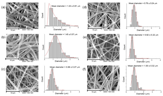
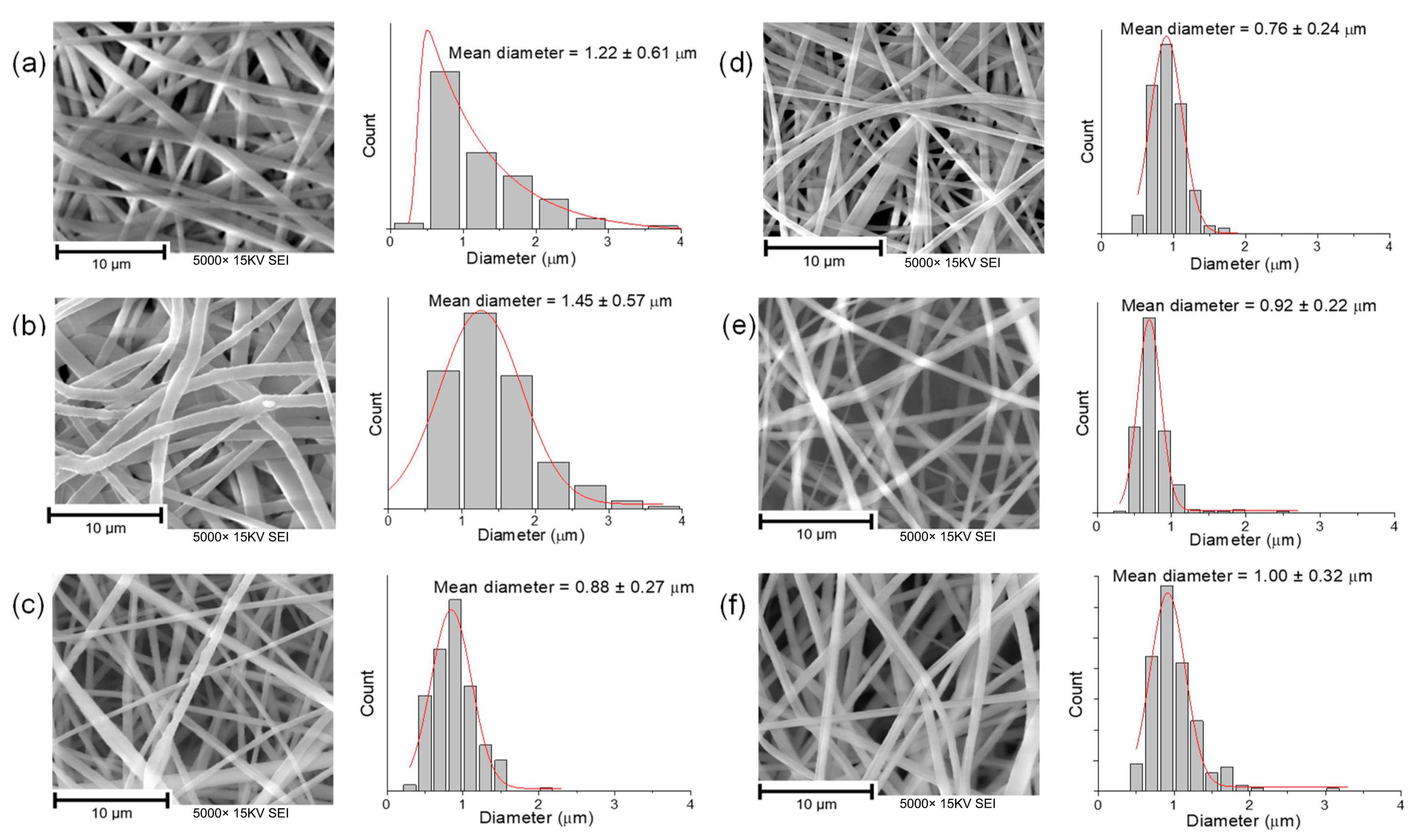
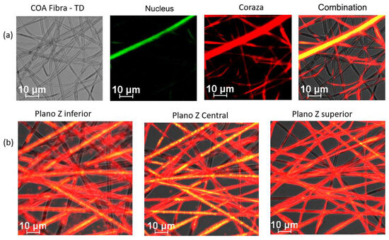
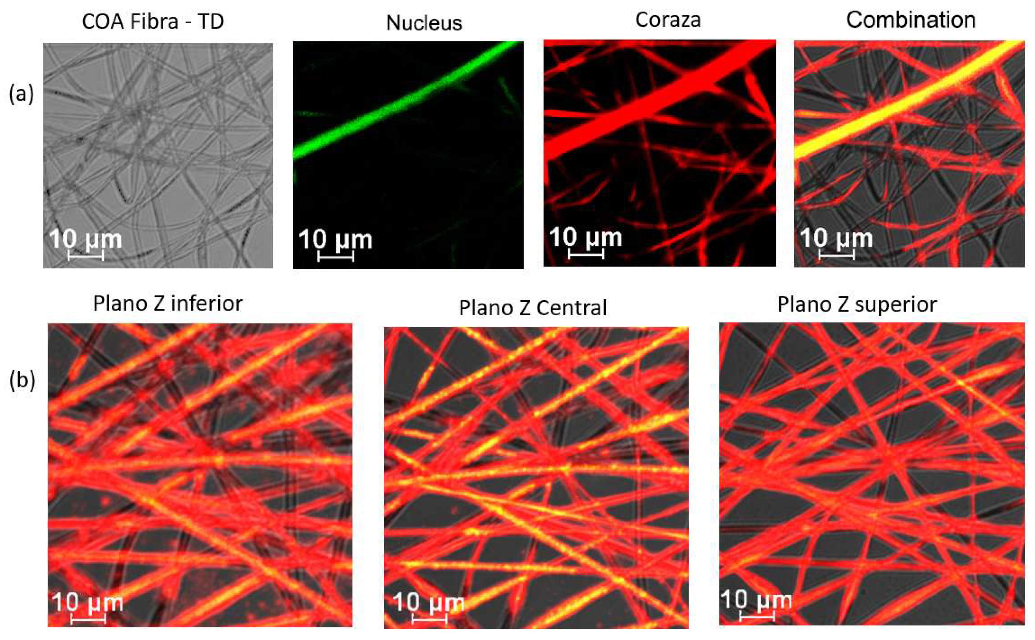
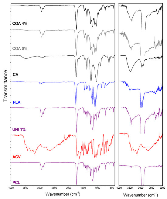
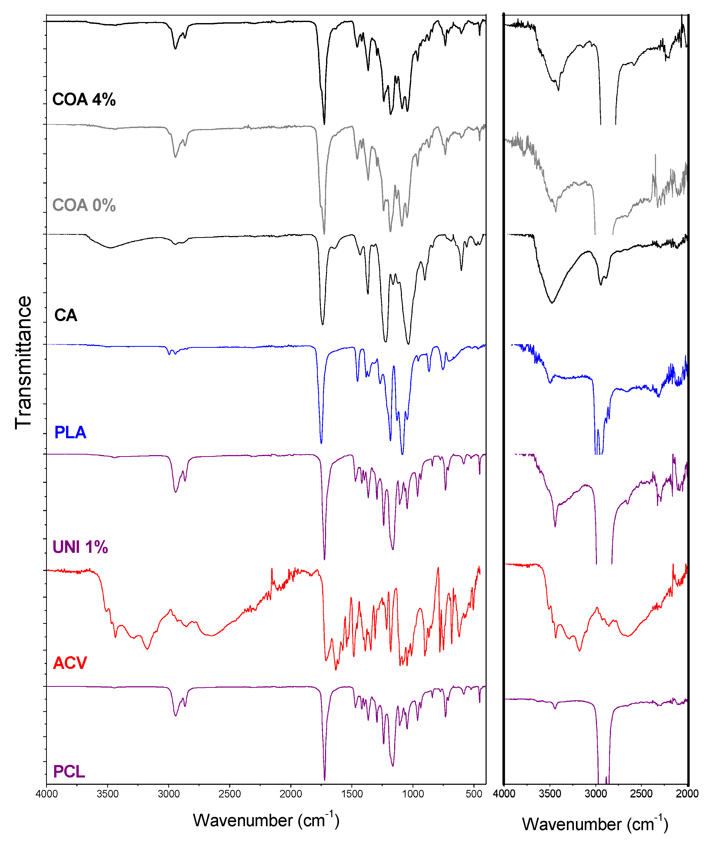
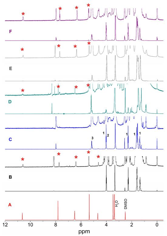
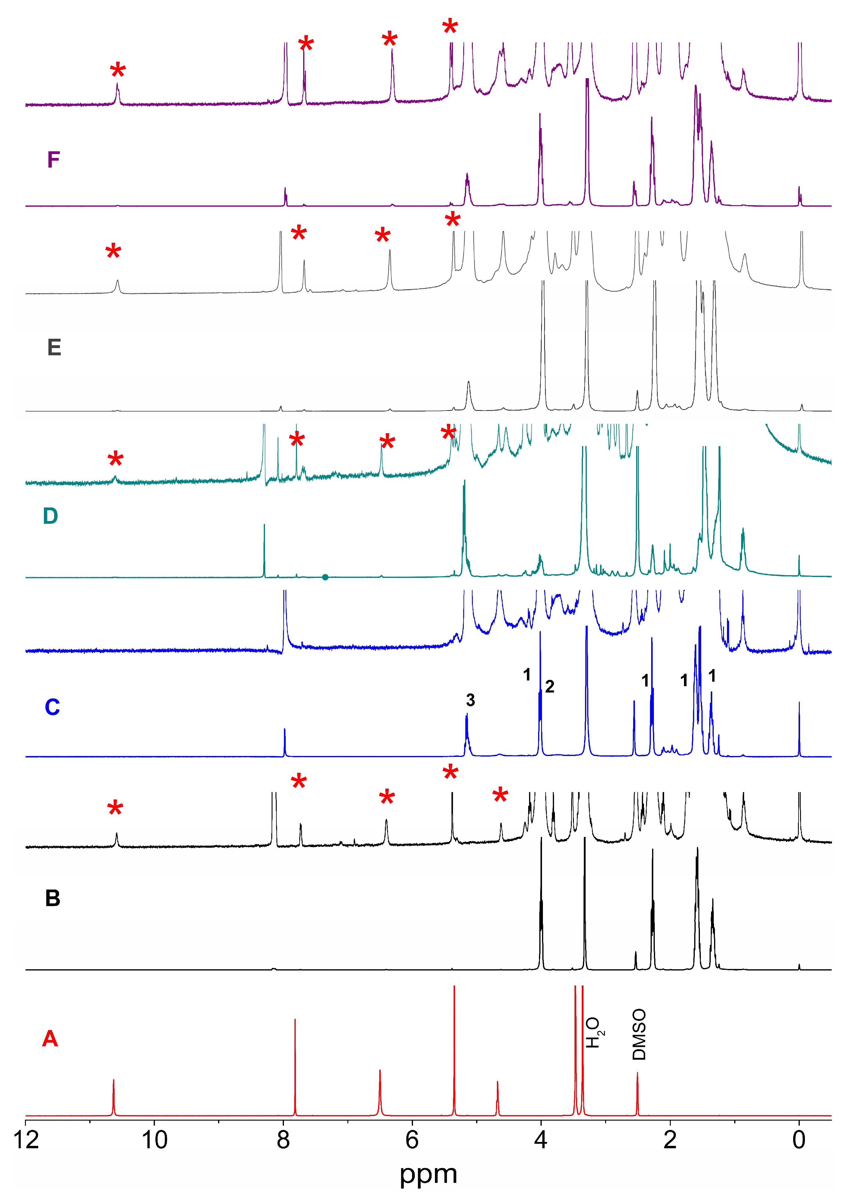
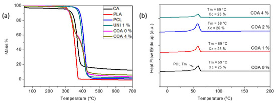
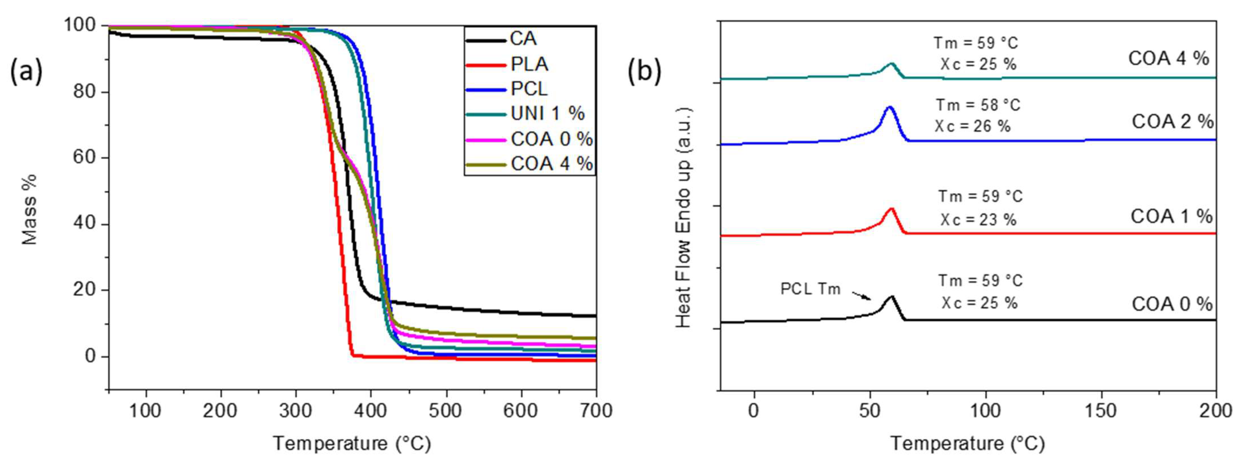
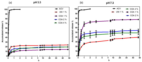
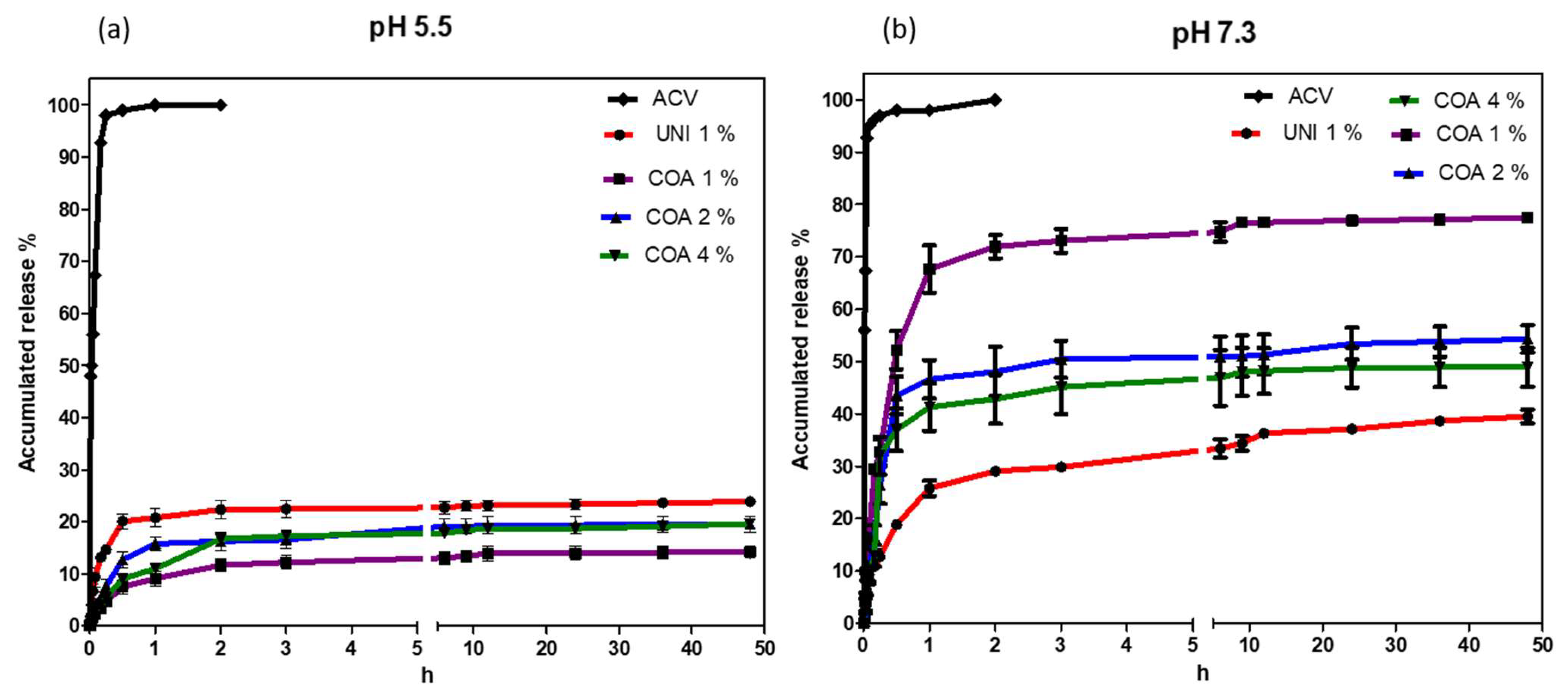
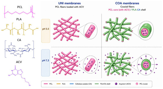
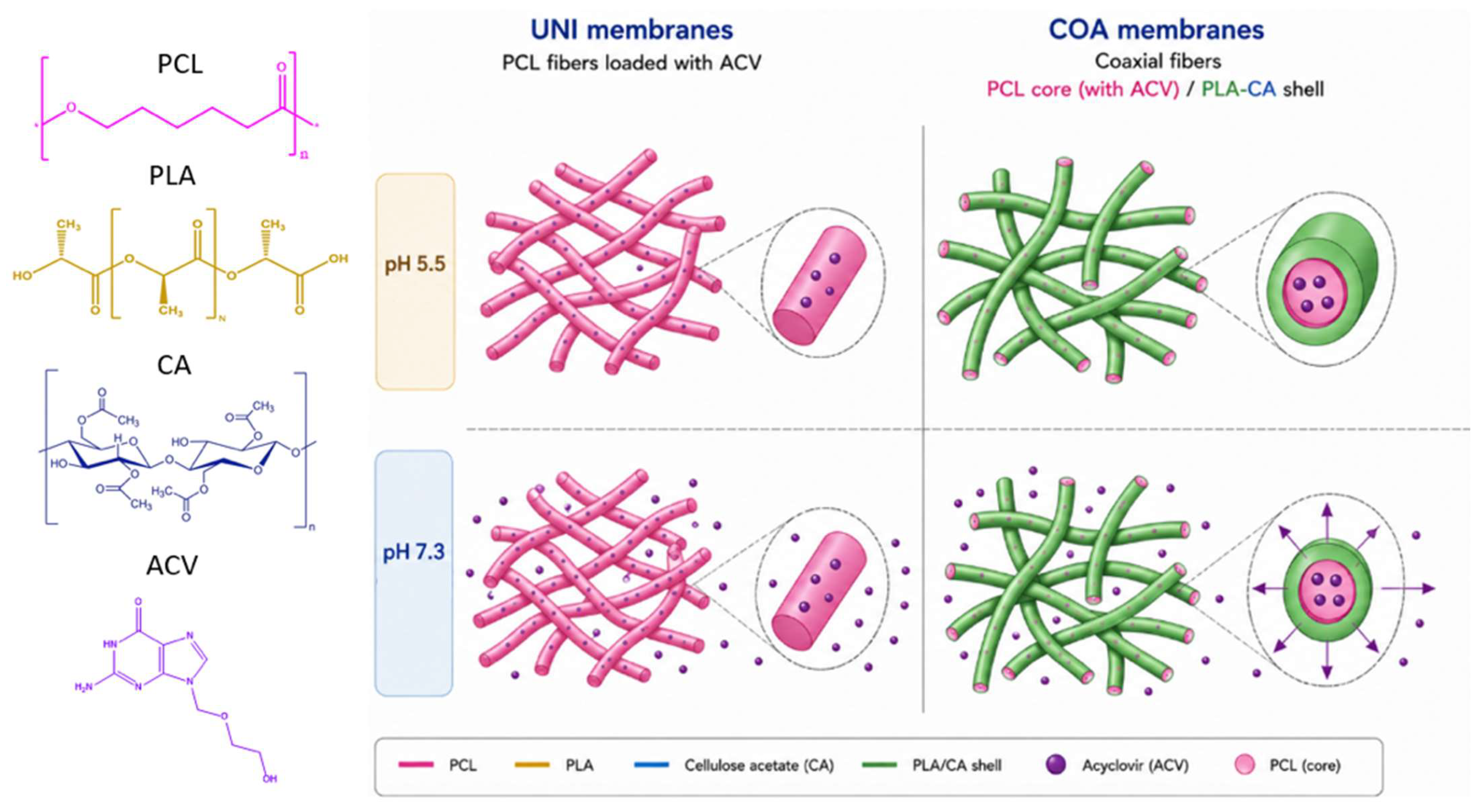
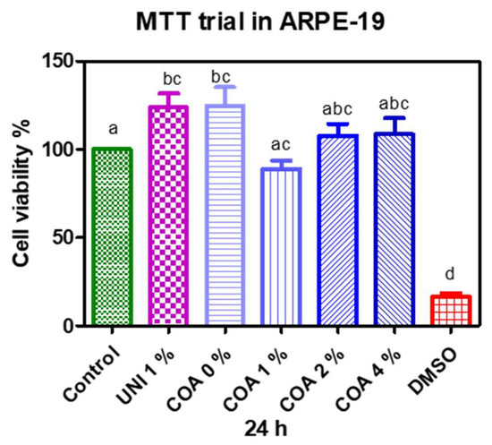
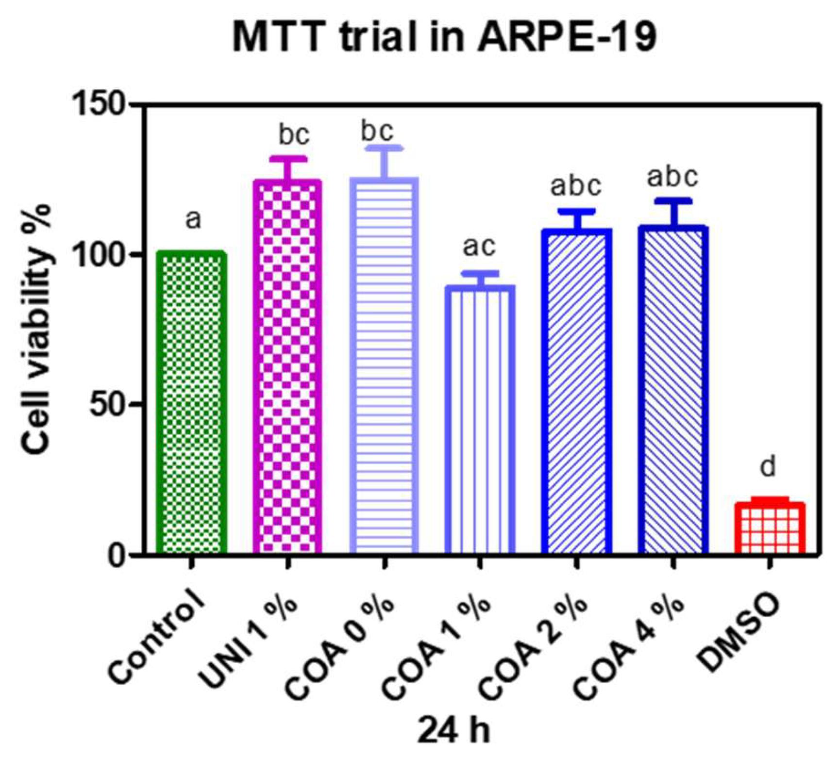

Disclaimer/Publisher’s Note: The statements, opinions and data contained in all publications are solely those of the individual author(s) and contributor(s) and not of MDPI and/or the editor(s). MDPI and/or the editor(s) disclaim responsibility for any injury to people or property resulting from any ideas, methods, instructions or products referred to in the content. |
© 2026 by the authors. Licensee MDPI, Basel, Switzerland. This article is an open access article distributed under the terms and conditions of the Creative Commons Attribution (CC BY) license.