


Abstract
The development of sustainable biopolymer-based active packaging materials is essential to replace single-use petroleum-derived plastics and reduce food deterioration. In this study, chitosan-based films incorporating green-synthesized silver microparticles (Ag microparticles) obtained from pecan nutshell extract rich in phenolic compounds were developed as multifunctional materials with antioxidant and antibacterial properties. Films were prepared by the casting method using chitosan solutions at different concentrations (1.5–2.5% w/v), with Ag microparticles incorporated at 0.25% (w/v). The phenolic profile of the extract (gallic acid, catechin, ferulic acid, and ellagic acid), determined by UPLC-DAD, confirmed its role as a reducing and stabilizing agent during Ag microparticle synthesis. All film-forming solutions exhibited non-Newtonian pseudoplastic behavior, and variations in viscosity and consistency were directly reflected in the mechanical behavior of the films. Strong antioxidant activity, mainly governed by single-electron transfer mechanisms, was observed in ABTS, DPPH, and FRAP assays. The films also showed pronounced antibacterial activity, achieving complete inhibition of Listeria monocytogenes. Finally, it is concluded that film mechanical properties are strongly governed by the rheological behavior of the chitosan-based film-forming solutions. The resulting chitosan-Ag microparticle films combine suitable mechanical behavior with antioxidant activity and antibacterial effects against Listeria monocytogenes, suggesting their potential for future application in active food-packaging systems.
1. Introduction
Food packaging plays a critical role in preserving food quality and extending shelf life by protecting products from physical, chemical, and microbiological deterioration [1,2,3,4]. Growing environmental concerns associated with petroleum-based plastics have stimulated the development of biodegradable packaging materials based on renewable biopolymers [5,6,7,8]. Among these materials, polysaccharides have attracted considerable attention due to their availability, biocompatibility, non-toxicity, and excellent film-forming properties, making them promising candidates for sustainable food packaging applications [9,10].
Among polysaccharides, chitosan has emerged as one of the most promising biopolymers for active food packaging applications due to its biodegradability, biocompatibility, non-toxicity, and excellent film-forming ability [11,12,13].
Several studies have demonstrated that the incorporation of metallic particles into chitosan films enhances their functional properties compared to neat chitosan films [14,15]. In particular, silver particles (Ag particles) have been shown to improve antimicrobial activity against a broad spectrum of microorganisms. Moreover, Ag particles can reinforce the polymer matrix, leading to improvements in mechanical strength and barrier performance through modifications in film microstructure and polymer–particle interactions [15,16,17].
The functional properties of chitosan–silver composites strongly depend on the characteristics of the incorporated silver particles, which are influenced by the synthesis method employed. Conventional chemical and physical synthesis methods are often expensive and involve toxic reagents that may leave harmful residues on the particle surface [18]. In contrast, green synthesis using biological resources such as plant extracts has emerged as an eco-friendly and cost-effective alternative. In these systems, naturally occurring biomolecules, including flavonoids, phenolic acids, and tannins, act as reducing and stabilizing agents, promoting particle formation while enhancing stability, biocompatibility, and antioxidant activity [19,20,21,22].
Among the different biological sources employed for green synthesis, pecan nutshell represents an abundant agro-industrial by-product rich in phenolic compounds such as gallic acid, catechin, and tannins, which can effectively participate in the reduction and stabilization of silver particles [23,24,25,26]. The incorporation of these biofunctionalized particles into polymeric matrices represents a promising strategy for developing active packaging materials that combine the antibacterial and antioxidant properties of silver with the stability and biocompatibility of chitosan-based films [27,28].
Polymer–particle interactions can significantly alter the mechanical behavior of active packaging materials. The intermolecular interactions that govern the structural organization of films originate at the solution stage. Therefore, the analysis of the rheological properties of film-forming solutions provides valuable information about these interactions and constitutes a predictive and non-destructive tool for assessing the mechanical behavior of the resulting films [29].
Despite the large number of studies focused on the development and characterization of chitosan–silver composite films, most investigations have primarily evaluated the properties of the final materials, including antimicrobial activity, antioxidant capacity, barrier performance, and mechanical behavior [11,30,31,32,33]. However, limited information is available regarding the relationship between the rheological properties of film-forming solutions and the mechanical performance of films containing silver particles. Furthermore, studies employing green-synthesized Ag microparticles obtained from pecan nutshell extract remain scarce. Therefore, the novelty of the present work lies in evaluating the relationship between the rheological properties of chitosan film-forming solutions and the mechanical properties of films containing green-synthesized Ag microparticles produced using an aqueous pecan nutshell extract as a reducing agent. In addition, the antioxidant and antibacterial activities of the resulting films were evaluated to assess their potential application as active food packaging materials.
Therefore, the aim of the present study was to develop and characterize chitosan films incorporated with Ag microparticles obtained through green synthesis using an aqueous pecan nutshell extract as a reducing agent. Particular emphasis was placed on evaluating the relationship between the rheological properties of film-forming solutions and the mechanical properties of the resulting films. Additionally, the antioxidant and antibacterial activities of the films were assessed to determine their potential application as active food packaging materials.
2. Materials and Methods
2.1. Materials
The reagents used in this study were medium molecular weight chitosan (190,000–310,000 Da) with a degree of deacetylation of 75–85%, silver nitrate (AgNO3) (99%), glacial acetic acid (99.94%), 2,2′-azinobis(3-ethylbenzothiazoline-6-sulfonic acid) (ABTS), 2,2-diphenyl-1-picrylhydrazyl (DPPH), 6-hydroxy-2,5,7,8-tetramethylchroman-2-carboxylic acid (Trolox), potassium persulfate (K2S2O8), iron(III) chloride hexahydrate (FeCl36H2O), 2,4,6-tris(2-pyridyl)-s-triazine (TPTZ), and sodium acetate (C2H3NaO2). All reagents were purchased from Sigma-Aldrich (St. Louis, MO, USA). Pecan nuts (Carya illinoinensis) were purchased from a local market; the shells were separated and pulverized using a blender to obtain the aqueous extract.
2.2. Green Synthesis of Ag Microparticles
2.2.1. Preparation of Pecan Nutshell Extract
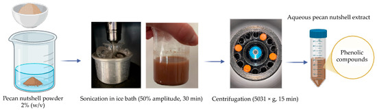
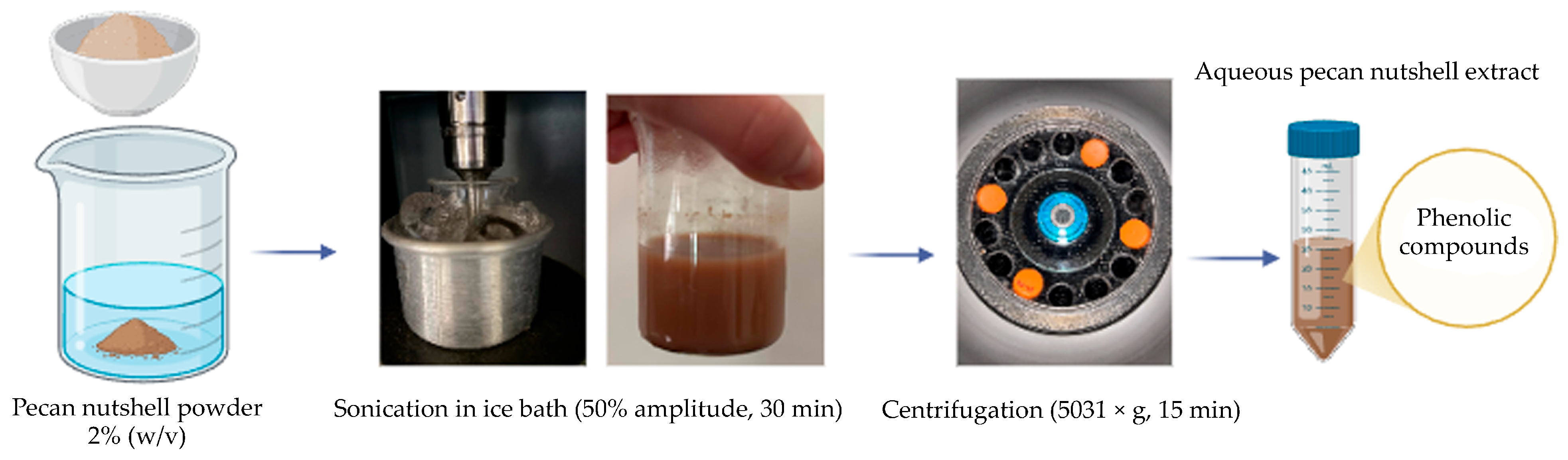
The pecan nuthsell extract was prepared following the methodology described by Ozuna-Valencia et al. (2025) [26]. 1 g of pulverized pecan nutshell was suspended in 50 mL of deionized water and subjected to sonication in an ice bath at 50% amplitude for 25 min using a digital probe sonicator (Sonifier® S-450D, 20 kHz, 400 W, Branson Ultrasounds TM, Danbury, CT, USA). The resulting solution was then centrifuged at 5031× g for 15 min at 4 °C. The supernatant was collected and stored under refrigeration for subsequent use in the synthesis of Ag microparticles (Figure 1).
Figure 1.
Schematic representation of the preparation of aqueous pecan nutshell extract by ultrasonic-assisted extraction followed by centrifugation to obtain a phenolic-rich extract used for the green synthesis of Ag microparticles.
2.2.2. Identification and Quantification of Individual Phenolic Compounds in the Pecan Nutshell Extract
Phenolic compounds (PCs) were extracted from 2 g of pulverized pecan nutshell with 10 mL of 80% (v/v) methanol. The suspension was stirred for 10 min, sonicated in an ice bath for 30 min, and centrifuged at 10,000× g for 15 min at 25 °C. Free PCs were analyzed in the supernatant, while the pellet was subjected to sequential acidic and alkaline hydrolysis to quantify chemically bound PCs. For acidic hydrolysis, 5 mL of 37% (v/v) HCl were added and the sample incubated at 85 °C for 3 h, stirred for 4 h, and the pH adjusted to 7.0 using 2 M NaOH.
Alkaline hydrolysis was performed by adding 10 mL of 2 M NaOH, purging the solution with nitrogen (N2) for 15 s to prevent oxidation, and stirring at 100 rpm for 4 h under dark conditions at 25 °C; the pH was then adjusted to 1.5–2.0 with HCl. The hydrolyzed sample was extracted twice with 15 mL of 50:50 (v/v) ethyl acetate/diethyl ether, and the combined organic phases were concentrated by rotary evaporation at 45 °C, resuspended in 5 mL of 80% methanol, and filtered through a 25 µm filter.
PCs were analyzed using an ultra-performance liquid chromatography (UPLC) system equipped with a diode array detector (DAD) (ACQUITY, Waters Corp, Milford, MA, USA). Separation was achieved on a BEH C18 column (1.7 µm, 3.0 × 100 mm) maintained at 60 °C. The mobile phase consisted of water containing 0.5% (v/v) formic acid (solvent A) and methanol (solvent B). Elution was performed using a gradient program expressed as percentage of solvent B (v/v) as follows: 0–0.25 min, 20% B (flow rate 0.4 mL/min); 5 min, 20% B (0.2 mL/min); 12 min, 45% B (0.18 mL/min); 25 min, 100% B (0.1 mL/min); 26 min, 40% B (0.2 mL/min); and 30 min, 20% B (0.4 mL/min).
PCs were identified by retention time and UV–Vis spectra compared to commercial standards, and quantified using external calibration curves, expressed as concentration in the extract [34].
2.2.3. Synthesis of Ag Microparticles
The synthesis of Ag microparticles was performed following the methodology described by Ozuna-Valencia et al. (2025) [26]. A 1:1 mixture was prepared by combining the pecan nutshell extract with an AgNO3 precursor solution. The resulting suspension was continuously stirred for 4 h at room temperature in the dark to prevent the degradation of bioactive compounds. The homogenized solution was then subjected to three washing cycles with distilled water by centrifugation at 11,180× g for 15 min at 20 °C. After each cycle, the supernatant was discarded, and the resulting precipitate was redispersed using an ultrasonic bath for 60 min. Finally, the material was dried in a convection oven (Yamato Scientific Co., Ltd. ADP310C, Tokyo, Japan) at 50 °C for 48 h to remove residual moisture.
2.3. Characterization of Ag Microparticles
2.3.1. Scanning Electron Microscopy (SEM)
The morphology of the Ag microparticles was analyzed using a scanning electron microscope (SEM) (JEOL JSM-5410LV, JEOL Ltd., Tokyo, Japan) operated at an accelerating voltage of 20 kV. The samples were mounted on aluminum stubs using double-sided carbon tape and coated with a thin gold layer to ensure electrical conductivity prior to morphological characterization. Elemental analysis of the Ag microparticles was performed by energy-dispersive X-ray spectroscopy (EDX) using a detector coupled to the microscope [35].
2.3.2. Dynamic Light Scattering (DLS) and Z-Potential Measurements
The hydrodynamic size and Z-potential of Ag microparticles were determined by Zetasizer Nano instrument (Malvern Panalytical, Model Nano-ZS90). For this purpose, Ag microparticles were dispersed in ultrapure water at a concentration of 1 mg/mL, and 3 mL of the resulting suspension were used for each measurement. The zeta potential was determined by electrophoretic light scattering, and the values were calculated using the Smoluchowski equation [36].
2.4. Evaluation of the Antioxidant Activity of Ag Microparticles
The antioxidant activity of Ag microparticles at a concentration of 0.25% (w/v) was evaluated to determine their potential as active agents for food packaging applications. The ABTS, DPPH, and FRAP assays were performed according to the methodology reported by García-Lárez et al. (2025) [37] with minor modifications.
2.4.1. ABTS Radical Scavenging Capacity
The ABTS radical was generated by mixing ABTS with 1.5 mL of potassium persulfate (K2S2O8) and allowing the mixture to react for 12–16 h in the dark. Subsequently, the solution was adjusted with ethanol to an absorbance of 0.70 ± 0.01 at 734 nm.
Then, 20 µL of a 0.25% (w/v) Ag microparticle suspension were added to a 96-well microplate containing 270 µL of the ABTS solution. The mixture was incubated for 30 min at 25 °C in the dark, and the absorbance was measured at 734 nm. Results were expressed as percentage inhibition (%) and micromoles of Trolox equivalents per gram of sample (µM TE/g).
2.4.2. DPPH Radical Scavenging Capacity
The DPPH radical was prepared in methanol, and the solution was adjusted to an absorbance of 0.70 ± 0.01 at 515 nm. Then, 20 µL of a 0.25% (w/v) Ag microparticle suspension were added to a 96-well microplate containing 200 µL of the DPPH solution. The mixture was incubated for 30 min at 15 °C in the dark, and the absorbance was measured at 515 nm. Results were expressed as percentage inhibition (%) and micromoles of Trolox equivalents per gram of sample (µM TE/g).
2.4.3. Ferric Reducing Antioxidant Power (FRAP)
The FRAP assay is based on the reduction of Fe3+ in the presence of 2,4,6-tripyridyl-s-triazine (TPTZ). The FRAP reagent was prepared by mixing acetate buffer (300 µM, pH 3.6), TPTZ (10 µM in 40 µM HCl), and FeCl3. Then, 20 µL of a Ag microparticle suspension at a concentration of 0.25% (w/v) were added to 280 µL of the FRAP solution in a 96-well microplate. After incubation for 30 min at 15 °C in the dark, the absorbance was measured at 638 nm using a microplate reader. The results were expressed as micromoles of Trolox equivalents per gram of sample (µM TE/g).
2.5. Antibacterial Activity
The bacterial growth inhibition of Ag microparticles was evaluated using a liquid diffusion method, with Mueller–Hinton broth as the culture medium. The antibacterial activity of the samples was tested against the Gram-positive bacterium Listeria monocytogenes (ATCC 7644).
A single colony from the bacterial culture was selected, and the turbidity of the suspension was adjusted to the 0.5 McFarland standard (equivalent to 1.5 × 10⁸ CFU/mL). The culture medium was prepared by dissolving 1.28 g of Mueller–Hinton broth in 160 mL of ultrapure water and subsequently sterilized by autoclaving at 121 °C for 15 min. After cooling to room temperature, aliquots of the sterile medium were dispensed into test tubes, to which 100 µL of the bacterial inoculum and 20 µL of Ag microparticle suspension (0.25% w/v) were added in triplicate. The tubes were incubated at 37 °C for 24 h, and bacterial growth was evaluated by measuring the absorbance at 600 nm.
The percentage of L. monocytogenes growth inhibition was calculated according to Moreno-Vásquez et al. (2017) [38] using the following equation:
where represents the absorbance of the culture medium containing the sample and the bacterial inoculum; and correspond to the initial absorbance of the sterilized culture medium; represents the absorbance of the sterile culture medium containing the sample without inoculum; and finally, represents the absorbance of the sterilized culture medium containing the bacterial inoculum (growth control).
2.6. Preparation of Chitosan Films with Ag Particles
Chitosan and chitosan-Ag microparticle films were prepared by the casting method according to the procedure reported by Ozuna-Valencia et al. (2025) [26], with minor modifications. Polymeric solutions at different concentrations (1.5, 2.0, and 2.5% w/v) were prepared by dissolving chitosan in a 2% v/v acetic acid solution. The mixtures were stirred for 24 h to ensure complete polymer dissolution. Subsequently, 16 mL of each solution were cast onto plastic Petri dishes with a diameter of 8.5 cm and dried in a convection oven at 50 °C for 48 h to allow controlled solvent evaporation and film formation.
For chitosan films containing Ag microparticles, 0.25% (w/v) of the previously dried microparticles was suspended in a volume corresponding to 50% of the total distilled water required (50 mL). This suspension was subjected to sonication at 50% amplitude for 45 min (three cycles of 15 min) in ice-cooled containers using a probe-type ultrasonic processor to ensure adequate microparticle dispersion. Subsequently, chitosan solutions at different concentrations (1.5, 2.0, and 2.5% w/v), prepared in 2% (v/v) acetic acid using the remaining 50 mL of distilled water, were added to the Ag microparticle suspension and stirred until homogeneous solutions were obtained. The final mixture was stirred under the same conditions previously described and then cast onto Petri dishes for drying in a convection oven at 50 °C for 48 h, following the same protocol used for films without microparticles. The Ag microparticle content (0.25% w/v) was selected based on our previous study using pecan nutshell extract-mediated green synthesis, which demonstrated that higher microparticle contents promoted aggregation and adversely affected film homogeneity [26]. Therefore, this concentration was employed to minimize aggregation effects while allowing the evaluation of the influence of chitosan concentration on the rheological and mechanical properties of the films. The resulting formulations are summarized in Table 1.
Table 1.
Composition of chitosan and chitosan-Ag microparticle films.
2.7. Flow Behavior and Rheological Properties of Film-Forming Solutions
The rheological behavior of the chitosan-Ag microparticle film-forming solutions was analyzed using an MCR-102 rheometer (Anton Paar, Ostfildern, Germany) equipped with a CC27 concentric cylinder geometry with a 1 mm gap. For each measurement, 19 mL of film-forming solutions were loaded into the measuring cell. Measurements were performed in controlled shear rate (CSR) mode by recording shear stress as a function of shear rate under a continuous shear-rate ramp from 0.1 to 100 s−1 at a constant temperature of 25 °C. A total of 100 data points were collected at 2 s intervals, resulting in a total measurement time of 200 s per sample. All measurements were carried out in triplicate.
The experimental flow curves were fitted to the Power Law model using the following equation:
where τ is the shear stress (Pa), K is the consistency index (Pa·sn), γ is the shear rate (s−1), and n is the flow behavior index. According to the Power Law model, solutions with n = 1 exhibit Newtonian behavior, whereas solutions with n < 1 and n > 1 exhibit pseudoplastic (shear-thinning) and dilatant (shear-thickening) non-Newtonian behavior, respectively. The model parameters were used to determine the flow behavior of the film-forming solutions. Determining the flow behavior of these systems is essential for linking their rheological properties to the mechanical performance of the resulting films [39].
τ = Kγn
2.8. Characterization of Chitosan-Ag Microparticles Films
2.8.1. Film Thickness
The thickness of chitosan and chitosan-Ag microparticle films was determined according to the ASTM D1708-18 standard [40], using a micrometer (C112XBS, Mitutoyo Corporation, Kawasaki, Japan). For each formulation, three independent films were analyzed, with ten random thickness measurements taken per film. The results were reported as mean ± standard deviation.
2.8.2. Mechanical Properties
Tensile strength, elongation at break, and Young’s modulus of chitosan and chitosan-Ag microparticle films were determined from stress–strain curves using a TA.XT.plus universal testing machine (Texture Analyzer, Stable Micro Systems, Surrey, UK).
The instrument was configured with an initial grip separation of 30 mm, a crosshead speed of 5 mm/s, an applied force of 0.1 N, and a sensitivity of 0.1 N. Data acquisition and analysis were performed using Exponent software (Stable Micro Systems Ltd., Godalming, Surrey, UK). Prior to mechanical testing, strips (10 mm × 5 mm) were cut from the films and conditioned for 72 h at a relative humidity of 57 ± 2% at 25 °C. For each formulation, three independent films were analyzed, and four strips were obtained from each film [41].
2.8.3. Fourier Transform Infrared (FTIR)
The structural analysis of chitosan and chitosan-Ag microparticle films was performed using a Subtech Spectrum FTIR spectrophotometer (PerkinElmer, Waltham, MA, USA) equipped with an attenuated total reflectance (ATR) accessory with a diamond crystal as the contact element. Spectra were recorded over the range of 4000–400 cm−1, accumulating 16 scans per spectrum [26].
2.8.4. Scanning Electron Microscopy (SEM)
The surface morphology of chitosan and chitosan-Ag microparticle films was analyzed to evaluate the dispersion of the active agent within the polymer matrix and its influence on surface homogeneity. Morphological characterization was carried out using scanning electron microscopy (SEM). Prior to analysis, the samples were mounted on aluminum stubs using double-sided conductive carbon tape and coated with a thin gold layer to ensure adequate electrical conductivity [26].
2.9. Determination of the Antioxidant Activity of Chitosan-Ag Microparticle Films
The antioxidant activity of chitosan-Ag microparticle films was evaluated using a direct contact method with ABTS, DPPH, and FRAP radicals, following the methodology reported by Acuña-Pacheco et al. (2024) [42]. The percentage inhibition (%) for the ABTS and DPPH assays was calculated using the following equation:
2.9.1. ABTS Radical Scavenging Capacity
The ABTS radical was prepared and adjusted according to the methodology previously described in Section 2.4.1. Subsequently, 48 mg of the Ch-2.0Ag film was placed in an amber glass container containing 10 mL of the ABTS solution and incubated at 25 °C in the dark for 30 min. After incubation, the absorbance was measured at 734 nm. The results were expressed as percentage inhibition (%) and micromoles of Trolox equivalents per gram of sample (µM TE/g).
2.9.2. DPPH Radical Scavenging Capacity
The DPPH radical was prepared and adjusted according to the methodology previously described in Section 2.4.2. Subsequently, 48 mg of the Ch-2.0Ag film was placed in an amber glass container containing 10 mL of the DPPH solution and incubated at 25 °C in the dark for 30 min. After incubation, the absorbance was measured at 515 nm. The results were expressed as percentage inhibition (%) and micromoles of Trolox equivalents per gram of sample (µM TE/g).
2.9.3. Ferric Reducing Antioxidant Power (FRAP)
The FRAP reagent was prepared according to the methodology previously described in Section 2.4.3. Subsequently, 48 mg of the Ch-2.0Ag film was placed in an amber glass container containing 10 mL of the FRAP solution and incubated at 25 °C in the dark for 30 min. After incubation, the absorbance was measured at 638 nm. The results were expressed as micromoles of Trolox equivalents per gram of sample (µM TE/g).
2.10. Antibacterial Activity
The bacterial growth inhibition of chitosan and chitosan-Ag microparticle films was evaluated using a liquid diffusion method, with Mueller–Hinton broth as the culture medium. The antibacterial activity of the samples was tested against Listeria monocytogenes (ATCC 7644).
A total of 10 mg of Ch-2.0 Ag films was cut into small pieces and added to sterile Mueller–Hinton broth. Subsequently, 100 µL of a Listeria monocytogenes inoculum adjusted to the 0.5 McFarland standard (1.5 × 10⁸ CFU/mL) were added. The cultures were incubated at 37 °C for 24 h, and bacterial growth was monitored by measuring the absorbance at 600 nm. The percentage of Listeria monocytogenes growth inhibition was calculated according to Moreno-Vásquez et al. (2017) [38], using the same equation previously described and considering the corresponding growth and sample blanks.
2.11. Statistical Analysis
The preparation of chitosan films incorporating Ag microparticles as the active agent was carried out following a 3 × 2 factorial experimental design. The first factor was the chitosan concentration, with three levels: 1.5%, 2.0%, and 2.5% (w/v); the second factor corresponded to the presence or absence of Ag microparticles at 0.25% (w/v).
The response variables evaluated included antibacterial activity, antioxidant activity, mechanical properties, and film thickness, which were analyzed in three independent replicates per treatment. Additionally, the chromatographic analysis of the pecan nutshell extract was performed in duplicate. All results are expressed as mean ± standard deviation.
Statistical analysis was conducted using analysis of variance (ANOVA), and significant differences among treatments were determined using Tukey’s multiple comparison test at a confidence level of p < 0.05, employing InfoStat software, version 2020.
3. Results and Discussion
3.1. Chromatographic Analysis
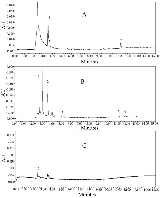
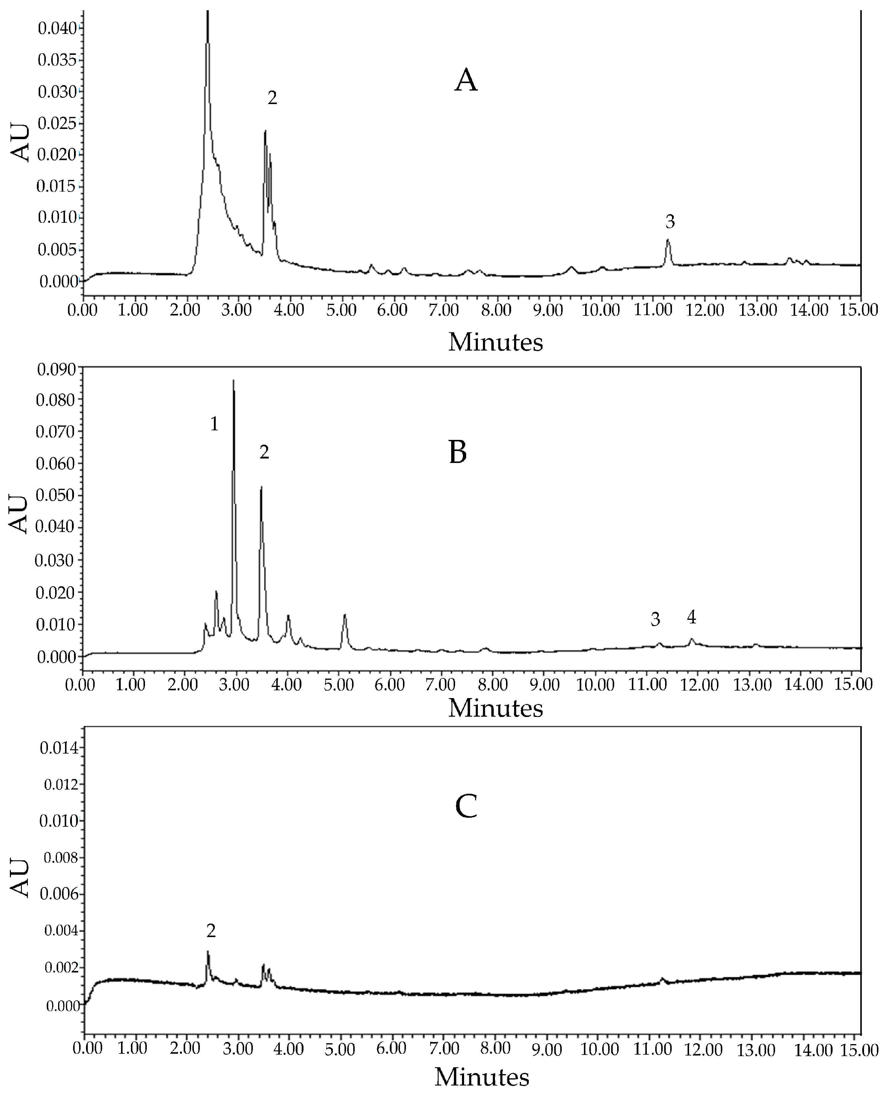
A detailed chromatographic analysis of the pecan nutshell extract was carried out (Figure 2). The chromatograms allowed the qualitative and quantitative identification of four PCs: gallic acid, catechin, ferulic acid, and ellagic acid. Table 2 shows the corresponding retention times, the fraction (free, acidic, or alkaline) in which each compound was detected, and their concentrations (mg/g). Some compounds showed signals that could be distinguished by comparing their absorbance spectra and retention times with those reported in the literature; however, they were not detected or quantified by the system (NQ, not quantified).
Figure 2.
Representative UPLC-DAD chromatograms used for the identification and quantification of PCs present in the pecan nutshell extract: (1) gallic acid, (2) catechin, (3) ferulic acid, and (4) ellagic acid, in three fractions: free compounds in the extract (A), alkaline hydrolysis (B), and acidic hydrolysis (C).
Table 2.
PCs present in pecan nutshell extract.
Ferulic acid was the compound present at the highest concentration, with a value of 9.01 ± 0.28 mg/g in the pecan nutshell extract. This compound is widely recognized for its remarkable antioxidant activity, which is attributed to the presence of an unsaturated side chain that facilitates electron delocalization. This structural feature enables the formation of a resonance-stabilized phenoxy radical, therefore significantly contributing to its ability to neutralize reactive oxygen species [43,44].
Catechin was the most frequently detected compound, as it was identified in the three fractions; however, it was only detected and quantified in the alkaline hydrolysis fraction (Figure 2B). According to Demir et al. (2025) [45], catechin is one of the main PCs involved in the reduction of silver ions, which makes it a fundamental reducing agent in the synthesis of Ag microparticles. This reducing capability is attributed to the presence of multiple hydroxyl groups in its structure, which facilitate electron donation due to the high electron density on the oxygen atoms of the O–H bonds. In addition, it has been reported that PCs such as catechin and gallic acid not only participate in the reduction process but also act as stabilizing agents, contributing to the morphological and dimensional control of Ag microparticles during their formation [46,47].
3.2. Characterization of Ag Microparticles
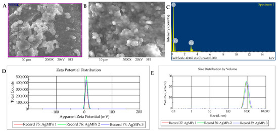
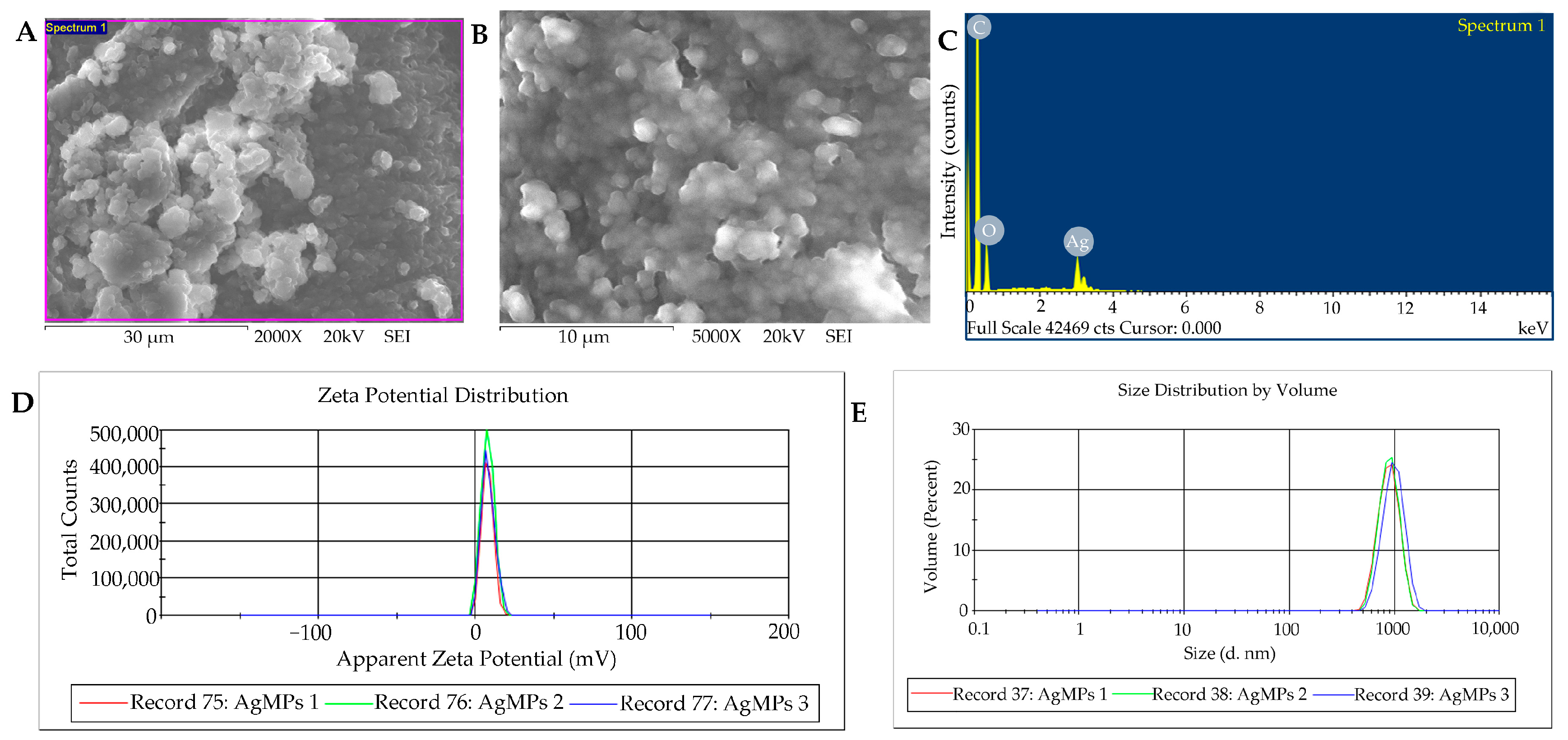
SEM analysis revealed that the synthesized Ag microparticles exhibited a strong tendency to form agglomerates, as observed at 2000× magnification (Figure 3A), while individual microparticles displayed spherical and quasi-spherical morphologies at higher magnification (5000×, Figure 3B). Due to the extensive aggregation and overlap of individual particles, a reliable and statistically representative particle-size distribution could not be obtained directly from the SEM micrographs. This observation is consistent with our previous findings, in which Ag microparticles synthesized using pecan nutshell extract also exhibited a marked tendency to aggregate [26]. The EDX spectrum (Figure 3C) confirmed the presence of carbon and oxygen, associated with PCs from the pecan nutshell extract, which remain adsorbed on the particle surface and are responsible for their antioxidant and antibacterial potential [48].
Figure 3.
Characterization of Ag microparticles: scanning electron microscopy (SEM) images at 2000× (A) and 5000× (B); EDX spectrum of the Ag microparticles (C); zeta potential distribution of Ag microparticles at 1 mg/mL (D); and hydrodynamic size distribution of Ag microparticles in solution (E).
The colloidal stability of the Ag microparticles was evaluated by zeta potential measurements, yielding a low value of 8.28 ± 0.17 mV (Figure 3D), which indicates weak electrostatic repulsion and, consequently, a high propensity for aggregation, since absolute ζ-potential values above ±30 mV are typically required for stable colloidal systems [49,50]. Consistently, DLS analysis (Figure 3E) revealed a large submicron hydrodynamic diameter (100 nm < d < 1 µm) of 896 ± 52 nm (n = 3), with a PDI of 0.59 ± 0.16, indicating a moderately polydisperse system dominated by agglomerated structures [51].
These results are in agreement with previous reports on Ag microparticles synthesized using pecan nutshell extract, where Argenziano et al. (2023) [52] obtained much smaller hydrodynamic sizes (20–150 nm) when employing a low AgNO3 concentration (0.01 M), whereas Ozuna-Valencia et al. (2025) [26] reported significantly larger microparticles (545–1043 nm) when using a higher precursor concentration (0.3 M), similar to that applied in the present study. This comparison supports that increasing the AgNO3 concentration leads to higher supersaturation and insufficient surface passivation, promoting particle growth and aggregation by coalescence, and thus resulting in larger hydrodynamic diameters.
In addition, the presence of PCs in the pecan nutshell extract (previously detected by UPLC), containing multiple hydroxyl groups that act as mild reducing agents, also contributed to the formation of larger Ag particles [53]. Although smaller Ag microparticles enhance their antioxidant and antibacterial activity due to their higher specific surface area, which promotes greater Ag+ release and more efficient radical scavenging, it also entails a higher toxicological risk, which is particularly relevant when this active agent is intended for use in food-contact active packaging applications [54,55].
3.3. Evaluation of the Antioxidant Activity of Ag Microparticles
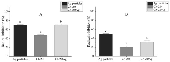
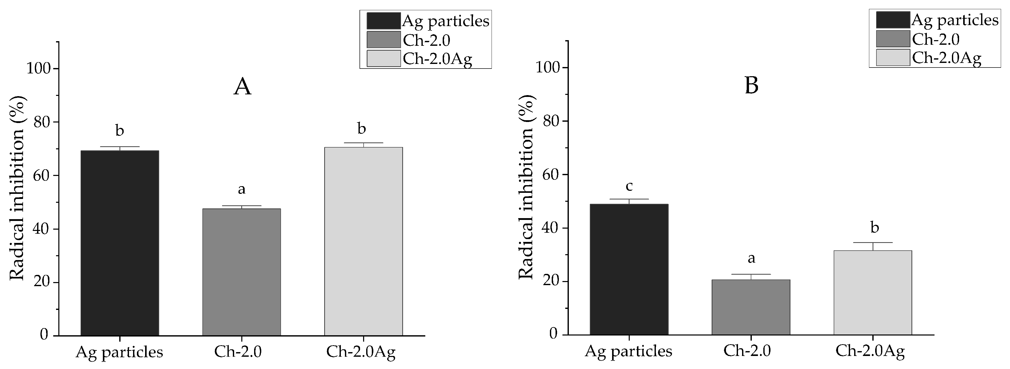
The results obtained from the evaluation of the antioxidant activity of Ag microparticles are expressed as percentage inhibition and as µM TE/g and are summarized in Table 3. The Ag microparticle solution at 0.25% (w/v) exhibited an inhibition of 69.26 ± 1.51% (141.59 ± 2.32 µM TE/g) against the ABTS radical (Figure 4A), whereas a lower inhibition of 48.86 ± 1.99% (126.43 ± 9.35 µM TE/g) was observed for the DPPH• radical (Figure 4B). Similar inhibition values have been reported by Ozuna-Valencia et al. (2025) [26], where Ag microparticles synthesized via a green route using pecan nutshell extract at a concentration of 500 µg/mL showed inhibition percentages of 79.84% for ABTS and 79.20% for DPPH. The higher scavenging efficiency toward ABTS compared to DPPH indicates that the antioxidant activity of the green-synthesized Ag microparticles is mainly governed by an electron-transfer mechanism, which can be associated with the presence of PCs from the pecan nutshell extract acting as surface capping agents. According to previous studies, ABTS neutralization predominantly proceeds via single-electron transfer (SET), whereas DPPH scavenging mainly occurs through hydrogen atom transfer (HAT) mechanisms [55,56,57].
Table 3.
Antioxidant activity of Ag microparticles, chitosan and chitosan-Ag microparticle films evaluated by ABTS, DPPH, and FRAP assays.
Figure 4.
Antioxidant activity of Ag microparticles, chitosan, and chitosan-Ag microparticle films evaluated by ABTS (A) and DPPH (B) assays. Different letters indicate statistically significant differences according to Tukey’s test (p < 0.05).
The ferric reducing antioxidant power (FRAP) of the Ag microparticles, corresponding to their ability to reduce Fe3+ ions to Fe2+, is also presented in Table 3. Antioxidant activity was confirmed by a color change in the FRAP reagent from transparent/yellow to intense blue upon sample addition and is expressed as µM TE/g. The Ag microparticles exhibited a ferric reducing activity of 281.15 ± 12.04 µmol TE/g.
Although the FRAP value obtained in this study is lower than that reported by dos Santos et al. (2025) [58] for Ag microparticles synthesized using an aqueous extract of Euterpe edulis (386.66 ± 6.50 µmol TE/g), the reducing capacity can be classified as high (100–400 µmol TE/g) according to the antioxidant power categories established by Fernandes et al. (2016) [59].
Overall, these results confirm that the green-synthesized Ag microparticles exhibit antioxidant activity, mainly governed by electron-transfer mechanisms, supporting their potential application as active agents in functional packaging systems.
3.4. Antibacterial Activity
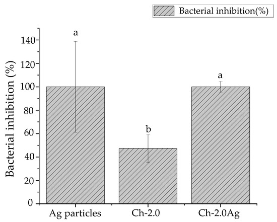
The antibacterial activity of Ag microparticles was evaluated against Listeria monocytogenes. The results demonstrated that Ag microparticles at 0.25% (w/v), synthesized using pecan nutshell extract as a reducing agent, achieved 100% bacterial inhibition after 24 h of incubation (Figure 5). L. monocytogenes possesses a cell wall mainly composed of peptidoglycan, which provides rigidity and structural protection [60]. Upon contact with Ag microparticles, an autolytic process of peptidoglycan may be induced, in which the polymer is degraded by endogenous enzymes, leading to cell wall disruption and preventing bacterial survival [61].
Figure 5.
Antibacterial activity against Listeria monocytogenes of Ag microparticles, chitosan, and chitosan-Ag microparticle films evaluated by the liquid diffusion method. Different letters indicate statistically significant differences according to Tukey’s test (p < 0.05).
Consistent with these findings, Amer et al. (2021) [62] reported that plantaricin alone did not exhibit antibacterial activity against Listeria monocytogenes, whereas its incorporation into Ag microparticles resulted in a marked inhibitory effect against this strain, highlighting the key role of Ag microparticles in enhancing antibacterial performance.
Overall, these results confirm the antibacterial effectiveness of Ag microparticles against Listeria monocytogenes under the conditions evaluated. However, additional studies involving a broader spectrum of pathogenic and spoilage microorganisms are necessary to better establish their potential application in active food-packaging systems aimed at enhancing food safety and extending shelf life.
3.5. Rheological Behavior
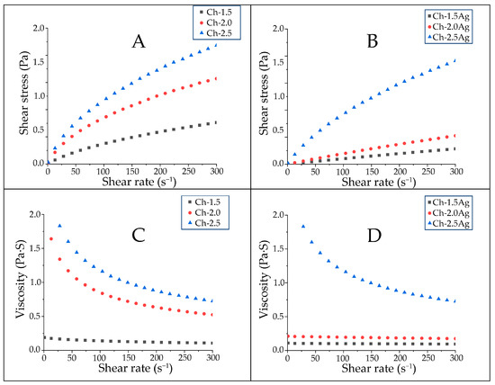
The rheological behavior of chitosan and chitosan-Ag microparticle film-forming solutions was evaluated to assess the effects of chitosan concentration and Ag microparticles incorporation. Figure 6A,B shows similar behavior for all solutions. The shear stress increased with increasing shear rate; however, this relationship was not linear, as observed for Newtonian fluids. Instead, a progressive decrease in the slope of the curves was observed, indicating a downward-concave behavior. Additionally, increasing the chitosan concentration resulted in higher shear stress values, which is attributed to restricted molecular mobility due to physicochemical interactions among chitosan chains [63]. In contrast, the incorporation of Ag microparticles led to a decrease in the shear stress of the film-forming solutions. This behavior is attributed to alterations in the chitosan microstructure, associated with modifications in the intermolecular interactions between polymer chains, which directly affect the apparent viscosity of the solutions as a function of shear rate.
Figure 6.
Shear stress–shear rate curves of the film-forming solutions of chitosan (A) and chitosan-Ag microparticles (B). Viscosity–shear rate curves of the film-forming solutions of chitosan (C) and chitosan-Ag microparticles (D).
Figure 6C,D illustrates the viscosity behavior of the film-forming solutions as a function of shear rate. A progressive decrease in apparent viscosity was observed with increasing shear rate, a phenomenon known as shear thinning, which is characteristic of pseudoplastic fluids. This behavior reflects the presence of an initial viscosity associated with chitosan chain entanglement in the resting state, which is gradually disrupted as the shear rate increases [64]. The apparent viscosity increased with increasing chitosan concentration; however, a decrease in apparent viscosity was observed after the incorporation of Ag microparticles, which is attributed to their interaction with the polymeric matrix and the disruption of intermolecular interactions among chitosan chains [65]. Several studies have demonstrated that changes in the rheological properties of film-forming solutions can influence the mechanical properties of the resulting films. In this system, the decrease in apparent viscosity following Ag microparticles incorporation is associated with reduced film rigidity, as confirmed by tensile testing.
Table 4 presents the rheological parameters obtained by fitting the experimental shear stress and viscosity data to the power-law model. The consistency index (K), flow behavior index (n), and coefficient of determination (R2) were determined for each formulation. All film-forming solutions exhibited n values lower than 1 (n < 1), confirming a non-Newtonian pseudoplastic behavior.
Table 4.
Rheological parameters obtained by fitting the experimental data to the power-law model for chitosan and chitosan-Ag microparticle film-forming solutions.
Understanding the rheological behavior of film-forming solutions is essential for optimizing the film production process and tailoring materials with specific mechanical properties. In subsequent analyses, the mechanical properties of films prepared from these non-Newtonian, pseudoplastic solutions are evaluated.
3.6. Characterization of Chitosan-Ag Microparticle Films
3.6.1. Film Thickness
Table 5 shows the variations in film thickness as a function of polymer concentration and incorporation of Ag microparticles. In chitosan films, increasing the polymer concentration from 1.5 to 2.0% (w/v) resulted in a significant increase (p < 0.05) in thickness, from 29 ± 2 to 52 ± 3 µm. However, a further increase in polymer concentration to 2.5% (w/v) led to only a slight change in thickness, reaching 53 ± 2 µm. This behavior is attributed to the increase in polysaccharide concentration, which leads to a higher mass of solid material per unit volume. Since all films were prepared using a constant casting volume (16 mL), increasing the polymer concentration results in a greater solid content, thereby increasing the film thickness [63].
Table 5.
Mechanical properties of chitosan and chitosan-Ag microparticle films.
When Ag microparticles were incorporated at 0.25% (w/v), the films exhibited a significant increase in thickness, from 29 ± 2 to 45 ± 4 µm for Ch-1.5Ag and from 53 ± 2 to 142 ± 9 µm for Ch-2.5Ag. In contrast, the Ch-2.0Ag film showed no significant difference, maintaining a thickness of 52 ± 3 µm. The increase in film thickness observed after the incorporation of Ag microparticles is mainly attributed to the higher solid content in the film-forming solution, which promotes the formation of thicker films. At higher chitosan concentration (2.5% w/v), the dispersion of Ag microparticles within the chitosan polymer matrix becomes more difficult, leading to the formation of several agglomerates, which significantly increase the film thickness [66].
3.6.2. Mechanical Properties
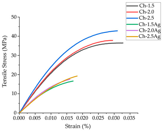
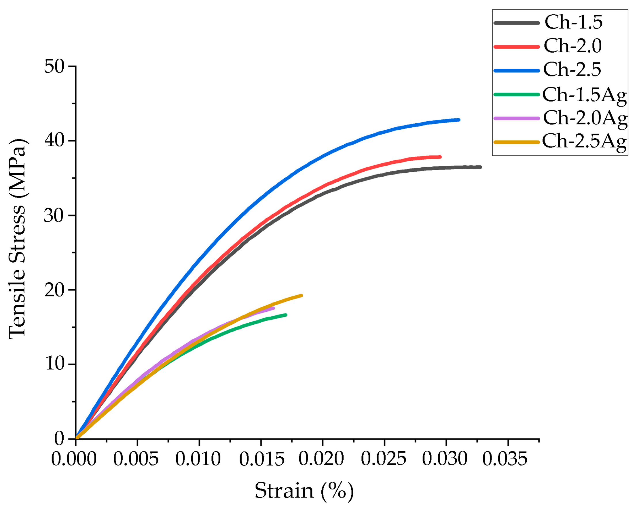
Adequate mechanical properties are essential for active food packaging materials to preserve product integrity under the physical stresses encountered during transportation and storage [67]. Table 5 summarizes the mechanical properties of the obtained chitosan-based films. For the Ch-1.5 film, a tensile strength of 35.02 ± 2.07 MPa and an elongation at break of 2.99 ± 0.44% were recorded. As the chitosan concentration increased, a progressive increase in tensile strength accompanied by a decrease in elongation at break was observed. This behavior is attributed to the higher density of intermolecular interactions and physical crosslinking among chitosan chains, which promote the formation of a more compact and rigid polymeric network. Consequently, the films exhibit enhanced mechanical resistance at the expense of reduced flexibility [68].
The incorporation of Ag microparticles at 0.25% (w/v) led to a pronounced reduction in tensile strength for the Ch-1.5Ag, Ch-2.0Ag, and Ch-2.5Ag films, with decreases of 51.57%, 52.82%, and 49.83%, respectively. A concomitant decrease in elongation at break was also observed, indicating a loss of deformation capacity. Previous studies have shown that PCs coating Ag microparticles, derived from green synthesis routes, can disrupt the crystalline organization of chitosan and weaken intermolecular interactions within the polymer matrix, ultimately compromising mechanical strength [69].
Similar trends have been reported by Ediyilyam et al. (2021) [32], who demonstrated that the incorporation of Ag microparticles at low concentrations (0.0075–0.025% w/v) enhanced the tensile strength of chitosan–gelatin composite films, whereas a further increase to 0.05% w/v resulted in a decrease in tensile strength. Notably, this concentration is still substantially lower than the Ag microparticle content employed in the present study (0.25% w/v), which promotes increased particle aggregation. This behavior is associated with the presence of hydrophilic phytochemical compounds from plant extracts used during particles synthesis, which stabilize Ag microparticles while modifying polymer–polymer interactions.
Although a decrease in tensile strength is often associated with an increase in elongation at break, this relationship does not necessarily apply to particle-reinforced systems. When particles exhibit a strong tendency to aggregate, rigid and non-deformable domains are formed within the polymer matrix [70]. These heterogeneities act as stress concentration sites, disrupt the continuity of the polymer matrix, and promote premature failure, thereby reducing the deformation capacity of the films.
Young’s modulus, which reflects the elastic stiffness of a material [71], increased with increasing chitosan concentration, indicating enhanced rigidity accompanied by reduced elongation at break. In contrast, the incorporation of Ag microparticles resulted in a decrease in Young’s modulus, suggesting a reduction in film stiffness (Figure 7).
Figure 7.
Tensile stress–strain curves of chitosan and chitosan-Ag microparticles films at different polymer concentrations.
These results indicate that Ag microparticles directly modify both the rheological and mechanical properties of the system, particularly tensile strength and Young’s modulus. Higher solution viscosity is associated with increased mechanical resistance and stiffness of the films, whereas lower viscosity, resulting from the incorporation of Ag microparticles, leads to reduced tensile strength and rigidity.
Based on the overall results obtained for thickness and mechanical properties, the Ch-2.0Ag formulation was selected for subsequent antioxidant and antibacterial evaluations. This formulation exhibited the most suitable balance between film-forming characteristics and mechanical performance among the Ag-containing films, making it the most promising candidate for potential food-packaging applications.
3.6.3. Fourier Transform Infrared (FTIR)
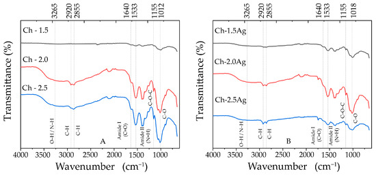
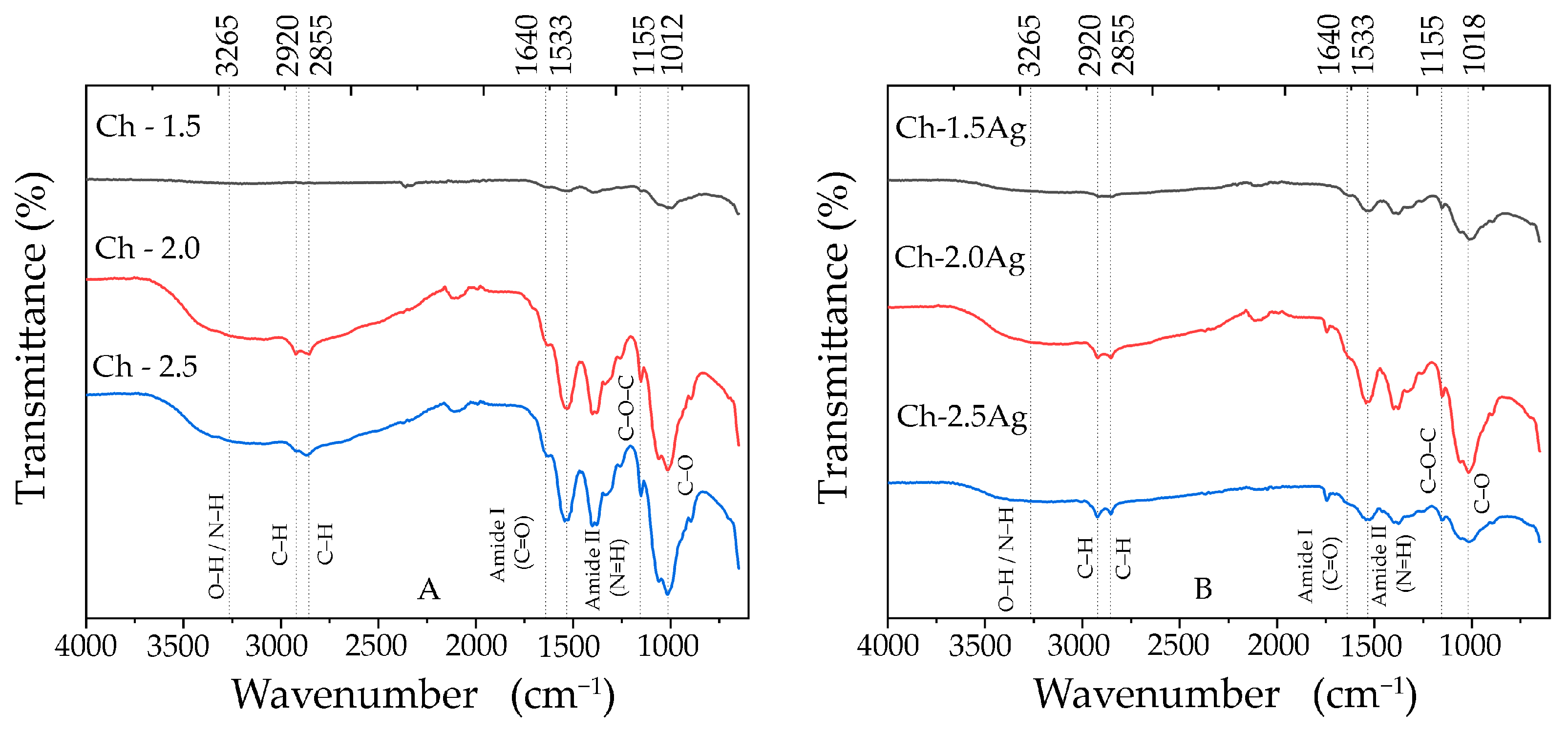
Chitosan and chitosan-Ag microparticle films were structurally characterized by Fourier transform infrared (FT-IR) spectroscopy (Figure 8). For all chitosan formulations, regardless of concentration, a broad band centered at approximately 3265 cm−1 was observed, which is attributed to the stretching vibrations of hydroxyl (–OH) and primary amino (–NH2) groups present in the chitosan structure [72]. Bands in the region of 2855–2920 cm−1 were also identified, corresponding to the stretching vibrations of C–H bonds. The absorption band located at 1640 cm−1 was associated with the C=O stretching of amide I groups, while the band at 1533 cm−1 was related to the N–H bending of amide II groups [42,72]. Additionally, a band at 1155 cm−1, attributed to the C–O–C stretching vibrations of the polysaccharide backbone, and a characteristic band around 1012 cm−1, assigned to C–O stretching vibrations, were detected [66].
Figure 8.
Fourier transform infrared (FT-IR) spectra of chitosan films (A) and chitosan-Ag microparticles films (B).
The incorporation of Ag microparticles into the chitosan polymer matrix induced noticeable changes in the characteristic FT-IR bands. In particular, the broad band centered around 3265 cm−1, attributed to the stretching vibrations of hydroxyl (–OH) and amino (–NH2) groups, exhibited a decrease in intensity, especially in the Ch-2.5Ag formulation. Similarly, a reduction in the intensity of the bands corresponding to amide I (1640 cm−1), associated with C=O stretching, and amide II (1533 cm−1), related to N–H bending, was observed. Finally, the band attributed to C–O stretching vibrations showed a decrease in intensity along with a slight shift toward higher wavenumbers, from 1012 cm−1 to 1018 cm−1. Notably, Mohamed and Madian (2020) [31] reported minor shifts in the band assigned to free amino groups from 1020.4 cm−1 to 1021–1021.5 cm−1 in chitosan films incorporating Ag microparticles synthesized using Aloe vera extract, which is comparable to the slight shift observed in the present study. The observed shifts and intensity variations in the FTIR bands are attributed to coordination interactions between Ag microparticles and the electron-donating (–NH) and (–OH) groups of chitosan, which alter the local electronic environment and bond lengths without the formation of new covalent bonds [73].
3.6.4. Scanning Electron Microscopy (SEM)
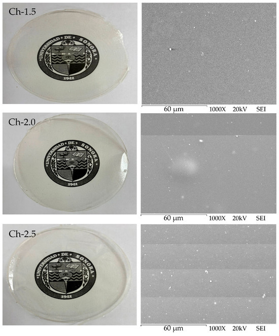
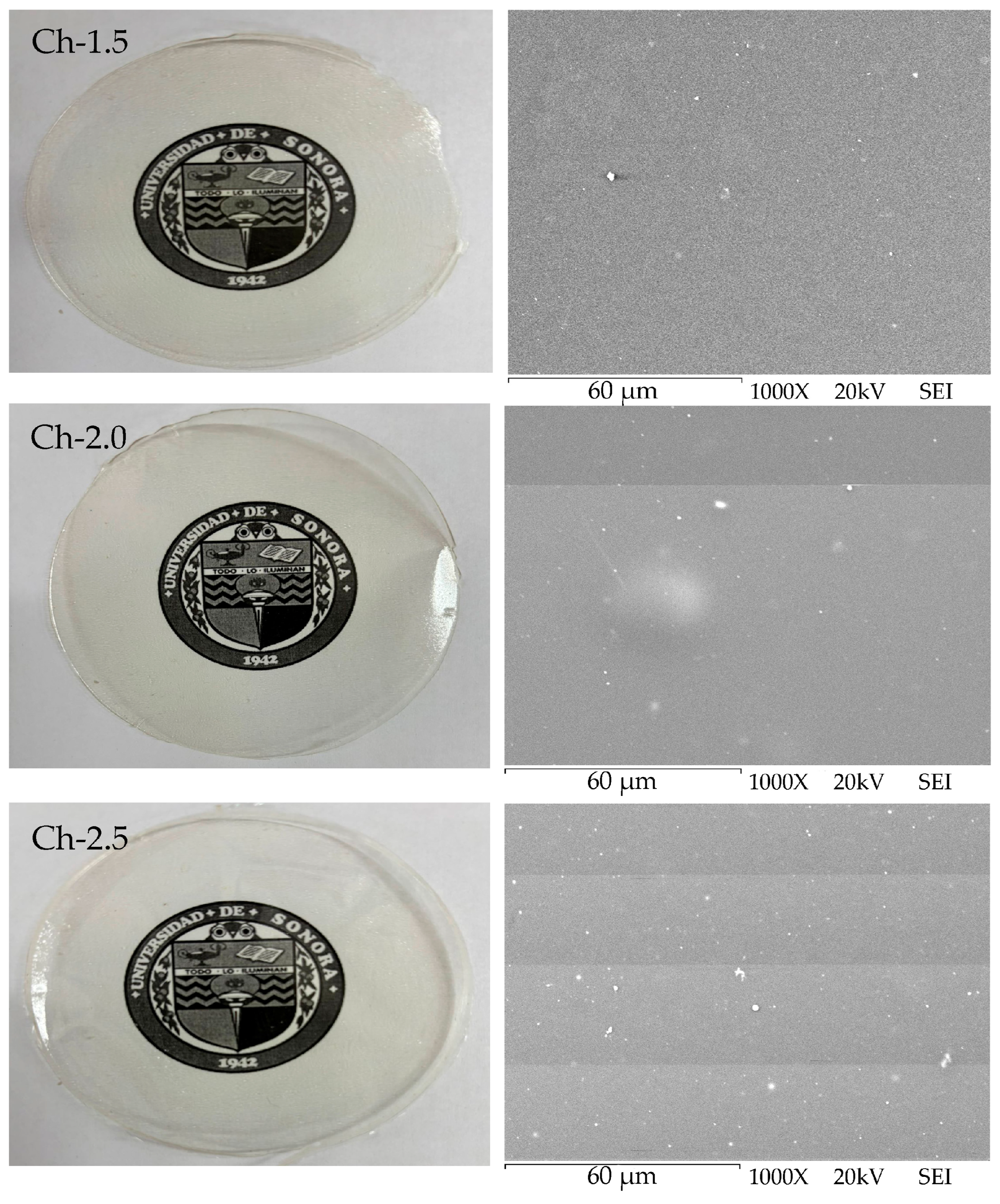
Figure 9 and Figure 10 show the macroscopic images and scanning electron microscopy (SEM) micrographs of chitosan and chitosan-Ag microparticle films. As shown in Figure 9, the Ch-1.5 films exhibited a translucent appearance at the macroscopic level, as the logo of the University of Sonora could be clearly observed beneath them.
Figure 9.
Logo of the University of Sonora covered with Ch-1.5, Ch-2.0, and Ch-2.5 films, along with their corresponding SEM micrographs at 1000× magnification.
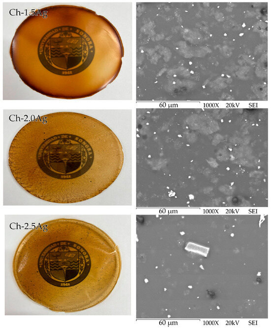
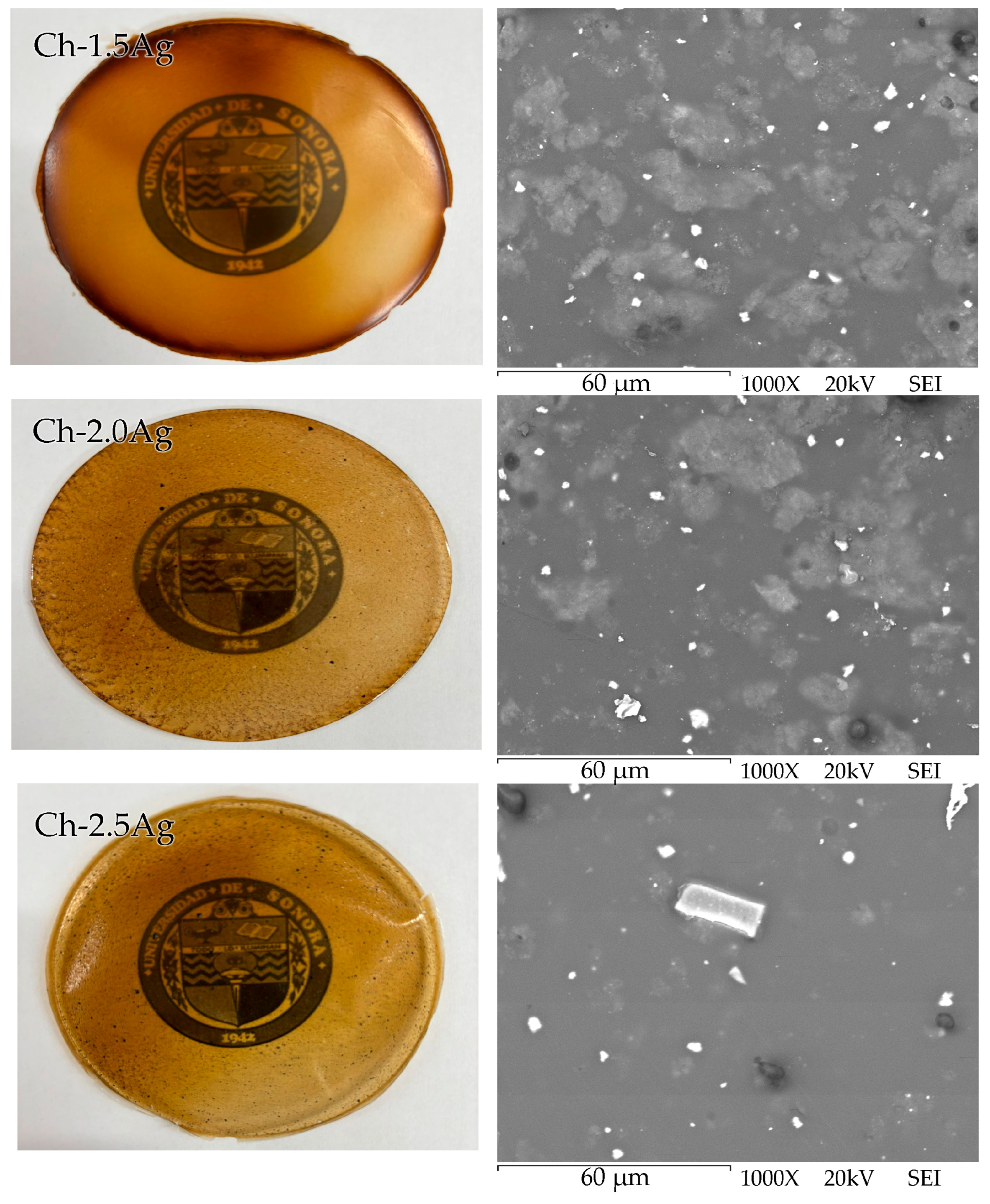
Figure 10.
Logo of the University of Sonora covered with Ch-1.5Ag, Ch-2.0Ag, and Ch-2.5Ag films, along with their corresponding SEM micrographs at 1000× magnification.
SEM analysis revealed a smooth, homogeneous, and continuous surface, with the presence of small white spots dispersed throughout the films, which are attributed to polymer distribution. Although no marked differences in surface appearance were visually detected at the macroscopic level with increasing chitosan concentration (up to 2.5% w/v), the SEM micrographs showed an increase in both the number and size of these white spots. This phenomenon indicates a tendency toward aggregate formation, which is attributed to the increase in solution viscosity at higher chitosan concentrations, hindering effective homogenization during the drying process [74].
SEM micrographs of the chitosan-Ag microparticle films (Figure 10) reveal the presence of white spots and agglomerates, indicating difficulties in achieving a homogeneous dispersion of the microparticles within the polymer matrix. This phenomenon is particularly pronounced in the Ch-2.5Ag formulations, where the highest accumulation of Ag microparticles is observed. This trend can be explained by the increased physical crosslinking between chitosan chains, which restricts chain mobility and hinder the effective dispersion of particles, thereby promoting agglomeration. These findings are consistent with those reported by Mohamed and Madian (2020) [31], who prepared chitosan films with Ag microparticles concentrations of 0, 2, 4, 6, and 8 µg/mL and observed phase separation at 8 µg/mL. Although this concentration is substantially lower than the Ag microparticles content used in the present study (0.25% w/v, equivalent to 2500 µg/mL), the similarity in behavior suggests that phase separation is governed not only by the nominal Ag microparticles concentration, but also by factors such as particles dispersion, polymer–particle interactions, and the presence of surface-capping phytochemicals derived from green synthesis routes. Based on morphological, mechanical, and macroscopic observations, the Ch-2.0Ag film was selected as the optimal formulation for further evaluation of its antioxidant and antibacterial properties.
3.7. Evaluation of the Antioxidant Activity
The results corresponding to the antioxidant activity of the Ch-2.0 and Ch-2.0Ag films are presented in Table 3. The chitosan films exhibited an inhibition of the ABTS radical of 47.52 ± 1.20%, corresponding to 58.04 ± 6.93 µM TE/g. The incorporation of Ag microparticles significantly enhanced this effect, reaching an inhibition of 70.57 ± 1.68%. Although Ag microparticles have been previously reported to exhibit inhibitory activity against the ABTS radical, the results of this study confirm that the polysaccharide itself also possesses antiradical capacity, which is further strengthened by the presence of the particles (Figure 4A). According to Zagloul et al. (2024) [75], the antioxidant activity of chitosan is attributed to the presence of amino and hydroxyl groups in the polymer, which enable the inhibition of chain reactions associated with reactive oxygen species.
Regarding the DPPH radical, the Ch-2.0 films exhibited an inhibition of 20.64 ± 2.05%, which increased to 31.53 ± 3.05% after the incorporation of Ag microparticles. In this case, the active packaging system did not show a marked inhibitory effect against the DPPH radical compared to the control films, likely due to limitations in the reaction mechanism involved in the neutralization of this radical (Figure 4B).
Regarding the FRAP evaluation of the chitosan films with incorporated Ag microparticles, a higher absorbance at 638 nm was expected compared to the films without the active agent. However, a slight decrease in absorbance was observed, resulting in values of 141.29 ± 0.57 µM TE/g for the pure chitosan films and 137.29 ± 0.58 µM TE/g for the chitosan films containing Ag microparticles. Although the Ag microparticles exhibited a strong reducing power when dispersed in solution and in direct contact with the reagent, their incorporation into the chitosan polymeric matrix hinders the interaction between the FRAP reagent and the antioxidant agents within the film, thereby limiting the measured reducing capacity.
The antioxidant activity of chitosan-Ag microparticle films was found to be predominantly governed by SET mechanisms, consistent with the behavior previously determined for the Ag microparticle suspension.
3.8. Antibacterial Activity
The antibacterial activity of Ch-2.0 and Ch-2.0Ag films were evaluated to assess their functionality as active food packaging materials (Figure 5). Ch-2.0 films showed 41.42% bacterial inhibition due to the intrinsic antibacterial activity of chitosan, which primarily involves electrostatic interactions with the cell wall of Gram-positive bacteria. However, this activity is generally insufficient to fulfill the requirements of more demanding antibacterial applications [76,77].
However, after incorporation of Ag microparticles at 0.25% (w/v), the antibacterial activity of the Ch-2.0Ag film increased significantly (p < 0.05), reaching complete (100%) inhibition. This enhancement is attributed to the direct interaction of Ag microparticles with the negatively charged bacterial cell membrane, promoting membrane destabilization and subsequent cell death [78].
These findings are consistent with previous studies reporting the strong antibacterial efficacy of Ag microparticles incorporated polymeric materials against Listeria monocytogenes. Shankar et al. (2021) [79] developed chitosan-based films containing essential oils and Ag microparticles for strawberry packaging, achieving complete inhibition of the pathogen after 16 h of incubation.
The obtained results confirm the excellent inhibitory capacity of chitosan-Ag microparticles films, as evidenced by the complete inhibition of the pathogen Listeria monocytogenes. This finding is particularly promising, as it positions this type of material as an effective alternative for the prevention of foodborne diseases, especially in foods susceptible to spoilage or nutritional quality loss, thereby contributing to food safety and the extension of the shelf life of perishable products.
4. Conclusions
In conclusion, the rheological parameters of the film-forming solutions are closely related to the mechanical behavior of the resulting films. An increase in apparent viscosity and consistency index (K), associated with greater polymer chain entanglement and intermolecular interactions in solution, leads to stiffer and mechanically stronger films with higher Young’s modulus and tensile strength, but lower elongation at break. In contrast, the reduction in these rheological parameters, as observed after the incorporation of Ag microparticles, is reflected in films with lower stiffness and mechanical resistance, indicating a less cohesive polymer network.
Despite these mechanical changes, the chitosan-Ag microparticle films exhibited enhanced functional performance. Antioxidant activity was mainly governed by single-electron transfer mechanisms associated with chitosan functional groups and surface phenolics on the Ag microparticles, while a complete (100%) antibacterial effect against Listeria monocytogenes was observed under the experimental conditions evaluated.
Overall, these results highlight the importance of controlling solution rheology and microparticle dispersion to obtain chitosan-based films with adequate mechanical integrity and enhanced antioxidant and antibacterial functionality. Furthermore, the use of green-synthesized Ag microparticles obtained from pecan nutshell extract represents a more sustainable approach for the development of bioactive materials by valorizing an agro-industrial byproduct and reducing the use of conventional chemical reducing agents. The developed films showed promising characteristics for potential application in active food-packaging systems. However, additional studies involving a broader spectrum of pathogenic and spoilage microorganisms, as well as food-storage evaluations, are required to further assess their contribution to food safety and shelf-life extension.
Author Contributions
Conceptualization, J.L.P.-R., F.R.-F. and D.E.R.-F.; Methodology, J.L.P.-R., S.E.B.-I., K.H.O.-V., A.M.P.-S. and B.M.-L.; Software, R.A.L.-C.; validation, F.R.-F. and D.E.R.-F.; Formal analysis, J.A.T.-H., F.R.-F. and D.E.R.-F.; Investigation, J.L.P.-R. and F.R.-F.; Resources, F.R.-F. and D.E.R.-F.; Data curation, J.L.P.-R., F.R.-F. and D.E.R.-F.; Writing—original draft, J.L.P.-R. and J.A.T.-H.; Writing—review and editing, F.R.-F., T.d.C.-C., M.J.M.-V., J.A.T.-H., C.G.B.-U. and M.A.R.-G.; Supervision, F.R.-F., M.J.M.-V. and B.M.-L.; Visualization, R.A.L.-C.; Project administration, F.R.-F. and D.E.R.-F.; Funding acquisition, F.R.-F. All authors have read and agreed to the published version of the manuscript.
Funding
This research was funded by the University of Sonora, through the Interdisciplinary Faculty of Biological and Health, project number USO313009573.
Institutional Review Board Statement
Not applicable.
Informed Consent Statement
Not applicable.
Data Availability Statement
The raw data supporting the conclusions of this article will be made available by the authors on request.
Acknowledgments
The authors are grateful to the University of Sonora and to Secretaría de Ciencia, Humanidades, Tecnología e Innovación (SECIHTI) for their support.
Conflicts of Interest
The authors declare no conflicts of interest.
References
- Choskit, T.; Gupta, N.; Singh, J.; Bhat, A.; Bandral, J.D.; Sood, M. An Overview on Food Spoilage Mechanism and Their Prevention. Chem. Sci. Rev. Lett. 2023, 12, 60–66. [Google Scholar]
- Fadiji, T.; Rashvand, M.; Daramola, M.O.; Iwarere, S.A. A review on antimicrobial packaging for extending the shelf life of food. Processes 2023, 11, 590. [Google Scholar] [CrossRef] [Scilit]
- Saha, N.C. Food packaging: Concepts and its significance. In Food Packaging: Materials, Techniques and Environmental Issues; Springer Nature: Singapore, 2022; pp. 1–45. [Google Scholar]
- Siddiqui, S.A.; Bahmid, N.A.; Karim, I.; Mehany, T.; Gvozdenko, A.A.; Blinov, A.V.; Lorenzo, J.M. Cultured meat: Processing, packaging, shelf life, and consumer acceptance. LWT 2022, 172, 114192. [Google Scholar] [CrossRef] [Scilit]
- Baniasadi, H.; Abidnejad, R.; Fazeli, M.; Niskanen, J.; Lizundia, E. Advances in polysaccharide-based food packaging: Functionalization strategies and sustainability considerations. Mater. Sci. Eng. R Rep. 2026, 167, 101128. [Google Scholar] [CrossRef] [Scilit]
- Kim, Y.; Yoon, J.; Kim, J.; Kim, H.; Park, S.; Jin, H.J.; Kwak, H.W. Multifunctional fructose-crosslinked fibroin film with a developed β-sheet structure for advanced food packaging. Int. J. Biol. Macromol. 2025, 286, 138370. [Google Scholar] [PubMed]
- Ma, K.; Yang, J.; Goksen, G.; Alamri, A.S.; Alhomrani, M.; Xia, G.; Zhang, W. Application of tragacanth gum as a potential food packaging film and its performance enhancement strategy. Food Hydrocoll. 2025, 162, 110894. [Google Scholar]
- Periyasamy, T.; Asrafali, S.P.; Lee, J. Recent advances in functional biopolymer films with antimicrobial and antioxidant properties for enhanced food packaging. Polymers 2025, 17, 1257. [Google Scholar] [CrossRef] [Scilit] [PubMed]
- Gamage, A.; Thiviya, P.; Liyanapathiranage, A.; Wasana, M.D.; Jayakodi, Y.; Bandara, A.; Madhujith, T. Polysaccharide-based bioplastics: Eco-friendly and sustainable solutions for packaging. J. Compos. Sci. 2024, 8, 413. [Google Scholar]
- Yao, Q.B.; Huang, F.; Lu, Y.H.; Huang, J.M.; Ali, M.; Jia, X.Z.; Huang, Y.Y. Polysaccharide-based food packaging and intelligent packaging applications: A comprehensive review. Trends Food Sci. Technol. 2024, 147, 104390. [Google Scholar] [CrossRef] [Scilit]
- Demirbas, A.; Karsli, B. Innovative chitosan-silver nanoparticles: Green synthesis, antimicrobial properties, and migration assessment for food packaging. Food Chem. 2025, 467, 142363. [Google Scholar] [PubMed]
- Liu, X.; Xu, F.; Yong, H.; Chen, D.; Tang, C.; Kan, J.; Liu, J. Recent advances in chitosan-based active and intelligent packaging films incorporated with flavonoids. Food Chem. X 2025, 25, 102200. [Google Scholar] [CrossRef] [Scilit] [PubMed]
- Ponnusamy, A.; SR, R.R.; Rajan, R.; Ashraf, F. Chitosan silver nanoparticle inspired seaweed (Gracilaria crassa) biodegradable films for seafood packaging. Algal Res. 2024, 78, 103429. [Google Scholar] [CrossRef] [Scilit]
- Li, Y.; Hua, Z.; Li, Y.; Chen, T.; Alamri, A.S.; Xu, Y.; Hu, J. Development of multifunctional chitosan-based composite film loaded with tea polyphenol nanoparticles for strawberry preservation. Int. J. Biol. Macromol. 2024, 275, 133648. [Google Scholar] [CrossRef] [Scilit] [PubMed]
- Sultan, M.; Youssef, A.; Baseer, R.A. Fabrication of multifunctional ZnO@tannic acid nanoparticles embedded in chitosan and polyvinyl alcohol blend packaging film. Sci. Rep. 2024, 14, 18533. [Google Scholar] [PubMed]
- Aliabbasi, N.; Emam-Djomeh, Z.; Rezaeinia, H. Thermally repairable and antimicrobial chitosan-gelatin films with natural wax and silver nanoparticles. Bioresour. Technol. Rep. 2026, 33, 102533. [Google Scholar] [CrossRef] [Scilit]
- Yin, W.; Lei, Y.; Wang, J.; Lei, Q.; Yu, W.; Ou, S. A green approach to bio-based active packaging: Grape skin extract-synthesized AgNPs for food preservation. Materials 2026, 19, 218. [Google Scholar] [PubMed]
- Aliero, A.S.; Hasmoni, S.H.; Haruna, A.; Isah, M.; Malek, N.A.N.N.; Zawawi, N.A. Bibliometric exploration of green synthesized silver nanoparticles for antibacterial activity. Emerg. Contam. 2025, 11, 100411. [Google Scholar] [CrossRef] [Scilit]
- Eker, F.; Akdaşçi, E.; Duman, H.; Bechelany, M.; Karav, S. Green synthesis of silver nanoparticles using plant extracts: A comprehensive review of physicochemical properties and multifunctional applications. Int. J. Mol. Sci. 2025, 26, 6222. [Google Scholar] [CrossRef] [Scilit] [PubMed]
- Ijaz, M.; Zafar, M.; Iqbal, T. Green synthesis of silver nanoparticles by using various extracts: A review. Inorg. Nano-Met. Chem. 2020, 51, 744–755. [Google Scholar] [CrossRef] [Scilit]
- Keshari, A.K.; Srivastava, R.; Singh, P.; Yadav, V.B.; Nath, G. Antioxidant and antibacterial activity of silver nanoparticles synthesized by Cestrum nocturnum. J. Ayurveda Integr. Med. 2020, 11, 37–44. [Google Scholar] [CrossRef] [Scilit] [PubMed]
- Zhuo, M.; Liu, C.; Wang, Q.; Wang, Z.; Wang, Y.; Yu, F.; Zhang, Y. Catharanthus roseus extract-assisted silver nanoparticles chitosan films with high antioxidant and antimicrobial properties for fresh food preservation. Int. J. Biol. Macromol. 2025, 309, 142771. [Google Scholar] [CrossRef] [Scilit] [PubMed]
- Neira-Vielma, A.A.; Meléndez-Ortiz, H.I.; García-López, J.I.; Sanchez-Valdes, S.; Cruz-Hernández, M.A.; Rodríguez-González, J.G.; Ramírez-Barrón, S.N. Green synthesis of silver nanoparticles using pecan nut shell extracts and evaluation of their antimicrobial activity. Antibiotics 2022, 11, 1150. [Google Scholar] [CrossRef] [Scilit] [PubMed]
- Omotonoko, J.L.Y.; Polozola, M.; Svyantek, A.; Wang, Z. Valorization and environmental impacts of pecan waste: A critical review. Foods 2026, 15, 168. [Google Scholar] [CrossRef] [Scilit] [PubMed]
- Oviedo-Medrano, M.; Obregón, S.; Ruiz-Gómez, M.A.; López, I.; Vázquez, A. Surface functionalization of silver nanoparticles with pecan nutshell extract for the colorimetric detection of vitamin B1. Mater. Lett. 2025, 403, 139484. [Google Scholar]
- Ozuna-Valencia, K.H.; Barreras-Urbina, C.G.; Tapia-Hernández, J.A.; Moreno-Vásquez, M.D.J.; Graciano-Verdugo, A.Z.; Robles-García, M.Á.; Rodríguez-Félix, F. Green synthesis of silver particles using pecan nutshell extract: Development and antioxidant characterization of zein/pectin active films. Processes 2025, 14, 4. [Google Scholar] [CrossRef] [Scilit]
- Harun-Ur-Rashid, M.; Foyez, T.; Krishna, S.B.N.; Poda, S.; Imran, A.B. Recent advances of silver nanoparticle-based polymer nanocomposites for biomedical applications. RSC Adv. 2025, 15, 8480–8505. [Google Scholar] [CrossRef] [Scilit] [PubMed]
- Raza, S.; Ansari, A.; Siddiqui, N.N.; Ibrahim, F.; Abro, M.I.; Aman, A. Biosynthesis of silver nanoparticles for the fabrication of non cytotoxic and antibacterial metallic polymer based nanocomposite system. Sci. Rep. 2021, 11, 10500. [Google Scholar] [CrossRef] [Scilit] [PubMed]
- Zhang, H.; Su, S.; Liu, S.; Qiao, C.; Wang, E.; Chen, H.; Li, T. Effects of chitosan and cellulose derivatives on sodium carboxymethyl cellulose-based films. Molecules 2023, 28, 5211. [Google Scholar] [CrossRef] [Scilit] [PubMed]
- Zhang, W.; Jiang, W. Antioxidant and antibacterial chitosan film with tea polyphenols-mediated green synthesis silver nanoparticle via a novel one-pot method. Int. J. Biol. Macromol. 2020, 155, 1252–1261. [Google Scholar] [PubMed]
- Mohamed, N.; Madian, N.G. Evaluation of the mechanical, physical and antimicrobial properties of chitosan thin films doped with greenly synthesized silver nanoparticles. Mater. Today Commun. 2020, 25, 101372. [Google Scholar] [CrossRef] [Scilit]
- Ediyilyam, S.; George, B.; Shankar, S.S.; Dennis, T.T.; Waclawek, S.; Cenik, M.; Padil, V.V.T. Chitosan/Gelatin/Silver Nanoparticles Composites Films for Biodegradable Food Packaging Applications. Polymers 2021, 13, 1680. [Google Scholar] [CrossRef] [Scilit] [PubMed]
- Benaissa, S.; Benmansour, K.; Zenagui, C.; Khelladi, M.B.; Belyagoubi, L. Development of antioxidant and antimicrobial chitosan nanocomposite films incorporating with eco-friendly silver nanoparticles synthesized with Arabic gum. Polym. Bull. 2025, 82, 9275–9301. [Google Scholar] [CrossRef] [Scilit]
- Preciado-Saldaña, A.M.; Abraham Domínguez-Avila, J.; Fernando Ayala-Zavala, J.; Villegas-Ochoa, M.A.; Sáyago-Ayerdi, S.G.; Wall-Medrano, A.; González-Aguilar, G.A. Formulation and characterization of an optimized functional beverage. Food Sci. Technol. Int. 2019, 25, 547–561. [Google Scholar] [CrossRef] [Scilit] [PubMed]
- Rodríguez-Félix, F.; López-Cota, A.G.; Moreno-Vásquez, M.J.; Graciano-Verdugo, A.Z.; Quintero-Reyes, I.E.; Del-Toro-Sánchez, C.L.; Tapia-Hernández, J.A. Sustainable-green synthesis of silver nanoparticles using safflower waste extract and its antibacterial activity. Heliyon 2021, 7, e06923. [Google Scholar] [PubMed]
- Alvarez-Moreno, M.G.; Rodríguez-Félix, F.; Barreras-Urbina, C.G.; Plascencia-Jatomea, M.; Rueda-Puente, E.O.; Reyes-Pérez, J.J.; Santos-Sauceda, I. Preparation and characterization of zein-phosphate nanoparticles. ACS Omega 2025, 10, 25746–25765. [Google Scholar] [CrossRef] [Scilit] [PubMed]
- García-Lárez, F.L.; Esquer, J.; Guzmán, H.; Zepeda-Quintana, D.S.; Moreno-Vásquez, M.J.; Rodríguez-Félix, F.; Del-Toro-Sánchez, C.L.; López-Corona, B.E.; Tapia-Hernández, J.A. Effect of Ultrasound-Assisted Extraction (UAE) parameters on the recovery of polyphenols from pecan nutshell waste biomass and its antioxidant activity. Biomass Convers. Biorefin. 2025, 15, 10977–10995. [Google Scholar] [CrossRef] [Scilit]
- Moreno-Vásquez, M.J.; Valenzuela-Buitimea, E.L.; Plascencia-Jatomea, M.; Encinas-Encinas, J.C.; Rodríguez-Félix, F.; Sánchez-Valdes, S.; Graciano-Verdugo, A.Z. Functionalization of chitosan by free radical reaction. Carbohydr. Polym. 2017, 155, 117–127. [Google Scholar] [CrossRef] [Scilit] [PubMed]
- Estrella-Osuna, D.E.; Ruiz-Cruz, S.; Rodríguez-Félix, F.; Figueroa-Enríquez, C.E.; González-Ríos, H.; Fernández-Quiroz, D.; Suárez-Jiménez, G.M. Rheological properties of gelatin coatings. Gels 2024, 10, 624. [Google Scholar] [PubMed]
- ASTM D1708-18; Standard Test Method for Tensile Properties of Plastics by Use of Microtensile Specimens. ASTM International: West Conshohocken, PA, USA, 2018.
- Canizales-Rodríguez, D.F.; Rodríguez-Félix, F.; Tapia-Hernández, J.A.; Del-Toro-Sánchez, C.L.; Ruíz-Cruz, S.; Aubourg, S.P.; Álvarez-Moreno, M.G. PLA fibrous film with betalains. Coatings 2024, 14, 1581. [Google Scholar]
- Acuña-Pacheco, L.V.; Moreno-Robles, A.L.; Plascencia-Jatomea, M.; Del Toro-Sánchez, C.L.; Ayala-Zavala, J.F.; Tapia-Hernández, J.A.; Graciano-Verdugo, A.Z. Alginate–chitosan bilayer film. Coatings 2024, 14, 1232. [Google Scholar]
- Blanco, F.D.; Geevers, M.P.; Hanefeld, U.; Six, J.L.; Chebil, L.; Humeau, C.; Guiavarc’h, Y. Maximized lipase-catalysed production of a monoester of ferulic acid derivatives and ethylene glycol: A key step toward intrinsically antioxidant biosourced polymers. Green Chem. 2025, 27, 11892–11902. [Google Scholar] [CrossRef] [Scilit]
- Jacobo-Velázquez, D.A. Ferulic acid: Mechanistic insights and multifaceted applications in metabolic syndrome, food preservation, and cosmetics. Molecules 2025, 30, 3716. [Google Scholar] [CrossRef] [Scilit] [PubMed]
- Demir, A. Green-synthesized silver nanoparticles from Camellia sinensis: Mechanistic insights into phenolic-mediated multifunctional biological activities. BMC Plant Biol. 2025, 25, 1734. [Google Scholar] [PubMed]
- Xue, S.; Tan, W.; Mao, S.; Pan, H.; Ye, X.; Donlao, N.; Tian, J. Polyphenol-based functional materials: Structural insights, composite strategies, and biomedical applications. Adv. Sci. 2025, 12, e08924. [Google Scholar]
- Serdar, G.; Gül Kılınç, G.; Mazlum Şen, T. Green One-Pot Synthesis of Silver and Gold Nanoparticles Using Catechin Extracts: Influence of Temperature and Antioxidant Activity Evaluation. Plasmonics 2025, 20, 10691–10710. [Google Scholar] [CrossRef] [Scilit]
- Geremew, A.; Carson, L.; Woldesenbet, S.; Wang, H.; Reeves, S.; Brooks, N., Jr.; Peace, E. Effect of zinc oxide nanoparticles synthesized from Carya illinoinensis leaf extract on growth and antioxidant properties of mustard (Brassica juncea). Front. Plant Sci. 2023, 14, 1108186. [Google Scholar] [CrossRef] [Scilit] [PubMed]
- Mubeena, S.A.; Preetha, R. Ultrasound-assisted microalgal docosahexaenoic acid (DHA) nanoemulsion preparation using casein, chitosan, and pectin as emulsifiers for enhanced oxidative stability and shelf life for food fortification. Sustain. Food Technol. 2026, 4, 930–946. [Google Scholar]
- Begum, T.; Follett, P.A.; Salmieri, S.; Jaiswal, L.; Lacroix, M. Development of nanocomposite bioactive diffusion films (BDFs) combined with gamma irradiation for control of the rice weevil Sitophilus oryzae, a stored product pest. J. Food Sci. 2026, 91, e70785. [Google Scholar] [CrossRef] [Scilit] [PubMed]
- Pandey, P.J. Multifunctional nanosponges for the treatment of various diseases: A review. Asian J. Pharm. Pharmacol. 2019, 5, 235–248. [Google Scholar] [CrossRef] [Scilit]
- Argenziano, R.; Agustin-Salazar, S.; Panaro, A.; Calarco, A.; Di Salle, A.; Aprea, P.; Napolitano, A. Combining the potent reducing properties of pecan nutshell with a solvent-free mechanochemical approach for synthesizing high Ag0 content silver nanoparticles: An eco-friendly route to an efficient multifunctional photocatalytic, antibacterial, and antioxidant material. Nanomaterials 2023, 13, 821. [Google Scholar] [CrossRef] [Scilit] [PubMed]
- Mallouka, G.; Karkoutly, O.; Karzoun, O.; AlHannan, F.; Akhtar, S.; Henari, F.; Deen, G.R. Room temperature green synthesis and time resolved kinetics of formation of xanthan gum stabilised silver nanoparticles with catalytic and antibacterial potential. Sci. Rep. 2025, 15, 31626. [Google Scholar] [CrossRef] [Scilit] [PubMed]
- Bélteky, P.; Rónavári, A.; Zakupszky, D.; Boka, E.; Igaz, N.; Szerencsés, B.; Kónya, Z. Are smaller nanoparticles always better? Understanding the biological effect of size-dependent silver nanoparticle aggregation under biorelevant conditions. Int. J. Nanomed. 2021, 16, 3021–3040. [Google Scholar] [CrossRef] [Scilit]
- Okur, E.E.; Eker, F.; Akdaşçi, E.; Bechelany, M.; Karav, S. Comprehensive review of silver nanoparticles in food packaging applications. Int. J. Mol. Sci. 2025, 26, 9842. [Google Scholar] [CrossRef] [Scilit] [PubMed]
- Wołosiak, R.; Drużyńska, B.; Derewiaka, D.; Piecyk, M.; Majewska, E.; Ciecierska, M.; Pakosz, P. Verification of the conditions for determination of antioxidant activity by ABTS and DPPH assays—A practical approach. Molecules 2021, 27, 50. [Google Scholar] [CrossRef] [Scilit] [PubMed]
- Fitrania, F.; Rahman, D.Y.; Indrasi, D.; Prangdimurti, E. Antioxidant activity of phycocyanin from Galdieria sulphuraria in autotrophic cultivation. AIP Conf. Proc. 2025, 3323, 020019. [Google Scholar] [CrossRef] [Scilit]
- dos Santos, V.H.M.; de Oliveira Costa, M.M.; Granero, F.O.; Figueiredo, C.C.M.; Santos, H.H.; Benevides, P.J.C.; da Silva, R.M.G. Green biosynthesis of silver nanoparticles using anthocyanins-rich extract from Euterpe edulis fruits (AnthocyanOx®): In vitro antioxidant and antiglycation activities, and in silico anti-aging activity. Food Bioprod. Process. 2025, 151, 189–201. [Google Scholar]
- Fernandes, R.D.P.P.; Trindade, M.A.; Tonin, F.G.; Lima, C.G.D.; Pugine, S.M.P.; Munekata, P.E.S.; de Melo, M.P. Evaluation of antioxidant capacity of 13 plant extracts by three different methods: Cluster analyses applied for selection of natural extracts with higher antioxidant capacity to replace synthetic antioxidant in lamb burgers. J. Food Sci. Technol. 2016, 53, 451–460. [Google Scholar] [PubMed]
- Rabbani, A.; Khaliq, A.; Mudgil, P.; Maqsood, S.; Nazir, A. Recent advances in lemongrass essential oil: Food safety, preservation, and bioactivity in food systems. Compr. Rev. Food Sci. Food Saf. 2026, 25, e70350. [Google Scholar] [PubMed]
- Kim, M.; Jung-Suk, S.; Jee, S.C.; Dae-Young, K.; Mehta, V.; Vadakkan, K.; Ghodake, G. Sustainable, scalable nanotechnology approach using filtrate from Raphanus sativus in combating multidrug-resistant pathogens and neglected tropical diseases. Front. Cell. Infect. Microbiol. 2026, 15, 1684292. [Google Scholar] [PubMed]
- Amer, S.A.; Abushady, H.M.; Refay, R.M.; Mailam, M.A. Enhancement of the antibacterial potential of plantaricin by incorporation into silver nanoparticles. J. Genet. Eng. Biotechnol. 2021, 19, 13. [Google Scholar] [CrossRef] [Scilit] [PubMed]
- do Amaral Sobral, P.J.; Gebremariam, G.; Drudi, F.; De Aguiar Saldanha Pinheiro, A.C.; Romani, S.; Rocculi, P.; Dalla Rosa, M. Rheological and viscoelastic properties of chitosan solutions prepared with different chitosan or acetic acid concentrations. Foods 2022, 11, 2692. [Google Scholar] [CrossRef] [Scilit] [PubMed]
- Sun, Y.; Liu, Z.; Zhang, L.; Wang, X.; Li, L. Effects of plasticizer type and concentration on rheological, physico-mechanical and structural properties of chitosan/zein film. Int. J. Biol. Macromol. 2020, 143, 334–340. [Google Scholar] [PubMed]
- Di Muzio, L.; Cairone, F.; Cesa, S.; Sergi, C.; Tirillò, J.; Angiolella, L.; Paolicelli, P. Gellan gum-based nanocomposite films containing bio-reduced silver nanoparticles: Synthesis, characterisation and antifungal activity. Carbohydr. Polym. Technol. Appl. 2024, 7, 100485. [Google Scholar]
- Alizadeh Sani, M.; Khezerlou, A.; Tavassoli, M.; Mohammadi, K.; Hassani, S.; Ehsani, A.; McClements, D.J. Bionanocomposite active packaging material based on soy protein isolate/Persian gum/silver nanoparticles: Fabrication and characteristics. Colloids Interfaces 2022, 6, 57. [Google Scholar] [CrossRef] [Scilit]
- Shah, Y.A.; Bhatia, S.; Al-Harrasi, A.; Afzaal, M.; Saeed, F.; Anwer, M.K.; Faisal, Z. Mechanical properties of protein-based food packaging materials. Polymers 2023, 15, 1724. [Google Scholar] [CrossRef] [Scilit] [PubMed]
- Ren, L.; Yan, X.; Zhou, J.; Tong, J.; Su, X. Influence of chitosan concentration on mechanical and barrier properties of corn starch/chitosan films. Int. J. Biol. Macromol. 2017, 105, 1636–1643. [Google Scholar] [CrossRef] [Scilit] [PubMed]
- Riaz, A.; Lei, S.; Akhtar, H.M.S.; Wan, P.; Chen, D.; Jabbar, S.; Zeng, X. Preparation and characterization of chitosan-based antimicrobial active food packaging film incorporated with apple peel polyphenols. Int. J. Biol. Macromol. 2018, 114, 547–555. [Google Scholar] [CrossRef] [Scilit] [PubMed]
- Irnadiastputri, S.F.; Tiurma, Y.M.; Rahman, S.F.; Gan, B.S.; Katili, P.A. Engineering the human tympanic membrane: Lessons from mechanics, modelling, and materials. Biomech. Model. Mechanobiol. 2026, 25, 7. [Google Scholar]
- Carpintero, M.; Marcet, I.; Cortizo, C.; Guerrero, P.; de la Caba, K.; Rendueles, M.; Díaz, M. Chitosan modification with octenyl succinic anhydride (OSA): Effect of the degree of substitution on structural, mechanical and barrier properties in synthetized bioplastics. Food Hydrocoll. 2026, 171, 111838. [Google Scholar]
- Chenah, M.; Karabulut, G.; Zannou, O.; Tosif, M.M.; Goksen, G. Multi-component chitosan–pectin films reinforced with Padina pavonica nanocellulose and Thymus algeriensis essential oil for red meat shelf-life enhancement. Food Chem. X 2026, 33, 103484. [Google Scholar] [CrossRef] [Scilit] [PubMed]
- An, J.; Guo, G.; Yin, R.; Luo, Q.; Li, X.; Liu, F.; Wang, D. Facile preparation of silver/reduced graphene oxide/chitosan colloid and application of the nanocomposite in antibacterial and catalytic activity. Polym. Int. 2018, 67, 515–527. [Google Scholar]
- Jafri, N.F.; Salleh, K.M.; Ghazali, N.A.; Mazlan, N.S.N.; Ab Halim, N.H.; Zakaria, S. Effects of carboxymethyl cellulose fiber formations with chitosan incorporation via coating and mixing processes. Int. J. Biol. Macromol. 2023, 253, 126971. [Google Scholar] [CrossRef] [Scilit] [PubMed]
- Zagloul, H.; Dhahri, M.; Bashal, A.H.; Khaleil, M.M.; Habeeb, T.H.; Khalil, K.D. Multifunctional Ag2O/chitosan nanocomposites synthesized via sol-gel with enhanced antimicrobial and antioxidant properties: A novel food packaging material. Int. J. Biol. Macromol. 2024, 264, 129990. [Google Scholar] [CrossRef] [Scilit] [PubMed]
- Pan, T.; Chen, H.; Gao, X.; Wu, Z.; Ye, Y.; Shen, Y. Engineering efficient artificial nanozyme based on chitosan grafted Fe-doped-carbon dots for bacteria biofilm eradication. J. Hazard. Mater. 2022, 435, 128996. [Google Scholar] [PubMed]
- Perinelli, D.R.; Fagioli, L.; Campana, R.; Lam, J.K.; Baffone, W.; Palmieri, G.F.; Bonacucina, G. Chitosan-based nanosystems and their exploited antimicrobial activity. Eur. J. Pharm. Sci. 2018, 117, 8–20. [Google Scholar] [CrossRef] [Scilit] [PubMed]
- Fan, L.; Dong, Y.; Ismail, B.B.; Zhang, L.; Shi, Y.; Wu, D.; Li, G. The antimicrobial activity and resistance evolution of nanomaterials: A review. ACS Mater. Lett. 2025, 7, 1085–1111. [Google Scholar] [CrossRef] [Scilit]
- Shankar, S.; Khodaei, D.; Lacroix, M. Effect of chitosan/essential oils/silver nanoparticles composite films packaging and gamma irradiation on shelf life of strawberries. Food Hydrocoll. 2021, 117, 106750. [Google Scholar]

Disclaimer/Publisher’s Note: The statements, opinions and data contained in all publications are solely those of the individual author(s) and contributor(s) and not of MDPI and/or the editor(s). MDPI and/or the editor(s) disclaim responsibility for any injury to people or property resulting from any ideas, methods, instructions or products referred to in the content. |
© 2026 by the authors. Licensee MDPI, Basel, Switzerland. This article is an open access article distributed under the terms and conditions of the Creative Commons Attribution (CC BY) license.