


Abstract
Microfluidics provides precise control of microscale fluid transport and has become central to biomedical, pharmaceutical, and industrial technologies. However, conventional fabrication methods such as photolithography and soft lithography require cleanroom facilities, use costly materials, and offer limited capability for constructing complex or multi-material architectures. This review highlights emerging manufacturing strategies, focusing on polymer-based micro-milling as an accessible and cost-effective alternative for microfluidic device production. Advances in micro-milling now enable the fabrication of microchannels and functional features with improved dimensional accuracy and surface quality, while additive manufacturing offers complementary rapid prototyping and design flexibility. Micro-milling is particularly promising for rapid prototyping of polymeric biosensor chips designed for point-of-care diagnostics. The technique supports diverse materials and eliminates reliance on cleanroom processing. Critical parameters, including tool geometry, spindle speed, and feeding rate, strongly influence fidelity and surface roughness, which directly affect biosensor sensitivity. Despite its advantages, challenges such as tool wear, burr formation, and limits on minimum feature size continue to hinder reproducibility. Recent progress in toolpath optimization, hybrid additive–subtractive methods, and real-time process monitoring shows the potential to overcome these barriers. Overall, micro-milling offers a scalable and economical route for fabricating accessible microfluidic and biosensing platforms, with future work needed to standardize processes and improve integration with surface functionalization methods.
1. Introduction
Biosensors have emerged as transformative tools in medical diagnostics, environmental monitoring, and food safety, enabling rapid, sensitive, and specific detection of biological analytes. The miniaturization of biosensors into microchip formats has further enhanced their portability, integration, and efficiency, facilitating point-of-care and decentralized testing. However, the widespread adoption of microchip-based biosensors is often hindered by the complexity and cost associated with their fabrication [1]. Traditional microfabrication techniques, including photolithography, soft lithography, and laser micromachining, while well established, frequently require cleanroom environments, expensive materials, and multistep processes, limiting accessibility, especially in resource-constrained settings. Micro-milling, technique has been evolved over the decades and gain the important upgrades and a high-precision subtractive manufacturing process, has recently gained attention as a promising alternative for the fabrication of microstructured biosensor chips (Figure 1) [2]. This technique offers several advantages, including reduced production costs, faster prototyping, material versatility, and minimal need for cleanroom facilities. Micro-milling enables the direct patterning of diverse substrates, such as polymers, metals, and composites, without extensive sample preparation, making it an attractive option for biosensor development [3].
Figure 1.
Evolution and development trajectory of micro-milling technologies from conventional machining to smart and hybrid micro-milling, highlighting key milestones, process understanding, and future research directions.
Microfluidics is a multidisciplinary field that integrates principles from physics, chemistry, and engineering to develop microscale systems capable of precise fluid manipulation. The development of micro-milling technology for microfluidic chip fabrication can be traced back to the early 2000s. In 2009, a study evaluated various micromechanical manufacturing processes, including micro-end milling, for producing microfluidic devices [4]. The study highlighted the potential of micro-milling in achieving high precision and surface quality suitable for microchannel fabrication. This marked a significant step in recognizing micro-milling as a viable method for microfluidic chip production. In the following years, advancements in micro-milling technology have led to its increased adoption in biosensor fabrication. For instance, in 2021, Rahim and Ehsan reviewed the use of micro-milling for the rapid prototyping of microfluidic devices, emphasizing its cost effectiveness and efficiency. Similarly, in 2022, ref. [5] discussed the application of micro-milling in the fabrication of microelectrode arrays for biosensing applications, highlighting its advantages over traditional microfabrication techniques [6].
The uncut chip thickness ranges from a few nanometers to hundreds of micrometers [7]. Micro-parts and miniaturized components from micro-milling contribute to microscale fuel cells, micro-holes, microfluidic systems, micro-holes for fiber optics, micronozzles for high-temperature jets, micro-molds, deep X-ray lithography masks, and many other areas [8]. Micro-milling not only increases the precision and provides material versatility but also is a cost-effective way to produce products with improved quality [9].
Limitations of micro-milling include its low productivity and uncertainty of the tool life and premature tool failures because of low stiffness and high stress concentrations in the micrometer tooling [10]. The process also differs in the result when given different operating conditions like temperature and tool–workpiece alignment, selection parameters like feed, depth of cut, and spindle speed, and materials such as metals, polymers, ceramics, composition, and others [11]. Recent advancement in technology includes the availability of various tool sets and allowance for complex fabrication [12]. These alterations brought to the traditional micro-milling technology have made it a promising alternative to other fabrication methods. Micro-milling can produce advanced, complex structures such as completely circular microchannels, modular microfluidic systems in injection-molded bricks, or channels with different size ranges, lengths, and depths. Micro-milling is not limited to any kind of polymer, unlike 3D printing, and hence can work on different plastics and metals [13].
Several reviews and research articles have comprehensively discussed micro-milling as a general fabrication strategy for microfluidic devices, focusing on tool development, machining physics, and surface integrity across metals and polymers [13,14]. Other reviews emphasize microfluidic fabrication techniques broadly, comparing soft lithography, molding, and additive manufacturing for lab-on-chip systems [15]. However, these studies largely lack a biosensor-centric perspective, particularly with respect to polymer micro-milling as an enabling route for economical, accessible biosensor chip fabrication, where surface roughness, bonding compatibility, functionalization feasibility, and repeatability directly govern sensing performance [16].
This review highlights the critical parameters and advanced fabrication process used to create smart microfluidic chips. The review also focuses on material selection for the fabrication of good devices in a cost-effective manner and tries to summarize and include microfluidic commercial applications and industry-oriented services. Additionally, the review addresses this gap by systematically synthesizing polymer micro-milling process parameters with biosensor-relevant performance metrics, including dimensional tolerance, surface roughness, failure modes, post-milling bonding strategies, and surface functionalization compatibility. Unlike prior narrative reviews, this work introduces quantitative decision criteria and comparative frameworks across application domains (POCT, pharmaceutical screening, environmental monitoring, and food safety), highlighting both the promise and the practical limitations of micro-milling for translational biosensor development [5]. By emphasizing process standardization, integration constraints, and scalability considerations, this review provides actionable guidance that bridges the gap between machining research and deployable biosensor platforms.
This review follows a structured narrative literature analysis focusing on polymer micro-milling for microfluidic and biosensor chip fabrication. The literature was collected from Web of Science, Scopus, PubMed, and Google Scholar, covering publications from 2000 to 2025. Search keywords included “micro-milling”, “polymer microfluidics”, “biosensor chip fabrication”, “micromachining”, and “point-of-care diagnostics”. Studies were selected based on their relevance to fabrication parameters, surface integrity, bonding strategies, functionalization compatibility, and biosensor integration. Articles unrelated to polymer-based micro-milling were excluded unless they provided comparative insights.
2. Design and Development of a Microfluidic Chip
The design of biosensor microchips via micro-milling integrates CAD-driven architectures, encompassing microchannels, sensing sites, and electrodes tailored to analyte detection. Process parameters like tool size, spindle speed, and feed rate critically influence machining accuracy and surface quality, impacting fluid dynamics and sensor sensitivity. Material choice, often biocompatible polymers (e.g., PMMA and COC) or metals, affects both fabrication feasibility and device performance.
2.1. Software
The design and fabrication of biosensor microchips via micro-milling rely heavily on sophisticated software tools that facilitate precise control over geometry, machining paths, and process optimization. When making a model of a microfluidic chip pattern, high-end software is used to ensure optimal performance, as microfluidic device development requires a high level of precision and accuracy. Several design software programs are frequently used, such as AutoCAD, SolidWorks, COMSOL, Fusion-360, Flui3d, etc. [12,13]. Among the available CAD tools, this review addresses a few software programs commonly used in micro-milling, as they operate efficiently with tool path generation, micro-feature modeling, and parameter-driven design processes. AutoCAD: Notable for its precision in 2D designing, it is often employed for creating complex schematics for microfluidic equipment and pathways [12]. FLUI’DEVICE: This is a web-based application for designing microfluidic devices. It offers a collection of frequently used microfluidic configurations in addition to incorporated multipliers for parameters such as the Reynolds number and flow rate. This makes it feasible to explore ideas and designs in versions and formats that are compatible with various manufacturing techniques. Flui3d: This creative online design application intended primarily for 3D printing applications interactively modifies microfluidic geometries for the production process. It improves the development procedure for devices by enabling multilayer designs while providing an identical standardized element repository [17].
Furthermore, the integration of Finite Element Analysis (FEA) and Computational Fluid Dynamics (CFD) software like ANSYS and COMSOL Multiphysics provides critical insights during the design phase. FEA helps assess mechanical stresses and thermal effects during milling, guiding the selection of optimal process parameters to reduce deformation. CFD simulations enable the evaluation of fluid flow behavior within microchannels, informing geometric refinements that enhance analyte transport and biosensor sensitivity.
2.2. Critical Parameters
Micro-milling is a specialized subtractive manufacturing method that is best applicable to the fabrication of microscale patterns in biosensor chips and microfluidic device components. The accuracy of machining, geometric accuracy, and surface quality require a delicate balance of several closely related process parameters. Essential elements such as the spindle rotational speed, feed per tooth, feed speed, axial depth of engagement, and axial depth of cut are the critical factors that dictate the cutting force levels, chip removal behavior, development of burr, quality of finish on the surface, and dimensional fidelity of the resultant microstructures. These parameters determine the change between shear-based material removal and ploughing-based deformation modes, a crucial difference in microchannels and sensor cavities, where even small changes in surface texture, burr properties, or edge shapes can significantly alter fluid flow patterns and biosensor functionality [18].
The tool diameter becomes one of the main factors of the attainable feature resolution and the process mechanical stability. Regular micro-end mills cover a diameter between 25 µm and 1000 µm, which has an edge radius between 5 and 20 µm and supports the fabrication of structure to 1–500 µm scales. Smaller tools, below 100 µm diameter, can be used, though this increases the risk of deflection, chipping, and premature failure since the local stresses exceed the transverse rupture strength of traditional fluted designs, which is frequently less than 3 Gpa in hostile conditions. Diameters greater than 400 µm in hard biomedical alloys like CoCrMo increase the tool stiffness and reduce deflection caused by elastic forces by 25–60% under 1–4 N loads and von Mises stresses to about 1 GPa [19].
2.2.1. Spindle Speed Effects
Spindle speed has a twofold effect on the micro-milling, which impacts thermal softening, force profiles, and chip morphology. As always, experiments show that high cutting speeds will result in improved shearing of the material, thus minimizing surface roughness, whereas high feed rates will increase roughness through higher uncut chip thickness. Best limits are 15,000–40,000 spindle speeds, which reduce forces and produce less than 1 mm roughness, and forces three to four times lower than other equivalent speeds, 31.4 m/min [18]. Rotations exceeding a 25,000-spindle speed cause local temperatures of 200–400 °C in thermoplastics such as PMMA at higher speeds, which causes thermal distortion and spikes in roughness of 0.2 µm to more than 1 µm between 3750 spindle speeds and higher. In the case of polymers, the forces are significantly reduced to a 30,000-spindle speed with cryogenic help, and chatter is prevented in the 4–5 kHz natural frequency ranges [20].
2.2.2. Feed and Depth Parameters
Feed per tooth and depth of axes control the material removal rates in addition to the quality of the machined microstructure. Operating windows are reported to be 0.2–5 µm/tooth feeds and 2–400 µm depths. Burrs of more than 10 mm/tooth or 500 µm depth activate strong burrs with more than 25 nm, roughness is above 1.8 mm, and there is a 50–70% force increase; Fy components, as an example, can triple at 200–400 µm depth, degrading topography and microfluidic performance [21]. Optimized biosensor conditions suggest 1–5 µm/tooth feeds and less than 50 µm depths when Ra is less than 0.8 µm, with up-milling slashing the top burrs of the tool by as much as 87%. Increasing feeds will enhance the instantaneous chips and increase the exit-edge plastic deformation, and lower feeds will cause ploughing over shearing. Too deep cuts worsen deflection of tools, chatter, and edge rounding [22].
2.2.3. Tool Materials and Coatings
The tool substrate and coating determine longevity, frictional response, and microscale stability. With a 3.2 µm diameter diamond-like carbon (DLC) coating, tungsten carbide end mills cut wear and friction, increasing life 267–640% compared to TiAlN in the dry condition and limiting flank wear at 15–20 µm before failure [23]. DLC WC tools (0.4–0.8 µm) have a life of more than 30 times longer in Ti6Al4V and wear less than 0.1 of the 800 µm tools of the cutting-edge contact area of approximately 700 µm 2 [24]. These improvements have been because of lower adhesion and more efficient evacuation of chips, which is essential in a long-run process in biomedical alloys [25].
2.2.4. Requirements of Machine Rigidity
The high precision required at micron dimensions of accuracy requires strong machine stiffness and vibration isolation, which causes features to be distorted by 20% and roughness to be doubled when chattering occurs in the machine. Peak instabilities are 30–121 Hz; high-stiffness designs have deflections of less than 3 µm, which attenuate amplitudes at frequencies above 64 Hz excitations. Micro-step motors and mini-dynamometers provide 0.1 mm resolution in precision setups, which record peak–valley forces at 7 kHz sampling [25].
2.2.5. Influences of Workpiece Material
The parameter strategies are different due to the properties of the substrates. PMMA gives low forces to be easily channeled, but it deforms at temperatures above 150 °C, preferring 5–10 mm/tooth feeds and 45–90 min tool life; MQL trimming forces are 11–30%. Cooling options are MQL, cryogenic N2/CO2, hybrid NanoMQL, and reducing heat; NanoMQL reduces Ra by 8.72%, decreases forces to 11.8–30, and multiplies life 23–50× compared to flood cooling. Moreover 25 Hz pulses are suitable with DLC machines, and cryogenics provide over 200 µm depths at Sa = 1 µm [21].
2.2.6. Size-Effect Phenomena
Microscale chip thickness competes with edge radius, and size effects are triggered by removal diminishing to below minimum chip thickness (~0.25× edge radius). This results in elastic recovery, finish degradation, energy spikes, and wear acceleration, and near-threshold instability varies forces and enhances roughness [26]. The effective rake angles are related to this threshold and enhance edge-dominated cutting, recovery, and deviations that affect channel uniformity, flow, and sensing in the biosensors [27]. The size effect fundamentally alters cutting mechanics because the uncut chip thickness (h, often 0.5–10 μm) becomes comparable to or smaller than the tool edge radius (re, typically 1–5 μm for micro-end mills), shifting dominance from ideal shear plane deformation (valid in macro-milling where tools are effectively sharp) to edge geometry-driven processes. This scale dependency invalidates conventional machining models like Merchant’s shear angle theory, as ploughing and rubbing negligible at macroscales now contribute >50% of cutting energy, elevating specific cutting pressures from ~2–5 GPa to 10–70 GPa [28]. The minimum uncut chip thickness (MUCT) defines the critical transition: below this threshold (empirically ~0.2–0.4 re, with 0.25 re commonly cited from stagnation point flow models assuming ~45° rake angles and ductile material behavior), the tool edge fails to initiate stable chip formation via shear. Instead, material undergoes elastic/plastic deformation regimes [29].
Ploughing: The rounded edge acts like a blunt indenter, displacing material laterally (side flow) and vertically (pile-up), generating subsurface plastic strain, recrystallized layers, and burr formation that degrade channel geometry.
Rubbing: Frictional contact along the tool flank produces stick–slip dynamics, localized heating (up to 30–50% higher than shear-dominated zones), and surface defects like feed marks or cracks. When h < MUCT (~0.25 re), these mechanisms trigger cascading effects: elastic recovery (spring-back of 5–20% of deformed depth due to stored strain energy release), finish degradation (Ra increasing 2–5× from ploughing ridges), energy spikes (intermittent force peaks from plough-to-chip transitions), and accelerated flank wear (edge chipping or abrasion under unstable loading). The effective rake angle (αeff = αtool − arctan (h/re)) becomes increasingly negative, further promoting edge-dominated cutting that amplifies dimensional deviations (tolerances worsening to ±2–5 μm), burr heights (>10 μm), and channel non-uniformity [30].
For biosensor microfabrication, these phenomena critically disrupt microfluidic performance: irregular walls induce turbulent microflow (Re > 1 in shallow channels), dead volumes, and inconsistent electrode spacing, compromising laminar flow uniformity, biomolecule adsorption, and electrochemical signal fidelity (e.g., 15–30% CV peak variation). Process windows must thus target h > 0.5 re with vibration control and edge honing to maintain sensing channel precision. As shown in Figure 2, an integrated experimental–modeling framework is employed to systematically link tool geometry, machining parameters, and material mechanical properties to microscale cutting behavior and size effects. Micro-milling slot experiments conducted using defined edge radii, effective rake angles, and machining conditions provide cutting force signatures, force magnitudes, surface morphology, specific cutting energy, and roughness metrics.
Figure 2.
Schematic illustration of the integrated experimental–modeling framework for micro-milling, showing the systematic linkage between tool geometry, processing parameters, and material properties with cutting force characteristics, surface integrity, specific cutting energy, size-effect analysis, and minimum uncut chip thickness determination through coupled mechanistic models (Model A and Model B) [31].
These force-related features are used to establish a force-based mechanistic model to identify optimal feed per tooth, calculate cutting and feed force components, and determine the MUCT separating ploughing-dominated deformation from true shearing. In parallel, surface roughness and specific cutting energy trends are analyzed through a complementary size-effect model to characterize chip-thickness-dependent microscale behavior. The outcomes of the size-effect analysis are subsequently used to verify and validate the force-based predictions, forming a closed-loop methodology that unifies cutting forces, surface integrity, energy consumption, and minimum uncut chip thickness into a coherent explanation of micro-milling mechanics [31].
2.2.7. Optimization Methodologies
Micro-milling comprises a family of high-precision subtractive micromachining techniques that differ in tool engagement, kinematic configuration, and achievable geometric complexity. In microfluidic and biosensor chip fabrication, 2.5-dimensional and three-axis micro-end milling are most commonly employed for planar microchannels, cavities, and electrode recesses, while four- and five-axis micro-milling enable the fabrication of curved channels, inclined features, and 3-dimensional interconnects that are difficult to achieve using planar lithographic techniques [13,14]. Compared to laser ablation or micro-engraving, micro-end milling offers deterministic material removal with well-defined cutting mechanics, which is particularly advantageous for polymer substrates used in biosensor platforms.
Despite these advantages, micro-milling faces several process-inherent challenges that limit surface integrity and reproducibility. Common issues include burr formation at channel edges, tool wear and premature failure due to low tool stiffness, thermal softening and melting of thermoplastics, and process instability caused by chatter and spindle runout [10,11]. These effects are amplified at small tool diameters where size-effect phenomena dominate, causing a transition from shearing- to ploughing-dominated material removal when the uncut chip thickness approaches the cutting-edge radius [16]. In polymer microfluidics, localized heat accumulation can result in surface smearing and dimensional distortion, which directly influence fluidic resistance, biomolecule adsorption, and biosensor signal repeatability.
To mitigate these limitations, optimization methodologies have evolved from single-parameter studies toward multi-objective optimization frameworks that balance surface roughness, dimensional accuracy, tool life, and productivity. Design-of-experiments approaches such as Taguchi methods, response surface methodology, and gray relational analysis are widely applied to identify stable operating windows for polymer micro-milling [18]. More recent studies incorporate physics-based force and temperature models as well as data-driven approaches to predict burr formation, tool wear, and surface quality, enabling parameter selection tailored to specific materials and biosensor geometries [13,14].
An increasingly important yet under-represented aspect of micro-milling is online process monitoring and adaptive control. Real-time monitoring using cutting force sensors, vibration and acoustic emission signals, spindle current analysis, and machine vision systems enables early detection of tool wear, chatter onset, and thermal drift [7,32]. Such feedback-driven strategies allow for dynamic adjustment of the spindle speed, feed rate, or depth of cut, significantly improving process stability and part-to-part consistency. For biosensor chip fabrication, where small geometric or surface deviations can lead to substantial sensing variability, online monitoring provides a critical pathway toward process standardization and quality assurance.
Micro-milling also plays a complementary role to additive manufacturing rather than acting as a competing fabrication method. While additive techniques such as stereolithography and fused deposition modeling enable the rapid generation of complex or enclosed geometries, they often suffer from surface roughness, limited material options, and anisotropic mechanical properties [33]. Micro-milling is therefore frequently employed as a post-processing or hybrid step to refine critical features, smooth channel surfaces, define electrode regions, or fabricate high-precision master molds for replication via hot embossing or injection molding [2,12]. Hybrid additive–subtractive workflows leverage the design flexibility of additive manufacturing with the dimensional accuracy and surface control of micro-milling, offering a pragmatic route for producing functional, polymer-based biosensor chips.
Overall, micro-milling should be viewed as a process ecosystem rather than a standalone technique, where tool design, parameter optimization, online monitoring, and hybrid integration collectively determine fabrication success. Addressing these interconnected factors is essential for translating micro-milled microfluidic biosensors from laboratory-scale prototypes to reliable and scalable diagnostic platforms [20].
2.3. Materials for Microfluidic Chips
Material selection plays a critical role in the micro-milling fabrication of biosensor chips, influencing machining feasibility, device performance, and biocompatibility. Polymers such as polymethyl methacrylate (PMMA), cyclic olefin copolymer (COC), and polydimethylsiloxane (PDMS) dominate due to their optical transparency, biocompatibility, and ease of machining. Rahim and Ehsan (2021) reported that micro-milled PMMA substrates yielded microchannels with smooth surfaces and dimensional accuracy suitable for optical biosensors [3]. COC has gained attention for its chemical resistance and low autofluorescence, improving signal-to-noise ratios in fluorescence-based sensors [5]. PDMS, while soft and flexible, is less commonly micro-milled directly but is often integrated post-milling as a sealing layer. Metals like stainless steel and aluminum are employed for their mechanical robustness and electrical conductivity, enabling direct integration of microelectrodes or sensor housings [2]. However, metals demand careful optimization of cutting parameters to minimize tool wear and thermal damage. Composite materials, combining polymers with conductive fillers (e.g., carbon nanotubes), are being explored to provide multifunctionality such as enhanced electrical properties alongside ease of fabrication [6]. Ceramics such as alumina and silicon carbide offer exceptional chemical stability and thermal resistance, making them ideal for harsh environmental biosensing [34]. Though challenging to machine, advances in micro-milling tooling and cooling techniques have expanded their usability. Additionally, recent studies have explored biodegradable polymers like polylactic acid (PLA) for disposable biosensors, fabricated via micro-milling for rapid prototyping [35]. This diverse material palette underlines micro-milling’s versatility, enabling tailored biosensor platforms across applications.
2.4. Molding Technologies
Micro-milling is frequently employed to fabricate master molds for replicating microfluidic biosensor chips through various molding techniques, enabling scalable and cost-effective production. Among these, hot embossing stands out as a prevalent method wherein a micro-milled master mold is pressed against a thermoplastic substrate such as polymethyl methacrylate (PMMA) or cyclic olefin copolymer (COC) under elevated temperature and pressure conditions. This process transfers intricate microstructures with high fidelity and surface smoothness. For example, Rahim and Ehsan (2021) demonstrated successful hot embossing replication of micro-milled PMMA molds, achieving precise microchannel geometries suitable for biosensing applications [3].
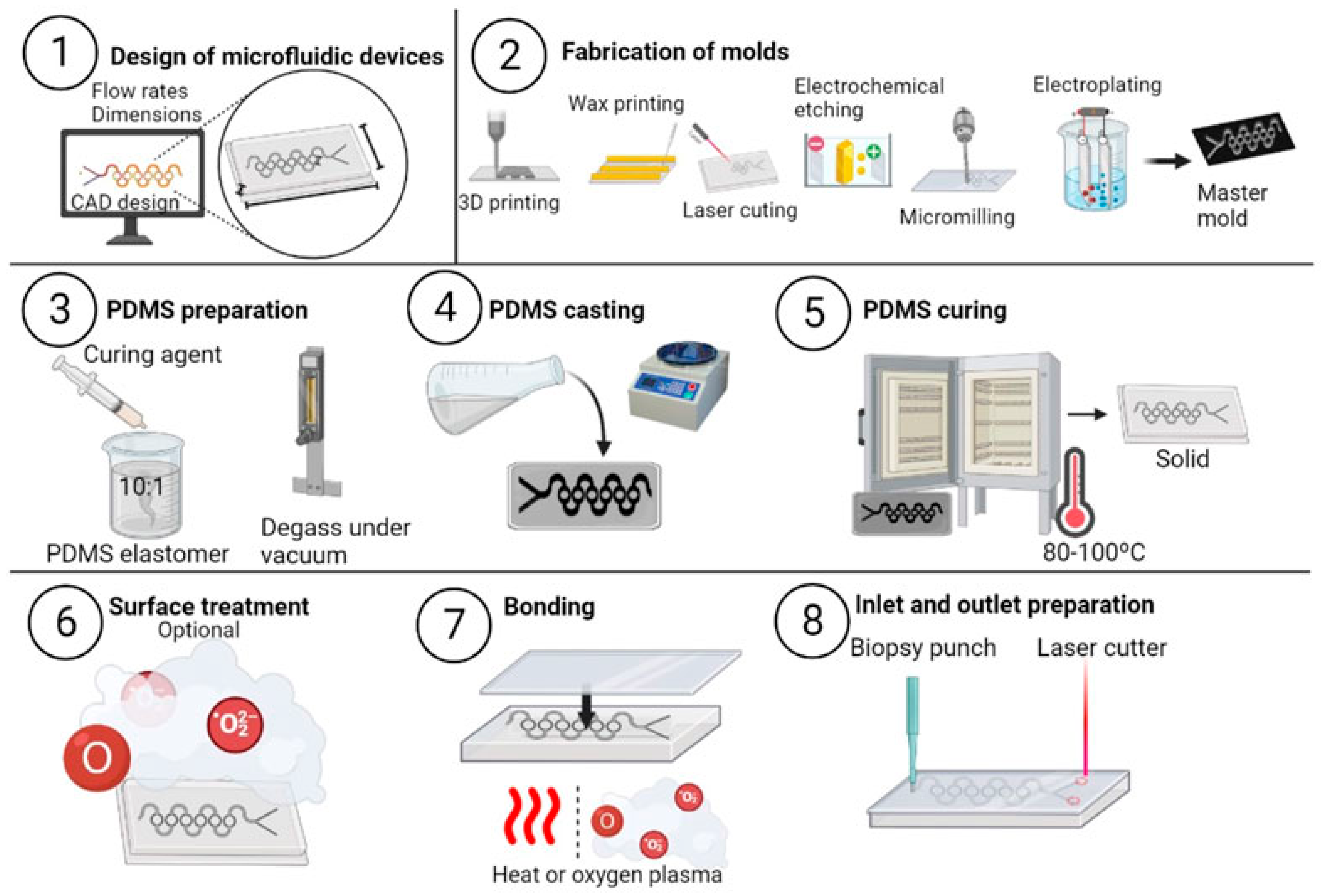
Injection molding: A type of thermoplastic manufacturing method has been developed for fabricating microfluidic gadgets in enormous quantities, exhibiting remarkable precision and uniformity, as shown in Figure 3. Injection molding offers another scalable fabrication route, where molten polymer is injected into micro-milled mold cavities, allowing for rapid mass production with excellent dimensional accuracy. Injection molding was utilized to replicate COC microfluidic chips from micro-milled masters, highlighting its potential for commercial biosensor manufacturing due to its short cycle times and reproducibility [5]. It consists of several stages: Mold fabrication: This is a technique that includes micro-milling and laser ablation, being built to generate a high-accuracy mold chamber typically made from nickel or stainless-steel material. The required microfluidic designs’ adverse effect can be seen in this type of mold [8]. Injection and melting of polymer: In a preheated container, pellets of thermoplastic material like COC and PMMA undergo melting prior to being injected inside the mold cavity with high levels of pressure. In particular, slim microchannel features are occupied with polymer due to pressure [36]. Freezing and ejection: For hardening of the polymer, the mold gets cooled down immediately following loading. The demolding technique is employed to get rid of the molded substance after it has completely hardened [8]. In contrast, Soft lithography employs elastomeric materials like polydimethylsiloxane (PDMS) cast against micro-milled molds to produce flexible replicas. While PDMS molding is advantageous for prototyping because of its biocompatibility and optical transparency, it is less suited for high-volume production due to mechanical wear and swelling. PDMS casting was showcased on micro-milled molds to fabricate disposable biosensor chips, demonstrating rapid prototyping flexibility. The PDMS casting of a master mold gets coated using a blend of PDMS base and healing substance, generally in a ratio of 10 to 1. To get rid of any air bubbles that may affect channel-related quality, the combined solution was decontaminated under a vacuum. Therefore, PDMS is cleaned and treated for a couple of hours at around 60 degrees Celsius until it settles [26].
Furthermore, hybrid molding techniques combining hot embossing with soft lithography have been explored to harness the strengths of both approaches, enabling multilayer chip fabrication with enhanced fluidic integration [2]. Surface treatments such as oxygen plasma and silanization are often applied post-molding to improve bonding and fluid compatibility. Punching and the process of demolding: Employing biopsy holes or punches leads to the accessibility of pores for liquid inflows, and outflows are created by punching once the fixed surface of PDMS is carefully stripped from the master mold. Bonding: Through the treatment of oxygen plasma, the PDMS surface is finally attached to an external planar substrate, generally a transparent glass plate or slide, or an additional slice of PDMS [37,38] (Figure 3, point no ⑥–⑧). Soft lithography is cost effective and simple to utilize, and its adaptability renders it an appealing option for enabling device development in educational and scientific environments. The utilization possibilities involving biological compliance and visibility of compounds such as PDMS seem advantageous; notably, those such as cell-based testing methods, organ-on-chip mechanisms, and vasculature designs (micro) are particularly appropriate for it. The generation of organic substances within miniature channels provides an additional advantage. This technique is additionally utilized in microreactor structures for efficient screening systems and droplet microfluidics for the research of those reactions that are involved in kinetics [38]. Its adaptability for biological analytics purposes is further enhanced by its ability to replicate sophisticated small structures using sub-micron accuracy and by its suitability to alterations of surface procedures, such as protein encapsulating and salinization as part [39]. Considering all this, vast applications in industry are restricted by problems such as a lack of mechanical resistance and the capacity to hold onto hydrophobic molecules [37].
Figure 3.
Soft lithography method to produce microfluidic devices. The procedure consists of the steps listed below: (1) Using CAD software, design the microfluidic device in consideration of the device’s specifications. (2) Create the master mold using various methods, such as 3D printing, wax printing, laser cutting, micro-milling, or electrochemical etching. (3) Create the PDMS solution by combining the PDM and curing agent with the PDMS. (4) Using a spin coater or by hand, evenly coat the mold with the PDMS mixture. (5) Cure the PDMS layer in an oven at temperatures between 80 °C and 100 °C for a few hours. The PDMS should then be removed from the mold and sized and shaped as desired. (6) Treat the surface of the PDMS device. Attach the PDMS device to the substrate’s lid using oxygen plasma or thermal bonding. Utilize a biopsy punch or a laser cutter to create the required inlets and outlets in the from design in software to fabrication of PDMS devise using soft lithography approach (reproduced from ref. [40] with permission, Frontiers in Bioengineering and Biotechnology, copyright 2023).
Particularly, in the general fabrication of recyclable, accessible, and financially effective devices, the process of injection molding is perfect, specifically for applications including POCT, referred to as point-of-care testing, in vitro diagnostics (IVD), and drug screening [33,36]. Furthermore, POCT technologies that include detectors or sensors for fluidic control in tiny cases and medical frameworks that require logical gadgets take advantage of injection molding. Moderate throughput, which makes it feasible to manufacture numerous gadgets each day, and multidimensional management must be achieved for accurate function of the devices and connectivity via an array of polymers, and this allows for modification of durability against chemicals, visual understanding, or biologic compatibility, with only a handful of the technologies having many positive aspects [33,41].
2.5. Decision Framework for Microfluidic Fabrication Selection
The Figure 4 presents a selection framework that guides users in choosing fabrication routes based on performance capability, cost, and application intent. Micro-milling enables ±5–20 µm tolerance for thermoplastic masters but may introduce burrs and chatter, mitigated through tool coatings and high-speed machining. Laser micromachining supports hard materials without tool wear, though melting and taper require fs/ps pulses and post-cleaning. Also, 3D printing offers rapid iteration but suffers layer artifacts, improved through orientation and post-curing. Hot embossing achieves high replication fidelity, while injection molding/ICM provides the lowest unit cost but requires strategies to control shrinkage and warpage.
Figure 4.
A systematic decision tree, which determines major performance criteria such as feature resolution, dimensional tolerance, material compatibility, roughness, volume of production, and the prevailing failure modes. The first step of the framework is the decision node prototyping versus production as the initial goal.
3. Biomedical and Clinical Diagnostics
3.1. Micro-Milling and Microfluidics in Point-of-Care Testing (POCT)
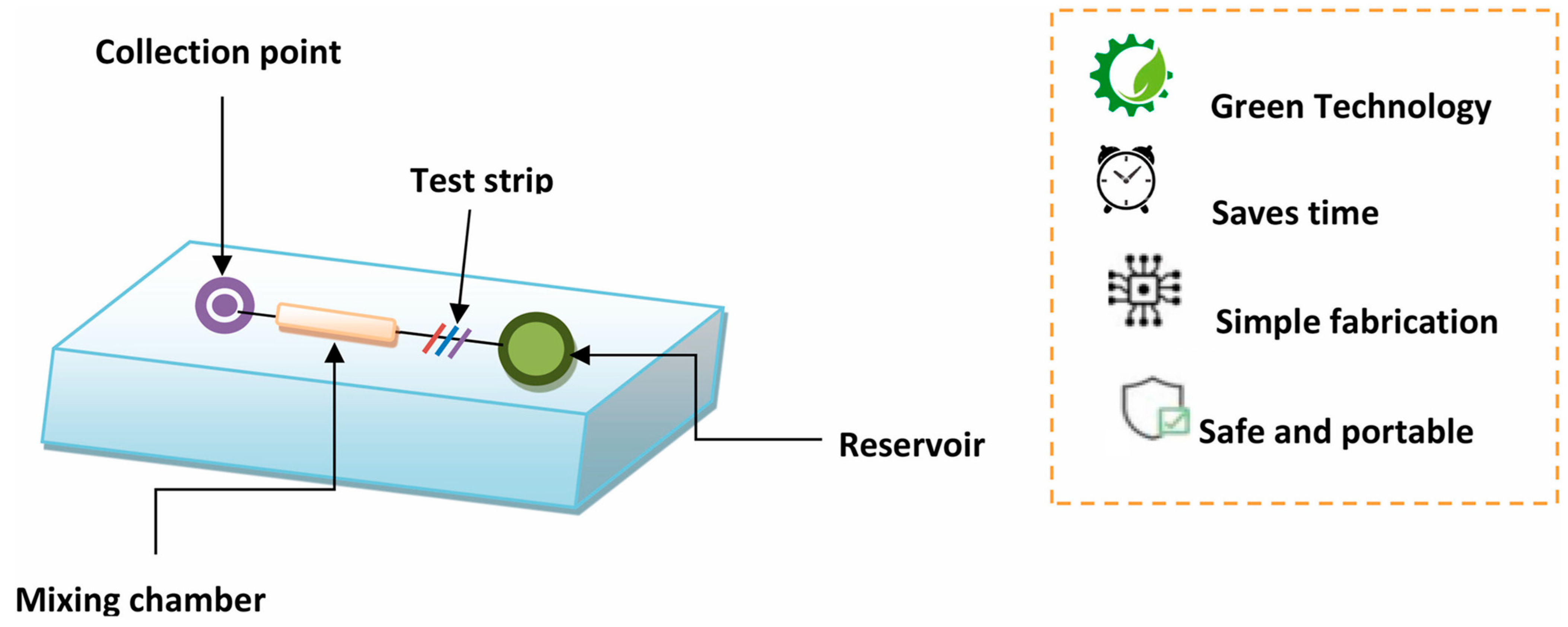
Point-of-care testing (POCT) demands rapid, sensitive, and portable diagnostic platforms, and micro-milling has become a prominent technique in fabricating microfluidic devices that meet these requirements. Micro-milling offers advantages such as low cost, rapid prototyping, and high precision, enabling the production of customizable microfluidic chips for diverse POCT applications. The researcher developed electrochemical immunosensors on micro-milled microfluidic chips capable of detecting protein biomarkers at picogram levels, facilitating early disease diagnosis Ref. [42]. Similarly, in another research the fabricated fluorescence and colorimetric microfluidic assays for the detection of infectious diseases with detection limits in the low nanogram per milliliter range [43]. The integration of lateral flow assays into micro-milled platforms further enhanced rapid biomarker detection, as shown in Figure 5, and it is demonstrated by researchers who reported improved sensitivity and specificity for hormone and pathogen detection [44].
Paper-based microfluidic devices fabricated by micro-milling have also been employed for enzymatic metabolite detection, providing a cost-effective solution with micromolar sensitivity suitable for metabolic monitoring [45]. One of the researcher team introduced aptamer-based electrochemical sensors on micro-milled chips that achieved ultrasensitive cancer biomarker detection at picomolar levels [46]. In the realm of nucleic acid diagnostics, scientist designed micro-milled microfluidic PCR chips enabling rapid viral RNA detection with reduced assay times [47]. Smartphone-integrated fluorescence detection systems combined with micro-milled microfluidic devices have allowed for portable and accurate HIV viral load quantification. The employed impedance spectroscopy within micro-milled microfluidics for precise electrolyte monitoring, achieving nanomolar sensitivity [20].
Beyond these, it was reported that surface plasmon resonance (SPR)-based micro-milled sensors for label-free, real-time detection of cardiac biomarkers, providing rapid diagnostics for acute myocardial infarction [39]. The fabricated colorimetric enzymatic assays on paper-based micro-milled microfluidic devices, enhancing POCT utility in resource-limited environments [36]. Scientist developed electrochemical DNA sensors on micro-milled chips for detecting genetic mutations with femtomolar sensitivity, emphasizing applications in personalized medicine [48]. Further advances include micro-milled ELISA chips for pregnancy hormone detection, CRISPR-integrated microfluidic chips for SARS-CoV-2 RNA detection with attomolar sensitivity, and loop-mediated isothermal amplification (LAMP)-based micro-milled devices for rapid bacterial pathogen detection. A team of researchers demonstrated optical biosensors fabricated via micro-milling for real-time enzymatic reaction monitoring in synthetic biology, highlighting the versatility of this fabrication method in diverse POCT contexts [49]. Collectively, these studies illustrate the significant role of micro-milling in advancing microfluidic POCT platforms by combining rapid fabrication, precision, and compatibility with various detection modalities. The ongoing innovation promises enhanced diagnostic speed, sensitivity, and accessibility crucial for global healthcare needs.
Figure 5.
Schematic of the microdevice showing the collection point, mixing chamber, test strip, and reservoir, along with key advantages including green technology, time efficiency, simple fabrication, and safe portability (reproduced from ref. [50] with permission, Elsevier, copyright 2024).
3.2. Drug Development and Screening
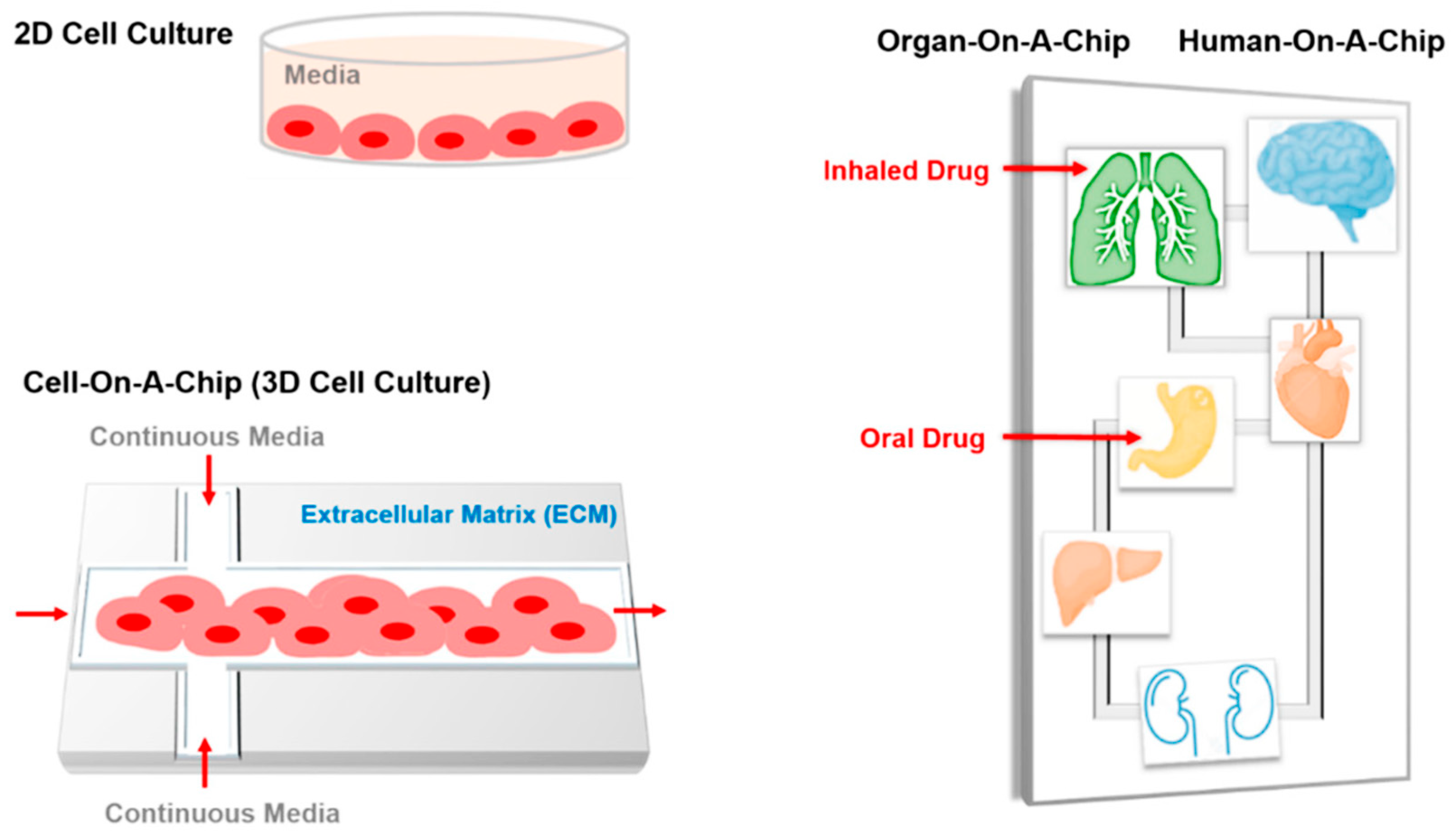
Micro-milling has emerged as a versatile and cost-effective fabrication technique in drug development and screening, enabling the rapid prototyping of microfluidic devices that closely mimic physiological environments. For example, CNC micro-milling has been used to create acrylic microfluidic devices for magnetic nanoparticle-based immunoassays, facilitating sensitive and low-cost detection of biomarkers like lysozyme, which is valuable for point-of-care testing in resource-limited settings [51]. Additionally, micro-milled PMMA chips have supported primary neuronal cultures and 3D neurospheroid growth, providing a controlled platform for evaluating neurotoxicity and drug efficacy [52]. In cancer research, low-cost micro-milled microfluidic devices have enabled high-throughput drug testing on static and dynamic 3D spheroid cultures, as shown in Figure 6, offering more accurate in vitro models that better represent in vivo conditions compared to traditional 2D cultures [53]. The precision, adaptability, and affordability of micro-milling make it a powerful tool for accelerating drug discovery and improving the reliability of preclinical testing, particularly in labs without access to expensive cleanroom facilities.
Figure 6.
Overview of cellular and organ-mimicking platforms: 2D cell culture in static media, 3D cell-on-a-chip culture integrating extracellular matrix and continuous media flow, and multi-organ/human-on-a-chip models enabling physiologically relevant testing of inhaled and oral drug pathways (reproduced from ref. [54] with permission, MDPI, copyright 2018).
3.3. Cell Culture and Analysis
Micro-milling has proven to be a valuable technique in the fabrication of microfluidic devices for cell culture and analysis, offering a cost-effective and precise alternative to traditional methods. For instance, a study developed a microfluidic spheroid-on-a-chip model using micro-milled PMMA and vapor-assisted thermal bonding, which supported the growth of 3D cell spheroids and provided a more accurate representation of in vivo conditions for drug testing and disease modeling.
Another research focused on creating low-cost CNC-milled PMMA microfluidic chips to support primary neuronal cultures and 3D neurospheroid growth, as shown in Figure 6, enabling a detailed study of neuronal development and response to stimuli [55]. Additionally, micro-milling was used to fabricate microfluidic devices capable of partially separating red blood cells from plasma, an important feature for cell sorting and analysis in hematology and diagnostics [56]. These examples underscore the versatility of micro-milling in creating microfluidic platforms that support cell culture and detailed cellular analysis in controlled environments.
3.4. Environmental Monitoring
Micro-milling has become an essential and versatile technique in developing microfluidic devices for environmental monitoring, offering both precision and cost effectiveness in fabricating intricate microchannels on materials such as PMMA and PDMS, as presented in Figure 7. These microfluidic platforms enable portable, sensitive, and high-throughput detection of environmental contaminants in air, water, and soil [57]. The researcher developed a micro-milled microfluidic system that uses magnetic microdiscs to tag Escherichia coli in large-volume water samples, achieving rapid and efficient bacterial capture critical for real-time water quality assessment [17]. These studies illustrate the growing impact of micro-milling in creating adaptable, low-cost, and portable microfluidic devices that enhance environmental monitoring by providing rapid, accurate detection and analysis of various contaminants. Furthermore, a fabricated low-cost CNC-milled PMMA chips capable of integrating biosensors to detect multiple bacterial contaminants simultaneously with high specificity and sensitivity, advancing multiplexed environmental monitoring capabilities [56]. Additionally, researcher showcased a micro-milled device designed for partial separation of red blood cells from plasma, a technique translating to cells in environmental bioanalysis, as shown in Figure 7 [58]. Moreover, scientists developed 3D high-quality PDMS microfluidic chips by micro-milling, enabling precise manipulation and detection of microparticles and pollutants in environmental samples [59].
Figure 7.
Illustration depicting the integration of micro-milled microfluidic technology for environmental monitoring. Airborne pollutants, including microbial contaminants (Staphylococcus aureus, Saccharomyces cerevisiae, and Escherichia coli).
3.5. Food Safety and Quality Control
Micro-milling has emerged as a pivotal fabrication technique in the development of microfluidic devices aimed at enhancing food safety and quality control. Its advantages, including rapid prototyping, high precision, and cost effectiveness, make it particularly well suited for producing customized microfluidic platforms capable of sensitive and on-site detection of food contaminants, pathogens, and spoilage indicators. One notable application is the detection of foodborne pathogens. Researcher reported the fabrication of a micro-milled polymethyl methacrylate (PMMA) microfluidic chip integrated with magnetic bead-based immunoassays for rapid and sensitive detection of Salmonella in food samples [60]. Their device achieved detection limits as low as 10 colony-forming units per milliliter (CFU/mL) within an hour, significantly shortening the analysis time compared to conventional laboratory techniques. In addition to microbial contamination, micro-milled microfluidic systems have been applied to chemical contaminant detection. Scientist developed a micro-milled device coupled with electrochemical sensors to detect organophosphorus pesticides in fruits [61]. The platform exhibited high sensitivity and selectivity for pesticide residues, enabling rapid, on-site screening that can help prevent contaminated produce from reaching consumers [62]. Such real-time detection is essential for minimizing public health risks associated with pesticide exposure. Furthermore, monitoring spoilage markers is vital for ensuring food freshness. Researchers designed a micro-milled microfluidic platform to detect biogenic amines, such as histamine, in seafood sample [63]. This system facilitates real-time assessment of spoilage levels, thereby supporting quality control and reducing food waste.
Beyond these examples, micro-milling has been utilized to fabricate microfluidic devices for detecting mycotoxins in grains through fluorescence-based assays [64] and for immunosensor-based allergen detection in diverse food matrices. These applications demonstrate the versatility of micro-milling in accommodating various detection modalities, including optical, electrochemical, and immunological methods [25]. As demand for on-site, real-time food quality monitoring grows, micro-milled microfluidic devices are poised to play a central role in safeguarding public health and ensuring food integrity across the supply chain [65]. Various approaches have been used to fabricate microfluidic devices, including soft lithography, injection molding, hot embossing, and 3D printing, each offering distinct advantages depending on the required resolutions, material properties, and the device’s function. Among these, high-precision CNC micro-milling has emerged as a robust and versatile technique, especially for rapid prototyping and the fabrication of complex three-dimensional microchannel architectures. The work demonstrated by the researcher presents an integrated digital-to-physical manufacturing pipeline [2]. The process begins with the construction of the microfluidic design in Fusion 360 CAD, where channel geometries, curvatures, and functional features are defined with micron-level control. This digital model is then transferred to Fusion 360 CAM, which generates optimized toolpaths that maintain accurate channel depths and smooth surfaces essential for laminar flow, predictable hydraulic resistance, and consistent mass transport within microfluidic systems. The CAM output is exported as a G-code, where every motion of the milling tool, including the spindle speed, feed rate, and axis positioning, is precisely encoded to ensure dimensional fidelity during fabrication. The G-code is subsequently executed through Mach3 CNC control software, which drives a five-axis micro-milling machine capable of producing intricate 3D geometries that are difficult or impossible to achieve with conventional 2D soft lithography [2]. Various approaches have been employed to fabricate modular and customizable microfluidic devices, ranging from soft lithography to micro-milling and multistep molding strategies. The construction of a PDMS-based microfluidic system for phenol sensing is depicted, beginning with the assembly of a PDMS channel layer bonded onto a glass substrate containing a screen-printed electrode. This configuration enables controlled fluid flow over the electrochemical sensing region. The modular dual-flow configuration enhances separation efficiency and improves sensor performance by allowing buffer and analyte streams to merge precisely at the catalytic detection interface. The process starts with a 3D-print master mold defining microchannels and structural studs. This master is used to create a tin-based silicone negative, which is subsequently replicated into a urethane mold. The final casting step yields a 2D-defined PDMS microfluidic block whose channels and geometric features match the original CAD design. This layered molding approach enables reproducible fabrication of microstructured components that can be integrated into larger modular systems [66]. The researcher expands this concept further by demonstrating the fabrication of fully 3D-defined PDMS building blocks that incorporate Lego-like interlocking features. A PDMS master mold is capped with a PDMS Lego® plate cap, and degassed PDMS is injected through a designated inlet to form the internal structure. Once cured, the mold is opened to expose the cast block. Minor defects or excess material are removed, yielding a clean 3D-defined block capable of physical interconnection with other units through its molded studs and sockets. These modular blocks enable rapid assembly of microfluidic networks without the need for permanent bonding, allowing channels, sensors, and functional modules to be rearranged or replaced as needed [67]. Together, these fabrication techniques highlight how multistep molding methods, PDMS casting, and Lego-like modularity provide a versatile platform for constructing customizable microfluidic systems. Such approaches support rapid prototyping, easy reconfiguration, and integration of sensing elements, making them valuable for phenol detection, biochemical assays, and a wide range of lab-on-chip applications (Figure 8) [68].
Figure 8.
Conceptual schematic of microfluidic device-based food safety monitoring, illustrating the detection of biological contaminants (viruses and bacteria) and chemical contaminants (pesticides and heavy metals) using integrated electrochemical, immunoassay, molecular, enzymatic, and chemical reaction-based sensing strategies within a single microfluidic platform [68].
4. Microfluidic Devices and Healthcare
The integration of artificial intelligence (AI) with microfluidic wearable devices represents a transformative step in the evolution of personalized healthcare and real-time healthcare/real-time diagnostics. Microfluidic wearables manipulate small volumes of biofluids such as sweat, interstitial fluid, or tears, providing continuous and minimally invasive access to biochemical markers. When combined with AI, particularly machine learning (ML) and deep learning algorithms, these systems can detect complex patterns, correct for noise, and offer predictive insights that traditional sensors cannot. Researcher developed a wearable microfluidic pressure sensor that enables real-time health monitoring [56]. Similarly, another team created a sweat glucose sensor integrated into a microfluidic colorimetric patch for diabetes management [69]. Scientists also demonstrated a soft smart contact lens for wireless ocular diagnostics, showcasing the integration of microfluidics with flexible electronics [70]. AI plays a crucial role by processing dynamic bio signals to detect physiological changes, anomalies, or disease onset. Scientist introduced its potential in point-of-care applications [36]. However, the adoption of these technologies faces several challenges: data security, device calibration process across populations, power constraints, and regulatory barriers. Studies like these are emphasize the need for standardized biomarker validations to ensure reliability [18]. Future directions include multi-modal sensing platforms, edge AI for local decision making, and closed-loop systems for drug delivery, such as smart insulin pumps. The synergy between AI and microfluidics is poised to shift healthcare from episodic to continuous and preventive models, but ethical deployment and robust clinical validation are essential. As these technologies evolve, AI-enhanced microfluidic wearables could redefine chronic disease management, athletic training, and even mental health monitoring on a global scale (Figure 9) [71].
Figure 9.
A schematic for what an idealized rapid and on-site microfluidic device can offer the field of diagnostics and the devices’ basic components [71].
Comparison and Analysis of Micro-Milling in Fields of Application
While micro-milling has been applied across diverse domains including point-of-care diagnostics, pharmaceutical screening, environmental monitoring, and food safety, its suitability as a fabrication strategy is governed by a common set of performance constraints and trade-offs that transcend individual applications. A critical comparison across these domains reveals that micro-milling’s primary strength lies in its ability to balance design flexibility, material compatibility, and cost efficiency at the prototyping to small-batch production scale rather than in ultra-high-resolution or mass-manufacturing scenarios [72].
From a fabrication capability perspective, micro-milling consistently enables rapid iteration of microfluidic layouts with dimensional tolerances typically in the ±5–20 µm range for thermoplastic polymers. This level of precision is sufficient for most electrochemical and biochemical biosensing platforms, including immunosensors, enzymatic assays, and nucleic acid amplification systems. In point-of-care testing and pharmaceutical screening, where channel dimensions primarily govern residence time, mixing efficiency, and electrode placement rather than optical diffraction limits, these tolerances are generally acceptable. In contrast, applications requiring sub-micron optical alignment or nanostructured surface features, such as advanced photonic biosensors, remain outside the practical resolution envelope of micro-milling and continue to favor lithographic approaches [73].
Surface roughness and burr formation emerge as cross-cutting limitations across all application areas. In POCT cartridges and environmental sensors, where capillary flow and reproducibility of fluid transport are critical, excessive roughness or edge burrs can introduce flow heterogeneity and signal variability. Pharmaceutical and cell-based platforms are particularly sensitive to these effects, as rough surfaces can alter cell adhesion, protein adsorption, and local concentration gradients. Although post-processing strategies such as secondary finishing passes, tool coatings, optimized feed-to-diameter ratios, and hybrid machining approaches have been shown to reduce roughness to sub-micron levels, these steps increase fabrication time and partially offset cost advantages. Thus, micro-milling represents a trade-off between achievable surface quality and fabrication throughput rather than a universally optimal solution [74].
Bonding and functionalization compatibility further differentiate micro-milling’s performance across domains. Thermoplastic substrates commonly used in micro-milling, such as PMMA and COC, offer broad compatibility with thermal, solvent, and adhesive bonding, enabling robust device sealing for POCT and environmental monitoring. Their surfaces can also be readily modified through plasma treatment, silanization, or electrode deposition, supporting immunoassays, nucleic acid detection, and electrochemical sensing. However, these same polymers may exhibit hydrophobic analyte adsorption or solvent sensitivity in long-term environmental or food monitoring applications, necessitating additional surface coatings or passivation layers. In comparison, PDMS-based soft lithography offers superior surface tunability but suffers from mechanical instability and poor scalability, highlighting micro-milling’s intermediate position between flexibility and robustness [75].
When examined from a repeatability and scalability standpoint, micro-milling demonstrates high device-to-device consistency at laboratory and pilot scales, making it well suited for research translation and early-stage commercialization. Nevertheless, extended production runs introduce variability due to tool wear, thermal drift, and burr accumulation, particularly when using ultra-small diameter tools [46]; These effects are largely absent in injection molding or hot embossing once molds are optimized, explaining why micro-milling is most effectively deployed as a master fabrication or bridge technology rather than a final high-volume manufacturing method. This hybrid role is consistently observed across biomedical, pharmaceutical, environmental, and food safety applications [76].
Importantly, a comparative view across application areas reveals that micro-milling’s value proposition is application dependent rather than universal. In POCT and decentralized diagnostics, its rapid prototyping capability and low infrastructure requirements directly support accessibility and customization. In pharmaceutical screening and cell-based assays, micro-milling enables complex three-dimensional channel architectures and modular designs that are difficult to achieve with planar lithography [77]. In environmental and food monitoring, its robustness and material versatility support field-deployable devices, albeit with additional surface engineering requirements. These distinctions underscore the need for decision-oriented fabrication selection rather than technique-driven adoption [66]. Various microfluidic biosensing platforms across application domains are summarized under Table 1.
Table 1.
This table summarizes representative examples of microfluidic biosensing platforms across application domains. For review articles, reported limits of detection correspond to the primary studies cited within the referenced reviews; where applicable, values are reported exactly as defined in the original experimental work to maintain unit consistency and contextual accuracy.
Overall, this comparative analysis demonstrates that micro-milling should not be viewed as a replacement for established microfabrication methods but rather as a strategically positioned technology that bridges the gap between rapid prototyping and scalable manufacturing. Its true promise lies in enabling economical, polymer-based biosensor chips where integration constraints, material compatibility, and process standardization are as critical as raw resolution, a perspective that is often underemphasized in prior narrative reviews [67].
5. Future Prospects and Biomedical Applications
Micro-milling has, over time, become a key component in microfluidic device fabrication because it is efficient and cost effective to produce rapid and precise prototypes. At present, this technology is experiencing its growth through its combination with the emerging field that includes, but is not limited to, those in healthcare and the personal well-being ecosystem. A notable step forward has been the intrusion of a variety of sensors, primarily electrochemical, optical, and biosensors, into the channel milled on a microscale. The smart lab-on-chip systems are primarily used in instances where devices are used for continuous glucose monitoring (CGM) and continuous electrolyte monitoring. The devices are often wireless and are therefore used by healthcare professionals for diagnostics in remote areas. In the meantime, combining micro-milling with 3D printing or laser ablation has resulted in the development of new hybrid fabrication methods capable of generating multi-material devices. As an example of this, the teaming up of our rigid PMMA with malleable PDMS has enabled the creation of a microfluidic platform for cardiovascular drug testing and the study of multiple organs that interact, thus stimulating in vivo conditions. With the commonly used PMMA material for developing POC devices, micro-milling should be carried out using sharp ultra-fine grain carbide end mills with polished, uncoated or DLC-coated flutes, which are preferred to minimize adhesion and burr formation. High cutting speeds of 150–300 m/min and spindle speeds between 20,000 and 60,000 rpm are recommended, combined with very low feed per tooth (0.3–4 µm) to maintain minimum chip thickness. Axial depth of cut should be limited to 5–10% of tool diameter, with radial engagement of 10–30%. Climb milling, air blast cooling, and effective chip evacuation are essential to prevent melting, cracking, and surface degradation.
In the field of personalized medicine, micro-milled chips are also a big help in patient-specific testing via the use of oncogenes or circulating tumor cells (CTC) for the reason that they assist in finding the treatment that will be the most productive and, in parallel, avoid unnecessary drug exposure. In other areas of biomedicine that constantly innovate, micro-milled chips are being designed both for infectious disease diagnosis, where parental screening can also come in, and, on the other hand, they provide a solution for controlled drug release, the question of implantable biosensors, and even the biodegradable devices made by utilizing materials that dissolve after use and do not require retrieval surgeries. Furthermore, the design of the chips and equipment and AI are taking place in such a way with the help of the software and AI that the design process is constantly changing, and this occurs in a faster way since the technology is so advanced that the software itself has the ability to find the appropriate channel and optimize fluidic dynamics, thus cutting down on the chances of error. Overall, the joining of micro-milling and electronic technology, material science, biomedicine, and AI will create the next generation of microfluidics, which is intelligent, responsive systems. Biodegradable micro-milled microfluidics designed for in vivo diagnostic or drug delivery then safely degrade inside the body (example via hydrolysis). This will lead to an eco-friendly breakthrough, which is ideal for temporary implants or remote diagnostics in resource-limited settings.
Furthermore, this review provides readers with a practical, decision-oriented framework that links micro-milling parameters and material choices to achievable dimensional tolerances, surface roughness, failure modes, and integration strategies across application domains such as point-of-care diagnostics, pharmaceutical screening, environmental monitoring, and food safety. This framework is intended to support rational fabrication strategy selection and process standardization, bridging the gap between laboratory-scale prototyping and translational biosensor deployment.
Author Contributions
Conceptualization, A.T. and S.P.; methodology, A.T.; formal analysis, A.M. and K.K., investigation, A.S., A.T. and S.P.; writing, A.T. and S.P.; writing—review and editing, A.T., S.P. and A.S.; supervision, K.K. and A.M. All authors have read and agreed to the published version of the manuscript.
Funding
This research received no external funding.
Data Availability Statement
The data presented in this study are available on request from the corresponding author.
Acknowledgments
The authors would like to acknowledge Bennett University for providing the infrastructure and resources to write this review.
Conflicts of Interest
The authors declare no conflicts of interest.
References
- Beebe, D.J.; Mensing, G.A.; Walker, G.M. Physics and Applications of Microfluidics in Biology. Annu. Rev. Biomed. Eng. 2002, 4, 261–286. [Google Scholar] [CrossRef] [Scilit]
- Modarelli, M.J.C.; Kot-Thompson, D.M.; Hoshino, K. 5-Axis CNC Micro-Milling Machine for Three-Dimensional Microfluidics. Lab Chip 2025, 25, 127–142. [Google Scholar] [CrossRef] [Scilit] [PubMed]
- Syafiq Rahim, M.; Annuar Ehsan, A. Micro Milling Process for the Rapid Prototyping of Microfluidic Devices. In Advances in Microfluidics and Nanofluids; IntechOpen: London, UK, 2021. [Google Scholar]
- Vázquez, E.; Rodríguez, C.A.; Elías-Zúñiga, A.; Ciurana, J. An Experimental Analysis of Process Parameters to Manufacture Metallic Micro-Channels by Micro-Milling. Int. J. Adv. Manuf. Technol. 2010, 51, 945–955. [Google Scholar] [CrossRef] [Scilit]
- Kulkarni, M.B.; Ayachit, N.H.; Aminabhavi, T.M. Biosensors and Microfluidic Biosensors: From Fabrication to Application. Biosensors 2022, 12, 543. [Google Scholar] [CrossRef] [Scilit] [PubMed]
- Ren, K.; Zhou, J.; Wu, H. Materials for Microfluidic Chip Fabrication. Acc. Chem. Res. 2013, 46, 2396–2406. [Google Scholar] [CrossRef] [Scilit]
- Gomes, M.C.; Brito, L.C.; Bacci da Silva, M.; Viana Duarte, M.A. Tool Wear Monitoring in Micromilling Using Support Vector Machine with Vibration and Sound Sensors. Precis. Eng. 2021, 67, 137–151. [Google Scholar] [CrossRef] [Scilit]
- Zhang, H.; Liu, H.; Zhang, N. A Review of Microinjection Moulding of Polymeric Micro Devices. Micromachines 2022, 13, 1530. [Google Scholar] [CrossRef] [Scilit]
- O’Toole, L.; Kang, C.-W.; Fang, F.-Z. Precision Micro-Milling Process: State of the Art. Adv. Manuf. 2021, 9, 173–205. [Google Scholar] [CrossRef] [Scilit]
- Balázs, B.Z.; Geier, N.; Takács, M.; Davim, J.P. A Review on Micro-Milling: Recent Advances and Future Trends. Int. J. Adv. Manuf. Technol. 2021, 112, 655–684. [Google Scholar] [CrossRef] [Scilit]
- Filiz, S.; Conley, C.M.; Wasserman, M.B.; Ozdoganlar, O.B. An Experimental Investigation of Micro-Machinability of Copper 101 Using Tungsten Carbide Micro-Endmills. Int. J. Mach. Tools Manuf. 2007, 47, 1088–1100. [Google Scholar] [CrossRef] [Scilit]
- Allen, M.C.; Lookmire, S.; Avci, E. An Alternative Micro-Milling Fabrication Process for Rapid and Low-Cost Microfluidics. Micromachines 2024, 15, 905. [Google Scholar] [CrossRef] [Scilit]
- Câmara, M.A.; Rubio, J.C.C.; Abrão, A.M.; Davim, J.P. State of the Art on Micromilling of Materials, a Review. J. Mater. Sci. Technol. 2012, 28, 673–685. [Google Scholar] [CrossRef] [Scilit]
- Chen, N.; Li, H.N.; Wu, J.; Li, Z.; Li, L.; Liu, G.; He, N. Advances in Micro Milling: From Tool Fabrication to Process Outcomes. Int. J. Mach. Tools Manuf. 2021, 160, 103670. [Google Scholar] [CrossRef] [Scilit]
- Whitesides, G.M. The Origins and the Future of Microfluidics. Nature 2006, 442, 368–373. [Google Scholar] [CrossRef] [Scilit]
- Boswell, B.; Islam, M.N.; Davies, I.J. A Review of Micro-Mechanical Cutting. Int. J. Adv. Manuf. Technol. 2018, 94, 789–806. [Google Scholar] [CrossRef] [Scilit]
- Shahbazi, M.-A.; Kant, K.; Kaplinsky, J.J.; Chidambara, V.A.; Bang, D.D.; Wolff, A. From 2D Fluidic Array Screening to 3D Bacterial Capturing Structures in a Point of Care System for Sepsis Diagnosis. In Proceedings of the 2017 IEEE 30th International Conference on Micro Electro Mechanical Systems (MEMS), Las Vegas, NV, USA, 23 January 2017; pp. 440–443. [Google Scholar]
- Ercetin, A.; Aslantaş, K.; Özgün, Ö.; Perçin, M.; Chandrashekarappa, M.P.G. Optimization of Machining Parameters to Minimize Cutting Forces and Surface Roughness in Micro-Milling of Mg13Sn Alloy. Micromachines 2023, 14, 1590. [Google Scholar] [CrossRef] [Scilit]
- O’Toole, L.; Fang, F.-Z. Optimal Tool Design in Micro-Milling of Difficult-to-Machine Materials. Adv. Manuf. 2023, 11, 222–247. [Google Scholar] [CrossRef] [Scilit]
- Lai, F.; Hu, A.; Mao, K.; Wu, Z.; Lin, Y. Effect of Milling Processing Parameters on the Surface Roughness and Tool Cutting Forces of T2 Pure Copper. Micromachines 2023, 14, 224. [Google Scholar] [CrossRef] [Scilit]
- Wu, X.; Kang, Q.; Jiang, X.; Fang, X. Machinability and Surface Properties of Cryogenic Poly(Methyl Methacrylate) Machined via Single-Point Diamond Turning. Materials 2024, 17, 866. [Google Scholar] [CrossRef] [Scilit]
- Ma, L.; Howard, I.; Pang, M.; Wang, Z.; Su, J. Experimental Investigation of Cutting Vibration during Micro-End-Milling of the Straight Groove. Micromachines 2020, 11, 494. [Google Scholar] [CrossRef] [Scilit]
- Aslantas, K.; Hascelik, A.; Çiçek, A. Performance Evaluation of DLC and NCD Coatings in Micro-Milling of Al7075-T6 Alloy. J. Manuf. Process. 2022, 81, 976–990. [Google Scholar] [CrossRef] [Scilit]
- Assurin, S.R.; Mativenga, P.; Rajab, F.; Cooke, K.; Field, S. Laser De-Coating of Hard DLC Coatings from Tungsten Carbide Cutting Tool. Proc. Inst. Mech. Eng. B J. Eng. Manuf. 2021, 235, 13–22. [Google Scholar] [CrossRef] [Scilit]
- Wang, L.; Hou, P.; Wang, S.; Wang, C.; Yan, X.; Wang, X. Study on the Effect of Rolling Mill Dynamic Stiffness on Coupled Vibration of Hydraulic Machine. J. Vibroeng. 2024, 26, 455–468. [Google Scholar] [CrossRef] [Scilit]
- Wu, X.; Liu, L.; Du, M.; Shen, J.; Jiang, F.; Li, Y.; Lin, Y. Experimental Study on the Minimum Undeformed Chip Thickness Based on Effective Rake Angle in Micro Milling. Micromachines 2020, 11, 924. [Google Scholar] [CrossRef] [Scilit]
- Wang, G.; Yu, T.; Zhou, X.; Guo, R.; Chen, M. Investigation on Minimum Uncut Chip Thickness and Size Effect in Micro Milling of Glow Discharge Polymer (GDP). J. Manuf. Process. 2022, 84, 786–797. [Google Scholar] [CrossRef] [Scilit]
- De Oliveira, F.B.; Rodrigues, A.R.; Coelho, R.T.; De Souza, A.F. Size Effect and Minimum Chip Thickness in Micromilling. Int. J. Mach. Tools Manuf. 2015, 89, 39–54. [Google Scholar] [CrossRef] [Scilit]
- Skrzyniarz, M. A Method to Determine the Minimum Chip Thickness during Longitudinal Turning. Micromachines 2020, 11, 1029. [Google Scholar] [CrossRef] [Scilit]
- Ducobu, F.; Filippi, E.; Rivière-Lorphèvre, E. Chip Formation and Minimum Chip Thickness in Micro-Milling. In Proceedings of the 12th CIRP Conference on Modelling of Machining Operations, Donostia-San Sebastian, Spain, 7–8 May 2009. [Google Scholar]
- Zhang, L.; He, X.; Chen, G.; Wang, T.; Tang, Y.; He, Z. Effects of Rf Power on Chemical Composition and Surface Roughness of Glow Discharge Polymer Films. Appl. Surf. Sci. 2016, 366, 499–505. [Google Scholar] [CrossRef] [Scilit]
- Wang, Z. Detection of Mycotoxin in Foods Using an MIP-Based Paper-Fluidic Device. Master’s Thesis, McGill University, Montreal, QC, Canada, 2023. [Google Scholar]
- Juang, Y.-J.; Chiu, Y.-J. Fabrication of Polymer Microfluidics: An Overview. Polymers 2022, 14, 2028. [Google Scholar] [CrossRef] [Scilit]
- Zhang, H.; Wu, Z.; Lu, H.; Hao, Z.; Liu, W.; Zhang, F.; Zhang, R.; Li, X.; Zhang, L. Efficient Bimetallic Nanozyme Precisely Tuned by DNA for High-Throughput Foldable Paper Biosensing. Chem. Eng. J. 2025, 503, 158504. [Google Scholar] [CrossRef] [Scilit]
- Zhao, B.; Han, M.; Wang, Q.; Huang, Z.; Liang, Y.; Tumba, K.; Li, M.; Chen, W.; Kamal, M.; Chang, Z.; et al. Polylactic Acid-Based Composite Filter with Multi-Gradient Structure Developed for High-Efficiency Particulate Matter Filtration and NH3 Purification. Sep. Purif. Technol. 2025, 354, 129354. [Google Scholar] [CrossRef] [Scilit]
- Shakeri, A.; Khan, S.; Didar, T.F. Conventional and Emerging Strategies for the Fabrication and Functionalization of PDMS-Based Microfluidic Devices. Lab Chip 2021, 21, 3053–3075. [Google Scholar] [CrossRef] [Scilit] [PubMed]
- Jenkins, G. Rapid Prototyping of PDMS Devices Using SU-8 Lithography. In Microfluidics Diagnostics Methods in Molecular Biology; Humana Press Inc.: Totowa, NJ, USA, 2013; Volume 949, pp. 153–168. [Google Scholar]
- Xu, J.; Harasek, M.; Gföhler, M. From Soft Lithography to 3D Printing: Current Status and Future of Microfluidic Device Fabrication. Polymers 2025, 17, 455. [Google Scholar] [CrossRef] [Scilit] [PubMed]
- Choudhary, S.; Altintas, Z. Development of a Point-of-Care SPR Sensor for the Diagnosis of Acute Myocardial Infarction. Biosensors 2023, 13, 229. [Google Scholar] [CrossRef] [Scilit]
- Rodríguez, C.F.; Andrade-Pérez, V.; Vargas, M.C.; Mantilla-Orozco, A.; Osma, J.F.; Reyes, L.H.; Cruz, J.C. Breaking the Clean Room Barrier: Exploring Low-Cost Alternatives for Microfluidic Devices. Front. Bioeng. Biotechnol. 2023, 11, 1176557. [Google Scholar] [CrossRef] [Scilit]
- Zhang, N.; Guan, T.; Zhang, H. Polymer Micro Fabrication. Encycl. Mater. Plast. Polym. 2022, 3, 548–564. [Google Scholar] [CrossRef] [Scilit]
- Ebrahimi, G.; Samadi Pakchin, P.; Shamloo, A.; Mota, A.; de la Guardia, M.; Omidian, H.; Omidi, Y. Label-Free Electrochemical Microfluidic Biosensors: Futuristic Point-of-Care Analytical Devices for Monitoring Diseases. Microchim. Acta 2022, 189, 252. [Google Scholar] [CrossRef] [Scilit]
- Lehnert, T.; Gijs, M.A.M. Microfluidic Systems for Infectious Disease Diagnostics. Lab Chip 2024, 24, 1441–1493. [Google Scholar] [CrossRef] [Scilit]
- Rubio-Monterde, A.; Quesada-González, D.; Merkoçi, A. Toward Integrated Molecular Lateral Flow Diagnostic Tests Using Advanced Micro- and Nanotechnology. Anal. Chem. 2023, 95, 468–489. [Google Scholar] [CrossRef] [Scilit]
- Nadar, S.S.; Patil, P.D.; Tiwari, M.S.; Ahirrao, D.J. Enzyme Embedded Microfluidic Paper-Based Analytic Device (ΜPAD): A Comprehensive Review. Crit. Rev. Biotechnol. 2021, 41, 1046–1080. [Google Scholar] [CrossRef] [Scilit]
- Sequeira-Antunes, B.; Ferreira, H.A. Nucleic Acid Aptamer-Based Biosensors: A Review. Biomedicines 2023, 11, 3201. [Google Scholar] [CrossRef] [Scilit]
- Xiao, M.; Tian, F.; Liu, X.; Zhou, Q.; Pan, J.; Luo, Z.; Yang, M.; Yi, C. Virus Detection: From State-of-the-Art Laboratories to Smartphone-Based Point-of-Care Testing. Adv. Sci. 2022, 9, 2105904. [Google Scholar] [CrossRef] [Scilit]
- Kalligosfyri, P.M.; Lamprou, E.; Kalogianni, D.P. Emerging Sensing Technologies for Liquid Biopsy Applications: Steps Closer to Personalized Medicine. Sensors 2024, 24, 7902. [Google Scholar] [CrossRef] [Scilit] [PubMed]
- Jarnda, K.V.; Dai, H.; Ali, A.; Bestman, P.L.; Trafialek, J.; Roberts-Jarnda, G.P.; Anaman, R.; Kamara, M.G.; Wu, P.; Ding, P. A Review on Optical Biosensors for Monitoring of Uric Acid and Blood Glucose Using Portable POCT Devices: Status, Challenges, and Future Horizons. Biosensors 2025, 15, 222. [Google Scholar] [CrossRef] [Scilit] [PubMed]
- Rafiq, S.M.; Majumder, R.; Joshi, D.; Dar, A.H.; Dash, K.K.; Pandey, V.K.; Sidiqi, U.S. Lab-on-a-Chip Device for Food Quality Control and Safety. Food Control 2024, 164, 110596. [Google Scholar] [CrossRef] [Scilit]
- Yen, D.P.; Ando, Y.; Shen, K. A Cost-Effective Micromilling Platform for Rapid Prototyping of Microdevices. Technology 2016, 4, 234–239. [Google Scholar] [CrossRef] [Scilit]
- Teixeira, M.I.; Amaral, M.H.; Costa, P.C.; Lopes, C.M.; Lamprou, D.A. Recent Developments in Microfluidic Technologies for Central Nervous System Targeted Studies. Pharmaceutics 2020, 12, 542. [Google Scholar] [CrossRef] [Scilit]
- Cardoso, B.D.; Castanheira, E.M.S.; Lanceros-Méndez, S.; Cardoso, V.F. Recent Advances on Cell Culture Platforms for In Vitro Drug Screening and Cell Therapies: From Conventional to Microfluidic Strategies. Adv. Healthc. Mater. 2023, 12, 2202936. [Google Scholar] [CrossRef] [Scilit]
- Damiati, S.; Kompella, U.B.; Damiati, S.A.; Kodzius, R. Microfluidic Devices for Drug Delivery Systems and Drug Screening. Genes 2018, 9, 103. [Google Scholar] [CrossRef] [Scilit]
- Behroodi, E.; Latifi, H.; Bagheri, Z.; Ermis, E.; Roshani, S.; Salehi Moghaddam, M. A Combined 3D Printing/CNC Micro-Milling Method to Fabricate a Large-Scale Microfluidic Device with the Small Size 3D Architectures: An Application for Tumor Spheroid Production. Sci. Rep. 2020, 10, 22171. [Google Scholar] [CrossRef] [Scilit]
- CNC Machining Surface Roughness: A Complete Guide. Available online: https://geomiq.com/blog/cnc-machining-surface-roughness-guide/ (accessed on 31 December 2025).
- Castillo-Torres, K.Y.; Arnold, D.P.; McLamore, E.S. Rapid Isolation of Escherichia coli from Water Samples Using Magnetic Microdiscs. Sens. Actuators B Chem. 2019, 291, 58–66. [Google Scholar] [CrossRef] [Scilit]
- Baillargeon, K.R.; Mace, C.R. Microsampling Tools for Collecting, Processing, and Storing Blood at the Point-of-care. Bioeng. Transl. Med. 2023, 8, e10476. [Google Scholar] [CrossRef] [Scilit] [PubMed]
- Mesquita, P.; Gong, L.; Lin, Y. Low-Cost Microfluidics: Towards Affordable Environmental Monitoring and Assessment. Front. Lab Chip Technol. 2022, 1, 1074009. [Google Scholar] [CrossRef] [Scilit]
- Zhao, X.; Li, M.; Liu, Y. Microfluidic-Based Approaches for Foodborne Pathogen Detection. Microorganisms 2019, 7, 381. [Google Scholar] [CrossRef] [Scilit] [PubMed]
- Hossain, M.I.; Hasnat, M.A. Recent Advancements in Non-Enzymatic Electrochemical Sensor Development for the Detection of Organophosphorus Pesticides in Food and Environment. Heliyon 2023, 9, e19299. [Google Scholar] [CrossRef] [Scilit]
- Kant, K.; Ngo, T. Solid Phase PCR on 3D Microstructure ArrayChip for Pathogen Detection Application. Bio. Protoc. 2019, 9, e3323. [Google Scholar] [CrossRef] [Scilit]
- Givanoudi, S.; Heyndrickx, M.; Depuydt, T.; Khorshid, M.; Robbens, J.; Wagner, P. A Review on Bio- and Chemosensors for the Detection of Biogenic Amines in Food Safety Applications: The Status in 2022. Sensors 2023, 23, 613. [Google Scholar] [CrossRef] [Scilit]
- Reynolds, C.B. Novel Microfluidic Device for In-Field Purification, Amplification, and Detection of Dna from Mycotoxin-Producing Fungi. Master’s Thesis, Purdue University, West Lafayette, IN, USA, 2023. [Google Scholar] [CrossRef]
- Bindu, A.; Bhadra, S.; Nayak, S.; Khan, R.; Prabhu, A.A.; Sevda, S. Bioelectrochemical Biosensors for Water Quality Assessment and Wastewater Monitoring. Open Life Sci. 2024, 19, 20220933. [Google Scholar] [CrossRef] [Scilit]
- Attia, U.M.; Alcock, J.R. Evaluating and Controlling Process Variability in Micro-Injection Moulding. Int. J. Adv. Manuf. Technol. 2011, 52, 183–194. [Google Scholar] [CrossRef] [Scilit]
- Rohr, T.; Ogletree, D.F.; Svec, F.; Fréchet, J.M.J. Surface Functionalization of Thermoplastic Polymers for the Fabrication of Microfluidic Devices by Photoinitiated Grafting. Adv. Funct. Mater. 2003, 13, 264–270. [Google Scholar] [CrossRef] [Scilit]
- Diep Trinh, T.N.; Trinh, K.T.L.; Lee, N.Y. Microfluidic Advances in Food Safety Control. Food Res. Int. 2024, 176, 113799. [Google Scholar] [CrossRef] [Scilit]
- Sun, X.; Chen, W.; Zhang, B. A Portable Microfluidic Device Integrated with Electrochemical Sensing Platform for Detection of Multiple Binders in Ancient Wall Paintings. Anal. Chim. Acta 2023, 1275, 341562. [Google Scholar] [CrossRef] [Scilit] [PubMed]
- Cho, D.; Park, J.; Kim, T.; Jeon, S.; Cho, D.; Park, J.; Kim, T.; Jeon, S. Recent Advances in Lithographic Fabrication of Micro-/Nanostructured Polydimethylsiloxanes and Their Soft Electronic Applications. J. Semicond. 2019, 40, 111605. [Google Scholar] [CrossRef] [Scilit]
- Wang, S.; Chinnasamy, T.; Lifson, M.A.; Inci, F.; Demirci, U. Flexible Substrate-Based Devices for Point-of-Care Diagnostics. Trends Biotechnol. 2016, 34, 909–921. [Google Scholar] [CrossRef] [Scilit] [PubMed]
- Jung, W.-C.; Heo, Y.-M.; Yoon, G.-S.; Shin, K.-H.; Chang, S.-H.; Kim, G.-H.; Cho, M.-W. Micro Machining of Injection Mold Inserts for Fluidic Channel of Polymeric Biochips. Sensors 2007, 7, 1643–1654. [Google Scholar] [CrossRef] [Scilit]
- Bodziak, S.; de Souza, A.F.; Rodrigues, A.R.; Diniz, A.E.; Coelho, R.T. Surface Integrity of Moulds for Microcomponents Manufactured by Micromilling and Electro-Discharge Machining. J. Braz. Soc. Mech. Sci. Eng. 2013. [Google Scholar] [CrossRef] [Scilit]
- Scott, S.; Ali, Z. Fabrication Methods for Microfluidic Devices: An Overview. Micromachines 2021, 12, 319. [Google Scholar] [CrossRef] [Scilit]
- Miranda Giraldo, M.; Serje, D.A.; Pacheco Bolívar, J.A.; Bris Cabrera, J.L. Burr Formation and Control for Polymers Micro-Milling: A Case Study with Vortex Tube Cooling. Dyna 2017, 84, 150–159. [Google Scholar] [CrossRef] [Scilit]
- Borók, A.; Laboda, K.; Bonyár, A. PDMS Bonding Technologies for Microfluidic Applications: A Review. Biosensors 2021, 11, 292. [Google Scholar] [CrossRef] [Scilit]
- Rodrigues, R.O.; Lima, R.; Gomes, H.T.; Silva, A.M.T. Polymer Microfluidic Devices: An Overview of Fabrication Methods. U. Porto J. Eng. 2017, 1, 67–79. [Google Scholar] [CrossRef] [Scilit]
- Cooper, R.M.; Leslie, D.C.; Domansky, K.; Jain, A.; Yung, C.; Cho, M.; Workman, S.; Super, M.; Ingber, D.E. A Microdevice for Rapid Optical Detection of Magnetically Captured Rare Blood Pathogens. Lab Chip 2014, 14, 182–188. [Google Scholar] [CrossRef] [Scilit]
- Kant, K.; Ngo, T.A.; Matteucci, M.; Wolff, A. Fabrication of 3D Microstructure Array on Chip for Rapid Pathogen Detection. Sens. Actuators B Chem. 2019, 281, 774–782. [Google Scholar] [CrossRef] [Scilit]
- Ding, Y.; Huang, C.; Zhang, Y.; Wang, J.; Wang, X. Magnetic Microbead Enzyme-Linked Immunoassay Based on Phage Encoded Protein RBP 41-Mediated for Rapid and Sensitive Detection of Salmonella in Food Matrices. Food Res. Int. 2023, 163. [Google Scholar] [CrossRef] [Scilit] [PubMed]
- Tripathi, A.; Bonilla-Cruz, J. Review on Healthcare Biosensing Nanomaterials. ACS Appl. Nano Mater. 2023, 6, 5042–5074. [Google Scholar] [CrossRef] [Scilit]
- Jin, N.; Xue, L.; Guo, R.; Wang, S.; Liu, Y.; Liao, M.; Li, Y.; Lin, J. Staggered Magnetic Bead Chains Enhanced Bacterial Colorimetric Biosensing. LWT 2022, 169, 114031. [Google Scholar] [CrossRef] [Scilit]
- Lin, C.; Li, Y.; Peng, Y.; Zhao, S.; Xu, M.; Zhang, L.; Huang, Z.; Shi, J.; Yang, Y. Recent Development of Surface-Enhanced Raman Scattering for Biosensing. J. Nanobiotechnol. 2023, 21, 1–37. [Google Scholar] [CrossRef] [Scilit]
- Macovei, D.G.; Irimes, M.B.; Hosu, O.; Cristea, C.; Tertis, M. Point-of-Care Electrochemical Testing of Biomarkers Involved in Inflammatory and Inflammatory-Associated Medical Conditions. Anal. Bioanal. Chem. 2023, 415, 1033–1063. [Google Scholar] [CrossRef] [Scilit]
- Raihan, M.F.; Septiani, N.L.W.; Gumilar, G.; Manurung, R.V.; Jenie, S.N.A.; Nuruddin, A.; Yuliarto, B. Developing Point-of-Care Diagnosis Using Electrochemical Biosensor: Mosquito-Borne Disease. Sens. Actuators Rep. 2025, 9, 100261. [Google Scholar] [CrossRef] [Scilit]
- Han, G.-R.; Goncharov, A.; Eryilmaz, M.; Joung, H.-A.; Ghosh, R.; Yim, G.; Chang, N.; Kim, M.; Ngo, K.; Veszpremi, M.; et al. Deep Learning-Enhanced Paper-Based Vertical Flow Assay for High-Sensitivity Troponin Detection Using Nanoparticle Amplification. ACS Nano 2024, 18, 27933–27948. [Google Scholar] [CrossRef] [Scilit]
- Nan, Y.; Zuo, P.; Ye, B. Paper-Based Microfluidic Device for Extracellular Lactate Detection. Biosensors 2024, 14, 442. [Google Scholar] [CrossRef] [Scilit]
- Guo, L.; Zhao, Y.; Huang, Q.; Huang, J.; Tao, Y.; Chen, J.; Li, H.Y.; Liu, H. Electrochemical Protein Biosensors for Disease Marker Detection: Progress and Opportunities. Microsyst. Nanoeng. 2024, 10, 65. [Google Scholar] [CrossRef] [Scilit]
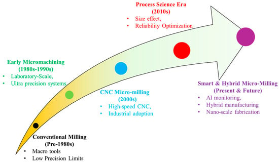
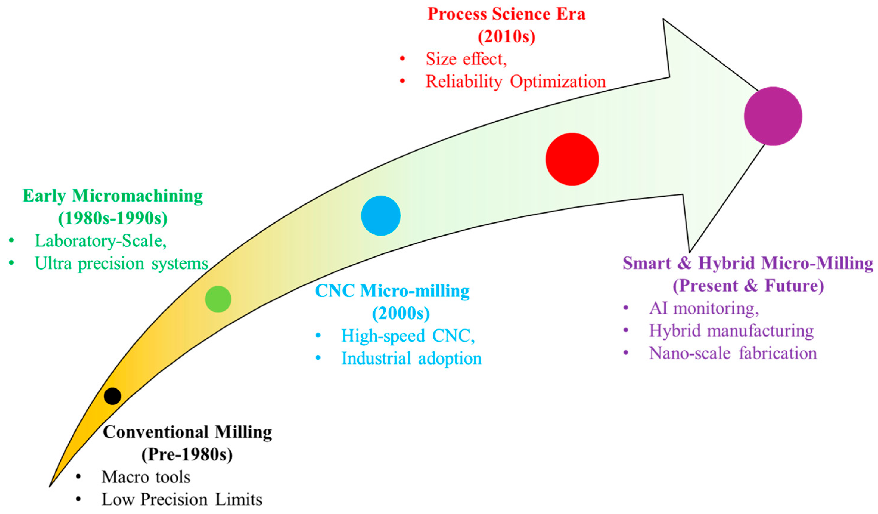
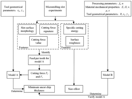
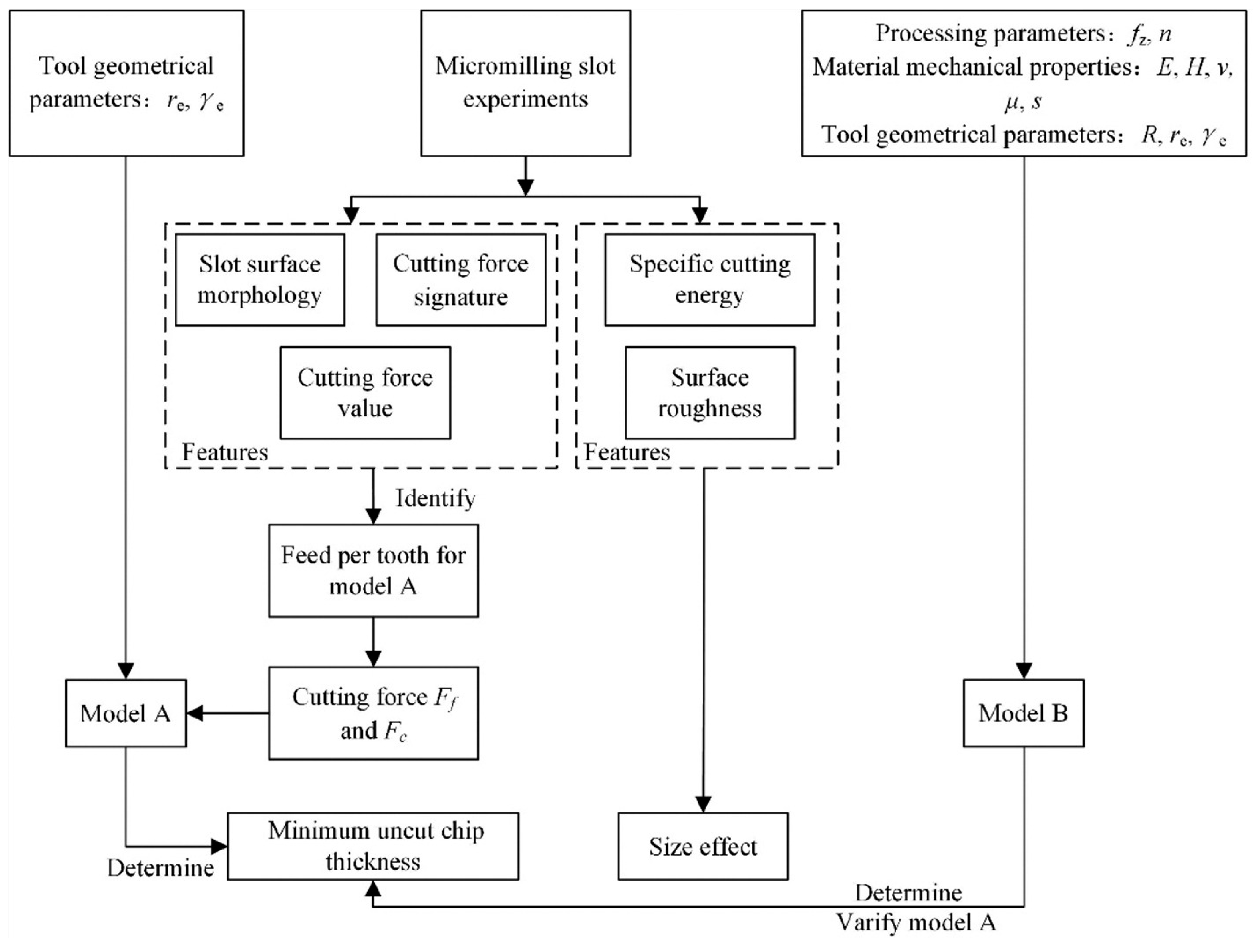
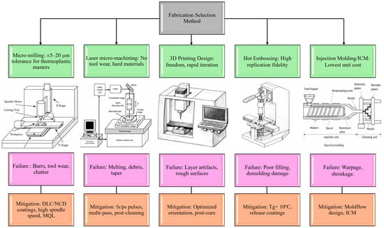
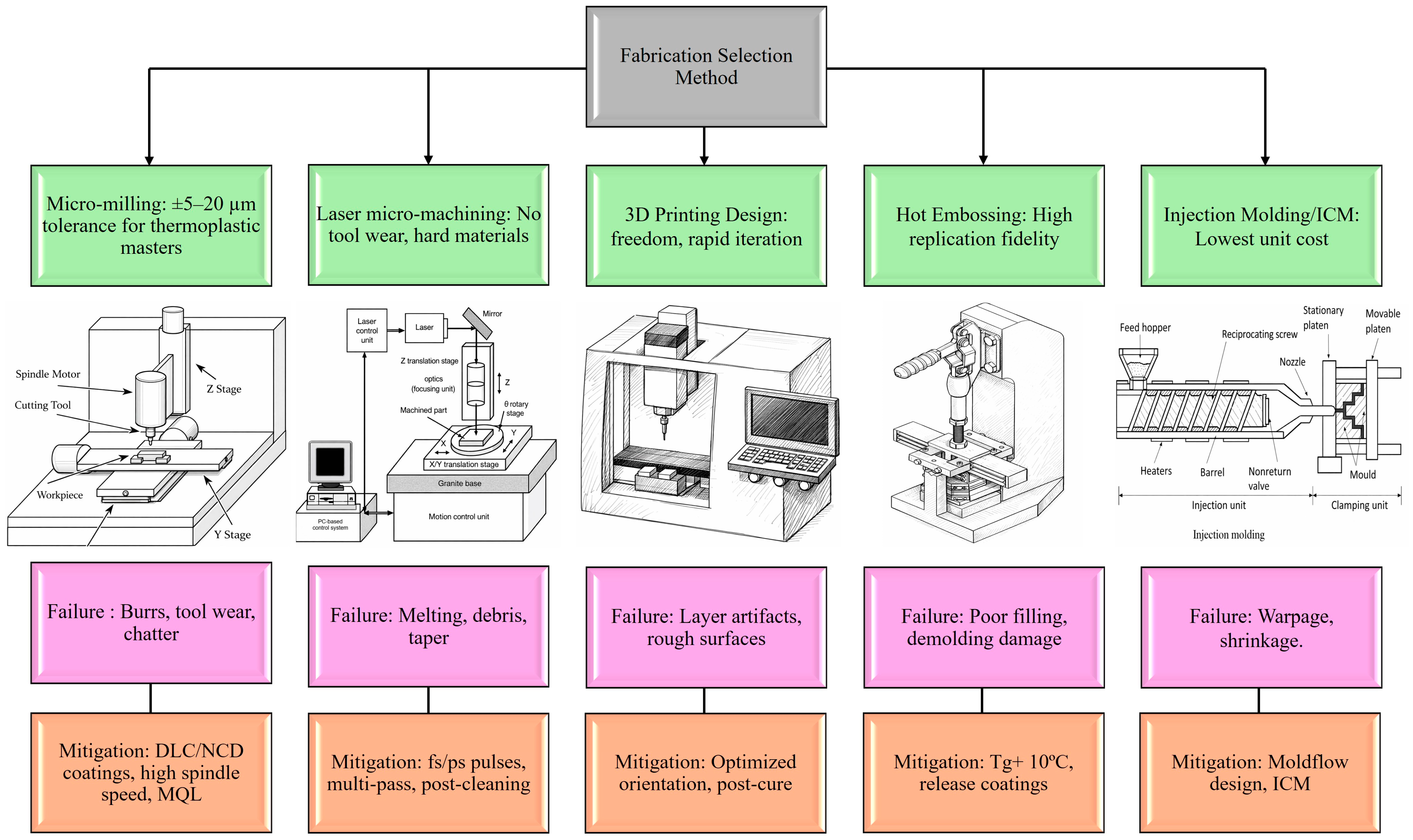
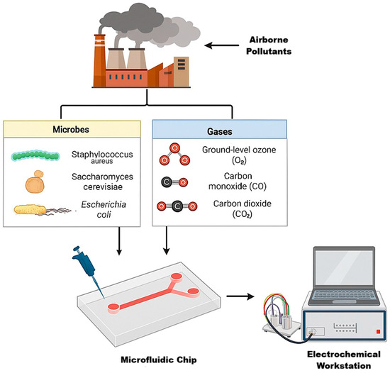
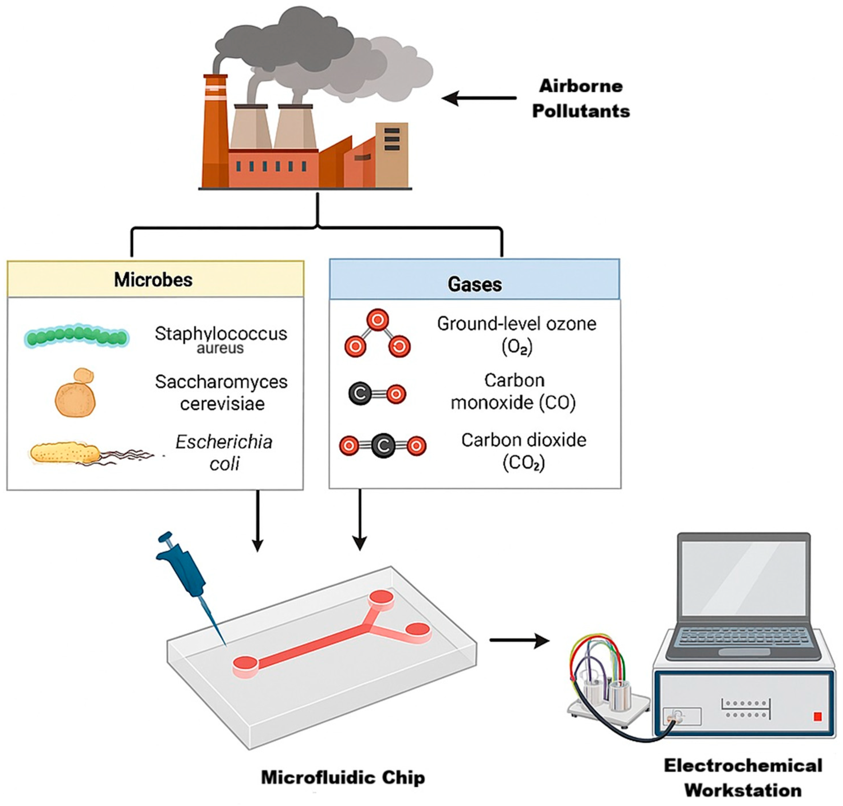
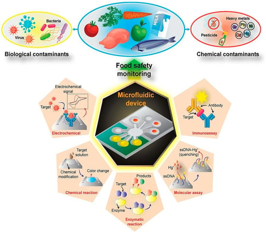
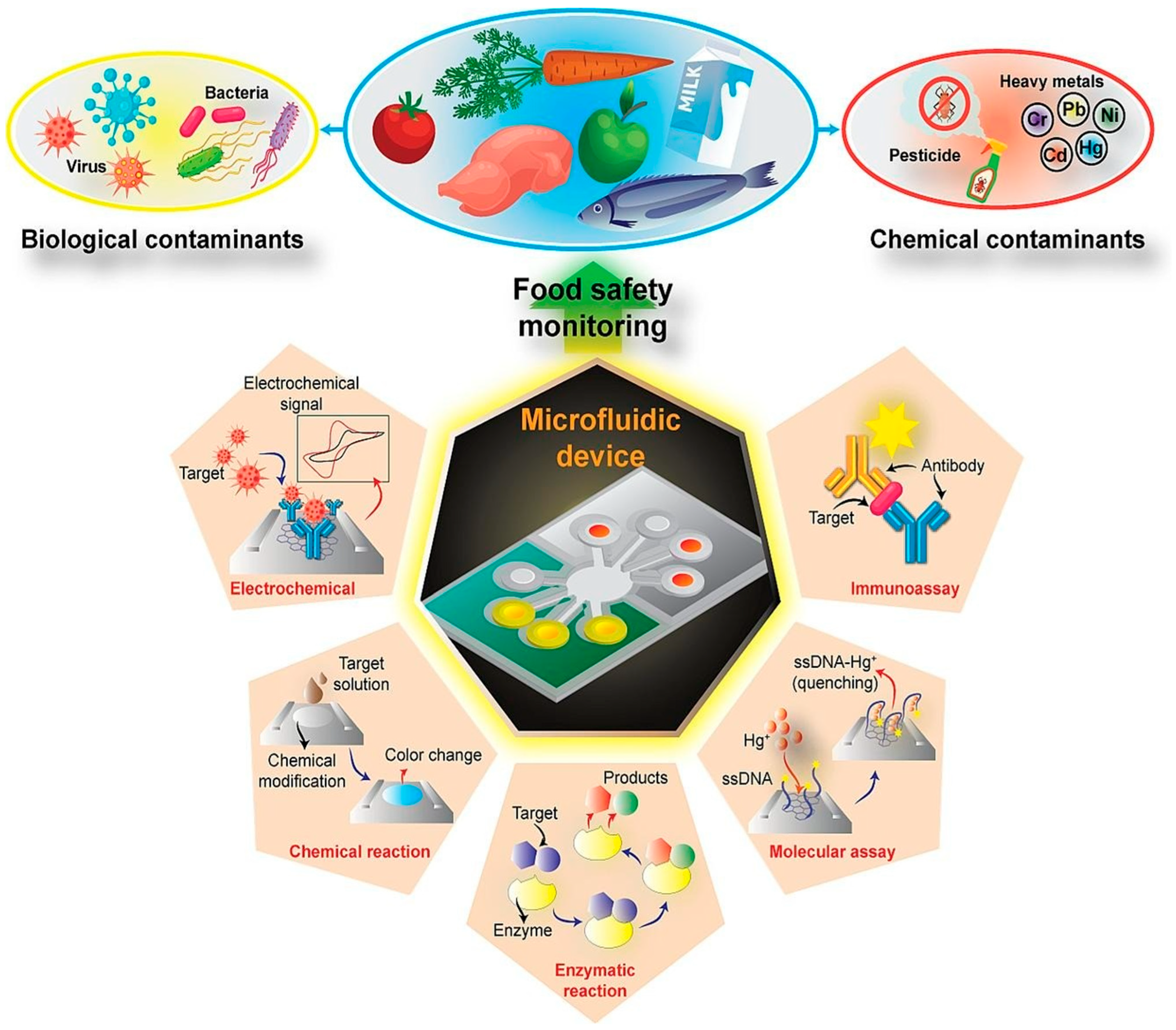
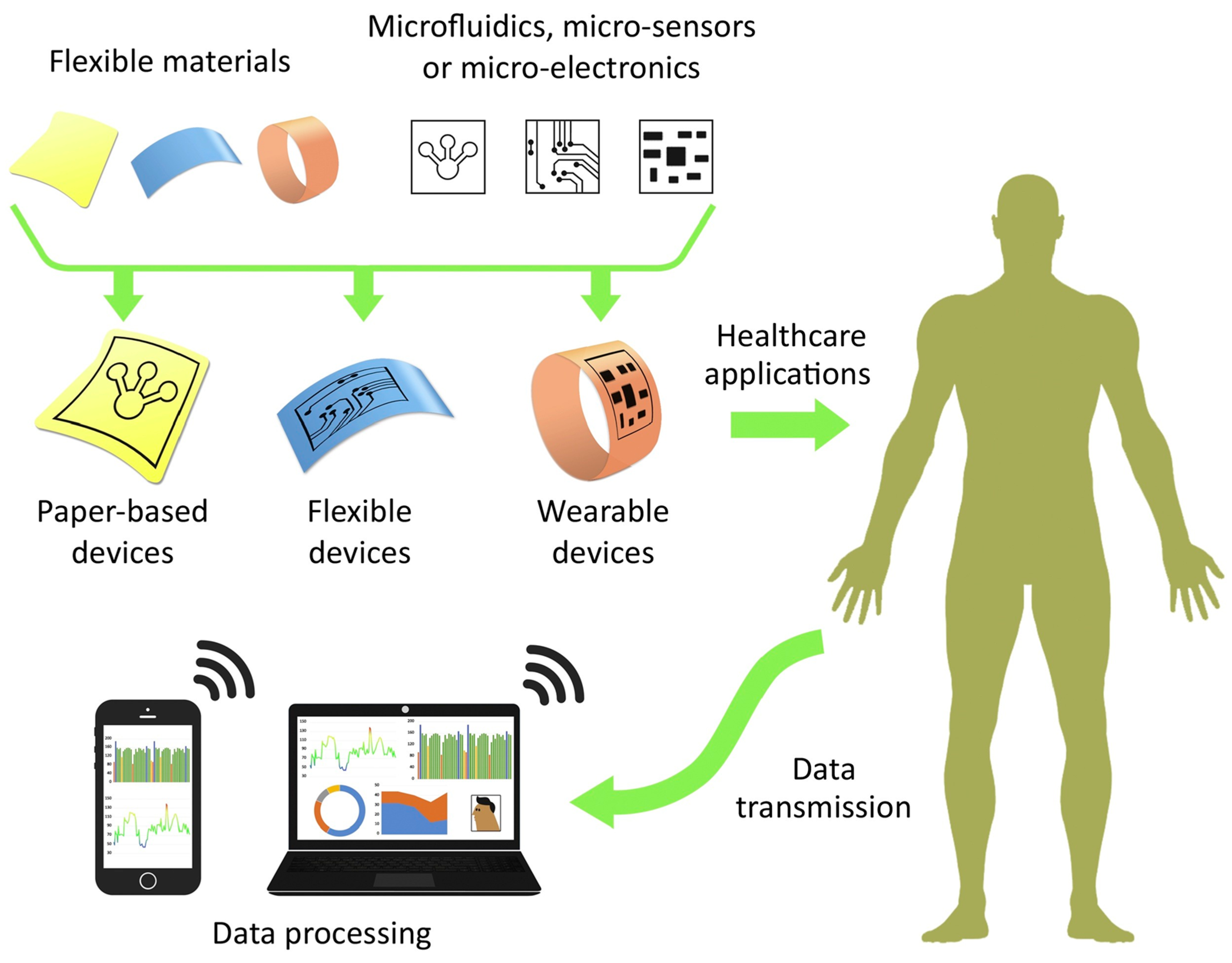

Disclaimer/Publisher’s Note: The statements, opinions and data contained in all publications are solely those of the individual author(s) and contributor(s) and not of MDPI and/or the editor(s). MDPI and/or the editor(s) disclaim responsibility for any injury to people or property resulting from any ideas, methods, instructions or products referred to in the content. |
© 2026 by the authors. Licensee MDPI, Basel, Switzerland. This article is an open access article distributed under the terms and conditions of the Creative Commons Attribution (CC BY) license.