


Abstract
Among the options to improve the establishment of jalapeno pepper (Capsicum annuum L.), physical biostimulants such as far-infrared bioceramics (FIR) and static magnetic fields (MF) have emerged as non-chemical alternatives. This study evaluated, under in vitro conditions, the individual and combined effects of FIR and positive or negative MF on seed germination dynamics, early seedling morphology, water status, and photosynthetic pigments. A completely randomized design with eight treatments was implemented, including FIR applied continuously throughout the entire experimental period, positive or negative MF applied for 24 h (MF+24, MF−24), and FIR + MF combinations under continuous or 24 h exposure regimes (n = 7). Germination percentage, mean germination time (MGT), mean germination rate (MGR), germination index (GI), morphological variables, water content (WC), and photosynthetic pigments were measured; ANOVA/alternative tests (a = 0.05), Principal Components Analysis (PCA) and exploratory Spearman’s correlations were used to assess relationships among the evaluated variables. Germination percentage did not change (97.64%), but kinetics did: FIR + MF−24 reduced MGT to 4.32 d, FIR increased MGR to 5.83 seeds day−1 (+11.69%), and FIR24 + MF+24 showed the highest GI (4.57). For morphological, MF+24 increased hypocotyl length (+16.29%), FIR increased collar diameter (+27.27%), and FIR + MF−24 increased cotyledon area (25%), and FIR increased chlorophyll a (+139%), chlorophyll b (+141%), and carotenoids (+114%). PCA explained 66.9% of the variance, grouping FIR with growth variables and FIR + MF combinations with WC and pigments. Inferences are limited to one cultivar and controlled in vitro conditions. This study provides novel quantitative evidence that continuous and short-term applications of FIR and MF modulate germination dynamics and early physiological traits without altering final germination, related to structure and pigments, without changing final germination percentage.
1. Introduction
The cultivation of jalapeño pepper (Capsicum annuum L.) in Mexico is among the most important crops, with a total planted area of 30,223.28 ha [1]. The states of Chihuahua, Sinaloa, and Michoacan account for more than half of this area, representing 59.45% of the total. Among them, Chihuahua stands out as the state with the largest cultivated area, reaching 11,154.40 ha, equivalent to 36.90% of the national surface. This state produced 383,423.24 tons of jalapeño peppers, with a production value of 3,097,349.57 thousand pesos during the 2022–2023 period [1]. Beyond its economic impact, jalapeño pepper cultivation has great social relevance, as its production requires more than 30 million workdays per year nationwide. Furthermore, its benefits extend to related sectors such as agrochemical industries, transportation, storage, and commerce in general [2].
The agronomic success of the crop depends on the optimization of resources and proper management at each phenological stage [3,4]. In particular, germination is essential for crop establishment and for ensuring high yields [5]. To enhance this process, various strategies have been explored, including the application of chemical, biological, or physical biostimulants, which have shown positive effects on germination and plant development [6,7,8]. Plant biostimulation is defined as the use of physical, chemical, or biological stimuli capable of inducing physiological responses in plants, with positive effects on their growth, development, and quality [8]. In this context, a plant biostimulant is a product that stimulates plant nutritional processes, independent of its nutrient content [9,10].
In jalapeño peppers, seed coating with biostimulants such as humic acid combined with fulvic acid has been shown to significantly increase the germination rate compared to untreated seeds [11]. Furthermore, these compounds promote growth, yield, and fruit quality not only through seed treatment but also via irrigation or foliar applications, highlighting their potential in the agronomic management of the crop [12,13,14].
On the other hand, biological biostimulants based on beneficial microorganisms have shown positive effects on plant development and fruit production. An example is Genifix® (Bacillus spp.), whose treatment increased the dry weight of seedlings by 39.9% and fruit production by 28.1% compared to the control, suggesting its effectiveness in improving growth, development, and yield of jalapeño peppers under protected cultivation conditions [15,16].
There are studies on chemical and biological biostimulants in jalapeño pepper [17,18]; However, the specific effects of physical biostimulants on species of the genus Capsicum remain poorly understood. In particular, few studies have evaluated jalapeño pepper seed responses to physical stimuli, such as static magnetic fields and bioceramic far-infrared radiation, during the early stages of development, especially under in vitro germination conditions. Furthermore, most available studies on physical biostimulation focus on single factors or on plant species other than Capsicum [19,20,21], which limits the extrapolation of their effects on germination kinetics, seedling morphology, and photosynthetic pigments in jalapeño pepper.
Exposure to magnetic fields of different intensities and durations has been associated with improved physiological performance in seeds and plants. For example, magnetic treatment increased germination rate and fresh biomass in sweet pepper (Capsicum annuum L.) seeds [22], while in sunflower (Helianthus annuus), the application of a 50 mT magnetic field for 45 min significantly enhanced germination and final emergence rates [20,23].
Meanwhile, far-infrared bioceramics technology based on metal oxide nanoparticles (aluminum oxide, ferric oxide, zinc oxide, magnesium oxide), combined with noble metals such as gold, silver, and platinum coated with ceramic material [19], attracted great interest due to their ability to induce significant biological activity [24]. Although research on plants is limited, existing studies suggest that far-infrared bioceramic treatment in the range of 8–14 µm can release antioxidant compounds, such as carotenoids and polyphenols, from natural sources like rice husks (Oryza sativa L.). Furthermore, far-infrared has been shown to exert a protective effect against oxidative stress induced by hydrogen peroxide (H2O2), reducing chlorophyll degradation and loss in Bidens pilosa L. leaves under adverse conditions [19,24].
From a physiological perspective, physical biostimulants such as far-infrared radiation bioceramics (FIR) and static magnetic fields (MF) are proposed to act through distinct but potentially complementary mechanisms during seed germination. FIR may primarily influence water-mediated processes by promoting the reorganization of water clusters and enhancing cellular hydration during imbibition [25,26]. In contrast, MF exposure has been associated with redox and ionic signaling pathways, including antioxidant activation, membrane stabilization, and the mobilization of elements essential for early seedling development [27,28,29]. Additionally, MF-induced responses may depend on physical factors such as seed orientation relative to field polarity, influencing water uptake and germination kinetics [30,31].
Although studies have focused on chemical and biological biostimulation in jalapeño peppers, information regarding the efficacy of physical biostimulants in early stages, particularly during in vitro germination, is still limited. Therefore, it is relevant to investigate these emerging alternatives to improve efficiency in agricultural production. Based on these gaps, this study tested the hypotheses that: the application of MF and FIR, individually or in combination, alters germination dynamics of jalapeño pepper seeds under in vitro conditions; and these physical biostimulants promote early seedling growth and photosynthetic pigment accumulation without necessarily affecting final germination percentage.
Thus, the objective of this study was to evaluate the effect of MF and FIR on the in vitro germination of jalapeño pepper (Capsicum annuum L.) cv. M seeds, determining their impact on germinative and vegetative parameters. Overall, the results indicate that although final germination percentage was not affected, specific MF and FIR treatments, applied individually or in combination, significantly improved germination dynamics, early seedling morphology, water status, and photosynthetic pigment accumulation, underscoring the potential of physical biostimulants at early development stages.
2. Materials and Methods
2.1. Experimental Site
The experiment was conducted at the Laboratory of Applied Microbiology, Phytopathology, and Postharvest Physiology (LAMPPP) of the Faculty of Agrotechnological Sciences, Universidad Autónoma de Chihuahua (UACH), Chihuahua, Mexico. The site is located at 28°39′24″ N, 106°05′12″ W.
2.2. Plant Material
Seeds of jalapeño pepper (Capsicum annuum L.) cultivar M (Southern Star Seeds®, Alamosa, CO, USA) were used. According to the supplier, this cultivar is described as semi-bushy, characterized by high productivity, dark green fruit color, and excellent pungency. It is recommended for both fresh consumption and processing.
2.3. Physical Biostimulators
The static magnetic field and far-infrared radiation were applied using a single commercial device integrating two functional discs: a barium ferrite magnet and a far-infrared-emitting bioceramic disc (FIR Power® band, e-energy by Nipponflex, Maringá, Brazil). Both discs measured 10 mm in diameter and 2 mm in thickness.
The barium ferrite magnet generated a magnetic flux density of 50,000 μT. The bioceramic disc was formulated from mineral oxides (Al2O3, SiO2, TiO2, Ag, Pt, Pd, Ir, and Rh) and exhibited high emissivity (≥0.90), with an emission spectrum within the 4–16 µm range, according to patent ES2257409T3 [32]. The disc surface was coated with an alumina–silica–zirconia base [19,33,34].
2.4. Experiment Preparation
Seeds of the jalapeño pepper cultivar M were surface-sterilized with 5% sodium hypochlorite for 5 min and then rinsed twice with sterile distilled water to remove any residual disinfectant. The seeds were placed on absorbent paper and allowed to dry before being distributed into sterile 100 × 15 mm borosilicate Petri dishes (KX-23061-10015, KIMAX®, Kimble Chase, Querétaro, Mexico) (Petri dishes were used due to their chemical inertness, thermal stability, and magnetic neutrality [34,35,36]. Each Petri dish contained two absorbent paper discs moistened with 5 mL of sterile distilled water, on which 25 seeds were evenly distributed with horizontal seed orientation. The Petri dishes were sealed with plastic film (Mybrand®, Chihuahua, Mexico).
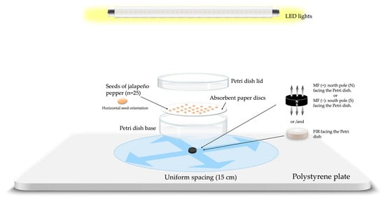
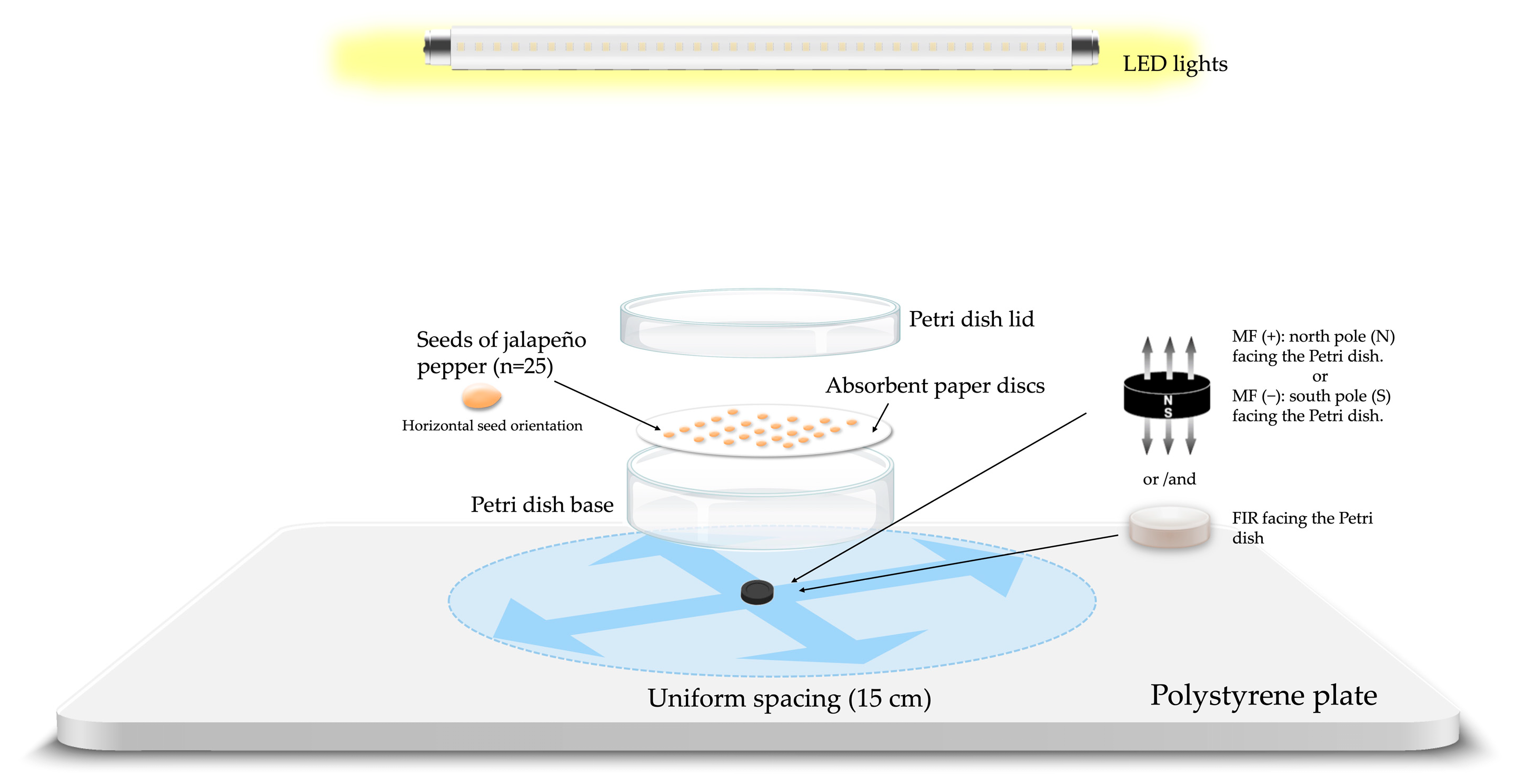
Subsequently, the Petri dishes were subjected to static magnetic field and/or far-infrared bioceramic radiation treatments, according to the experimental setup illustrated in Figure 1. Similarly, FIR was applied using a bioceramic disc positioned beneath the Petri dish, either alone or in combination with MF. In all treatments, Petri dishes were placed on a 1-inch-thick polystyrene plate to ensure uniform support and spacing (15 cm).
Figure 1.
Schematic of the experimental setup for the application of static magnetic field (MF) and bioceramic far-infrared radiation (FIR), individually or combined, to jalapeño chili seeds grown in vitro under controlled conditions. The blue arrows represent the equidistant position between the experimental units.
Petri dishes were placed in a plant growth room of the micropropagation and plant production laboratory (LAMPP), equipped with environmental control provided by a minisplit air-conditioning system. A photoperiod of 16 h of light (3500 lux provided by full-spectrum white LED lights; Goodwill az-energy®, model 20460, 30 W, Tlaxcalancingo, Mexico) at 28 °C was established and a dark period of eight hours at 18 °C, with a controlled relative humidity of 80 ± 2%. Watering was performed every 5 days by adding 2 mL of sterile distilled water to each Petri dish.
2.5. Evaluated Variables
2.5.1. Germination Parameters
Seed germination was monitored daily up to day eight and expressed as a germination percentage using Formula (1), as described by Al-Ansari and Kiski [37]. Seeds were considered germinated when the hypocotyls had emerged from the seed coat.
The mean germination time was calculated using Formula (2) proposed by Ranal and Garcia [38].
where MGT = mean germination time; Nᵢ = number of germinated seeds on day Ti, Tᵢ = time from the beginning of the experiment to the i observation, and k = last day of germination.
The mean germination rate was determined using Formula (3) proposed by Wardle et al. [39].
where N1, …Nn = number of seeds germinated on day i, and T1…Tn = time from sowing (number of seeds that germinated at a specific time).
The germination index (GI) was calculated using the Formula (4) proposed by Scott et al. [40]:
where nᵢ = number of seeds germinated on day i, tᵢ = number of days after sowing, and N = total number of seeds sown.
2.5.2. Photosynthetic Parameters
The quantification of chlorophyll a, chlorophyll b, total chlorophyll content and carotenoids was performed on day 22 after the establishment of the experiment, using the methodology described by Lichtenthaler and Wellburn [41]. Pigments were extracted from cotyledonary leaves (0.1 g) by homogenizing them in 4 mL of 80% (v/v) acetone (Sigma-Aldrich, St. Louis, MO, USA). Subsequently, the extract was centrifuged at 3000 rpm for 5 min. The absorbance of the supernatant was measured using a UV-Visible spectrophotometer (Evolution 60S, Thermo Scientific®, Madison, WI, USA) at 663, 645, and 470 nm. Pigment concentrations were calculated using the equations proposed by Lichtenthaler and Wellburn [41] for chlorophyll a, chlorophyll b, and carotenoids (Equations (5)–(7)), while total chlorophyll content was calculated according to Arnon [42] (Equation (8)).
where V = volume in mL of 80% acetone (v/v), W = fresh weight (FW) of cotyledonary leaves of the samples.
2.5.3. Morphological Parameters
Morphological variables of the seedlings were evaluated on day 22 the establishment of the experiment, using five randomly selected seedlings from each replicate. Hypocotyl length was measured from the base of the collar to the point of insertion of the cotyledonary leaves. The collar diameter, cotyledonary leaf length and width, as well as cotyledonary area, were determined by scanning the seedlings (1200 dpi; Epson EcoTank® L5590, CIICK57301, Seiko Epson Corp., Lipa, BTG, Philippines) and analyzing the images with ImageJ software version 1.46r.
The cotyledon shape index was calculated based on direct measurements, considering the maximum length and width of the cotyledonary leaves. The calculation was performed using the Formula (9) proposed by Weetman [43]:
The robustness index was determined according to Formula (10) proposed by Thompson [44]:
2.5.4. Biometric Parameters
Aerial fresh and dry biomass, as well as water content, were evaluated on day 22 after the establishment of the experiment. Biomass was determined after drying the seedlings in a forced-air convection oven (SMO3, Shel Lab®, Cornelius, OR, USA) at 40 °C for 48 h. Subsequently, biomass was measured using an analytical balance (XT-220A, Precisa Instruments®, Zurich, Switzerland). Weights were expressed in milligrams per plant (mg plant−1) [45].
Water content in seedlings was calculated using the Formula (11) proposed by Ben et al. [46]:
2.6. Experimental Design
To evaluate the effectiveness of the magnetic field and far-infrared radiation, both individually and in combination, on the germination and development of jalapeño pepper seedlings, treatments were applied either for 24 h periods or continuously throughout the entire experimental period. A total of seven treatments and one control were established under a completely randomized experimental design (Table 1).
Table 1.
Treatments with far-infrared bioceramic and ferrite magnetic field applied to jalapeño pepper seeds (Capsicum annuum L.) under in vitro conditions.
2.7. Statistical Analysis
The data obtained from all evaluated parameters were subjected to the Shapiro–Wilk and Levene tests to verify normality and homogeneity of variances, respectively. Based on these results, the data were analyzed using analysis of variance followed by Tukey’s test, Welch’s ANOVA followed by the Games–Howell test, or the nonparametric Kruskal–Wallis test followed by Conover–Iman Test, with a significance level of p < 0.05.
A Principal Component Analysis (PCA) was performed at the replicate level as an exploratory multivariate approach to identify dominant patterns of variation among germination, morphological, physiological, and biomass-related variables and to explore their association with the different treatments. Prior to PCA, Bartlett’s sphericity test was applied to assess the validity of the dataset (p < 0.01). The adequacy of the data for PCA was evaluated using the Kaiser–Meyer–Olkin (KMO) test, with values ≥ 0.60 considered acceptable [47].
A correlation analysis was performed to explore the relationships among germination, morphological, physiological, biomass, and water-related variables, and the results were visualized using a correlation heatmap. Prior to analysis, inter-variable relationships were examined to identify highly collinear parameters. Variables showing strong redundancy (|r| ≥ 0.85) were excluded, and a reduced set of biologically representative variables was retained to improve interpretability and avoid inflation of correlation coefficients [48]. Data normality was evaluated using the Shapiro–Wilk test, which indicated a significant deviation from multivariate normality (W = 0.871, p < 0.001); Therefore, correlations were calculated using Spearman’s rank correlation coefficient. It should be noted that the correlation analysis was employed as an exploratory tool to describe associations among variables, rather than for causal inference within individual treatments.
Statistical analyzes were performed using Jamovi (The Jamovi Project, 2024; version 2.5.2.0) and JASP (JASP, 2024; version 0.19, Intel®).
3. Results
3.1. Effects of Physical Biostimulants on Germination Parameters
The evaluation of the physical biostimulators, as analyzed by the analysis of variance, revealed significant differences (p < 0.05) in most germination variables, both in individual treatments and in the combinations applied in vitro (Table 2). No significant statistical differences were detected in germination percentage between the MF and FIR treatments compared to the control, with an overall average of 97.90%.
Table 2.
Germination parameters of jalapeño pepper seeds (cultivar M) treated in vitro with far-infrared bioceramic and a magnetic field of ferrite magnet, evaluated eight days after experiment establishment.
In this study, the magnetic field applied individually had a positive effect on the Mean Germination Time (MGT); However, only the MF+24 treatment reduced the MGT by 5.38% compared to the control. Similarly, the individual application of FIR decreased the MGT by 5.81%. However, the most significant reduction was achieved with the FIR + MF−24 combination, resulting in a 7.11% decrease compared to the control, with an average germination time of 4.31 days. These results indicate that both FIR and the positive magnetic field, when applied individually, tend to reduce the MGT.
Regarding the Mean Germination Rate (MGR), the FIR treatment showed the highest value (5.83 seeds day−1), representing an 11.69% increase compared to the control. Similarly, the FIR + MF+24 treatment increased by 10.15%, and FIR + MF−24 by 9.96%, followed by MF+24 with 9.19%. This pattern suggests a synergistic effect of the FIR and MF combination in improving the MGR of jalapeno pepper seeds.
In relation to the Germination Index (GI), significant statistical differences were observed among treatments, with FIR24 + MF+24 (4.57 ± 0.27) showing the highest GI and FIR + MF−24 (4.21 ± 0.08) the lowest. In contrast, the remaining treatments and the control did not differ statistically from each other. This pattern suggests that the temporal coincidence of FIR (24 h) with MF+ enhances the GI. In contrast, the combination with MF– may attenuate the effect, indicating an interaction dependent on both polarity and exposure timing.
3.2. Effects of Physical Biostimulants on Photosynthetic Parameters
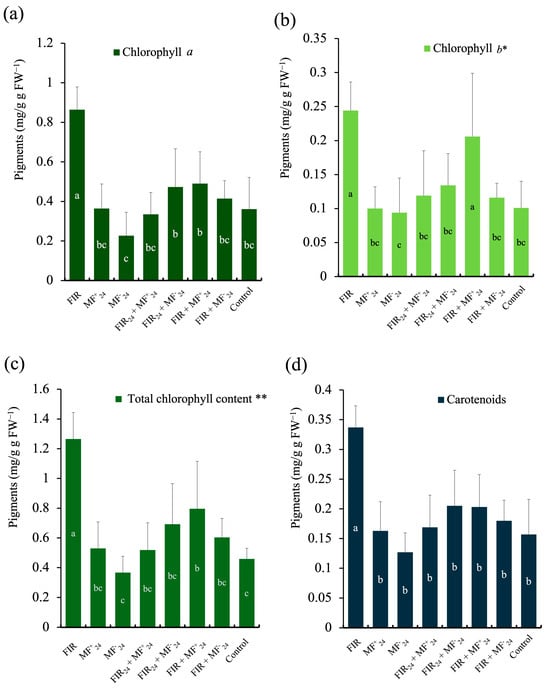
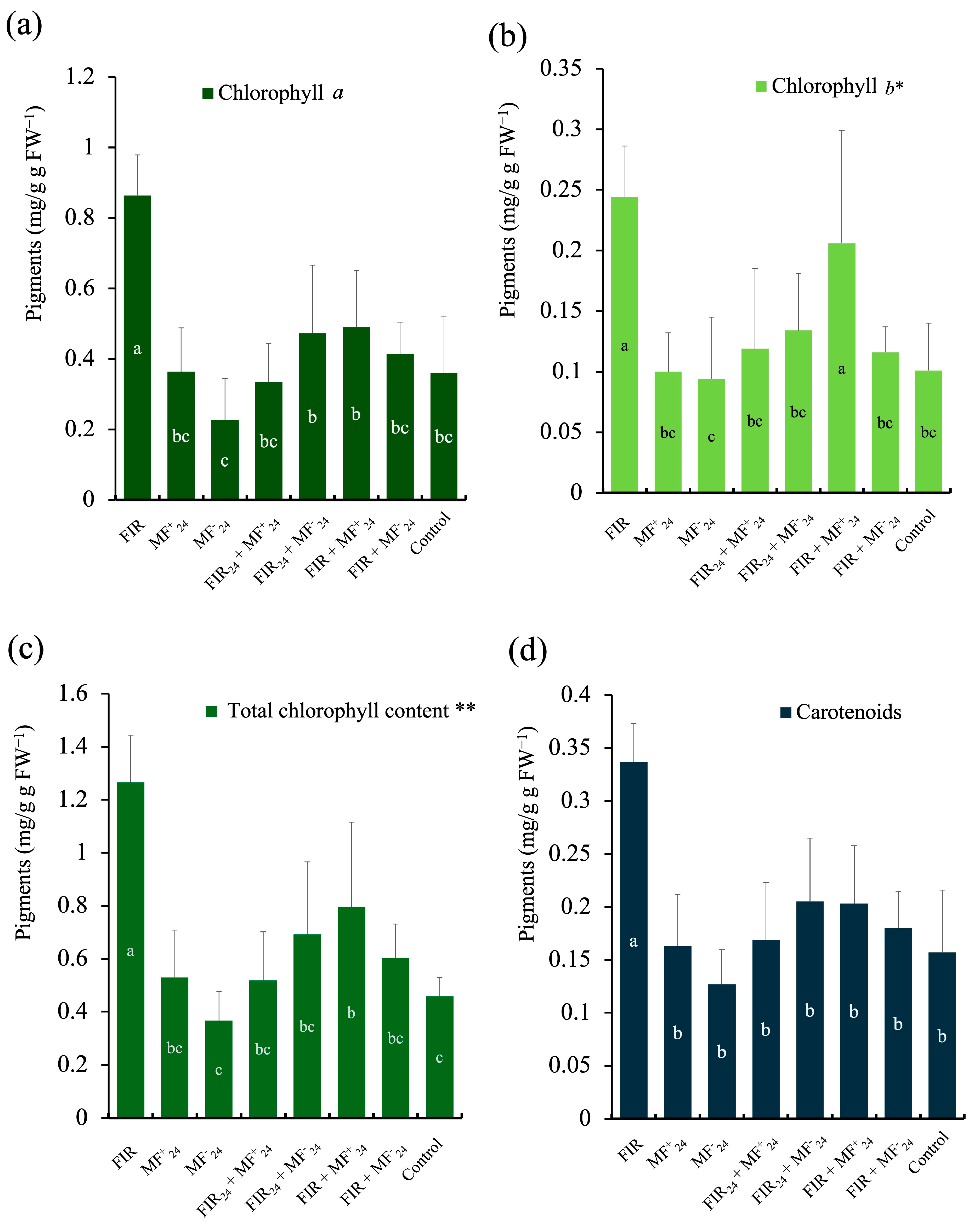
Significant differences were observed in the photosynthetic pigment content of chili pepper seedlings treated with MF and FIR (Figure 2). Chlorophyll a content was significantly higher in seedlings treated exclusively with FIR (0.864 mg g−1 FW), representing an increase of 139.3% compared with the control (0.361 mg g−1 FW) (p < 0.05). In contrast, the lowest chlorophyll a concentration was recorded under the MF−24 treatment (0.227 mg g−1 FW), which differed significantly from the FIR treatment but not from the control. Combined treatments involving FIR and MF showed intermediate values, ranging from 0.335 to 0.490 mg g−1 FW, indicating partial modulation of the FIR effect by magnetic field exposure (Figure 2a).
Figure 2.
Effects of far-infrared bioceramic radiation (FIR) and static magnetic fields (MF) on photosynthetic pigments of jalapeño pepper seedlings (Capsicum annuum L.) grown under in vitro conditions. (a) Chlorophyll a, (b) chlorophyll b, (c) total chlorophyll content, and (d) carotenoids. Treatments included continuous far-infrared radiation (FIR), positive or negative static magnetic field applied for 24 h (MF+24, MF−24), and their combinations with FIR applied continuously or for 24 h (FIR24). Bars represent mean values ± standard deviation (n = 7). Different lowercase letters indicate significant differences among treatments according to Tukey, Games–Howell (*) or Conover–Iman (**) post hoc tests (p < 0.05).
Pattern was observed for chlorophyll b. Seedlings exposed to FIR exhibited the highest chlorophyll b concentration (0.244 mg g−1 FW), significantly exceeding all MF-only treatments and the control (0.101 mg g−1 FW). The FIR + MF+24 treatment also showed elevated chlorophyll b levels (0.206 mg g−1 FW), comparable to FIR alone and significantly higher than MF−24 (0.094 mg g−1 FW), which presented the lowest values (Figure 2b).
Total chlorophyll content followed the same trend observed for individual chlorophyll fractions. The FIR-only treatment resulted in the highest total chlorophyll concentration (1.265 mg g−1 FW), which was significantly higher than all other treatments and represented an increase of 175.6% relative to the control (0.459 mg g−1 FW). Seedlings exposed to MF−24 showed the lowest total chlorophyll content (0.367 mg g−1 FW), while combined FIR + MF treatments exhibited intermediate values (0.519–0.796 mg g−1 FW), without significant differences among them (Figure 2c).
Carotenoid concentration was also significantly affected by the treatments. Seedlings exposed to FIR showed the highest carotenoid content (0.337 mg g−1 FW), differing significantly from all other treatments (p < 0.05). In contrast, the combined FIR + MF+24 and FIR24 + MF−24 treatments also showed moderately higher values than the control, although without statistical differences (Figure 2d).
3.3. Effects of Physical Biostimulants on Morphological Parameters
Significant differences (p < 0.05) were observed in some of the vegetative parameters analyzed (Table 3). Significant statistical differences were found in hypocotyl length. The MF+24 treatment recorded the highest value (2.07 cm), representing a 16.29% increase compared to the control, followed by the FIR treatment (2.05 cm) with a 15.16% increase. In contrast, the combined treatments FIR + MF+24 and FIR + MF−24 showed no significant differences compared to the control, with an overall average of −7.30%. Meanwhile, the MF−24, FIR24 + MF+24, and FIR24 + MF−24 treatments showed a more pronounced reduction in hypocotyl length, with an average decrease of –14.60% relative to the control.
Table 3.
Vegetative parameters of jalapeño pepper seeds (cultivar M) treated in vitro with far-infrared bioceramic and a magnetic field of ferrite magnet, evaluated 20 days after experiment establishment.
In the case of collar diameter, the FIR treatment showed the most significant effect, reaching a value of 1.43 mm, equivalent to a 27.27% increase compared to the control. This was followed by the FIR + MF−24 treatment, which resulted in an 18.18% increase. The remaining treatments were statistically equivalent to the control.
Regarding cotyledonary area, the FIR + MF−24 treatment produced the greatest effect, with a value of 0.80 cm2, representing 25% increase over the control, followed by the FIR treatment with 14.92% increase. The remaining treatments were not significantly different from the control.
Cotyledon shape index showed significant differences among treatments. The FIR treatment reached a value of 0.178, representing a 67.92% increase over the control, followed by FIR + MF−24 with 62.26% and FIR + MF+24 with 54.71%.
Finally, for the robustness index, there were statistically significant differences: the most favorable values were observed in MF−24 (1.19 cm), followed by FIR24 + MF+24 and FIR + MF−24, both with 1.24 cm.
3.4. Effects of Physical Biostimulants on Biometric Parameters
Significant differences (p < 0.05) were observed in some of the biometric parameters analyzed (Table 4).
Table 4.
Biometric parameters of jalapeño pepper seeds (cultivar M) treated in vitro with far-infrared bioceramic and a magnetic field of ferrite magnet, evaluated 20 days after experiment establishment.
For fresh matter, no significant differences were detected between the treatments evaluated with MF and FIR versus the control, with an overall mean of 47.97 g.
For dry weight, differences were observed among treatments: FIR24 + MF−24 (2.8 mg) had the highest dry weight, whereas MF−24 (1.8 mg) had the lowest. In contrast, the remaining treatments and the control did not differ statistically from each other.
WC did show differences among treatments. The highest value corresponded to the MF−24 treatment (1.31%) compared with the control. The remaining treatments showed a decrease in water content of up to 0.51%.
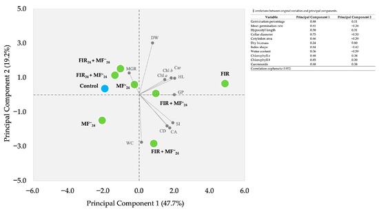
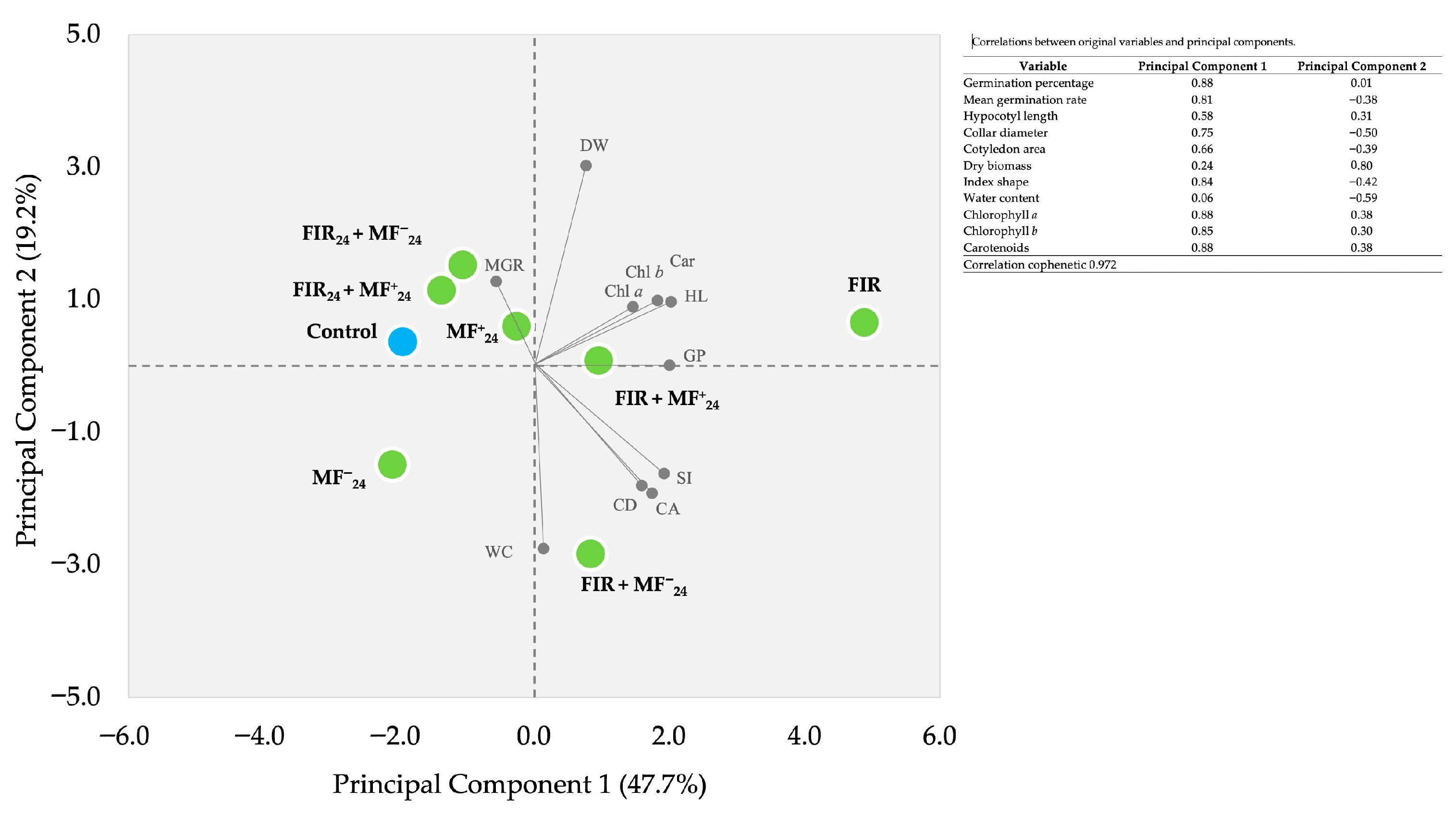
Principal component analysis (PCA) jointly explained 67% of the total variability in the data, with Principal Component 1 accounting for the most significant proportion (47.7%), followed by Principal Component 2 at 19.2% (Figure 3). These two components clearly differentiated the effects of the MF and FIR treatments on jalapeño pepper seedlings.
Figure 3.
Principal Component Analysis plot of the growth promotion in jalapeño pepper seedlings cultivar M treated with treated in vitro with far-infrared bioceramic and a magnetic field of ferrite magnet. (KMO 0.664, Bartlett’s χ2 = 336, p < 0.001). Control = non-treated seedlings; MF+24 = magnetic field positive for 24 h; MF−24 = magnetic field negative for 24 h; FIR = Far-infrared continuous; FIR24 = Far-infrared for 24 h; HL = hypocotyl length; GP = germination percentage; Chl a = chlorophyll a; Chl b = chlorophyll b; Car = carotenoids; DW = dry weight; MGR = mean germination rate; SI = shape index; CA = cotyledon area; CD = collar diameter; WC = water content.
In the biplot, the FIR treatment was located in the right quadrant, closely associated with hypocotyl length (HL), dry weight, and photosynthetic pigments (Chl a, Chl b, and carotenoids). This suggests a synergistic effect between FIR and the magnetic field, which not only stimulates the synthesis of chlorophylls and carotenoids but also contributes to maintaining a more favorable water balance, promoting good growth and development.
The combined treatments FIR + MF+24 and FIR + MF−24 were grouped in the lower-right quadrant, showing associations with variables such as water content (WC), shape index (SI), and collar diameter (CD). In contrast, the MF−24 treatment was positioned in the lower-left quadrant, without a clear association with the main variables, indicating a limited effect compared to the other treatments. The control and MF+24 treatments were located near the center of the plot, reflecting intermediate responses and no defined effect on seeding performance.
Overall, the PCA supports the results obtained from the univariate analyses, highlighting that FIR applied individually and in combination with the positive magnetic field (MF+24) exerted the most significant positive impact on physiological responses. The evidence above reinforces the notion that physical biostimulators act as modulators of growth and photosynthesis, thereby improving seedling quality and vigor under in vitro conditions.
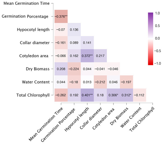
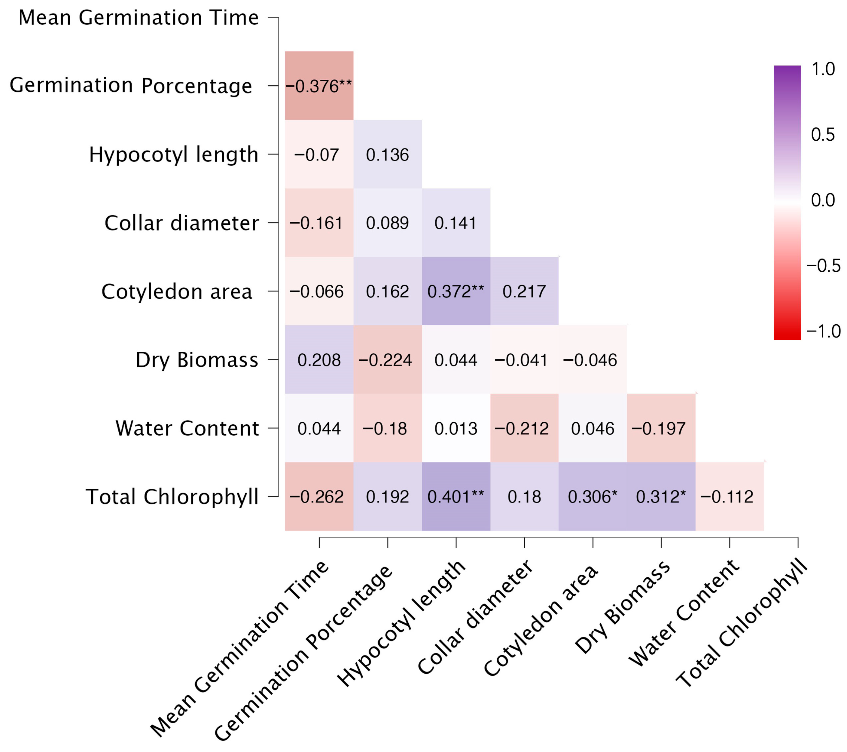
Spearman’s correlation analysis, shown in a heat map (Figure 4), revealed associations among germination, growth, and physiological parameters in jalapeño pepper seedlings under in vitro conditions. Mean germination time showed a significant negative correlation with germination percentage (ρ = −0.376, p < 0.01). Hypocotyl length showed significant positive correlations with cotyledon area (ρ = 0.372, p < 0.01) and with total chlorophyll content (ρ = 0.401, p < 0.01). Total chlorophyll content showed significant positive correlations with cotyledon area (ρ = 0.306, p < 0.05) and with dry biomass (ρ = 0.312, p < 0.05). No significant correlations were observed between water content and the other variables evaluated (p > 0.05), nor between collar diameter and most of the parameters analyzed under the study’s experimental conditions.
Figure 4.
Heatmap of Spearman’s correlation coefficients among germination, morphological, and physiological variables of jalapeño pepper (Capsicum annuum L.) under in vitro conditions. Color intensity indicates the strength and direction of the correlations (red = negative, purple = positive). Significant correlations are indicated by asterisks (* = p < 0.05; ** = p < 0.01).
The results of this study open new perspectives for the agricultural application of physical biostimulators. The integration of FIR and MF into seed pretreatment strategies could represent a sustainable alternative to enhance germination, vigor, and early photosynthesis in economically valuable crops. However, further evaluation under greenhouse and field conditions is required, taking into account environmental interactions, substrate type, and management practices, to establish reproducible protocols that enhance crop productivity and quality.
4. Discussion
The responses observed in jalapeño pepper seeds differ from those reported by Ahmed, Elzaawely, and Bayoumi [22], who documented a 44.9% increase in germination rate in Capsicum annuum seeds exposed to magnetic fields of 57,000–60,000 μT. Likewise, Gutiérrez-Villegas et al. [49] reported a doubling of germination percentage in Phaseolus vulgaris seeds subjected to an electromagnetic field of 250,000 μT. Taken together, these contrasts reinforce the notion that magnetic field effects on seed germination are highly species-dependent and strongly influenced by field intensity, exposure duration, and experimental context.
Mean germination time is widely recognized as an indicator of germination speed and uniformity, with lower values reflecting higher seed vigor and physiological efficiency, as discussed by Acuña et al. [50]. In this regard, the reduction in MGT observed in the present study is consistent with previous reports in lettuce and soybean. For instance, Acuña et al. [50] observed reductions of 9.3% and 13.5% in Lactuca sativa seeds exposed to electromagnetic fields for 24 h at 6.4 and 25.5 kJ m−3 s−1, respectively, while Torres-Osorio et al. [51] reported a 17.65% decrease in MGT in soybean seeds treated with a 300,000 μT magnetic field for 10 min. Notably, unlike magnetic stimulation, the influence of far-infrared radiation (FIR) on germination parameters has been scarcely documented, underscoring the novelty of the present findings. The pronounced reduction in MGT under the FIR + MF−24 treatment suggests a potential synergistic interaction, in which FIR may act as a germination modulator, while negative magnetic polarity enhances this response.
Regarding mean germination rate, although increases were detected, these were lower than those reported by Torres-Osorio et al. [51], who described up to a 42% increase in soybean exposed to 200,000 μT for 60 min. Such differences likely reflect variability among species, magnetic dose, exposure time, and experimental conditions. However, the observed trends support the idea that FIR and magnetic fields jointly modulate early germination processes, leading to faster, more synchronized germination dynamics.
Previous studies indicate that magnetic stimulation can serve as a biophysical pre-priming treatment, accelerating seed imbibition and synchronizing germination. Improvements in water uptake have been associated with increased cell membrane permeability and higher mass of swollen seeds, as described by Podleśny et al. [29]. At the molecular level, Dhawi [52] proposed that magnetic fields increase hydrogen bonding and activation energy in electrolyte solutions, thereby influencing enzymatic kinetics and solute transport, while Pazur [53] highlighted their role in modulating ion fluxes and biochemical equilibria. These processes may collectively accelerate seed metabolism and enhance biochemical signaling pathways associated with germination, as reviewed by Govindaraj et al. [54]. Furthermore, hormonal changes involving IAA, GA3, trans-zeatin, and trans-zeatin riboside have been documented in magnetically treated seeds and seedlings, together with higher imbibition rates, as reported by Podleśna, Bojarszczuk, and Podleśny [55]. Additional effects on mitotic activity and the G1 phase of the cell cycle have also been described by several authors [56,57,58,59,60], providing further support for faster and more synchronized seedling emergence.
In contrast, FIR is thought to interact primarily with water molecules and biomolecules by exciting O–H bonds, weakening hydrogen bonds, and reducing water cluster size, as demonstrated by Shimokawa et al. [25]. This mechanism may enhance solute mobility and diffusion toward the embryo, thereby facilitating germination.
The increase in seeding length observed here is in agreement with the findings of Koukounaras et al. [21], who reported improvements of 2.5–9.5% in tomato seedling length and vigor following exposure to a 45 μT magnetic field for 60–90 min. These observations suggest that improvements in early physiological performance are reflected in the morphology of key juvenile tissues.
Cotyledons play a central role during early seedling development by supplying nutrients and enabling initial photosynthetic activity, as emphasized by Snider et al. [61]. Their morphology among various species and cultivars [62], and indices describing cotyledon shape have long been associated with growth symmetry and vigor, as initially proposed by Weetman [43]. In this context, wider cotyledons may indicate more balanced growth, whereas elongated forms may reflect stronger polarity patterns that persist during subsequent development.
Seedling morphological quality can also be assessed through the sturdiness index, defined as the ratio between shoot height and root collar diameter. Values ≤ 6 are generally considered optimal, indicating compact and stress-resilient seedlings [44,63,64]. Such favorable proportions have been linked to hormonal regulation under magnetic field exposure, particularly involving auxins and cytokinins [65], which promote coordinated cell division and elongation and enhance photosynthetic capacity [66,67].
With respect to biomass accumulation, the present results differ from those reported by Efthimiadou et al. [68], who observed a 114% increase in fresh weight in tomato plants exposed to a 12.5 μT magnetic field for 15 min. Here, biomass responses suggest that the temporal coincidence of continuous FIR exposure with a 24 h negative static magnetic field enhances biomass gain, whereas MF−24 applied alone may attenuate this effect. This indicates that plant responses are influenced not only by field intensity but also by magnetic polarity and exposure timing, contrasting with the conclusions of Vashisth and Joshi [69] and highlighting the relevance of interactions among physical biostimulants.
Complementarily, magnetic polarity appears to differentially affect tissue hydration. Abhary and Akhkha [70] reported higher total water content in plants exposed to negative magnetic fields (150 μT), supporting the notion that the combination of negative magnetic polarity and FIR may improve cellular water availability and retention. As discussed by Shimokawa et al. [25] and Zhang et al. [26], FIR-induced water reorganization enhances hydration and solute diffusion, particularly toward chloroplasts.
From a biochemical perspective, FIR-induced water restructuring may facilitate controlled decomposition of H2O2, contributing to redox balance and preventing excessive ROS accumulation, as suggested by Jeon et al. [71]. Improved hydration and redox homeostasis support higher photochemical efficiency and more stable electron transport, resulting in increased ATP and NADPH production [72,73]. These conditions favor chloroplast biogenesis and pigment stability during early development [74,75,76].
Accordingly, the up to 140% increase in chlorophyll content observed in this study may reflect the synergistic action of FIR and static magnetic fields on redox signaling, nutrient bioavailability, and chloroplast functionality. Magnetic stimulation has been shown to induce hormetic responses involving antioxidant enzymes, preserving thylakoid membrane integrity and chlorophyll stability [29,77,78]. Furthermore, enhanced uptake and redistribution of Mg and Fe under magnetic exposure [27,28] may be particularly relevant under nutrient-free conditions, where these elements derive mainly from cotyledonary reserves [53]. The combined improvement in hydration and ionic signaling may thus facilitate tetrapyrrole biosynthesis and chlorophyll accumulation during early seedling growth.
The concurrent accumulation of carotenoids may represent a complementary photoprotective strategy, mitigating singlet oxygen formation and stabilizing photosystems under enhanced electron transport activity, as discussed by Havaux [79] and Ashraf and Harris [80]. Overall, the interaction between magnetic field polarity and FIR-mediated water reorganization appears to create a favorable physiological environment for enhanced photosynthetic pigment biosynthesis and functionality in jalapeño pepper seedlings.
Principal component analysis was used as an exploratory approach to visualize multivariate patterns among germination, growth, physiological, and biomass-related variables. Although the first two components explained 67% of the total variance, the relatively low KMO value indicates moderate sampling adequacy; Therefore, the ordination should be interpreted descriptively rather than as confirmatory evidence of treatment effects. The PCA revealed that FIR was mainly associated with growth and pigment-related variables, whereas combined FIR + MF treatments clustered with water content and structural traits, suggesting coordinated response profiles. In contrast, MF−24 and the control showed weaker associations. These multivariate trends are consistent with the univariate analyses, emphasizing patterns of association rather than causality.
Correlation analysis further indicated that treatments reducing MGT tended to increase germination percentage, reflecting improved physiological readiness. Likewise, enhanced hypocotyl elongation was associated with greater cotyledon area and chlorophyll content, suggesting improved early photosynthetic capacity. Together, these coordinated responses indicate that the treatments influenced not only individual traits but also the functional relationships among germination, growth, and physiological performance.
The present findings open new perspectives for the agricultural application of physical biostimulants. The integration of FIR and magnetic fields as seed pretreatments could represent a sustainable strategy to enhance germination, seedling vigor, and early photosynthetic performance in economically important crops. However, further validation under greenhouse and field conditions is required, considering environmental interactions, substrate type, and management practices, to establish reproducible and scalable protocols for commercial production.
5. Conclusions
This study demonstrates that the application of physical biostimulants, bioceramic far-infrared radiation (FIR), and static magnetic fields (MF) modulates germination dynamics and early physiological performance of Capsicum annuum L. cv. M under in vitro conditions, without altering the final germination percentage. Specifically, continuous FIR increased the mean germination rate by 11.69% and promoted substantial accumulation of photosynthetic pigments, increasing chlorophyll a, chlorophyll b, and carotenoids by 139.3%, 141.6%, and 114.6%, respectively, relative to the control. Short-term MF exposure (MF+24) reduced mean germination time and enhanced hypocotyl elongation (+16.3%). In comparison, the combined FIR + MF−24 treatment produced the most significant reduction in mean germination time (−7.1%) and increased cotyledon area by 25.0%. This treatment also improved early seedling performance under controlled conditions.
Multivariate analyses reinforced these findings by identifying consistent patterns linking FIR treatments with enhanced pigment content, biomass accumulation, and seedling vigor. In contrast, MF effects were more variable and dependent on polarity and exposure duration. These results confirmed treatment-specific responses rather than universal stimulation effects.
Although the observed responses may be associated with biophysical interactions involving water structure, membrane permeability, and metabolic activation, such mechanisms remain speculative and were not directly tested in this study. Therefore, conclusions are strictly limited to experimentally demonstrated physiological and morphological responses.
From an applied perspective, these findings suggest that FIR-based seed pretreatments or MF short-term exposure could be integrated into controlled nursery or seedling production systems to improve early vigor and photosynthetic capacity, particularly in high-value horticultural crops. However, direct extrapolation to commercial production should be approached cautiously.
Future research should validate these responses under greenhouse and field conditions, evaluate different jalapeño cultivars and other Capsicum types, and assess long-term effects on plant establishment, yield, and fruit quality. Additionally, mechanistic studies integrating molecular, hormonal, and water-structure analyses are required to clarify the underlying modes of action of these physical biostimulants.
This work provides quantitative, cultivar-specific evidence supporting the potential of FIR and MF as sustainable, non-chemical tools for improving early seeding performance under controlled conditions.
Author Contributions
Conceptualization, J.H.-H. and M.E.V.-P.; methodology, A.G.-C., L.R.O.-M. and L.R.-H.; validation, A.G.-C., M.E.V.-P. and J.H.-H.; formal analysis, M.E.V.-P., L.R.O.-M. and M.C.E.D.-G.; investigation, M.E.V.-P. and J.H.-H.; resources, A.G.-C., L.R.-H. and J.H.-H.; data curation, A.G.-C. and J.H.-H.; writing—original draft preparation, M.E.V.-P., L.R.O.-M. and J.H.-H.; writing—review and editing, M.C.E.D.-G., A.C.G.-F. and L.R.-H.; visualization, M.C.E.D.-G.; supervision, J.H.-H.; project administration, J.H.-H. and M.E.V.-P. All authors have read and agreed to the published version of the manuscript.
Funding
This research received no external funding.
Data Availability Statement
The data presented in this study are available upon request from the corresponding authors.
Conflicts of Interest
The authors declare no conflicts of interest.
References
- Servicio de Información Agroalimentaria y Pesquera (SIAP). Cierre Agrícola. Available online: https://nube.agricultura.gob.mx/cierre_agricola/ (accessed on 24 September 2024).
- Ramírez Meraz, M.; Arcos Cavazos, G.; Méndez Aguilar, R.; Meneses Márquez, I. Variedades e híbridos de chile para el trópico de México. In Producción Agropecuaria: Un Enfoque Integrado, 1st ed.; Meza-Villalvazo, V.M., Chay-Canul, A.J., Ramírez-Seañez, A.R., Palacios-Torres, R.E., Valenzuela-Jiménez, N., Alcántar-Vázquez, J.P., Kido-Cruz, M.T., Eds.; Universidad del Papaloapan: Oaxaca, Mexico, 2019; pp. 29–37. [Google Scholar]
- Fischer, R.A.; Connor, D.J. Issues for cropping and agricultural science in the next 20 years. Field Crops Res. 2018, 222, 121–142. [Google Scholar] [CrossRef] [Scilit]
- Neupane, J.; Guo, W. Agronomic basis and strategies for precision water management: A review. Agronomy 2019, 9, 87. [Google Scholar] [CrossRef] [Scilit]
- Han, C.; Yang, P. Studies on the molecular mechanisms of seed germination. Proteomics 2015, 15, 1671–1679. [Google Scholar] [CrossRef] [Scilit]
- Juárez Maldonado, A.; Ortega Ortíz, H.; Morales Díaz, A.B.; González Morales, S.; Morelos Moreno, Á.; Cabrera De la Fuente, M.; Sandoval Rangel, A.; Cadenas Pliego, G.; Benavides Mendoza, A. Nanoparticles and nanomaterials as plant biostimulants. Int. J. Mol. Sci. 2019, 20, 162. [Google Scholar] [CrossRef] [Scilit] [PubMed]
- Gupta, S.; Doležal, K.; Kulkarni, M.G.; Balázs, E.; Van Staden, J. Role of non-microbial biostimulants in regulation of seed germination and seedling establishment. Plant Growth Regul. 2022, 97, 271–313. [Google Scholar] [CrossRef] [Scilit]
- Garza Alonso, C.A.; Olivares Sáenz, E.; González Morales, S.; Cabrera De la Fuente, M.; Juárez Maldonado, A.; González Fuentes, J.A.; Tortella, G.; Valdés Caballero, M.V.; Benavides Mendoza, A. Strawberry biostimulation: From mechanisms of action to plant growth and fruit quality. Plants 2022, 11, 3463. [Google Scholar] [CrossRef] [Scilit] [PubMed]
- European Union. Regulation (EU) 2019/1009 of the European Parliament and of the Council of 5 June 2019 laying down rules on the making available on the market of EU fertilising products and amending Regulations (EC) No 1069/2009 and (EC) No 1107/2009 and repealing Regulation (EC) No 2003/2003. Off. J. Eur. Union 2019, 29, 1–129. Available online: https://faolex.fao.org/docs/pdf/eur187729.pdf (accessed on 18 November 2025).
- Ricci, M.; Tilbury, L.; Daridon, B.; Sukalac, K. General principles to justify plant biostimulant claims. Front. Plant Sci. 2019, 10, 494. [Google Scholar] [CrossRef] [Scilit] [PubMed]
- Pavani, B.; Sujatha, P.; Madhavi, M.; Bharathi, Y.; Peter, J. Biostimulants coating for enhanced seed vigour and longevity in chilli (Capsicum annuum L.). Plant Arch. 2024, 24, 2373–2378. [Google Scholar] [CrossRef] [Scilit]
- Jan, J.A.; Nabi, G.; Khan, M.; Ahmad, S.; Shah, P.S.; Hussain, S.; Sehrish. Foliar application of humic acid improves growth and yield of chilli (Capsicum annuum L.) varieties. Pak. J. Agric. Res. 2020, 33, 461–472. [Google Scholar] [CrossRef] [Scilit]
- Zamljen, T.; Grohar, M.C.; Slatnar, A. Effects of pre- and post-transplantation humic acid biostimulant treatment and harvest date on yield quantity and quality parameters of sweet peppers (Capsicum annuum L.). Sci. Hortic. 2024, 338, 113–747. [Google Scholar] [CrossRef] [Scilit]
- Zamljen, T.; Veberič, R.; Slatnar, A. Fertilization with humic acids in production changes the quality of fresh and stored sweet pepper fruits (Capsicum annuum L.). J. Plant Growth Regul. 2025, 44, 3965–3973. [Google Scholar] [CrossRef] [Scilit]
- Adame García, J.; Murillo Cuevas, F.D.; Fernández Viveros, J.A.; Cabrera Mireles, H.; Cornejo Castillo, R. Efecto de bioestimulantes microbianos en plántulas y frutos de chile jalapeño (Capsicum annuum L.) producidos en macrotúnel. Rev. Bio. Cienc. 2014, 11, e1566. [Google Scholar] [CrossRef] [Scilit]
- Urías Salazar, A.A.; Ayil Gutiérrez, B.A.; López Santillán, J.A.; Estrada Drouaillet, B.; Cano González, M.Á.; Hernández Martínez, J.G.; Villanueva Alonso, H.J.; Poot Poot, W.A. Crecimiento y rendimiento de Capsicum annuum L. inoculado con endomicorriza y rizobacterias. Rev. Mex. Cienc. Agríc. 2016, 7, 1539–1550. [Google Scholar]
- Buelna-Tarín, S.; Romero-Félix, C.S.; Bojórquez-Ramos, C.; Lugo-García, G.A.; Sánchez-Soto, B.H. Bioestimulantes y solución Steiner en crecimiento y producción de Capsicum annuum L. Rev. Mex. Cienc. Agríc. 2024, 15, e3255. [Google Scholar] [CrossRef] [Scilit]
- Ramírez-Pérez, C.; Ramírez, H.; Carrillo-Lomelí, D.A.; Moo-Huchin, V.M.; Hernández-Pérez, A.; Peña-Ramos, F.M.; Encina-Domínguez, J.A.; de Rodríguez, D.J. Biostimulant from semi-desert plant extracts for root and aerial growth of Capsicum annuum L. and C. chinense Jacq. seedlings. Ind. Crops Prod. 2025, 236, 121881. [Google Scholar] [CrossRef] [Scilit]
- Leung, T.K.; Shang, H.F.; Chen, D.C.; Chen, J.Y.; Chang, T.M.; Hsiao, S.Y.; Ho, C.K.; Lin, Y.S. Effects of far infrared rays on hydrogen peroxide scavenging capacity. Biomed. Eng.-Appl. Basis Commun. 2011, 23, 99–105. [Google Scholar] [CrossRef] [Scilit]
- Bukhari, S.A.; Tanveer, M.; Mustafa, G.; Zia-Ud-Den, N. Magnetic field stimulation effect on germination and antioxidant activities of presown hybrid seeds of sunflower and its seedlings. J. Food Qual. 2021, 1, 5594183. [Google Scholar] [CrossRef] [Scilit]
- Koukounaras, A.; Boursianis, A.; Kostas, S.; Theopoulos, A.; Bantis, F.; Samaras, T. Presowing static magnetic field treatment of vegetable seeds and its effect on germination and young seedlings development. Seeds 2023, 2, 394–405. [Google Scholar] [CrossRef] [Scilit]
- Ahmed, M.E.M.; Elzaawely, A.A.; Bayoumi, Y. Effect of magnetic field on seed germination, growth and yield of sweet pepper (Capsicum annuum L.). Asian J. Crop Sci. 2013, 5, 286–294. [Google Scholar] [CrossRef] [Scilit]
- Salehi, A.H.; Sharafi, S. Effect of Magnetic Field on Seed Germination and Early Growth of Calendula officinalis L. J. Ornam. Plants 2015, 5, 91–96. [Google Scholar]
- Azad, M.O.K.; Kim, W.W.; Park, C.H.; Cho, D.H. Effect of artificial LED light and far-infrared irradiation on phenolic compounds, isoflavones, and antioxidant capacity in soybean (Glycine max L.) sprout. Foods 2018, 7, 174. [Google Scholar] [CrossRef] [Scilit]
- Shimokawa, S.; Yokono, T.; Mizuno, T.; Tamura, H.; Erata, T.; Araiso, T. Effect of far-infrared light irradiation on water as observed by X-ray diffraction measurements. Jpn. J. Appl. Phys. 2004, 43, L545. [Google Scholar] [CrossRef] [Scilit]
- Zhang, M.; Han, F.; Li, C.; Wang, P.; Yang, Y.; Yu, K. Combined effect of weak electric field and ions on critical water cluster: Insight from molecular dynamics simulation. Chem. Phys. 2020, 539, 110932. [Google Scholar] [CrossRef] [Scilit]
- Eşitken, A.; Turan, M. Alternating magnetic field effects on yield and plant nutrient element composition of strawberry (Fragaria × ananassa cv. Camarosa). Acta Agric. Scand. Sect. B Soil Plant Sci. 2004, 54, 135–139. [Google Scholar] [CrossRef] [Scilit]
- Dhawi, F.; Al-Khayri, J.M.; Hassan, E. Static magnetic field influence on elements composition in date palm (Phoenix dactylifera L.). Res. J. Agric. Biol. Sci. 2009, 5, 161–166. [Google Scholar]
- Podleśny, J.; Podleśna, A.; Gładyszewska, B.; Bojarszczuk, J. Effect of pre-sowing magnetic field treatment on enzymes and phytohormones in pea (Pisum sativum L.) seeds and seedlings. Agronomy 2021, 11, 494. [Google Scholar] [CrossRef] [Scilit]
- Kevin, K.K.; Bernard, N.K.; Laurent, K.K.; Ignace, K.K.; Pierre, B.J.; Arsene, Z.B.I. Effects of Seed Orientation and Sowing Depths on Germination, Seedling Vigor and Yield in Oleaginous Type of Bottle Gourd, Lagenaria siceraria (Molina) Standl. Int. Res. J. Biol. Sci. 2015, 4, 46–53. [Google Scholar]
- Masilamani, P.; Venkatesan, S.; Navamaniraj, K.N.; Rajarathinam, P.; Alagesan, A.; Thiagu, K. Impact of the Orientation of Seed Placement and Depth of Its Sowing on Germination: A Review. J. Appl. Nat. Sci. 2023, 15, 314–324. [Google Scholar] [CrossRef] [Scilit]
- Komuro, T. Composition for Far-Infrared Radiation with Excellent Antistatic Properties, and Fiber and Textile Product Containing the Same. Spain Patent ES2257409T3, 17 May 2021. Available online: https://worldwide.espacenet.com/patent/search/family/018654745/publication/ES2257409T3?q=ES2257409T3 (accessed on 24 September 2024).
- Inoue, S.; Kabaya, M. Biological activities caused by far-infrared radiation. Int. J. Biometeorol. 1989, 33, 145–150. [Google Scholar] [CrossRef] [Scilit]
- Leung, T.K. In vitro and in vivo studies of the biological effects of bioceramic (a material of emitting high performance far-infrared ray) irradiation. Chin. J. Physiol. 2015, 58, 147–155. [Google Scholar] [CrossRef] [Scilit]
- Coey, J.M.D. Magnetism and Magnetic Materials; Cambridge University Press: Cambridge, UK, 2010. [Google Scholar] [CrossRef] [Scilit]
- Cullity, B.D.; Graham, C.D. Introduction to Magnetic Materials, 2nd ed.; Wiley-IEEE Press: Hoboken, NJ, USA, 2011. [Google Scholar]
- Al-Ansari, F.; Ksiksi, T. A quantitative assessment of germination parameters: The case of Crotalaria persica and Tephrosia apollinea. Open Ecol. J. 2016, 9, 13–21. [Google Scholar] [CrossRef] [Scilit]
- Ranal, M.A.; Santana, D.G. How and why to measure the germination process? Braz. J. Bot. 2006, 29, 1–11. [Google Scholar] [CrossRef] [Scilit]
- Wardle, D.A.; Ahmed, M.; Nicholson, K.S. Allelopathic influence of nodding thistle (Carduus nutans L.) seeds on germination and radicle growth of pasture plants. N. Z. J. Agric. Res. 1991, 34, 185–191. [Google Scholar] [CrossRef] [Scilit]
- Scott, S.J.; Jones, R.A.; Williams, W.A. Review of data analysis methods for seed germination. Crop Sci. 1984, 24, 1192–1199. [Google Scholar] [CrossRef] [Scilit]
- Lichtenthaler, H.K.; Wellburn, A.R. Determinations of total carotenoids and chlorophylls a and b of leaf extracts in different solvents. Biochem. Soc. Trans. 1983, 11, 591–592. [Google Scholar] [CrossRef] [Scilit]
- Arnon, D.I. Copper enzymes in isolated chloroplasts, polyphenolexidase in Beta vulgaris. Plant Physiol. 1949, 24, 1–15. [Google Scholar] [CrossRef] [Scilit] [PubMed]
- Weetman, L.M. Correlation of shape of fruits, cotyledons, and seeds in melons. Bot. Gaz. 1935, 97, 388–398. [Google Scholar] [CrossRef] [Scilit]
- Thompson, B.E. Seedling morphological evaluation: What you can tell by looking. In Evaluating Seedling Quality: Principles, Procedures and Predictive Abilities of Major Tests; Duryea, M.L., Ed.; Forest Research Laboratory, Oregon State University: Corvallis, OR, USA, 1985; pp. 59–71. [Google Scholar]
- Patrignani, A.; Ochsner, T.E. Canopeo: A powerful new tool for measuring fractional green canopy cover. Agron. J. 2025, 107, 2312–2320. [Google Scholar] [CrossRef] [Scilit]
- Ben Amor, N.; Ben Hamed, K.; Debez, A.; Grignon, C.; Abdelly, C. Physiological and antioxidant responses of the perennial halophyte Crithmum maritimum to salinity. Plant Sci. 2005, 168, 889–899. [Google Scholar] [CrossRef] [Scilit]
- Tabachnick, B.G.; Fidell, L.S. Using Multivariate Statistics, 5th ed.; Pearson Education, Inc.: Boston, MA, USA, 2007. Available online: https://lccn.loc.gov/2017040173 (accessed on 12 December 2025).
- Dormann, C.F.; Elith, J.; Bacher, S.; Buchmann, C.; Carl, G.; Carré, G.; Marquéz, J.R.G.; Gruber, B.; Lafourcade, B.; Leitão, P.J.; et al. Collinearity: A review of methods to deal with it and a simulation study evaluating their performance. Ecography 2013, 36, 27–46. [Google Scholar]
- Gutiérrez Villegas, J.N.; Gutiérrez Villegas, I.I.; Martínez Flores, V.H.; Gutiérrez Armenta, E.M.; Gutiérrez Villegas, M.A.; Niño Monroy, L. Germinación de semillas utilizando campos electromagnéticos. RICT 2024, 2, 24–29. [Google Scholar] [CrossRef]
- Acuña, R.; Naguelquin, F.; García, F.; Torres, J. Aplicación de campos magnéticos (CM) y su relación con la recuperación de la viabilidad y vigor en semillas envejecidas de Lactuca sativa L. Agro Sur 2019, 47, 9–21. [Google Scholar] [CrossRef] [Scilit]
- Torres Osorio, J.I.; Aranzazu Osorio, J.E.; Carbonell Padrino, M.V. Efecto del campo magnético estático homogéneo en la germinación y absorción de agua en semillas de soja. TecnoLógicas 2015, 18, 11–20. [Google Scholar] [CrossRef] [Scilit]
- Dhawi, F. Why are magnetic fields used to enhance a plant’s growth and productivity? Annu. Res. Rev. Biol. 2014, 4, 886–896. [Google Scholar] [CrossRef] [Scilit]
- Pazur, A. Calcium ion cyclotron resonance in dissipative water structures. Electromagn. Biol. Med. 2018, 37, 100–113. [Google Scholar] [CrossRef] [Scilit]
- Govindaraj, M.; Masilamani, P.; Albert, V.A.; Bhaskaran, M. Effect of physical seed treatment on yield and quality of crops: A review. Agric. Rev. 2017, 38, 1–14. [Google Scholar] [CrossRef] [Scilit]
- Podleśna, A.; Bojarszczuk, J.; Podleśny, J. Effect of presowing magnetic field treatment on some biochemical and physiological processes in faba bean (Vicia faba L. spp. minor). J. Plant Growth Regul. 2019, 38, 1153–1160. [Google Scholar] [CrossRef] [Scilit]
- Dul’binskaya, D.A. Effect of a constant magnetic field on growth of corn seedlings. Fiziol. Rast. 1973, 20, 183–186. [Google Scholar]
- Lebedev, S.I.; Baranskii, P.I.; Litvinenko, L.G.; Shiyan, L.T. Physiobiochemical characteristics of plants after presowing treatment with a permanent magnetic field. Sov. Plant Physiol. 1975, 22, 84–89. [Google Scholar]
- Govorun, R.D.; Danilov, V.I.; Fomicheva, V.M.; Belyavskaya, N.A.; Zinchenko, S.Y. Influence of geomagnetic field fluctuations and its shielding on early phases of higher plant development. Biofizika 1992, 37, 738–743. [Google Scholar]
- Fomicheva, V.M.; Zaslavskii, V.A.; Govorun, R.D.; Danilov, V.I. Dynamics of RNA and protein synthesis in the cells of the root meristems of the pea, lentil and flax. Biofizika 1992, 37, 750–758. [Google Scholar]
- Paul, A.L.; Ferl, R.J.; Meisel, M.W. High magnetic field induced changes of gene expression in Arabidopsis thaliana. Biomagn. Res. Technol. 2006, 4, 7. [Google Scholar] [CrossRef] [Scilit] [PubMed]
- Snider, J.L.; Pilon, C.; Virk, G. Seed characteristics and seedling vigor. In Cotton Seed Seedlings; The Cotton Foundation: Cordova, TN, USA, 2020; pp. 9–23. [Google Scholar]
- Han, J.; Li, Y.; Zhao, Y.; Sun, Y.; Li, Y.; Peng, Z. How does light regulate plant regeneration? Front. Plant Sci. 2025, 15, 1474431. [Google Scholar] [CrossRef] [Scilit]
- Opio, C.; Jacob, N.; Coopersmith, D. Height to diameter ratio as a competition index for young conifer plantations in northern British Columbia, Canada. For. Ecol. Manag. 2000, 137, 245–252. [Google Scholar] [CrossRef] [Scilit]
- Rodríguez, T.D.A. Indicadores de Calidad de Planta Forestal; Mundi-Prensa México: Mexico City, Mexico, 2008; 156p. [Google Scholar]
- Galland, P.; Pazur, A. Magnetoreception in plants. J. Plant Res. 2005, 118, 371–389. [Google Scholar] [CrossRef] [Scilit]
- Ross, J.; O’Neill, D.P.; Wolbang, C.M.; Symons, G.M.; Reid, J.B. Auxin–gibberellin interactions and their role in plant growth. J. Plant Growth Regul. 2001, 20, 346–353. [Google Scholar] [CrossRef] [Scilit]
- Spaninks, K.; Lamers, G.; van Lieshout, J.; Offringa, R. Light quality regulates apical and primary radial growth of Arabidopsis thaliana and Solanum lycopersicum. Sci. Hortic. 2023, 317, 112082. [Google Scholar] [CrossRef] [Scilit]
- Efthimiadou, A.; Katsenios, N.; Karkanis, A.; Papastylianou, P.; Triantafyllidis, V.; Travlos, I.; Bilalis, D.J. Effects of presowing pulsed electromagnetic treatment of tomato seed on growth, yield, and lycopene content. Sci. World J. 2014, 2014, 369745. [Google Scholar] [CrossRef] [Scilit]
- Vashisth, A.; Joshi, D.K. Growth characteristics of maize seeds exposed to magnetic field. Bioelectromagnetics 2017, 38, 151–157. [Google Scholar] [CrossRef] [Scilit]
- Abhary, M.K.; Akhkha, A. Effects of neodymium magneto priming on seed germination and salinity tolerance in tomato. Bioelectromagnetic 2023, 44, 47–56. [Google Scholar] [CrossRef] [Scilit]
- Jeon, K.I.; Park, E.; Park, H.R.; Jeon, Y.J.; Cha, S.H.; Lee, S.C. Antioxidant activity of far-infrared radiated rice hull extracts on reactive oxygen species scavenging and oxidative DNA damage in human lymphocytes. J. Med. Food 2006, 9, 42–48. [Google Scholar] [CrossRef] [Scilit]
- Shanker, A.K.; Amirineni, S.; Bhanu, D.; Yadav, S.K.; Jyothilakshmi, N.; Vanaja, M.; Singh, J.; Sarkar, B.; Maheswari, M.; Singh, V.K. High-resolution dissection of photosystem II electron transport reveals differential response to water deficit and heat stress in isolation and combination in pearl millet (Pennisetum glaucum (L.) R. Br.). Front. Plant Sci. 2022, 13, 892676. [Google Scholar] [CrossRef] [Scilit] [PubMed]
- Qiao, M.; Hong, C.; Jiao, Y.; Hou, S.; Gao, H. Impacts of drought on photosynthesis in major food crops and related mechanisms. Plants 2024, 13, 1808. [Google Scholar] [CrossRef] [Scilit] [PubMed]
- Yoshida, K.; Hisabori, T. Current insights into the redox regulation network in plant chloroplasts. Plant Cell Physiol. 2023, 64, 704–715. [Google Scholar] [CrossRef] [Scilit]
- Lee, K.P.; Kim, C. Photosynthetic ROS and retrograde signaling pathways. New Phytol. 2024, 244, 1183–1198. [Google Scholar] [CrossRef] [Scilit]
- Mahapatra, K.; Mukherjee, A.; Suyal, S.; Dar, M.A.; Bhagavatula, L.; Datta, S. Regulation of chloroplast biogenesis, development, and signaling by endogenous and exogenous cues. Physiol. Mol. Biol. Plants 2024, 30, 167–183. [Google Scholar] [CrossRef] [Scilit] [PubMed]
- Saletnik, B.; Saletnik, A.; Słysz, E.; Zaguła, G.; Bajcar, M.; Puchalska-Sarna, A.; Puchalski, C. The static magnetic field regulates the structure, biochemical activity, and gene expression of plants. Molecules 2022, 27, 5823. [Google Scholar] [CrossRef] [Scilit]
- Liu, Y.; Hu, J.; Duan, X.; Ding, W.; Xu, M.; Xiong, Y. Target of rapamycin (TOR): A master regulator in plant growth, development, and stress responses. Annu. Rev. Plant Biol. 2025, 76, 341–371. [Google Scholar] [CrossRef] [Scilit]
- Havaux, M. Carotenoid oxidation products as stress signals in plants. Plant J. 2014, 79, 597–606. [Google Scholar] [CrossRef] [Scilit]
- Ashraf, M.H.P.J.C.; Harris, P.J. Photosynthesis under stressful environments: An overview. Photosynthetica 2013, 51, 163–190. [Google Scholar] [CrossRef] [Scilit]

Disclaimer/Publisher’s Note: The statements, opinions and data contained in all publications are solely those of the individual author(s) and contributor(s) and not of MDPI and/or the editor(s). MDPI and/or the editor(s) disclaim responsibility for any injury to people or property resulting from any ideas, methods, instructions or products referred to in the content. |
© 2026 by the authors. Licensee MDPI, Basel, Switzerland. This article is an open access article distributed under the terms and conditions of the Creative Commons Attribution (CC BY) license.