


Abstract
Microencapsulation has become an indispensable technique across various industries that require the controlled release and stability of bioactive agents. In this study, the encapsulation of cinnamon essential oil (CEO), known for its antibacterial and anti-inflammatory properties, was study to enable controlled release when applied to textile substrates. This procedure involves defining and examining several steps to establish a stable, scalable complex coacervation methodology. To form a stable microcapsule matrix, CEO was emulsified using a combination of surfactants (Span 80, Tween 20, and Sodium Dodecyl Sulfate). After forming micelles containing CEO, two biopolymers (chitosan and gum Arabic) were used at various proportions to form a microcapsule shell via a layer-by-layer approach. Advanced characterization techniques, such as spectrophotometry and laser scattering, were used to evaluate microcapsule stability, size, and release kinetics, as well as to assess potential antibacterial activity. The presence of oil-containing microcapsules was confirmed using fourier transform infrared (FTIR) spectroscopy and thermogravimetric analysis (TGA). The results demonstrate that Span 80 concentrations of 0.4 and 0.7 g/L provided the most stable encapsulation environment and enabled a controlled CEO release profile after being applied to cotton substrates. In addition, the influence of the fabric’s chemical characteristics was clearly illustrated in the drug delivery experiments. However, antibacterial efficacy was limited due to the low CEO concentration within the microcapsules, indicating the need for further optimization. These findings provide valuable insights into the broader application of essential oil encapsulation, particularly within the pharmaceutical, textile, and cosmetic sectors.
1. Introduction
In recent years, the microencapsulation method, which consists of enclosing bioactive substances (core) in a polymeric coat (shell) for protection against external influences such as light, oxygen, and temperature, has become increasingly important in sectors that require the controlled release and stability of active ingredients [1]. Microencapsulation is used across multiple industries, in applications such as pharmaceuticals, food technology, agriculture, cosmetics, and textiles [2,3]. In the textile sector, there is a growing consumer demand for “green” and sustainable functional products that ensure health and environmental safety [4,5]. This shift has driven the development of smart and functional textiles using natural alternatives like lemon, cinnamon, thyme, lavender, and clove oils to impart aromatic, cosmetic, and medical properties [5]. Common microencapsulation techniques used for these applications include coacervation, spray-drying, and in situ polymerization [4,6]. The ability to control the release rate and protect active substances during storage and use has expanded the possibilities for microencapsulated products [3].
Essential oils, such as cinnamon essential oil (CEO), have demonstrated their usefulness due to their diverse bioactive characteristics, which include antibacterial, anti-fungal, and anti-inflammatory properties [7,8,9,10]. CEO notably contains cinnamaldehyde and eugenol, which possess considerable therapeutic promise but are highly volatile and prone to environmental degradation [11]. Encapsulating CEO can improve the stability and retention of its more volatile compounds and offers a means for regulated release, thereby optimizing its usefulness for diverse applications [12,13].
Microencapsulation procedures are usually performed using direct contact between CEO and polymers through the formation or modification of intricate interfaces. Yang Y. et al. [14], for example, developed microcapsules using starch nanoparticles as carriers and encapsulated them via complex coacervation with chitosan (CS) and gum Arabic (GA) for food preservation applications. However, this direct contact method results in microcapsules with a nonuniform size distribution and cannot ensure the inclusion of all components in the oil.
In this study, two clear steps were followed to develop microcapsules using the previously mentioned polymers [14]. We use two stages during this procedure due to CEO’s high complexity: many different components with varied chemical characteristics must be stabilized together within the microcapsule core. One of the new proposals in this work is the investigation of micelle formation using various surfactants. The global hydrophobic–lipophilic balance of CEO can be used to choose specific surfactants capable of closing all components of the oil inside their micelles. These micelles constitute the basic size of the final microcapsule, as this microcapsule is formed when the biopolymers of the shell wrap the micelles during this complex coacervation method.
This method first requires a parametrization of the micelles formed during this process. A greater affinity between the surfactant and CEO will result in smaller micelles, whereas a larger size of the micelles indicates poor affinity. In addition, every micelle formed will modify the interface between the chemical groups of the surfactant and water, eliciting the formation of an electrical field called the zeta potential, which can be used as an index of stability for the colloidal structure that is formed.
After characterizing the colloidal system, biopolymers are added. CS, which acts as a cationic species, increases the antibacterial properties of the microcapsule, interacting with the previously formed micelles.
Once the first layer of CS has been added, it is necessary to select a new colloidal system that meets the required size and zeta potential, after which the second biopolymer, gum Arabic, can be added at different mass ratios. GA acts as the external shell that can or cannot be cross-linked using multifunctional carboxylic agents via a catalyzed reaction [15].
GA, a polysaccharide obtained from the Acacia tree, possesses remarkable film-forming properties and is often used in encapsulation techniques due to its biodegradability and excellent solubility.
In the present work, three surfactants were assessed to determine their compatibility and stability during the emulsifying procedure. These surfactants, Span 80 (S80) (Sorbitan monooleate), Tween 20 (T20) (Polysorbate 20), and Sodium Dodecyl Sulfate (SDS), contributed distinct characteristics to the formulation. S80, a nonionic surfactant, is extensively used to stabilize water-in-oil emulsions and is compatible with some of the hydrophobic substances present in CEO. T20, an additional nonionic surfactant, improves oil distribution in water-based systems and is effective for lowering interfacial tension, thereby enhancing the stability of CEO emulsions. Conversely, SDS is an anionic surfactant known for its potent emulsifying properties and is frequently used in pharmaceutical and cosmetic formulations for its ability to improve dispersions [16].
The presence of cinnamon oil encapsulated within the microcapsule core was confirmed using fourier transform infrared (FTIR) spectroscopy and thermogravimetric analysis (TGA). For both techniques, the assessment was positive, and the results showed similarities to previous studies [17,18].
When an active ingredient is encapsulated and functionalized within a textile substrate, its release behavior can be studied and quantified. Drug delivery systems are commonly employed to extend the therapeutic effect of a single dose while minimizing concentrations that exceed therapeutic needs [17]. In pharmaceutical or cosmetic applications, predicting the release profile of active agents is essential for ensuring efficacy and safety.
In this study, a series of experiments were conducted to derive a mathematical model governing drug release. The goal of controlled release is to deliver an appropriate amount of an active agent at the right time and location [18]. To assess the retention capacity resulting from microencapsulation, it is necessary to analyze drug delivery dynamics. When applying microcapsules to textile substrates, the reservoir effect of the textile substrate should be taken into account [18]. This effect involves a physicochemical retention of the active ingredient by the textile fibers, which directly assists in controlling and sustaining its release. Depending on the chemical characteristics of the active agent and the specific structure and surface chemistry of the textile substrate, the fibers of the textile can interact with the core material, acting as a temporary storage medium that modulates the subsequent delivery kinetics of the material [19]. In the context of this work, this phenomenon is driven by the interaction between the diffusing CEO components and the specific textile matrices, where the highly hydrophilic nature of cotton (COT) fabric plays a critical role in trapping the released active agent.
The microcapsules obtained using this approach showed differences during essential oil emulsification and during the addition of the two biopolymers, CS and AG, and their end behavior and transport models were quantified through mathematical modeling. The Korsmeyer–Peppas model was used to identify the main mechanisms of the system formed by the microcapsules and the textile substrate, including the reservoir effect of the fabric’s chemical characteristics. Meanwhile, the Higuchi model and its mathematical analogies approach allowed us to consider the short times of Fick’s second law and estimate the apparent global diffusion coefficient.
The main objective of our study was to develop stable CEO microcapsules using a complex coacervation method modified by incorporating CEO into surfactant micelles. Unlike previous general applications, this work systematically investigated how the specific ratios between the essential oil, the surfactant, and the polymers influence microcapsule formation and stability. Furthermore, a release mechanism for these formulations was established. In addition, this research investigated the integration of CEO-loaded microcapsules into cotton and polyester textile fabrics to evaluate their potential for long-term applications in functional and antibacterial textiles. The release behavior of the encapsulated oil was experimentally determined, together with an assessment of its antibacterial activity. Cotton fibers were selected for their hydrophilic nature and widespread use in materials that are in direct contact with the skin.
2. Materials and Methods
2.1. Materials
Cinnamon essential oil (CEO) was provided by Aromatika (Baarn, The Netherlands); the surfactants Tween 20 (T20), Span 80 (S80), and SDS were supplied by Sigma-Aldrich (St. Louis, MO, USA); chitosan polymer (CS) 200–600 mPa.s was from TCI (Haven, Belgium); and gum Arabic (GA) was kindly supplied by the Carinsa Group (Sant Quirze del Vallès, Spain).
Cotton 100% (COT) (plain weave, 227 g/m2, yarn density of 19 threads/cm (warp) and 10 threads/cm (weft); industrially scoured and bleached to ensure a hydrophilic surface free of natural waxes) and polyester 100% (PES) (plain weave, 110 g/m2, yarn density of 21 ends/cm (warp) and 19 picks/cm (weft)) were used for the drug delivery study, both supplied by Surfactant and Detergency Laboratory at INTEXTER-UPC (Terrassa, Spain). For antibacterial tests, two specimens from Essays Laboratory (Barcelona, Spain) were used: Escherichia coli (Gram-negative) and Staphylococcus aureus (Gram-positive).
2.2. Microcapsule Preparation
The encapsulation process started with the formation of an oil-in-water emulsion. CEO was dispersed in surfactant solutions (1% w/w) at different mass ratios (1 g/L–0.01 g/L) to identify optimal conditions for stable microcapsule formation. Using 1% solutions (w/w) of S80, T20, and SDS, a specific volume corresponding to the desired mass ratios (1:1, 1:0.5, 1:1.5, Woil/Wsurf) was added to 50 mL of a 1.5% CEO solution with deionized water under magnetic stirring (300 rpm) at pH 7 for 15 min. The mixture was processed in a temperature-controlled bath (30 °C) under constant stirring (300 rpm) to ensure uniformity. These samples, freshly prepared, were used to analyze particle size and zeta potential.
After obtaining a stable emulsion, the initial encapsulation layer was created by adding CS solution (1% w/w) at different mass ratios (1:1:0.5, 1:1:1, 1:1:1.5, 1:0.5:0.5, 1:0.5:1, 1:0.5:1.5, 1:1.5:1.5, 1:1.5:1, and 1:1.5:0.5; Woil/Wsurf/WCS) versus the CEO. Due to their positive character, CS chains were absorbed electrostatically at the interface of the micelles and interacted with the emulsion at a colloidal scale, establishing a unified layer surrounding the CEO–surfactant droplets. This foundational layer offers crucial support to the microcapsules, reducing CEO diffusion and improving the retention of all chemical components of the oil in the core.
After the CS layer was established, the best simple coacervation samples were chosen, and the second coacervate layer, i.e., the GA solution (1% w/w), was added following a similar method. This solution was incorporated at different mass ratios (1:1:1:AG, 1:1:0.5:AG, 1:1:1.5:AG; WCEOl:Wsurf:WCS:WAG) dropwise into the former solutions under the same magnetic stirring conditions and was kept at 30 °C, resulting in a bilayered encapsulation system. This two-layer design enhanced the mechanical durability and chemical resilience of the microcapsules, thus enabling regulated CEO release. By adjusting the proportion of CEO to surfactants and biopolymers, we aimed to enhance the microcapsules’ stability and release kinetics.
2.3. Microcapsule Characterization
2.3.1. Size and Zeta Potential
The hydrodynamic diameter (size) and the zeta potential (ZP) of the emulsions and microcapsules were analyzed using a dynamic light scattering (DLS) Nano ZS Zetasizer ZEN3600 (Malvern Instruments Ltd., Malvern, UK). Zeta potential measurements, based on electrophoretic mobility, were conducted using the same instrument, considering the dielectric constant of water (78.5 F/m). All measurements were carried out in triplicate at room temperature (25 °C). The samples were diluted in Milli-Q water at a volume ratio of 1:50 (v/v).
2.3.2. Optical Microscopy Characterization
To analyze the morphology of the created structures, 5 mL of the microcapsule solution was placed on a Petri dish and left to dry at room temperature for 24 h. Observations were carried out using an Infinityx ZEISS Stemi 2000-C microscope (Jena, Germany).
2.3.3. FTIR Spectroscopy
The presence of microcapsules was demonstrated by the existence of molecular interactions and the disappearance of some functional groups corresponding to the essential oil, with the oil forming part of the core and chitosan/gum Arabic as the shell structure. FT-IR spectroscopy was performed using a Perkin-Elmer Spectrum 65 (Shelton, CT, USA) with a Universal ATR device.
2.3.4. Thermogravimetric Analysis
The final microstructure of the microcapsules was analyzed using Mettler-Toledo Thermo Gravimetric Analysis (Columbus, OH, USA). Samples were heated at a rate of 10 °C/min, and the weight loss was recorded. To more accurately determine the decomposition temperature, the DTGA (Derivative TGA) approach was used.
2.4. Impregnation of Textile Substrates with Microcapsules
Prior to microcapsule application, cotton (COT) and polyester (PES) fabrics were subjected to a standard scouring pretreatment to remove any commercial impurities, sizing agents, or spinning oils. The fabrics (100% cotton, plain weave, weight: 227 g/m2; 100% polyester, weight: 110 g/m2) were washed with a 1% Triton X-100 (Sigma Aldrich, St. Louis, MO, USA) solution at a 1:20 liquor ratio, followed by rinsing and drying in a controlled environment to ensure standardized, reproducible fiber surfaces. The scoured textile substrates were cut into equal rectangular pieces (5 cm × 10 cm) and impregnated by dipping them into the microcapsule solutions for 30 min, followed by drying for 24 h under controlled ambient conditions (23 °C ± 2 °C and 65% ± 5% HR) [3]. The mass of the microcapsules deposited onto the fabric was evaluated through weight difference.
2.5. Drug Delivery Test
CEO release was evaluated via a series of drug delivery experiments mimicking physiological conditions [3,18]. The primary objective of these experiments was to assess CEO release behavior when the microcapsules were used on textile substrates, focusing on COT due to its widespread potential for biofunctional antimicrobial textiles.
To initiate the testing procedure, pre-conditioned and microcapsule-treated COT and PES fabric samples were placed into the release medium. In this setup, hydrophobic PES samples were introduced specifically as a chemical control group, allowing for a direct comparison of absorption/retention capabilities and an evaluation of how a hydrophobic matrix influences release dynamics, in contrast to the highly hydrophilic nature of COT.
The modified fibers were subsequently soaked in a buffer solution at a pH of 7.4, simulating human skin conditions and offering a stable aqueous setting for release assessment. The immersion arrangement featured continuous stirring at a regulated temperature (37 °C, simulating body temperature) to mimic conditions that could occur during usage.
Impregnated fabric samples (1 g) were immersed in 50 mL of distilled water (semi-infinite bath conditions). Over a period of 180 min, 2 mL aliquots were withdrawn at intervals of 0.5, 2, 5, or 10 min. After each sampling, the extracted volume was replaced with fresh distilled water to maintain a constant bath ratio. This sampling method at a specific time enabled the monitoring of CEO concentration alterations over time, which is essential for identifying the release profile and kinetics. UV–Visible (UV-VIS) spectrophotometry (Shimadzu M-40, Shimadzu Corporation, Kyoto, Japan) was employed to measure the CEO concentration in each sample, relying on absorbance at the CEO-specific wavelength of 294 nm. This wavelength was selected because it aligns with the absorbance peak of cinnamaldehyde, the main active ingredient in CEO, allowing for the precise detection of CEO in the solution.
The release data obtained via UV-VIS spectrophotometry were subsequently examined to construct the CEO release profile. Two semiempirical kinetic models were utilized to analyze the release mechanisms: the Korsmeyer–Peppas model was used to differentiate between Fickian diffusion and non-Fickian diffusion, and the Peppas–Sahlin model was used to determine the apparent diffusion coefficient (D) modifications [18,20].
2.6. Scanning Electron Microscopy (SEM)Analysis
The surface morphology of the microcapsules was examined with Scanning Electron Microscopy (SEM), performed on treated and untreated COT and PES fibers using a JEOL JSM (5610 instrument, Peabody, MA, USA). For SEM, the textile articles were covered with gold using a sputtering chamber.
2.7. Microbiological Test
The antimicrobial effectiveness of the CEO microcapsules was assessed via microbiological assays employing two prevalent bacterial strains: Staphylococcus aureus ATCC 6538 (CECT 239) and Escherichia coli ATCC 25922 (CECT 434). These strains exemplify Gram-positive and Gram-negative bacteria, respectively, and were selected to thoroughly evaluate CEO’s antimicrobial effects on various bacterial cell wall structures, which might react differently to essential oil constituents. Since these samples were textiles, the normative Test Method for Antibacterial Activity of Textile Materials: Parallel Streak from the American Association of Textile Chemists and Colorists (AATCC) TM147-2011(2016)e was followed.
To begin the microbiological analysis, each fabric sample was transferred to Tryptic Soy Agar (TSA) plates, which provide a nutrient-rich environment conducive to bacterial growth. A consistent bacterial culture was inoculated onto each plate near the treated textile samples, ensuring uniform bacterial distribution around the CEO-treated areas. The TSA plates were incubated at 37 °C, mimicking human body temperature and real-world conditions that antibacterial fabrics may encounter.
Following a 24 h incubation, the TSA plates were inspected for evidence of bacterial inhibition surrounding the CEO-treated textile samples. The primary sign of antimicrobial effectiveness was the presence of a distinct inhibition zone, characterized by a space surrounding the fabric where bacterial growth was noticeably diminished or nonexistent. This inhibition zone directly indicated the microcapsule’s ability to release adequate CEO amounts to hinder bacterial growth in the nearby region.
3. Results and Discussion
3.1. Microcapsule Stability and Characterization
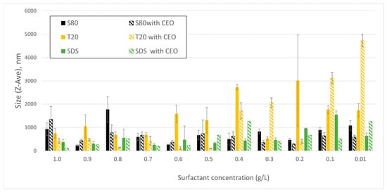
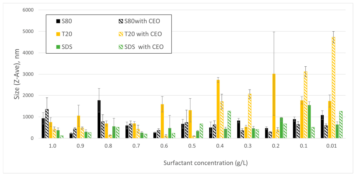
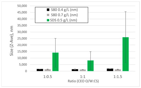
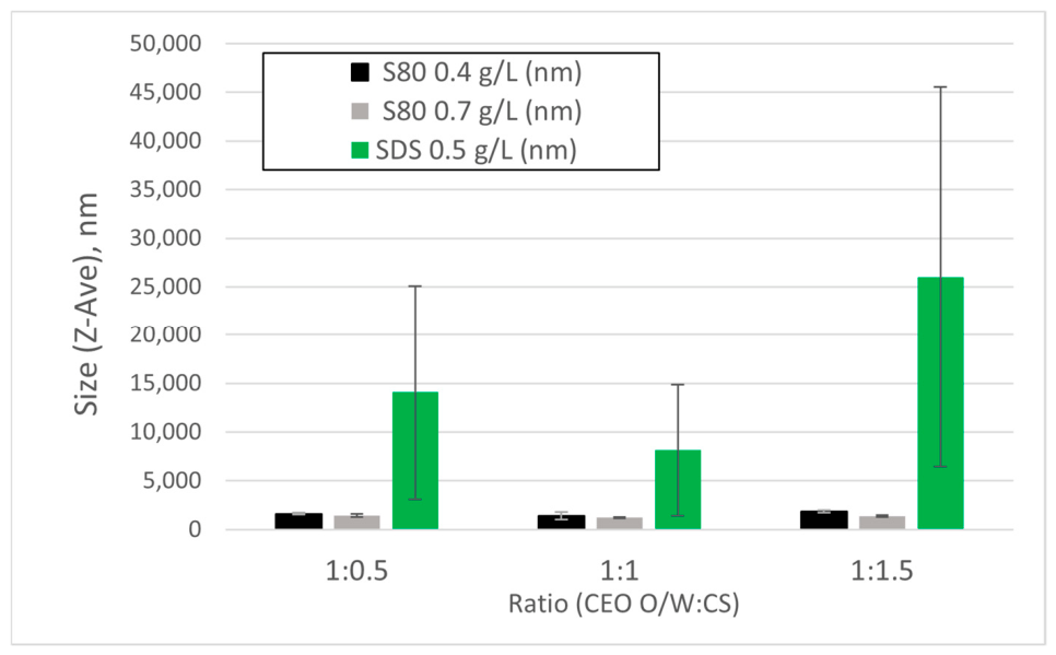
Dynamic light scattering (DLS) measurements indicated that the particle size of microcapsules changed depending on the surfactant and biopolymer concentrations used during the encapsulation procedure. The particle sizes presented in Figure 1 highlight the critical role of surfactant concentration and type on emulsion stability. S80 emulsions at concentrations of 0.4 g/L and 0.7 g/L (black columns) had significantly smaller particle sizes compared to those formed using T20 and SDS, with an average diameter of less than one micron. This result can be attributed to the lower interfacial tension achieved by S80, a nonionic surfactant with excellent hydrophobic compatibility [16]. Additionally, the inclusion of CEO further influenced the droplet sizes, suggesting enhanced emulsion stability under optimal conditions for hydrophobic essential oils. This limited size range is ideal for maintaining stable dispersion in water-based environments, as it minimizes the likelihood of sedimentation and clustering [16].
Figure 1.
Emulsion size (Z-Ave) obtained with the use of the various surfactants.
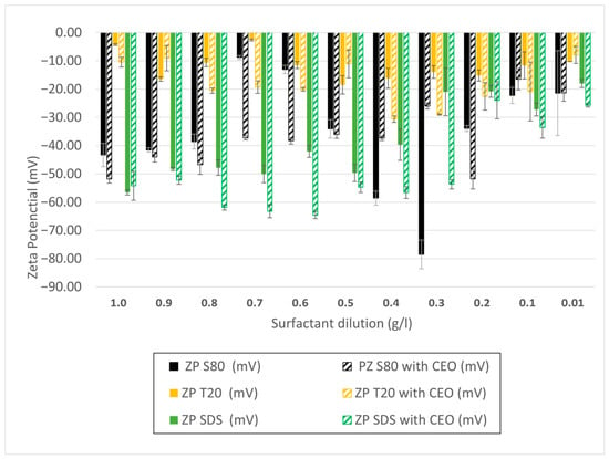
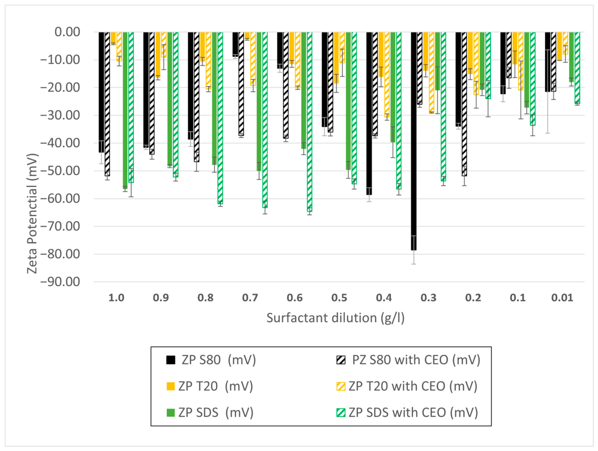
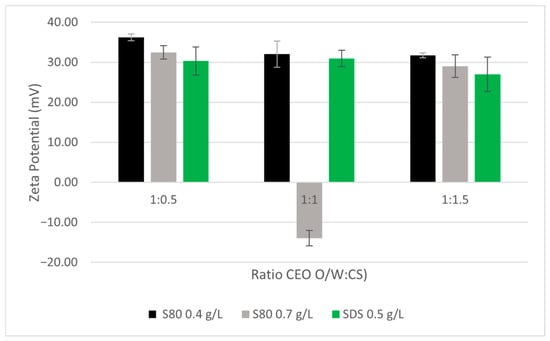
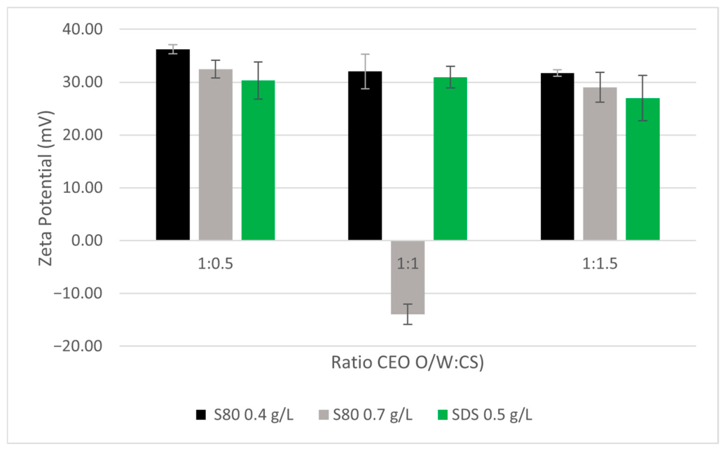
The zeta potential assessments additionally confirmed the stability of these emulsions (Figure 2). Emulsions containing S80 exhibited elevated zeta potential values, suggesting significant electrostatic repulsion among the micelles. This repulsion hinders clustering, keeping the micelles in a steady colloidal suspension. The ideal zeta potential range noted was from −30 mV to −40 mV, generally linked to very stable suspensions, reducing the likelihood of aggregation over time. In contrast, T20 and SDS resulted in emulsions with reduced zeta potential values, which were associated with an increased likelihood of aggregation, especially at lower concentrations [16]. The zeta potential behavior aligns with the molecular characteristics of S80, which supports stable water-in-oil emulsions critical for the encapsulation process.
Figure 2.
Zeta potential measurements of the emulsions prepared using different surfactants.
Based on the size and zeta potential results, three emulsions were selected: S80 0.4 g/L, S80 0.7 g/L, and SDS 0.5 g/L. These emulsions had small diameters and a high electrical charge after the CEO was incorporated.
Introducing CS as the primary coating (Figure 3) led to stable microcapsules with consistent size distributions. CS, known for its inherent antimicrobial properties and biocompatibility, effectively adsorbed onto the emulsion surface through electrostatic interactions with the negatively charged CEO emulsions (Figure 2). The average sizes of the microcapsules formed with S80 at 0.4 g/L and 0.7 g/L remained within the submicron range, which is ideal for controlled release applications.
Figure 3.
Size distribution of S80 microcapsules coated with a CS layer.
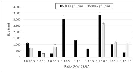
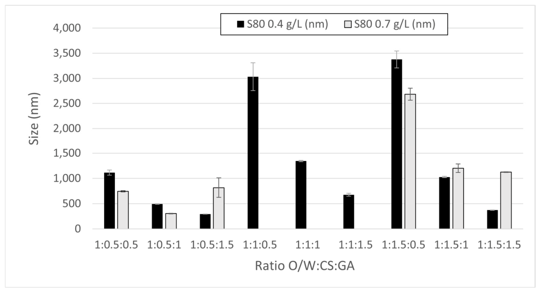
Adding a secondary Gum Arabic (GA) layer (Figure 4) further enhanced microcapsule stability and mechanical strength. The dual-layer design, as expected, effectively increased microcapsule diameters due to the additional polymer deposition, with ratios such as 1:0.5:1 (CEO O/W:CS:GA) and 1:1:1.5 demonstrating an optimal balance between size and structural uniformity. These combinations are particularly important for applications requiring prolonged CEO release, as the secondary layer mitigates premature release by reinforcing the barrier properties of the microcapsule [21].
Figure 4.
Emulsion size results (Z-Ave) obtained with the various surfactants used.
Figure 5 presents the zeta potential distribution of microcapsules with a single CS coating. The data obtained highlight that microcapsules prepared with S80 at a concentration of 0.4 g/L exhibit a zeta potential in the range of −30 mV to −40 mV, suggesting electrostatic repulsion between particles. This indicates a highly stable colloidal system, where the aggregation has been minimized, ensuring the uniform dispersion of microcapsules. The strong interaction between the positively charged CS and the negatively charged CEO-loaded micelles enhances the stability of the emulsion.
Figure 5.
Zeta potential distribution of S80 microcapsules coated with a CS layer.
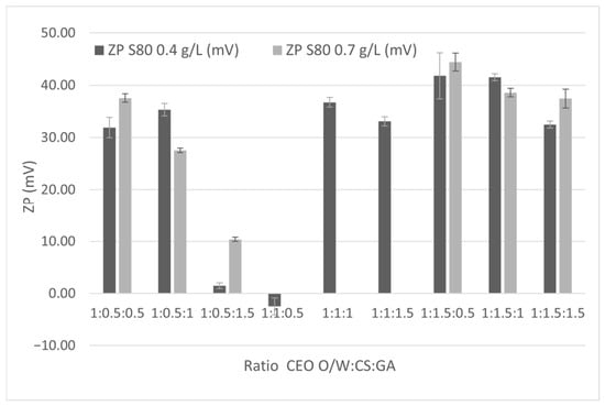
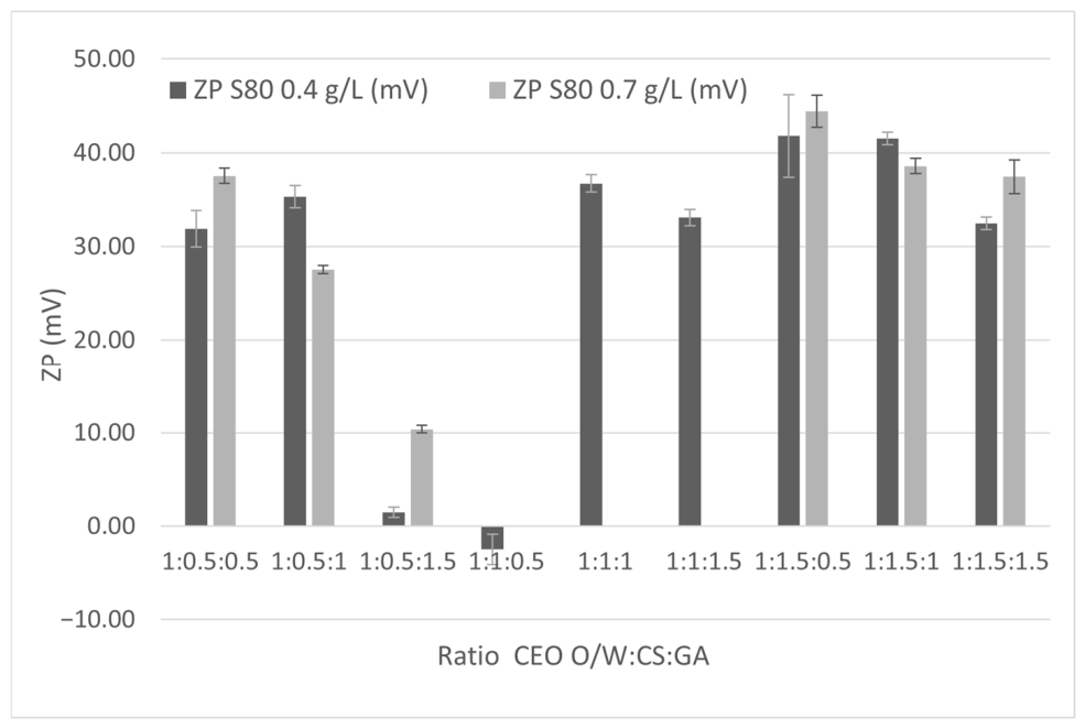
Figure 6 extends the analysis to microcapsules coated with both CS and GA. The addition of GA as a secondary layer further stabilizes the microcapsules, with zeta potential values becoming less negative compared to those in Figure 5. This reduction is due to the neutralizing effect of the GA layer, which reduces the net surface charge of the microcapsules while enhancing their mechanical integrity.
Figure 6.
Zeta potential distribution of S80 microcapsules coated with CS and GA layers.
For microcapsules formed with S80 at 0.4 g/L, the zeta potential remained within a range of −30 mV to −40 mV, which is still indicative of good colloidal stability but reflects a slight reduction in electrostatic repulsion. This shift aligns with GA’s role in creating a compact, cohesive matrix that stabilizes the microcapsule structure [21,22].
3.2. Optical Microscopy
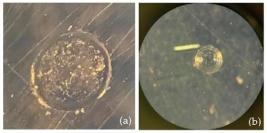
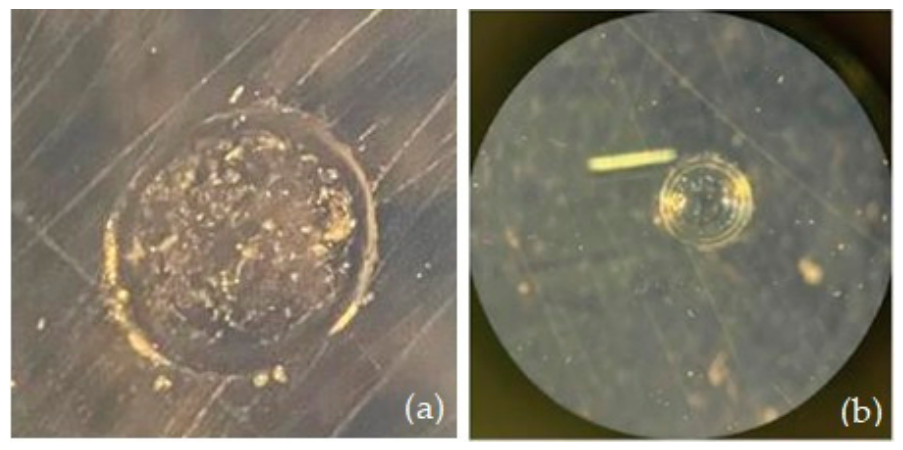
Morphological examination using an optical microscope further revealed the microcapsules’ structure (Figure 7). These images show that the S80 microcapsules were spherical and displayed a smooth surface texture, suggesting the presence of a cohesive biopolymer layer surrounding the CEO. The dual-layer configuration, comprising CS and GA, established a compact matrix enveloping the CEO core, improving structural strength and potentially regulating CEO release. These results emphasize the efficiency of S80 in creating stable and structurally robust CEO microcapsules appropriate for extended use.
Figure 7.
S80 microcapsules. (a) Microcapsule coated with a single biopolymer layer of CS; (b) microcapsule with a dual biopolymer layer comprising CS and GA.
3.3. FTIR Spectra
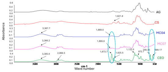
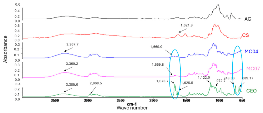
The main signals that characterize cinnamon essential oil (green line in Figure 8) are 689 cm−1, corresponding to the specific C-H out-of-plane bending vibration of alkenes, 1673.7 cm−1, attributed to the C=O carbonyl stretching from the aldehyde group, and 1625.5 cm−1, corresponding to C=C stretching. Both the carbonyl and alkene signals are characteristic indicators of cinnamic aldehyde, which is a marker of cinnamon oil [23,24]. Additionally, a minor peak at 1734 cm−1 was observed, which can be attributed to the C=O stretching of trace ester components within the essential oil.
Figure 8.
FTIR spectra of CS, AG, CEO, and two samples of microcapsules (0.4 g/L, MC04 and 0.7 g/L MC07).
As shown in Figure 8, the microcapsules showed a shift in the 3385 cm−1 peak, indicating a molecular interaction between the -OH groups of the oil and the -OH groups of chitosan through a hydrogen bond. This can be attributed to the formation of oil microcapsules, which is confirmed by the disappearance of oil peaks in the 748.1–688.9 cm−1 range and the significant decrease at 1673.7 cm−1 masked by the biopolymer shell.
3.4. TGA and DTGA
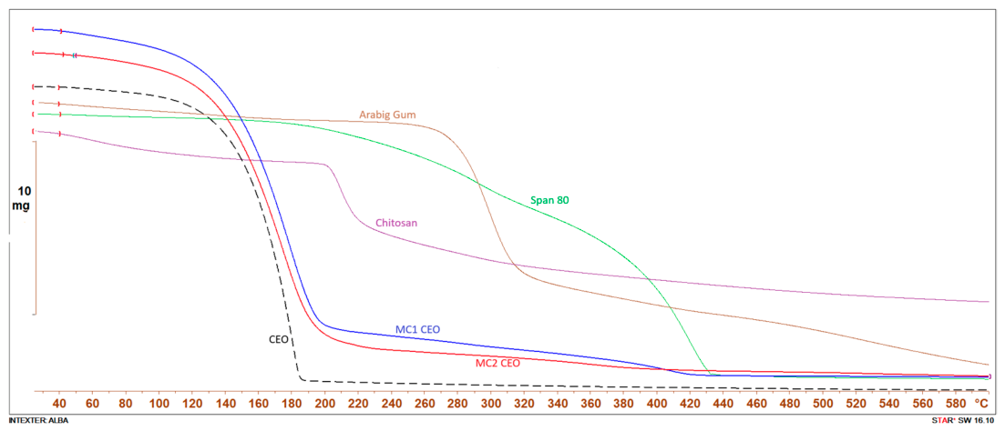
During thermogravimetric analysis, an average mass of 10 mg of the sample (polymers, surfactant, and microcapsules, all previously dehydrated for 24 h at 80 °C) was placed in an alumina cup and heated from 25 °C to 600 °C at a heating rate of 10 K min−1 under a nitrogen flow of 50 mL min−1. The derivative thermogravimetric (DTGA), the initial degradation temperature (Tonset), and the peak temperature were evaluated to understand the thermal properties of the samples and are presented in Table 1.
Table 1.
Parameters of the TGA assays. MC1 CEO and MC2 CEO are the two types of microcapsules obtained and used in the delivery experiments (0.4 g/L and 0.7 g/L).
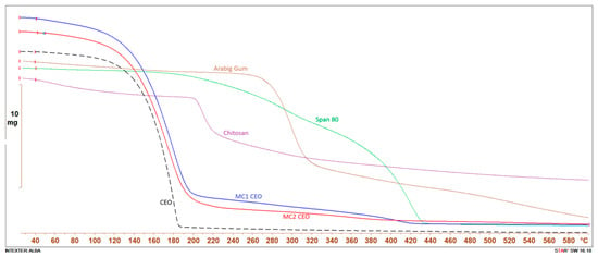
As shown in Figure 9, the weight loss profiles of the cinnamon oil and microcapsules are very similar. Biopolymers and other components of the analyzed system strongly deviated from this behavior. It is only possible to detect the influence of Gum Arabic or chitosan after long periods of time, when the oil has volatilized from the microcapsules over the period of 12 to 16 min. In contrast, when the oil was inside the microcapsules, this joint decomposition occurred over longer periods of time, showing the stability of the microcapsules and demonstrating the presence of the essential oil inside their structure as part of the core.
Figure 9.
TGA graphs for each of the components of the system, separated and together as microcapsules.
3.5. Textile Application
Fabric samples of 1 g (5 cm × 10 cm) were impregnated using the dipping procedure. The textile weight before and after application was used to calculate the impregnation percentage (see Table 2).
Table 2.
Impregnation of textiles with S80 microcapsules.
Table 2 indicates that, although both fabrics were impregnated with microcapsules, the COT fabric retained a higher amount. This is because COT fibers are cellulosic, with a porous and absorbent structure, hydrophilic properties, capillary absorption capacity, and a higher affinity for chemical bonds (hydrogen bonds) and physical interactions with microcapsules. In other words, the texture and roughness of COT fibers provide a larger surface area and more anchoring points for the microcapsules compared to the smoother surface of PES fibers. Therefore, COT samples were used for the delivery experiments because the number of microcapsules in the PES samples was too low.
The 1:0.5:1 ratio was selected not only for its superior impregnation efficiency (3.76% for COT) but also due to its optimal colloidal characteristics. This formulation maintained a submicron particle size and a high zeta potential (−30 to −40 mV), ensuring a stable suspension that facilitates uniform distribution within the porous structure of cotton fibers. In the next step, the 1:0.5:1 ratio was used to study CEO drug delivery from both textiles and both S80 microcapsule concentrations (0.4 g/L and 0.7 g/L).
3.6. Regulated Release Patterns
CEO release from S80 microcapsules, particularly the release kinetics when applied to COT, was examined under simulated physiological conditions.
The release profiles of each fiber type were assessed using UV–Visible spectrophotometry to measure the CEO concentration at 294 nm, with the calibration curve shown in Equation (1).
where y is absorbance, and x is the CEO concentration (g/L).
y = 5.36x + 0.153 R2 = 0.998
Fickian diffusion, the conventional diffusion model, explains a release mechanism that relies entirely on concentration gradients, in which the CEO moves through the microcapsule barrier into the adjacent medium. Anomalous diffusion, also known as non-Fickian diffusion, refers to a release process affected by other parameters, such as relaxation of the polymer matrix or structural changes within the surrounding layers promoted by intense molecular interactions.
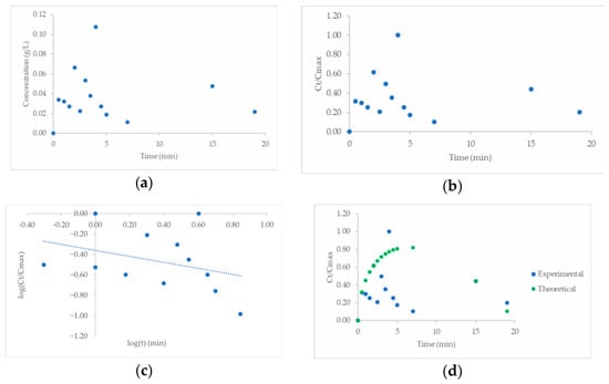
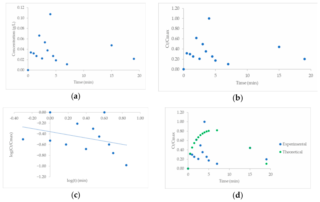
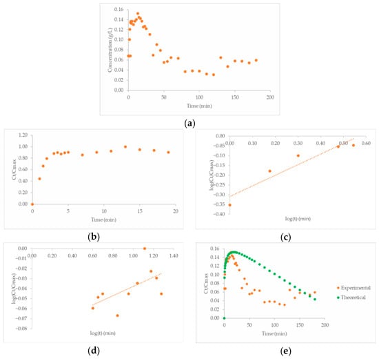
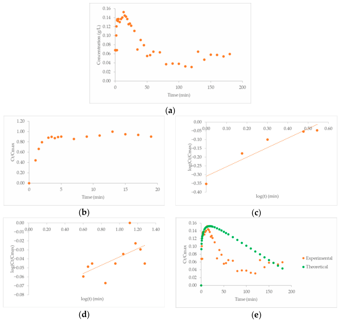
The CEO release pattern (Figure 10 and Figure 11) showed a biphasic profile marked by an initial quick release phase followed by a gradual, prolonged release. This biphasic pattern is frequently seen in controlled-release systems, where an initial “burst release” ensures the immediate availability of the active ingredient, followed by a sustained diffusion phase that enables extended delivery. During the early phase, Fickian diffusion was the primary mechanism for CEO release, as deduced from the rise in concentration within the buffer solution. This phase is attributed to CEO molecules being positioned in the outer layer of the microcapsule matrix, allowing them to disperse more easily into the surrounding environment.
Figure 10.
Drug delivery profiles of microcapsules formulated with S80 at 0.4 g/ ratio 1:0.5:1 on COT. (a) Experimental drug release data, showing the concentration of the compound over time. (b) Fitting of the Korsmeyer–Peppas model to the experimental data. (c) Logarithmic representation of the Korsmeyer–Peppas model, illustrating the non-conventional release mechanism; R2 = 0.445. (d) Comparison of experimental data with the Peppas–Sahlin model.
Figure 11.
Drug delivery profiles of microcapsules formulated with S80 at 0.7 g/L in COT: (a) experimental drug release data showing the concentration of the active compound over time; (b) fitting of the Korsmeyer–Peppas model to the experimental data, illustrating the general release behavior; (c) application of the Korsmeyer–Peppas model highlighting the diffusion-based release mechanism; R2 = 0.778; (d) representation of the Korsmeyer–Peppas model demonstrating a non-conventional release mechanism; (e) comparison of experimental results with the Peppas–Sahlin model.
In the next phase, the CEO released profiles presented in Figure 10 and Figure 11, which highlight the sustained release capabilities of the microcapsules formulated with S80 at 0.4 g/L and 0.7 g/L from COT fabrics. The release kinetics were analyzed using established models, such as the Korsmeyer–Peppas (Equation (2)) and Peppas–Sahlin (Equation (3)) models.
The Korsmeyer–Peppas model (Figure 10c and Figure 11c) indicated a non-Fickian release mechanism, characterized by both diffusion and polymer relaxation processes. This release behavior underscores the influence of the dual biopolymer layers in controlling CEO diffusion rates.
where Ct is the amount of active principle (AP) released at time t, C∞ is the maximum amount of the released AP at infinite time, k is the rate constant of AP released, and n is the diffusional exponent that depends on the system geometry. The value of n is indicative of the release mechanism of the AP [20].
Ct/C∞ = k · tn
Ct/C∞ = k1 · tm + k2 · t2m
The application of the Peppas–Sahlin model (Figure 10d and Figure 11e) further confirmed the presence of anomalous diffusion, as reflected in the low k2 values (Table 2). These results emphasize the role of polymer matrix relaxation in modulating CEO release and the strong influence of the chemical characteristics of the textile fabric where the capsules deliver the CEO.
The transport in swelling systems (such as CS and GA) can be described using Fick’s second law, where the diffusion of the active agent is assumed to be a plane surface for short times of liberation. The Higuchi equation can be used for apparent diffusion coefficient approximation (Equation (4)), where D is the apparent diffusion coefficient of drug release and δ is the width of the planar matrix [25,26].
The Higuchi model’s (Equation (4)) parameters, such as the diffusion coefficient D/δ2, also demonstrated higher values for S80 microcapsules at 0.7 g/L, suggesting enhanced CEO mobility through the matrix under optimal conditions.
Ct/C∞ = KH · t0.5
The experimental data (Figure 11a,b) indicate that formulations with 0.7 g/L S80 and a 1:0.5:1 (CEO O/W: CS: GA) ratio provided the most consistent CEO release profile, achieving a sustained release over an extended period. The use of dual biopolymer layers not only delayed the release of CEO but also ensured that its therapeutic concentration remained within an effective range, thus enhancing its potential for long-term applications [15].
In essence, the release rate of the CEO decreases, exhibiting an unusual (non-Fickian) diffusion pattern. This sustained-release phase is affected by the encapsulation matrix, as the structural characteristics of the CS-GA bilayer regulate CEO diffusion. The thick, stratified composition limits the movement of the CEO, thus prolonging its release. This regulated release characteristic is particularly advantageous for applications that require a sustained antibacterial effect, such as therapeutic or antimicrobial fabrics. It is also noticeable that the prolonged decrease is caused by the reservoir effect, which is observed when the textile acts as an interface between the bath and the microcapsules. Depending on the chemical interactions that the active agent undergoes in the bath, it tends to adhere to the textile due to mutual affinity, especially when the essential oil is poorly soluble in water.
For the S80 0.4 g/L and 0.7 g/L samples, a 1:0.5:1 ratio was applied to the COT, and the results for these samples are shown in Table 2. The samples with a higher amount of surfactant showed a higher mass transfer coefficient, indicating that their retention was less strong. The absorption of the bath, as a triggering mechanism, reduced the effect of retention when the amount of surfactant was 0.7 g/L, which is consistent with the type of interactions that the surfactant can form when the quantity of water is high. A deformation of the micelles occurred, improving the release of the oil compounds. Specifically, the observed trend in impregnation efficiency with varying surfactant concentrations (Table 2) suggests that a concentration of 0.7 g/L of S80 enhances the mass transfer coefficient. This higher concentration likely promotes micellar deformation during the drying process, improving the interaction between the biopolymer shell and the cellulosic hydroxyl groups and thereby optimizing the initial reservoir for subsequent delivery.
The phenomena described here exhibit two different mechanisms. On the one hand, a mass gradient exists between the microcapsules and the tissue, which can control CEO delivery outside of the capsule. By contrast, the mass gradient between the CEO retained in the tissue and the semi-infinite bath will depend on the chemical affinity of the CEO chemical components and the hydrophilicity/hydrophobicity of the tissue. Over a short period of time, apparent diffusion controls the delivery to the bath. When the oil concentration increases considerably in the bath, some oil components return from the bath to the tissue, especially when the hydrophilic COT acts as the transitional reservoir substrate. This effect is physically responsible for the existence of two steps in the delivery to the bath and accounts for the lower single-phase R2 coefficients, as the system transitions between these regimes. Consequently, the Korsmeyer–Peppas equation provides an average value of n because the main mechanism depends on time. Meanwhile, the values obtained using the Higuchi model applied to short times allow us to calculate the apparent diffusion coefficient of the microcapsule–textile substrate system. The Peppas–Sahlin model (Figure 11e), designed to discriminate the separate behavior of the microcapsule core content gradient from the mobility of polymers in the shell, clearly shows the best correlation with the experimental results.
3.7. Scanning Electron Microscopy (SEM)
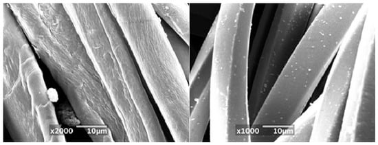
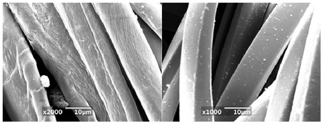
Scanning Electron Microscopy (SEM) images revealed the presence and distribution of S80-based microcapsules in both the COT and PES fibers. The dual-layered microcapsules exhibited enhanced mechanical attachment, particularly in formulations with optimized polymer ratios. This adhesion suggests that the microcapsules remain attached to the fibers, maintaining their functional properties throughout use and enabling controlled CEO release.
A comparative analysis of the CEO release profiles of these fibers highlighted distinct differences. In COT fibers, the release was more gradual and sustained, attributed to their higher porosity and absorbency, as well as the favorable surface chemistry of cotton (hydroxyl groups) that promotes hydrogen bonding with the encapsulation matrix. This may have allowed closer interactions between the microcapsules within the fiber network. In contrast, PES fibers, with their lower porosity and hydrophobic nature, retained the microcapsules primarily on the surface, leading to a faster but shorter release. These findings suggest that COT fibers are better suited for applications requiring prolonged CEO release, while PES fibers are more appropriate for scenarios demanding rapid, transient delivery (Figure 12).
Figure 12.
SEM images showing CEO microcapsules adhering to the surface and embedded within the structure of (a) COT fiber and (b) PES fiber.
3.8. Bacterial Inhibition Effectiveness
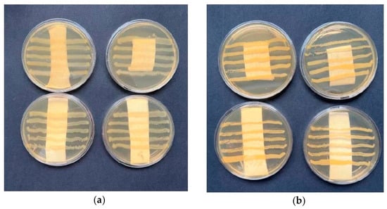
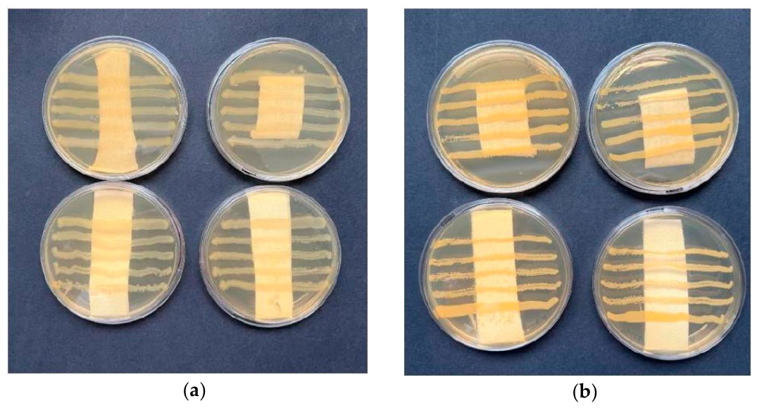
The antibacterial effectiveness of the CEO microcapsules was evaluated by determining their inhibition zones against E. coli and S. aureus, representing Gram-negative and Gram-positive bacteria, respectively. The formation of inhibition zones around the textile samples treated with CEO shows that CEO was effectively released from the microcapsules in adequate amounts to influence bacterial growth (Figure 13). The antibacterial assay period was only 24 h (AATCC TM147-2011-2016e), which was not long enough to deliver a larger quantity of active principles, as supported by the low apparent diffusion coefficients in Table 3. Consequently, the observed effects at 24 h are limited by diffusion, and the effect should be qualified as an “influence”.
Figure 13.
Photographs of antibacterial tests for CEO microcapsules adhered to textile samples against (a) Escherichia coli and (b) Staphylococcus aureus. In both panels, the top images correspond to PES and the bottom images to COT.
Table 3.
Parameters derived from various mathematical models applied for the analysis of the drug release kinetics.
This suggests that the antibacterial efficacy of the CEO microcapsules, assessed using E. coli and S. aureus, was limited. Despite strong microcapsule adhesion to textile fibers, a potentially limited CEO concentration within the capsules might have restricted the formation of significant bacterial inhibition zones. This limitation underscores the need to optimize CEO loading during the encapsulation process to enhance antimicrobial performance.
Although CEO has shown inherent antibacterial qualities in earlier studies [17,18], the encapsulation method presents certain constraints. The bilayer encapsulation matrix, although successful in regulating CEO release, might limit CEO’s diffusion into the adjacent medium, particularly at minimal CEO concentrations. Additionally, as a hypothesis, the stability of the microcapsules tested might immobilize the chitosan chains in the microcapsule, potentially reducing their mobility and their antimicrobial effects. Therefore, although the microcapsules provided certain antibacterial protection, the inhibition zones were smaller than those observed in free CEO applications in comparable studies. This discovery indicates that future research should aim to enhance the loading of CEOs into microcapsules to boost antimicrobial effectiveness while maintaining structural stability.
Future strategies for enhancing the antibacterial performance of the developed microcapsules should focus on increasing both the effective concentration and the bio-availability of CEO at the textile–microorganism interface. This could be achieved by optimizing the core loading capacity of the microcapsules, either by increasing the oil-to-polymer ratio or by modifying the micellar pre-encapsulation step to improve the retention of active volatile compounds.
4. Conclusions
In this study, stable cinnamon essential oil (CEO) microcapsules were successfully produced using Span 80 (0.4–0.7 g/L) and a chitosan–Gum Arabic dual layer. The capsules exhibited submicron diameters and high zeta potential values, ensuring colloidal stability and minimizing aggregation.
The release kinetics followed a biphasic pattern, characterized by an initial burst phase followed by sustained diffusion regulated by the polymeric matrix. In textile applications, cotton fibers demonstrated significantly higher microcapsule retention and more effective release than polyester, which can be attributed to their porous and absorbent nature.
The enhanced thermal stability observed in the TGA further supports the formation of a compact biopolymeric shell, which is consistent with the reduced diffusion coefficients and the sustained release behavior observed during the drug delivery experiments.
Finally, the microcapsules exhibited limited antibacterial activity against E. coli and S. aureus, as their effectiveness was constrained by the CEO concentration within the core. These findings indicate that future optimization strategies should focus on increasing the loading capacity to enhance antimicrobial performance in functional textile applications.
Author Contributions
Conceptualization, M.J.L.; methodology and experimental part, P.C. and L.M.; validation, G.M., H.L. and S.S.; data curation, P.C., H.L. and R.M.; writing—original draft preparation, P.C.; writing—review and editing, M.J.L., M.M. and G.M.; supervision, M.J.L. and G.M. All authors have read and agreed to the published version of the manuscript.
Funding
This research received no external funding.
Institutional Review Board Statement
Not applicable.
Data Availability Statement
The original contributions presented in the study are included in the article, further inquiries can be directed to the corresponding author.
Acknowledgments
The authors would like to thank Mercedes Escusa for her technical assistance with the SEM analysis and Bettina Vallés for her support during the antibacterial tests.
Conflicts of Interest
The authors declare no conflicts of interest.
References
- Bakry, A.M.; Abbas, S.; Ali, B.; Majeed, H.; Abouelwafa, M.Y.; Mousa, A.H.; Liang, L. Microencapsulation of Oils: A Comprehensive Review of Benefits, Techniques, and Applications. Compr. Rev. Food Sci. Food Saf. 2015, 15, 143–182. [Google Scholar] [CrossRef] [Scilit] [PubMed]
- Fuchs, M.; Turchiuli, C.; Bohin, M.; Cuvelier, M.; Ordonnaud, C.; Maillard, M.; Dumoulin, É. Encapsulation of oil in powder using spray drying and fluidized bed agglomeration. J. Food Eng. 2006, 75, 27–35. [Google Scholar] [CrossRef] [Scilit]
- Sisqueira, R.; Borges, J.; Bezerra, F.; Da Costa, C.; Correira, J.; Marti, M.; Coderch, L.; López, A.; Lis, M. Application of lavender-oil microcapsules to functionalized PET fibers. Polymers 2023, 15, 917. [Google Scholar] [CrossRef] [Scilit] [PubMed]
- Tariq, H.; Rehman, A.; Ali Raza, Z.; Kishwar, F.; Abid, S. Recent progress in the microencapsulation of essential oils for sustainable functional textiles. Polym. Bull. 2024, 81, 7585–7629. [Google Scholar] [CrossRef] [Scilit]
- Georgievska, T.; Trajkovikj, S.; Atkovska, K.; Lisichkov, K. Recent Advances in Textile Functionalization Using Essential Oil-Based-Microcapsules with Antimicrobial Properties. Tekstilec 2025, 68, 121–131. [Google Scholar] [CrossRef] [Scilit]
- Lezeck, H.; Martí, M.; Saxena, S.; Lis, M.J. Essential Oils Applied to Textile Substrates with Emphasis on Antibacterial Properties: Review Article. Molecules 2026, 31, 1077. [Google Scholar] [CrossRef] [Scilit] [PubMed]
- Benita, S. Microencapsulation: Methods and Industrial Applications, 2nd ed.; CRC Press: Boca Raton, FL, USA, 2005; Volume 158. [Google Scholar]
- Han, Y.; Yu, M.; Wang, L. Physical and antimicrobial properties of sodium alginate/carboxymethyl cellulose films incorporated with cinnamon essential oil. Food Packag. Shelf Life 2018, 15, 35–42. [Google Scholar] [CrossRef] [Scilit]
- Marqués, L.; Cota, P.; Cano, F.; Marti, M.; Martínez, A.; Lis, M. Chapter 1: Development and Characterisation of PLA-Nanofiber Scaffolds with Cinnamon Oil-Lanolin Emulsions for Antibacterial Human Skin Grafts. In Chemical and Materials Sciences: Research Findings Vol. 4; Bhowmik, P.K., Ed.; BP International: Kolkata, India, 2025; pp. 1–18. [Google Scholar] [CrossRef] [Scilit]
- Shu, C.; Ge, L.; Li, Z.; Chen, B.; Liao, S.; Lu, L.; Wu, Q.; Jiang, X.; An, Y.; Wang, Z.; et al. Antibacterial activity of cinnamon essential oil and its main component of cinnamaldehyde and the underlying mechanism. Front. Pharmacol. 2024, 15, 1378434. [Google Scholar] [CrossRef] [Scilit] [PubMed]
- Culas, M.S.; Popovich, D.G.; Rashidinejad, A. Recent advances in encapsulation techniques for cinnamon bioactive compounds: A review on stability, effectiveness, and potential applications. Food Biosci. 2024, 57, 103470. [Google Scholar] [CrossRef] [Scilit]
- Peluche, Y.I.P.; Correa, D.A.; Villadiego, A.M.D. Efecto antimicrobiano del clavo y la canela sobre patógenos. Biotecnol. Sect. Agropecu. Agroind. 2017, 15, 56–65. [Google Scholar] [CrossRef] [Scilit]
- Haputhanthri, R.; Dehigahawatta, S.; Sandaruwan, C.; Kanchanamali Perera, K.; Pieris, C. Preparation, Characterization and Sustained-release Study of Encapsulated Cinnamon Essential Oil Microcapsules. Nat. Prod. J. 2025, 15, e22103155292898. [Google Scholar] [CrossRef] [Scilit]
- Yang, Y.; Yang, W.; Chen, X.; Xiao, H.; Liu, G.-Q.; Han, W.; Li, J. Starch nanoparticle loading cinnamon essential oil encapsulated by complex coacervation of chitosan and gum Arabic improve the stability and antibacterial potential of the microcapsules. Int. J. Biol. Macromol. 2025, 310, 143613. [Google Scholar] [CrossRef] [Scilit] [PubMed]
- Teixeira-Costa, B.E.; Andrade, C.T. Chitosan as a valuable biomolecule from seafood industry waste in the design of green food packaging. Biomolecules 2021, 11, 1599. [Google Scholar] [CrossRef] [Scilit] [PubMed]
- Cullum, D.C. (Ed.) Surfactant types; classification, identification, separation. In Introduction to Surfactant Analysis; Springer: Dordrecht, The Netherlands, 1994. [Google Scholar] [CrossRef] [Scilit]
- Sáez, V.; Hernáez, E.; Angulo, L.S. Sistemas de liberación controlada de medicamentos. Rev. Iberoam. Polím. 2002, 3, 16–35. [Google Scholar]
- Martí, M.; Martínez, V.; Lis, M.J.; Coderch, L. Mathematical models for drug delivery from textile. J. Ind. Text. 2021, 50, 1225–1238. [Google Scholar]
- Valle, J.A.B.; Curto Valle, R.D.C.S.; da Costa, C.; Maestá, F.B.; Lis Arias, M.J. Reservoir Effect of Textile Substrates on the Delivery of Essential Oils Microencapsulated by Complex Coacervation. Polymers 2024, 16, 670. [Google Scholar] [CrossRef] [Scilit] [PubMed]
- Ravindra, B.B.; Naveen, B.K. Unified Mathematical Expression for Ideal Peppas Model: Prospective Estimation of percent deviation. Asian J. Pharm. Tech. 2016, 6, 189–196. [Google Scholar] [CrossRef] [Scilit]
- Tang, Y.; Gao, C.; Tang, X. In situ rapid conjugation of chitosan-gum Arabic coacervated complex with cinnamaldehyde in cinnamon essential oil to stabilize high internal phase Pickering emulsion. Food Hydrocoll. 2023, 134, 108103. [Google Scholar] [CrossRef] [Scilit]
- Narambuena, C.; Ausar, F.S.; Bianco, I.D.; Beltramo, D.M.; Leiva, E.P.M. Aggregation of Casein Micelles by Interactions with Chitosans: A Study by Monte Carlo Simulations. J. Agric. Food Chem. 2005, 53, 459−463. [Google Scholar] [CrossRef] [Scilit]
- Boughendjioua1, H.; Amoura, N.; Boughendjioua, Z. Purity Specifications of Constituents of Cinnamon Essential Oil by Fourier Transformed Infrared Spectroscopy Analysis. Indian J. Pharm. Biol. Res. 2017, 5, 36–40. [Google Scholar] [CrossRef] [Scilit]
- Li, Y.-Q.; Kong, D.-X.; Wua, H. Analysis and evaluation of essential oil components of cinnamon barks using GC–MS and FTIR spectroscopy. Ind. Crops Prod. 2013, 41, 269–278. [Google Scholar] [CrossRef] [Scilit]
- Brazel, C.S.; Peppas, N.A. Modeling of drug release from swellable polymers. Eur. J. Pharm. Biopharm. 2000, 49, 47–58. [Google Scholar] [CrossRef] [Scilit] [PubMed]
- Peppas, N.A.; Keys, K.B.; Torres-Lugo, M.; Lowman, A.M. Poly (ethylene glycol)-containing hydrogels in drug delivery. J. Control. Release 1999, 62, 81–87. [Google Scholar] [CrossRef] [Scilit]

Disclaimer/Publisher’s Note: The statements, opinions and data contained in all publications are solely those of the individual author(s) and contributor(s) and not of MDPI and/or the editor(s). MDPI and/or the editor(s) disclaim responsibility for any injury to people or property resulting from any ideas, methods, instructions or products referred to in the content. |
© 2026 by the authors. Licensee MDPI, Basel, Switzerland. This article is an open access article distributed under the terms and conditions of the Creative Commons Attribution (CC BY) license.