


Abstract
The Oriental stork, Ciconia boyciana Swinhoe, 1873 (Ciconiiformes: Ciconiidae), once common as a sedentary stork species across Japan, experienced a marked population decline during the last century, reaching extinction in the country in 1971 as a result of anthropogenic alterations to the natural environment. After the reintroduction of five captive descendants of continental pairs in 2005, the naturalized population of Oriental storks has steadily increased in Japan, reaching >500 wild storks by 2025. We recently encountered a case of apparent intestinal fluke infection with Chaunocephalus ferox (Echinostomata; Echinostomatidae) in a 2-month-old stork fledgling within a few days after leaving the nest on 29 June 2025. The parasite was subjected to morphological and molecular identification based on nucleotide sequences of the small- to large-subunit ribosomal RNA gene and mitochondrial cytochrome c oxidase subunit 1 gene and was identified as C. ferox, a parasite exhibiting high host specificity for ciconiid birds worldwide. This finding suggests the re-establishment of its natural transmission cycle in the field in Japan, accompanied by the dispersal of definitive hosts after a gap of at least 30 years. Following the establishment of a reintroduced sedentary Oriental stork population in Japan, this health-threatening chaunocephalosis may, by chance, have a pronounced impact on infection-susceptible stork chicks cared for and fed by parent birds inhabiting fields with dense C. ferox endemicity (hotspots). Another Oriental stork fledgling from the same nest in June 2025 survived and was observed in March 2026 in a paddy field at a distant locality. Parasites are natural components of healthy ecosystems that contribute to biodiversity and trophic balance; therefore, inappropriate anthropogenic habitat modification may create scenarios in which parasites become overly destructive rather than serving merely as ecological regulators.
Keywords:
Chaunocephalus ferox; Japan; Ciconiidae; conservation; rDNA; cox1; morphology; phylogeny; parasite conservation 1. Introduction
The Oriental stork, Ciconia boyciana Swinhoe, 1873 (Ciconiiformes: Ciconiidae), is a large bird (weighing up to 4–5 kg) at the apex of the wetland food webs. It is carnivorous, feeding on snails, crustaceans, amphibians, reptiles, and fish [1]. Currently, the Oriental stork, which is distributed in East Asia, including the southern Russian Far East, eastern China, Taiwan, Korea, and Japan, is classified as “Endangered” according to the International Union for Conservation of Nature Red List of Threatened Species [2,3,4]. It has a small metapopulation of <2500 individuals. Stork populations have undergone rapid declines due to deforestation, wetland reclamation for agriculture, excessive pesticide use in paddy fields, overfishing, and human activity-induced changes in ecosystems [1,2,5,6]. Multipronged conservation efforts are underway to secure the future of Oriental storks in East Asian countries [5,7,8,9,10,11,12,13,14,15].
In Japan, the Oriental stork, designated as a special national natural monument as in China and South Korea, once commonly inhabited throughout the country except for Hokkaido and Okinawa. However, the sedentary population in Japan drastically declined during the last century, particularly toward the 1960s, and became completely extinct in 1971 despite conservation efforts by local citizens [1,6,9,10,11,16,17]. After the first reintroduction of five captive descendants from continental pairs in 2005 (originating from Khabarovsk Krai and China) by the Hyogo Park of the Oriental White Stork, Toyooka City, Hyogo Prefecture, the naturalized population of Oriental storks has steadily increased in Japan, reaching >500 wild storks in 2025. Overall, 55 field nests of reintroduced Oriental storks have been identified in 14 prefectures in western Japan, and 146 stork chicks have left the nests [10,11,18]. For individual identification, color rings have been attached to all released storks and wild-fledged storks in Japan since 2005 [18,19], and tracking stork movements using leg bands is feasible through cooperation among local communities and local governments.
Reintroduced Oriental storks in Japan may face various challenges to survival in newly created natural environments, including dried paddy fields from autumn to spring, widespread pesticide use, concrete-covered waterways, deforestation, and impoverished ecosystems in the modern era. Matsumoto et al. [19] attempted to clarify the causes of injury and death in a newly naturalized population of Oriental storks in Japan and analyzed 153 problematic cases among 412 extant storks (78 released and 334 wild-fledged storks) between 2005 and 2021. Cases of injury and death caused by various human activities accounted for 49.7% (76 cases), with entanglement in pest control measures (bird- and beast-proofing nets) (35 cases) and accidents involving electrical and telecommunication lines (31 cases) being the two major causes. Other causes of injury and death among reintroduced Oriental storks included straying immediately after fledging or release (seven cases), injuries caused by attacks from other storks (seven cases), falls into waterways, ponds, and pools (seven cases), and illness or weakness (six cases). In South Korea, Oh et al. [20] examined 12 dead wild and captive Oriental storks (six cases each) between 2019 and 2023. A high prevalence of fluke nodules in the gastrointestinal tract (chaunocephalosis) was observed in five wild storks, including a fledgling chick. The gastrointestinal lesions described in these wild storks corresponded to the pathological changes caused by Chaunocephalus ferox (Rudolphi, 1795) Dietz, 1909 (Digenea: Plagiorchiida: Echinostomata; Echinostomatidae), which has been widely reported in ciconiid birds from Europe and Asia, including South Korea [21]. Chaunocephalosis was not mentioned in the Japanese report that analyzed the causes of 153 cases of injury or death after reintroduction into the country [19]. Zhou et al. [22] suggested that C. ferox infection may have a serious health impact on Oriental storks because this fluke infection showed a distinctly high prevalence in deceased storks in China (96.3% of 28 individuals examined), compared with a lower prevalence (1.4–5.6%) among field individuals examined using environmental fecal samples.
In this study, we present a case of chaunocephalosis in a fledgling chick in Japan and characterize the newly obtained flukes based on morphological observations and sequences of the small- and large-subunit ribosomal RNA genes (SSU and LSU rDNA), and the mitochondrial cytochrome c oxidase subunit 1 gene (cox1). We further discuss the potential impact of C. ferox infection on the reintroduced population residing in Japan and its implications for stork conservation.
2. Materials and Methods
2.1. Case Report
One of the two stork chicks that hatched on 25 April 2025, in Manno Town, Kagawa Prefecture, was a female and was found weakened on the side of a farm road on 30 June 2025, and immediately rescued (ID: J0907; 66 days old). It weighed 3.57 kg and exhibited severe dehydration, with a 12–15% reduction in body weight. Upon transportation, the chick was lying on its side, unable to stand, and remained recumbent. The weakened chick was treated with intravenous infusion of Ringer’s acetate solution and a 25% glucose solution (420, 450, and 350 mL on the first, second, and third days of care, respectively) and was fed fish paste (horse mackerel). On the fourth day of care, the ill stork chick was found dead. The chick’s body temperature was 39.4, 39.8, and 36.8 °C on the first, second, and third days of hospitalization, respectively. No antibiotics were administered.
Necropsy revealed dehydration; however, no obvious physical injuries were observed. All visceral organs were examined histologically after the trimmed samples were embedded in 27 paraffin blocks, sectioned, and stained with hematoxylin and eosin (HE) according to a standard methodology. Chronic enteritis with fluke infection was evident.
The nest, where a rescued female chick (ID: J0907) hatched with another female chick (ID: J0906) in 2025, was built by a pair of parent storks (ID: J0126 and J0203; cf. Supplementary Figure S1): a male (ID: J0126), which hatched at a field nest in Toyooka City, Hyogo Prefecture, in April 2016 and fledged in June 2016 under a pair of parent storks (ID: J0391 and J0294) that had hatched, been released, and settled in Toyooka, and a female (ID: J0203), which hatched in captivity in May 2018 and was released in September 2018 in Echizen City, Fukui Prefecture. This parent pair built their nest atop a utility pole beside a paddy field in Manno Town, Kagawa Prefecture, in 2023, and one to three chicks (eight fledglings in total up to 2026, excluding the deceased J0907 stork reported here) flew to distant locations, including Shimane and Fukui Prefectures. Before building their nest there, the male stork (ID: J0126) was observed in several areas of western Japan (Naruto, Tokushima Prefecture; and Unnan, Shimane Prefecture), and the female stork (ID: J0203) was observed in Tokushima Prefecture. Another female stork fledged from the same nest on 29 June 2025 (ID: J0906) survived, and was observed in February 2026 in a paddy field in Toyooka City, Hyogo Prefecture, along with three other storks, and in March 2026 in a paddy field in Yosano Town, Kyoto Prefecture, along with 12 other storks. One male stork that fledged in July 2023 (ID: J0671) and three female storks that fledged in July 2024 (IDs: J0789–J0791) from the same nest in Manno Town were widely observed in western Japan (Naruto City, Tokushima Prefecture; Yasugi City, Shimane Prefecture; Inami Town, Hyogo Prefecture; Yosano Town, Kyoto Prefecture). This information is available on the website of the Hyogo Park of the Oriental White Stork, Toyooka (https://satokouen.jp/, accessed on 15 April 2026), and in social media posts by birdwatchers, based on unique individual identification numbers on the colored leg rings attached to all Oriental storks released from captivity or fledged in the field [18,19].
2.2. Parasitological Examination
Parasitological examination was conducted using mid-ileum fragments containing fluke nodules in the mucosa, which were preserved at −20 °C until use. The recovered specimens, except for the one used for molecular analysis, were flattened between two glass plates, stained with either acetocarmine or Weigert’s iron hematoxylin, and cleared with 30% glycerin in 70% alcohol solution. The fixed fluke specimens were deposited in the Meguro Parasitological Museum, Tokyo, Japan (collection no. 25518). Intestinal contents were microscopically examined using a sedimentation technique.
2.3. DNA Extraction, PCR Amplification and Sanger Sequencing
One fluke specimen was washed in physiological saline, and genomic DNA was extracted using an Illustra™ tissue and cells genomicPrep Mini Spin Kit (GE Healthcare UK, Buckinghamshire, UK) according to the manufacturer’s instructions. Overlapping rDNA fragments were amplified by polymerase chain reaction (PCR) using multiple primer pairs reported by Sato et al. [23] (cf. Supplementary Table S1). The resulting PCR products were purified using a FastGene Gel/PCR Extraction Kit (NIPPON Genetics Co., Bunkyo-ku, Tokyo, Japan) and directly sequenced using PCR amplification and sequencing primers (Supplementary Table S1) by Sanger sequencing through a commercial service (FASMAC, Atsugi, Kanagawa, Japan). When direct sequencing was unsatisfactory, the purified PCR products were cloned into the plasmid vector pTA2 (TArget CloneTM; TOYOBO, Kita-ku, Osaka, Japan) and transformed into Escherichia coli JM109 cells (TOYOBO) according to the manufacturer’s instructions. Following propagation, plasmid DNA was extracted using a FastGene Plasmid Mini Kit (NIPPON Genetics Co.), and inserts from at least three independent clones were sequenced using universal M13 forward and reverse primers.
Overlapping cox1 fragments were amplified by PCR using four combinations of newly prepared primers (Table 1), which were designed using the online software ‘Primer3web ver.4.0.0’ [24] based on cox1 sequences of C. ferox (DDBJ/EMBL/GenBank accession no. PV685907), Echinostoma hortense (KR062182), and Echinostoma miyagawai (MN116740). The PCR cycling protocol consisted of an initial denaturation at 94 °C for 3 min, followed by 35 cycles of denaturation at 94 °C for 30 s, annealing at 52 °C for 30 s, and extension at 72 °C for 1 min, with a final extension at 72 °C for 7 min. Purification of PCR amplicons, and direct sequencing using PCR amplification primers were conducted as described above for the rDNA fragments.
Table 1.
Primers used to amplify the Chaunocephalus ferox cox1 sequence.
rDNA contigs were constructed by alignment with closely related rDNA sequences retrieved from the GenBank database of the National Center for Biotechnology Information website (https://www.ncbi.nlm.nih.gov/) using ClustalW [25]. The nucleotide sequences reported in the current study are available in the DDBJ/EMBL/GenBank databases under accession numbers LC934762 (rDNA) and LC934763 (cox1).
2.4. Phylogenetic Analysis
Fragments of the newly obtained rDNA sequences and partial cox1 sequences were subjected to Basic Local Alignment Search Tool (BLAST) analysis of the National Center for Biotechnology Information website (NCBI; https://blast.ncbi.nlm.nih.gov/Blast.cgi, accessed on 15 April 2026) to identify closely related nucleotide sequences. The newly generated sequences, together with homologous sequences of related species within the order Plagiorchiida retrieved from the GenBank databases, were used for phylogenetic analysis. Fifty-two LSU rDNA sequences from 43 species and 40 cox1 sequences from 30 species were aligned using ClustalW in MEGA11 [26] and manually adjusted. Poorly aligned regions and characters with gaps in any sequence were manually excluded. After trimming, the LSU rDNA alignment comprised 1123 characters, of which 412 were variable, whereas the cox1 alignment comprised 1315 characters, of which 753 were variable. The accession numbers of the sequences analyzed in this study are shown in the phylogenetic trees. Phylogenetic trees were reconstructed separately for each gene using the maximum likelihood (ML) method with the HKY85 substitution model implemented in PhyML [27,28] via the Phylogeny.fr website (http://phylogeny.lirmm.fr/phylo_cgi/alacarte.cgi, accessed on 15 April 2026). The probability of inferred branches was assessed using the approximate likelihood ratio test, an alternative to the nonparametric bootstrap estimation of branch support [29]. Graphical representation and editing of the phylogenetic trees were performed using TreeDyn (v198.3) implemented in PhyML, and cleaned illustrations were prepared using Adobe® Photoshop® Elements 2024 (Adobe Systems, San Jose, CA, USA).
3. Results
3.1. Histological Findings
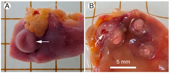
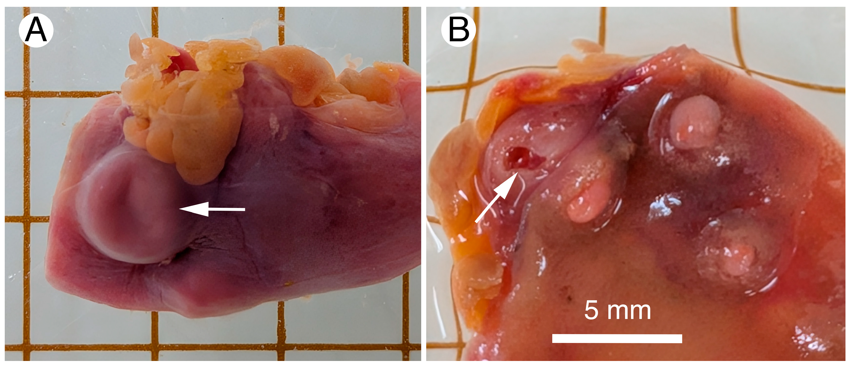
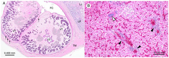
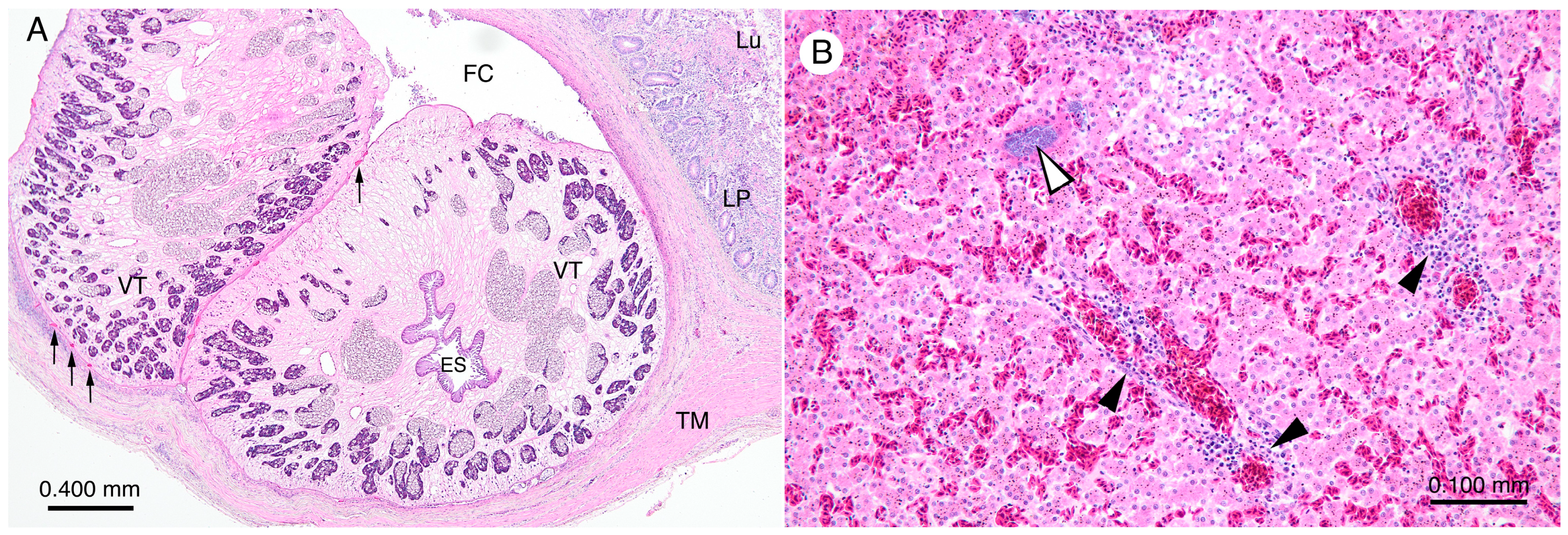
According to municipal records, the dead fledgling stork chick (ID: J0907; 66 days old) was rescued the day after leaving the nest. Municipal officials suspected that its weakened condition resulted from injuries sustained in a traffic accident because it was found on the side of a farm road. However, necropsy did not reveal any physical injuries to the body or viscera. The most evident gross findings were dehydration and 10 globular protrusions on the ileum’s serosal surface (Figure 1A). On the mucosal side corresponding to the serosal protrusions, well-developed globular sacs enclosing two adult flukes, approximately 3.6–4.8 mm in diameter (n = 5), were evident as fluke crypts opening into the lumen (Figure 1B). Histologically, marked infiltration of granulocytes (heterophils/eosinophils), lymphocytes, and monocytes was evident in and around the fibrous fluke crypts (Figure 2A). In general, the mucosal lamina propria, not limited to the areas around the fluke crypts, was thickened with evident leukocyte infiltration, suggesting chronic enteritis. Bacterial clumps between the flukes and fibrous crypt walls were frequently observed. In the liver, mild-to-intermediate focal leukocyte aggregation consisting of lymphocytes (specifically, plasmocytes) and several granulocytes was observed along with multifocal bacteria clumps (Figure 2B), and some small granulomata were found in the spleen. No histological changes were observed in the other organs.
Figure 1.
Gross findings of chaunocephalosis in the ileum of an Oriental stork fledgling. (A) Serosal protrusion (arrow) associated with a mucosal fluke crypt of Chaunocephalus ferox. (B) Mucosal view of fluke crypts of C. ferox, where two flukes are embedded. A vacant fluke crypt after removal of flukes is shown by an arrow.
Figure 2.
Histology of a fluke crypt containing two flukes (A) and the liver (B) of a stork fledgling infected with Chaunocephalus ferox. HE staining. Abbreviations: ES, esophagus; FC, fluke crypt; LP, lamina propria; Lu, intestinal lumen; TM, tunica muscularis; and VT, vitelline glands. Thin arrows in (A) indicate tegumental spines of flukes. White and black arrowheads in (B) indicate bacterial clumps and leukocyte aggregation, respectively.
3.2. Morphological Observation of Flukes
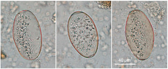
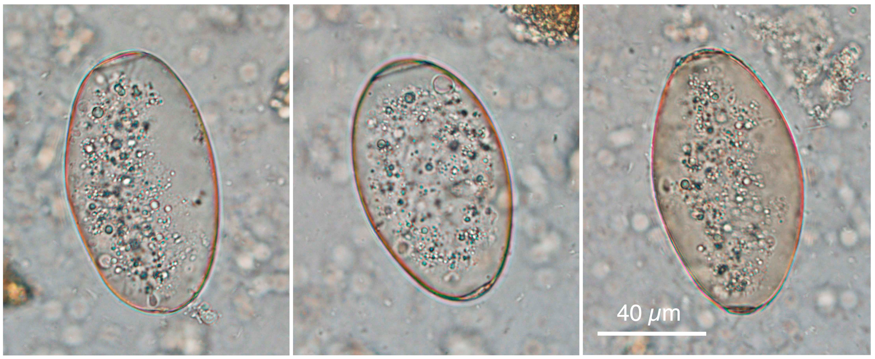
Ovoid eggs produced by the adult flukes were found in the intestinal contents (Figure 3). The egg shells, which were thin and colorless, measured 94.8 ± 6.3 (80.6–107.5) µm in length and 57.4 ± 2.7 (49.3–62.7) µm in width (n = 30) and exhibited uniform thickness (approximately 0.45 µm). Micropyles located at the rounded poles of the egg shells measured 19.9 ± 1.7 (17.2–23.9) µm in diameter. The eggs contained a single oocyte surrounded by multiple vitellocytes.
Figure 3.
Lateral view of Chaunocephalus ferox eggs in the intestinal contents. Micropyles are located on the upper side of each egg. A single oocyte and multiple vitellocytes in the eggs are artificially degenerated in this photograph due to the use of thawed eggs from a frozen tissue sample.
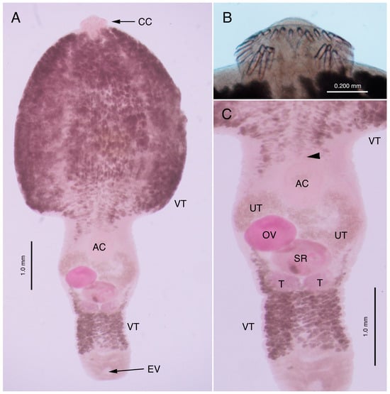
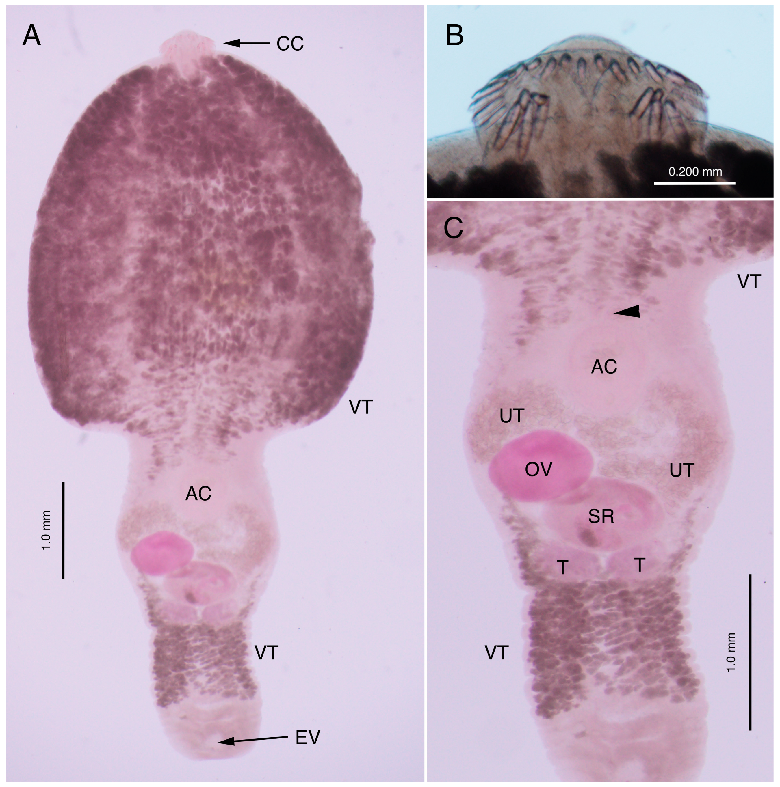
Serosal protrusions of the small intestine were attributed to fluke cysts opening into the lumen, i.e., fluke crypts, as described above (Figure 1B). Two mature flukes were localized within a single crypt. The collected adult flukes (n = 8) were tadpole-shaped, measuring 5.9–7.7 (7.1) mm in length and 2.5–4.2 (3.5) mm in maximum width around the mid-forebody (Figure 4A). The forebody was bulbous, measuring 3.3–4.3 (3.9) mm in length, and the subcylindrical hindbody measured 2.6–3.6 (3.2) mm in length and 1.1–1.8 (1.5) mm in maximum width at the level of the ovary in the second fifth of the hindbody. The reniform cephalic crown protruded from the anterior end, measuring 0.17–0.29 (0.23) mm in length and 0.31–0.59 (0.44) mm in width, and the oral sucker, measuring 0.19–0.29 (0.25) mm in width, was surrounded by 27 spines, except on its ventral side (Figure 4B). Four angle spines were clustered ventrolaterally on both sides, measuring 0.144–0.164 mm in length and 0.026–0.031 mm in width, and shorter marginal collar spines were uninterruptedly aligned along the lateral and dorsal portions of the crown, measuring 0.108–0.125 mm in length and 0.020–0.021 mm in width. The pharynx was not clearly observed because of the dense distribution of vitelline glands throughout the forebody of the fluke. Vitelline glands were densely distributed in the third quarter of the hindbody (Figure 4C). The esophagus extended to the posterior margin of the forebody and bifurcated into the intestines.
Figure 4.
Chaunocephalus ferox adult fluke. (A) Ventral view of the whole body. Acetocarmine staining. (B) High-magnification view of the cephalic crown with 27 collar spines. (C) High-magnification view of the hindbody. Acetocarmine staining. Abbreviations: AC, acetabulum; CC, cephalic crown; EV, excretory vesicle; OV, ovary; SR, seminal receptacle; T, testis; UT, uterus filled with eggs; and VT, vitelline glands. The arrowhead in (C) indicates the genital pore.
In the first fifth of the hindbody, a small genital pore was situated immediately anterior to the ventral sucker, which measured 0.39–0.57 (0.49) mm in length and 0.45–0.58 (0.52) mm in width, on the midline. In the second and third fifths of the hindbody, a short, convoluted uterus containing numerous eggs, an oval to round ovary located in the dextral field, a seminal receptacle, and two testes were observed (Figure 4C). Measurements of these organs are shown and compared with those reported previously in Table 2. The morphological features of the collected flukes corresponded to those of Chaunocephalus ferox.
Table 2.
Morphometric comparison of Chaunocephalus ferox with different origins (in mm) *.
3.3. Molecular Characterization of Recovered Flukes
A newly collected C. ferox specimen was subjected to molecular characterization based on its rDNA (LC934762; 6836 bp) and mitochondrial cox1 (LC934763; 1318 bp) sequences.
The obtained rDNA sequence of C. ferox comprised SSU rDNA (1965 bp), internal transcribed spacer 1 (ITS1; 443 bp), 5.8S rDNA (162 bp), ITS2 (445 bp), and LSU rDNA (3821 bp). The LSU rDNA sequence showed 100% (1265/1265) identity with that of the Chinese C. ferox isolate (PQ676287) and 99.92% (1291/1292) identity with that of the Ukrainian C. ferox isolate (KT447522), followed by Petasiger sp. (KM191803) and other flukes, which showed 98.31% (1164/1184) identity or lower. The 5.8S rDNA sequences of various Echinostomatoidea flukes, including C. ferox, were identical. The newly obtained ITS1–5.8S rDNA–ITS2 sequence was completely identical (1017/1017) to those of C. ferox isolates from China (PZ282482–PZ282485) and South Korea (PZ282482) and was almost identical to that of a C. ferox isolate from Aswan, Egypt (PP660909), after excluding unreliable dozens of nucleotides at the 5′- and 3′-ends: 99.77% (433/434) for the ITS1 region, 100% for the 5.8S rDNA region, and 99.72% (356/357) for the ITS2 region. The ITS2 sequence of a C. ferox isolate from the white stork in Romania (PV151458) showed 100% identity (428/428) with the newly obtained sequence of the Japanese C. ferox isolate. The ITS1 and ITS2 sequences of various echinostomatid flukes other than C. ferox showed higher identities with those of the newly obtained C. ferox sequences in this study; however, these identities were <93% and <96%, respectively.
The obtained cox1 sequence (LC934763; 1318 bp) was identical (675/675) to that of C. ferox flukes collected in China (PQ676277). Similarly, compared with the complete cox1 sequence of a Chinese C. ferox isolate (PV685907; 1557 bp), the nucleotide identity was 99.47% (1311/1318), whereas the amino acid identity was 100% (439/439). Flukes classified in multiple families (Echinostomatidae, Fasciolidae, and Cyclocoelidae) within the superfamily Echinostomatoidea showed higher cox1 sequence identities than other flukes; however, their identities were <83%.
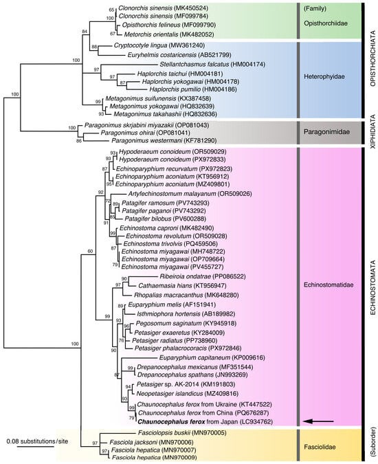
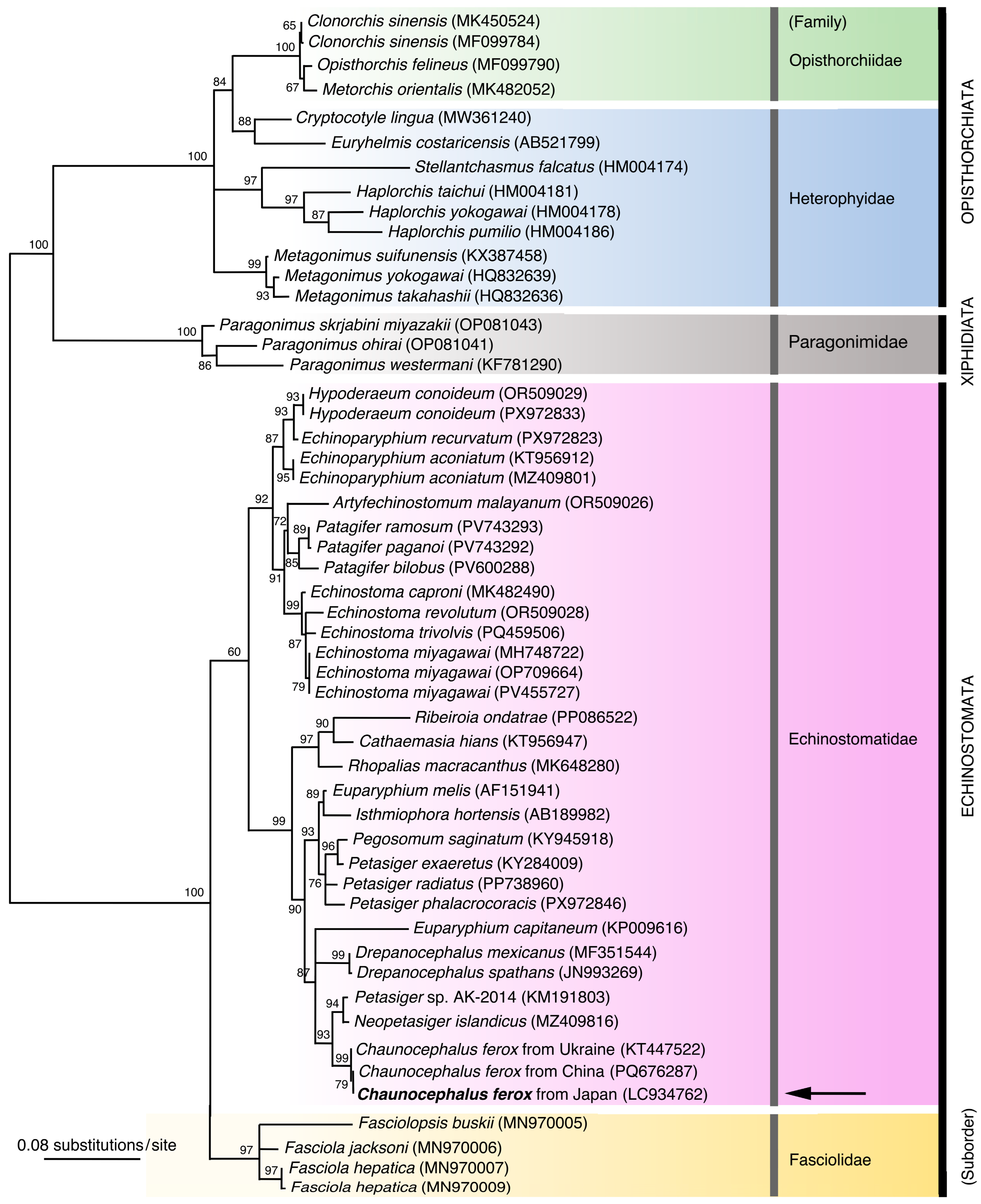
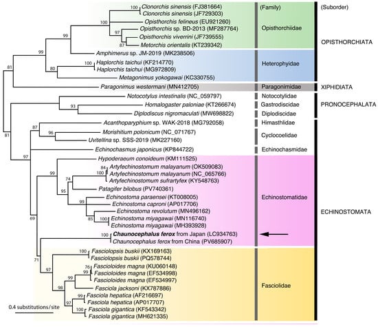
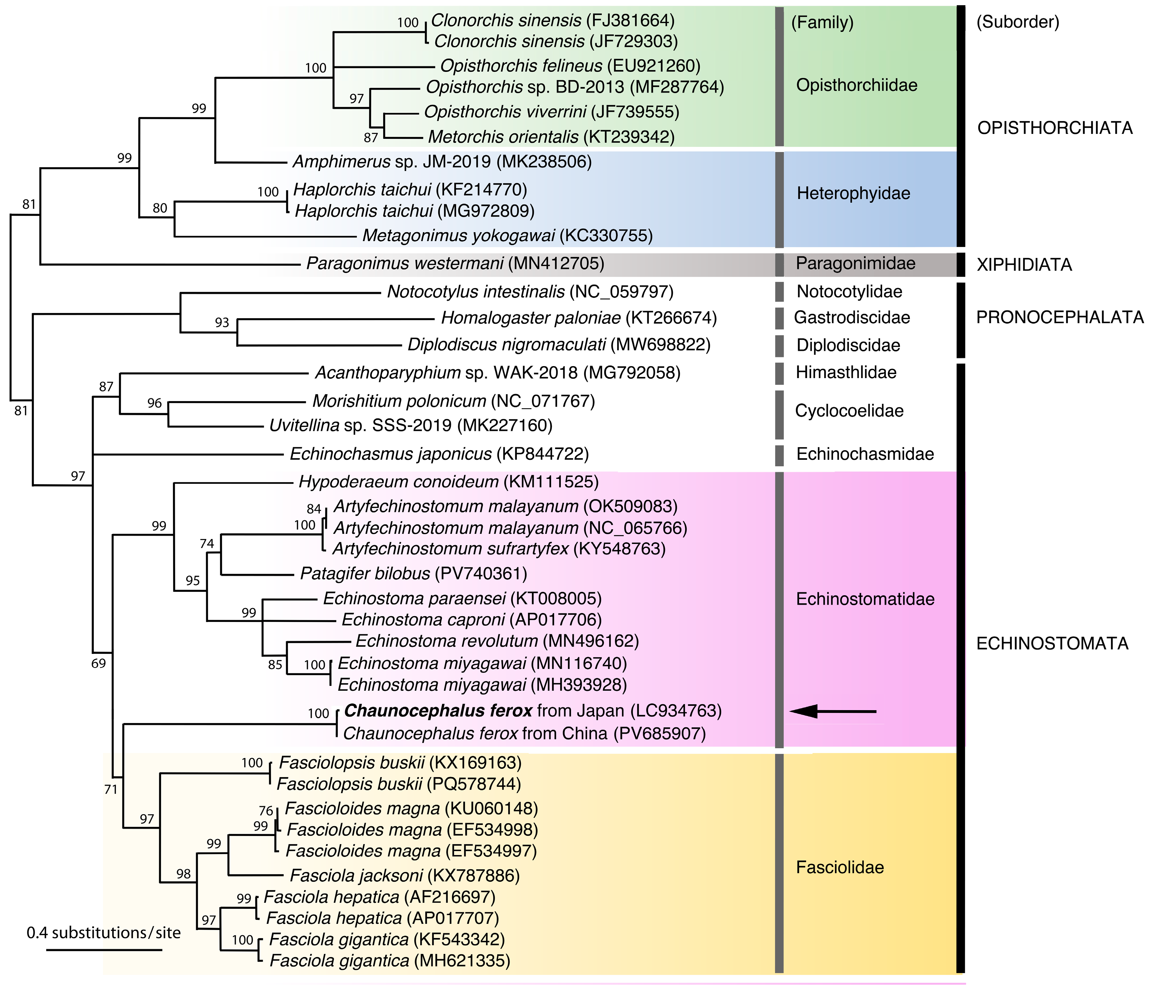
Genetic relationships among various flukes were robust, as shown by the phylogenetic trees constructed based on LSU rDNA (Figure 5) and cox1 (Figure 6). In the ML phylogenetic tree based on LSU rDNA, flukes classified in the families Echinostomatidae and Fasciolidae, both belonging to the suborder Echinostomata, formed separate clades, and C. ferox occupied a position within the former family, showing a sister relationship with Neopetasiger flukes (Figure 5). In the ML phylogenetic tree based on cox1, various flukes from the two aforementioned families similarly clustered into two separate clades, except for C. ferox (Echinostomatidae), which showed an apparent sister relationship with fasciolid flukes (Figure 6).
Figure 5.
Maximum likelihood (ML) phylogenetic tree based on LSU rDNA sequences of flukes classified in the suborders Echinostomata, Xiphidiata and Opisthorchiata. Species names are followed by GenBank accession numbers in parentheses. The newly obtained sequence of Chaunocephalus ferox from Japan is indicated in bold and with an arrow. Sequences of fluke species belonging to the same family are placed on a single background color.
Figure 6.
Maximum likelihood (ML) phylogenetic tree based on cox1 sequences of flukes classified in the suborders Echinostomata, Pronocephalata, Xiphidiata, and Opisthorchiata. Species names are followed by GenBank accession numbers in parentheses. The newly obtained sequence of Chaunocephalus ferox from Japan is indicated in bold and with an arrow. Sequences of fluke species belonging to the same family (Fasciolidae, Echinostomatidae, Paragonimidae, Heterophyidae, and Opisthorchiidae) are placed on a single background color.
4. Discussion
Fluke specimens isolated from the ileum of the Oriental stork chick within a few days after fledgling (at two months of age) exhibited the typical morphology of Chaunocephalus Dietz, 1909, including a bulbous forebody, shorter and narrow subcylindrical hindbody, uroproct, and reniform cephalic crown with collar spines [31,32]. Chaunocephalus flukes with 27 collar spines and vitelline glands densely distributed throughout the forebody and in two lateral fields around the mid-hindbody are identified as C. ferox, which has been reported from various storks of the family Ciconiidae, such as the white stork Ciconia ciconia (Linnaeus, 1758); black stork C. nigra Linnaeus, 1758; Oriental stork; black-necked stork Ephippiorhynchus asiaticus (Latham, 1790); and Asian openbill stork Anastomus oscitans (Boddaert, 1783) [21,22,33,34,35,36,37,38]. Evident pathogenicity of C. ferox parasitism has been reported in captive and wild host storks [21,33,38,39,40]. Patnaik et al. [38] noted that storks infected with C. ferox exhibited signs of diarrhea, listlessness, loss of appetite, and convulsions before death. Furthermore, Zhou et al. [22] suggested a high prevalence of C. ferox infection in weakened Oriental storks compared with a lower prevalence in healthy wild storks. Although chaunocephalosis is often associated with high mortality rates, the infection itself is not always fatal in storks. A case of a five-month-old male black stork in good physical condition (2.2 kg) traveling from Central Europe (Latvia, Poland, and Switzerland) to the Iberian Peninsula (southeastern Spain) was reported to have 143 parasite nodules caused by C. ferox infection (chaunocephalosis with 275 flukes) in the small intestine; however, the direct cause of death was electrocution [37]. Furthermore, Santoro et al. [34] collected 545 C. ferox flukes from parasite nodules throughout the intestine, from the duodenum to the ceca, of a debilitated young white stork (male, 2045 g) rescued in Southern Italy. The prevalence of C. ferox in black storks, white storks, and Asian openbill storks ranged from 9.8% to 80.0%, with 1–86 intestinal fluke nodules, among 10–91 deceased storks examined for various causes (geometric mean, 34; n = 8) [33,38,39,41,42,43,44]. In this study, the weakened stork fledgling had 10 fluke nodules, and fibrous fluke crypts were well formed in response to C. ferox infection. Although histologically evident bacterial clumps at the bottom of the fluke crypts and in the liver were suspected to have formed after its death due to the absence of host reactions, chronic enteritis and focal inflammatory responses in the liver and spleen may have been resulted from concomitant bacterial infection, potentially weakening the fledgling. However, the relative contributions of C. ferox infection, dehydration, possible secondary bacterial infection, and other stressors to the death of the fledgling remain uncertain.
Characterization of the SSU rDNA to LSU rDNA regions, including the ITS1 and ITS2 regions, and mitochondrial cox1 sequences of the present isolate facilitated the specific confirmation of C. ferox and helped assess the possible extent of intraspecific genetic variation in C. ferox using different molecular markers. rDNA and cox1 markers showed minimal variation among C. ferox isolates from different hosts and/or geographical origins (Europe, the Middle East, and East Asia). Zhou et al. [45] attempted to identify cox1 variants (haplotypes) of C. ferox from Oriental storks at different wintering sites (33 samples from three localities) but found no variation in cox1 sequences. Molecular differentiation of C. ferox from echinostomatid species belonging to different genera is possible using molecular markers; however, we found no data regarding interspecific variation based on different molecular markers within the genus Chaunocephalus. Indeed, not only for taxonomic identification of flukes but also for taxonomic revision of all described species (currently 12 nominal species [46]) within the genus Chaunocephalus, is required to assess their validity, particularly for those from India, as suggested previously. For instance, possible synonymy of C. kirai Vrat, 1947; C. odhneri Vrat, 1947; and C. similiferox Verma, 1936 with C. ferox has been suggested by Patnaik et al. [38] and Greben et al. [35]. Although distinct distributions of vitelline glands (separated into two fields: throughout the forebody and lateral fields in the mid-hindbody; throughout the whole body except for both cephalic and posterior ends) have been reported for C. ferox, no molecular genetic differences have been observed between these two morphotypes to date [35] (the present study). In this context, C. sinensis Ku, Chiu, Li & Chu, 1974, recorded from the small intestine of the black stork in Tianjin, China, resembles the general morphology of the present isolate but has widely distributed vitelline glands, similar to the C. ferox specimen reported by Greben et al. [35]. The taxonomic significance of vitelline gland distribution (interrupted between the forebody and hindbody versus continuous throughout the body) for taxonomic differentiation requires molecular verification using rDNA and/or mitochondrial genes, such as cox1.
Tkach et al. [47] and Zhou et al. [45] conducted phylogenetic analyses using ML and Bayesian inference (BI) methods based on long LSU rDNA and cox1 sequences. Both phylogenetic trees based on the ML and BI methods showed consistent topologies. In a phylogenetic tree based on LSU rDNA sequences of the superfamily Echinostomatoidea, C. ferox showed a sister relationship with Neopetasiger spp. and was a member of the robust clade of Echinostomatidae [47], as observed in this study (Figure 5). Similarly, in a phylogenetic tree based on cox1 sequences of four orders (Caryophyllidea, Plagiorchiida, Opisthorchiida, and Strigeidida), C. ferox showed a higher affinity for a clade of species in the family Fasciolidae than for the clade of the family Echinostomatidae [45], as observed in this study (Figure 6). Zhou et al. [45] reported that the topologies inferred from the concatenated dataset (12 mitochondrial protein-coding genes) were largely congruent with those from individual gene trees, suggesting an overall stable phylogenetic signal at higher taxonomic levels; however, the echinostomatid C. ferox showed a higher affinity to the fasciolid clade than to the major echinostomatid clade. Therefore, Zhou et al. [45] suggested that mitochondrial DNA data alone may be insufficient to resolve the evolutionary position of C. ferox, considering that morphological characteristics clearly supported the classification of C. ferox within Echinostomatidae. Alternatively, additional mitochondrial DNA sequences from other species of the genus Chaunocephalus and/or species of closely related genera, such as Balfouria Leiper, 1908 (the sibling genus in the subfamily Chaunocephalinae Travassos, 1922), may result in changes in phylogenetic analyses.
Uchida et al. [30] recorded C. ferox in the small intestine of a dead Oriental stork recovered in January 1977 in Oga City, Akita Prefecture, northern Japan. Considering that the last sedentary Oriental stork in Japan died in May 1971 in Toyooka City, Hyogo Prefecture, central Japan, and that the release of Oriental storks bred in captivity began in 2005 [10], the host stork examined by Uchida et al. [30] may have been a migratory stork from the continent, which are rarely found in Japan [10]. Although no cases of chaunocephalosis have been recorded despite the presence of grossly apparent globular nodules in the intestinal wall of the reintroduced population of Oriental storks released from breeding facilities or fledged in the field [10,19], the parasite was most likely maintained in the breeding colony at the conservation facilities for Oriental storks. The limited number of C. ferox crypts in the intestinal wall seems not to threaten the health of adult Oriental storks, because the inflammatory lesions around the flukes are well bordered by granulomatous fibrous tissues. Alternatively, migratory Oriental storks from the continent may have brought the fluke to Japan, resulting in the formation of C. ferox-endemic areas. Subsequently, either or both parent storks may have brought the flukes to their breeding site in Manno Town, Kagawa Prefecture. Chaunocephalosis in chicks before fledging, as in this case, is attributed to food items parasitized with C. ferox metacercariae (second or paratenic hosts) brought by parent storks. The parent storks collected food within the territory surrounding their nest, providing the first evidence consistent with the establishment of the local transmission cycle of C. ferox in Japan after the reintroduction of Oriental storks.
However, the life cycle of C. ferox remains unknown. Zhou et al. [22] detected metacercariae in crucian carp collected from stopover sites in China. Patnaik et al. [38] noted that their C. ferox-infected Asian open-billed storks in captivity at the Nandankanan Zoo were fed only snails (Viviparus sp.). As with many echinostomatid flukes, C. ferox may have at least two intermediate hosts and often paratenic hosts [48,49]. The first intermediate hosts must be snails, and the second intermediate hosts may include snails, fish, or amphibians. Paratenic hosts could include larger fish and reptiles. Oriental storks have a voracious appetite, requiring approximately ≥500 g of animal food per day per stork, and are more likely to ingest metacercariae from second intermediate or paratenic hosts. In natural fields with healthy ecosystems, intermediate and paratenic hosts highly infected with metacercariae are sparsely distributed, and the infection status of final hosts is not homogeneous, showing an overdispersed distribution of infection intensity among hosts; heavily infected hosts are rare, and most hosts harbor a limited number of worms [50,51]. However, in artificial ecosystems within relatively limited areas, concentrated contact among all hosts (first and second intermediate hosts, paratenic hosts, and final hosts) may result in heavy infections, creating highly endemic areas of C. ferox infection [52]. Conservation of endangered Oriental storks is ongoing despite the modernization of agricultural systems, including controlled irrigation of paddy fields, and deforestation. Limited use of natural environments for foraging (i.e., restricted foraging areas) can increase the endemicity of parasitic diseases. It remains uncertain whether the fledgling stork chick case reported here represents a suspected emerging problem or a common instance of natural parasite exposure through parental feeding. It is important, however, to note that its siblings survived and flew to multiple distant locations in western Japan, suggesting a healthy condition. Nevertheless, the current fragmented knowledge regarding invertebrate and vertebrate hosts involved in the life cycle of C. ferox in endangered ciconiid birds, including the Oriental stork, must be urgently improved to assess the suitability of available habitats in modern global ecosystems from the perspective of infection dynamics.
In captive rearing programs for the conservation of endangered free-living animal species, their parasites, which are commonly believed to be harmful to host, are intentionally eliminated through veterinary care [53,54,55,56,57,58]. This phenomenon is referred to as “conservation-driven extinction,” and examples of extinct parasites include the lice Neotrichodectes sp. and Colpocephalum californici Price et Beer, 1963, from the black-footed ferret (Mustela nigripes (Audubon et Bachman, 1851)) and Californian condor (Gymnogyps californianus (Shaw, 1797)), respectively, which were eliminated through delousing protocols in captive breeding programs [56,57,59,60]. Recently, however, arguments supporting parasite conservation alongside their endangered hosts have been emphasized because the elimination of parasites that have co-evolved with endangered host animals (specialist parasites) may destabilize ecological systems and result in unintended consequences [57,61,62,63,64,65,66,67,68,69]. Parasites are important components of ecological networks (ecosystems) and biodiversity, contributing to community structure by influencing host populations in various ways, including regulation of host behavior, growth, and reproduction; regulation of food web stability; and maintenance of the genetic diversity of host populations, which is critical for the survival of endangered species [57,58,62,63,67,68,69,70,71]. Researchers [59,72] speculate that the loss of “specialist” parasites may predispose hosts to infection by their “generalist” competitors or emerging parasites. Accordingly, most cases in which diseases cause significant mortality in rare species are attributable to generalist rather than specialist parasites [73,74,75].
As mentioned above, Zhou et al. [45] reported a unique phylogenetic position of C. ferox, with its strict host specificity to storks (family Ciconiidae), within the order Plagiorchiida. Furthermore, they estimated the divergence time of Chaunocephalus trematodes (75–39 million years ago), which coincided with the origin of modern storks approximately 40 million years ago during the early Eocene, suggesting a long co-evolutionary history. Considering these co-evolutionary relationships, along with the biological and ecological importance of specialist parasites to their host, the re-establishment of the C. ferox transmission cycle in a reintroduced Oriental stork population in Japan may be beneficial for the health of the reintroduced stork population and the ecosystem, despite its adverse effects at the individual level. A co-evolved feather mite, Pelargolichus orientalis Waki, Mironov et Shimano, 2023 (Acariformes: Pterolichidae), associated with Oriental storks was unintentionally reintroduced with host storks in Japan [76,77]. Except for these examples, no information on parasites is available for reintroduced Oriental storks in Japan, although multiple parasite species have been reported globally in storks [41,43,44,78,79,80,81,82].
The primary concern is that these well-adapted parasites may cause serious problems for the reintroduced stork population in artificially modified natural environments. For instance, limited wetland areas available for feeding may form transmission hotspots, and anthropogenic stress may reduce immunity to pathogens, increasing susceptibility to infectious diseases and chronic inflammation. Thus, continued monitoring of disease incidence is necessary to assess the health of ecosystems supporting reintroduced Oriental stork populations. To date, we have diagnosed only one case of chaunocephalosis in Japan, whereas the number of wild sedentary storks in Japan has increased to ≥500, with 55 field nests in 14 prefectures by 2025 [10,11,18]. Similarly, conservation efforts for continental Oriental stork populations, including strict protection of wintering and important stopover sites, have increased the population size to more than 9600 individuals in 2022 [13]. Zhou et al. [45] raised concerns about increased cases of chaunocephalosis due to increased host density and overlap of available habitats. Therefore, a proper understanding of infection conditions, whether they represent natural ecological processes or problematic conditions, is necessary. Parasites and parasitology could contribute further to the conservation efforts of storks and other wild birds through health assessments of provided ecosystems and inference of intergroup interactions [64], in addition to improving understanding of the ecological significance of parasites in host population survival and stability [57,58,62,63,67,68,69,70,71].
Supplementary Materials
The following supporting information can be downloaded at https://www.mdpi.com/article/10.3390/conservation6030097/s1, Supplementary Table S1: Primers used to amplify and sequence 11 overlapping fragments of C. ferox rDNA. Supplementary Figure S1: Family tree of Oriental storks related to a nest established atop a utility pole beside a paddy field in Manno Town, Kagawa Prefecture, Japan. Each individual box includes the stork ID number and sex in parentheses (M, male; F, female, and ?, unknown). A deceased fledgling reported in this study is indicated by an arrow. References [83,84] are cited in the supplementary materials.
Author Contributions
Conceptualization, T.K. and H.S.; methodology, T.K., C.K., I.N.V., T.W.P.J. and H.S.; software, I.N.V., T.W.P.J. and H.S.; validation, T.K., C.K. and H.S.; investigation, C.K., I.N.V., T.W.P.J. and H.S.; data curation, T.K., C.K. and T.W.P.J.; writing—original draft preparation, T.W.P.J., I.N.V. and H.S.; writing—review and editing, T.K., C.K. and H.S.; visualization, C.K., T.W.P.J. and H.S.; supervision, H.S. All authors have read and agreed to the published version of the manuscript.
Funding
This research received no external funding.
Institutional Review Board Statement
Ethics review and approval were waived for this study because we examined a deceased stork in accordance with the general regulations (Law for the Protection of Cultural Properties (1950) [https://laws.e-gov.go.jp/law/325AC0100000214] (accessed 15 April 2026); Act on Conservation of Endangered Species of Wild Fauna and Flora (Act No. 75 of 1992) [https://laws.e-gov.go.jp/law/404AC0000000075] (accessed 15 April 2026)) and municipal wildlife conservation plan issued by Manno Town, Kagawa Prefecture, Japan (“Special Feature: The Oriental Storks of Manno Town” in Manno Town Newsletter vol. 222, September 2024 [https://www.town.manno.lg.jp/uploaded/attachment/4017.pdf] (accessed 15 April 2026)).
Informed Consent Statement
Not applicable.
Data Availability Statement
The parasite specimens collected in the present study were deposited at the Meguro Parasitological Museum, Tokyo, Japan (collection no. 25518). The nucleotide sequences obtained in the present study are available from the DDBJ/EMBL/GenBank databases under the accession numbers LC934762 (rDNA) and LC934763 (cox1).
Acknowledgments
We thank the municipal officers and related citizens of Manno Town, Kagawa Prefecture, Japan, for their understanding and cooperation in conducting pathological and parasitological examinations of a deceased stork, which is a special national natural monument in Japan. Constructive comments and suggestions by three anonymous referees considerably improved the manuscript.
Conflicts of Interest
The authors declare no conflicts of interest.
References
- Ohsako, Y. New progress and reinforcement target in the reintroduction project of the oriental white stork. Reintroduction 2012, 2, 21–25. (In Japanese) [Google Scholar]
- Luthin, C.S. Status of and conservation priorities for the world’s stork species. Col. Waterbirds 1987, 10, 181–202. [Google Scholar] [CrossRef] [Scilit]
- BirdLife International. Ciconia boyciana. In The IUCN Red List of Threatened Species; BirdLife International: Tokyo, Japan, 2018; Volume 2018, p. e.T22697695A131942061. [Google Scholar] [CrossRef] [Scilit]
- Gula, J.; Sundar, K.S.G.; Willows-Munro, S.; Downs, C.T. The state of stork research globally: A systematic review. Biol. Conserv. 2023, 280, 109969. [Google Scholar] [CrossRef] [Scilit]
- Park, S.-R.; Yoon, J.; Kim, S.-K. Captive propagation, habitat restoration, and reintroduction of Oriental white storks (Ciconia boyciana) extirpated in South Korea. Reintroduction 2011, 1, 31–36. [Google Scholar]
- Ohsako, Y. From extinction to reintroduction: Conservation, breeding, and restoration of the Oriental white stork. Jpn. J. Ornithol. 2012, 61, 91–93. (In Japanese) [Google Scholar]
- Shimazaki, H.; Tamura, M.; Higuchi, H. Migration routes and important stopover sites of endangered oriental white storks (Ciconia boyciana) as revealed by satellite tracking. Mem. Natl. Inst. Polar Res. 2004, 58, 162–178. [Google Scholar]
- Onuma, M.; Kocherga, M.; Tyagunin, V.; Parilov, M.; Sasin, A.; Edyta, S.; Kuwana, T. Evaluation of genetic diversity of wild Oriental white stork (Ciconia boyciana) in Russia and their phylogenetic relationship with extinct populations in Japan. Jpn. J. Zoo Wildl. Med. 2011, 16, 139–144. [Google Scholar] [CrossRef] [Scilit]
- Ezaki, Y. Reintroduction of the Oriental white stork Ciconia biyciana aiming at reconstructing its meta-population structure in Japan. Reintroduction 2012, 2, 3–10, (In Japanese with English summary). [Google Scholar]
- Ezaki, Y.; Mitsuhashi, Y.; Ohsako, Y. Survival, mortality and skewed sex ration of the Japanese Oriental white stork reintroduced population at the end of 2014. Reintroduction 2016, 4, 95–102. [Google Scholar] [CrossRef] [Scilit]
- Ezaki, Y.; Ohsako, Y. Reintroduction and successful re-establishment of a breeding population of the Oriental White Stork Ciconia boyciana in Japan. Jpn. J. Ornithol. 2019, 68, 183–192, (In Japanese with English summary). [Google Scholar] [CrossRef] [Scilit]
- Xu, X.; Lu, X.; Wang, Q.; Liu, C.; Song, C.; Wang, H.; Wang, G.; Liu, B.; Sun, K.; Zheng, H. Exploring conservation strategies for oriental white stork fledglings (Ciconia boyciana) across the breeding wetland landscape: Hints from tracking movement patterns. Glob. Ecol. Conserv. 2021, 26, e01531. [Google Scholar] [CrossRef] [Scilit]
- Yang, Z.; Chen, L.; Jia, R.; Xu, H.; Wang, Y.; Wei, X.; Liu, D.; Liu, H.; Liu, Y.; Yang, P.; et al. Migration routes of the endangered Oriental Stork (Ciconia boyciana) from Xingkai Lake, China, and their repeatability as revealed by GPS tracking. Avian Res. 2023, 14, 100090. [Google Scholar] [CrossRef] [Scilit]
- Yang, S.; Liu, Y.; Zhao, X.; Chen, J.; Li, H.; Liang, H.; Fan, J.; Zhou, M.; Wang, S.; Zhang, X.; et al. Genomic exploration of the endangered oriental stork, Ciconia boyciana, sheds light on migration adaptation and future conservation. Gigascience 2024, 13, giae081. [Google Scholar] [CrossRef] [Scilit]
- Yang, F.; Shao, M.; Wang, J. Distributional and behavioral responses of the wintering Oriental Storks to drought in China’s largest freshwater lake. Avian Res. 2024, 15, 100176. [Google Scholar] [CrossRef] [Scilit]
- Murata, K. Reintroduction plan for the Oriental white stork (Ciconia boyciana) in Japan and its veterinan’s role. J. Jpn. Zoo Wild. Med. 1997, 2, 117–122. [Google Scholar] [CrossRef] [Scilit]
- Murata, K.; Satou, M.; Matushima, K.; Satake, S.; Yamamoto, Y. Retrospective estimation of genetic diversity of an extinct Oriental White Stork (Ciconia boyciana) population in Japan using mounted specimens and implications for reintroduction programs. Conserv. Genet. 2004, 5, 553–560. [Google Scholar] [CrossRef] [Scilit]
- Hyogo Park of the Oriental White Stork. Leg Band Catalog of Oriental White Stork for Individual Identification. 2026. Available online: https://satokouen.jp/downloads/ind_identif/ind_identif.pdf (accessed on 23 April 2026).
- Matsumoto, R.; Horie, M.; Mitsuhashi, Y.; Ohsako, Y. Anthropogenic threats to the reintroduced population of Oriental storks in Japan: Analysis of the causes of injury and death of 153 storks from 2005 to 2021. J. Vet. Med. Sci. 2024, 86, 400–408. [Google Scholar] [CrossRef] [Scilit] [PubMed]
- Oh, B.; Yang, M.S.; Ha, D.S.; Kim, S.K.; Park, H.J.; Kim, B.; Lim, C.W.; Oh, S.I. Retrospective analysis of postmortem findings in Oriental stork (Ciconia boyciana) from Korea (2019–2023). J. Vet. Clin. 2024, 41, 323–329. [Google Scholar] [CrossRef] [Scilit]
- Choe, S.; Lee, D.; Park, H.; Jeon, H.K.; Lee, Y.; Na, K.J.; Park, S.R.; Eom, K.S. A case of chaunocephalosis by Chaunocephalus ferox (Digenea: Echinostomatidae) in an oriental white stork, Ciconia boyciana, in Korea. Korean J. Parasitol. 2016, 54, 659–665. [Google Scholar] [CrossRef] [Scilit] [PubMed]
- Zhou, M.C.; Yu, M.Y.; Liu, M.Q.; Liang, H.R.; Zhao, M.R.; Lu, Y.X.; Qin, S.Y.; Zhang, X.T.; Yu, H.Y.; Tian, H.; et al. Chaunocephalus ferox (Digenea: Echinostomatidae): A potential threat to the health of Oriental stork (Ciconia boyciana). Eur. J. Wildl. Res. 2025, 71, 56. [Google Scholar] [CrossRef] [Scilit]
- Sato, H.; Ihara, S.; Inaba, O.; Une, Y. Identification of Euryhelmis costaricensis metacercariae in the skin of Tohoku hynobiid salamanders (Hynobius lichenatus), northeastern Honshu, Japan. J. Wildl. Dis. 2010, 46, 832–842. [Google Scholar] [CrossRef] [Scilit] [PubMed]
- Untergasser, A.; Cutcutache, I.; Koressaar, T.; Ye, J.; Faircloth, B.C.; Remm, M.; Rozen, S.G. Primer 3—New capabilities and interfaces. Nucleic Acids Res. 2012, 40, e115. [Google Scholar] [CrossRef] [Scilit] [PubMed]
- Thompson, J.D.; Higgins, D.G.; Gibson, T.J. CLUSTALW: Improving the sensitivity of progressive multiple sequence alignment through sequence weighting, position-specific gap penalties and weight matrix choice. Nucleic Acids Res. 1994, 22, 4673–4680. [Google Scholar] [CrossRef] [Scilit]
- Tamaru, K.; Stecher, G.; Kumar, S. MEGA11: Molecular evolutionary genetics analysis version 11. Mol. Biol. Evol. 2021, 38, 3022–3027. [Google Scholar] [CrossRef] [Scilit]
- Guindon, S.; Gascuel, O. A simple, fast, and accurate algorithm to estimate large phylogenies by maximum likelihood. Syst. Biol. 2003, 52, 696–704. [Google Scholar] [CrossRef] [Scilit] [PubMed]
- Dereeper, A.; Guignon, V.; Blanc, G.; Audic, S.; Buffet, S.; Chevenet, F.; Dufayard, J.F.; Guindon, S.; Lefort, V.; Lescot, M.; et al. Phylogeny.fr: Robust phylogenetic analysis for the non-specialist. Nucleic Acids Res. 2008, 36, W465–W469. [Google Scholar] [CrossRef] [Scilit]
- Anisimova, M.; Gascuel, O. Approximate likelihood-ratio test for branches: A fast, accurate, and powerful alternative. Syst. Biol. 2006, 55, 539–552. [Google Scholar] [CrossRef] [Scilit] [PubMed]
- Uchida, A.; Itagaki, H.; Ishida, K.; Yarime, M.; Ito, T.; Shimura, S.; Kondo, S. The trematode, Chaunocephalus sp. found in a stork, Ciconia Ciconia boyciana in Akita Prefecture. Juui-Chikusan-Shinpou 1978, 687, 565–566. with figures on 559 and 561(In Japanese) [Google Scholar]
- Dietz, E. Die Echinostomiden der Vogel. Zool. Anz. 1909, 34, 180–192. [Google Scholar]
- Kostadinova, A. Family Echinostomatidae Looss, 1899. In Keys to the Trematoda; Jones, A., Bray, R.A., Gibson, D.I., Eds.; CABI Publishing: Boston, MA, USA; The Natural History Museum: Cambridge, MA, USA, 2005; Volume 2, pp. 9–64. [Google Scholar]
- Poonswad, P.; Chatikavanij, P.; Thamavit, W. Chaunocephalosis in a wild population of Asian open-billed storks in Thailand. J. Wildl. Dis. 1992, 28, 460–466. [Google Scholar] [CrossRef] [Scilit]
- Santoro, M.; Degli Uberti, B.; Galiero, G.; Di Prisco, F.; D’Alessio, N.; Veneziano, V. Chaunocephalus ferox (Digenea: Echinostomatidae) infection associated with fatal cachexia in a white stork (Ciconia ciconia). Helminthologia 2013, 50, 181–184. [Google Scholar] [CrossRef] [Scilit]
- Greben, O.B.; Kudlai, O.; Korol, E.N.; Kornyushin, V.V.; Vasilkovska, I.B.; Kobylinsky, V.V. A new record of Chaunocephalus ferox (Digenea, Echinostomatidae) from Ciconia nigra in Ukraine including morphological and molecular data. Vestn. Zool. 2016, 50, 99–104. [Google Scholar] [CrossRef] [Scilit]
- Islam, S.; Ali, S.; Kakoty, P.; Deka, P.; Basumatary, P.; Das, D.; Deka, A.; Begum, S.A. Pathology of Chaunocephalus ferox (Rudolphi, 1795) Dietz, 1909 infection in Asian openbill stork (Anastomus oscitans Boddaert, 1783). Vet. Pract. 2020, 21, 123–128. [Google Scholar]
- Gonzálvez, M.; Muñoz-Hernández, C.; Gómez de Ramón, A.; Buendía, A.; Escribano, F.; Martínez-Carrasco, C. Flying across Europe: The case of the spread of Chaunocephalus ferox on a black stork (Ciconia nigra). J. Helminthol. 2022, 96, e80. [Google Scholar] [CrossRef] [Scilit]
- Patnaik, M.M.; Rao, A.T.; Acharjyo, L.N.; Mohanty, D.N. Notes on a nodular disease of the intestine of the open-billed stork—(Anastomus oscitans) caused by Chaunouephalus ferox. J. Wildl. Dis. 1970, 6, 64–66. [Google Scholar] [CrossRef] [Scilit]
- Höfle, U.; Krone, O.; Blanco, J.M.; Pizarro, M. Chaunocephalus ferox in free-living white storks in central Spain. Avian Dis. 2003, 47, 506–512. [Google Scholar] [CrossRef] [Scilit]
- Iliev, P.T.; Dimitorov, K.; Ivanov, A. A case of nodular enteritis due to Chaunocephalus ferox in a white stork (Ciconia ciconia) in Bulgaria. Bulg. J. Vet. Med. 2023, 26, 660–667. [Google Scholar] [CrossRef] [Scilit]
- Sitko, J.; Heneberg, P. Composition, structure and pattern of helminth assemblages associated with central European storks (Ciconiidae). Parasitol. Int. 2015, 64, 130–134. [Google Scholar] [CrossRef] [Scilit] [PubMed]
- Girisgin, A.O.; Birlik, S.; Senlik, B.; Yildirimhan, H.S. Intestinal helminths of white stork (Ciconia ciconia Linnaeus 1758) from an inter-route site in Turkey. Acta Vet. Hung. 2017, 65, 221–233. [Google Scholar] [CrossRef] [Scilit] [PubMed]
- Michalczyk, M.; Sokół, R.; Gesek, M.; Mączyński, M.; Będzłowicz, D. Internal parasites and associated histopathological changes in deceased white storks from Poland. Belg. J. Zool. 2020, 150, 71–80. [Google Scholar]
- Tylkowska, A.; Stapf, A.; Rząd, I.; Sałamatin, R.; Panicz, R.; Jeżewski, W.; Kaczmarek, A.; Eljasik, P. Helminths of white stork Ciconia ciconia in north-eastern Poland. Sci. Rep. 2026, 16, 2824. [Google Scholar] [CrossRef] [Scilit]
- Zhou, M.; Liang, H.; Lv, Z.; Zhang, X.; Liu, Y.; Lu, Y.; Yu, H.; Tian, H.; Han, L.; Xu, Y.; et al. Complete mitochondrial genome and phylogenetic implications of Chaunocephalus ferox (Digenea: Echinostomatidae), a critical pathogen for Oriental Stork conservation. Int. J. Parasitol. Parasites Wildl. 2025, 28, 101129. [Google Scholar] [CrossRef] [Scilit]
- Marine Species Traits Editorial Board. Marine Species Traits. Chaunocephalus Dietz, 1909. Available online: https://www.marinespecies.org/traits/aphia.php?p=taxdetails&id=1596471 (accessed on 31 March 2026).
- Tkach, V.V.; Kudlai, O.; Kostadinova, A. Molecular phylogeny and systematics of the Echinostomatoidea Looss, 1899 (Platyhelminthes: Digenea). Int. J. Parasitol. 2016, 46, 171–185. [Google Scholar] [CrossRef] [Scilit]
- Shoop, W.L. Trematode transmission patterns. J. Parasitol. 1988, 74, 46–59. [Google Scholar] [CrossRef] [Scilit]
- Esch, G.W.; Barger, M.A.; Fellis, K.J. The transmission of digenetic trematodes: Style, elegance, complexity. Integr. Comp. Biol. 2002, 42, 304–312. [Google Scholar] [CrossRef] [Scilit]
- Churcher, T.S.; Ferguson, N.M.; Basáñez, M.G. Density dependence and overdispersion in the transmission of helminth parasites. Parasitology 2005, 131, 121–132. [Google Scholar] [CrossRef] [Scilit]
- Warburton, E.M.; Vonhof, M.J. From individual heterogeneity to population-level overdispersion: Quantifying the relative roles of host exposure and parasite establishment in driving aggregated helminth distributions. Int. J. Parasitol. 2018, 48, 309–318. [Google Scholar] [CrossRef] [Scilit]
- Budria, A.; Candolin, U. How does human-induced environmental change influence host-parasite interactions? Parasitology 2014, 141, 462–474. [Google Scholar] [CrossRef] [Scilit]
- Pizzi, R. Veterinarians and taxonomic chauvinism: The dilemma of parasite conservation. J. Exot. Pet Med. 2009, 18, 279–282. [Google Scholar] [CrossRef] [Scilit]
- Loker, E.S. This de-wormed world? J. Parasitol. 2013, 99, 933–942. [Google Scholar] [CrossRef] [Scilit]
- Jørgensen, D. Conservation implications of parasite co-reintroduction. Conserv. Biol. 2015, 29, 602–604. [Google Scholar] [CrossRef] [Scilit] [PubMed]
- Rózsa, L.; Vas, Z. Co-extinct and critically co-endangered species of parasitic lice, and conservation-induced extinction: Should lice be reintroduced to their hosts? Oryx 2015, 49, 107–110. [Google Scholar] [CrossRef] [Scilit]
- Milotic, M.; Lymbery, A.; Thompson, A.; Doherty, J.-F.; Godfrey, S. Parasites are endangered by the conservation of their hosts: Meta-analyses of the effect of host captivity on the odds of parasite infection. Biol. Conserv. 2020, 248, 108702. [Google Scholar] [CrossRef] [Scilit]
- Lymbery, A.J.; Smit, N.J. Conservation of parasites: A primer. Int. J. Parasitol. Parasites Wildl. 2023, 21, 255–263. [Google Scholar] [CrossRef] [Scilit]
- Gompper, M.E.; Williams, E.S. Parasite conservation and the black-footed ferret recovery program. Conserv. Biol. 1998, 12, 730–732. [Google Scholar] [CrossRef] [Scilit]
- Koh, L.P.; Dunn, R.R.; Sodhi, N.S.; Colwell, R.K.; Proctor, H.C.; Smith, V.S. Species coextinctions and the biodiversity crisis. Science 2004, 305, 1632–1634. [Google Scholar] [CrossRef] [Scilit]
- Rózsa, L. Points in question. Endangered parasite species. Int. J. Parasitol. 1992, 22, 265–266. [Google Scholar] [CrossRef] [Scilit]
- Windsor, D.A. Equal rights for parasites. Conserv. Biol. 1995, 9, 1–2. [Google Scholar] [CrossRef] [Scilit]
- Durden, L.A.; Keirans, J.E. Host-parasite coextinction and the plight of tick conservation. Am. Entomol. 1996, 42, 87–91. [Google Scholar] [CrossRef] [Scilit]
- Whiteman, N.K.; Parker, P.G. Using parasites to infer host population history: A new rationale for parasite conservation. Anim. Conserv. 2005, 8, 175–181. [Google Scholar] [CrossRef] [Scilit]
- Hudson, P.J.; Dobson, A.P.; Lafferty, K.D. Is a healthy ecosystem one that is rich in parasites? Trends Ecol. Evol. 2006, 21, 381–385. [Google Scholar] [CrossRef] [Scilit]
- Gómez, A.; Nichols, E. Neglected wild life: Parasitic biodiversity as a conservation target. Int. J. Parasitol. Parasites Wildl. 2013, 2, 222–227. [Google Scholar] [CrossRef] [Scilit]
- Stringer, A.P.; Linklater, W. Everything in moderation: Principles of parasite control for wildlife conservation. BioScience 2014, 64, 932–937. [Google Scholar] [CrossRef] [Scilit]
- Dougherty, E.R.; Carlson, C.J.; Bueno, V.M.; Burgio, K.R.; Cizauskas, C.A.; Clements, C.F.; Seidel, D.P.; Harris, N.C. Paradigms for parasite conservation. Conserv. Biol. 2016, 30, 724–733. [Google Scholar] [CrossRef] [Scilit]
- Wood, C.L.; Johnson, P.T. A world without parasites: Exploring the hidden ecology of infection. Front. Ecol. Environ. 2015, 13, 425–434. [Google Scholar] [CrossRef] [Scilit]
- Colwell, D.D.; Otranto, D.; Stevens, J.R. Oestrid flies: Eradication and extinction versus biodiversity. Trends Parasitol. 2009, 25, 500–504. [Google Scholar] [CrossRef] [Scilit]
- Strona, G. Past, present and future of host-parasite co-extinctions. Int. J. Parasitol. Parasites Wildl. 2015, 4, 431–441. [Google Scholar] [CrossRef] [Scilit]
- Almberg, E.S.; Cross, P.C.; Dobson, A.P.; Smith, D.W.; Hudson, P.J. Parasite invasion following host reintroduction: A case study of Yellowstone’s wolves. Philos. Trans. R. Soc. Lond. B Biol. Sci. 2012, 367, 2840–2851. [Google Scholar] [CrossRef] [Scilit]
- Dobson, A.; Foufopoulos, J. Emerging infectious pathogens of wildlife. Philos. Trans. R. Soc. Lond. B Biol. Sci. 2001, 356, 1001–1012. [Google Scholar] [CrossRef] [Scilit]
- Gog, J.; Woodroffe, R.; Swinton, J. Disease in endangered metapopulations: The importance of alternative hosts. Proc. Biol. Sci. 2002, 269, 671–676. [Google Scholar] [CrossRef] [Scilit]
- De Castro, F.; Bolker, B. Mechanisms of disease-induced extinction. Ecol. Lett. 2005, 8, 117–126. [Google Scholar] [CrossRef] [Scilit]
- Waki, T.; Mironov, S.V.; Nakajima, A.; Shimano, S. A new feather mite of the genus Pelargolichus (Acariformes: Pterolichidae) from the Oriental White Stork Ciconia boyciana (Ciconiiformes: Ciconiidae) in Japan. Syst. Appl. Acarol. 2023, 28, 471–482. [Google Scholar] [CrossRef] [Scilit]
- Waki, T.; Nishiumi, I.; Shimano, S. Report of a feather mite species (Acariformes: Astigmata) from the Oriental White Stork, Ciconia boyciana (Ciconiiformes: Ciconiidae), belonging to the Japanese native population. Species Divers. 2023, 28, 225–229. [Google Scholar] [CrossRef] [Scilit]
- Grünberg, W.; Kutzer, E. Die Pathologie verschiedener Trematodeninfektionen bei Störchen (Ciconia ciconia L., Ciconia nigra L.). Zentralbl. Veterinarmed. 1964, 11, 712–727. [Google Scholar] [CrossRef] [Scilit]
- Pinto, R.M.; Barros, L.A.; Tortelly, L.; Teixeira, R.F.; Gomes, D.C. Prevalence and pathology of helminths of ciconiiform birds from the Brazilian swamplands. J. Helminthol. 2004, 78, 259–264. [Google Scholar] [CrossRef] [Scilit] [PubMed]
- Liptovszky, M.; Majoros, G.; Perge, E. Cathaemasia hians in a black stork (Ciconia nigra) in Hungary. J. Wildl. Dis. 2012, 48, 809–811. [Google Scholar] [CrossRef] [Scilit] [PubMed]
- Ramilo, D.W.; Caetano, I.; Brazio, E.; Mira, M.; Antunes, L.; Pereira da Fonseca, I.; Cardoso, L. Presence of one ecto- and two endoparasite species of the black stork (Ciconia nigra) in Portugal. BMC Vet. Res. 2021, 17, 21. [Google Scholar] [CrossRef] [Scilit] [PubMed]
- Meister, S.L.; Wenker, C.; Wyss, F.; Zühlke, I.; Veiga, I.B.; Basso, W.U. Syngamus trachea in free-ranging white stork (Ciconia ciconia) nestlings in Switzerland. Int. J. Parasitol. Parasites Wildl. 2022, 18, 76–81. [Google Scholar] [CrossRef] [Scilit]
- Chisholm, L.A.; Morgan, J.A.; Adlard, R.D.; Whittington, I.D. Phylogenetic analysis of the Monocotylidae (Monogenea) inferred from 28S rDNA sequences. Int. J. Parasitol. 2001, 31, 1537–1547. [Google Scholar] [CrossRef] [Scilit] [PubMed]
- Van de Peer, Y.; Robbrecht, E.; de Hoog, S.; Caers, A.; De Rijk, P.; De Wachter, R. Database on the structure of small subunit ribosomal RNA. Nucleic Acids Res. 1999, 27, 179–183. [Google Scholar] [CrossRef] [Scilit] [PubMed]

Disclaimer/Publisher’s Note: The statements, opinions and data contained in all publications are solely those of the individual author(s) and contributor(s) and not of MDPI and/or the editor(s). MDPI and/or the editor(s) disclaim responsibility for any injury to people or property resulting from any ideas, methods, instructions or products referred to in the content. |
© 2026 by the authors. Licensee MDPI, Basel, Switzerland. This article is an open access article distributed under the terms and conditions of the Creative Commons Attribution (CC BY) license.