


Abstract
In vertebrates, corticosterone and cortisol are glucocorticoid (GC) steroid hormones central to the vertebrate stress response, but their relative contribution depends on context and life history. We used water-borne hormone sampling in lesser sirens, Siren intermedia, to examine whether corticosterone or cortisol predominates in stress responses and evaluated whether dermal secretions reliably track acute corticosterone changes. First, we measured corticosterone via dermal swabbing over a 2 h period following a hand-restraint stressor. Dermal corticosterone did not increase relative to pre-stressor baseline levels during the 2 h sampling period, suggesting that dermal measurements may not reflect acute circulating GC changes. We then measured cortisol and corticosterone across the GC profile (baseline, stressed, and recovery) using water-borne sampling techniques in juveniles. Cortisol release rates were significantly higher than corticosterone and were elevated during the recovery phase relative to baseline (p = 0.08), whereas stressed samples did not differ from baseline. Additionally, cortisol release rates were positively associated with temperature, while no effect of temperature was detected on corticosterone. Although cortisol and corticosterone were strongly correlated, cortisol responded to environmental temperatures and was repeatable, whereas corticosterone was not. Finally, we validated the use of water-borne sampling techniques to measure cortisol and corticosterone in S. intermedia. Cortisol was higher in juvenile sirens, highlighting that each GC may play functionally distinct roles across contexts. Together, these findings indicate that cortisol is the more environmentally responsive GC in juvenile S. intermedia and that water-borne sampling provides a more reliable method for assessing acute stress physiology in this species.
Keywords:
amphibian; aquatic; corticosterone; cortisol; dermal; lesser siren; salamander; stress; water-borne 1. Introduction
Glucocorticoids (GCs), specifically corticosterone and cortisol, are steroid hormones central to the vertebrate stress response. In vertebrates, they are released via the hypothalamic–pituitary–adrenal/interrenal (HPA/I) axis and bind to glucocorticoid and mineralocorticoid receptors to regulate downstream processes [1,2]. During acute stress, GCs mobilize energy and suppress non-essential functions such as growth and reproduction, while prolonged activation of the HPA/I axis is linked to immune suppression, reduced reproductive output, and long-term fitness costs [3,4,5,6]. In amphibians, glucocorticoids also mediate key developmental processes, including the timing of metamorphosis. Because of their broad physiological roles, GCs are widely used as biomarkers of health and environmental stress [7,8]. However, baseline GC release rates alone do not fully capture how organisms respond to environmental challenges. An understanding of an organism’s capacity to cope helps contextualize these physiological responses. Coping capacity refers to the physiological and behavioral flexibility that allows an organism to maintain homeostasis and support survival and reproduction under environmental stress [9]. Individuals with robust coping capacity can mount a stress response and effectively regulate that negative feedback response after the threat has passed, thereby avoiding the harmful effects of chronic stress [10]. According to the reactive scope model of stress, a shorter acute stress response reduces the likelihood that recurrent or prolonged stressors will induce phenotypic damage. Efficient negative feedback can facilitate a robust stress response while limiting pathological consequences. Alternatively, repeated exposure to stress without adequate recovery can lead to cumulative wear and tear over time [11]. By measuring coping capacity via the GC profile (e.g., baseline, stress and recovery), we gain insight into a key component of resilience and can evaluate when an organism’s stress response crosses the threshold from adaptive to maladaptive. Additionally, multiple GC measures within an individual allow for the assessment of repeatability. GC repeatability provides insights into an animal’s ability to respond to a changing environment and offers an upper bound estimate of the heritability of a trait [12,13].
In amphibians, it is essential to identify which hormone best reflects stress responsiveness and recovery to evaluate coping capacity. Corticosterone is traditionally considered the dominant stress hormone in amphibians, largely because it is often produced at higher concentrations than cortisol in many species [12,14,15]. As a result, most amphibian studies focus on corticosterone [16,17]. However, emerging evidence challenges this assumption in fully aquatic salamanders. In the fully aquatic hellbender, Cryptobranchus alleganiensis, cortisol, not corticosterone, was the primary glucocorticoid elevated in response to stress [18]. Likewise, recent work in several poison frogs, Dendrobates spp., found cortisol release rates exceeding corticosterone in water-borne samples. Additionally, in Dendrobates tinctorius, corticosterone was significantly higher than cortisol during tadpole stages. In adults, corticosterone and cortisol were almost equally abundant, indicating the need for context-dependent studies that span multiple stages [19,20]. Alternatively, cortisol may be more abundant in fully aquatic species, similar to fishes that primarily rely on cortisol [21,22,23]. However, these patterns likely reflect more complex regulatory dynamics than habitat alone would predict, as fully aquatic Xenopus frogs exhibit corticosterone dominance [24]. These findings complicate efforts to generalize stress physiology across taxa and underscore the need for species-specific assessments of the dominant hormone, cortisol or corticosterone. While representative studies have documented GCs across most salamander suborders, Sirenoidea has remained notably underrepresented. Lesser sirens, Siren intermedia, belong to the family Sirenidae, which is the sister lineage to all other salamander families [25]. As such, their distinct evolutionary history makes Sirenidae a valuable opportunity to explore GC profiles across varying amphibian lineages and to better understand differences in GC regulation across amphibians.
Lesser sirens are large, fully aquatic salamanders that can reach high population densities in southeastern wetlands in the United States [26,27]. Despite their ecological importance, almost nothing is known about their stress physiology. Clarifying which GC is more strongly elevated under stress is essential for studying coping capacity to evaluate their response to environmental change. Additionally, this question is especially relevant given that some aquatic vertebrates exhibit contrasting patterns of GC dominance.
Non-invasive sampling methods are increasingly important for studying amphibian steroid hormones, particularly for assessing stress physiology because it prevents the need to euthanize individuals, which is sometimes required during blood sampling due to size. Water-borne hormone sampling is widely used, especially in aquatic and semi-aquatic species, and provides an integrated measure of GCs [7]. Water-borne hormone methods have been fully validated (e.g., correlation between plasma and water-borne hormones) in many amphibian species as adults and larvae [12,28]. This method is especially valuable as it allows for repeated measures on individuals that are too small to draw blood from or in threatened or endangered species that cannot be sacrificed. However, water-borne assays can take hours to complete and require transporting large volumes of water and appropriate-sized containers to fit the species being studied, making them logistically challenging for large species in field settings comparative to non-invasive swabbing. Because S. intermedia produces a lot of mucus and has large variation in size, from 40 mm as larvae to 68.6 cm as adults [29], the water-borne hormone collection method can be difficult, as larger containers and much more water are needed to obtain water-borne hormones from full-grown adults. Dermal swabbing could provide a quicker and less labor-intensive alternative to water-borne sampling. This method, however, has not been fully validated, and it is unclear whether swab samples reflect integrated hormone output or a snapshot of circulating levels, making the method suitable for assessing coping capacity. Results are mixed: swabbing has been physiologically and biologically validated in some species (e.g., Pyxicephalus edulis, Ambystoma tigrinum, and A. laterale) but failed in others, including Cryptobranchus alleganiensis and Notophthalmus viridescens [30,31,32]. Importantly, water-borne hormone sampling reflects integrated hormone release over time, whereas dermal swabbing may capture more localized or transient hormone secretion at the skin surface. This distinction is critical for interpreting differences between these methods. Together, these findings highlight the usefulness of directly comparing both methods in amphibians.
In this study, we obtained dermal swabs after acute stress exposure in lesser sirens to evaluate the efficacy of swabbing as a non-invasive method to measure corticosterone. We also measured the GC profile of individuals and assayed both corticosterone and cortisol to explore which steroid is the dominant stress hormone in this species using water-borne assays. Specifically, Experiment 1 evaluated whether dermal swabbing reliably detects acute corticosterone responses. Experiment 2 compared cortisol and corticosterone release rates using validated water-borne assays in juvenile sirens, allowing us to assess hormone dominance at a defined developmental stage and relate it to patterns reported in other taxa. Given the cortisol-dominated stress responses of teleost fishes [22] and emerging evidence from other fully aquatic salamanders [18], we hypothesized that sirens would exhibit greater cortisol involvement than is typical for amphibians. In this case, we predict higher release rates of cortisol compared to corticosterone. We also predict that the water-borne method will more reliably measure GCs given past issues with swabbing in some salamanders [32] and the success of water-borne methods for a variety of aquatic taxa [7,23]. By measuring GC profiles and evaluating non-invasive sampling approaches, this work establishes species-specific GC profiles for S. intermedia and informs broader efforts to develop reliable, low-impact monitoring tools for amphibians. Additionally, measuring GC profiles in the underrepresented suborder Sirenoidea contributes to a broader understanding of differences in GC regulation across amphibians.
2. Materials and Methods
2.1. Experiment 1: Dermal Swabbing
To explore if dermal swabbing techniques are effective at measuring corticosterone release rates driven by stress in the skin of the aquatic salamander S. intermedia, we collected 14 lesser sirens from two ponds in Davy Crockett National Forest, TX, between 14 July 2023 and 22 July 2023. We captured sirens from the field using fyke, minnow, and modified trashcan traps [33] and checked traps every morning at 07:00 h. We captured a variety of sizes due to our use of multiple trap types. Sirens averaged 20.34 ± 7.56 g (range: 9.5–39.7 g) in mass and averaged 211 ± 21.57 mm (range: 178–267 mm) in total length. For each siren, we recorded how long it took to move them from the trap and into their first swab collection. Temperature was recorded from data loggers (HOBO) (Onset Computer Corporation, Bourne, MA, USA) that were set at the substrate of the pond and recorded the temperature every hour. We used temperature at time of capture. Dermal swabs largely followed the procedures of Santymire et al. [31]; briefly, we rinsed sirens with distilled water before swabbing. Within 4 min of capture, we obtained a “pre-stress” swab sample from individuals using a Puritan sterile cotton tipped applicator. We took a swab sample by swabbing the back of their dorsal side 5 times, covering a 50 mm section. We placed swabs in microcentrifuge tubes that contained 1 mL of 70% ethanol and stored them on ice before placing them at −4 °C for later processing. We wore gloves throughout the hormone collection process to limit human contamination and changed gloves for every individual. After the pre-stress sample, we exposed sirens to a novel stress of manual hand restraint for 5 min. Once the 5 min had passed, we took another dermal swab, which was considered the “minute 0” sample. Dermal swabs were obtained from all sirens at 0, 15, 30, 45, 60, 90 and 120 min post-stress exposure. Including the pre-stress sample, each siren was sampled 8 times for this experiment, and then we recorded their mass.
Corticosterone extraction from swabs followed a modified version of Neely et al. [34]; to extract corticosterone from the swab, we transferred the 1 mL of ethanol plus the swab itself to a 13 mL borosilicate test tube. We washed the microcentrifuge tubes 4 times with 750 µL of 70% ethanol and added the remnants to the test tube containing the swab. Lastly, we added an additional 2 mL of 70% ethanol to bring the total volume of each culture tube to 6 mL. We shook the samples for 5 min to liberate any hormonal remnants from the swab. Then, we removed the swab from the test tube using forceps that had been prewashed in ethanol and combined the sample with 20 mL of distilled water in a clean beaker to bring the final volume to 26 mL. We extracted corticosterone from the sample using C18 solid phase extraction columns (SepPak Vac3 cc/500 mg; Waters, Inc., Milford, MA, USA) with a vacuum manifold. We conditioned solid-phase extraction columns (SPEs) with 4 mL of HPLC-grade methanol followed by 4 mL of distilled water before the water sample was passed through the SPE. Afterward, we passed 4 mL of distilled water through the SPE. We placed test tubes beneath the SPEs, and 4 mL of methanol was added to elute the hormone from the SPE into the test tubes. Once eluted, we dried the samples by placing them in a water bath set to 37 °C and applying a gentle nitrogen stream. After drying, we stored the samples at −20 °C until use for plating. We resuspended samples to 500 uL using 25 uL of 95% ethanol and 475 uL of enzyme immunoassay (EIA) buffer. After resuspension, we shook the samples for 90 min, and corticosterone samples were plated in duplicate using a Corticosterone EIA kit (Cayman Chemicals Inc. #500360, Ann Arbor, MI, USA). We analyzed samples using a plate reader (BioTek ELX800, Biotek Instruments Inc., Winooski, VT, USA) set to 405 nm. Across 4 plates our interplate variation was 12.66% and intraplate variation ranged from 6.29 to 15.73%.
To understand how corticosterone changed across 2 h following exposure to a handling stressor, we fit linear mixed-effects models (LMMs) using the lme4 package in R. The model included a random intercept for an individual (ID) nested within a pond to account for repeated measures of individuals and non-independence among individuals from the same pond. We coded “pre-stress” samples as −1 to reflect their position prior to time zero (immediately post-restraint), allowing time (8 levels: −1, 0, 15 30, 45, 60, 90, and 120 min) to be treated as a continuous predictor centered on stress exposure in order to maintain statistical power that would be lost if time was considered as categorical. We did not see a significant effect of mass or time from trap to sampling, so we excluded those variables from the final model. We also compared a model including an interaction between mass and time to a model including only time using AICc and retained the time-only model, which was indicated to have the best fit. We calculated corticosterone concentrations as swab corticosterone (pg/mL) multiplied by the resuspension amount (0.5 mL), resulting in the final units (pg/sample). Corticosterone values were natural log (ln)-transformed to meet the assumption of normality and homoskedasticity. We did not correct by body mass for dermal samples because we swabbed their dorsal side 5 times, covering a 50 mm section, regardless of size. Post hoc pairwise comparisons between timepoints were conducted using the emmeans package, with false discovery rate (FDR) correction applied to control for multiple comparisons. All analyses were performed in R version (4.4.2), and statistical significance was evaluated at 0.05. Model assumptions were assessed by inspecting residual plots and Q-Q plots.
2.2. Experiment 2: Comparing Cortisol and Corticosterone via Water-Borne Sampling
To compare the hormonal stress indicators cortisol and corticosterone in juvenile lesser sirens we collected 18 individuals from Davy Crockett National Forest, TX, between 27 May 2023 and 30 July 2023 from three sites. One site was reused from Experiment 1; however, no individuals were reused across experiments. The sirens we tested were juveniles based on an average 77.32 ± 30.21 mm (range: 40–200 mm) for total length. We focused on juveniles in this study because the dominant glucocorticoid can shift across life stages, and restricting our sampling to a single stage allowed us to obtain sufficient sample sizes to characterize juvenile hormone profiles robustly. Using dipnets, we captured juvenile sirens and immediately brought them to the hormonal assay area. We recorded what time sirens were pulled from dipnets and how long until we moved them into hormone sampling. For each siren, we took three measures of water-borne hormones to obtain their GC profile: baseline, agitation stress and recovery.
To collect hormones we followed methods established in Gabor et al. and Forsburg et al. [7,12,35]. We placed each siren in a small container (141.75 g) with holes at the bottom inserted into another same-sized container. We used 150 mL of spring water to fully submerge the sirens for all samples. For baseline water-borne hormones, sirens remained in their water containers for 1 h. Following that hour, we removed the insert with holes, which allowed us to easily remove the animal from the sample and leave the water behind without having to touch the sample (or the individual). Following baseline samples, sirens were moved into a second container of the same size. We shook containers with the individuals in them for 1 min every 3 min throughout the sampling hour to obtain the stressed samples. This protocol mimics the physical disturbance of the water column, such as increased flow and turbulence during storm events. Lastly, we removed individuals and placed them into a third container to obtain the recovery sample for 1 h following Bókony et al. [36]. We measured water-borne hormones between 06:30 and 10:30 h. We wore gloves throughout the process to prevent contamination from our skin hormones. Samples were then placed on ice and transported back to the lab. Samples were stored at −20 °C until later processing. Following hormone sampling, we measured individuals for total length (mm). After processing, sirens were released near where they were captured.
To extract cortisol and corticosterone from the water sample, we thawed samples and then filtered them twice using coffee filters (similar to filter 4 level). We extracted steroids from the water sample using C18 solid-phase extraction (SPEs) columns (SepPak Vac3 cc/500 mg; Waters, Inc., Milford, MA, USA) with a vacuum manifold following [35]. We conditioned SPEs and dried the samples with the same methods we used for swabbing. We resuspended samples to 1000 ul using 50 ul ETOH and 950 ul EIA buffer. After resuspension, we shook samples for 90 min before plating them in duplicate on both cortisol and corticosterone EIA kits (Cayman Chemicals Inc., #500360 for cortisol and #501320 for corticosterone, Ann Arbor, MI, USA). We analyzed samples using a plate reader (BioTek ELX800; Biotek Instruments Inc., Winooski, VT, USA) set to 405 nm. For cortisol, the interplate variation was 7.53% and intraplate range was 2.56–4.21%. For corticosterone, the interplate variation was 9.34% and ranged between 0.21 and 8.57%. The assay sensitivity for corticosterone was 30 pg/mL and for cortisol it was 35 pg/mL, as reported by Cayman Chemicals.
We analyzed cortisol and corticosterone release rates separately using LMMs to evaluate the effect of stress exposure on hormones. We calculated release rates as water-borne hormone measured over 1 h, multiplied by the resuspension amount and divided by the total length of the individual (pg/mm/h). We ln-transformed the hormone concentration (pg/mm/h) and included the fixed effect and sample type, a categorical variable with three levels: baseline, stressed, and recovery. We also included temperature as a continuous fixed effect. To account for potential individual-level variation, we included siren ID as a random intercept in each model. We used the lmer function in the lme4 package in R (version 4.4.2) to fit the models. We compared models with and without temperature and sample type interactions using AICc. Although the interaction between temperature and sample type was statistically significant for cortisol, model comparison using AICc indicated that the interaction model did not substantially improve fit; therefore, we retained the simpler additive model. When a significant main effect of sample type was detected, we conducted post hoc pairwise comparisons using the emmeans function from the emmeans package, applying a false discovery rate (FDR) correction to control for multiple testing. Model assumptions were assessed by inspecting residual plots and Q-Q plots.
We calculated the magnitude of stress responses and negative feedback to understand relative release rates and an individual’s ability to respond and recover from exposure to stressors. The magnitude of stress response was calculated as 100 × (stressed − baseline)/baseline. Similarly, negative feedback was quantified using the following formula: 100 × (stressed − recovery)/stressed following Lattin and Kelly [37]. We used separate LMMs to analyze stress responses and negative feedback. Because negative feedback values occasionally included zero or negative values, we added a constant (150) prior to ln-transformation to meet model assumptions. This transformation does not alter relative differences among individuals. Alternative transformations were considered; however, adding a constant before log transformation provided the most interpretable and stable model residuals.
LMMs were fitted with siren ID as a random effect. For fixed effects, we included an interaction between temperature and hormone (cortisol and corticosterone) to understand if temperature regulates these GCs differently. Individuals were sampled across three timepoints (baseline, stress, and recovery) for two hormones (cortisol and corticosterone). Samples with high coefficients of variation (>30%) were excluded (2 baseline and 1 recovery), resulting in 51 samples. Mixed-effects models examining hormone × temperature interactions were then fit using all complete derived observations, resulting in final datasets of 32 observations from 16 individuals for each model. Model assumptions were assessed by inspecting residual plots and Q-Q plots.
To evaluate the relationship between ln cortisol and ln corticosterone release rates, we used Pearson’s correlation. We also used paired t-tests to compare cortisol and corticosterone release rates within individuals. We sampled 18 individual sirens, each of which was sampled up to three times (baseline, stressed, and recovery). To avoid pseudoreplication resulting from repeated sampling of individuals, we calculated individual mean ln cortisol and ln corticosterone values across available samples and used these means for between-individual comparisons. We also tested for repeatability of cortisol and corticosterone across baseline, stress and recovery using estimated adjusted repeatability (r) using a linear mixed-model approach fitted with Restricted Maximum Likelihood (REML) [38,39].
To validate our S. intermedia water-borne hormone samples with the Cayman Chemical enzyme immunoassay (EIA) kits (Cayman Chemical Company, Ann Arbor, MI, USA), we serially diluted one of the siren samples down to 1:32 to test if the serial dilution curve was parallel to the standard curve. We found that the dilution curve for corticosterone and cortisol was not significantly different from the dilution curve for the water-borne hormone samples from S. intermedia (corticosterone: comparison of slopes: t = 1.23, df = 9, p = 0.250; cortisol: comparison of slopes: t = −0.73, df = 9, p = 0.4834). We measured the quantitative recovery of water-borne corticosterone and cortisol samples by spiking a siren sample with each of the six standards in equal parts plus running the siren sample unmanipulated. Based on the known amount of corticosterone or cortisol in the standards and the siren sample, we determined expected recovery concentrations. For corticosterone, the minimum percent recovery for the siren was 101%. We found a linear relationship between the observed and expected slopes (slope = 1.20; F1,6 = 457.38; r2 = 0.99; p < 0.0001). For cortisol, the minimum recovery was 90%, and there was a linear relationship between the observed and expected slopes (slope = 1.01; F1,6 = 248.62; r2 = 0.98; p < 0.0001).
We considered statistical significance at α = 0.05. Raw datasets are provided in the Supplementary Materials. AI-assisted tools (ChatGPT—GPT-4 and GPT-5 series, OpenAI) were used solely to generate an example R code for visualization and to clarify statistical procedures; all analyses and interpretations were reviewed and verified by the authors.
3. Results
3.1. Experiment 1: Dermal Swabbing
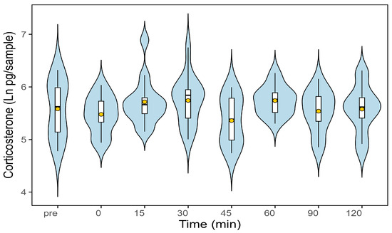
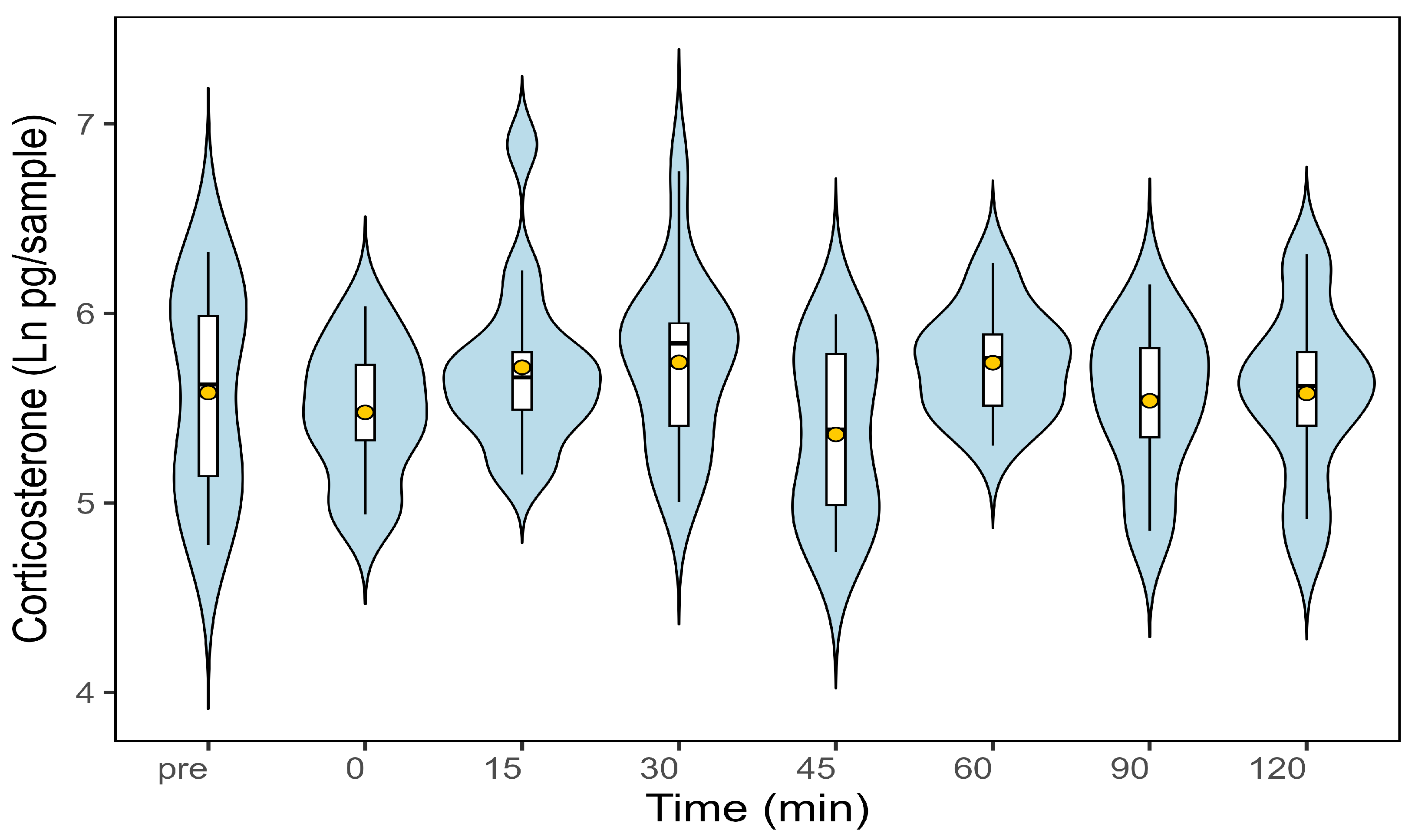
Dermal swab corticosterone concentrations averaged 5.56 ± 0.14 pg/sample prior to the acute hand-restraint stressor (Figure 1, Table 1). Time since stress exposure did not significantly predict ln-transformed corticosterone concentrations (t = −0.12, p = 0.90), and no post-stressor timepoint differed from the pre-stressor baseline (Figure 1, Table 1). Thus, we did not detect a significant acute corticosterone response to the hand-restraint stressor within the 120 min sampling window.
Figure 1.
Dermal swab ln corticosterone concentrations (pg/sample) in S. intermedia before and after exposure to an acute hand-restraint stressor (n = 14). Violin plots show the distribution of corticosterone values at each sampling timepoint (pre-stressor, 0, 15, 30, 45, 60, 90, and 120 min). Embedded boxplots indicate the median and interquartile range, and yellow points represent the model-estimated means. Corticosterone concentrations did not significantly differ across timepoints (p = 0.90), indicating no detectable acute endocrine response to the hand-restraint challenge.
Table 1.
Results of a linear mixed-effects model of dermal ln corticosterone (pg/sample) before and following an exposure to an acute hand-restraint stressor.
3.2. Experiment 2: Comparing Cortisol and Corticosterone via Water-Borne Sampling
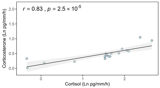
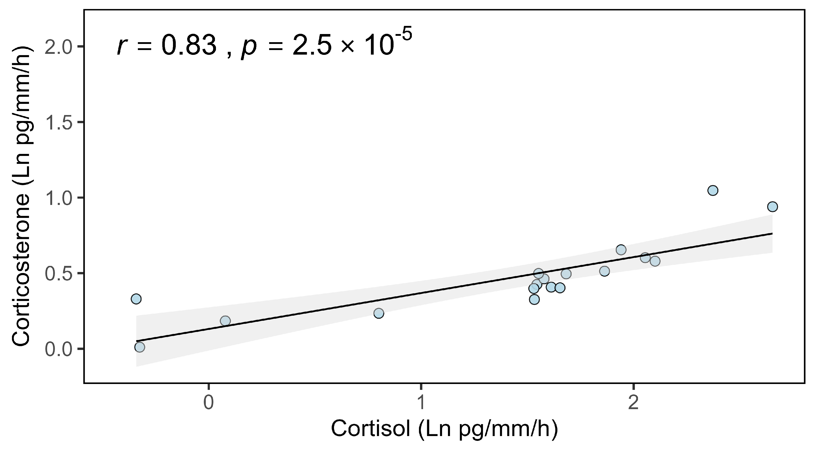
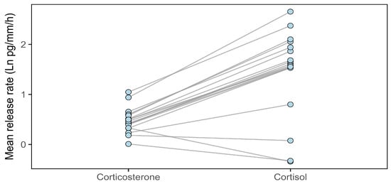
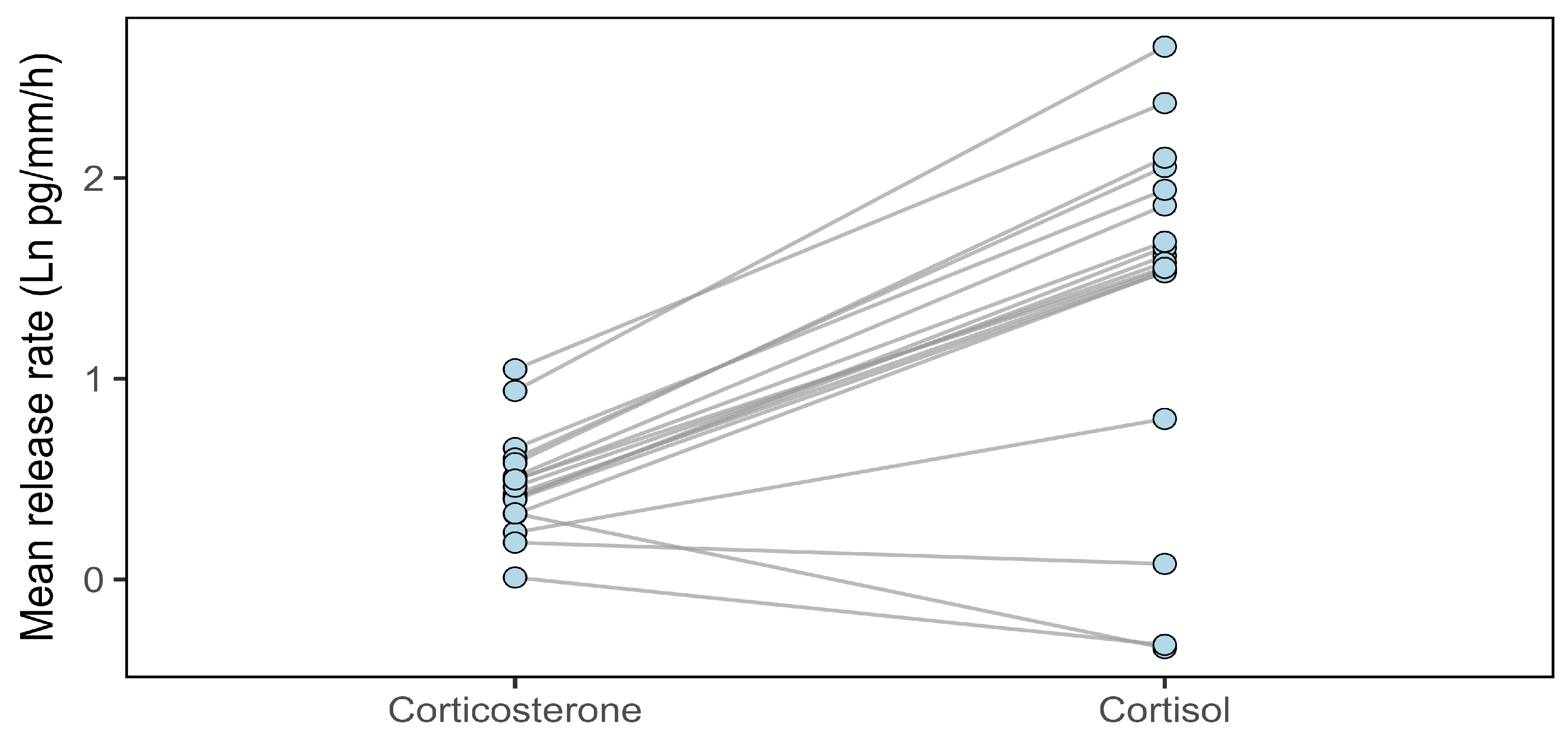
We found a strong significant positive correlation (r = 0.83, df = 16, t = 5.84, p < 0.0001, Figure 2) between mean water-borne ln cortisol and ln corticosterone release rates averaged across samples (baseline, stressed and recovery) within individuals. Cortisol release rates were significantly higher than corticosterone within individuals (t17 = 6.17, p < 0.0001, Figure 3). Cortisol release rates were repeatable across sample time (r = 0.233 ± 0.100, 95% CI: 0.024, 0.416; p = 0.0039) but not corticosterone release rates (r = 0.152 ± 0.142, 95% CI: 0, 0.468; p = 0.215).
Figure 2.
Relationship between ln cortisol and ln corticosterone (pg/mm/h) water-borne release rates in lesser sirens (n = 18). Shaded region represents 95% confidence intervals. Each point represents individual mean ln cortisol and corticosterone values across available samples. Ln cortisol and corticosterone were strongly positively correlated (p < 0.0001).
Figure 3.
Relationship between average cortisol and corticosterone (ln pg/mm/h) water-borne release rates in lesser sirens (n = 18). Each point represents the mean hormone release rate for an individual siren, averaged across baseline, stressed, and recovery samples. Lines connect paired hormone measurements within the same individual, illustrating within-individual differences in GCs. Cortisol release rates were higher than corticosterone within individuals (p < 0.0001).
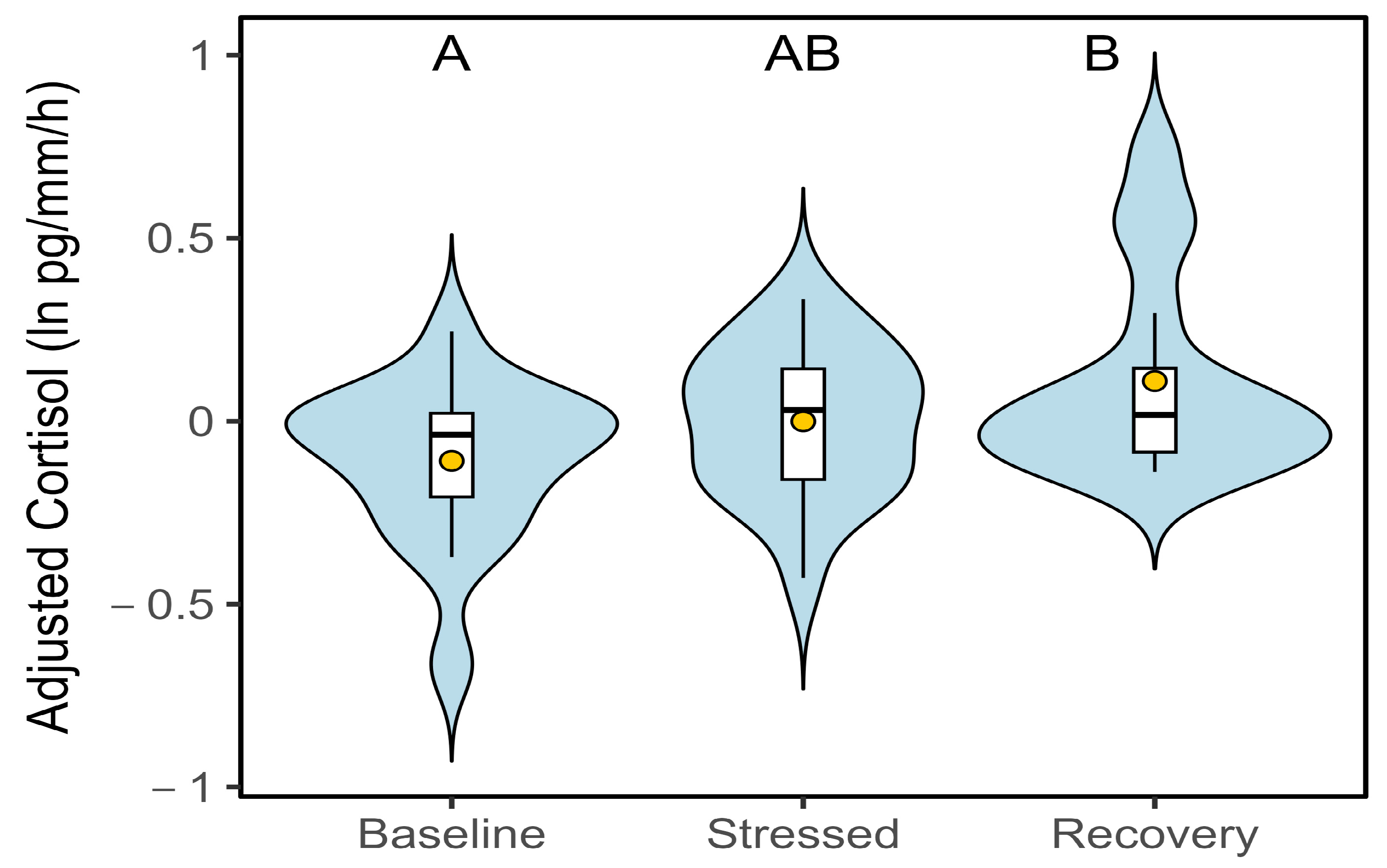
For cortisol, temperature and sample type affected release rates (Table 2). After accounting for temperature, sample type explained limited additional variation: cortisol during the recovery phase was marginally higher relative to baseline, as indicated by post hoc contrasts (pairwise comparison: estimate = –0.235 ± 0.101, t(30.2) = –2.32, p = 0.08, Figure 4), whereas stressed samples did not differ significantly from baseline (p = 0.24). Random intercepts for individuals accounted for substantial among-individual variation (SD = 0.74). For corticosterone, neither sample type nor temperature predicted release rates (Table 3). Individual variation was minimal (SD = 0.13), and the fixed effects explained little of the model variance.
Table 2.
Results of a linear mixed model of cortisol (ln pg/mm/h) release rates across sample type (baseline, stressed and recovery) and temperature. The intercept represents individuals in baseline samples. Effects with p < 0.05 denoted with *.
Figure 4.
Cortisol (ln pg/mm/h) release rates across sample types (baseline, stressed and recovery samples; n = 51) adjusted for temperature effects. Connecting letters represent results of pairwise comparisons. Differences found with marginal significance (p = 0.08).
Table 3.
Results of a linear mixed model of corticosterone (ln pg/mm/h) water-borne hormone release rates across sample type (baseline, stressed and recovery) and temperature. The intercept represents individuals in baseline samples.
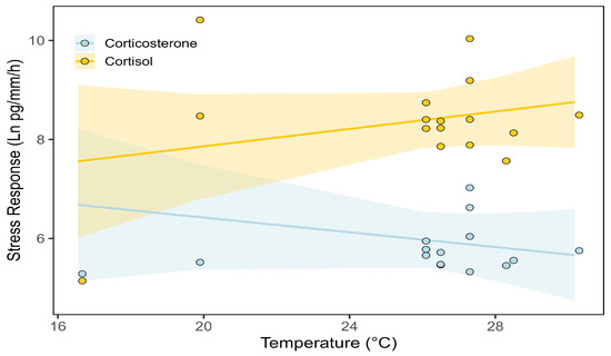
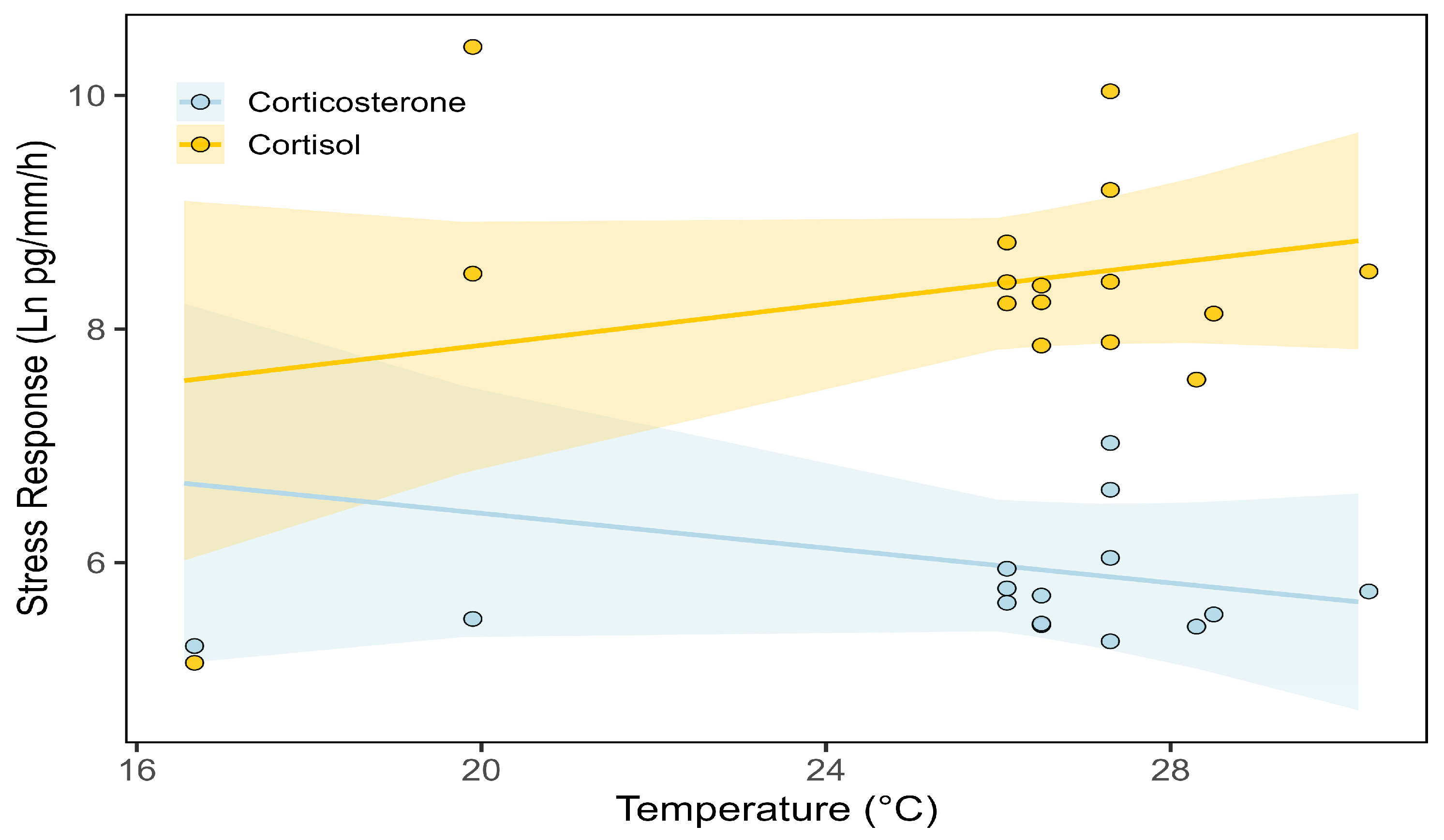
Temperature has hormone-specific effects on stress response and negative feedback. For stress response, temperature interacted significantly with hormone type (Figure 5). Cortisol samples showed substantially higher stress responses than the corticosterone samples (estimate = 2.36 ± 0.15, p < 0.0001). Temperature did not significantly influence stress response for corticosterone (estimate = −0.07 ± 0.08, p = 0.35). Cortisol stress responses were positively associated with temperature (estimate = 0.16 ± 0.04, p < 0.0018, Figure 5).
Figure 5.
Relationship between corticosterone and cortisol (ln pg/mm/h) release rates when exposed to a stressor and temperature. Cortisol is represented by yellow and corticosterone is represented by blue (n = 32 observations from 16 individuals). Shaded regions represent 95% confidence intervals. Temperature interacted significantly with hormone type in predicting stress response. Cortisol exhibited higher stress responses than corticosterone (p < 0.0001). Temperature did not significantly influence corticosterone stress responses (p = 0.35), whereas cortisol stress responses increased with temperature (p < 0.0018).
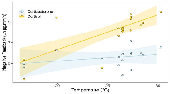
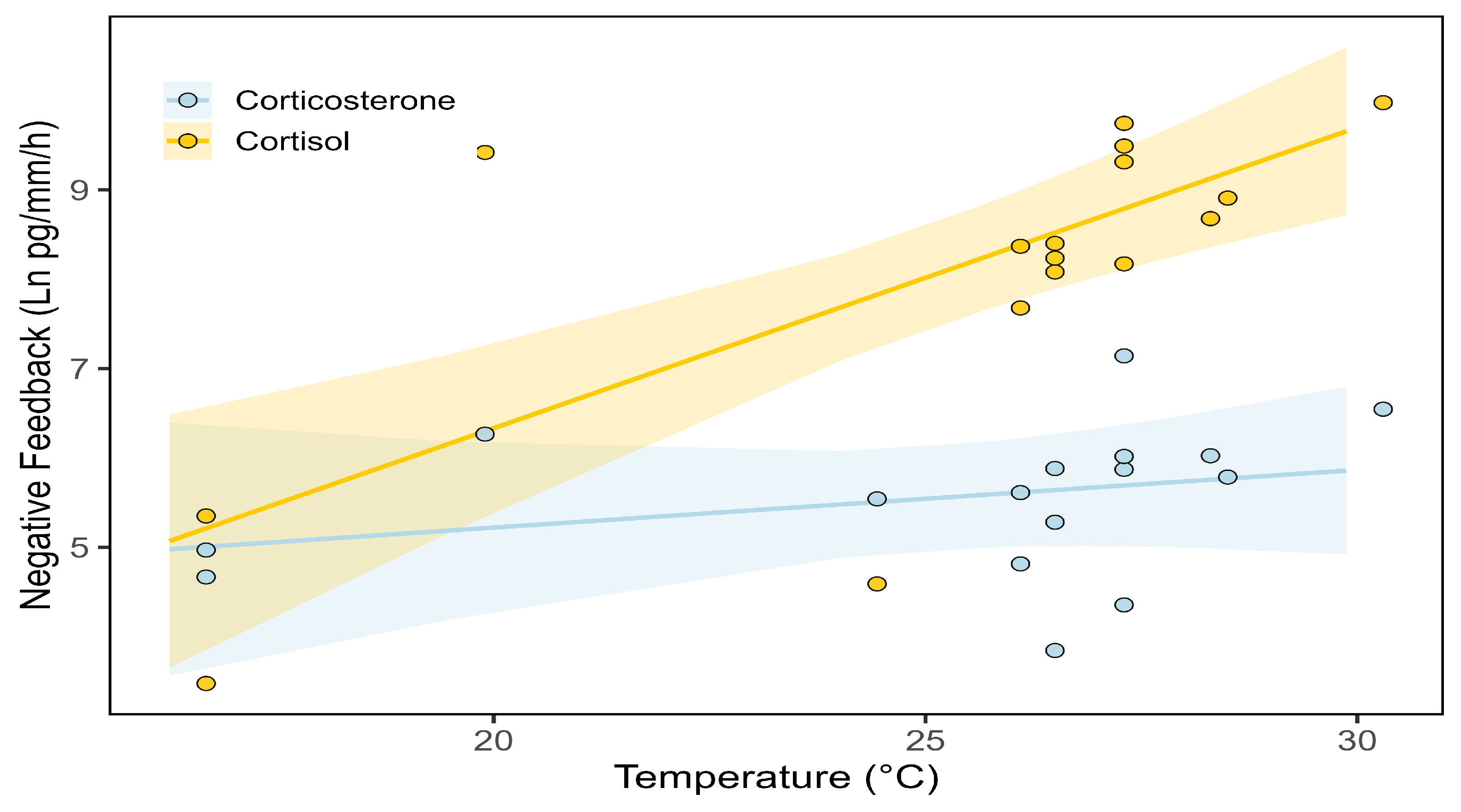
Negative feedback exhibited a similar hormone-specific temperature pattern. Negative feedback increased with temperature with cortisol but not with corticosterone (Temp × Hormone: estimate = 2.57 ± 0.36, p < 0.0001, Figure 6). This led to higher estimates of negative feedback at temperatures surpassing 25 °C for cortisol (estimate = 0.27 ± 0.09, p = 0.01). Temperature did not significantly influence corticosterone negative feedback (estimate = 0.06 ± 0.07, p = 0.38).
Figure 6.
Relationship between corticosterone and cortisol (ln pg/mm/h) negative feedback and temperature (n = 32 observations from 16 individuals). Cortisol is represented by yellow and corticosterone is represented by blue. Shaded regions represent 95% confidence intervals. Temperature interacted significantly with hormone type in predicting negative feedback. Cortisol negative feedback increased with temperature (p = 0.01), whereas corticosterone negative feedback did not change significantly with temperature (p = 0.38).
4. Discussion
Approaches to GC hormone collection and the specific hormone measured had strong effects on both the quantity of hormone detected and its apparent responsiveness to stressors in lesser sirens. Corticosterone concentrations measured with dermal swabs failed to detect an acute response to the hand-restraint challenge, whereas water-borne sampling revealed clear differences between cortisol and corticosterone dynamics. In juvenile sirens, cortisol and corticosterone measured from water-borne samples were strongly positively correlated, suggesting shared physiological regulation. However, cortisol occurred at substantially higher release rates and was the only hormone that varied with temperature or sampling type (baseline, stressed, or recovery). Water-borne cortisol exhibited a positive relationship with temperature, with both stress response magnitude and negative feedback increasing as temperatures rose. Corticosterone stress response and negative feedback showed no detectable temperature dependence. These results suggest that cortisol may be more sensitive to short-term environmental variation in juveniles of this species than corticosterone. In amphibians, GCs are well known to be temperature sensitive, reflecting the strong influence of environmental temperature on metabolic rate, endocrine activity, and HPI axis function in ectotherms [40]. We also found that only cortisol release rates were repeatable across sampling type. Together, these findings support a framework in which cortisol and corticosterone contribute to stress regulation in distinct but overlapping ways, with cortisol mediating rapid, context-dependent responses in S. intermedia.
We failed to detect effects of acute stress assays with dermal swabs targeting corticosterone. Although it remains possible that the hand-restraint protocol did not produce a strong corticosterone response in S. intermedia, the broader evidence suggests that the lack of change may also be attributable to limitations of dermal swabbing. Notably, individuals exhibited frequent wriggling and escape-oriented behaviors during restraint, suggesting that the protocol elicited a behavioral stress response, despite the absence of detectable changes to dermal corticosterone release rates. However, behavioral responses were not quantified in this study, and future work incorporating systematic behavioral measurements would help confirm the extent of behavioral stress responses associated with this protocol. A similar lack of response to acute stressors has been reported in northern leopard frogs, Lithobates pipens, and eastern red-spotted newts, N. viridescescens, where dermal corticosterone failed to increase even after adrenocorticotrophic hormone injections, despite clear expectations of elevated internal circulating levels [32]. This pattern is consistent with known biological mechanisms: in humans and other vertebrates, keratinocytes synthesize corticosterone locally through pathways independent of the HPA/I axis [32,41,42], meaning dermal hormone levels can be partially decoupled from circulating concentrations. The thick mucosal layer characteristic of aquatic salamanders may delay or modulate hormone diffusion between the epidermis and surrounding water, potentially reducing the sensitivity of dermal swabs to detect acute changes. Thus, the failure of dermal swabbing to detect an acute response could be explained by sampling limitations, potentially including local skin synthesis and dilution by the thick mucosal layer rather than by a lack of corticosterone reactivity. However, we did not measure cortisol from the dermal swabs so we cannot eliminate that method as useful. Taken together, the available evidence supports the idea that methodological constraints, rather than an absence of physiological reactivity, underlie the lack of detectable corticosterone responses observed via dermal swabbing.
Water-borne cortisol release rates were substantially higher than corticosterone and differed across the GC profile (sample type). Cortisol was also positively associated with temperature, whereas corticosterone showed no such association with temperature or sample type, indicating that cortisol may be more responsive to environmental regulation in S. intermedia. Higher cortisol release rates mirror results found in other amphibians, such as C. alleganiensis and several poison frog species, where cortisol is also the dominant glucocorticoid [18,19], challenging long-standing assumptions that corticosterone universally serves as the primary stress hormone in amphibians. It is worth noting that exceptions to corticosterone dominance in amphibians were found in the suborders Cryptobranchoidea and Sirenoidea. This pattern raises the possibility that cortisol dominance may characterize certain early-diverging salamander lineages, although testing this hypothesis will require broader phylogenetic sampling. Together, these findings suggest that larval Siren intermedia exhibits a cortisol-dominant glucocorticoid profile, underscoring the importance of species-specific perspectives when interpreting GC function. This pattern may reflect (1) an association with a fully aquatic lifestyle, consistent with many aquatic vertebrates, (2) evolutionary conservation within certain lineages, or (3) species-specific physiological or ecological factors that are not readily predicted by habitat or taxonomy alone. Further work is needed to explore these hypotheses, as we did not test adult sirens. Although cortisol appears to be the primary responder in juveniles of this species, measuring both hormones in future studies, despite higher costs, may help clarify potential functional differences or context-dependent roles that would otherwise be obscured by focusing on a single GC. For example, in tadpoles, corticosterone typically increases across development and reaches its highest levels near the metamorphic climax, where it interacts with thyroid hormones to drive tissue remodeling [43]. Larval amphibians can suppress corticosterone elevation early in development to avoid premature metamorphosis, thereby reducing potentially maladaptive developmental costs [44]. Lesser sirens are neotenic and retain larval characteristics throughout life, yet they still undergo partial metamorphosis involving skin restructuring and dorsal fin regression [45]. All individuals in the comparative water-borne study were juveniles, as they were relatively small (<200 mm). Reduced corticosterone responsiveness in these individuals may therefore reflect low HPI axis activity and reduced thyroid sensitivity, both characteristic of neotenic species [46]. The predominance of cortisol in juveniles suggests the possibility that stress responsiveness in this life stage may rely more heavily on cortisol-mediated pathways, though this interpretation remains speculative without direct thyroid-axis measurements.
Elevated corticosterone can actively oppose paedomorphosis, as demonstrated in facultatively paedomorphic salamanders such as mole salamanders, Ambystoma talpoideum, where paedomorph frequency declines with increasing corticosterone exposure [47]. By shifting stress responsiveness toward cortisol, sirens may be able to mount and regulate stress responses without activating corticosterone-linked pathways. These pathways could be developmentally constrained by their obligately paedomorphic life history. Within this framework, repeatability provides insight into the consistency of physiological traits and is often interpreted as an upper-bound estimate of heritability, reflecting the potential for traits to respond to selection [13]. In juvenile sirens, cortisol release rates were repeatable across sampling events, whereas corticosterone was not. This pattern suggests that cortisol represents a more stable, individual-level trait during this life stage and further supports cortisol as the primary GC-mediating stress responses in juvenile sirens. In contrast, studies in amphibian larvae show a different pattern: corticosterone is repeatable during tadpole stages of Rana berlandieri [12], suggesting that corticosterone plays a more dominant role earlier in their development. Additionally, in D. tinctorius, corticosterone release rates exceed cortisol during tadpole stages, whereas corticosterone and cortisol occur at nearly equal levels in adults [19,20]. In mice, corticosterone functions as the primary glucocorticoid involved in longer-term stress adaptation, whereas cortisol acts as a faster responder to severe acute stressors [48]. Evidence from other vertebrates supports the idea that cortisol and corticosterone can serve complementary, rather than redundant, roles in stress regulation, with their relative importance shifting across species, developmental and ecological contexts.
The positive association between cortisol and temperature has important ecological and conservation implications for lesser sirens. Because GC production in ectotherms is tightly linked to metabolic rate [49], elevated cortisol at higher temperatures may reflect increased energetic demands or heightened physiological strain under warming conditions [11]. This pattern aligns with growing evidence that amphibian stress physiology is sensitive to thermal variation, which can alter immune function, behavior, and disease susceptibility [40]. For aquatic species such as lesser sirens that persist in shallow wetlands, rising temperatures and increased frequency of thermal extremes may therefore lead to chronically elevated cortisol levels, potentially reducing fitness or resilience [11]. However, longitudinal studies are needed to evaluate this possibility. Although we focused on temperature as a key environmental covariate, other factors such as dissolved oxygen and pH may also influence endocrine responses and should be considered in future work. Identifying GC dynamics in this species provides a valuable tool for monitoring environmental stress in natural populations, particularly in the context of climate-driven shifts in hydroperiod, temperature regimes, and habitat quality. Integrating GC indicators like coping capacity into conservation assessments may help managers detect sublethal stress before population-level declines occur, thereby improving early intervention and habitat protection strategies.
Although our study provides new insight into GC profiles and sampling methods in S. intermedia, several limitations of our study design warrant consideration. Sample sizes were constrained for the dermal swab experiment, which may limit statistical power. Additionally, temperature variation in our study was observational rather than experimentally manipulated, preventing us from isolating causal effects of temperature on GC dynamics. Our study also did not include water-borne hormone sampling from adult sirens, limiting our ability to evaluate how GC profiles compare across all life stages and to determine whether adults also exhibit cortisol dominance over corticosterone. The acute stressors we employed may not fully capture the range of ecologically relevant challenges experienced by sirens in the wild. However, we validated the use of EIA kits, and we confirmed the parallelism of the serial dilution curves against the standard curve and found acceptable recovery for each hormone. This validation supports the conclusion that observed differences between cortisol and corticosterone reflect true biological variation rather than matrix effects or assay bias. Future research that incorporates repeated sampling of multiple GCs across environmental gradients and life stages will be essential for clarifying context-dependent differences in cortisol and corticosterone stress regulation. Establishing species-specific GC patterns provides an important foundation for future studies examining how environmental stressors influence amphibian physiology across life stages and ecological contexts.
Supplementary Materials
The following supporting information can be downloaded at https://www.mdpi.com/article/10.3390/conservation6020047/s1, Dataset S1: Experiment 1; Dataset S2: Experiment 2.
Author Contributions
Conceptualization, M.F. and C.R.G.; methodology, M.F. and C.R.G.; validation, M.F. and C.R.G.; formal analysis, M.F.; resources, T.L. and C.R.G.; data curation, M.F.; writing—original draft preparation, M.F.; writing—review and editing, M.F., C.R.G., and T.L.; visualization, M.F.; supervision, T.L. and C.R.G.; funding acquisition, M.F. and T.L. All authors have read and agreed to the published version of the manuscript.
Funding
This research was funded by the Texas Herpetological Society, Texas State Graduate Fellowship, and the Wild Animal Initiative (grant number C-2022-00032).
Institutional Review Board Statement
All protocols and housing were approved by the Institutional Animal Care and Use Committee of Texas State University (IACUC #8805, Approval Date: 1 March 2023).
Data Availability Statement
Data is contained within the article or Supplementary Material.
Acknowledgments
We thank Sydney Falcon, Jacob Kearns and Caroline Crisp for their help with data collection. We also thank Carmen Montaña for letting us use her lab to store samples. We thank Christopher Shalk for helping us identify siren populations. Lastly, thanks to the sirens for their invaluable contribution to this work. During the preparation of this manuscript/study, the authors used AI-assisted tools (ChatGPT—GPT-4 and GPT-5 series, OpenAI) for the purposes of R code generation and clarifying statistical processes. We thank the three reviewers for their helpful comments and edits on this manuscript. The authors have reviewed and edited the output and take full responsibility for the content of this publication.
Conflicts of Interest
The authors declare no conflicts of interest.
Abbreviations
The following abbreviations are used in this manuscript:
| GCs | Glucocorticoids |
| LMMs | Linear Mixed-Effect Models |
| FDR | False Discovery Rate |
| REML | Restricted Maximum Likelihood |
| ln | Natural Logarithm |
| HPI | Hypothalamic–Pituitary–Interrenal |
| EIA | Enzyme Immunoassay |
References
- Sapolsky, R.M.; Romero, L.M.; Munck, A.U. How Do Glucocorticoids Influence Stress Responses? Integrating Permissive, Suppressive, Stimulatory, and Preparative Actions. Endocr. Rev. 2000, 21, 55–89. [Google Scholar] [CrossRef] [Scilit]
- Romero, L.M.; Gormally, B.M.G. How Truly Conserved Is the “Well-Conserved” Vertebrate Stress Response? Integr. Comp. Biol. 2019, 59, 273–281. [Google Scholar] [CrossRef] [Scilit]
- McEwen, B.S.; Wingfield, J.C. The Concept of Allostasis in Biology and Biomedicine. Horm. Behav. 2003, 43, 2–15. [Google Scholar] [CrossRef] [Scilit]
- Apaydin, D.C.; Jaramillo, P.A.M.; Corradi, L.; Cosco, F.; Rathjen, F.G.; Kammertoens, T.; Filosa, A.; Sawamiphak, S. Early-Life Stress Regulates Cardiac Development through an IL-4-Glucocorticoid Signaling Balance. Cell Rep. 2020, 33, 108404. [Google Scholar] [CrossRef] [Scilit] [PubMed]
- Vitousek, M.N.; Taff, C.C.; Hallinger, K.K.; Zimmer, C.; Winkler, D.W. Hormones and Fitness: Evidence for Trade-Offs in Glucocorticoid Regulation Across Contexts. Front. Ecol. Evol. 2018, 6, 42. [Google Scholar] [CrossRef] [Scilit]
- Dantzer, B.; Fletcher, Q.E.; Boonstra, R.; Sheriff, M.J. Measures of Physiological Stress: A Transparent or Opaque Window into the Status, Management and Conservation of Species? Conserv. Physiol. 2014, 2, cou023. [Google Scholar] [CrossRef] [Scilit] [PubMed]
- Gabor, C.R.; Bosch, J.; Fries, J.N.; Davis, D.R. A Non-Invasive Water-Borne Hormone Assay for Amphibians. Amphibia-Reptilia 2013, 34, 151–162. [Google Scholar] [CrossRef] [Scilit]
- Gabor, C.R.; Fisher, M.C.; Bosch, J. Elevated Corticosterone Levels and Changes in Amphibian Behavior Are Associated with Batrachochytrium Dendrobatidis (Bd) Infection and Bd Lineage. PLoS ONE 2015, 10, e0122685. [Google Scholar] [CrossRef] [Scilit]
- Koolhaas, J.M.; Korte, S.M.; De Boer, S.F.; Van Der Vegt, B.J.; Van Reenen, C.G.; Hopster, H.; De Jong, I.C.; Ruis, M.A.W.; Blokhuis, H.J. Coping Styles in Animals: Current Status in Behavior and Stress-Physiology. Neurosci. Biobehav. Rev. 1999, 23, 925–935. [Google Scholar] [CrossRef] [Scilit]
- Vitousek, M.N.; Johnson, M.A.; Downs, C.J.; Miller, E.T.; Martin, L.B.; Francis, C.D.; Donald, J.W.; Fuxjager, M.J.; Goymann, W.; Hau, M.; et al. Macroevolutionary Patterning in Glucocorticoids Suggests Different Selective Pressures Shape Baseline and Stress-Induced Levels. Am. Nat. 2019, 193, 866–880. [Google Scholar] [CrossRef] [Scilit]
- Romero, L.M.; Dickens, M.J.; Cyr, N.E. The Reactive Scope Model—A New Model Integrating Homeostasis, Allostasis, and Stress. Horm. Behav. 2009, 55, 375–389. [Google Scholar] [CrossRef] [Scilit] [PubMed]
- Forsburg, Z.R.; Goff, C.B.; Perkins, H.R.; Robicheaux, J.A.; Almond, G.F.; Gabor, C.R. Validation of Water-Borne Cortisol and Corticosterone in Tadpoles: Recovery Rate from an Acute Stressor, Repeatability, and Evaluating Rearing Methods. Gen. Comp. Endocrinol. 2019, 281, 145–152. [Google Scholar] [CrossRef] [Scilit] [PubMed]
- Dohm, M.R. Repeatability Estimates Do Not Always Set an Upper Limit to Heritability. Funct. Ecol. 2002, 16, 273–280. [Google Scholar] [CrossRef] [Scilit]
- Moore, I.T.; Jessop, T.S. Stress, Reproduction, and Adrenocortical Modulation in Amphibians and Reptiles. Horm. Behav. 2003, 43, 39–47. [Google Scholar] [CrossRef] [Scilit]
- Norris, D.O.; Carr, J.A. Vertebrate Endocrinology; Academic Press: Cambridge, MA, USA, 2020. [Google Scholar]
- Koren, L.; Whiteside, D.; Fahlman, Å.; Ruckstuhl, K.; Kutz, S.; Checkley, S.; Dumond, M.; Wynne-Edwards, K. Cortisol and Corticosterone Independence in Cortisol-Dominant Wildlife. Gen. Comp. Endocrinol. 2012, 177, 113–119. [Google Scholar] [CrossRef] [Scilit]
- Hancock, T. A Duality in Mammalian Glucocorticoid Signaling: Evidence Against the Dominant Glucocorticoid/Interchangeability Assumption. Master’s Thesis, Queen’s University, Kingston, ON, Canada, 2010. [Google Scholar]
- Hopkins, W.A.; DuRant, S.E.; Beck, M.L.; Ray, W.K.; Helm, R.F.; Romero, L.M. Cortisol Is the Predominant Glucocorticoid in the Giant Paedomorphic Hellbender Salamander (Cryptobranchus Alleganiensis). Gen. Comp. Endocrinol. 2020, 285, 113267. [Google Scholar] [CrossRef] [Scilit]
- Westrick, S.E.; Paitz, R.T.; Fischer, E.K. Why Not Both? Measuring Cortisol and Corticosterone in Poison Frogs. bioRxiv 2025. [Google Scholar] [CrossRef] [Scilit]
- Surber-Cunningham, L.L.; Jimenez, L.S.; Mobo, L.W.; Westrick, S.E.; Fischer, E.K. Early Development of the Glucocorticoid Stress Response in Dyeing Poison Frog Tadpoles. Gen. Comp. Endocrinol. 2025, 371, 114774. [Google Scholar] [CrossRef] [Scilit]
- Jungreis, A.M.; Huibregtse, W.H.; Ungar, F. Corticosteroid Identification and Corticosterone Concentration in Serum of Rana Pipiens during Dehydration in Winter and Summer. Comp. Biochem. Physiol. 1970, 34, 683–689. [Google Scholar] [CrossRef] [Scilit]
- Sadoul, B.; Geffroy, B. Measuring Cortisol, the Major Stress Hormone in Fishes. J. Fish Biol. 2019, 94, 540–555. [Google Scholar] [CrossRef] [Scilit]
- Scott, A.P.; Ellis, T. Measurement of Fish Steroids in Water—A Review. Gen. Comp. Endocrinol. 2007, 153, 392–400. [Google Scholar] [CrossRef] [Scilit] [PubMed]
- Chan, S.T.H.; Edwards, B.R. Kinetic Studies on the Biosynthesis of Corticosteroids in Vitro from Exogenous Precursers by the Interrenal Glands of the Normal Corticotrophin-Treated and Adenohypophysectomized Xenopus Laevis Daudin. J. Endocrinol. 1970, 47, 183–195. [Google Scholar] [CrossRef] [Scilit] [PubMed]
- Weisrock, D.W.; Harmon, L.J.; Larson, A. Resolving Deep Phylogenetic Relationships in Salamanders: Analyses of Mitochondrial and Nuclear Genomic Data. Syst. Biol. 2005, 54, 758–777. [Google Scholar] [CrossRef] [Scilit] [PubMed]
- Gehlbach, F.R.; Kennedy, S.E. Population Ecology of a Highly Productive Aquatic Salamander (Siren Intermedia). Southwest. Nat. 1978, 23, 423–429. [Google Scholar] [CrossRef] [Scilit]
- Fauth, J.E. Identifying Potential Keystone Species from Field Data—An Example from Temporary Ponds. Ecol. Lett. 1999, 2, 36–43. [Google Scholar] [CrossRef] [Scilit]
- Baugh, A.T.; Bastien, B.; Still, M.B.; Stowell, N. Validation of Water-Borne Steroid Hormones in a Tropical Frog (Physalaemus Pustulosus). Gen. Comp. Endocrinol. 2018, 261, 67–80. [Google Scholar] [CrossRef] [Scilit]
- Conant, R.; Collins, J.T. A Field Guide to Reptiles & Amphibians: Eastern and Central North America; Houghton Mifflin Harcourt: Boston, MA, USA, 1998. [Google Scholar]
- Scheun, J.; Greeff, D.; Medger, K.; Ganswindt, A. Validating the Use of Dermal Secretion as a Matrix for Monitoring Glucocorticoid Concentrations in African Amphibian Species. Conserv. Physiol. 2019, 7, coz022. [Google Scholar] [CrossRef] [Scilit]
- Santymire, R.M.; Manjerovic, M.B.; Sacerdote-Velat, A. A Novel Method for the Measurement of Glucocorticoids in Dermal Secretions of Amphibians. Conserv. Physiol. 2018, 6, coy008. [Google Scholar] [CrossRef] [Scilit]
- Quadros, V.; Inman, B.; McDonnell, N.; Williams, K.; Romero, L.M.; Woodhams, D.C. Dermal Glucocorticoids Are Uncoupled from Stress Physiology and Infection. Conserv. Physiol. 2025, 13, coaf005. [Google Scholar] [CrossRef] [Scilit]
- Luhring, T.M.; Jennison, C.A. A New Stratified Aquatic Sampling Technique for Aquatic Vertebrates. J. Freshw. Ecol. 2008, 23, 445–450. [Google Scholar] [CrossRef] [Scilit]
- Neely, W.J.; Martins, R.A.; da Silva, C.M.M.; da Silva, T.F.; Fleck, L.E.; Whetstone, R.D.; Woodhams, D.C.; Cook, W.H.; Prist, P.R.; Valiati, V.H.; et al. Linking Microbiome and Stress Hormone Responses in Wild Tropical Treefrogs across Continuous and Fragmented Forests. Commun. Biol. 2023, 6, 1261. [Google Scholar] [CrossRef] [Scilit] [PubMed]
- Gabor, C.R.; Zabierek, K.C.; Kim, D.S.; da Barbiano, L.A.; Mondelli, M.J.; Bendik, N.F.; Davis, D.R. A Non-Invasive Water-Borne Assay of Stress Hormones in Aquatic Salamanders. Ichthyol. Herpetol. 2016, 104, 172–181. [Google Scholar] [CrossRef] [Scilit]
- Bókony, V.; Ujhegyi, N.; Hamow, K.Á.; Bosch, J.; Thumsová, B.; Vörös, J.; Aspbury, A.S.; Gabor, C.R. Stressed Tadpoles Mount More Efficient Glucocorticoid Negative Feedback in Anthropogenic Habitats Due to Phenotypic Plasticity. Sci. Total Environ. 2021, 753, 141896. [Google Scholar] [CrossRef] [Scilit] [PubMed]
- Lattin, C.R.; Kelly, T.R. Glucocorticoid Negative Feedback as a Potential Mediator of Trade-Offs between Reproduction and Survival. Gen. Comp. Endocrinol. 2020, 286, 113301. [Google Scholar] [CrossRef] [Scilit]
- Nakagawa, S.; Schielzeth, H. Repeatability for Gaussian and Non-Gaussian Data: A Practical Guide for Biologists. Biol. Rev. 2010, 85, 935–956. [Google Scholar] [CrossRef] [Scilit]
- Dingemanse, N.J.; Dochtermann, N.A. Quantifying Individual Variation in Behaviour: Mixed-Effect Modelling Approaches. J. Anim. Ecol. 2013, 82, 39–54. [Google Scholar] [CrossRef] [Scilit]
- Rollins-Smith, L.A.; Le Sage, E.H. Heat Stress and Amphibian Immunity in a Time of Climate Change. Philos. Trans. R. Soc. B Biol. Sci. 2023, 378, 20220132. [Google Scholar] [CrossRef] [Scilit]
- Jozic, I.; Stojadinovic, O.; Kirsner, R.S.; Tomic-Canic, M. Stressing the Steroids in Skin: Paradox or Fine-Tuning? J. Investig. Dermatol. 2014, 134, 2869–2872. [Google Scholar] [CrossRef] [Scilit]
- Phan, T.S.; Schink, L.; Mann, J.; Merk, V.M.; Zwicky, P.; Mundt, S.; Simon, D.; Kulms, D.; Abraham, S.; Legler, D.F.; et al. Keratinocytes Control Skin Immune Homeostasis through de Novo–Synthesized Glucocorticoids. Sci. Adv. 2021, 7, eabe0337. [Google Scholar] [CrossRef] [Scilit]
- Glennemeier, K.A. Developmental Changes in Interrenal Responsiveness in Anuran Amphibians. Integr. Comp. Biol. 2002, 42, 565–573. [Google Scholar] [CrossRef] [Scilit]
- Forsburg, Z.R.; Guzman, A.; Gabor, C.R. Artificial Light at Night (ALAN) Affects the Stress Physiology but Not the Behavior or Growth of Rana Berlandieri and Bufo Valliceps. Environ. Pollut. 2021, 277, 116775. [Google Scholar] [CrossRef] [Scilit]
- Noble, G.K.; Marshall, B.C. The Validity of Siren Intermedia Leconte, with Observations on Its Life History; American Museum Novitates, No. 532; The American Museum of Natural History: New York, NY, USA, 1932; Volume No. 532. [Google Scholar]
- Lofts, B. Physiology of the Amphibia; Elsevier: Amsterdam, The Netherlands, 2012. [Google Scholar]
- Bohenek, J.R.; Leary, C.J.; Resetarits, W.J., Jr. Exposure to Glucocorticoids Alters Life History Strategies in a Facultatively Paedomorphic Salamander. J. Exp. Zool. Part Ecol. Integr. Physiol. 2021, 335, 329–338. [Google Scholar] [CrossRef] [Scilit]
- Gong, S.; Miao, Y.-L.; Jiao, G.-Z.; Sun, M.-J.; Li, H.; Lin, J.; Luo, M.-J.; Tan, J.-H. Dynamics and Correlation of Serum Cortisol and Corticosterone under Different Physiological or Stressful Conditions in Mice. PLoS ONE 2015, 10, e0117503. [Google Scholar] [CrossRef] [Scilit]
- Gangloff, E.J.; Telemeco, R.S. High Temperature, Oxygen, and Performance: Insights from Reptiles and Amphibians. Integr. Comp. Biol. 2018, 58, 9–24. [Google Scholar] [CrossRef] [Scilit]

Disclaimer/Publisher’s Note: The statements, opinions and data contained in all publications are solely those of the individual author(s) and contributor(s) and not of MDPI and/or the editor(s). MDPI and/or the editor(s) disclaim responsibility for any injury to people or property resulting from any ideas, methods, instructions or products referred to in the content. |
© 2026 by the authors. Licensee MDPI, Basel, Switzerland. This article is an open access article distributed under the terms and conditions of the Creative Commons Attribution (CC BY) license.