


Abstract
Antioxidants, such as phenolic compounds, are essential for mammal physiology. Significant research made on the gut–brain axis has produced volumes of evidence indicating that some plant-derived phenolic compounds can reach brain cells to exert protective effects on them, mainly by maintaining and/or restoring redox homeostasis. Their systemic uptake and transport might be determined by the phenolic’s physicochemical properties, along with complex interactions with protein transporters and carriers, including GLUT, SGLT1, ABC transporters (P-glycoprotein, breast cancer resistance protein), albumin, fibrinogen, organic anion and cation transporters, and MATE1. The present work focuses on the chemical interactions and transport pathways of some phenolic compounds to reach brain cells.
1. Introduction
Plants possess complex metabolic and signaling pathways that maintain and regulate various physiological processes [1], that can be up- and downregulated by environmental conditions (abiotic stress) and biotic stress. As a result, many metabolites are produced to help the plant maintain homeostasis [2]. One of the secondary metabolites produced in plants and largely distributed in leaves, roots, seeds, stems, and fruits are phenolic compounds (PCs) [3], which participate in several physiological processes like plant development, environmental protection, and adaptation [4].
Throughout history, humans have consumed fruits and vegetables in their meals as part of their daily diet, as well as in the form of beverages, tea, and others without a full understanding of their nutritional benefits. Considerable efforts have now been made to demonstrate the functionality of plant-derived bioactive compounds in human physiology, for instance, dietary consumption of PCs improves health, mainly by controlling the redox environment within tissues, in addition to modulating physiological processes like inflammation, apoptosis, cellular communication, and genetic expression, among others [5]. These bioactivities reduce the risk of developing chronic diseases, particularly those that affect the central nervous system (CNS), such as Alzheimer’s and Parkinson’s diseases [6].
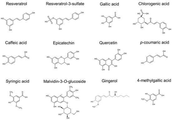
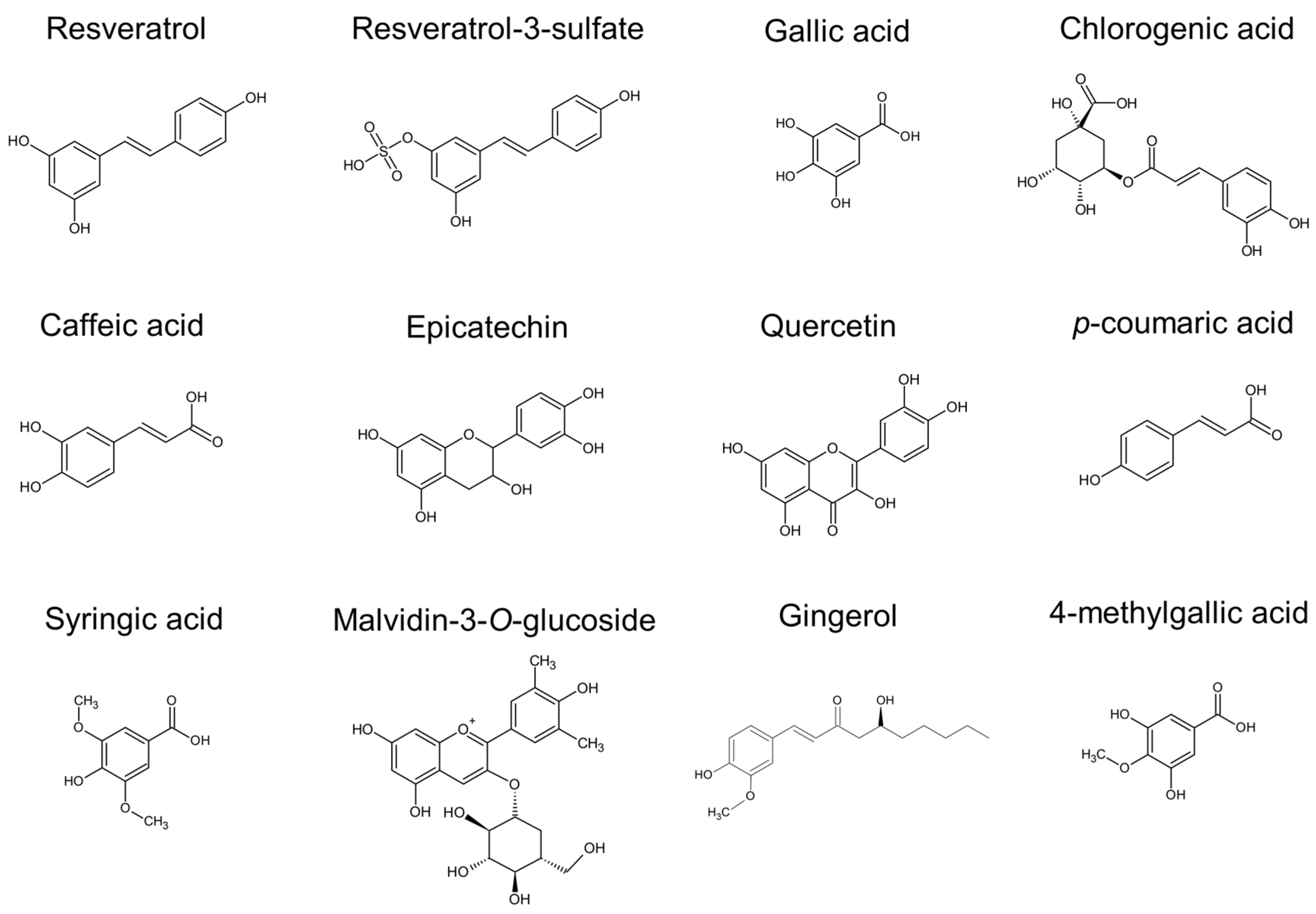
The journey from PC intake to final bioactivity has been studied in different cell lines, animal models, and human subjects due to their biological importance; however, PC transport is a particularly complex process that depends on several factors, but two of them stand out. The first factor is the differences in their chemical structure (Figure 1), since they can differ in the number, position, and types of functional groups attached to one or more phenolic rings [7]. The second factor is the interactions formed with membrane proteins (receptors and transporters) or with biomolecules found throughout the body (lipids, serum proteins, DNA) [8,9]. The pharmacokinetics and pharmacodynamics of PCs are determined by their chemical structure and may be influenced by human physiology (e.g., metabolism, cell division, gene expression) at the moment of their consumption [10]. For instance, it is unclear if PC assimilation is determined by nutritional requirements that promote higher absorption of glucuronidated PCs, or by detecting a specific PC that enterocytes use to modulate the intestine’s redox homeostasis. Nevertheless, it is important to study why some PCs may have one or several transport mechanisms to understand their potential role in human physiology. The purpose of the present work is to discuss PC transport throughout the different cell types and organs that form the gastrointestinal tract, molecules of the bloodstream, the blood–brain barrier (BBB), and brain cells.
Figure 1.
Chemical structure of some PCs that interact with membrane receptors, transporters and blood proteins.
2. Oral and Gut Digestion: The Path for Phenolic Compound Absorption
The journey of PC digestion starts in the mouth with the mastication of food, in order to reduce its size and form the alimentary bolus. α-amylase (E.C. 3.2.1.1) is the predominant digestive enzyme present in saliva, along with proteins with lipase, peptidase, and hydrolase activities that catabolize food macromolecules [11,12,13]. Although mastication time is short (as compared to later stages), enzymatic processes in the oral cavity allow the release of some PCs from the alimentary matrix or from a pool of complex phenolic compounds present in foods and beverages, respectively. In fermented beverages like wine, bacterial β-glucosidase (E.C. 3.2.1.21) can hydrolyze the monosaccharide residue conjugated to PCs (e.g., flavonoid glycosides), releasing the aglycone form [14,15]. For example, high-performance liquid chromatography was able to detect and quantify 25.33 mg of gallic acid, 0.35 mg of resveratrol, 0.11 of syringic acid, 0.50 mg of caffeic acid, and 0.83 mg of p-coumaric acid in 150 mL of red wine, of which only 0.94–6.89 µg of gallic acid, 0.29 µg of resveratrol, and 0.53–83 µg of p-coumaric acid were detected in 2 mL of saliva (males and females, 25 to 41 years old) after consuming it; the other documented compounds were detected below the quantification limit (0.25 µg) [16]. The authors sampled from 0 to 5 h after wine consumption; the highest concentration peaks were found in the first 5 and 10 min, indicating that PCs in beverages could be accessible to the enzymatic environment of the mouth, and be potentially absorbed by oral epithelia and mucosa to be distributed systemically. For instance, studying the permeation of chestnut shells phenolic extract (37 mg GAE/g) in human tongue squamous carcinoma (HSC-3) and human neck metastasis of buccal carcinoma (TR146) cell lines, Ferreira et al. (2025) [17] found that epicatechin (>96%) and protocatechuic acid (>85%) showed higher cell permeability in comparison with other PCs like vanillin. These studies suggest that oral PC absorption may contribute to their systemic distribution, although the evidence is scarce and does not elucidate the internalization pathway in oral tissue [18].
According to in vitro mouth models made with HSC-3 and TR146 cells, two types of phenolic mixtures, one with flavonoids (0.5, 1.0, and 3.0 mg/mL) and the other one with tannins (0.1, 0.3, and 0.5 mg/mL), PCs are retained in the intracellular space. Gallic acid showed the highest bound phenolic concentration, with 50.9 mg/L in TR146 cells; however, Guerreiro et al. (2022) [19] suggest that salivary proteins may influence the interactions between cell lines and PCs, mainly by physical sequestration that promotes a delay in the early absorption of tannin-derived compounds (gallic acid, digallic acid, trigallic acid) and decrease the affinity of those containing glucose residues in their structure. This may indicate that PCs with simple structures are easier to absorb than conjugated ones, and that early chemical interactions with oral proteins may impact their absorption in the next stages in the gastrointestinal tract.
The digestive environment of the oral cavity paves the way for PC release during the gastric phase, wherein the acidic medium (pH < 3) generated mainly by gastric acid results in the hydrolysis of chemical bonds, which allows the release of polysaccharide-esterified PCs [20]. The digested alimentary bolus then enters the duodenum and turns into chyme, which is mixed with pancreatic lipases (E.C. 3.1.1.3), amylases (E.C. 3.2.1.1), and proteases (E.C. 3.4.21), as well as bile from the gallbladder, which altogether allow the breakdown of molecular bonds to release nutrients (e.g., vitamins, sugars, minerals) [21]. PCs are also released during this stage and are now known as bioaccessible phenolic compounds (BAPCs); they can be found as monomers, dimers, and oligomers, and can be complexed with other BAPCs, starch, fibers, or carbohydrates [22]. These BAPCs may be metabolized by the host’s enzymes, or be absorbed, transformed, or used by the gut microbiota.
The diversity of cells along the gastrointestinal tract promotes specific microenvironments that may favor differences in the uptake of nutrients and several molecules in the duodenum, ileum, jejunum, and colon [23,24]. The absorption of PCs in the gut is also considered to be low (in the range of micro- and picograms), as compared to other nutrients, and depends on the concentration of PCs in chyme, biotransformation and metabolism by gut microbiota, intermolecular interactions that PCs form with chyme molecules (e.g., PCs oligomers or polysaccharides), and the return of these compounds to the lumen by efflux pumps to be excreted through the fecal route [25,26]. Moreover, the transport and distribution of absorbed PCs may be modulated by physiological needs (e.g., redox balance, to counter pathological conditions, for cellular recovery processes) in every organ of the body. For this, the systemic PCs that were not used by any cell are excreted through urine and feces [27,28,29], suggesting that their gut and systemic absorption can be up- and downregulated by physiological needs, a hypothesis that remains unproven so far.
As previously mentioned, the structure of BAPCs may be central for their absorption and distribution due to chemical interactions with proteins, receptors, and transporters of intestinal cells. This may have an impact on the pharmacokinetics and pharmacodynamics of BAPCs, biotransformed PCs (BTPCs), and bioavailable PCs (BPCs). For example, in crossbred female pigs, oral feeding with a 1% grape seed extract formulation alters their gut microbiome by increasing the bacterial species Lachnospiraceae, Clostridales, Lactobacillus and Ruminococcacceae [30]. Interestingly, the authors report a high fecal excretion of proanthocyanidins (400 mg/kg) after feeding in a period no longer than 48 h, with 4-hydroxyphenylvaleric acid (30 mg/kg) and 3-hydroxybenzoic acid (3 mg/kg) being the main compounds identified and quantified. The identification of fecal BTPCs indicates the participation of the gut microbiota in the metabolism and the biotransformation of PCs, suggesting its important role in PC digestion and absorption.
It is important to consider that BAPCs may form intermolecular complexes with alimentary molecules (e.g., mono- and disaccharides), digestion-derived products present in chyme (e.g., glucuronides), as well as those from the host, which may modulate the free fraction of BAPCs and thereby modulate their intestinal absorption [31]. For example, in vitro experiments using red wine methanolic extracts showed that malvidin-3-O-glucoside (250 µM) and malvidin-3,5-O-diglucoside (300 µM) can be transported through a simulated intestinal barrier using a Caco-2 cell monolayer. Further molecular docking experiments demonstrate the interaction of these PCs with GLUT1 and GLUT3, where the glucoside-conjugated anthocyanin interacts mainly with threonine, glutamine, asparagine, and glutamic acid residues of the binding cleft [32]. However, further studies are needed to understand the effect of these interactions, because it is unclear if they promote PC absorption or GLUT modulation.
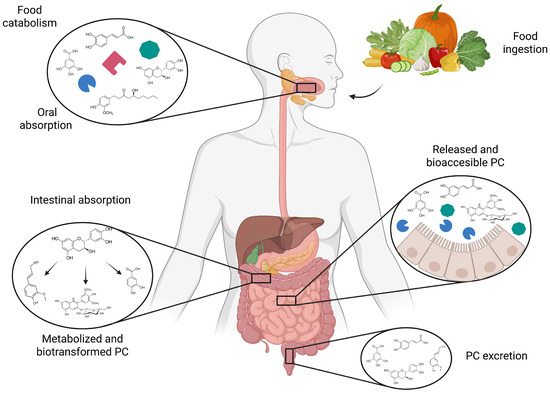
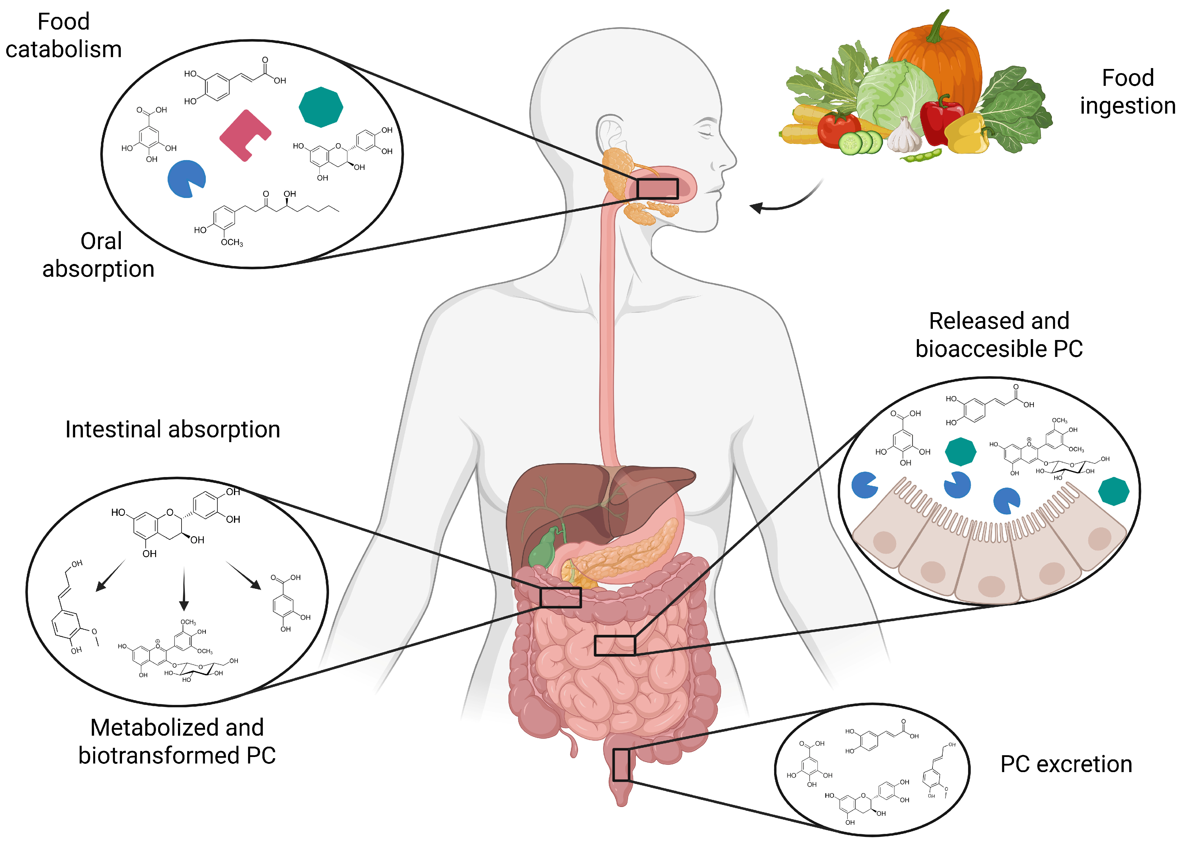
Unconjugated PCs can be biotransformed by the host’s enzymatic activity during their journey through the gastrointestinal tract. Intestinal catechol-O-methyltransferase (COMT; EC 2.1.1.6), sulfotransferase (SULT; EC 2.8.2.1), and UDP-glucuronosyltransferase (UGT; EC 2.4.1.17) are some of the main enzymes that produce BTPCs [33,34]. For example, Han et al. (2021) [35] used an in vivo male rat model where anthocyanins were metabolized in the intestine and transformed, mainly into anthocyanidins and phenolic acids, which favors their absorption through passive diffusion; moreover, this metabolism also converts flavonoids into isoflavonoid glucosides. Shi et al. (2016) [36] showed that flavonoid hydrolysis by action of lactase–phloridzin hydrolase (E.C. 3.2.1.108 for lactase activity, and E.C. 3.2.1.62 for glycosylceramidase activity) produces mainly two compounds, calycosin-3′-glucuronide and calycosin-7-O-β-d-glucoside; these metabolites are transported by the sodium–glucose cotransporter 1 (SGLT1) in an in vitro Caco-2 digestion model. Hydrophilic BAPCs, such as chicoric acid, can be transported in Caco-2 monolayers by P-glycoprotein (P-gp, ABCB1) and organic anion transport polypeptide 2B1 (OATP2B1) [37]. It has been demonstrated that hydrophobic BAPCs can pass the cellular membranes by active transport through ABC transporters. For example, studies performed in the small intestine of mice showed the participation of multidrug resistance protein 3 (MRP3, ABCC3) in the transport of resveratrol and its derivatives from the apical to the basolateral side [38,39]. Nonetheless, BAPCs may follow different routes during the digestive process that will depend on the chemical properties of the molecules and their interactions with receptors and transporters found throughout the digestive system (Figure 2). BAPCs can also be metabolized by gut microbiota, absorbed in their native form, or enzymatically biotransformed by enterocytes. Once BAPCs are absorbed, they reach the cytoplasmatic space, where they can be used to fulfill the physiological needs of the enterocyte (e.g., redox processes, gene expression, cell division modulation) or be transported into the bloodstream, where they are now known as BPCs; these travel through the circulatory system and reach different tissues and cells to perform various biological activities [40]. Once the BPCs reaches the liver, they may undergo chemical modifications by action of cellular metabolism producing BTPCs; these are then returned to the bloodstream and interact with carrier proteins to travel to other organs, including the brain [41], as discussed in the following sections.
Figure 2.
Phenolic compounds in the gastrointestinal tract: digestion, uptake, metabolism, and excretion.
3. Transport of Bioavailable and Biotransformed Phenolic Compounds Through the Bloodstream
The bloodstream delivers BPCs and BTPCs to different organs, but BAPCs can also suffer various chemical modifications during this process that alter their pharmacokinetic and pharmacodynamic properties [42]. The liver is particularly responsible for these modifications, mainly by the activity of three enzymes that add methyl, glucuronate, and sulfate groups to BPCs. The first type of chemical modification is mediated by COMT, which is expressed in a variety of tissues and organs and catalyzes the incorporation of a methyl group into BPCs and various other compounds [43]. Computational studies made by Cao et al. (2014) [44] showed that COMT preferred meta and para positions of the phenyl ring to attach methyl groups, as permitted by the charge distribution in its oxygen atoms. This chemical modification changes the physical and chemical properties of the molecule, mainly by increasing its hydrophobicity. Hence, BTPCs may increase their affinity for lipid structures like those of the phospholipid membrane, where specific regions of lipid rafts can interact with different BTPCs. In this sense, it has been shown that monounsaturated and polyunsaturated phosphatidylcholine-enriched microdomains of cellular membranes favor the uptake of hydrophobic PCs, BPCs, and BTPCs [45]. In vitro experiments carried out in SW480, SW620, and HT29 colon cancer cell lines showed that methylated derivatives of 3H-resveratrol (30 μmol/L) and 3H-trans-resveratrol (30 μmol/L) were time- and concentration-dependent, and both were internalized faster (1 min after exposure) than the unmodified PCs (6 min after exposure) through lipid raft-mediated endocytosis [46]. Interestingly, the authors showed that PCs remained inside the cells less than 48 h, suggesting a two-day window to be metabolized.
The functional groups attached to the BTPCs will determine the type of interactions they can form with different biomolecules; for example, the presence of glucuronate groups may favor their transport and internalization due to intermolecular interactions and mimetic processes, respectively. In mammals, glucuronate groups are incorporated by UGT, which is expressed predominantly in the liver, kidney, and intestines [47,48]. UGT catalyzes the covalent attachment of a glucuronate molecule onto BPCs, particularly on the hydroxyl or alkoxyl groups found in meta and ortho positions of the C3 of the A ring of 7-hydroxycoumarins [49]. This modification increases the molecule’s hydrophilicity [50], and may facilitate its cellular absorption by glucose transporters.
Sulfate groups play an important role in cellular physiology due to their conversion to sulfur-based groups (like thiol, sulfoxides, disulfides and sulfones) and their ability to interact with the host’s biomolecules [51]. In mammals, SULT is expressed in the gastrointestinal tract and liver [52,53], where it catalyzes the incorporation of a sulfate group into BPCs [54]. In rat liver, SULT modifies resveratrol to produce different molecules, including resveratrol-3-O-4’-O-disulfate and resveratrol-3-O-sulfate as major metabolites [55]. Although the expression of SULT isoforms may differ between tissues, their activity and the resulting products are similar; for instance, experiments with mice liver showed that these resveratrol derivatives are mainly produced by the SULT1A1, SULT1A2, SULT1A3, and SULT1E1 isoforms [56].
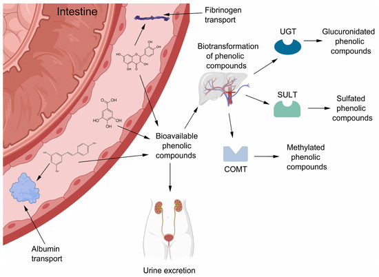
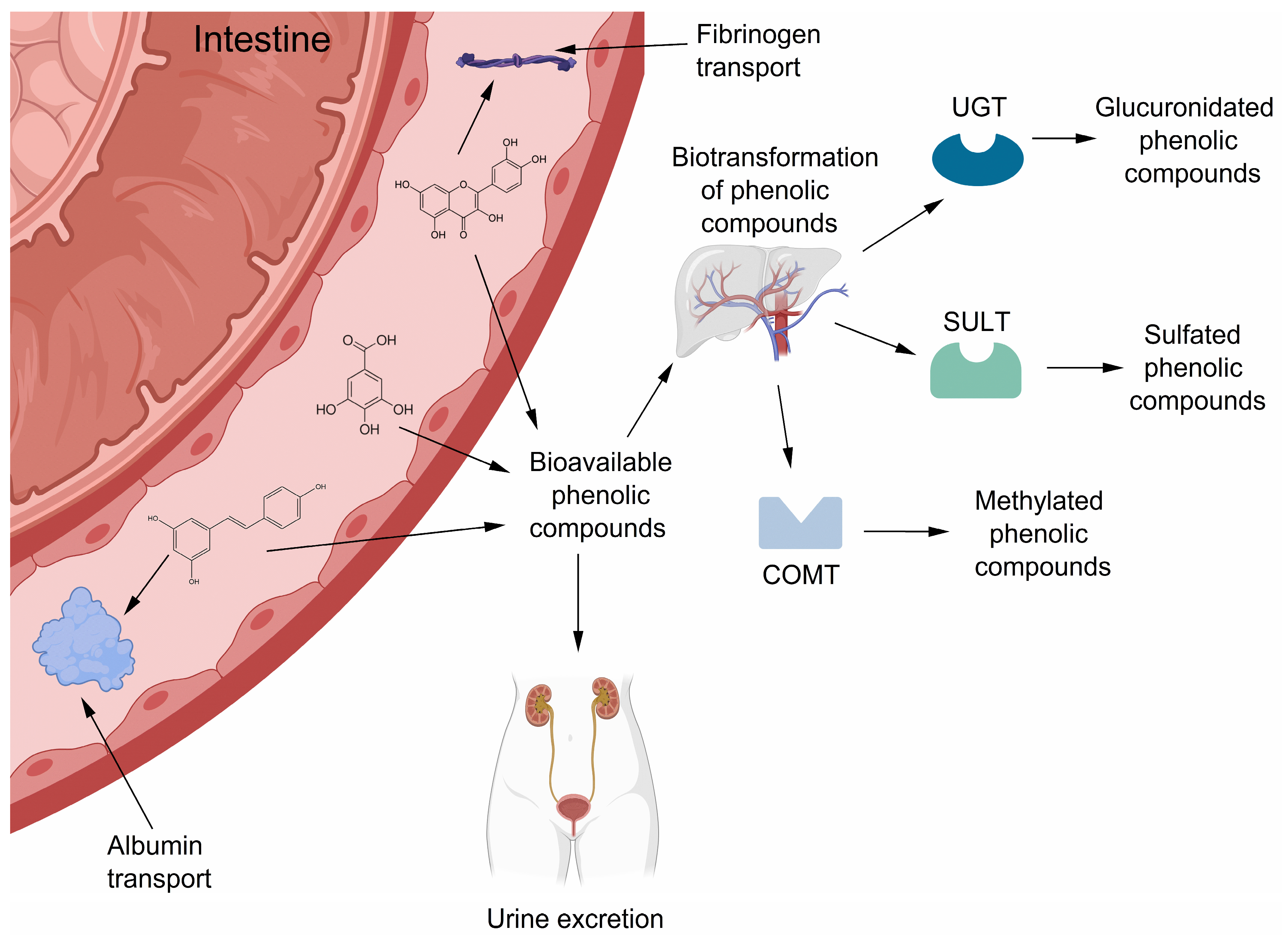
A fraction of circulating BPCs are biotransformed; those that are not absorbed or excreted may stay in the bloodstream and be potentially transported to other organs. In the bloodstream, some BTPCs (e.g., sulfated resveratrol derivatives) can interact with blood proteins, including albumin and fibrinogen [39,57]. The interactions formed between BTPCs and globulins or fibrinogen have been shown to be related to BTPC transport, through non-covalent interactions on specific sites of the proteins. According to in silico and in vitro studies made by Kim et al. (2021) [58,59], BTPCs interact with specific amino acid residues through intermolecular hydrophobic, thiol, and hydrogen interactions. These allow the formation of protein complexes for the transport and delivery of BTPCs and BPCs, mainly with albumin and fibrinogen. Figure 3 shows the journey of PCs from the gut to the bloodstream and the participation of the liver in their biotransformation.
Figure 3.
Simplified representation of PC transport through the bloodstream by serum proteins, and their hepatic biotransformation.
3.1. Albumin-Mediated PC Transport
Albumin is the most abundant blood protein in mammals; it is encoded by the ALB gene, and participates in many physiological processes, such as pH homeostasis, oncotic pressure, and solute and pharmacological transport [60]. The protein is composed of different domains with specific and non-specific binding sites that allow interactions with diverse types of molecules, mainly through non-covalent intermolecular interactions [61]. Several studies demonstrated that the albumin–BPC and albumin–BTPC complexes show specific interactions that are attributed to their chemical structures; for instance, flavonoids, phenolic acids, stilbenes, and hydrolyzable tannins that suffer chemical modifications by liver enzymes (e.g., COMT, SULT) may increase or decrease their affinity to albumin binding sites [62]. Xiao et al. (2011) [63] studied the binding constants of PCs with human serum albumin, finding that the binding affinity between hydroxylated flavonoids and hydrophilic amino acidic residues of albumin was increased when the C7 and C6 positions of the A ring were hydroxylated. The prosthetic group of albumin interacts through its Leu430 residue with free hydroxyl moieties of BPCs and BTPCs through hydrogen bonds [60]. The hydroxyl group in C3 of quercetin interacts with a Lys195 residue in the pocket of the IIA subdomain of albumin through hydrogen bonding [62,64,65], although some flavonoids can interact with albumin through bonds other than hydrogen bonds, including hydrophobic interactions. For example, in vitro assays showed an average binding constant of logK = 4.5–4.7 between quercetin, isorhamnetin, tamarixetin, quercetin-3’-sulfate, quercetin-3-glucuronide, and isorhamnetin-3-glucuronide with human serum albumin [66]. Altogether, this indicates that these compounds possess a high affinity and potentially strong interactions with the prosthetic group and subdomains of albumin, and are potentially mediated by chemical modifications that BPCs suffer during their blood transport to other organs.
The chemical structure of BPCs and BTPCs determines the types of interactions they may form with albumin, while several factors are also involved in the formation of these complexes. For example, double bonds in the structure and chemical modifications with monosaccharides, methyl moieties, and sulfate groups change the polarity of the BTPCs and BPCs, increasing or decreasing their chemical interactions with specific and non-specific sites of albumin domains. The number of hydroxyl groups and their position in the benzene ring of BPCs and BTPCs will determine their attraction force with albumin [67]. Fibrinogen is another serum protein with significant physiological relevance, and which has been recently associated with possible PC transport due to the molecular interactions that occur between its amino acidic residues and the hydroxyl groups of phenolics, as described in the following section.
3.2. Fibrinogen Interactions with PCs
Fibrinogen is a blood protein encoded by 3 genes (FGA, FGB, and FGG), and is considered an acute-phase protein involved in vascular processes and others pertaining to blood coagulation [68]. It also participates in neuroinflammation processes through receptor-mediated immune response to interleukins secreted during brain cell damage, and microglia or astrocyte reactivity [69,70]. In this sense, fibrinogen was recently shown to bind some PCs from natural extracts; for instance, Marchelak et al. (2021) [71] showed in vitro interactions between kaempferol, avicularin, juglanin, p-coumaric acid, kaempferitrin, proanthocyanidin A2, chlorogenic acid, miquelianin, protocatechuic acid, and dihydrocaffeic acid with human fibrinogen. This work evaluated the protective effect of these PCs against oxidative conditions that normally affect fibrinogen in the human body; SDS-PAGE analyses demonstrated a reduction in oxidated Aα chains, showing that the redox potential of these compounds may have a stronger role in the blood because PCs diminish the oxidation rate of the protein and, consequently, their removal from the bloodstream.
Intermolecular interactions between BPCs and BTPCs with fibrinogen facilitate PC transport to different organs and tissues, due to the strength and stability of their bonding [59]. In vitro and in silico studies performed by Shafreen et al. (2020) [72] using low-fermented lager beers showed the interaction between beer PCs with different human serum proteins. Most notably, molecular docking simulations showed that ferulic and caffeic acid interact with Cys19, Thr21, Thr22, and Tyr22 on the E site of fibrinogen by Van der Waals forces and hydrogen bonding. These computational simulations suggest that some potential transport pathways may be mediated by serum proteins, fibrinogen in particular. The interactions of PCs with albumin and fibrinogen have been studied in silico; the proteins and specific carbons and hydroxyl groups of the PC interaction sites are shown in Table 1.
Table 1.
In silico interactions between phenolic compounds and serum proteins.
The interactions between fibrinogen and BPCs influence the chemical characteristics of both molecules, increasing their solubility and interactions with other compounds. For example, Gligorijević et al. (2020) [76] tested the antioxidant and binding properties of a complex formed by human fibrinogen with resveratrol, where quenching analyses showed that resveratrol does not change some chemical properties of fibrinogen, such as thermal stability. However, ethanol precipitation (13%) showed that fibrinogen (9 µM) alters the solubility of resveratrol in aqueous solution by changing it from 0.57 to 1.13 mM when the fibrinogen–resveratrol complex is formed, showing changes in polarity and an increased antioxidant activity. This may potentially facilitate the transport and delivery of BPCs and BTPCs through the bloodstream.
Albumin and fibrinogen cross tissue membranes and can even be found in cerebrospinal fluid; for example, fibrinogen can be used as a clinical marker to identify brain inflammation and endothelial permeability in traumatic injury and neurodegenerative diseases [77]. However, serum proteins can cross the BBB through transcytosis processes under any physiological and pathological conditions. This idea was tested using PCs12 cells from rat pheochromocytoma to study the cationic form of albumin crossing the BBB, showing that changing the surface charge of the protein produced metabolites that could be easily transported through cell membranes, as compared to transport in its native configuration [78]. The reported in vitro and in silico interactions of PCs, BPCs, and BTPCs with albumin and fibrinogen may explain why some of these compounds can or cannot cross the BBB, independently of a transporter or endocytic mechanisms.
During the journey of BPCs and BTPCs through the bloodstream, several cellular types can absorb and utilize them, while those that are not absorbed or used will reach the kidney, where they can be reabsorbed by tubular cells or excreted via urine. For example, Glaeser et al. (2014) [79] studied the participation of OATP1A2, OATP2B1, and OCT1 in specific quercetin uptake in HEK293 cell lines, showing that the inhibition of these transporters modulated the uptake of quercetin. According to this, the participation of these transporters may be a central factor in the reabsorption of BPCs and BTPCs, ensuring a new journey through the bloodstream where cells that need these compounds can internalize them.
4. Transport of BPCs and BTPCs Through the Blood–Brain Barrier (BBB)
The delivery of BPCs and BTPCs to the brain is an interesting and complex process, due to the “high impermeability dogma” of the BBB to diverse molecules and xenobiotics that mammals consume in their diet [80]. The neurovascular unit oversees the passage of these molecules from the blood to the brain. This cellular niche is made of brain endothelial cells, pericytes, neurons, astrocyte endfeet, and smooth muscle cells [81]. Neurovascular unit cells also have a highly complex communication network that leads to decision-making for the uptake of nutrients and molecules from the bloodstream; this communication network also participates in the efflux of non-essential molecules [80,82,83]. Modern studies of nutrient transport across the BBB have led to questions about how natural compounds like PCs could reach brain cells; some of them have focused on phenolic transport to understand why these compounds are taken by brain cells to produce important biological effects that can help prevent and treat diseases of the CNS.
The potential of some phenolics to cross the BBB has been clearly demonstrated mainly by in vitro BBB models and the quantification of PCs in brain parenchyma; for example, caffeic acid has been detected in human cerebrospinal fluid. Since this compound is of dietary origin and not synthesized endogenously, it is clear that at least some compounds are indeed able to cross it [84]. However, this is not the case for all compounds and some of these are apparently not able to cross this barrier. Therefore, this cannot be generalized to all the PCs (over 8000 PCs) since the their transport is dependent on multiple variables, such as its molecular weight, polarity, metabolism, and other factors [41]. This highlights the necessity of further research to clarify the complex transport of dietary compounds, particularly into the brain. For this reason, multiple PC transport mechanisms are discussed nowadays to explain how these compounds are distributed systematically. The scientific consensus indicates that the passage of BPCs and BTPCs through the BBB may follow two main pathways. First, free BPCs and BTPCs are absorbed and transported into the brain by the brain endothelial cells through membrane transporters or endocytosis. The second is mediated by facilitated transport due to interactions of BPCs and BTPCs with serum proteins, including albumin and fibrinogen. It should also be mentioned that these molecules can be taken up by other, less-used transport mechanisms, such as transcellular and passive diffusion, which are being studied to understand the role of the physical and chemical properties of PCs.
Absorption processes were studied by Simon et al. (2020) [85] using a parallel artificial membrane permeability assay to analyze the crossing of ginger constituents through an in vitro BBB model. Particularly, 1 mM solutions of [6]-gingerol, [8]-gingerol, [10]-gingerol, [6]-shogaol, [10]-shogaol, 1-dehydro-[6]-gingerdione, and 1-dehydro-[10]-gingerdione were used to show that PC lipophilicity has a significant impact on their absorption by the vascular endothelial cells that form the BBB. According to these analyses, the high lipophilicity of these seven PCs resulted in a >95% average retention in the membranes of the BBB model, indicating that only a very low fraction of these lipophilic PCs cross them. HPLC-DAD quantification showed that approximately 2 µg of [6]-gingerol crosses the BBB model, consistent with in vivo and in silico data that showed [6]-gingerol cross cellular membranes by passive diffusion. This suggests that some BPCs and BTPCs may cross the BBB without being bound to a protein or by the action of membrane transporters; however, their concentration is key in the specific absorption pathway used, since lower concentrations could not be easily transported into the brain.
The metabolic activity of the vascular endothelial cells that form the BBB plays an important role in metabolite absorption. Brain endothelial cells express enzymes like COMT, UGT, and methyltransferases that produce chemical modifications by the addition of different functional groups [86,87]. For example, Faria et al. (2011) [88] showed that catechin and epicatechin are chemically modified by UGT enzymes of the BBB; these compounds were found in their glucuronide form on the apical side. This potentially facilitates the entry of BTPCs into the neurovascular unit and, therefore, the brain. Non-metabolized BPCs and BTPCs are recognized not only by membrane transporters and proteins of vascular endothelial cells, but also by their intracellular proteins that may help to deliver them from the basolateral side to the brain parenchyma [41,89].
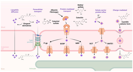
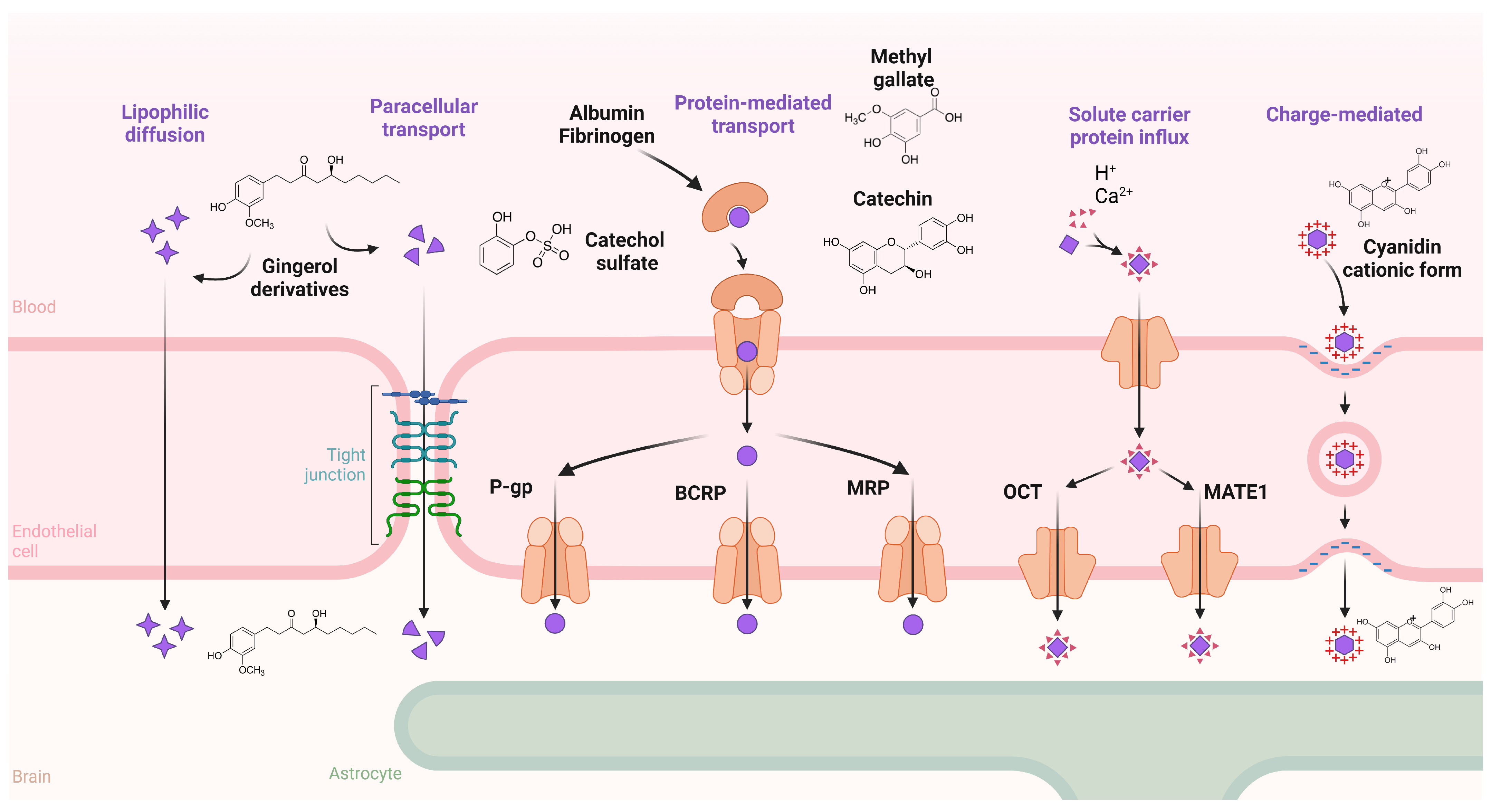
The location of transporters and membrane proteins also plays a critical role in metabolite influx and efflux. One of the major transporters in charge of metabolite flux in cells is the ABC transporter superfamily, which is widely expressed in several tissues and cell types. For instance, transporters expressed on the apical side are used to return metabolites to the extracellular space [90], and potentially used as an elimination mechanism. Meanwhile, transporters expressed on the basolateral side are used to deliver intracellular metabolites into the bloodstream and maintain homeostasis [91]. Some of the ABC transporters that are related to BPCs and BTPCs are described in the following sections, while Figure 4 depicts the mechanisms for PC, BPC, and BTPC transport through the BBB.
Figure 4.
Mechanisms used by brain endothelial cells of the blood–brain barrier to uptake phenolic compounds from the bloodstream according to their chemical properties (polarity), chemical structure, intermolecular interactions with serum proteins, and ion-carried fluxes. Active transport and ion-carried efflux of PCs from the intracellular space to the brain parenchyma. P-glycoprotein (P-gp); breast cancer resistance protein (BCRP); multidrug resistance protein (MRP); organic cation transporter (OCT); multidrug and toxic extrusion protein 1 (MATE1).
5. ABC Transporter Superfamily
Active transport is an essential mechanism to up- and downregulate the concentration of various metabolites in the intracellular and extracellular space. For instance, the ATP binding cassette transporter (ABC) superfamily includes 48 genes that encode for 48 proteins, 44 of which are located in the plasmatic membrane [92]. These proteins are widely expressed in different mammalian organs, tissues, and cells [83], including in neurons, glia, and vascular endothelial cells (Table 2) of the neurovascular unit in the central and peripheral nervous system, where they serve several physiological mechanisms [93,94]. The particular expression pattern of ABC transporters in the BBB tends to be species-specific; for example, the relative expression of BCRP in the BBB is reported to be higher in humans than in rodents [95,96]. In vitro and in silico studies performed by Figueira et al. (2017) [97] with human brain microvascular endothelial cells showed that BCRP, MRP, and P-gp are involved in the transport of 5–10 µM of 4-O-methylgallic acid-3-O-sulfate, 4-methylgallic acid, 4-methylcatechol-O-sulfate, catechol-O-sulfate, 1-O-methylpyrogallol-O-sulfate, and 2-O-methylpyrogallol-1-O-sulfate, showing that BTPCs with sulfate, methyl, and glucuronide groups are metabolized in the brain endothelial cell membranes, and then transported to the apical side of the cell monolayer. This result indicates that ABC transporters may play an alternative role in the transport of BTPCs, at least in vitro, to transport PCs to the vascular lumen into the brain parenchyma.
Table 2.
Transporters that are expressed in the human brain and that interact with PCs to promote their uptake.
As previously mentioned, the transport of BTPCs is influenced by their physicochemical properties, which also applies to ABC transporters. For example, Faria et al. (2011) [88] studied the transport of catechin and epicatechin in rat capillary cerebral endothelial cells (RBE4) and in immortalized human cerebral microvessel endothelial cells (hCMEC/D3). This study showed that 30 µM of epicatechin and catechin are transported across cellular models in a time-dependent manner. However, the percentage of transport of epicatechin was significantly higher (27.5%) than the transport of catechin (15.4%). According to this result, the transport efficiency in chemically similar molecules like catechin and epicatechin could be strongly determined by the stereochemical bonds between the central pyran ring, A or B phenolic rings with wedge-and-dashed or wedged catechol groups.
6. Solute Carrier (SLC) Proteins
The SLC superfamily transporters includes 55 genes that encode 362 proteins; this is a widely expressed family whose function is to transport and exchange several cellular metabolites, nutrients, drugs, and xenobiotics in all cells, playing a major role in the absorption, distribution, metabolism, and excretion (ADME) of these molecules [108]. They can be found in different cell types of the BBB, blood–cerebrospinal fluid barrier, and arachnoid barrier (Table 2), where the cells of these sites can uptake, excrete, and re-uptake several molecules from the bloodstream and cerebrospinal fluid, as well as passing them to the brain parenchyma to fulfill multiple biological activities [109]. Diverse transporters from the SLC family have recently gained interest due to their impact on metabolite transport, particularly for xenobiotics, drugs, and natural compounds that can be transported across the BBB, as discussed in the following sections.
6.1. Organic Cation Transporters (OCTs)
Organic cation transporters (OCTs) are a subfamily (SLC22A) of 23 genes that encode for 13 membrane proteins [110]. These are expressed in several cellular lineages of the liver, intestine, kidney, central and peripheral nervous system (Table 2). According to computational modeling studies of OCT structure, these transporters have 12 transmembrane domains and their N- and C-termini in the intracellular space [111]. OCTs have a huge impact on the physiology of mammals; they can be expressed as an inward and outward control to transport several molecules, and they can be found in the luminal and abluminal sides in the physical barriers of the organism. For instance, OCTs were reported to be widely expressed in the choroid plexus, the barrier between the bloodstream and cerebrospinal fluid, allowing the exchange of metabolites, nutrients, and substrates that are taken by endothelial cells and then transported to the brain [109,112].
The physical and chemical properties of the substrates that can be recognized by OCT may determine their transport across the cellular membranes in an ion-independent manner. Furthermore, it has been described that OCT configuration may change from outward- to inward-facing according to their affinity for the specific compound recognized in the binding pocket. For example, molecules that contain aromatic benzene rings (e.g., PCs) can be transported by the action of hydrophobic interactions with hydrophobic phenylalanine and tyrosine residues that are in the pore-forming domain of the OCT. Moreover, positively charged molecules with hydroxyl moieties form electrostatic and hydrogen bond interactions with hydroxyl or amino groups of polar amino acids, such as glutamine [113].
6.2. Multidrug and Toxic Extrusion Protein 1 (MATE1)
In humans, the MATE1 transporter is encoded by the SLC47A1 gene, which is translated into a 12-transmembrane helix protein [114] in the adrenal gland, cerebral cortex, liver, kidney, lungs, and testes [115,116]. These transporters are H+/organic cation antiporters used to transport cationic molecules across the cellular membrane [117], although they can also transport neutral compounds. For example, Lee et al. [118] showed that MATE1 transporters are involved in quercetin efflux and influx through the cellular membranes, mainly promoting the accumulation of this compound in the endoplasmic reticulum and peroxisomes of HEK293T and HepG2 cells. Furthermore, specific molecular interactions between PCs and MATE1 proteins were studied by Liu et al. (2024) [119] using computational modeling and accumulation assays in intracellular vacuoles. In the study, they identified the specific sites where catechin, epicatechin gallate, and epigallocatechin gallate interact with amino acid residues of the protein. For catechin, it interacts with Asn64, Ser68, Ala228, Gln94, and Gln166, and Ile88, while epicatechin gallate interacts with Ser68, Ile88, Gly92, Tyr98, Ala228, and Asn314, and epigallocatechin gallate interacts with Tyr65, Ser68, Gln94, Gly95, Ile286, Cys312, and Asn314. This suggests that MATE1 preferentially transports flavonoids with more hydroxyl groups on its structure, such as those with gallates.
7. Conclusions
Phenolic compounds play an essential role in mammalian cell homeostasis. These compounds are transported systemically (mainly by protein carriers like albumin and fibrinogen), reach the brain, and are then internalized by the neurovascular unit through several cell membrane proteins, endocytosis-like processes, and even cellular passive diffusion. These transport processes are influenced by the phenolic compounds’ chemical structure and their interactions with membrane receptors, lipids, and transporters. Further in vivo studies are needed to better understand the physiological effects of dietary phenolic compounds (~8000 compounds described so far) and their potential as promoters of human brain health.
Author Contributions
Conceptualization, writing, and revision: J.A.D.-A., G.A.G.-A., M.A.V.-O., J.F.A.-Z., A.M.P.-S., A.M.-M., F.J.P.-D., D.G.-V. and M.M.-H. All authors wrote the manuscript. All authors have read and agreed to the published version of the manuscript.
Funding
This research was funded by the Secretaría de Ciencia, Humanidades, Tecnología e Innovación (SECIHTI), CF-2023-I-905, CVU 1006147, CF-2023-I-672. And the APC was funded by the University of Sonora.
Data Availability Statement
No new data were created or analyzed in this study. Data sharing is not applicable to this article.
Conflicts of Interest
The authors declare no conflicts of interest.
Abbreviations
The following abbreviations are used in this manuscript:
| PCs | Phenolic Compounds |
| BAPCs | Bioaccessible phenolic compounds |
| BPCs | Bioavailable phenolic compounds |
| BTPCs | Biotransformed phenolic compounds |
| CNS | Central nervous system |
| BBB | Blood–brain barrier |
| GLUT | Glucose transporter |
| SGLT | Sodium–glucose cotransporter |
| P-gp | P-glycoprotein |
| OATP | Organic anion transport polypeptide |
| OCT | Organic cation transporter |
| MRP | Multidrug resistance protein |
| COMT | Catechol o-methyl transferase |
| UGT | UDP-glucuronosyltransferase |
| SULT | Sulfotransferase |
| MATE1 | Multidrug and toxic extrusion protein 1 |
References
- Khan, N. Decoding Phytohormone Signaling in Plant Stress Physiology: Insights, Challenges, and Future Directions. Environ. Exp. Bot. 2025, 231, 106099. [Google Scholar] [CrossRef] [Scilit]
- Saijo, Y.; Loo, E.P.-I. Plant Immunity in Signal Integration between Biotic and Abiotic Stress Responses. New Phytol. 2020, 225, 87–104. [Google Scholar] [CrossRef] [Scilit]
- Erb, M.; Kliebenstein, D.J. Plant Secondary Metabolites as Defenses, Regulators, and Primary Metabolites: The Blurred Functional Trichotomy. Plant Physiol. 2020, 184, 39–52. [Google Scholar] [CrossRef] [Scilit]
- Singh, P.; Arif, Y.; Bajguz, A.; Hayat, S. The Role of Quercetin in Plants. Plant Physiol. Biochem. PPB 2021, 166, 10–19. [Google Scholar] [CrossRef] [Scilit]
- Pannucci, E.; Spagnuolo, L.; De Gara, L.; Santi, L.; Dugo, L. Phenolic Compounds as Preventive and Therapeutic Agents in Diabetes-Related Oxidative Stress, Inflammation, Advanced Glycation End-Products Production and Insulin Sensitivity. Discov. Med. 2023, 35, 715–732. [Google Scholar] [CrossRef] [Scilit] [PubMed]
- Murai, T.; Matsuda, S. The Chemopreventive Effects of Chlorogenic Acids, Phenolic Compounds in Coffee, against Inflammation, Cancer, and Neurological Diseases. Molecules 2023, 28, 2381. [Google Scholar] [CrossRef] [Scilit] [PubMed]
- González, Y.; Mojica-Flores, R.; Moreno-Labrador, D.; Pecchio, M.; Rao, K.S.J.; Ahumedo-Monterrosa, M.; Fernández, P.L.; Larionov, O.V.; Lakey-Beitia, J. Tetrahydrocurcumin Derivatives Enhanced the Anti-Inflammatory Activity of Curcumin: Synthesis, Biological Evaluation, and Structure-Activity Relationship Analysis. Molecules 2023, 28, 7787. [Google Scholar] [CrossRef] [Scilit]
- Hossain, S.I.; Saha, S.C.; Deplazes, E. Phenolic Compounds Alter the Ion Permeability of Phospholipid Bilayers via Specific Lipid Interactions. Phys. Chem. Chem. Phys. 2021, 23, 22352–22366. [Google Scholar] [CrossRef] [Scilit]
- Shahidi, F.; Dissanayaka, C.S. Phenolic-Protein Interactions: Insight from in-Silico Analyses—A Review. Food Prod. Process. Nutr. 2023, 5, 2. [Google Scholar] [CrossRef] [Scilit]
- Taylor, S.R.; Ramsamooj, S.; Liang, R.J.; Katti, A.; Pozovskiy, R.; Vasan, N.; Hwang, S.-K.; Nahiyaan, N.; Francoeur, N.J.; Schatoff, E.M.; et al. Dietary Fructose Improves Intestinal Cell Survival and Nutrient Absorption. Nature 2021, 597, 263–267. [Google Scholar] [CrossRef] [Scilit] [PubMed]
- Alhajj, M.; Babos, M. Physiology, Salivation. In StatPearls; StatPearls Publishing: Treasure Island, FL, USA, 2025. Available online: https://www.ncbi.nlm.nih.gov/books/NBK542251/ (accessed on 20 January 2026).
- Brignot, H.; Feron, G. Oral Lipolysis and Its Association with Diet and the Perception and Digestion of Lipids: A Systematic Literature Review. Arch. Oral Biol. 2019, 108, 104550. [Google Scholar] [CrossRef] [Scilit] [PubMed]
- Heda, R.; Toro, F.; Tombazzi, C.R. Physiology, Pepsin. In StatPearls; StatPearls Publishing: Treasure Island, FL, USA, 2025. Available online: https://www.ncbi.nlm.nih.gov/books/NBK537005/ (accessed on 20 January 2026).
- Braune, A.; Blaut, M. Bacterial Species Involved in the Conversion of Dietary Flavonoids in the Human Gut. Gut Microbes 2016, 7, 216–234. [Google Scholar] [CrossRef] [Scilit] [PubMed]
- Zhang, P.; Zhang, R.; Sirisena, S.; Gan, R.; Fang, Z. Beta-Glucosidase Activity of Wine Yeasts and Its Impacts on Wine Volatiles and Phenolics: A Mini-Review. Food Microbiol. 2021, 100, 103859. [Google Scholar] [CrossRef] [Scilit]
- Tartaglia, A.; Romasco, T.; D’Ovidio, C.; Rosato, E.; Ulusoy, H.I.; Furton, K.G.; Kabir, A.; Locatelli, M. Determination of Phenolic Compounds in Human Saliva after Oral Administration of Red Wine by High Performance Liquid Chromatography. J. Pharm. Biomed. Anal. 2022, 209, 114486. [Google Scholar] [CrossRef] [Scilit]
- Ferreira, A.S.; Silva, A.M.; Macedo, C.; Laveriano-Santos, E.P.; Lozano-Castellón, J.; Lamuela-Raventós, R.M.; Švarc-Gajíc, J.; Costa, P.C.; Delerue-Matos, C.; Rodrigues, F. Orodispersible Films Containing Chestnut Shell Phenolics for Buccal Delivery: A Preclinical Approach for Oral Mucositis Prevention. Front. Med. Technol. 2025, 7, 1675082. [Google Scholar] [CrossRef] [Scilit]
- Guo, Y.; Li, Z.; Chen, F.; Chai, Y. Polyphenols in Oral Health: Homeostasis Maintenance, Disease Prevention, and Therapeutic Applications. Nutrients 2023, 15, 4384. [Google Scholar] [CrossRef] [Scilit]
- Guerreiro, C.; Brandão, E.; de Jesus, M.; Gonçalves, L.; Pérez-Gregório, R.; Mateus, N.; de Freitas, V.; Soares, S. New Insights into the Oral Interactions of Different Families of Phenolic Compounds: Deepening the Astringency Mouthfeels. Food Chem. 2022, 375, 131642. [Google Scholar] [CrossRef] [Scilit]
- Greenwood-Van Meerveld, B.; Johnson, A.C.; Grundy, D. Gastrointestinal Physiology and Function. Handb. Exp. Pharmacol. 2017, 239, 1–16. [Google Scholar] [CrossRef] [Scilit]
- Boland, M. Human Digestion—A Processing Perspective. J. Sci. Food Agric. 2016, 96, 2275–2283. [Google Scholar] [CrossRef] [Scilit] [PubMed]
- Rocchetti, G.; Gregorio, R.P.; Lorenzo, J.M.; Barba, F.J.; Oliveira, P.G.; Prieto, M.A.; Simal-Gandara, J.; Mosele, J.I.; Motilva, M.-J.; Tomas, M.; et al. Functional Implications of Bound Phenolic Compounds and Phenolics-Food Interaction: A Review. Compr. Rev. Food Sci. Food Saf. 2022, 21, 811–842. [Google Scholar] [CrossRef] [Scilit]
- Takahashi, M.; Tahara, Y. Timing of Food/Nutrient Intake and Its Health Benefits. J. Nutr. Sci. Vitaminol. 2022, 68, S2–S4. [Google Scholar] [CrossRef] [Scilit] [PubMed]
- Yu, L.-E.; Yang, W.-C.; Liang, Y.-C. Crosstalk Within the Intestinal Epithelium: Aspects of Intestinal Absorption, Homeostasis, and Immunity. Biomedicines 2024, 12, 2771. [Google Scholar] [CrossRef] [Scilit]
- Mahdi, L.; Graziani, A.; Baffy, G.; Mitten, E.K.; Portincasa, P.; Khalil, M. Unlocking Polyphenol Efficacy: The Role of Gut Microbiota in Modulating Bioavailability and Health Effects. Nutrients 2025, 17, 2793. [Google Scholar] [CrossRef] [Scilit]
- Rossi, L.D.; Rocchetti, G.; Lucini, L.; Rebecchi, A. Antimicrobial Potential of Polyphenols: Mechanisms of Action and Microbial Responses—A Narrative Review. Antioxidants 2025, 14, 200. [Google Scholar] [CrossRef] [Scilit]
- Burgo-Gutiérrez, C.D.; Tosi, N.; Cid, C.; Rio, D.D.; Bresciani, L.; Ludwig, I.A.; Mena, P.; Peña, M.-P.D. Urinary Excretion Kinetics of (Poly)Phenolic Metabolites Derived from the Consumption of Microwaved Piquillo Pepper (Capsicum annuum Cv. Piquillo). Food Funct. 2025, 16, 5744–5756. [Google Scholar] [CrossRef] [Scilit] [PubMed]
- Kalt, W. Anthocyanins and Their C6-C3-C6 Metabolites in Humans and Animals. Molecules 2019, 24, 4024. [Google Scholar] [CrossRef] [Scilit]
- Scott, M.B.; Styring, A.K.; McCullagh, J.S.O. Polyphenols: Bioavailability, Microbiome Interactions and Cellular Effects on Health in Humans and Animals. Pathogens 2022, 11, 770. [Google Scholar] [CrossRef] [Scilit]
- Choy, Y.Y.; Quifer-Rada, P.; Holstege, D.M.; Frese, S.A.; Calvert, C.C.; Mills, D.A.; Lamuela-Raventos, R.M.; Waterhouse, A.L. Phenolic Metabolites and Substantial Microbiome Changes in Pig Feces by Ingesting Grape Seed Proanthocyanidins. Food Funct. 2014, 5, 2298–2308. [Google Scholar] [CrossRef] [Scilit]
- Czubinski, J.; Dwiecki, K. A Review of Methods Used for Investigation of Protein–Phenolic Compound Interactions. Int. J. Food Sci. Technol. 2016, 52, 573–585. [Google Scholar] [CrossRef] [Scilit]
- Han, F.; Oliveira, H.; Brás, N.F.; Fernandes, I.; Cruz, L.; De Freitas, V.; Mateus, N. In Vitro Gastrointestinal Absorption of Red Wine Anthocyanins—Impact of Structural Complexity and Phase II Metabolization. Food Chem. 2020, 317, 126398. [Google Scholar] [CrossRef] [Scilit]
- Domínguez-Avila, J.A.; Villa-Rodriguez, J.A.; Montiel-Herrera, M.; Pacheco-Ordaz, R.; Roopchand, D.E.; Venema, K.; González-Aguilar, G.A. Phenolic Compounds Promote Diversity of Gut Microbiota and Maintain Colonic Health. Dig. Dis. Sci. 2021, 66, 3270–3289. [Google Scholar] [CrossRef] [Scilit]
- Murota, K.; Nakamura, Y.; Uehara, M. Flavonoid Metabolism: The Interaction of Metabolites and Gut Microbiota. Biosci. Biotechnol. Biochem. 2018, 82, 600–610. [Google Scholar] [CrossRef] [Scilit]
- Han, H.; Liu, C.; Gao, W.; Li, Z.; Qin, G.; Qi, S.; Jiang, H.; Li, X.; Liu, M.; Yan, F.; et al. Anthocyanins Are Converted into Anthocyanidins and Phenolic Acids and Effectively Absorbed in the Jejunum and Ileum. J. Agric. Food Chem. 2021, 69, 992–1002. [Google Scholar] [CrossRef] [Scilit] [PubMed]
- Shi, J.; Zheng, H.; Yu, J.; Zhu, L.; Yan, T.; Wu, P.; Lu, L.; Wang, Y.; Hu, M.; Liu, Z. SGLT-1 Transport and Deglycosylation inside Intestinal Cells Are Key Steps in the Absorption and Disposition of Calycosin-7-O-β-D-Glucoside in Rats. Drug Metab. Dispos. 2016, 44, 283–296. [Google Scholar] [CrossRef] [Scilit] [PubMed]
- Wang, G.N.; Li, Y.P.; Yuan, S.K.; Zhang, H.; Ren, J.; Ren, X.; Liu, J.X. The Intestinal Absorption Mechanism of Chicoric Acid and Its Bioavailability Improvement with Chitosan. Heliyon 2022, 8, e09955. [Google Scholar] [CrossRef] [Scilit]
- Prakash, V.; Bose, C.; Sunilkumar, D.; Cherian, R.M.; Thomas, S.S.; Nair, B.G. Resveratrol as a Promising Nutraceutical: Implications in Gut Microbiota Modulation, Inflammatory Disorders, and Colorectal Cancer. Int. J. Mol. Sci. 2024, 25, 3370. [Google Scholar] [CrossRef] [Scilit]
- Springer, M.; Moco, S. Resveratrol and Its Human Metabolites-Effects on Metabolic Health and Obesity. Nutrients 2019, 11, 143. [Google Scholar] [CrossRef] [Scilit]
- Tufarelli, V.; Casalino, E.; D’ALessandro, A.G.; Laudadio, V. Dietary Phenolic Compounds: Biochemistry, Metabolism and Significance in Animal and Human Health. Curr. Drug Metab. 2017, 18, 905–913. [Google Scholar] [CrossRef] [Scilit]
- Velásquez-Jiménez, D.; Corella-Salazar, D.A.; Zuñiga-Martínez, B.S.; Domínguez-Avila, J.A.; Montiel-Herrera, M.; Salazar-López, N.J.; Rodrigo-Garcia, J.; Villegas-Ochoa, M.A.; González-Aguilar, G.A. Phenolic Compounds That Cross the Blood-Brain Barrier Exert Positive Health Effects as Central Nervous System Antioxidants. Food Funct. 2021, 12, 10356–10369. [Google Scholar] [CrossRef] [Scilit] [PubMed]
- Katada, S.; Watanabe, T.; Mizuno, T.; Kobayashi, S.; Takeshita, M.; Osaki, N.; Kobayashi, S.; Katsuragi, Y. Effects of Chlorogenic Acid-Enriched and Hydroxyhydroquinone-Reduced Coffee on Postprandial Fat Oxidation and Antioxidative Capacity in Healthy Men: A Randomized, Double-Blind, Placebo-Controlled, Crossover Trial. Nutrients 2018, 10, 525. [Google Scholar] [CrossRef] [Scilit]
- Poór, M.; Zrínyi, Z.; Kőszegi, T. Structure Related Effects of Flavonoid Aglycones on Cell Cycle Progression of HepG2 Cells: Metabolic Activation of Fisetin and Quercetin by Catechol-O-Methyltransferase (COMT). Biomed. Pharmacother. Biomedecine Pharmacother. 2016, 83, 998–1005. [Google Scholar] [CrossRef] [Scilit]
- Cao, Y.; Chen, Z.-J.; Jiang, H.-D.; Chen, J.-Z. Computational Studies of the Regioselectivities of COMT-Catalyzed Meta-/Para-O Methylations of Luteolin and Quercetin. J. Phys. Chem. B 2014, 118, 470–481. [Google Scholar] [CrossRef] [Scilit]
- Hazarosova, R.; Momchilova, A.; Vitkova, V.; Yordanova, V.; Kostadinova, A.; Angelova, M.I.; Tessier, C.; Nuss, P.; Staneva, G. Structural Changes Induced by Resveratrol in Monounsaturated and Polyunsaturated Phosphatidylcholine-Enriched Model Membranes. Membranes 2023, 13, 909. [Google Scholar] [CrossRef] [Scilit]
- Colin, D.; Limagne, E.; Jeanningros, S.; Jacquel, A.; Lizard, G.; Athias, A.; Gambert, P.; Hichami, A.; Latruffe, N.; Solary, E.; et al. Endocytosis of Resveratrol via Lipid Rafts and Activation of Downstream Signaling Pathways in Cancer Cells. Cancer Prev. Res. Phila. Pa 2011, 4, 1095–1106. [Google Scholar] [CrossRef] [Scilit]
- Rowland, A.; Miners, J.O.; Mackenzie, P.I. The UDP-Glucuronosyltransferases: Their Role in Drug Metabolism and Detoxification. Int. J. Biochem. Cell Biol. 2013, 45, 1121–1132. [Google Scholar] [CrossRef] [Scilit]
- Hashiba, S.; Nakano, M.; Yokoseki, I.; Takahashi, E.; Kondo, M.; Jimbo, Y.; Ishiguro, N.; Arakawa, H.; Fukami, T.; Nakajima, M. Cytochrome P450 and UDP-Glucuronosyltransferase Expressions, Activities, and Induction Abilities in 3D-Cultured Human Renal Proximal Tubule Epithelial Cells. Drug Metab. Dispos. Biol. Fate Chem. 2024, 52, 949–956. [Google Scholar] [CrossRef] [Scilit]
- Juvonen, R.O.; Rauhamäki, S.; Kortet, S.; Niinivehmas, S.; Troberg, J.; Petsalo, A.; Huuskonen, J.; Raunio, H.; Finel, M.; Pentikäinen, O.T. Molecular Docking-Based Design and Development of a Highly Selective Probe Substrate for UDP-Glucuronosyltransferase 1A10. Mol. Pharm. 2018, 15, 923–933. [Google Scholar] [CrossRef] [Scilit] [PubMed]
- Wu, B.; Kulkarni, K.; Basu, S.; Zhang, S.; Hu, M. First-Pass Metabolism via UDP-Glucuronosyltransferase: A Barrier to Oral Bioavailability of Phenolics. J. Pharm. Sci. 2011, 100, 3655–3681. [Google Scholar] [CrossRef] [Scilit] [PubMed]
- Hill, C.R.; Shafaei, A.; Balmer, L.; Lewis, J.R.; Hodgson, J.M.; Millar, A.H.; Blekkenhorst, L.C. Sulfur Compounds: From Plants to Humans and Their Role in Chronic Disease Prevention. Crit. Rev. Food Sci. Nutr. 2023, 63, 8616–8638. [Google Scholar] [CrossRef] [Scilit] [PubMed]
- Chen, J.; Zheng, H.; Zeng, S.; Xie, C.; Li, X.; Yan, T.; Gong, X.; Lu, L.; Qi, X.; Wang, Y.; et al. Profiles and Gender-Specifics of UDP-Glucuronosyltransferases and Sulfotransferases Expressions in the Major Metabolic Organs of Wild-Type and Efflux Transporter Knockout FVB Mice. Mol. Pharm. 2017, 14, 2967–2976. [Google Scholar] [CrossRef] [Scilit]
- Fritz, A.; Busch, D.; Lapczuk, J.; Ostrowski, M.; Drozdzik, M.; Oswald, S. Expression of Clinically Relevant Drug-Metabolizing Enzymes along the Human Intestine and Their Correlation to Drug Transporters and Nuclear Receptors: An Intra-Subject Analysis. Basic Clin. Pharmacol. Toxicol. 2019, 124, 245–255. [Google Scholar] [CrossRef] [Scilit]
- Feliciano, R.P.; Istas, G.; Heiss, C.; Rodriguez-Mateos, A. Plasma and Urinary Phenolic Profiles after Acute and Repetitive Intake of Wild Blueberry. Molecules 2016, 21, 1120. [Google Scholar] [CrossRef] [Scilit]
- Maier-Salamon, A.; Hagenauer, B.; Reznicek, G.; Szekeres, T.; Thalhammer, T.; Jäger, W. Metabolism and Disposition of Resveratrol in the Isolated Perfused Rat Liver: Role of Mrp2 in the Biliary Excretion of Glucuronides. J. Pharm. Sci. 2008, 97, 1615–1628. [Google Scholar] [CrossRef] [Scilit]
- Böhmdorfer, M.; Szakmary, A.; Schiestl, R.H.; Vaquero, J.; Riha, J.; Brenner, S.; Thalhammer, T.; Szekeres, T.; Jäger, W. Involvement of UDP-Glucuronosyltransferases and Sulfotransferases in the Excretion and Tissue Distribution of Resveratrol in Mice. Nutrients 2017, 9, 1347. [Google Scholar] [CrossRef] [Scilit] [PubMed]
- Barber, T.M.; Kabisch, S.; Randeva, H.S.; Pfeiffer, A.F.H.; Weickert, M.O. Implications of Resveratrol in Obesity and Insulin Resistance: A State-of-the-Art Review. Nutrients 2022, 14, 2870. [Google Scholar] [CrossRef] [Scilit] [PubMed]
- Kim, Y.M.; Park, Y.S.; Park, Y.-K.; Ham, K.-S.; Kang, S.-G.; Shafreen, R.M.B.; Lakshmi, S.A.; Gorinstein, S. Characterization of Bioactive Ligands with Antioxidant Properties of Kiwifruit and Persimmon Cultivars Using In Vitro and in Silico Studies. Appl. Sci. 2020, 10, 4218. [Google Scholar] [CrossRef] [Scilit]
- Kim, Y.-M.; Abas, F.; Park, Y.-S.; Park, Y.-K.; Ham, K.-S.; Kang, S.-G.; Lubinska-Szczygeł, M.; Ezra, A.; Gorinstein, S. Bioactivities of Phenolic Compounds from Kiwifruit and Persimmon. Molecules 2021, 26, 4405. [Google Scholar] [CrossRef] [Scilit]
- De Simone, G.; di Masi, A.; Ascenzi, P. Serum Albumin: A Multifaced Enzyme. Int. J. Mol. Sci. 2021, 22, 10086. [Google Scholar] [CrossRef] [Scilit]
- di Masi, A.; Trezza, V.; Leboffe, L.; Ascenzi, P. Human Plasma Lipocalins and Serum Albumin: Plasma Alternative Carriers? J. Control. Release Off. J. Control. Release Soc. 2016, 228, 191–205. [Google Scholar] [CrossRef] [Scilit]
- López-Yerena, A.; Perez, M.; Vallverdú-Queralt, A.; Escribano-Ferrer, E. Insights into the Binding of Dietary Phenolic Compounds to Human Serum Albumin and Food-Drug Interactions. Pharmaceutics 2020, 12, 1123. [Google Scholar] [CrossRef] [Scilit]
- Xiao, J.; Zhao, Y.; Wang, H.; Yuan, Y.; Yang, F.; Zhang, C.; Yamamoto, K. Noncovalent Interaction of Dietary Polyphenols with Common Human Plasma Proteins. J. Agric. Food Chem. 2011, 59, 10747–10754. [Google Scholar] [CrossRef] [Scilit]
- Rimac, H.; Tandarić, T.; Vianello, R.; Bojić, M. Indomethacin Increases Quercetin Affinity for Human Serum Albumin: A Combined Experimental and Computational Study and Its Broader Implications. Int. J. Mol. Sci. 2020, 21, 5740. [Google Scholar] [CrossRef] [Scilit]
- Zsila, F.; Bikádi, Z.; Simonyi, M. Probing the Binding of the Flavonoid, Quercetin to Human Serum Albumin by Circular Dichroism, Electronic Absorption Spectroscopy and Molecular Modelling Methods. Biochem. Pharmacol. 2003, 65, 447–456. [Google Scholar] [CrossRef] [Scilit]
- Poór, M.; Boda, G.; Needs, P.W.; Kroon, P.A.; Lemli, B.; Bencsik, T. Interaction of Quercetin and Its Metabolites with Warfarin: Displacement of Warfarin from Serum Albumin and Inhibition of CYP2C9 Enzyme. Biomed. Pharmacother. 2017, 88, 574–581. [Google Scholar] [CrossRef] [Scilit] [PubMed]
- Zhang, Y.; Wu, S.; Qin, Y.; Liu, J.; Liu, J.; Wang, Q.; Ren, F.; Zhang, H. Interaction of Phenolic Acids and Their Derivatives with Human Serum Albumin: Structure-Affinity Relationships and Effects on Antioxidant Activity. Food Chem. 2018, 240, 1072–1080. [Google Scholar] [CrossRef] [Scilit]
- Stang, L.J.; Mitchell, L.G. Fibrinogen. In Methods in Molecular Biology; Springer: Clifton, NJ, USA, 2013; Volume 992, pp. 181–192. [Google Scholar] [CrossRef] [Scilit]
- Merlini, M.; Rafalski, V.A.; Rios Coronado, P.E.; Gill, T.M.; Ellisman, M.; Muthukumar, G.; Subramanian, K.S.; Ryu, J.K.; Syme, C.A.; Davalos, D.; et al. Fibrinogen Induces Microglia-Mediated Spine Elimination and Cognitive Impairment in an Alzheimer’s Disease Model. Neuron 2019, 101, 1099–1108.e6. [Google Scholar] [CrossRef] [Scilit] [PubMed]
- Sulimai, N.; Lominadze, D. Fibrinogen and Neuroinflammation During Traumatic Brain Injury. Mol. Neurobiol. 2020, 57, 4692–4703. [Google Scholar] [CrossRef] [Scilit] [PubMed]
- Marchelak, A.; Kolodziejczyk-Czepas, J.; Wasielewska, P.; Nowak, P.; Olszewska, M.A. The Effects of Prunus spinosa L. Flower Extracts, Model Polyphenols and Phenolic Metabolites on Oxidative/Nitrative Modifications of Human Plasma Components with Particular Emphasis on Fibrinogen In Vitro. Antioxidants 2021, 10, 581. [Google Scholar] [CrossRef] [Scilit]
- Shafreen, R.M.B.; Lakshmi, S.A.; Pandian, S.K.; Park, Y.S.; Kim, Y.M.; Paśko, P.; Deutsch, J.; Katrich, E.; Gorinstein, S. Unraveling the Antioxidant, Binding and Health-Protecting Properties of Phenolic Compounds of Beers with Main Human Serum Proteins: In Vitro and In Silico Approaches. Molecules 2020, 25, 4962. [Google Scholar] [CrossRef] [Scilit]
- Cao, H.; Liu, X.; Ulrih, N.P.; Sengupta, P.K.; Xiao, J. Plasma Protein Binding of Dietary Polyphenols to Human Serum Albumin: A High Performance Affinity Chromatography Approach. Food Chem. 2019, 270, 257–263. [Google Scholar] [CrossRef] [Scilit]
- Shafreen, R.M.B.; Lakshmi, S.A.; Pandian, S.K.; Kim, Y.-M.; Deutsch, J.; Katrich, E.; Gorinstein, S. In Vitro and In Silico Interaction Studies with Red Wine Polyphenols against Different Proteins from Human Serum. Molecules 2021, 26, 6686. [Google Scholar] [CrossRef] [Scilit]
- Rezende, J.d.P.; Hudson, E.A.; De Paula, H.M.C.; Meinel, R.S.; Da Silva, A.D.; Da Silva, L.H.M.; Pires, A.C.D.S. Human Serum Albumin-Resveratrol Complex Formation: Effect of the Phenolic Chemical Structure on the Kinetic and Thermodynamic Parameters of the Interactions. Food Chem. 2020, 307, 125514. [Google Scholar] [CrossRef] [Scilit] [PubMed]
- Gligorijević, N.; Radomirović, M.; Rajković, A.; Nedić, O.; Ćirković Veličković, T. Fibrinogen Increases Resveratrol Solubility and Prevents It from Oxidation. Foods 2020, 9, 780. [Google Scholar] [CrossRef] [Scilit]
- Ahishali, B.; Kaya, M. Evaluation of Blood-Brain Barrier Integrity Using Vascular Permeability Markers: Evans Blue, Sodium Fluorescein, Albumin-Alexa Fluor Conjugates, and Horseradish Peroxidase. Methods Mol. Biol. Clifton NJ 2021, 2367, 87–103. [Google Scholar] [CrossRef] [Scilit]
- Kamalinia, G.; Khodagholi, F.; Shaerzadeh, F.; Tavssolian, F.; Chaharband, F.; Atyabi, F.; Sharifzadeh, M.; Amini, M.; Dinarvand, R. Cationic Albumin-Conjugated Chelating Agent as a Novel Brain Drug Delivery System in Neurodegeneration. Chem. Biol. Drug Des. 2015, 86, 1203–1214. [Google Scholar] [CrossRef] [Scilit]
- Glaeser, H.; Bujok, K.; Schmidt, I.; Fromm, M.F.; Mandery, K. Organic Anion Transporting Polypeptides and Organic Cation Transporter 1 Contribute to the Cellular Uptake of the Flavonoid Quercetin. Naunyn. Schmiedebergs Arch. Pharmacol. 2014, 387, 883–891. [Google Scholar] [CrossRef] [Scilit]
- Sanchez-Cano, F.; Hernández-Kelly, L.C.; Ortega, A. The Blood-Brain Barrier: Much More Than a Selective Access to the Brain. Neurotox. Res. 2021, 39, 2154–2174. [Google Scholar] [CrossRef] [Scilit]
- Iadecola, C. The Neurovascular Unit Coming of Age: A Journey through Neurovascular Coupling in Health and Disease. Neuron 2017, 96, 17–42. [Google Scholar] [CrossRef] [Scilit]
- Latif, S.; Kang, Y.-S. Blood-Brain Barrier Solute Carrier Transporters and Motor Neuron Disease. Pharmaceutics 2022, 14, 2167. [Google Scholar] [CrossRef] [Scilit] [PubMed]
- Mahringer, A.; Fricker, G. ABC Transporters at the Blood-Brain Barrier. Expert Opin. Drug Metab. Toxicol. 2016, 12, 499–508. [Google Scholar] [CrossRef] [Scilit]
- Grabska-Kobylecka, I.; Kaczmarek-Bak, J.; Figlus, M.; Prymont-Przyminska, A.; Zwolinska, A.; Sarniak, A.; Wlodarczyk, A.; Glabinski, A.; Nowak, D. The Presence of Caffeic Acid in Cerebrospinal Fluid: Evidence That Dietary Polyphenols Can Cross the Blood-Brain Barrier in Humans. Nutrients 2020, 12, 1531. [Google Scholar] [CrossRef] [Scilit]
- Simon, A.; Darcsi, A.; Kéry, Á.; Riethmüller, E. Blood-Brain Barrier Permeability Study of Ginger Constituents. J. Pharm. Biomed. Anal. 2020, 177, 112820. [Google Scholar] [CrossRef] [Scilit]
- Dauchy, S.; Dutheil, F.; Weaver, R.J.; Chassoux, F.; Daumas-Duport, C.; Couraud, P.-O.; Scherrmann, J.-M.; De Waziers, I.; Declèves, X. ABC Transporters, Cytochromes P450 and Their Main Transcription Factors: Expression at the Human Blood-Brain Barrier. J. Neurochem. 2008, 107, 1518–1528. [Google Scholar] [CrossRef] [Scilit] [PubMed]
- Shawahna, R.; Uchida, Y.; Declèves, X.; Ohtsuki, S.; Yousif, S.; Dauchy, S.; Jacob, A.; Chassoux, F.; Daumas-Duport, C.; Couraud, P.-O.; et al. Transcriptomic and Quantitative Proteomic Analysis of Transporters and Drug Metabolizing Enzymes in Freshly Isolated Human Brain Microvessels. Mol. Pharm. 2011, 8, 1332–1341. [Google Scholar] [CrossRef] [Scilit]
- Faria, A.; Pestana, D.; Teixeira, D.; Couraud, P.-O.; Romero, I.; Weksler, B.; de Freitas, V.; Mateus, N.; Calhau, C. Insights into the Putative Catechin and Epicatechin Transport across Blood-Brain Barrier. Food Funct. 2011, 2, 39–44. [Google Scholar] [CrossRef] [Scilit]
- Figueira, I.; Tavares, L.; Jardim, C.; Costa, I.; Terrasso, A.P.; Almeida, A.F.; Govers, C.; Mes, J.J.; Gardner, R.; Becker, J.D.; et al. Blood-Brain Barrier Transport and Neuroprotective Potential of Blackberry-Digested Polyphenols: An in Vitro Study. Eur. J. Nutr. 2019, 58, 113–130. [Google Scholar] [CrossRef] [Scilit]
- Juan-Carlos, P.-D.M.; Perla-Lidia, P.-P.; Stephanie-Talia, M.-M.; Mónica-Griselda, A.-M.; Luz-María, T.-E. ABC Transporter Superfamily. An Updated Overview, Relevance in Cancer Multidrug Resistance and Perspectives with Personalized Medicine. Mol. Biol. Rep. 2021, 48, 1883–1901. [Google Scholar] [CrossRef] [Scilit] [PubMed]
- Nigam, S.K.; Granados, J.C. OAT, OATP, and MRP Drug Transporters and the Remote Sensing and Signaling Theory. Annu. Rev. Pharmacol. Toxicol. 2023, 63, 637–660. [Google Scholar] [CrossRef] [Scilit] [PubMed]
- Alam, A.; Locher, K.P. Structure and Mechanism of Human ABC Transporters. Annu. Rev. Biophys. 2023, 52, 275–300. [Google Scholar] [CrossRef] [Scilit]
- Feng, J.; Wang, J.; Bu, T.; Ge, Z.; Yang, K.; Sun, P.; Wu, L.; Cai, M. Structural, in Vitro Digestion, and Fermentation Characteristics of Lotus Leaf Flavonoids. Food Chem. 2023, 406, 135007. [Google Scholar] [CrossRef] [Scilit]
- Kido, Y.; Nanchi, I.; Fusamae, Y.; Matsuzaki, T.; Akazawa, T.; Sawada, H.; Iwasaki, M.; Nishida, K.; Tsuchiya, E.; Okuda, T. Species Difference in Brain Penetration of P-Gp and BCRP Substrates among Monkey, Dog and Mouse. Drug Metab. Pharmacokinet. 2022, 42, 100426. [Google Scholar] [CrossRef] [Scilit]
- Al Feteisi, H.; Al-Majdoub, Z.M.; Achour, B.; Couto, N.; Rostami-Hodjegan, A.; Barber, J. Identification and Quantification of Blood–Brain Barrier Transporters in Isolated Rat Brain Microvessels. J. Neurochem. 2018, 146, 670–685. [Google Scholar] [CrossRef] [Scilit]
- Taggi, V.; Schäfer, A.M.; Dolce, A.; Meyer Zu Schwabedissen, H.E. A Face-to-Face Comparison of the BBB Cell Models hCMEC/D3 and hBMEC for Their Applicability to Adenoviral Expression of Transporters. J. Neurochem. 2024, 168, 2611–2620. [Google Scholar] [CrossRef] [Scilit] [PubMed]
- Figueira, I.; Garcia, G.; Pimpão, R.C.; Terrasso, A.P.; Costa, I.; Almeida, A.F.; Tavares, L.; Pais, T.F.; Pinto, P.; Ventura, M.R.; et al. Polyphenols Journey through Blood-Brain Barrier towards Neuronal Protection. Sci. Rep. 2017, 7, 11456. [Google Scholar] [CrossRef] [Scilit] [PubMed]
- SLC2A1 ENSG00000117394. Available online: https://www.bgee.org/gene/ENSG00000117394?expression=anat&data_type=RNA_SEQ&pageNumber=1 (accessed on 20 January 2026).
- SLC2A3 ENSG00000059804. Available online: https://www.bgee.org/gene/ENSG00000059804?data_type=RNA_SEQ&pageNumber=2 (accessed on 20 January 2026).
- SLC5A1 ENSG00000100170. Available online: https://www.bgee.org/gene/ENSG00000100170?expression=anat&data_type=RNA_SEQ&pageNumber=3 (accessed on 20 January 2026).
- SLC22A1 ENSG00000175003. Available online: https://www.bgee.org/gene/ENSG00000175003?expression=anat&data_type=RNA_SEQ&pageNumber=4 (accessed on 20 January 2026).
- SLC22A2 ENSG00000112499. Available online: https://www.bgee.org/gene/ENSG00000112499?data_type=RNA_SEQ&pageNumber=3 (accessed on 20 January 2026).
- SLC47A1 ENSG00000142494. Available online: https://www.bgee.org/gene/ENSG00000142494?data_type=RNA_SEQ&pageNumber=3 (accessed on 20 January 2026).
- SLCO2B1 ENSG00000137491. Available online: https://www.bgee.org/gene/ENSG00000137491?expression=anat&data_type=RNA_SEQ&pageNumber=2 (accessed on 20 January 2026).
- ABCB1 ENSG00000085563. Available online: https://www.bgee.org/gene/ENSG00000085563?data_type=RNA_SEQ&pageNumber=2 (accessed on 20 January 2026).
- ABCC2 ENSG00000023839. Available online: https://www.bgee.org/gene/ENSG00000023839 (accessed on 20 January 2026).
- ABCG2 ENSG00000118777. Available online: https://www.bgee.org/gene/ENSG00000118777 (accessed on 20 January 2026).
- Li, Y.; Lu, J.; Paxton, J.W. The Role of ABC and SLC Transporters in the Pharmacokinetics of Dietary and Herbal Phytochemicals and Their Interactions with Xenobiotics. Curr. Drug Metab. 2012, 13, 624–639. [Google Scholar] [CrossRef] [Scilit]
- Betterton, R.D.; Davis, T.P.; Ronaldson, P.T. Organic Cation Transporter (OCT/OCTN) Expression at Brain Barrier Sites: Focus on CNS Drug Delivery. Handb. Exp. Pharmacol. 2021, 266, 301–328. [Google Scholar] [CrossRef] [Scilit]
- Koepsell, H. The SLC22 Family with Transporters of Organic Cations, Anions and Zwitterions. Mol. Aspects Med. 2013, 34, 413–435. [Google Scholar] [CrossRef] [Scilit]
- Samodelov, S.L.; Kullak-Ublick, G.A.; Gai, Z.; Visentin, M. Organic Cation Transporters in Human Physiology, Pharmacology, and Toxicology. Int. J. Mol. Sci. 2020, 21, 7890. [Google Scholar] [CrossRef] [Scilit]
- Ishimoto, T.; Nakamichi, N.; Taguchi, T.; Nishiyama, M.; Kato, Y. Functional Expression of Carnitine/Organic Cation Transporter 1 in Murine Choroid Plexus. Biol. Pharm. Bull. 2024, 47, 1484–1486. [Google Scholar] [CrossRef] [Scilit] [PubMed]
- Zeng, Y.C.; Sobti, M.; Quinn, A.; Smith, N.J.; Brown, S.H.J.; Vandenberg, J.I.; Ryan, R.M.; O’Mara, M.L.; Stewart, A.G. Structural Basis of Promiscuous Substrate Transport by Organic Cation Transporter 1. Nat. Commun. 2023, 14, 6374. [Google Scholar] [CrossRef] [Scilit]
- Motohashi, H.; Inui, K. Multidrug and Toxin Extrusion Family SLC47: Physiological, Pharmacokinetic and Toxicokinetic Importance of MATE1 and MATE2-K. Mol. Aspects Med. 2013, 34, 661–668. [Google Scholar] [CrossRef] [Scilit] [PubMed]
- Kim, M.-S.; Pinto, S.M.; Getnet, D.; Nirujogi, R.S.; Manda, S.S.; Chaerkady, R.; Madugundu, A.K.; Kelkar, D.S.; Isserlin, R.; Jain, S.; et al. A Draft Map of the Human Proteome. Nature 2014, 509, 575–581. [Google Scholar] [CrossRef] [Scilit] [PubMed]
- Wilhelm, M.; Schlegl, J.; Hahne, H.; Gholami, A.M.; Lieberenz, M.; Savitski, M.M.; Ziegler, E.; Butzmann, L.; Gessulat, S.; Marx, H.; et al. Mass-Spectrometry-Based Draft of the Human Proteome. Nature 2014, 509, 582–587. [Google Scholar] [CrossRef] [Scilit]
- Yonezawa, A.; Inui, K. Importance of the Multidrug and Toxin Extrusion MATE/SLC47A Family to Pharmacokinetics, Pharmacodynamics/Toxicodynamics and Pharmacogenomics. Br. J. Pharmacol. 2011, 164, 1817–1825. [Google Scholar] [CrossRef] [Scilit]
- Lee, J.H.; Lee, J.E.; Kim, Y.; Lee, H.; Jun, H.-J.; Lee, S.-J. Multidrug and Toxic Compound Extrusion Protein-1 (MATE1/SLC47A1) Is a Novel Flavonoid Transporter. J. Agric. Food Chem. 2014, 62, 9690–9698. [Google Scholar] [CrossRef] [Scilit]
- Liu, Y.; Sun, C.; Wu, X.; Chen, W.; Luo, Z.; Xu, L.; Zhang, Q. DkDTX1/MATE1 Mediates the Accumulation of Proanthocyanidin and Affects Astringency in Persimmon. Plant Cell Environ. 2024, 47, 5205–5219. [Google Scholar] [CrossRef] [Scilit] [PubMed]

Disclaimer/Publisher’s Note: The statements, opinions and data contained in all publications are solely those of the individual author(s) and contributor(s) and not of MDPI and/or the editor(s). MDPI and/or the editor(s) disclaim responsibility for any injury to people or property resulting from any ideas, methods, instructions or products referred to in the content. |
© 2026 by the authors. Licensee MDPI, Basel, Switzerland. This article is an open access article distributed under the terms and conditions of the Creative Commons Attribution (CC BY) license.