


Abstract
The growing demand for sustainable, functional food ingredients has increased interest in algae, particularly for their bioactive compounds. Gracilaria birdiae is mainly used for agar production, but its protein potential is underexplored. Conventional extraction methods require high temperatures and long durations, whereas emerging technologies, such as ultrasound (US), could be more efficient alternatives when combined with other strategies. This study is the first to evaluate the co-extraction of carbohydrates and proteins from G. birdiae using US combined with different extraction methods. The proximate composition of the algae was as follows: 63.97% carbohydrates, 5.20% proteins, and 19.65% lipids. Ethanol pretreatment did not improve US co-extraction but enabled phycobiliprotein recovery. Higher US power (500 W, 373 W·cm−2) and biomass concentration enhanced recovery, achieving up to 64.43 mg/g of carbohydrates and 10.28 mg/g of proteins. Sequential extraction using hot water and US at 60 °C produced 97.51 and 74.70 mg/g of carbohydrates and 5.67 and 5.08 mg/g of proteins, respectively. Acid treatment combined with US 60 °C achieved the highest recovery of 396.15 mg/g carbohydrate (1% v/v H2SO4) and 38.49 mg/g of protein (7% v/v H2SO4). Alkali extraction yielded lower amounts: 190.51 mg/g of carbohydrates and 33.20 mg/g of protein using 5% w/v NaOH. Microscopy revealed that the chemical treatments fully disrupted the cell wall, promoting compound release. Thus, combining the US with chemical extraction effectively enhances carbohydrates and protein recovery from G. birdiae.
1. Introduction
The increasing global demand for sustainable and functional food ingredients has driven significant interest in exploring novel natural resources. In this context, algae are a sustainable alternative to traditional food ingredient sources due to their rich nutritional profile which includes macronutrients, micronutrients, and bioactive compounds. Over 50,000 species of algae, with diverse characteristics, are used to produce more than 13,000 algae-based food products worldwide [1,2]. Among the diverse algal species, red macroalgae, particularly Gracilaria birdiae, are gaining attention for their high biomass production and established industrial use, primarily for agar extraction. G. birdiae contains approximately 73% carbohydrates, 8% protein, and 0.46% lipids [3,4], varying according to environmental conditions, highlighting its potential as a nutrient-rich resource. However, despite this rich composition, the full potential of G. birdiae regarding its protein fraction remains largely underexplored.
Algal compounds, such as carbohydrates, proteins, lipids, phenolic compounds, vitamins, fatty acids, and minerals, are used across various industries due to their broad range of applications. In the food industry, carbohydrates such as agar, alginate, and carrageenan are used for their thickening, gelling, and stabilizing properties. Aside from enhancing texture and nutritional value, they can act as functional ingredients, promoting health benefits [5,6]. Algae protein, which can range from 8 to 47% dry weight, represents a competitive alternative to other plant-based protein sources. These proteins include phycobiliproteins, a protein–pigment that is used as a natural food dye; they also contain glycoproteins and phycolectins, carbohydrate-binding proteins, and enzymes (alkaline phosphatase, oxidases, fibrinolytic enzyme, and rubisco) [7,8].
Despite their potential, the efficient extraction of these valuable compounds is challenging due to the complex and robust cell wall structure of macroalgae, which consists of strong polysaccharide chains (e.g., agar, carrageenan, alginate, cellulose, and hemicellulose) that hinder the release of intracellular carbohydrates and proteins, often leading to lower process yields [9,10]. Traditional extraction methods frequently suffer from drawbacks such as high temperatures, prolonged processing times, and elevated operational costs. To overcome these limitations, green technologies such as ultrasound (US), microwave, pulsed electrical field, and supercritical fluid extraction are used to disrupt the algae cell wall and release the desired compounds [11,12,13,14].
US extraction has been reported as a promising technology that uses acoustic cavitation to mechanically disrupt cell walls, enhancing permeability and mass transfer, thereby increasing extraction yields for various compounds with reduced extraction time and energy requirements. For example, US has been used to improve the extraction of phenolic compounds from brown algae [11], to decrease the temperature and extraction time in the extraction of alginate from Sargassum muticum [15], and to increase the yield of carrageenan from Chondracanthus canaliculatus with or without heating [16]. This approach is also suitable for the simultaneous (co-extraction) release of both carbohydrates and proteins, which are entrapped within the same complex matrices. However, the composition of the algal cell wall and extraction parameters (biomass concentration, solvent, time, temperature, and US input power) play critical roles in achieving higher extraction yields [17]. For example, differences in algal cell wall composition result in varying phenolic compound extraction yields after US extraction. Sargassum wightii (brown) obtained the highest recovery of phenolic compounds compared to Ulva rigida (green) and Gracilaria edulis (red) [14].
Moreover, combining multiple extraction methodologies can improve cell wall disruption and the recovery of diverse compounds. For example, the combination of acid and US improved the alginate properties (viscosity and antioxidant capacity) and the extraction yield from Sargassum cymosum [18]. Mechanical stirring was applied to obtain phycobiliprotein, followed by alkali hot extraction to obtain agar from Gracilaria birdiae [19]. Phenolic compounds were released after ethanolic extraction, and enzyme-assisted extraction (cellulase) was applied to obtain alginate and fucoidan from Ecklonia maxima [20]. Similarly, protein was obtained after ultrasound-assisted enzymatic extraction from Gracilaria dura [4]. The co-extraction of carbohydrates and proteins from Gelidium sesquipedale was feasible using US [21].
Despite the economic importance of G. birdiae for agar production in Brazil, comprehensive strategies for the simultaneous co-extraction of its diverse carbohydrate and protein fractions, particularly using green technologies, remain unexplored. Current research focuses on single-compound extraction using conventional methods, leaving a significant gap in understanding the optimal conditions for multi-component recovery from this specific species [3,22]. Thus, this work aims to study extraction strategies, including the effects of ethanol pretreatment, biomass concentration, ultrasound power, and sequential chemical treatments (acid and alkali), to maximize the simultaneous recovery of carbohydrate and protein fractions from Gracilaria birdiae.
2. Materials and Methods
2.1. Materials
Sulfuric acid for carbohydrate analyses, Folin and Ciocalteu’s phenol 2 N, serum bovine albumin (BSA), and acetone were purchased from Sigma-Aldrich, St. Louis, MO, USA) All other chemicals used were of analytical grade and were used as received without any further purification. Sodium carbonate (Na2CO3), sodium hydroxide (NaOH), copper sulfate (CuSO4), and potassium sodium tartrate tetrahydrate (KNaC4H4O6·4H2O) were purchased from VetecTM (Rio de Janeiro, Brazil); ethanol (96%) and sulfuric acid from Neon (São Paulo, Brazil); phenol from Synth (São Paulo, Brazil); maltose from Acros organics (São Paulo, Brazil); sodium acetate (CH3COONa) from Dinâmica (São Paulo, Brazil); and sodium cacodylate, osmium tetroxide, formaldehyde and glutaraldehyde from Electron Microscopy Sciences (São Paulo, Brazil).
2.2. Macroalgae Preparation
The macroalgae G. birdiae were cultivated at marine farms at “Praia da Volta do Rio” (Acarau, CE, Brazil) in long-line structures. The dried macroalgae (moisture content 11%) were milled (Wiley TE 650-1, mesh 30, Tecnal Equipamentos Científicos, Piracicaba, Brazil), packaged, and stored for the extraction process [22].
2.3. Proximate Composition
The dried G. birdiae was characterized by carbohydrates, protein and lipids according to the methods described by AOAC International on dry biomass basis [23]. Protein was determined by the micro-Kjeldahl method system (TECNAL, TE-0366, Piracicaba, Brazil), using a conversion factor of 6.25. Lipids were extracted from the dried G. birdiae biomass using a Soxhlet extractor (TECNAL, TE-1881/3, Piracicaba, Brazil) at 70 °C for 6 h using hexane as a solvent. Carbohydrate content was determined by weight difference with the following correlation: Carbohydrate (%) = [100%—total percentage of lipid, protein, ash, and moisture].
2.4. Evaluation of Carbohydrate and Protein Extraction from Gracilaria birdiae Using Different Strategies
2.4.1. Pretreatment with Ethanol
Pretreatment with ethanol to remove lipids and pigments from G. birdiae was evaluated by adding an ethanol solution (80% v/v) to a tube containing algae biomass (1% and 5%, w/v). The suspension was kept in the dark for 18 h [17]. Then, the suspension was centrifuged at 7000 g for 10 min at 25 °C (Excelsea® II 206 BL FANEM, São Paulo, Brazil). The residue was used for carbohydrate and protein extraction, and the supernatant was evaluated to determine phycobiliprotein content.
2.4.2. The Effect of Pretreatment with Ethanol, Biomass Concentration, and Ultrasound Power
After the pretreatment with ethanol 80% (v/v) (see Section 2.4.1), the biomass residue without ethanol was added with distilled water (1% or 5%, w/v), and the ultrasound extraction was conducted using a probe-type ultrasound device (Unique® DES500, São Paulo, Brazil), operating at 19 kHz. The sonication was carried out in a jacketed glass vessel (50 mL, external diameter 45 mm × height 120 mm, and internal diameter 27 mm × depth 110 mm) with a 3 mm titanium probe immersed 15 mm below the liquid surface. The process was maintained at a constant temperature of 25 °C using a thermostatic water bath (TE-2005, TECNAL, São Paulo, Brazil). Ultrasonic treatment was applied for 2 min at an acoustic intensity of 75 W·cm−2 (100 W) or 373 W·cm−2 (500 W). Then, the suspension was centrifuged (2202 g, 10 min, 25 °C, Excelsea® II 206 BL FANEM, São Paulo, Brazil), and the supernatant was evaluated for carbohydrate and protein content. The results were expressed as carbohydrate and protein content in mg/g dry biomass basis (Equations (1) and (2)) and carbohydrate and protein yield % (Equations (3) and (4)) related to the carbohydrate and protein found in the dry biomass.
2.4.3. The Effect of Biomass Concentration and Ultrasound Application Time
The increment in biomass concentration (8% and 10%, w/v) and the US application time (10 min) were evaluated. The biomass diluted in distilled water was added to a jacketed glass vessel (50 mL, external diameter 45 mm × height 120 mm, and internal diameter 27 mm × depth 110 mm) with a 3 mm titanium probe immersed 15 mm below the liquid surface. The ultrasound extraction was conducted using a probe-type ultrasound device (Unique® DES500, São Paulo, Brazil), operating at 19 kHz with 500 W at an acoustic intensity of 373 W·cm−2. The process was maintained at 25 °C using a thermostatic water bath (TE-2005, TECNAL, São Paulo, Brazil). Then, the suspension was centrifuged (2202 g, 10 min, 25 °C, Excelsea® II 206 BL FANEM, São Paulo, Brazil), and the supernatant was evaluated for carbohydrate and protein content and yield as presented in Section 2.4.2.
2.4.4. The Effect of Hot Water-Assisted Extraction Followed by Ultrasound
First, biomass (10%, w/v) and distilled water were added to a screw tube (50 mL) and subjected to hot-water-assisted extraction in an autoclave (120 °C, 0.101 MPa, 20 min; Phoenix, São Paulo, Brazil). The suspension was then placed on an ice-water bath to lower the temperature. The sequential extraction was performed in a jacketed glass vessel (50 mL, external diameter 45 mm × height 120 mm, and internal diameter 27 mm × depth 110 mm) with a 3 mm titanium probe immersed 15 mm below the liquid surface. The ultrasound extraction was conducted using a probe-type ultrasound device (Unique® DES500, São Paulo, Brazil), operating at 19 kHz with 500 W at an acoustic intensity of 373 W·cm−2. The process was maintained at a constant temperature of 25 °C or 60 °C using a thermostatic water bath (TE-2005, TECNAL, São Paulo, Brazil). Then, the suspension was centrifuged (2202 g, 10 min, 25 °C, Excelsea® II 206 BL FANEM, São Paulo, Brazil), and the residue was submitted to a second extraction in the US equipment using 20 mL of distilled water. Therefore, the supernatants were evaluated for carbohydrate and protein content and yield as presented in Section 2.4.2.
2.4.5. The Effect of Acid Extraction Followed by Ultrasound
First, the effect of extraction with 7% (v/v) H2SO4 followed by ultrasound treatment at either 25 °C or 60 °C was evaluated. An acid solution (7% v/v H2SO4) was prepared and added to the algal biomass at a concentration of 10% (w/v). After 20 min of acid treatment, the pH of the suspension was measured with pH indicator strips and found to be 1 [3]. The sequential extraction was performed in a jacketed glass vessel (50 mL, external diameter 45 mm × height 120 mm, and internal diameter 27 mm × depth 110 mm) with a 3 mm titanium probe immersed 15 mm below the liquid surface. The ultrasound extraction was conducted using a probe-type ultrasound device (Unique® DES500, São Paulo, Brazil), operating at 19 kHz with 500 W at an acoustic intensity of 373 W·cm−2. The process was maintained at a constant temperature of 25 °C or 60 °C using a thermostatic water bath (TE-2005, TECNAL, São Paulo, Brazil). Then, the suspension was centrifuged (2202 g, 10 min, 25 °C, Excelsea® II 206 BL FANEM, São Paulo, Brazil), and the residue was submitted to a second extraction in the US equipment using 20 mL of distilled water. Therefore, the supernatants were evaluated for carbohydrate and protein content.
Secondly, the effect of H2SO4 concentration (1%, 3%, or 5% v/v) followed by ultrasound treatment at 60 °C was evaluated. Thus, different acid solutions (1%, 3%, or 5% v/v, H2SO4) were prepared and added to the algal biomass at a concentration of 10% (w/v). After 20 min of acid treatment, the pH of all suspensions was measured with pH indicator strips and found to be 1. The sequential extraction was performed in a jacketed glass vessel, as mentioned previously. The process was maintained at a constant temperature of 60 °C using a thermostatic water bath (TE-2005, TECNAL, São Paulo, Brazil). Then, the suspension was centrifuged (2202 g, 10 min, 25 °C, Excelsea® II 206 BL FANEM, São Paulo, Brazil), and the residue was submitted to a second extraction process in the US equipment by adding 20 mL of distilled water. Therefore, the supernatants were evaluated for carbohydrate and protein content and yield as presented in Section 2.4.2.
2.4.6. The Effect of Different NaOH Concentrations on Alkaline Extraction Followed by Ultrasound
The effect of NaOH concentration (1%, 3%, or 5% w/v) followed by ultrasound treatment at 60 °C was evaluated. Thus, different alkali solutions (1%, 3%, or 5% w/v, NaOH) were prepared and added to the algal biomass at a concentration of 10% (w/v). After 20 min of alkali treatment, the pH of all suspensions was measured with pH indicator strips and found to be 12 [24]. The sequential extraction was performed in a jacketed glass vessel (50 mL, external diameter 45 mm × height 120 mm, and internal diameter 27 mm × depth 110 mm) with a 3 mm titanium probe immersed 15 mm below the liquid surface. The ultrasound extraction was conducted using a probe-type ultrasound device (Unique® DES500, São Paulo, Brazil), operating at 19 kHz with 500 W at an acoustic intensity of 373 W·cm−2. The process was maintained at 60 °C using a thermostatic water bath (TE-2005, TECNAL, São Paulo, Brazil). Then, the suspension was centrifuged (2202 g, 10 min, 25 °C, Excelsea® II 206 BL FANEM, São Paulo, Brazil), and the residue was submitted to a second extraction in the US equipment using 20 mL of distilled water. Therefore, the supernatants were evaluated for carbohydrates and protein content and yield as presented in Section 2.4.2.
2.5. Determination of Total Carbohydrate
Total carbohydrates were determined by the phenol-sulfuric acid method miniaturized for a 96-well plate [25]. Diluted samples (25 µL) were placed on a 96-well plate, followed by 25 µL of phenol solution (5% w/v). After mixing for 30 s, 125 µL of sulfuric acid was added, and the plate was remixed for 30 s. The 96-well plate was covered with a lid and incubated in an 80 °C water bath for 30 min. After incubation, the plate was dried with paper, and absorbance was measured at 490 nm wavelength using a BioTek Epoch microplate reader (Agilent, Santa Clara, CA, USA) with Gen6 software (v1.04). Total carbohydrate concentration (g/L) was calculated from a maltose calibration curve.
2.6. Determination of Total Protein
The protein was determined using the Lowry method adapted for a 96-well plate [26,27]. The samples were diluted (1:10) and 100 μL were placed in a 96-well plate, mixed with 200 μL of Lowry reagent (Reagent A: 2% Na2CO3, NaOH 0,1 N, and Reagent B: B1-5% CuSO4 and B2-2% KNaC4H4O6·4H2O in a 100:1:1 ratio) prepared right before the assay. After 10 min, Folin and Ciocalteu’s phenol reagent (1 N, prepared by dilution of the commercial 2 N stock solution) was added (20 μL), mixed, and incubated in the dark for 20 min. The protein content was determined from a calibration curve with BSA standard (g/L) against absorbance at 750 nm wavelength using a BioTek Epoch microplate reader (Agilent, Santa Clara, CA, USA) with Gen6 software (v1.04).
2.7. Determination of Phycobiliprotein
Phycobiliproteins were determined colorimetrically. The pigment was extracted with ethanol from G. birdiae (1% and 5%, w/v), placed on a 96-well plate (200 μL). Its absorbance (ABS) was measured at wavelengths of 562, 615, and 652 nm using a BioTek Epoch microplate reader (Agilent, Santa Clara, CA, USA) with Gen6 software (v1.04) [28]. Concentrations of C-phycocyanin (C-PC), allophycocyanin (APC), and phycoerythrin (PE) were calculated as total phycobiliprotein (TPB) concentration according to the following equations:
2.8. Morphological Analysis Under Scanning Electron Microscopy of Gracilaria birdiae After Different Extraction Processes
For SEM morphological analysis, samples were collected and fixed in a solution containing 2.5% (v/v) glutaraldehyde and 4.0% (v/v) formaldehyde in 0.1 mol/L sodium cacodylate buffer (pH 7.2, 25 °C, 24 h). Then, the material was rinsed in 0.1 mol/L sodium cacodylate buffer, pH 7.2, three times for 45 min each, and post-fixed (1 h, 25 °C) with 1.0% osmium tetroxide in 0.1 mol/L sodium cacodylate buffer, pH 7.2. Subsequently, the samples were dehydrated with acetone in an increasing series for 45 min each step. Finally, the dehydrated samples were critical-point dried (Q150T ES), mounted on stubs, and sputtered with 20 nm of gold. Observation and documentation were performed in the scanning electron microscope (Quanta FEG 450, FEI, Hillsboro, OR, USA) [29].
2.9. Statistical Analysis
The results are presented as a means of two samplings with three replicates ± SD (n = 6). For statistical analysis, one-way analysis of variance (ANOVA) with Tukey HSD (Honestly Significant Difference) post hoc tests were applied.
3. Results
3.1. Effect of Ethanol Pretreatment, Biomass Concentration, and Ultrasound Input on Biocompounds Extraction
The macroalgae G. birdiae had 63.97 ± 1.20% carbohydrates, 5.20 ± 0.01% proteins, and 19.65 ± 0.41% lipids. Thus, feasible strategies are needed to extract these compounds from robust cell walls. Different parameters were evaluated for carbohydrate and protein extraction, including ethanol pretreatment, biomass concentration, and ultrasound power. Table 1 shows the carbohydrate and protein content extracted by ultrasound from the pigmented (without pretreatment with ethanol) and depigmented (after pretreatment with ethanol) macroalgae G. birdiae. Pretreatment with ethanol decreased protein extraction, possibly because phycobiliproteins were removed from the macroalgae before the ultrasound processing. Increasing ultrasound power from 100 W (75 W·cm−2) to 500 W (373 W·cm−2) improved protein extraction (5.77 mg/g), with the highest value observed with 5% (w/v) biomass, corresponding to 11.10% yield.
Table 1.
Carbohydrate and protein content extracted from Gracilaria birdiae pigmented or depigmented by ultrasound (2 min, 25 °C).
Pretreatment with ethanol did not show a statistically significant difference (p ≤ 0.05) in carbohydrate extraction at US 100 W (75 W·cm−2) and 5% (w/v) biomass. Conversely, higher biomass concentration and ultrasound power applied increased the carbohydrate extraction, reaching 31.42 mg/g (pigmented, 5% w/v, 500 W (373 W·cm−2), corresponding to 4.91% yield. Therefore, new tests were performed to evaluate the effects of biomass concentration on carbohydrate and protein extraction without ethanol pretreatment.
3.2. Pigments Extracted from Gracilaria birdiae
Table 2 shows the pigments extracted from the macroalgae G. birdiae with ethanol (80%, v/v) added to 1% or 5% (w/v) biomass. The blue pigments C-phycocyanin (C-PC) and Allophycocyanin (APC), the red pigment Phycoerythrin (PE), and total Phycobiliprotein (TPB) were evaluated.
Table 2.
Pigments extracted from Gracilaria birdiae with ethanol: C-phycocyanin (C-PC), Allophycocyanin (APC), Phycoerythrin (PE), and total phycobiliprotein (TPB).
Regarding biomass, a lower biomass concentration (1%, w/v) improved pigment extraction, likely by providing better contact between biomass and ethanol. APC had the highest concentration, above 0.910 mg/g. C-PC and PE showed values above 0.342 mg/g and 0.279 mg/g, respectively.
3.3. Effect of Biomass Concentration and Ultrasound Time on Biocompounds Extraction
The effects of biomass concentration (5, 8, and 10%, w/v) and increased US process time (10 min) on the extraction of carbohydrates and proteins were evaluated (Table 3). Compound extraction increased with both longer US process time and higher biomass concentration. Using 5% (w/v) biomass, carbohydrate release reached 42.20 mg/g, whereas 10% (w/v) biomass promoted a release of 64.43 mg/g (10.07% carbohydrate yield). Protein release reached 10.28 mg/g (19.76% protein yield) at a biomass concentration of 10% (w/v), which is 1.78-fold higher than the value obtained with 2 min of US processing and 5% (w/v) biomass. Thus, a US process time of 10 min and a biomass concentration of 10% (w/v) were selected for subsequent extraction strategies.
Table 3.
Carbohydrate and protein content extracted from Gracilaria birdiae using different biomass concentrations by ultrasound (10 min/25 °C/500 W/373 W·cm−2).
3.4. Effect of Sequential Hot-Water (Autoclave) and Ultrasound on Biocompounds Extraction
To increase the release of biocompounds, a sequential extraction using hot water (autoclave) extraction (120 °C/20 min) followed by US extraction (10 min/500 W/373 W·cm−2) at different temperatures (25 °C or 60 °C) was evaluated (Table 4). It was observed that sequential extraction, followed by hot water extraction at 25 °C, increased the extracted carbohydrates by 25%. Furthermore, repeating the US 25 °C process increased the carbohydrate extraction by 16% compared to the first extraction, rising from 33.66 mg/g to 38.75 mg/g. The sequential extraction using hot water followed by US at 60 °C increased carbohydrate extraction by approximately three times, resulting in 97.51 mg/g in the first extraction and 74.70 mg/g in the second, yielding 26.92% of carbohydrate extracted.
Table 4.
Carbohydrate and protein content extracted from Gracilaria birdiae using sequential extraction by autoclave (120 °C/20 min) and ultrasound (10% (w/v)/10 min/500 W/373 W·cm−2/25 or 60 °C).
A similar result was obtained for protein extraction, reinforcing the idea that the combination of methods favors a greater release of intracellular content. After the US 25 °C application, protein extraction increased by 22%. A second US 25 °C process resulted in lower protein extraction than the first, from 9.35 mg/g to 6.60 mg/g. Interestingly, higher US extraction temperature (60 °C) with US decreased protein extraction, yielding 5.67 mg/g in the first extraction and 5.08 mg/g in the second, yielding 20.67% of protein extracted.
3.5. Effect of Sequential Acid and Ultrasound Biocompounds Extraction
Chemical sequential extraction was evaluated using acid pretreatment with H2SO4 (7%, v/v) followed by US (25 °C or 60 °C) to increase the release of algae cell wall compounds (Table 5). Carbohydrate extraction increased the extracted carbohydrate content to 244.54 mg/g, a 3-fold increase. The concentration obtained in the second processing (93.37 mg/g) was lower than that in the first extraction, but still higher than that obtained with only US at 60 °C (48 mg/g). Increasing the temperature increases the rupture of the macroalgae cell wall, leading to a greater release of compounds, increasing the yield from 10 to 17.74%.
Table 5.
Carbohydrate and protein content extracted from Gracilaria birdiae after chemical sequential extraction with acid (H2SO4 7%, v/v) and ultrasound (10% (w/v)/10 min/500 W/373 W·cm−2/25 or 60 °C).
The extraction of protein was more efficient in the second extraction using US at 60 °C (without pretreatment), increasing the content from 2.75 mg/g to 8.98 mg/g, compared with the autoclave followed by US at 60 °C (Table 4). The sequential H2SO4 7% (v/v) pretreatment and US at 25 °C favored protein release, increasing protein levels 5-fold compared to US 60 °C (without pretreatment). However, the H2SO4 (7%, v/v) pretreatment followed by US 60 °C extracted approximately 2-fold more proteins than processing at 25 °C, obtaining 38.49 mg/g, reaching 98% yield with two sequential extractions. This result suggests that chemical sequential extraction with H2SO4 (7%, v/v) and US 60 °C favored the release of compounds from the algae cell wall.
3.6. Effect of Different Acid Concentrations on Sequential Acid and Ultrasound Extraction
Different concentrations of H2SO4 (1%, 3%, and 5% v/v) were evaluated during the pretreatment step (Table 6). Regarding carbohydrate content, increasing the H2SO4 concentration (1–5%, v/v) for 20 min, followed by US treatment at 60 °C, resulted in a decrease in carbohydrate yield from 396.15 mg/g to 330.00 mg/g. In the second extraction, significant differences were observed (p ≤ 0.05), with carbohydrate contents of 225.58 mg/g, 252.63 mg/g, and 167.24 mg/g for 1%, 3%, and 5% (v/v) H2SO4, respectively. The carbohydrate yield ranged from 77.73 to 97.19 for 1% and 5% (v/v) H2SO4 sequential extraction, respectively.
Table 6.
Carbohydrate and protein content extracted from Gracilaria birdiae after chemical sequential extraction using different acid concentrations (H2SO4 1, 3, and 5%, v/v) and ultrasound (10% (w/v)/10 min/500 W/373 W·cm−2/60 °C).
The content of extracted protein increased approximately 8-, 10-, and 8-fold with increasing acid concentration from 1% to 5% (v/v) in the acid pretreatment compared with the sequential treatment with US at 60 °C. The most effective was the H2SO4 5% (v/v) acid pretreatment followed by US 60 °C, obtaining a protein content of 35.99 mg/g, reaching a protein yield of 86.87% with two sequential extractions. The second extraction process using US yielded a protein content of 5–9 mg/g, demonstrating that applying additional US cycles alone is not sufficient to extract more protein than in the first processing.
3.7. Effect of Different Alkali Concentrations on the Sequential Alkali and Ultrasound Extraction
The sequential alkaline (NaOH 1%, 3%, and 5%, w/v) at US 60 °C was evaluated (Table 7). When comparing only the increase in NaOH concentration over 20 min of exposure, the carbohydrate content extraction ranged from 23 to 92 mg/g. Once again, combining extraction methods increased carbohydrate extraction, and increasing the NaOH concentration from 1% to 3% (w/v) improved it by 28%. However, when using 5% (w/v) NaOH, the extracted carbohydrate content increased by only 8%, compared to 3% (w/v) NaOH. This indicates that NaOH concentration influences carbohydrate extraction. The second extraction yielded carbohydrate content ranging from 49 to 84 mg/g, with the highest value observed at the lowest NaOH concentration (1%, w/v). Moreover, the carbohydrate extraction content was lower than that obtained with acid pretreatment, reaching carbohydrate yield from 14 to 34% in the sequential extraction.
Table 7.
Carbohydrate and protein content extracted from Gracilaria birdiae after chemical sequential extraction using alkali (NaOH 1%, 3%, and 5%, w/v) and ultrasound (10% (w/v)/10 min/500 W/373 W·cm−2/60 °C).
Increasing the NaOH concentration increased the content of extracted proteins 4-fold at 3% (w/v) and 5% (w/v) compared to 1% (w/v), reaching 12.97 mg/g. However, comparing the alkaline treatment with the sequential process (alkaline treatment followed by US at 60 °C), increases of 8-, 2-, and 2.6-fold were observed for the respective alkaline treatments (1%, 3%, and 5%, w/v). The sequential alkaline treatments with 1% and 3% (w/v) NaOH obtained 24.49 mg/g and 25.33 mg/g, respectively, while that with 5% (w/v) NaOH obtained 33.20 mg/g. The second extraction yielded a protein content of 10–16 mg/g, while the extraction yield varied from 67 to 95%.
3.8. Effect of Different Acid, Alkali, and Ultrasound Extraction Processes on Gracilaria birdiae Cell Wall
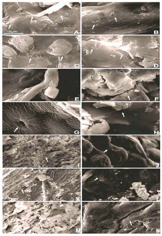
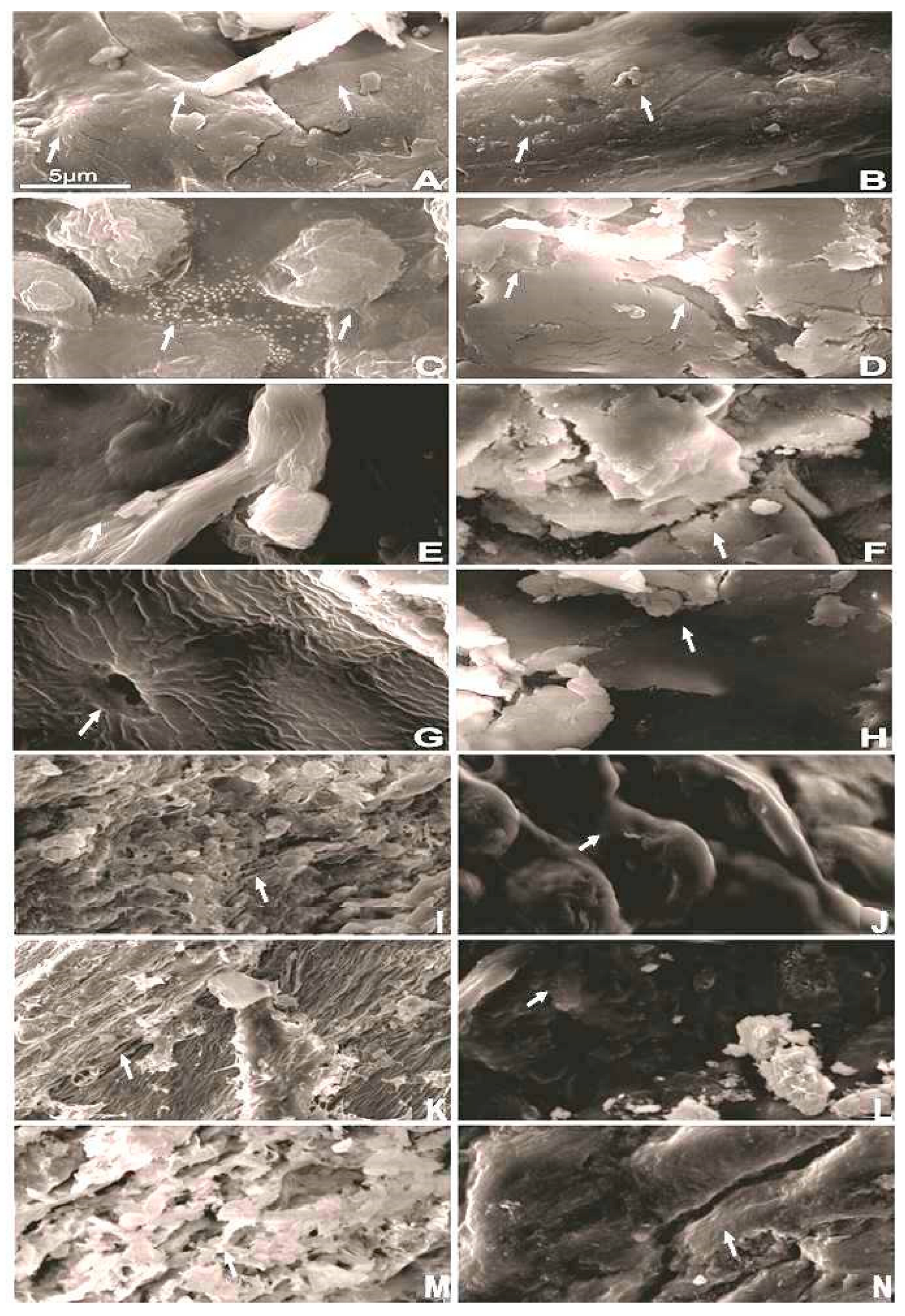
Figure 1 shows that macroalgae have a slightly irregular surface, polymer chains with asymmetrical contours and variable sizes, fissures, and lighter spots. After the US at 60 °C (Figure 1B), a more irregular surface can be observed. Pretreatment with 1% (v/v) H2SO4 (Figure 1C) increased the number of granules on the surface, resulting in lighter spots. The morphology of the macroalgae after the combination of pretreatment with 1% (v/v) H2SO4 and US 60 °C (Figure 1D) completely changed. The surface presents fissures of various sizes and depths with no lighter spots. With an increase in the H2SO4 concentration to 3% (v/v) and 5% (v/v) (Figure 1F,H), more degradation of the macroalgae cell wall was observed.
Figure 1.
Scanning electron micrographs of Gracilaria birdiae after different extraction strategies: (A) Gracilaria birdiae in natura, (B) Gracilaria birdiae after ultrasound 60 °C, (C) Gracilaria birdiae after H2SO4 (1%, v/v), (D) Gracilaria birdiae after H2SO4 (1%, v/v) and ultrasound 60 °C, (E) Gracilaria birdiae after H2SO4 (3%, v/v), (F) Gracilaria birdiae after H2SO4 (3%, v/v) and ultrasound 60 °C, (G) Gracilaria birdiae after H2SO4 (5%, v/v), (H) Gracilaria birdiae after H2SO4 (5%, v/v) and ultrasound 60 °C, (I) Gracilaria birdiae after NaOH (1%, w/v), (J) Gracilaria birdiae after NaOH (1%, w/v) and ultrasound 60 °C, (K) Gracilaria birdiae after NaOH (3%, w/v), (L) Gracilaria birdiae after NaOH (3%, w/v) and ultrasound 60 °C, (M) Gracilaria birdiae after NaOH (5%, w/v), (N) Gracilaria birdiae after NaOH (5%, w/v) and ultrasound 60 °C. Ultrasound (10% (w/v)/10 min/500 W/373 W·cm−2/60 °C).
Pretreatment of the macroalgae with NaOH further exposed the polymer chains (Figure 1I). Increasing the NaOH concentration from 1% to 5% w/v (Figure 1K,M) caused more damage to the cell walls with larger cracks. After combined treatment at US 60 °C (Figure 1J,L,N), the processed biomass presented a less irregular surface.
4. Discussion
The macroalgae Gracilaria birdiae had 63.97 ± 1.20% carbohydrates, 5.20 ± 0.01% proteins, and 19.65 ± 0.41% lipids. The higher lipid content obtained in this study can be explained by the widely variation in red macroalgae due to environmental, physiological, and methodological factors [6]. Analyses performed by our research group on biomass cultivated at Praia da Volta do Rio (Acaraú, CE, Brazil) revealed lipid contents ranging from 3.92% to 19.65% (dry weight), indicating pronounced seasonal variability. In addition, lipid recovery is strongly affected by extraction conditions, while a previous study reported a lipid content of 0.46% using Soxhlet extraction with acetone (80 °C, 4 h) [3], the present study employed hexane (70 °C, 6 h), as differences in solvent polarity and processing conditions significantly affect the recovery of neutral and non-polar lipid fractions. Given this compositional variability and the structural complexity of the G. birdiae cell wall, the efficiency of compound recovery is highly dependent on the extraction strategy employed. In this context, extraction parameters are a key step in improving the release of cell wall compounds from macroalgae.
In this study, increasing the US power from 100 W (75 W·cm−2) to 500 W (373 W·cm−2) increased the release of compounds. A similar result was obtained in the extraction of carbohydrates from Sargassum angustifolium, which improved the extraction yield from 5% to 7% by increasing the US power from 400 W to 800 W. However, increasing the power to 1200 W reduced the extraction yield to 6%. The authors suggested that increasing the US power improves the permeability of the algae cell wall, releasing carbohydrates; however, excess energy can cause aggregation and hydrolysis of carbohydrates [17]. Nonetheless, other factors can influence protein extraction, such as ionic interactions between the cell wall and proteins, as well as the high viscosity of polysaccharides in aqueous solutions [6].
The effect of pigment removal as a pretreatment step varies among macroalgae. Although previous studies have reported improved carbohydrate extraction following depigmentation [20], this effect was not observed for G. birdiae, where ethanol treatment extracted phycobiliproteins. G. birdiae presented more than 1.5 mg/g of TPB, with the highest content of APC (blue pigment), followed by PE (red pigment), and C-PC (blue pigment). These pigments are considered valuable natural colorants owing to their technological and bioactive properties [30]. Various strategies have been employed to obtain those compounds from red macroalgae. PE was extracted (25 mM phosphate buffer, pH 6.8, 1.3 w/v) under mechanical stirring from G. birdiae, obtaining 0.26 mg/mL after partial purification (ammonium sulfate (NH4)2SO4, 25/45) [19]. After ultrasound treatment (20 kHz, 400 W, 10 min), R-phycoerythrin 0.09 mg/g dry matter and R-phycocyanin 0.08 mg/g dry matter were extracted from Gracilaria chilensi [24].
The process time and biomass concentration also affected the extraction of biocompounds from G. birdiae. Increasing the process time from 2 to 10 min increased the recovery of biocompounds from G. birdiae, confirming that extraction time is a critical parameter influencing the extent of cell wall disruption and mass transfer. Higher exposure times generally enhance compound release via cavitation, thereby increasing membrane permeability and solvent penetration [17]. This result is consistent with that of Braspaiboon et al. [31] who reported increased protein extraction from Arthrospira platensis, Cladophora glomerata, Porphyra tenera, Laminaria japonica and Undaria pinnatifida when the US time was increased from 10 to 30 min. Although increasing biomass has improved the release of carbohydrates and proteins in G. birdiae, other studies have reported the opposite behavior due to diffusion limitations. For example, in Sargassum angustifolium, decreasing biomass concentration from 5% to 3% increased carbohydrate yield from 5.72% to 6.70%, attributed to improved diffusion of solubilized polysaccharides [17]. According to previous reports, a higher water-to-solid ratio typically enhances mass transfer and polysaccharide extraction efficiency. However, excessively large solvent volumes may increase the diffusion distance and reduce overall dissolution efficiency [32,33]. The complexity of carbohydrates present in the macroalgae cell wall influences compound extraction. For example, after ultrasound application (ethanol 50% w/v, 10 min, and 1/10 algae/solvent), it was observed that the brown macroalgae Fucus vesiculosus showed a yield similar to that of the control (same conditions under magnetic stirring), yielding 130.7 mg/g of total sugar. In contrast, Pelvetia canaliculata showed lower sugar content but a 66% increase in total sugar compared to the control (59.1 mg/g) [11]. P. canaliculata and F. vesiculosus have cell walls mainly composed of fucan (fucosan), a sulfated polysaccharide rich in L-fucose, with a carbohydrate content of 65.76% and 34.53%, respectively [34,35].
With US time (10 min) and biomass concentration (10%, w/v) set, the use of sequential extraction with hot water (autoclave) followed by US showed good recovery of biocompounds. Hot-water treatment likely weakens the cell wall and disrupts polysaccharide–protein interactions, allowing ultrasound to promote cavitation rupture [13,21]. At 25 °C, US increased carbohydrate extraction by 25%, and the US second cycle released an additional 16%, indicating residual material still accessible after the first treatment. When the US was applied at 60 °C, carbohydrate extraction increased threefold, reflecting the combined effects of elevated temperature, reduced viscosity, and enhanced diffusion; however, the lower yield in the second cycle suggests that most extractable material had already been released, reaching 26.92% yield in the sequential extraction. In the extraction of protein and agar from Gelidium sesquipedale by US (4% w/v, 20 kHz, 100% intensity, 85 °C, 60 min), it was observed that a second extraction process (5% w/v, 2 h, 95 °C), without US, doubled the agar yield, reaching 14.23%. In comparison, protein increased from 10.59% to 12.73% (related to biomass residue) [21]. The extraction of carrageenan from Chondracanthus canaliculatus by US (2 h under agitation in 1M Na2CO4, followed by US 40 Hz, 30 min, 25 °C) increased the extraction yield by 10%, reaching a 45% yield [16].
Protein extraction exhibited different behaviors. While the US at 25 °C increased protein recovery by 22%, a second US extraction yielded less protein, likely due to reduced or partial denaturation [36]. At US 60 °C, protein extraction was consistently lower, indicating that high temperatures may promote protein denaturation or reduced solubility, counteracting the benefits of cavitation. Incubation at elevated temperatures weakens the cell wall through thermally induced pressure, hydrogen bond breakage, or modification of components; however, it may also cause protein denaturation and promote unwanted aggregation [37]. Overall, mild ultrasound conditions favored both carbohydrate and protein recovery, whereas high-temperature ultrasound strongly enhanced carbohydrate release, but impaired protein extraction.
The sequential extraction strategy using 7% (v/v) H2SO4 followed by ultrasound significantly enhanced the release of cell wall compounds. Acid pretreatment acts directly on the cellulose and hemicellulose components of algal biomass, hydrolyzing glycosidic bonds, promoting their breakdown into oligosaccharides and monosaccharides. This depolymerization increases cell wall porosity, allowing greater solvent penetration into the matrix and enhancing compound release [38]. Carbohydrate recovery was highest with US at 60 °C, tripling the yield (52.82%) compared to lower temperatures (25 °C). This result indicates that US elevated-temperature and acid pretreatment act synergistically to disrupt the G. birdiae cell wall and facilitate biocompound recovery. Protein was affected differently; US at 25 °C after acid pretreatment increased protein fivefold compared to US alone, while US at 60 °C combined with acid pretreatment further increased protein recovery thirteenfold, suggesting that higher temperature and acid enhanced protein solubilization with lower degradation. Overall, the sequential acid and ultrasound 60 °C strategy proved effective for maximizing both carbohydrate (52.82% yield) and protein release (98% yield), with temperature and pretreatment type being the key factors. These results are consistent with those of other studies showing that sequential chemical and physical treatments, including acid, alkali, or enzymatic pretreatments combined with US, significantly improve the extraction of carbohydrates, proteins, and bioactive compounds from macroalgae. For example, ultrasound-assisted extraction of citric acid from Sargassum cymosum C. Agardh (176 W, 15 min, pH 1) yielded an alginate yield of 54.20% [18]. The sequential extraction of alginate and fucoidan using acid (HCl, pH 1, 42 °C, 159 min) and alkali (0.2 M Na2CO3, 45 °C, 120 min) from different brown algae showed that Macrocystis pyrifera, Durvillaea potatorum, and Ecklonia radiata yielded 30–40% of total available fucoidans and 80–94% of total available alginates. In contrast, Seirococcus axillaris yielded only 5.5% fucoidans and 74% alginates [39]. The application of sequential extraction using US and enzymatic hydrolysis (1% w/v, 53 kHz, 65% intensity, 165 s, hemicellulase enzyme/substrate 2.5, 35 °C, and extraction time of 20 h) obtained a protein content of 212.57 mg/g from Gracilaria dura [4]. Drying and defatting were employed as pretreatments in Ecklonia maxima to extract polyphenols (11%), followed by enzymatic-assisted extraction (Cellic® Ctec 2 and Viscozyme L) of carbohydrates (alginate 35% and fucoidan 18%) [20].
The evaluation of lower H2SO4 concentrations showed that increasing the acid concentration from 1% to 5% (v/v) during pretreatment followed by US at 60 °C decreased carbohydrate recovery, likely due to acid-induced polysaccharide degradation. Under these conditions, the combined acid and US treatments may promote extensive depolymerization of cell wall polysaccharides by altering their secondary and tertiary structures and modifying both main and branched chains. Such structural changes can increase polysaccharide solubility and lead to a reduction in the molecular weight of carbohydrates, thereby affecting their recoverability [39]. In contrast, protein extraction increased with acid concentration, achieving 35.99 mg/g using 5% (v/v) H2SO4, indicating that stronger acid promotes protein solubilization from the cell wall. The second US extraction obtained significantly lower protein and carbohydrate content, suggesting that repeated cycles alone cannot compensate for the initial extraction efficiency. Nonetheless, the sequential extraction using 1% (v/v) H2SO4 obtained 97.19% of carbohydrate yield, while using 5% (v/v) H2SO4 obtained 86.87% of protein yield. These results highlight that acid concentration is a critical factor, with higher concentrations favoring protein release but jeopardizing carbohydrate stability. The extraction of different carbohydrates from G. birdiae using hot acid extraction was related to the different concentrations of sulfuric acid. The most suitable conditions to produce glucose (28.56 g/L) and galactose (108.03 g/L) were at 121 °C, 1.3 mol of sulfuric acid, 841.59 g/L of biomass for 20 min; cellobiose (25.39 g/L) was 0.6 mol/L of sulfuric acid, 680 g/L of biomass for 10 min [3]. In the extraction of carbohydrates from the microalgae Scenedesmus sp., it was observed that extraction increased with increasing acid concentration (HCl) from 0.1 N to 2.82 N. However, for values above 2.8 N, a lower carbohydrate yield was obtained [40]. The carbohydrate extraction yield of Chlorella vulgaris increased with increasing sulfuric acid (H2SO4) concentrations up to 4%. However, values above 4% decreased the extraction yield. This effect was associated with the degradation of monosaccharides into other sugar degradation products, such as furfural, acetic acid, formic acid, and lactic acid [41]. The algae/acid interaction can also affect carbohydrate extraction yield, as different authors have shown that HCl can be more effective than H2SO4, or vice versa [42,43].
Similar behavior was observed using alkaline pretreatment. Sequential alkaline pretreatment (NaOH 1%, 3%, and 5%, w/v) followed by US at 60 °C significantly enhanced the extraction of both carbohydrates and proteins, reaching yields of 42.06% and 95.27%, respectively, at 5% w/v NaOH. This improvement can be attributed to the disruption of the rigid cell wall structures and complex interactions between proteins and carbohydrates, such as β-(1 → 3)/β-(1 → 4)-D-xylans and cellulose. Alkaline treatment is known to weaken or cleave hydrogen bonds, van der Waals interactions, and glycosidic linkages within cell wall carbohydrates, leading to cell wall swelling. While subsequent ultrasound further promotes cell wall disintegration through mechanical shear and cavitation effects, thereby facilitating the release of compounds [44]. However, the findings highlight that NaOH concentration and sequential US application are critical for maximizing protein release, whereas carbohydrate extraction benefits more from moderate alkaline conditions. Previous studies have shown similar results. For example, US treatments (2 min, 5 s pulses, and 15 s interval) combined with alcalase (0.5 mg/g, pH 8, and 60 °C) under alkaline conditions yielded the highest protein extraction yield, reaching 90 g/100 g of protein in the microalgae Chlorella vulgaris [45]. This enhanced protein recovery can be explained by structural changes in macroalgal proteins under both acidic and alkaline conditions. Variations in pH alter the ionization state of amino acid side chains, disrupting hydrogen bonding and electrostatic interactions, which can lead to protein denaturation and changes in secondary and tertiary structures [46]. For example, under alkaline conditions, higher protein extraction was observed, likely because the elevated pH increases protein solubility, facilitating protein release from the cell matrix [47]. Meanwhile, the lower carbohydrate extraction was associated with carbohydrate degradation by hydroxide ions. Increasing the NaOH concentration decreased the concentrations of carbohydrates such as arabinose, xylose, and galactose extracted from sesame (Sesamum indicum L.), indicating a greater loss of heteropolysaccharides with 2 mol/L NaOH [48]. Alkaline pretreatment (NaOH 3 N) resulted in a low sugar yield from the microalgae Scenedesmus obliquus, due to severe sugar degradation caused by a high alkali concentration [42].
These extraction results were supported by morphological changes in the processed G. birdiae. Pretreatment with acid or alkali induces irreversible changes in the G. birdiae cell wall, resulting in swelling, cracks, and loss of structural integrity. These effects intensified with increasing H2SO4 or NaOH concentration, reflecting hemicellulose hydrolysis and cellulose exposure. Acid pretreatment is used to hydrolyze hemicellulose in the cell wall and make cellulose more accessible to subsequent extraction methods, such as enzymatic extraction [49]. Pretreatment of macroalgae with NaOH further exposed the polymer chains. The NaOH solution swells cellulose in the cell wall, weakening hydrogen bonds between cellulose and hemicellulose, leading to the dissolution of hemicellulose and the formation of cracks on the sample surface [48]. Ultrasound further widened the pores and deepened the fissures, amplifying the disruption initiated by the chemical pretreatments. Together, these changes explain the higher extraction yields, as the combined treatments greatly improved the cell wall permeability and compound release. Ultrasound causes pore widening due to the shock of ultrasonic waves and increases solvent penetration by diffusion, promoting cell rupture and the concomitant release of compounds [16].
Finally, the combined chemical and physical treatments proved highly effective at extracting pigments, carbohydrates, and proteins from G. birdiae, offering valuable insights for future studies aimed at optimizing the recovery and application of these natural compounds in the food industry.
Despite the promising results obtained in this study, some limitations should be acknowledged. The extraction strategies were evaluated at laboratory scale under controlled conditions, which may limit the direct extrapolation of the results to larger-scale or continuous systems. In addition, the study primarily focused on the recovery yields of carbohydrates and proteins, without an in-depth structural characterization of the extracted fractions. Therefore, these aspects should be considered when interpreting the results and defining optimal processing conditions.
5. Conclusions
Gracilaria birdiae was successfully disrupted using a combination of extraction strategies (hot-water, alkali, and acid treatments) and ultrasound to obtain carbohydrates and proteins simultaneously. Pretreatment with ethanol did not improve co-extraction by ultrasound, but did extract different phycobiliproteins (c-phycocyanin, allophycocyanin, and phycoerythrin). Moreover, the acid treatment (5% v/v H2SO4) followed by ultrasound (500 W, 373 W·cm−2) at 60 °C proved to be the most feasible method for the simultaneous co-extraction of carbohydrates and proteins, reaching 330.00 mg/g and 35.99 mg/g, respectively. Nevertheless, the highest carbohydrate content (396.15 mg/g) was obtained using 1% (v/v) H2SO4, whereas the highest protein content (38.49 mg/g) was achieved using 7% (v/v) H2SO4 combined with ultrasound (500 W, 373 W·cm−2) at 60 °C. Thus, combining ultrasound with other extraction techniques simultaneously recovers carbohydrates and proteins; however, to achieve higher recovery yields, different strategies are needed for each compound, at 60 °C.
Future studies may further explore the structural and functional characterization of the extracted carbohydrate and protein fractions, as well as their potential techno-functional properties for food applications. Additionally, further optimization of the extraction conditions and evaluation of ultrasound-assisted processes at larger scales could contribute to improving process feasibility and sustainability.
Author Contributions
Conceptualization S.R., F.A.N.F., P.H.C., G.S.A., R.M.d.S. and R.S.B.d.N.; methodology, formal analysis, writing—original draft preparation, R.M.d.S. and R.S.B.d.N.; scanning electron microscopy, T.B.A.R.M. and E.d.C.M.; proximate composition analysis, P.H.C.; Algae donation, G.S.A.; writing review and supervision, S.R.; project administration, S.R.; funding acquisition S.R., F.A.N.F. and G.S.A. All authors have read and agreed to the published version of the manuscript.
Funding
This study was financed in part by the Coordenação de Aperfeiçoamento de Pessoal de Nível Superior—Brasil (CAPES)—Finance Code 001. This research was also funded by Fundação Cearense de Apoio ao Desenvolvimento Científico e Tecnológico (FUNCAP) postdoctoral grant and Conselho Nacional de Desenvolvimento Científico e Tecnológico CNPq, research grant.
Data Availability Statement
The original contributions presented in this study are included in the article. Further inquiries can be directed to the corresponding author.
Acknowledgments
To Analytical Central/UFC for microscopy analysis.
Conflicts of Interest
The authors declare no conflicts of interest.
Abbreviations
The following abbreviations are used in this manuscript:
| US | Ultrasound |
| C-PC | C-phycocyanin |
| APC | Allophycocyanin |
| PE | Phycoerythrin |
| TPB | Total phycobiliprotein |
| BSA | Bovine Serum Albumin |
| ABS | Absorbance |
References
- Boukid, F.; Castellari, M. Food and Beverages Containing Algae and Derived Ingredients Launched in the Market from 2015 to 2019: A Front-of-Pack Labeling Perspective with a Special Focus on Spain. Foods 2021, 10, 173. [Google Scholar] [CrossRef] [Scilit]
- Guiry, M.D. How Many Species of Algae Are There? A Reprise. Four Kingdoms, 14 Phyla, 63 Classes and Still Growing. J. Phycol. 2024, 60, 214–228. [Google Scholar] [CrossRef] [Scilit]
- Albuquerque, J.C.S.; Araújo, M.L.H.; Rocha, M.V.P.; de Souza, B.W.S.; de Castro, G.M.C.; Cordeiro, E.M.S.; Silva, J.d.S.; Benevides, N.M.B. Acid Hydrolysis Conditions for the Production of Fine Chemicals from Gracilaria birdiae Alga Biomass. Algal Res. 2021, 53, 102139. [Google Scholar] [CrossRef] [Scilit]
- Bozdemir, A.; Şensu, E.; Okudan, E.Ş.; Özçelik, B.; Yücetepe, A. Ultrasound-Assisted Enzymatic Extraction of Proteins from Gracilaria dura: Investigation of Antioxidant Activity and Techno-Functional Properties. J. Food Process. Preserv. 2022, 46, e16803. [Google Scholar] [CrossRef] [Scilit]
- Sarkar, P.; Bandyopadhyay, T.K.; Gopikrishna, K.; Nath Tiwari, O.; Bhunia, B.; Muthuraj, M. Algal Carbohydrates: Sources, Biosynthetic Pathway, Production, and Applications. Bioresour. Technol. 2024, 413, 131489. [Google Scholar] [CrossRef] [Scilit]
- Mandalka, A.; Cavalcanti, M.I.L.G.; Harb, T.B.; Toyota Fujii, M.; Eisner, P.; Schweiggert-Weisz, U.; Chow, F. Nutritional Composition of Beach-Cast Marine Algae from the Brazilian Coast: Added Value for Algal Biomass Considered as Waste. Foods 2022, 11, 1201. [Google Scholar] [CrossRef] [Scilit]
- Gordalina, M.; Pinheiro, H.M.; Mateus, M.; da Fonseca, M.M.R.; Cesário, M.T. Macroalgae as Protein Sources—A Review on Protein Bioactivity, Extraction, Purification and Characterization. Appl. Sci. 2021, 11, 7969. [Google Scholar] [CrossRef] [Scilit]
- Suresh Kumar, K.; Ganesan, K.; Selvaraj, K.; Subba Rao, P.V. Studies on the Functional Properties of Protein Concentrate of Kappaphycus alvarezii (Doty) Doty—An Edible Seaweed. Food Chem. 2014, 153, 353–360. [Google Scholar] [CrossRef] [Scilit]
- Bleakley, S.; Hayes, M. Algal Proteins: Extraction, Application, and Challenges Concerning Production. Foods 2017, 6, 33. [Google Scholar] [CrossRef] [Scilit]
- Jönsson, M.; Allahgholi, L.; Sardari, R.R.R.; Hreggviosson, G.O.; Karlsson, E.N. Extraction and Modification of Macroalgal Polysaccharides for Current and Next-Generation Applications. Molecules 2020, 25, 930. [Google Scholar] [CrossRef] [Scilit] [PubMed]
- Garcia-Vaquero, M.; Ravindran, R.; Walsh, O.; O’Doherty, J.; Jaiswal, A.K.; Tiwari, B.K.; Rajauria, G. Evaluation of Ultrasound, Microwave, Ultrasound–Microwave, Hydrothermal and High Pressure Assisted Extraction Technologies for the Recovery of Phytochemicals and Antioxidants from Brown Macroalgae. Mar. Drugs 2021, 19, 309. [Google Scholar] [CrossRef] [Scilit] [PubMed]
- Kashyap, M.; Ghosh, S.; Steinbruch, E.; Levkov, K.; Israel, Á.; Bala, K.; Livney, Y.; Golberg, A. Extracting Water-Soluble Proteins from the Red Macroalgae Gracilaria Sp. with Pulsed Electric Field in a Continuous Process. ACS Food Sci. Technol. 2023, 3, 562–575. [Google Scholar] [CrossRef] [Scilit]
- Rudke, A.R.; da Silva, M.; de Andrade, C.J.; Vitali, L.; Ferreira, S.R.S. Green Extraction of Phenolic Compounds and Carrageenan from the Red Alga Kappaphycus alvarezii. Algal Res. 2022, 67, 102866. [Google Scholar] [CrossRef] [Scilit]
- Kumar, Y.; Singhal, S.; Tarafdar, A.; Pharande, A.; Ganesan, M.; Badgujar, P.C. Ultrasound Assisted Extraction of Selected Edible Macroalgae: Effect on Antioxidant Activity and Quantitative Assessment of Polyphenols by Liquid Chromatography with Tandem Mass Spectrometry (LC-MS/MS). Algal Res. 2020, 52, 102114. [Google Scholar] [CrossRef] [Scilit]
- Flórez-Fernández, N.; Domínguez, H.; Torres, M.D. A Green Approach for Alginate Extraction from Sargassum Muticum Brown Seaweed Using Ultrasound-Assisted Technique. Int. J. Biol. Macromol. 2019, 124, 451–459. [Google Scholar] [CrossRef] [Scilit]
- Martín-del-Campo, A.; Fermín-Jiménez, J.A.; Fernández-Escamilla, V.V.; Escalante-García, Z.Y.; Macías-Rodríguez, M.E.; Estrada-Girón, Y. Improved Extraction of Carrageenan from Red Seaweed (Chondracantus Canaliculatus) Using Ultrasound-Assisted Methods and Evaluation of the Yield, Physicochemical Properties and Functional Groups. Food Sci. Biotechnol. 2021, 30, 901–910. [Google Scholar] [CrossRef] [Scilit] [PubMed]
- Norouzi, A.; Mehrgan, M.S.; Roomiani, L.; Islami, H.R.; Raissy, M. Ultrasound-Assisted Extraction of Polysaccharides from Brown Alga (Sargassum angustifolium): Structural Characterization, Antioxidant, and Antitumor Activities. J. Food Meas. Charact. 2023, 17, 6330–6340. [Google Scholar] [CrossRef] [Scilit]
- Chica, L.R.; Yamashita, C.; Nunes, N.S.S.; Negreiros, A.T.; Moraes, I.C.F.; Ferreira, A.G.; Mayer, C.R.M.; Haminiuk, C.W.I.; Branco, C.C.Z.; Branco, I.G. Optimizing Alginate Extraction Using Box-Behnken Design: Improving Yield and Antioxidant Properties through Ultrasound-Assisted Citric Acid Extraction. Food Chem. Adv. 2024, 5, 100813. [Google Scholar] [CrossRef] [Scilit]
- de Aguiar, A.L.L.; Araújo, M.L.H.; Benevides, N.M.B.; Mattos, A.L.A.; Araújo, I.M.d.S.; da Silva, E.M.C. Sequential Extraction Process and Physicochemical Characterization of R-Phycoerythrin and Agar from Red Macroalgae Gracilaria birdiae. Algal Res. 2023, 69, 102920. [Google Scholar] [CrossRef] [Scilit]
- Mabate, B.; Pletschke, B.I. Sequential and Enzyme-Assisted Extraction of Algal Bioproducts from Ecklonia maxima. Enzyme Microb. Technol. 2024, 173, 110364. [Google Scholar] [CrossRef] [Scilit]
- Gómez Barrio, L.P.; Cabral, E.M.; Zhao, M.; Álvarez García, C.; Senthamaraikannan, R.; Padamati, R.B.; Tiwari, U.; Curtin, J.F.; Tiwari, B.K. Comparison Study of an Optimized Ultrasound-Based Method versus an Optimized Conventional Method for Agar Extraction, and Protein Co-Extraction, from Gelidium sesquipedale. Foods 2022, 11, 805. [Google Scholar] [CrossRef] [Scilit]
- Oliveira, L.C.B.P.; Queiroz, M.F.; Fidelis, G.P.; Melo, K.R.T.; Câmara, R.B.G.; Alves, M.G.C.F.; Costa, L.S.; Teixeira, D.I.A.; Melo-Silveira, R.F.; Rocha, H.A.O. Antioxidant Sulfated Polysaccharide from Edible Red Seaweed Gracilaria birdiae Is an Inhibitor of Calcium Oxalate Crystal Formation. Molecules 2020, 25, 2055. [Google Scholar] [CrossRef] [Scilit] [PubMed]
- AOAC. Books in Brief. J. AOAC Int. 1997, 80, 127A–128A. [Google Scholar] [CrossRef] [Scilit]
- Brain-Isasi, S.; Correa, S.; Amado-Hinojosa, J.; Buschmann, A.H.; Camus, C.; Lienqueo, M.E. Combined Extraction Methodology for Simultaneous Recovery of Phycobiliproteins and Agar from the Red Alga Gracilaria chilensis C. J. Bird, McLachlan & E. C. Oliveira. Algal Res. 2022, 67, 102821. [Google Scholar] [CrossRef] [Scilit]
- Fox, J.D.; Robyt, J.F. Miniaturization of Three Carbohydrate Analyses Using a Microsample Plate Reader. Anal. Biochem. 1991, 195, 93–96. [Google Scholar] [CrossRef] [Scilit]
- Lowry, O.; Rosebrough, N.; Farr, A.L.; Randall, R. Protein Measurement with the Folin Phenol Reagent. J. Biol. Chem. 1951, 193, 265–275. [Google Scholar] [CrossRef] [Scilit]
- da Silva, R.M.; Gonçalves, L.R.B.; Rodrigues, S. Different Strategies to Co-Immobilize Dextransucrase and Dextranase onto Agarose Based Supports: Operational Stability Study. Int. J. Biol. Macromol. 2020, 156, 411–419. [Google Scholar] [CrossRef] [Scilit] [PubMed]
- Pan-utai, W.; Iamtham, S.; Boonbumrung, S.; Mookdasanit, J. Improvement in the Sequential Extraction of Phycobiliproteins from Arthrospira platensis Using Green Technologies. Life 2022, 12, 1896. [Google Scholar] [CrossRef] [Scilit]
- da Silva, R.M.; Miguel, T.B.A.R.; de Castro Miguel, E.; Campelo, P.H.; Fernandes, F.A.N.; Rodrigues, S. Protective Effect of Ultrasound-Processed Amazonian Sapota-Do-Solimões (Quararibea cordata) Juice on Artemia salina Nauplii. Processes 2022, 10, 1880. [Google Scholar] [CrossRef] [Scilit]
- Castro-Varela, P.; Celis-Pla, P.S.M.; Figueroa, F.L.; Rubilar, M. Highly Efficient Water-Based Extraction of Biliprotein R-Phycoerythrin From Marine the Red-Macroalga Sarcopeltis skottsbergii by Ultrasound and High-Pressure Homogenization Methods. Front. Mar. Sci. 2022, 9, 877177. [Google Scholar] [CrossRef] [Scilit]
- Braspaiboon, S.; Osiriphun, S.; Surawang, S.; Jirarattanarangsri, W.; Kanha, N.; Laokuldilok, T. Ultrasound-Assisted Alkaline Extraction of Proteins in Several Algae and Their Nutritional Characteristics. Int. J. Food Sci. Technol. 2022, 57, 6143–6154. [Google Scholar] [CrossRef] [Scilit]
- Sorourian, R.; Khajehrahimi, A.E.; Tadayoni, M.; Azizi, M.H.; Hojjati, M. Ultrasound-Assisted Extraction of Polysaccharides from Typha domingensis: Structural Characterization and Functional Properties. Int. J. Biol. Macromol. 2020, 160, 758–768. [Google Scholar] [CrossRef] [Scilit]
- Ren, B.; Chen, C.; Li, C.; Fu, X.; You, L.; Liu, R.H. Optimization of Microwave-Assisted Extraction of Sargassum thunbergii Polysaccharides and Its Antioxidant and Hypoglycemic Activities. Carbohydr. Polym. 2017, 173, 192–201. [Google Scholar] [CrossRef] [Scilit] [PubMed]
- Pires, D.; Passos, R.; do Carmo, B.; Tchobanov, C.F.; Forte, S.; Vaz, M.; Antunes, M.; Neves, M.; Tecelão, C.; Baptista, T. Pelvetia canaliculata as an Aquafeed Supplement for Gilthead Seabream Sparus Aurata: A Biorefinery Approach for Seaweed Biomass Valorisation. Sustainability 2022, 14, 11469. [Google Scholar] [CrossRef] [Scilit]
- Agregán, R.; Munekata, P.E.; Domínguez, R.; Carballo, J.; Franco, D.; Lorenzo, J.M. Proximate Composition, Phenolic Content and in Vitro Antioxidant Activity of Aqueous Extracts of the Seaweeds Ascophyllum nodosum, Bifurcaria bifurcata and Fucus vesiculosus. Effect of Addition of the Extracts on the Oxidative Stability of Canola Oil under Accelerated Storage Conditions. Food Res. Int. 2017, 99, 986–994. [Google Scholar] [CrossRef] [Scilit] [PubMed]
- Antunez-Medina, J.R.; Suárez-Jiménez, G.M.; Ocano-Higuera, V.M.; Tolano-Villaverde, I.d.J.; Ornelas-Paz, J.d.J.; Torres-Arreola, W.; Márquez-Ríos, E. Application of Ultrasound in Proteins: Physicochemical, Structural Changes, and Functional Properties with Emphasis on Foaming Properties. Process 2025, 13, 1646. [Google Scholar] [CrossRef] [Scilit]
- Li, R.; Mejdahl, T.K.B.; Jørgensen, M.L.; Corredig, M.; Gregersen, S.B. Effect of Processing on Protein Solubility of Chlorella Sorokiniana Dispersions. Sustain. Food Proteins 2024, 2, 215–222. [Google Scholar] [CrossRef] [Scilit]
- Srivastava, H.; Bisht, B.; James, J.; Malhotra, R.K.; Kurbatova, A.; Dabral, A.; Upadhyay, S.; Kumar, V. Advanced Extraction Technologies and Functional Applications of Algal Polysaccharides in Modern Food Systems. Discov. Food 2025, 5, 272. [Google Scholar] [CrossRef] [Scilit]
- Lorbeer, A.J.; Charoensiddhi, S.; Lahnstein, J.; Lars, C.; Franco, C.M.M.; Bulone, V.; Zhang, W. Sequential Extraction and Characterization of Fucoidans and Alginates from Ecklonia radiata, Macrocystis pyrifera, Durvillaea potatorum, and Seirococcus axillaris. J. Appl. Phycol. 2017, 29, 1515–1526. [Google Scholar] [CrossRef] [Scilit]
- Yirgu, Z.; Leta, S.; Hussen, A.; Khan, M.M.; Aragaw, T. Optimization of Microwave-Assisted Carbohydrate Extraction from Indigenous Scenedesmus sp. Grown in Brewery Effluent Using Response Surface Methodology. Heliyon 2021, 7, e07115. [Google Scholar] [CrossRef] [Scilit]
- Hammann, W.; Ross, A.; Seames, W. Sequential Extraction of Carbohydrates and Lipids from Chlorella Vulgaris Using Combined Physical and Chemical Pre-Treatments. ChemEngineering 2024, 8, 11. [Google Scholar] [CrossRef] [Scilit]
- Miranda, J.R.; Passarinho, P.C.; Gouveia, L. Pre-Treatment Optimization of Scenedesmus obliquus Microalga for Bioethanol Production. Bioresour. Technol. 2012, 104, 342–348. [Google Scholar] [CrossRef] [Scilit]
- Shokrkar, H.; Ebrahimi, S.; Zamani, M. Bioethanol Production from Acidic and Enzymatic Hydrolysates of Mixed Microalgae Culture. Fuel 2017, 200, 380–386. [Google Scholar] [CrossRef] [Scilit]
- Wijethunga, A.M.; He, Q.S.; Prithiviraj, B.; Sun, X. Acid Hydrolysis and Microwave Digestion Enhanced Protein Extraction from Red Seaweed Palmaria palmata. Food Chem. X 2025, 25, 102222. [Google Scholar] [CrossRef] [Scilit]
- Martins, P.L.; Duarte, L.C.; Pereira, H.; Reis, A.; Carvalheiro, F. Evaluation of Different Fractionation Methods for the Simultaneous Protein and Carbohydrate Extraction from Microalgae. Biomass Convers. Biorefinery 2024, 15, 999–1011. [Google Scholar] [CrossRef] [Scilit]
- Zhu, L.; Liu, M.; Wang, Y.; Zhu, Z.; Zhao, X. Euglena gracilis Protein: Effects of Different Acidic and Alkaline Environments on Structural Characteristics and Functional Properties. Foods 2024, 13, 2050. [Google Scholar] [CrossRef] [Scilit]
- Álvarez, C.; Lélu, P.; Lynch, S.A.; Tiwari, B.K. Optimised Protein Recovery from Mackerel Whole Fish by Using Sequential Acid/Alkaline Isoelectric Solubilization Precipitation (ISP) Extraction Assisted by Ultrasound. LWT 2018, 88, 210–216. [Google Scholar] [CrossRef] [Scilit]
- Zhang, R.Y.; Liu, C.; Wang, X.D.; Liu, H.M.; Zhu, W.X. Effects of Different Concentrations of NaOH on the Structure and In Vitro Digestion of Cellulose from Sesame Kernel. LWT 2022, 168, 113956. [Google Scholar] [CrossRef] [Scilit]
- Kim, S.W.; Hong, C.H.; Jeon, S.W.; Shin, H.J. High-Yield Production of Biosugars from Gracilaria Verrucosa by Acid and Enzymatic Hydrolysis Processes. Bioresour. Technol. 2015, 196, 634–641. [Google Scholar] [CrossRef] [Scilit] [PubMed]

Disclaimer/Publisher’s Note: The statements, opinions and data contained in all publications are solely those of the individual author(s) and contributor(s) and not of MDPI and/or the editor(s). MDPI and/or the editor(s) disclaim responsibility for any injury to people or property resulting from any ideas, methods, instructions or products referred to in the content. |
© 2026 by the authors. Licensee MDPI, Basel, Switzerland. This article is an open access article distributed under the terms and conditions of the Creative Commons Attribution (CC BY) license.