


Abstract
Monitoring hematological indicators and the dynamics of hemotropic agents in sheep production is essential for diagnosing the animals’ health status under field conditions. The aim of this study was to evaluate the frequency of hemotropic agents and the hematophysiological variability associated with intrinsic and extrinsic factors in sheep from the “Lanero” Foothills (Colombian Orinoquia). One hundred and forty-eight Creole sheep (43 males aged 2 to 36 months and 105 females aged 2 to 48 months) from seven production units were sampled. Blood samples were collected by jugular venipuncture to determine the complete hematological profile and the presence of hemotropic agents using Wright-stained blood smears. Five classification criteria (farm, sex, age, body condition score, and FAMACHA® scale) were evaluated based on three physiological variables and eight hematological parameters. Sex was the only intrinsic factor with a significant influence (p < 0.05) on hematocrit, hemoglobin, eosinophils, and platelets (p < 0.05), which remained within the reference ranges for the species. Females exhibited higher values for hematocrit (30.5 vs. 28.0%), hemoglobin (10.5 vs. 9.8 g/dL), and eosinophils (0.66 vs. 0.52 × 10⁹/L), while males presented higher platelet values (467.7 vs. 323.6 × 10⁹/L). Anaplasma spp. was the most frequent (52.03%). An interaction (p < 0.05) was observed between the “farm” factor and the frequency of Anaplasma spp. Sex and age were not associated with the frequency of any hemotropic agent. (p > 0.05). Evaluation using FAMACHA® was consistent with the absence of clinical anemia in the evaluated population. The results demonstrate the remarkable clinical and hematological resilience of the region’s Creole sheep against hemotropic agents, reinforcing a scenario consistent with a state of enzootic stability.
1. Introduction
Sheep production in Colombia has grown in recent years, especially in regions with favorable agroecological characteristics. Sheep production has become a strategy for food security, income generation, and efficient use of available resources for farmers. In the department of Meta, Colombia, it is estimated that 70% of sheep production takes place on farms smaller than 5 hectares, particularly in the “Llanero” Foothills region [1].
In the tropical and subtropical ecosystems of Latin America, vector-borne parasitic diseases represent one of the greatest structural challenges for livestock development. These ecosystems offer optimal environmental conditions for the proliferation of arthropods, particularly ticks and biting flies, which are vectors of hemotropic agents [2]. These infections constitute an often-overlooked problem, despite their capacity to cause significant losses in animal health and predispose flocks to other comorbidities [3].
Hemotropic agents have been shown to cause the destruction of red blood cells, leading to anemia, anorexia, jaundice, weight loss, and a consequent decrease in production indexes [4]. Furthermore, several of these pathologies can affect small ruminants [5]. The dynamics of hemoparasitosis are influenced by a complex interaction of factors associated with the physiological state of the host, the ecological characteristics of the environment, and environmental variables that determine the development of their vectors [6].
The hemotropic species frequently detected in sheep are Anaplasma ovis, Babesia ovis, and Trypanosoma vivax [7,8]. Monitoring for these hemotropic agents can be strengthened by directly assessing the flock’s health status using laboratory diagnostic tools such as blood smears and complete blood counts, or molecular analyses. These techniques allow for the identification of anemia, hemoconcentration, and other abnormalities suggestive of an infectious process [9,10]. However, these methods remain underutilized by sheep producers, despite their critical value for early detection and timely zootechnical and therapeutic decisions [10]. A study in sheep conducted in Colombia reported an overall prevalence of 1.6% for Anaplasma spp., 1.0% for Trypanosoma spp., and 0.3% for Rickettsia spp. [11].
Most reports on hemotropic agents in Colombia have focused on cattle; however, research shows that these infections in small ruminants often go unnoticed, with subclinical infections, causing silent production losses [3]. The results of studies in the department of Meta indicate that there is a situation of enzootic stability for tick-borne hemotropic agents in cattle, characterized by a mild primary infection that leads to a reduction in hematocrit with subsequent recovery and transition to a state of co-infective immunity [2].
The “Llanero” Foothills region in the Meta department of Colombia is characterized by high relative humidity, seasonal rainfall, and elevated temperatures. This ecological niche accelerates the life cycles of macroparasites and hematophagous vectors [2]. Determining parasitic status through direct microscopy is fundamental for estimating local prevalence, allowing for early detection to mitigate negative economic impacts and optimize animal welfare, especially since these animals can be reservoirs of hemotropic agents of parasitic origin due to asymptomatic infections [12]. Staining techniques are a fundamental analytical tool in microscopy for optimizing the contrast of blood smears. Among these techniques, Wright’s stain, belonging to the Romanowsky group of dyes, stands out. Wright’s stain is widely used for processing blood smears and observing them under optical microscopes [13].
Despite the existence of the international literature on blood cell profiles and the dynamics of hemotropic agents in sheep [9,14,15,16], in Colombia this type of approach is scarce, especially those that simultaneously integrate parasite prevalence, physiological behavior, hematological variables, and general health status. Particularly in the department of Meta, information on hemotropic agents affecting sheep farms is limited, even though these pathologies constitute one of the main causes of decreased productivity due to deficiencies in traditional farm management.
Blood profile analysis is widely used to determine health status and guide diagnosis in domestic animal medicine [17], as the study of these variables reveals early systemic abnormalities [15,18,19]. It is emphasized that these biological parameters are susceptible to variations due to environmental, genetic, physiological, and pathological factors, such as age and sex [15,19,20,21,22]. Reference ranges published in the standard literature often differ from findings in local populations due to the collateral influence of breed, geographic area, climate, seasonal management, and type of forage consumed, with each sheep biotype exhibiting its own cellular characteristics [23,24].
Due to the scarcity of comprehensive studies in Colombia that simultaneously evaluate the frequency of hemotropic agents and their association with local hematophysiological variables in animals, this research is justified by the imperative need to generate baseline epidemiological information. This will allow producers and health organizations to abandon empirical treatments and adopt zootechnical management decisions based on regional scientific evidence. Given the constant exposure to vectors, epidemiological surveillance of these agents is critically important within comprehensive health management, as some of the agents are zoonotic. The significance of this research lies in generating updated and region-specific data. This aim of descriptive cross-sectional study was to evaluate the frequency of hemotropic agents and the physiological and hematological variations in sheep from the Colombian Orinoquia.
2. Materials and Methods
2.1. Study Area
The study was structured using a descriptive, cross-sectional research design in the “Llanero” Foothills region of the Meta department (Colombian Orinoquia), located between 04°54′25″ and 01°36′52″ N, and 71°4′38″ and 74°53′57″ W. The region has two climatic seasons: the rainy season extends from April to November, and the dry season occurs between December and March. Precipitation varies from 2000 to over 6000 mm per year. The temperature in the Foothills region ranges between 24 and 31 °C, relative humidity is greater than 60%, and the average altitude is 423 m [25].
2.2. Farms and Animals
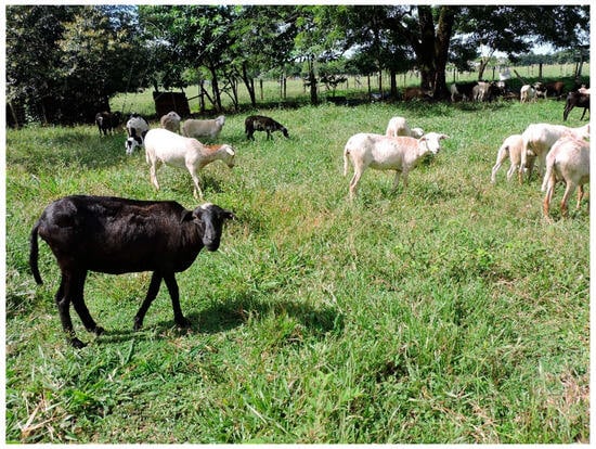
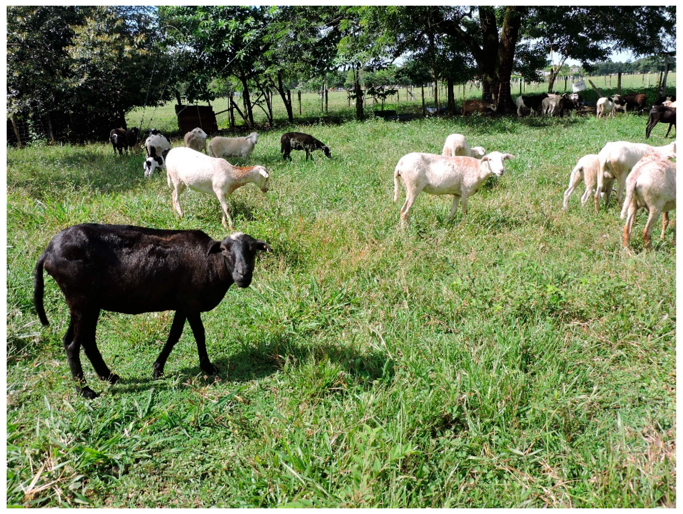
A sample of 148 sheep located on different farms in the “Llanero” Foothills of the Meta department was studied: Farm 1 (n = 18), Farm 2 (n = 3), Farm 3 (n = 13), Farm 4 (n = 31), Farm 5 (n = 17), Farm 6 (n = 13), and Farm 7 (n = 53). The sample comprised 43 males (age range 2 to 36 months) and 105 females (age range 2 to 48 months). The animals’ ages were reported by the producers. Farm sampling was conducted using convenience sampling [26] after a meeting with each producer. At each farm, the number of animals made available by the producer at the time of the visit was sampled. The sheep population consisted of Creole animals, classified based on their external phenotypic characteristics [27] (Figure 1), as well as information provided by the producers.
Figure 1.
Creole sheep from the “Llanero” Foothills, Colombia. Photography: La Vitrina Farm.
The farms belong to small producers with low levels of technology, whose main activity is raising, breeding, and fattening sheep. The farming system is extensive, typical of the traditional management practices of the “Llanero” Foothills. Deworming is infrequent, and ectoparasites—mainly ticks—were present on most farms, although the genus was unknown (this variable was not included in the study and is recognized as a limitation). Vector density and species identification were beyond the scope of this study. The farms lack genealogical, production, and reproductive records. Data collection took place from November 2025 to April 2026, coinciding with the dry season. The data were collected by students who participated in field activities and laboratory analyses.
2.3. Blood Sample Collection
At the time of sample collection, a clinical examination was performed to determine the general health status of each animal. The examination included body condition, mucous membrane color, rectal temperature, heart rate, and respiratory rate. A blood sample was drawn from each animal by venipuncture of the jugular vein, after disinfecting the area, using 21G Vacutainer® (Franklin Lakes, NY, USA) multi-needles and 4 mL vacuum tubes with anticoagulant (EDTA). The samples were stored in a polystyrene container and kept at a temperature of 2 °C to 5 °C, following standard procedures [28]. The inclusion criteria were animals older than 2 months that had not received antiparasitic treatment in the last three months. Prior to sample collection, an open oral interview was conducted with the farm owner to inform them about the project’s aim. The owners gave their informed consent to participate in the study. Sampling activities were carried out in the morning (8:00–11:00 a.m.).
2.4. Hematological Evaluation
Eight quantitative variables of the red blood cell, white blood cell, and platelet counts were determined: hematocrit (%), hemoglobin (g/dL), total platelet count, and differential leukocyte counts (neutrophils, lymphocytes, monocytes, eosinophils, and basophils). Hematological evaluation was performed at the Animalitos Veterinary Clinic Laboratory using a GenVet VH-50® hematology analyzer (Shenzhen, China).
2.5. Parasitological Evaluation
For the diagnosis of hemotropic agents, the Wright-stained whole blood smear technique was used, following standard procedures [13,28]. Microscopic evaluation of the smears was performed in duplicate and double-blind using a 100X oil immersion objective. The identification of hemotropic agents was established by morphological characteristics for Anaplasma spp., Trypanosoma spp. and Babesia spp., according to criteria established up to the taxon Genus [29,30]. One hundred microscopic fields were analyzed.
2.6. Mucosal Color Assessment
The FAMACHA® criteria were used to assess mucosal color: 1: red (non-anemic), 2: pale red (moderate anemia), 3: pink (intermediate anemia), 4: pale pink (severe anemia), 5: white (severe anemia) [31,32]. Although the FAMACHA® criteria are used to diagnose Haemonchus contortus anemia, in this study they were used only to determine mucosal color. Body condition was established according to the following reference criteria: 1–3: thin, 4–5: normal/ideal [33]. Age was grouped according to the following criteria: 2–7 months: suckling/weaned lambs; 8–11 months: growing sheep; 12–24 months: adults; >24 months: rams [31].
2.7. Statistical Analysis
Using the recorded variables, five classification criteria were established as potential factors of variation: farm, sex, age, body condition score, and FAMACHA®. Fourteen response variables were considered: heart rate (beats/minute), respiratory rate (breaths/minute), and rectal temperature (°C) as physiological variables; hematocrit (Hto, %), Hemoglobin (Hb, g/dL), neutrophils (109/L), lymphocytes (109/L), monocytes (109/L), eosinophils (109/L), basophils (109/L), and platelets (109/L) as hematological variables; and the presence of Babesia, Anaplasma, and Trypanosoma as hemotropic variables. The genus of Babesia, Anaplasma, and Trypanosoma were categorical variables, and the rest were numerical.
Initially, the quantitative response variables were subjected to descriptive statistics, while the categorical variables were subjected to frequency tables. Subsequently, the effect of the classification criteria on the quantitative response variables was evaluated using a mixed linear model that included the farm as a random effect to control for the intrinsic variability of the microenvironment and the management of each farm.
The following model was used:
where
Yijklm = µ + Si + Aj + Ck + Fl + Zm + ℇijklm
- Yijklm = Response variables
- µ = Overall mean of all observations
- Si = Fixed effect of sex (i = male, female)
- Aj = Fixed effect of age (j = 2–7 months, 8–11 months, 12–24 months, >24 months)
- Ck = Fixed effect of body condition (k = 1–3, 4–5)
- Fl = Fixed effect of FAMACHA® (l = 1, …, 4)
- Zm = Random farm effect (m = F1, …, F7)
- ℇijklm = Random error associated with all observations
Given the skewed distribution and extreme values of the neutrophil, lymphocyte, monocyte, eosinophil, basophil, and platelet counts, the data were log10 transformed prior to analysis to normalize the distribution of residuals [34]. The assumptions of normality and residual heteroscedasticity were reviewed and adjusted as necessary. The Di Rienzo, Guzmán, and Casanoves (DGC) test [35] was used to differentiate the means (p < 0.05). To assess the relationship between the presence of hemotropic agents and the classification criteria, the Chi-square test of independence was performed (χ2; p < 0.05). To facilitate statistical testing and due to the low number of observations in some categories, certain categorical variables were regrouped as follows: age (2–11, 12–24, and >24 months), farm (1–5, 6, and 7), and FAMACHA® criteria (1, 2, and 3–4). All data were processed using the free 2020 version of InfoStat software [35].
3. Results
Table 1 presents the descriptive statistics of the physiological and hematological variables analyzed.
Table 1.
Descriptive statistics of criteria, physiological and hematological variables of Creole sheep in the “Llanero” Foothills, Meta (Colombia) (n = 148).
According to the linear models evaluated, sex was the only effect that influenced hematocrit, hemoglobin, log10 eosinophil, and log10 platelet levels (p < 0.05). Females showed higher hematocrit (30.5 vs. 28.0%), hemoglobin (10.5 vs. 9.8 g/dL), and eosinophil (0.66 vs. 0.52 × 10⁹/L) values, while males had higher platelet values (467.7 vs. 323.6 × 10⁹/L). The other effects evaluated were not significant (p > 0.05).
Table 2 presents the frequency of anemia cases according to the FAMACHA® criteria. It is observed that 22.30% of the evaluated sheep showed normal values according to criteria 1 (non-anemic animals), and 47,92% showed moderate anemia. The remaining 29.7% suggest anemia levels ranging from intermediate to severe.
Table 2.
Overall frequency of anemia according to FAMACHA® criteria in Creole sheep from the “Llanero” Foothills, Meta (Colombia).
Table 3 shows the frequency of hemotropic agents in the evaluated animals. A frequency of 5.41% was found for Babesia spp. and Trypanosoma spp., while for Anaplasma spp. the value was 52.03%.
Table 3.
Overall frequency of hemotropic agents in Creole sheep from the “Llanero” Foothills, Meta (Colombia).
The Chi-square (χ2) test revealed a statistically significant association between the studied farms and the presence of Anaplasma spp. (p = 0.0001). FAMACHA® system significantly influenced the presence of Babesia spp. (p = 0.0439), with a higher frequency observed in criteria 2, and the presence of Anaplasma spp. (p = 0.0193), with a higher frequency in criteria 3–4. Babesia spp., Anaplasma spp., and Trypanosoma spp. was independent of sex, body condition, and age (p > 0.05) (Table 4).
Table 4.
Frequency of hemotropic agents in Creole sheep on farms in the “Llanero” Foothills, Meta (Colombia) (n = 148), according to classification criteria.
4. Discussion
4.1. Hematophysiological Variations
The analysis of physiological and hematological variables provides substantial information on the health status of animals and is useful for assessing the evolution and prognosis of a disease [16,20]. In the present study, hematological and physiological values are within the reference range for sheep. Only sex influenced hematological values, with higher hematocrit, hemoglobin, and eosinophil levels in females, and higher platelet levels in males.
These differences can be attributed to variations in physiological state, such as higher hemoglobin and hematocrit in pregnant animals [18], although pregnancy status was not reported in the present study. Hormonal responses and tissue composition also influence physiological and hematological variations: for example, it has been shown that androgen levels increase erythropoiesis [22,37,38]; which may be due to males having a more active metabolism that induces greater oxygen transport, thereby increasing red blood cell production [14,39], among other things. However, studies evaluating sex-related hematological variations in sheep remain limited [40].
4.1.1. Effect of Sex on Hematocrit
Studies conducted on sheep of different breeds in Iraq found no significant differences in hematocrit between sexes, nor in the hematological profiles studied, although females exhibited higher values. However, hematological values remained within the normal range for sheep [41]. In semi-arid areas of Algeria, no differences between sexes were reported for hematocrit, hemoglobin, and platelets, although variations were observed during sheep growth [15]. In dwarf sheep from West Africa, hematocrit values and red blood cell count were found not to differ significantly between males and females [40]. In the present study, hematocrit values were within the reference ranges, despite the high frequency of Anaplasma spp. (52.03%, with a higher value in males). This finding differs from that reported in recent studies, which indicate that sheep infected by Anaplasma spp. usually present a significant decrease in hematocrit [42].
Other reports have shown sex differences in hematological values. In Nigeria, hematological indicators in sheep varied according to sex and age, with higher hematocrit values observed in females [19]. In other contexts, such as in humans and mice, older males have been reported to have increased hemoglobin and hematocrit levels, which may be associated with greater muscle mass and the influence of the erythropoiesis-stimulating hormone testosterone; however, this information is not universal [43,44]. The effect of sex does not act independently on hematological values, but rather in association with other factors such as age, nutritional and physiological status, health, and environmental conditions, such as climate, altitude, and heat stress [22,45]. This may be the case regarding the results observed in the present study, where the sheep are exposed to the heat stress characteristic of the warm-temperate tropics in the foothills of Meta.
4.1.2. Sex and Hemoglobin Effect
Hemoglobin and hematocrit values can vary with sex; however, these differences are not uniform across breeds. In general, a tendency toward higher values in males has been described, influenced by genetic and environmental factors. This difference may decrease in adult animals and is more pronounced in young animals [46,47]. Studies report that sheep with anaplasmosis caused by A. ovis show significantly lower hemoglobin and hematocrit levels compared to unaffected animals [48,49]. In our study, no significant interaction was found between sex and the frequency of the three hemotropic agents studied.
Hematocrit and hemoglobin levels may indicate greater tissue oxygenation capacity and hematopoietic regeneration, while increased basophils could be associated with a more effective immune response. This suggests that productive longevity in sheep is not a random trait, but may be biologically supported by specific hematological and biochemical profiles. Therefore, these could be considered biomarkers of survival and tolerance associated with the efficiency, sustainability, and profitability of the production system for native breeds [50].
4.1.3. Effect of Sex on Eosinophils and Platelets
Eosinophils and platelets are important components of the ovine blood count, and their variation depends on physiological and environmental factors, with sex being one of the most studied factors. In the case of eosinophils, the variation may be related to a secondary response associated with immune activation, the presence of parasites, and inflammatory and allergic processes, given their phagocytic capacity [51]. In the present study, males showed higher platelet counts, which could be related to sex-related physiological differences. Environmental, management, or nutritional variables may contribute to this differentiation, suggesting a possible combined influence of environmental and hormonal factors [52]. On the other hand, transient thrombocytosis can occur during sampling, as a result of physical activity, which stimulates the secretion of epinephrine and produces splenocontraction and, therefore, a mobilization of stored platelets into the circulation [53].
Studies conducted on Barbary sheep report that sex did not influence platelet count, indicating that variations may depend on management conditions, age, and breed [52]. Furthermore, platelet count was similar to that reported in other studies [14,54]. In Kashmiri sheep, India, age and sex did not influence hematological parameters in apparently healthy animals, indicating (in that study) that hematological values are considered relatively independent of sex [21]. Similar results were reported for Iranian red sheep where no significant changes in parameters were observed based on sex, although differences in the biochemical profile were observed [23]. However, studies on the influence of sex on platelets and eosinophils in sheep are limited [22].
In the current study, eosinophil levels remained within reference ranges despite the high frequency of Anaplasma spp. observed. This finding is relevant, considering that the frequency of this agent can induce alterations in hematological values [42]. It is highly likely to find values within the normal range in cases of subclinical anaplasmosis in both small and large ruminants. Studies conducted on Wonosobo sheep in Java found no significant effects of sex on eosinophils, although the total count was higher in young and pregnant ewes; no significant difference was found in hemoglobin or platelet count either [24]. In a preliminary study conducted on sheep from Colombian farms, females were observed to have higher eosinophil, hematocrit, and hemoglobin levels compared to males [55]. In any case, eosinophil and platelet counts can be considered in future evaluations as potential biomarkers of adaptive immune response.
It is important to highlight that geographic location, determined by climate and food availability, can affect the hematological values of locally raised sheep [54]. Measuring hematological values can provide objective information about an animal’s condition, revealing its nutritional status, diseases, or the stress it has been subjected to [56]. In one study, lambs kept under individual housing conditions with shade showed higher hematocrit concentrations (p < 0.05) but lower hemoglobin concentrations compared to ewes housed without shade [57]. It is important to consider that the environmental effects of heat stress can influence hematological parameters; therefore, it is essential to continue developing future research.
The variation in red blood cell and platelet count linked exclusively to sex (where females showed higher hematocrit and hemoglobin, and males a higher platelet count) is due to divergent endocrine and immunological profiles described in domestic ruminants [20]. What is truly relevant from a pathogenic perspective is the non-anemic status determined by FAMACHA®, even though this technique was developed to evaluate the intensity of blood-sucking nematode infection in sheep.
4.2. FAMACHA® Assessment
The majority of sheep evaluated in the clinical examination using the FAMACHA® technique with direct observation of conjunctival mucosal color indicate that they are in good health [31]. The results for animals in good health can be explained by the low frequency of Babesia spp. and Trypanosoma spp., and the cases of anemia found can be attributed mainly to Anaplasma spp., which had a frequency of 52.03%. However, it is important to highlight that the FAMACHA® method was designed for the field evaluation of H. contortus infections [31]. It is also known that sheep can tolerate high parasite loads of hemotropic agents without showing clinical signs or apparent anemia [58] and therefore may have had mucous membranes of acceptable color. Furthermore, the assessment can be influenced by the operator’s subjective interpretation and by external factors such as ambient lighting. Natural light conditions, dust in the eyes or stress in the animal can alter the tone of the conjunctival mucosa and cause errors in assessment [58,59].
This animal response likely indicates that the parasite load is not producing obvious signs of anemia reflected in the color of the mucous membranes, which can be corroborated by the mean hematocrit (29.24%) and hemoglobin (10.20 g/dL) values, which remain within normal reference ranges. However, further analysis of the association with hematocrit, hemoglobin, coproparasitology, or ectoparasite load is necessary, but this was not the aim of the present study. The low frequency of anemic animals may be due to subclinical infection, chronic carriers, low parasitemia, low smear sensitivity, or varying degrees of tolerance to hemotropic agents.
4.3. Frequency of Hemotropic Agents
4.3.1. Frequency of Anaplasma spp.
Regarding the high frequency of Anaplasma spp. (52.03%), the lack of quantification of parasitemia is recognized as a methodological limitation, since the mere presence of the hemotropic agent is insufficient as the sole evaluation criterion. In A. ovis infections, hosts in the chronic phase maintain low-level, fluctuating rickettsiemia; while immune-mediated tolerance prevents severe anemia, these levels are sufficient to ensure the infection of ticks and mechanical vectors. Quantifying intraerythrocytic load is essential for defining actual thresholds of vector infectivity and assessing epidemiological risk in heterogeneous livestock operations [8]. In the mixed livestock farming systems of the “Llanero” Foothills (where sheep and cattle share pastures, water sources, and pens or paddocks) the absence of clinical signs leads to the omission of treatment, thereby establishing small ruminants as persistent subclinical reservoirs. It is imperative to determine the intraerythrocytic parasite load to correlate it with the host’s clinical signs. Despite this high value, it would correspond to a state of enzootic stability in the studied sheep population. This hypothesis is supported by the fact that the other hematophysiological variables evaluated were within normal ranges, consistent with low parasitemia in the infected animals. This assumption agrees with the findings reported by other authors [2,7].
The results found in this study are similar to those reported for sheep in northern Colombia, where the frequency of Anaplasma spp. was 73.7%, but with negative results for Babesia spp. and Trypanosoma spp. [7]. In Nicaragua, the prevalence of Trypanosoma spp. in Creole sheep reached 47% using Giemsa-stained blood smears, a higher value than that obtained in the present study. Females were more affected than males [60]. More recent studies using molecular biology as a diagnostic method confirm the role of hematophagous vectors as reservoirs for anaplasmosis. Therefore, it is important to analyze the hematological response in both cell lines, red blood cells (erythrocytes) and white blood cells (leukocytes) [61]. Studies conducted on sheep in Iraq reported that Anaplasma spp. was the most prevalent hemotropic agent (4%), and its prevalence was influenced by environmental factors such as precipitation and relative humidity [4]; while in sheep in Turkey, a prevalence of A. ovis of 16.05% was recorded [5]. The prevalence values of Anaplasma described in both countries were lower than those observed in sheep from the “Llanero” Foothills of Colombia.
Research conducted on sheep in China using molecular detection showed a prevalence of Anaplasma spp. of 63.8% [62]; while in sheep in Colombia, the prevalence of Anaplasma spp. was 73.7%, with higher values in lactating and pregnant females [7]. In our study, no symptomatic animals were observed, which may indicate that the absence of clinical signs in small ruminants suggests a subclinical carrier state, which could act as a reservoir for Anaplasma spp. The frequency of Anaplasma spp. is consistent with the classic epidemiology of the tropical “Llanero” regions [2]. A. ovis has been determined to be an intraerythrocytic Rickettsia of low primary virulence in adapted Creole sheep [3,9], which explains why, despite its high frequency (52.03%), the animals maintained hematological parameters within the reference ranges.
4.3.2. Frequency of Babesia spp. and Trypanosoma spp.
The frequency of Babesia spp. and Trypanosoma spp. was similar in the sheep studied (5.41%). In Nigeria, the prevalence of Babesia spp. in the sheep population shows marked geographical fluctuation. In the southwestern region, 12% positivity for B. ovis was recorded, while in Ibadan, 1.9% was reported [63]. In northern Nigeria, the prevalence rates of Babesia spp. remain low (0.97% in Kano; 1% in Bauchi; 3.1% in Nasarawa) [64] and 1% in Borno [65]. In Egypt, microscopic examination determined an overall prevalence of 44% for hemotropic in the analyzed sheep population. When broken down by sex, Theileria spp. was the most frequent with 30% positivity, followed by Babesia spp. with 14% [66]. The previous results contrast with those recorded in the present study, where the frequency of Babesia spp. was 5.41%. Although the presence of ticks was evident, their taxonomic identification was not considered in the aim of this research.
Other studies mention that restricting grazing in young animals tends to reduce contact with vectors that transmit the disease and that young animals are less prone to Babesia spp. infections [67]. A critical limitation of these historical studies lies in their exclusive dependence on direct microscopic evaluation, a methodology whose diagnostic sensitivity is limited for the accurate morphological identification of Babesia, frequently omitting chronic carriers.
The absence of trypomastigote forms of T. vivax in the analyzed blood smears contrasts with what has been reported in Venezuela, where active infections of 4.35% were indicated using low-sensitivity parasitological methods [68]. These small ruminants could harbor not only T. vivax but also other trypanosome species, which could be a significant cause of reduced productivity. Furthermore, sheep could act as a reservoir and source of infection for other animals in production systems [68]. In another study, the positivity rate for T. vivax was higher in lambs (33.3%), males (15%), and mosaic animals (undefined crosses) (50%), compared to adults (7.3%), females (12.1%), and West African crossbreeds (13.4%). Conversely, no positive reactors were recorded in the Barbados Black Belly, Persian Black Head, Bergamasca (pure and crossbred) or Dorset breeds; which confirms an active and selective circulation of the hemotropic agent at the end of the dry season in the region, highlighting that in these farms sheep lived with bovines [69].
Through serological tests using the Indirect Immunofluorescence technique, a general seroprevalence of T. vivax of 12.7% was reported in sheep from livestock farms in Venezuela, evaluated at the end of the dry season (March-April). The greatest agroecological distribution of the infection was observed in the Tropical Dry Forest Zone and in mountainous regions with semi-arid and sub-humid climates, where seroprevalence reached 100%. No evidence of the hemotropic agent was observed in mountainous areas with sub-humid to humid climatic conditions [69]. The results demonstrated a high seroprevalence of T. vivax in sheep, even during the dry season, on these farms in the central states of Venezuela, with a typical agroecological distribution.
Several field studies have demonstrated the trypanotolerant nature of biotypes of native African sheep, such as the Djallonké and Red Maasai breeds, and of indigenous goats, such as the West African Dwarf Goat and the East African Small Goat. The available evidence suggests that this physiological phenomenon is based primarily on resilience mechanisms (i.e., the ability to maintain productive and hematological indices despite parasitemia) rather than on absolute immunological resistance to the establishment of the parasite [9,12,70].
It is important to note that, in the present study, it was not possible to differentiate between the species T. vivax and T. evansi, as neither morphometric nor structural characterization analyses of the observed parasitic forms were performed. Therefore, taxonomic identification was restricted to the Genus level of Trypanosoma spp. Furthermore, the limited diagnostic sensitivity of direct parasitological methods using whole blood smears when applied to animals in the chronic phase of the disease is a significant drawback [12]. Therefore, concentration methods, such as the Woo technique (capable of identifying the Trypanosoma species present based on its motility) and the Buffy coat stain, are required to increase the sensitivity of conventional direct methods [71].
Under this epidemiological scenario, it is very likely that the sheep overcame the acute phase through spontaneous clinical recovery, transitioning to a chronic and asymptomatic state. However, despite the low circulating parasite load, these individuals persist as key reservoirs and latent sources of infection for other ruminants on the farm [72]. It is worth remembering that the peripheral blood smear offers an optimal sensitivity window almost exclusively during the initial stages of infection (acute phase), when the host experiences high parasitemia peaks that are easily detectable under a microscope [73].
Iatrogenic transmission should be considered among the modes of transmission of hemotropic agents, especially in infections caused by Anaplasma spp. and Trypanosoma spp., since for Babesia spp. the transmission route involves biological vectors (ticks). Because small ruminants can act as reservoirs for A. marginale, it is important to identify these animals in mixed production units where cattle, sheep, and goats are raised, as well as to specify the particular hemotropic agent in order to activate effective epizootiological surveillance [74].
4.4. Interaction Between Age and Frequency of Hemotropic Agents
Age did not show a significant interaction with the frequency of hemotropic agents (p > 0.05); however, a higher frequency of Trypanosoma spp. was observed in sheep aged 2–11 months, Anaplasma spp., and Babesia spp in sheep older than 24 months. This interaction reflects a process of cumulative exposure over time or differential susceptibility, where adult animals chronically exposed to horseflies and biting flies while grazing maintain fluctuating parasitemia without exhibiting the classic acute syndrome of bovine trypanosomiasis. Although the frequency of Anaplasma spp. was high (52.03%), the animals did not present evident signs of clinical hemotropic disease, which in sheep is more frequently the chronic form [58,75], as has also been described in cattle in areas of enzootic stability [2,7]. In the Neotropical region, where lambs exhibited higher reactivity to T. vivax than adults, serological methods confirm that the loss of maternal protection and early exposure render the youngest age group the most vulnerable to active infections; however, it has been shown that this difference is not significant statistically [69].
Several studies have shown that age is a determining factor in the prevalence of hemotropic agents. Reports from Africa in small ruminants indicated that young animals had a lower prevalence than adult animals (p < 0.05) and that the frequency was higher in females than in males [76]. In a research of 14 farms in two municipalities in the Sisaleira region of Bahia (Brazil), trypomastigote forms of trypanosomes were not observed in blood smears; however, the specific molecular analysis of TviCatL detected 7.4% positive for T. vivax in the sampled sheep; this technique allowed for greater sensitivity in the diagnosis of T. vivax [77]. When analyzing intrinsic factors in a study of sheep in Egypt, age did not influence the dynamics of hemotropic prevalence, remaining without significant differences between young (50%) and adult (38.5%) animals (p = 0.246). In contrast, sex did show a significant variation: males demonstrated greater susceptibility to both pathogens, reaching 63.3% positivity compared to 35.7% recorded in females (p = 0.011) [66].
Similar results were reported in sheep and goats from the coastal region of Egypt, where certain factors were observed to have differential effects on infection in the sheep evaluated. Microscopic examination of Giemsa-stained blood smears revealed that 73% of the samples (from sheep and goats) were infected with at least one of the three hemotropic species studied. The only factor significantly associated with the prevalence of hemotropics was the sex of the animal (49.4% of infected females) compared to only 19% of males (p = 0.013) [78]. This finding of a higher frequency of hemotropics in females is consistent with the literature and can be explained by the possible immunosuppression associated with physiological stress factors such as gestation and lactation, conditions that males do not experience [79].
4.5. Interaction Between Farm and Frequency of Hemotropic Agents
The present study revealed a significant interaction between farms and the frequency of Anaplasma spp (p < 0.05), with a frequency exceeding 47%. This suggests that management and environmental conditions influence the presence of the disease; for example, flooding, temperature, humidity, among other factors common in the “Llanero” Foothills. It could also be assumed that the variation between farms may be due to an imbalance in the number of animals per farm. Several studies report that prevalence interactions can be due to the movement of animals and vectors between geographic areas [12], environmental conditions, and differences in management practices [80].
Statistical interaction reveals the predominance of extrinsic factors in the pathogenesis of these hemotropic vectors. The “Llanero” Foothills are suffering a significant environmental impact from climate change, deforestation, and the establishment of pastures that homogenize the landscape. Each farm manages its own specific stocking rates, chemical control of ectoparasites, and tree shade; these management differences could explain the uneven distribution of biotic frequency observed among farms. Alterations in farm microclimates can modify the density and distribution of vectors such as the tick Rhipicephalus microplus and hematophagous dipterans of the genera Tabanus and Stomoxys [6].
In the present study, no significant interaction was found between sex and the frequency of the three hemotropic agents (p > 0.05). It was observed that the frequency of Babesia spp. and Trypanosoma spp. was higher in females, while Anaplasma spp. predominated in males. However, in Iraq, a general prevalence of 66.19% was reported, showing a significant influence of sex with a higher value in females [42]; as well as in female sheep from the northern coastal region of Egypt (42.10%) [78]. Several studies have reported that sex does not always exert a significant influence on the prevalence of hemotropic agents; however, other studies have identified a significant association, in some cases with a greater tendency in males. The prevalence of hemotropic agents can vary widely depending on environmental factors (e.g., climate), management practices, exposure to vectors, nutritional status, and the animal’s immune condition, among others [4,7].
Body condition score did not show a significant interaction with any hemotropic agent, whereas the Famacha® criteria showed an interaction with the frequency of the Babesia spp. (p = 0.0439) and Anaplasma spp. (p = 0.0193). However, some studies have reported that animals in high body condition had lower infection rates (38.6%) than weak and thin animals (53.3%) [78]. The same authors reported that pale mucous membranes were associated with an infection rate of 68.2%, normal mucous membranes with 56.2%, and congested mucous membranes with 52.2%; while 50% of febrile animals were infected compared to 42.2% of those with normal temperature, with no evidence of statistically significant differences [78]. These results are consistent with the findings obtained in the present study.
The differences observed in the frequencies of hemotropic agents can be partly attributed to the diagnostic techniques used. Whole blood smears are direct methods with low sensitivity, which decreases in chronic or subclinical cases. Blood smears stained with the rapid panoptic method are effective for diagnosis in the initial stages, when there is a high level of parasites in the blood. Therefore, it is essential to include parasitemia testing to assess the status and extent of the hemotropic infection [9].
In general, the studied sheep population showed the presence of hemotropic agents (Babesia spp., Anaplasma spp., and Trypanosoma spp.) with hematological indicators within reference ranges, which may indicate adaptation to or resistance to their environment. The results demonstrate that Creole sheep, despite their resilience, can be asymptomatic carriers or subclinical reservoirs of hemotropic agents, as well as other parasitic species. Because they do not exhibit evident clinical symptoms, producers often omit treatment, turning sheep flocks into persistent reservoirs of infection for mechanical and biological vectors present in mixed farming operations in the Orinoquia region. Consequently, the regional epidemiological risk increases, making monitoring and control essential to maintain enzootic stability and as a comprehensive biosecurity measure. For the variables body temperature and body condition, the animals were within the values considered normal, therefore, according to the results, it would be possible to analyze patients infected subclinically, without presenting evident signs.
The “One Health” approach in the “Llanero” Foothills lies in the integration of animal health, environmental monitoring, and the protection of rural livelihoods to achieve genuine productive stability. Animal health management cannot be isolated from the environment. Extrinsic factors such as climate change, vector load, and the specific management conditions of each farm are critical determinants that regulate the risk of infection. By addressing these interconnections holistically, food security and the economic sustainability of small-scale sheep producers are strengthened, transforming epidemiological surveillance into an essential pillar for the conservation of genetic diversity and the balance of tropical ecosystems [28]. This study demonstrates that Creole sheep, thanks to their resilience and evolutionary adaptation, can act as subclinical reservoirs of hemotropic agents, representing a potential epidemiological risk of silent transmission to other economically important species in mixed production systems.
A limitation of this study is the uneven size of animal groups across farms and the fact that sampling only look place during one season. The marked imbalance in sample size and age distribution, along with the low number of positive animals for each hemotropic agent, may have affected the statistical power of the mixed model and the chi-square tests of independence. Nevertheless, these results are of interest for the “Llanero” Foothills, where no health studies on sheep have been conducted. Therefore, future research should include a larger number of animals and cover both seasons. Another limitation is the limited number of ticks and other vectors, such as biting flies, on the animals. It is recommended that future studies include parasitological blood tests, corroborated by more sensitive microscopy techniques, for the taxonomic identification of hemotropic agent species. Similarly, molecular biology studies are recommended to improve the specificity and sensitivity of identifying etiological agents and to correlate these findings with other blood chemistry analyses. Coprological examinations are required to assess the presence of gastrointestinal parasites. Furthermore, it is necessary to compare hematological values with those of other sheep breeds to determine if the differences are due to physiological and health status, breed-related factors, or nutritional deficiencies.
This research is fundamental because it not only establishes an epidemiological baseline for a species often overlooked in hemotropic studies, but also highlights the resilience and adaptation of Creole sheep. Understanding how these animals achieve a state of enzootic stability is key to developing more sustainable production systems that are less dependent on chemical treatments in the tropics, thus contributing to food security and rural sustainability in the “Llanero” Foothills, connecting basic science with social impact. Protecting Creole sheep through proactive surveillance not only ensures food security, but also preserves a genetic reservoir of hardiness that is essential in the face of future global climate change scenarios. Farm management practices and vector load can modulate the risk associated with hemotropic agents, and a pattern of susceptibility or increased exposure linked to animal maturity is observed.
5. Conclusions
In the sheep population evaluated in the “Llanero” Foothills of Meta, the Genus Anaplasma spp. was established as the predominant hemotropic agent. An influence of production units on the frequency of hemotropic agents observed by microscopy was noted, confirming that the microenvironment and technical management regulate the vector-host contact rate. Sex acted as the main intrinsic determinant of variation in hematocrit, hemoglobin, eosinophil, and platelet levels, although these fluctuations remained within the physiological limits of the species. These findings, combined with reference criteria of the FAMACHA® conjunctival mucosa indicator as non-anemic, demonstrate the remarkable clinical and hematological resilience of the regional Creole sheep to hemotropic agents, consolidating a scenario compatible with a state of enzootic stability.
Author Contributions
Writing—original draft preparation, methodology, and formal analysis, writing—review and editing, A.S.-C. and M.V.-T.; writing—review and editing, investigation, D.E.R.-P., A.V., P.M.P.-C., R.T. and N.I.M.-O. All authors have read and agreed to the published version of the manuscript.
Funding
This research was funded by the Research Committee -CONADI (ID3641)- of the Universidad Cooperativa de Colombia.
Institutional Review Board Statement
Bioethical Concept No. BIO790, issued by the Research Bioethics Subcommittee of Bogotá Universidad Cooperativa de Colombia (Act No. 8, 1 October 2025, prior approval).
Data Availability Statement
Data are available upon reasonable request to the second author. The data will not be published because it is needed for another analysis.
Acknowledgments
Thanks to the farmers for providing the animals for blood sampling and to “Clinica Animalitos” for providing the equipment for processing the samples. We thank the students who collaborated with data collection. All persons included in this section have consented to their acknowledgement.
Conflicts of Interest
The authors declare that the research was conducted in the absence of any commercial or financial relationships that could be construed as potential conflicts of interest.
References
- ICA. Instituto Colombiano Agropecuario. Estimaciones Poblacionales del Sector Pecuario. Censos Caprinos y Ovinos. Available online: https://www.ica.gov.co/areas/pecuaria/servicios/epidemiologia-veterinaria/censos-2016/censo-2018 (accessed on 21 April 2026).
- Benavidez Ortiz, E.; Polanco Palencia, N. Epidemiología de hemoparásitos y endoparásitos en bovinos de zonas de reconversión ganadera en La Macarena (Meta, Colombia). Rev. Med. Vet. 2017, 34, 115–136. [Google Scholar] [CrossRef] [Scilit]
- Benavides Ortiz, E.; López Rozo, M.; Alayón Flórez, L.E. Enfermedades del ganado en la región de La Macarena (Meta). Rev. Med. Vet. 2011, 21, 41–62. [Google Scholar] [CrossRef] [Scilit]
- Abdullah, D.; Ali, M.; Omer, S.; Fadunsin, S.; Ali, F.; Gimba, F. Prevalence and climatic influence on hemoparasites of cattle and sheep in Mosul, Iraq. J. Adv. Vet. Anim. Res. 2019, 6, 492–498. [Google Scholar] [CrossRef] [Scilit] [PubMed]
- Ceylan, O.; Byamukama, B.; Ceylan, C.; Galon, E.M.; Liu, M.; Masatani, T.; Xuan, X.; Sevinc, F. Tick-Borne Hemoparasites of Sheep: A Molecular Research in Turkey. Pathogens 2021, 10, 162. [Google Scholar] [CrossRef] [Scilit] [PubMed]
- Rymaszewska, A.; Grenda, S. Bacteria of the genus Anaplasma-characteristics of Anaplasma and their vectors: A review. Czech Acad. Agric. Sci. 2008, 53, 573–584. [Google Scholar] [CrossRef] [Scilit]
- Ávila Pulgarín, L.S.; Acevedo Restrepo, A.; Jurado Guevara, J.A.; Polanco Echeverry, D.; Velásquez Vélez, R.; Zapata Salas, R. Hemoparasite infection in goats and sheep at five municipalities in north and northeastern Antioquia (Colombia). CES Med. Vet. Zoot. 2013, 8, 14–24. [Google Scholar]
- Cabezas Cruz, A.; Gallois, M.; Fontugne, M.; Allain, E.; Denoual, M.; Moutailler, S.; Devillers, E.; Zientara, S.; Memme, M.; Chauvin, A.; et al. Epidemiology and genetic diversity of Anaplasma ovis in goats in Corsica, France. Paras. Vect. 2019, 12, 3. [Google Scholar] [CrossRef] [Scilit] [PubMed]
- Yasini, S.P.; Khaki, Z.; Rahbari, S.; Kazemi, B.; Salar Amonli, J.; Gharabaghi, A.; Jajali, S.M. Hematologic and Clinical Aspects of Experimental Ovine Anaplasmosis Caused by Anaplasma ovis in Iran. Iran. J. Parasitol. 2012, 7, 91–98. [Google Scholar] [PubMed]
- Zhao, X.; Liao, Q.; Wang, D.; Meijeing, E.; Brosnahan, C.; Keeling, S.; Tian, R.; Li, W.; Sutherland, M.; Kang, J. Multimodal animal health monitoring in extensive livestock production systems. Front. Vet. Sci. 2026, 13, 1832869. [Google Scholar] [CrossRef] [Scilit] [PubMed]
- AGROSAVIA. Corporación Colombiana para la Investigación Agropecuaria. Capitulo 4 Salud Animal en Ovinos. Manual para la Producción de Ovinos en la Región Caribe de Colombia, 2018. Available online: https://editorial.agrosavia.co/index.php/publicaciones/catalog/download/316/327/1850-1?inline=1 (accessed on 9 July 2026).
- Stuen, S. Haemoparasites—Challenging and Wasting Infections in Small Ruminants: A Review. Animals 2020, 10, 2179. [Google Scholar] [CrossRef] [Scilit] [PubMed]
- Samanthi, P. Difference Between Giemsa Stain and Wright Stain, 2017. Available online: https://www.differencebetween.com/difference-between-giemsa-stain-and-vs-wright-stain/ (accessed on 7 July 2026).
- Lima, M.B.; Monteiro, M.V.B.; Jorge, E.M.; Campello, C.C.; Rodrígues, L.F.S.; Viana, R.B.; Monteiro, F.O.B.; Costa, C.T.C. Intervalos de referência sanguíneos e a influência da idade e sexo sobre parâmetros hematológicos e bioquímicos de ovinos da raça Santa Inês criados na Amazônia Oriental. Acta Amaz. 2015, 45, 317–322. [Google Scholar] [CrossRef] [Scilit][Green Version]
- Aicha, Z.; Djahida, M.; Amel, A.S.; Salim, R. Effect of gender on hematological parameters in growing Ouled Djellal sheep of semi arid zones of Northeastern Algeria. South Asian J. Experim. Biol. 2021, 11, 407–417. [Google Scholar] [CrossRef] [Scilit]
- Torres Rodríguez, A.; Izquierdo Pérez, N.; Arenal Cruz, A.; López Zaldivar, Y. Relación entre los valores hematológicos del hemograma y la carga parasitaria presente en ovinos Pelibuey resilientes en condiciones de pastoreo. Rev. Prod. Anim. 2023, 53, 5–21. [Google Scholar]
- Polizopoulou, Z.S. Haematological tests in sheep health management. Sm. Rum. Res. 2010, 92, 88–91. [Google Scholar] [CrossRef] [Scilit]
- Guzmán Medina, L.E.; Callacná Custodio, M.A. Valores hematológicos de cabras criollas en dos estados fisiológicos reproductivos. Sci. Agropec. 2013, 4, 285–292. [Google Scholar]
- Nafisat, A.; Boyi, B.; Mbap, S.T.; Elizabeth, T.; Ibrahim, Y.; Ja’afar, A.M.; Shuaibu, A. Effect of breed, sex, age and season on the haematological parameters of sheep in Bauchi state, Nigeria. Nig. J. Anim. Sci. 2021, 23, 28–37. [Google Scholar]
- Douglas, J.W.; Wardrop, K.J. Schalm’s Veterinary Hematology, 6th ed.; Wiley-Blackwwell: Ames, IA, USA, 2011; p. 1232. [Google Scholar]
- Bhat, S.; Mir, M.; Reshi, A.; Ahmad, S.; Husain, I.; Bashir, S.; Khan, M. Impact of Age and Gender on Some Blood Biochemical Parameters of Apparently Healthy Small Ruminants of Sheep and Goats in Kashmir Valley India. Int. J. Agric. Sci. Vet. Med. 2014, 2, 22–27. [Google Scholar]
- Korelidou, V.; Simitzis, P.; Massouras, T.; Gelasakis, A.I. Hematological Parameters in Sheep: Variability, Determinants, and Applications in Flock Health Management. Animals 2026, 16, 1295. [Google Scholar] [CrossRef] [Scilit] [PubMed]
- Azimzadeh, K.; Javadi, A. Serum Biochemistry and Haematology of Iranian Red Sheep (Ovis orientalis gmelini) in Sorkhabad Protected Area, Zanjan, Iran: Comparison with Age and Sex. Iran. J. Vet. Med. 2020, 14, 76–84. [Google Scholar] [CrossRef] [Scilit]
- Sarmin, S.S.; Astuti, P.; Airin, C.M. The Hematological and Biochemical Profiles of Wonosobo Sheep Blood in Various Physiological Conditions. Bul. Petern. 2022, 46, 169–178. [Google Scholar] [CrossRef] [Scilit]
- Departamento del Meta. Instituto de Turismo del Meta, 2025. Available online: https://www.turismometa.gov.co/el-meta/informacion-general.html (accessed on 12 June 2026).
- Otzen, T.; Manterola, C. Técnicas de muestreo sobre una población a estudio. Int. J. Morph. 2017, 35, 227–232. [Google Scholar] [CrossRef] [Scilit]
- Florez Murillo, J.; Hernández Pérez, M.D.J.; Bustamante Yánez, M.D.J.; Vergara Garay, O.D. Caracterización morfológica y faneróptica de hembras Ovino de Pelo Criollo Colombiano “OPC” Sudán. Rev. MVZ Córd. 2020, 25, e1263. [Google Scholar] [CrossRef] [Scilit]
- WOAH. World Organisation for Animal Health. Manual de Recoleccion, Conservacion y Envio de Muestras al Laboratorio Para Diagnostico de Enfermedades Comunes de los Animales. 2018. Available online: https://www.woah.org/fileadmin/Home/esp/Animal_Health_in_the_World/docs/pdf/Self-declarations/Archives/Anexo_4._Manual_de_toma_y_remision_de_muestras.pdf (accessed on 20 March 2026).
- Soulsby, E. Parasitología y Enfermedades Parasitarias en los Animales Domésticos; Interamericana: México, España, 1988; p. 823. [Google Scholar]
- Quiróz, H. Parasitología y Enfermedades Parasitarias de Animales Domésticos, Primera ed.; Limusa: Mexico City, Mexico, 2005. [Google Scholar]
- Morales, G.; Guillen, A.T.; Pinho, A.; Pino, L.; Barrios, F. Clasificación por el método Famacha y su relación con el valor de hematocrito y recuento de h.p.g. de ovinos criados en condiciones de pastoreo. Zoot. Trop. 2010, 28, 545–555. [Google Scholar]
- URI. The University of Rhode Island. Northeast Small Ruminant Parasite Control. FAMACHA© Programa de Entrenamiento, 2026. Available online: https://web.uri.edu/sheepngoat/famacha-programa-de-entrenamiento/ (accessed on 22 April 2026).
- Motossi, F.; Martínez, Y.; Mezquita, L.; Leivas, R. Escala de Condición Corporal en Ovinos-CC. INIA Uruguay. Cartilla No 102, 2023. Available online: https://inia.uy/sites/default/files/publications/2024-10/Cartilla-INIA-102-2023.pdf (accessed on 14 April 2026).
- Sokal, R.R.; Rohlf, F.J. Biometry: The Principles and Practice of Statistics in Biological Research, 4th ed.; W.H. Freeman: New York, NY, USA, 2012; p. 937. [Google Scholar]
- Di Rienzo, J.A.; Casanoves, F.; Balzarini, M.G.; Gonzalez, L.A.; Tablada, E.M. InfoStat, Versión 2021. Universidad Nacional de Córdoba, Argentina. 2021. Available online: http://www.infostat.com.ar/index.php?mod=page&id=34 (accessed on 9 March 2026).
- Saldaña-Ríos, C.I.; Ortega-Ríos, H.; Díaz-Granados, D. Constantes fisiológicas de ovinos Pelibuey, Dorper y Katahdin en ecosistemas de bosque húmedo tropical. Cienc. Agropec. 2016, 25, 118–130. [Google Scholar]
- Patel, M.D.; Lateef, A.; Das, H.; Patel, A.S.; Patel, A.G.; Joshi, A.B. Effect of age, sex and physiological stages on hematological indices of Banni buffalo (Bubalus bubalis). Vet. Wor. 2016, 9, 38–42. [Google Scholar] [CrossRef] [Scilit] [PubMed]
- Karaşahin, T.; Aksoy, N.H.; Dursun, Ş.; Bulut, G.; Haydardedeoğlu, A.E.; Çamkerten, G.; Çamkerten, İ.; İlgün, R. Effects of age and sex on some hematological and biochemical parameters in Hair goats. Vet. Res. Forum 2022, 3, 15–19. [Google Scholar] [CrossRef] [Scilit] [PubMed]
- Onasanya, G.O.; Oke, F.O.; Sanni, T.M.; Muhammad, A.I. Parameters Influencing Haematological, Serum and Bio-Chemical References in Livestock Animals under Different Management Systems. Op. J. Vet. Med. 2015, 5, 181–189. [Google Scholar]
- Fadare, A.O.; Peters, S.O.; Yakubu, A.; Sonibare, A.O.; Adeleke, M.A.; Ozoje, M.O.; Imumorin, I.G. Physiological and haematological indices suggest superior heat tolerance of white-coloured in the hot humid tropics. Trop. Anim. Health Prod. 2012, 45, 157–165. [Google Scholar] [CrossRef] [Scilit] [PubMed]
- Oramari, R.A.S.; Bamerny, A.O.; Zebary, H.M.H. Factors Affecting Some Hematology and Serum Biochemical Parameters in Three Indigenous Sheep Breeds. Adv. Life Sci. Techn. 2014, 21, 56–63. [Google Scholar]
- Abdullah, D.A.; Ali, F.F.; Jasim, A.Y.; Ola-Fadunsin, S.; Gimba, F.I.; Ali, M.S. Clinical signs, prevalence, and hematobiochemical profiles associated with Anaplasma infections in sheep of North Iraq. Vet. Wor. 2020, 13, 1524–1527. [Google Scholar] [CrossRef] [Scilit] [PubMed]
- Coviello, A.D.; Kaplan, B.; Lakshman, K.M.; Chen, T.; Singh, A.B.; Blasin, S. Effects of Graded Doses of Testosterone on Erythropoiesis in Healthy Young and Older Men. J. Clin. Endocrin. Metab. 2008, 93, 914–919. [Google Scholar] [CrossRef] [Scilit] [PubMed]
- McMartin, M.C.; Savkovic, S.; Romano, A.; Lim, S.; Muir, C.A.; Jayadev, V.; Conway, A.J.; Seccombe, L.; Handelsman, D.J. Testosterone and Erythrocyte Lifespan. J. Clin. Endocrin. Metab. 2025, 110, 114–122. [Google Scholar] [CrossRef] [Scilit] [PubMed]
- García y González, E.C.; Pineda-Burgos, B.C.; Ruiz-Ortega, M.; Cortez-Romero, C.; Paredes-Alvarado, M.; Ponce-Covarrubias, J.L. El estrés calórico afecta a las hembras ovinas Blackbelly durante el verano en el trópico. Rev. MVZ Córd. 2024, 29, e3186. [Google Scholar] [CrossRef] [Scilit]
- Njidda, A.A.; Shuai’bu, A.A.; Isidahomen, C.E. Haematological and Serum Biochemical Indices of Sheep in Semi-Arid Environment of Northern Nigeria. Glob. J. Sci. Front. Res. D Agric. Vet. 2014, 14, 49–56. [Google Scholar]
- Seixas, L.; Peripolli, V.; Façanha, D.A.E.; Fischer, V.; Poli, C.H.E.C.; Melo, C.B.; Louvandini, H.; McManus, C.M. Physiological and hematological parameters of sheep reared in the tropics and subtropics. Arch. Brasil. Med. Vet. Cienc. Anim. 2021, 73, 622–630. [Google Scholar] [CrossRef] [Scilit]
- Jiménez, C.; Benito, A.; Arnal, J.L.; Ortín, A.; Gómez, M.; López, A.; Villanueva-Saz, S.; Lacasta, D. Anaplasma ovis in sheep: Experimental infection, vertical transmission and colostral immunity. Sm. Rumin. Res. 2019, 178, 7–14. [Google Scholar] [CrossRef] [Scilit]
- Lacasta, D.; Ferrer, L.M.; Sanz, S.; Labanda, R.; González, J.M.; Benito, A.A.; Ruiz, H.; Rodríguez-Largo, A.; Ramos, J.J. Anaplasmosis Outbreak in Lambs: First Report Causing Carcass Condemnation. Animals 2020, 10, 1851. [Google Scholar] [CrossRef] [Scilit] [PubMed]
- Iglesias Pastrana, C.; Marín Marín, L.; Ruiz, D.E.; Aguilera Ropero, M.; Delgado Bermejo, J.V.; Camacho Vallejo, M.E. Análisis de biomarcadores hematológicos y bioquímicos en ovejas de raza Lojeña para la identificación de longevidad productiva. In Proceedings of the XXVI Simposio Iberoamericano Sobre Conservación de Recursos Zoogenéticos Criollos y Autóctonos, Montería y Cereté, Córdoba, Colombia, 20–24 October 2025; CONBIAND: Córdoba, Colombia, 2025; p. 81. [Google Scholar]
- Jenvey, C.J.; Alenizi, D.; Almasi, F.; Cairns, C.; Holmes, A.; Sloan, S.; Stear, M.J. Bioinformatic analysis of eosinophil activity and its implications for model and target species. Parasitology 2019, 147, 393–400. [Google Scholar] [CrossRef] [Scilit] [PubMed]
- Sitmo, M.S.M. Influence of Age and Gender on Haematological Parameters of Apparently Healthy Barbary Sheep in Aljabal Alakhdar, Libya. Int. J. Sci. Res. 2018, 7, 1278–1282. [Google Scholar]
- Plaza Cuadrado, A.; Hernández Padilla, E.; Rugeles Pinto, C.; Vergara Garay, O.; Herrera Benavides, Y. Perfil hematológico durante la gestación de Ovinos de Pelo Criollos en el departamento de Córdoba, Colombia. Rev. Colomb. Cienc. Anim. 2019, 11, 42–50. [Google Scholar] [CrossRef] [Scilit]
- Šimpraga, M.; Smuc, T.; Metanović, K.; Radin, L.; Shek-vugrovečki, A.; Ljubičić, I.; Vojta, A. Reference intervals for organically raised sheep: Effects of breed, location and season on hematological and biochemical parameters. Sm. Rumin. Res. 2013, 112, 1–6. [Google Scholar] [CrossRef] [Scilit]
- Rangel-Pachón, D.; Vélez-Terranova, M.; Salamanca-Carreño, A.; Valverde, A.; Parés-Casanova, P.M.; Monroy, N. Comportamiento de variables fisiológicas y hematológicas en ovinos criollos con diferentes puntuaciones según sistema Famacha©. In Proceedings of the XXVI Simposio Iberoamericano Sobre Conservación de Recursos Zoogenéticos Criollos y Autóctonos, Montería-Cereté, Colombia, 20–24 October 2025; p. 250. [Google Scholar]
- Vojta, A.; Shek-Vugrovečki, A.; Radin, L.; Efendić, M.; Pejaković, J.; Šimpraga, M. Hematological and biochemical reference intervals in Dalmatian ematological and biochemical reference intervals in Dalmatian. Vet. Arh. 2011, 81, 25–33. [Google Scholar]
- Vicente-Pérez, A.; Avendaño-Reyes, L.; Barajas-Cruz, R.; Macías-Cruz, U.; Correa-Calderón, A.; Vicente-Pérez, R.; Corrales-Navarro, J.L.; Guerra-Liera, J.E. Parámetros bioquímicos y hematológicos en ovinos de pelo con y sin sombra bajo condiciones desérticas. Ecosist. Rec. Agropec. 2018, 5, 259–269. [Google Scholar] [CrossRef] [Scilit]
- Windsor, P.A. Anaemia in Lambs Caused by Mycoplasma ovis: Global and Australian Perspectives. Animals 2022, 12, 1372. [Google Scholar] [CrossRef] [Scilit] [PubMed]
- Arece, J.; López, Y. Validación del método FAMACHA© en la detección de anemia en ovejas Pelibuey en Cuba. Past. For. 2013, 36, 479–484. [Google Scholar]
- Mora Sánchez, B.d.S.; Castro Rodríguez, K.A. Infección por Trypanosoma spp. en ovinos sintomáticos en el Municipio de León, Nicaragua. Rev. Cient. UNAN-León 2015, 6, 1–10. [Google Scholar]
- Zhang, Q.-X.; Wang, Y.; Li, Y.; Han, S.-Y.; Wang, B.; Yuan, G.-H.; Zhang, P.-Y.; Yang, Z.-W.; Wang, S.-L.; Chen, J.-Y.; et al. Vector-Borne Pathogens with Veterinary and Public Health Significance in Melophagus ovinus (Sheep Ked) from the Qinghai-Tibet Plateau. Parasitology 2021, 10, 249. [Google Scholar] [CrossRef] [Scilit] [PubMed]
- Li, Y.; Galon, E.M.; Guo, Q.; Rizk, M.A.; Adjou Moumouni, P.F.; Liu, M.; Li, J.; Ji, S.; Chahan, B.; Xuan, X. Molecular Detection and Identification of Babesia spp., Theileria spp., and Anaplasma spp. in Sheep. Front. Vet. Sci. 2020, 7, 630. [Google Scholar] [CrossRef] [Scilit] [PubMed]
- Adewumi, T.S.; Takeet, M.I.; Akande, F.A.; Sonibare, A.O.; Okpeku, M. Prevalence and molecular characterization of Babesia ovis Infecting sheep in Nigeria. Sustainability 2022, 14, 16974. [Google Scholar] [CrossRef] [Scilit]
- Opara, M.N.; A, S.; Mohammed, B.R.; Jegede, O.C. Prevalence of Haemoparasites of small ruminants in Lafia Nassarawa state: A guinea savannah zone of Nigeria. J. Vet. Adv. 2016, 6, 1251–1257. [Google Scholar] [CrossRef] [Scilit]
- Egbe-Nwiyi, T.N.; Sheriff, G.A.; Paul, B.T. Prevalence of tick-borne haemoparasiticdiseases (TBHDS) and Hhematological changes in sheep and goats in Maiduguri Abattoir. J. Vet. Med. Anim. Health 2018, 10, 28–33. [Google Scholar] [CrossRef] [Scilit]
- Kamal Dyab, A.; Abdel-Aal Mohamed, S.; Mohamed Abdel-Aziz, F.; Gare, A.; Osman, F.; Elgohary, F.A.; Ahmed Hassa, E.; Alsowayeh, N.; de Cierva, A.; Ahmed, A.A.-R.S.; et al. Detección Microscópica y Molecular de Piroplasmas en Ovejas. Noticias AXON VET, 2024. Available online: https://axoncomunicacion.net/deteccion-microscopica-y-molecular-de-piroplasmas-en-ovejas/ (accessed on 8 July 2026).
- BBello, A.M.; Lawal, J.R.; Dauda, J.; Wakil, Y.; Mshellia, E.S.; Abubakar, M.I.; Biu, A.A. Prevalence of haemoparasites in Balami sheep from Maiduguri, Northeasten Nigeria. Dir. Res. J. Vet. Med. Anim. Sci. 2017, 2, 28–35. [Google Scholar]
- García, H.; Rangel-Rivas, A.; Contreras, I.; García, M.-E.; García, F.; Perrone, T. Caracterización molecular de Trypanosoma vivax en ovinos naturalmente infectados en dos hatos de los Municipios San Fernando y Biruaca, Estado Apure, Venezuela. Rev. Científ. 2009, 19, 230–237. [Google Scholar]
- Tamasaukas, R.; Ruiz, H.; Aguirre, A.; Roa, N.; Cobo, M.; Aso, P.M. Agriecoepidemiology of trypanosomosis due to Trypanosoma vivax in ruminants of some farms located in Venezuela: Technical note. Rev. Científ. 2000, 10, 453–457. [Google Scholar]
- Geerts, S.; Osäer, S.; Goossens, B.; Faye, D. Trypanotolerance in small ruminants of sub-Saharan Africa. Tren. Parasit. 2009, 25, 132–138. [Google Scholar] [CrossRef] [Scilit] [PubMed]
- Woo, P.T.K. The haematocrit centrifuge technique for the diagnosis of African trypanosomiasis. Act. Trop. 1970, 27, 384–386. [Google Scholar]
- Bezerra, N.B.; Teófilo, T.S.; Araújo Júnior, H.N.; Silva, J.B.; Moura, G.H.F.; Costa, K.M.F.M.; Paiva, K.A.R.; Batista, J.S. Experimental infection by Trypanosoma vivax in goats in the Brazilian semiarid: Detection of T. vivax DNA in colostrum and assessment of lactogenic transmission. Pesq. Vet. Brasil. 2023, 43, e07119. [Google Scholar] [CrossRef] [Scilit]
- Batista, J.S.; Oliveira, A.F.; Rodrigues, C.M.F.; Damasceno, C.A.R.; Oliveira, I.R.S.; Alves, H.M.; Paiva, E.S.; Brito, P.D.; Medeiros, J.M.F.; Rodrigues, A.C.; et al. Infection by Trypanosoma vivax in goats and sheep in the Brazilian semiarid region: From acute disease outbreak to chronic cryptic infection. Vet. Parasitol. 2009, 165, 131–135. [Google Scholar] [CrossRef] [Scilit] [PubMed]
- Tavares Marques, L.M.; Núñez, C.; Rey Valeirón, C.; Reyna-Bello, A. Serological evidence of Anaplasma spp. in small ruminants from Venezuela using recombinant MSP5 in immunoenzymatic assay. Rev. Científ. 2010, 20, 506–511. [Google Scholar]
- Tagawa, M.; Yamakawa, K.; Aoki, T.; Matsumoto, K.; Ishii, M.; Inokuma, H. Effect of Chronic Hemoplasma Infection on Cattle Productivity. J. Vet. Med. Sci. 2013, 75, 1271–1275. [Google Scholar] [CrossRef] [Scilit] [PubMed]
- Mebanga Sassa, A.; Diezoumbe Wassah, R.; Ndukum Awah, J. Prévalence et facteurs de risque des hémoparasitoses chez les petits ruminants abattus dans la ville de Ngaoundéré au Cameroun. Int. J. Biol. Chem. Sci. 2019, 13, 157–165. [Google Scholar] [CrossRef] [Scilit]
- Brandão, J.Q.A.; Fehlberg, H.F.; Ribeiro, C.M.; Barbosa, T.F.; Souza Oliveira, E.; Oliveira Vieira, R.M.; Morais, Q.D.S.; Pizauro, L.J.L.; Perinotto, W.M.S.; Costa, J.N.; et al. First detection of Trypanosoma vivax in small ruminants in the state of Bahia. Rev. Brasil. Parasitol. Vet. 2025, 34, e009325. [Google Scholar] [CrossRef] [Scilit] [PubMed]
- Abdelsalam, M.-A.; Felefel, W.; Fadl, S.; Bessat, M. Molecular prevalence and associated infection risk factors of tick-borne protozoan and rickettsial blood pathogens in small ruminants. BMC Vet. Res. 2023, 19, 138. [Google Scholar] [CrossRef] [Scilit] [PubMed]
- Abu-Raya, B.; Michalski, C.; Sadarangani, M.; Lavoie, P.M. Maternal Immunological Adaptation During Normal Pregnancy. Front. Immunol. 2020, 11, 575197. [Google Scholar] [CrossRef] [Scilit] [PubMed]
- Mor, N.H.; Montenegro Tavera, J.V.; Tobón, J.C.; Guzmán Barragán, B.L.; Beltrán López, G.; Vargas Duarte, J.J.; Sanjuanelo Corredor, D.W.; Tafur-Gómez, G.A. Hemoparasitism in grazing cattle and risk factors associated with husbandry management in an endemic area of Eastern Colombia. J. Parasit. Dis. 2024, 48, 924–935. [Google Scholar] [CrossRef] [Scilit] [PubMed]

Disclaimer/Publisher’s Note: The statements, opinions and data contained in all publications are solely those of the individual author(s) and contributor(s) and not of MDPI and/or the editor(s). MDPI and/or the editor(s) disclaim responsibility for any injury to people or property resulting from any ideas, methods, instructions or products referred to in the content. |
© 2026 by the authors. Licensee MDPI, Basel, Switzerland. This article is an open access article distributed under the terms and conditions of the Creative Commons Attribution (CC BY) license.