


Abstract
Sea lice (Caligus rogercresseyi) are major ectoparasites in Chilean salmon aquaculture, causing economic losses and increasing fish susceptibility to secondary infections. Tenacibaculosis has recently emerged as the second leading cause of mortality in farmed salmon. Understanding whether sea lice may harbor or transmit bacterial pathogens is therefore highly relevant. A sea lice survey was conducted at an Atlantic salmon (Salmo salar) farm in the Los Lagos Region, Chile. Twenty adult parasites (10 ovigerous females and 10 males) were analyzed using an integrated approach combining immunohistochemistry (IHC) with specific antibodies and conventional PCR with appropriate controls. IHC revealed positive signals for Tenacibaculum maritimum in the gastrointestinal tract and cuticle, and for Tenacibacuum dicentrarchi in the cuticle and ovigerous sac of C. rogercresseyi. A specific PCR test confirmed the presence of both pathogens, yielding amplification products of 1088 bp for T. maritimum and 284 bp for T. dicentrarchi. This study provides the first dual evidence, by PCR and IHC, of Tenacibaculum species in C. rogercresseyi. These findings suggest that sea lice may play a role in pathogen dynamics in salmon farming, highlighting the need for further research on their contribution to disease transmission within aquaculture systems.
1. Introduction
In Chile, Caligidosis caused by the fish ectoparasite Caligus rogercresseyi is one of the salmon farming pathologies that generates significant economic and health impacts, with Atlantic salmon and rainbow trout (Oncorhynchus mykiss) being the most susceptible cultured species to infestations. In addition to the economic impacts on salmon farming, derived from the cost of preventive and palliative treatments against the parasite, the effects of infestation on fish cause skin damage, chronic stress, and possibly an increased susceptibility to secondary infections [1,2,3].
Tenacibaculosis is currently considered one of the most important emerging bacterial diseases affecting marine fish and salmon aquaculture worldwide. The disease, caused by several Tenacibaculum species, is characterized by severe ulcerative skin lesions, impaired osmoregulation, reduced feeding, and elevated mortalities, particularly in juvenile fish [4,5]. In Chile, Tenacibaculosis has become the second most important bacterial disease affecting Atlantic salmon production, with mortality associated with this condition increasing from approximately 1% in 2018 to 8.1% in 2025 [6]. Since the implementation of the Passive Surveillance Program (PVP), T. dicentrarchi has accounted for more than 70% of confirmed cases, followed by T. maritimum [6]. In addition to its growing sanitary impact, the disease remains largely dependent on antimicrobial treatments, as no commercial vaccine is currently registered for its control in Chile [4,6]. Consequently, understanding the epidemiology, transmission routes, and potential biological reservoirs of Tenacibaculum spp. has become a priority for improving disease prevention and reducing antibiotic use in salmon aquaculture [6,7].
Although Tenacibaculum spp. are recognized as important fish pathogens, their natural reservoirs and transmission pathways remain incompletely understood. These bacteria have been isolated from seawater, sediments, fish mucus, and infected tissues, suggesting multiple environmental and biological reservoirs [4,8]. In addition to waterborne and direct fish-to-fish transmission, marine ectoparasites have been proposed as potential carriers of Tenacibaculum spp. Tenacibaculosis itself is characterized by severe ulcerative dermatitis and gill necrosis, conditions that overlap with the damage inflicted by sea lice parasitism and may exacerbate disease outcomes [5,9]. Recent epidemiological evidence from Chile showed that Caligus rogercresseyi infestations increase the risk of Tenacibaculosis-associated mortality more than threefold (OR = 3.23) [10]. Furthermore, sea lice and other aquatic invertebrates have been reported to harbor fish pathogens, including Tenacibaculum spp. [11,12], although pathogen detection alone does not necessarily demonstrate transmission capability [13,14,15].
Several authors have suggested that fish ectoparasites may participate in the dissemination of pathogens either as mechanical carriers or, in some cases, as biological vectors. However, demonstrating vector competence requires evidence of pathogen acquisition, persistence and successful transmission to a susceptible host [16,17]. While L. salmonis has been associated with the transmission of bacterial pathogens such as Aeromonas salmonicida [18], salmon alphavirus [19], and infectious salmon anemia virus (ISAV) in Norway [20,21], in Canada L. salmonis has been linked to the transmission of bacterial pathogens such as A. salmonicida, T. maritimum, Pseudomonas fluorescens, and Vibrio spp. [22]. Recent evidence has also identified the presence of economically important bacterial pathogens such as T. maritimum, T. dicentrarchi and Vibrio anguillarum within sea lice microbiomes, highlighting their potential relevance in the epidemiology of Tenacibaculosis and vibriosis in salmon aquaculture in Ireland [11]. It has also been reported that L. salmonis may act as a mechanical vector of infectious hematopoietic necrosis virus (IHNV) [13].
In the case of the fish ectoparasite C. rogercresseyi, it has been reported that although it does not act as a vector of Piscirickettsia salmonis [14], the concomitant effect of both pathogens is synergistic, increasing the probability of mortality due to Piscirikettsiosis caused by Piscirikettsia salmonis in Atlantic salmon farming in Chile [2,23,24]. Moreover, microbiome analysis of C. rogercresseyi revealed that the most represented bacterial genome belonged to the fish pathogen Tenacibaculum ovolyticum [25]. Additionally, it has been reported that C. rogercresseyi has the potential to be a vector of ISAV [26] and other viruses such as the rhabdovirus present in the genome of C. rogercresseyi, detected through parasite genome analysis [27]. We hypothesized that C. rogercresseyi may harbor Tenacibaculum species within distinct anatomical structures, supporting its potential role as a bacterial carrier in salmon aquaculture.
In this study, we report for the first time the molecular detection by conventional PCR and immunohistochemistry (IHC) of the fish pathogens T. maritimum and T. dicentrarchi in parasites collected from a salmon farming site in the Los Lagos Region, Chile. A sea lice survey was conducted using both histopathological and molecular methods, where IHC and PCR were applied with specific antibodies and DNA extraction protocols under appropriate controls. This integrated approach provides novel and valuable insights into the interactions between sea lice and bacterial pathogens in aquaculture.
2. Materials and Methods
This study was designed as a preliminary observational survey aimed at detecting the presence of Tenacibaculum spp. in Caligus rogercresseyi using complementary molecular and histological techniques. The combined use of PCR and immunohistochemistry (IHC) enabled both pathogen detection and anatomical localization within the parasite. Although the sample size was limited, the study was intended as a proof-of-concept investigation to generate baseline evidence for future studies on the epidemiological role of C. rogercresseyi in salmon aquaculture.
2.1. Sampling
In January 2021, twenty adult Caligus rogercresseyi specimens (10 ovigerous females and 10 males) were randomly collected from five Atlantic salmon (Salmo salar) from a rearing site located in the Los Lagos Region, southern Chile. Parasites were recovered during a routine fish health inspection and were carefully removed using tweezers. Each specimen was handled individually to avoid cross contamination. At the time of sampling, fish did not present clinical signs compatible with Tenacibaculosis. Five parasites of each sex were fixed in 10% buffered formalin maintained at room temperature and processed for histological analyses within 48 h of collection. The remaining specimens were preserved individually in RNAlater® (Thermo Fisher Scientific, Waltham, MA, USA), transported to the laboratory within 48 h of collection and stored at −20 °C until further molecular analyses.
2.2. Immunohistochemistry (IHC)
Sea lice samples were fixed in 10% buffered formalin, dehydrated, and embedded in paraffin wax via standard procedures [28]. Each tissue was sectioned at 5 μm, and three sections per fish were stained with hematoxylin and eosin (H&E) to describe histopathological alterations. Sections were observed under a Leica DM2000 microscope (Leica Microsystems, Wetzlar, Germany) at different levels of magnification (20). The IHC staining was performed on 3–4 μm sections of paraffin-embedded tissues. This protocol began by blocking the endogenous peroxidase. Then non-specific binding was blocked with 2.5% horse normal serum from the ImmPRESS reagent kit Anti–mouse IgG/Anti–rabbit IgG (Vector Laboratories, Newark, CA, USA) for 20 min. The primary antibodies used in this study (T. maritimum isolate Tm–035 from rainbow trout [8] and T. dicentrarchi isolate Td–005 obtained from diseased Atlantic salmon during an outbreak in the year 2018) were added at 1:2000 dilution and incubated overnight at 4 °C. The next day, the samples were incubated with the ImmPRESS Polymer HRP Anti–Rabbit IgG Reagent (Vector Laboratories, Newark, CA, USA) for 30 min at 20 °C. The peroxidase reaction was visualized using the NovaRED kit (Vector Laboratories, CA, USA). Sections were each stained with Harris hematoxylin and Sodium Tetraborate for a few seconds. Then they were examined using Leica DM1000 microscopy coupled with a Leica ICC50HD digital camera (Leica Microsystems GmbH, Wetzlar, Germany) and Motic Easy Scanner (Motic, Xiamen, China) for digitalization of samples.
2.3. Specific PCR Test for Identification of Tenacibaculum Species
Extraction of DNA was performed with the E.Z.N.A total DNA KIT (Omega BIOTEK, Norcross, GA, USA), in accordance with the manufacturer’s instructions. Negative controls were processed with only phosphate-buffered saline (no C. rogercresseyi). Additionally, DNA obtained from the T. maritimum-type strain NCIMB 2154T and T. dicentrarchi NCIMB 15498T were included as a positive control for each PCR assay. Extracted DNA samples were stored at −20 °C until analysis. All PCR amplifications were performed using a SURECYCLER 880 Thermocycler (Agilent Technologies Inc., Santa Clara, CA, USA) and the GoTaq Green Master Mix Kit (Promega, Madison, WI, USA) according to the manufacturers’ instructions. PCR reactions were carried out in a final volume of 25 μL containing 2.5 μL of template DNA (10 ng/μL) and 0.75 μL of each primer (10 μM), resulting in a final primer concentration of 0.3 μM. All samples, including positive and negative controls, were analyzed in duplicate. The GoTaq kit included all of the reagents needed for PCR (buffer, nucleotides, and Taq DNA polymerase) except for the specific primers and DNA template. Sequences of the primer pairs used for each PCR and the expected size of PCR products generated with these primers are presented in Table 1.
Table 1.
Primers used in this study.
The amplification cycles used for denaturation, primer annealing, and extension were carried out according to pathogen-specific PCR protocols [29,30]. For T. maritimum, PCR cycling conditions were performed as described by [29], consisting of an initial denaturation at 94 °C for 2 min followed by 35 cycles of 94 °C for 2 min, 45 °C for 1.5 min and 72 °C for 2 min. For T. dicentrarchi, PCR cycling conditions were performed as described by [30], consisting of an initial denaturation at 95 °C for 2 min, followed by 35 cycles of 95 °C for 30 s, 60 °C for 30 s and 72 °C for 1 min, with a final extension step at 72 °C for 5 min. Amplified products were detected by horizontal 1% (w/v) agarose gel electrophoresis for 60 min at 100 V in 1× tris–acetate–EDTA buffer (0.04 M Tris, 0.0001 M EDTA, pH 8.0). The products were visualized using SafeView (NBS Biologicals, Huntingdon, UK), photographed under ultraviolet light, and digitally recorded using a Gel Doc 100 system (Bio-Rad, Berkeley, CA, USA). A 100–3000 bp DNA ladder (AccuRuler 100 bp Plus, Maestrogen Inc., Hsinchu, Taiwan) was used as a molecular size marker.
3. Results
3.1. Immunohistochemical Detection of Tenacibaculum in Caligus rogercresseyi
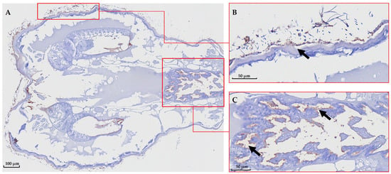
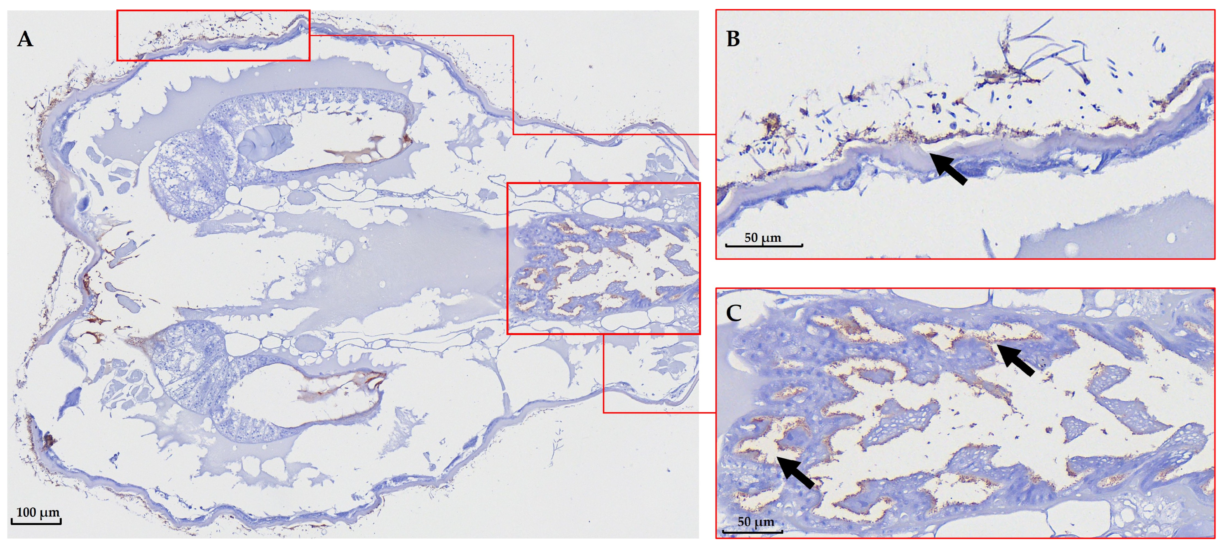
Immunohistochemical analysis revealed the presence of T. maritimum in distinct anatomical structures of the sea lice C. rogercresseyi. Mild to moderate positive signals were observed in the cuticle of both male and female parasites, particularly in superficial regions, indicating that the bacterium can adhere to or colonize external parasite tissues (Figure 1B and Figure 2B).
Figure 1.
Immunohistochemical detection of T. maritimum in Sea Lice (male). (A) Whole sea lice slide, magnification 12×. (B) Magnification 80×; a mild positive reaction to T. maritimum is observed in the superficial area of the cuticle (arrows). (C) Magnification 80×; a moderate positive reaction is observed in the gastrointestinal tract. The scale bar for each image is indicated in micrometers (µm) in the lower left corner of the corresponding panel. The arrows indicate positive reactions by IHC.
Figure 2.
Immunohistochemical detection of T. maritimum in Sea Lice (female). (A) Whole sea lice slide, magnification 1.51×. (B) Magnification 40×; a positive reaction to T. maritimum is observed in the gastrointestinal tract. (C) Magnification 40×; additional positive signal in the gastrointestinal tract. Scale bars are shown in micrometers (µm) in the lower left or right corner of the corresponding panel. The arrows indicate positive reactions by IHC.
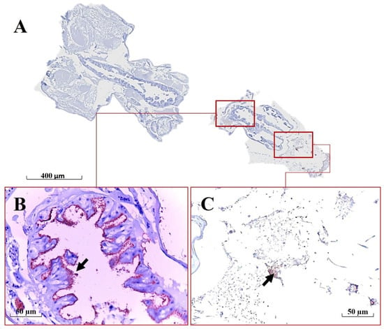
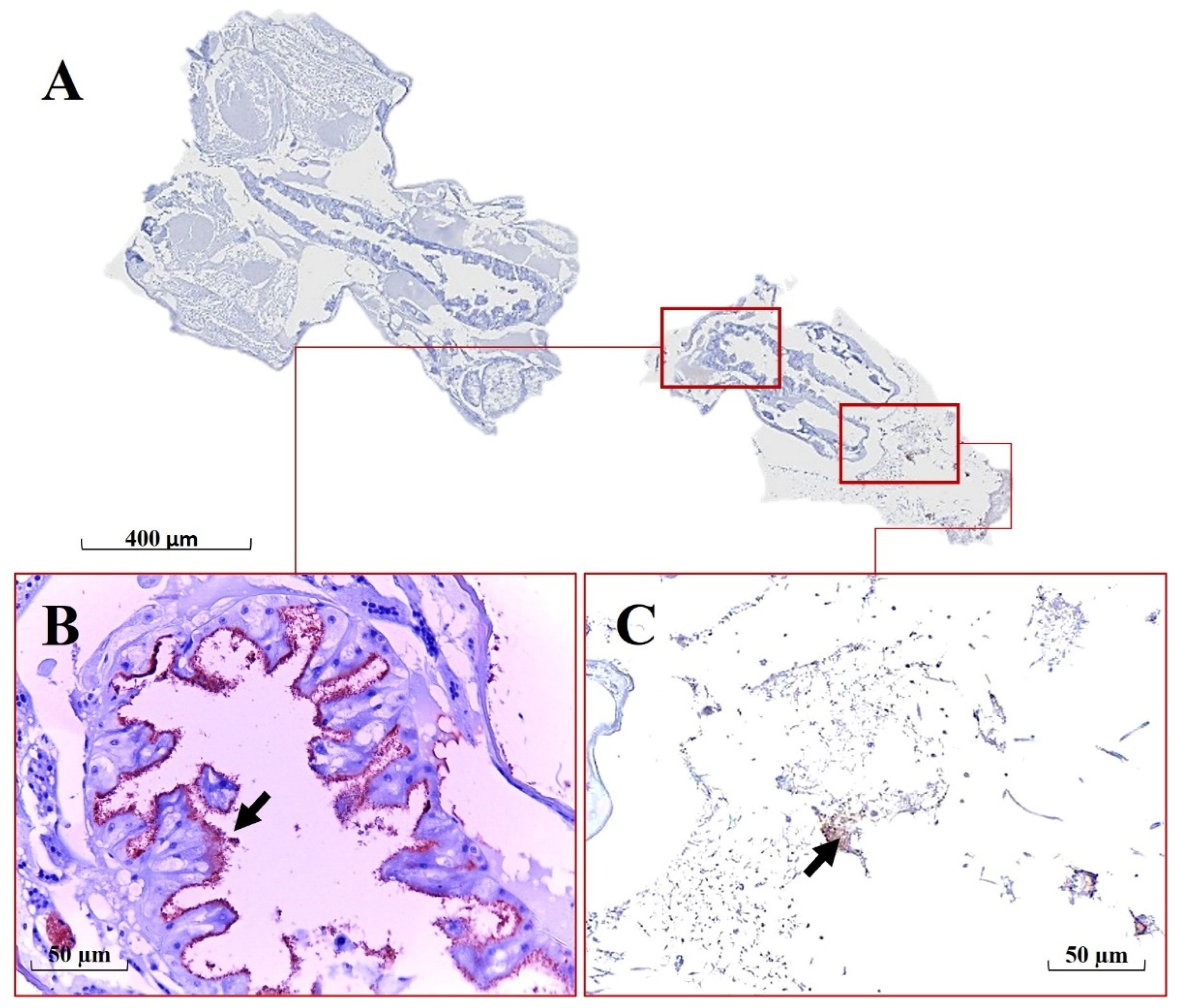
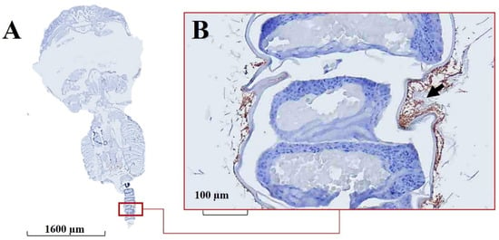
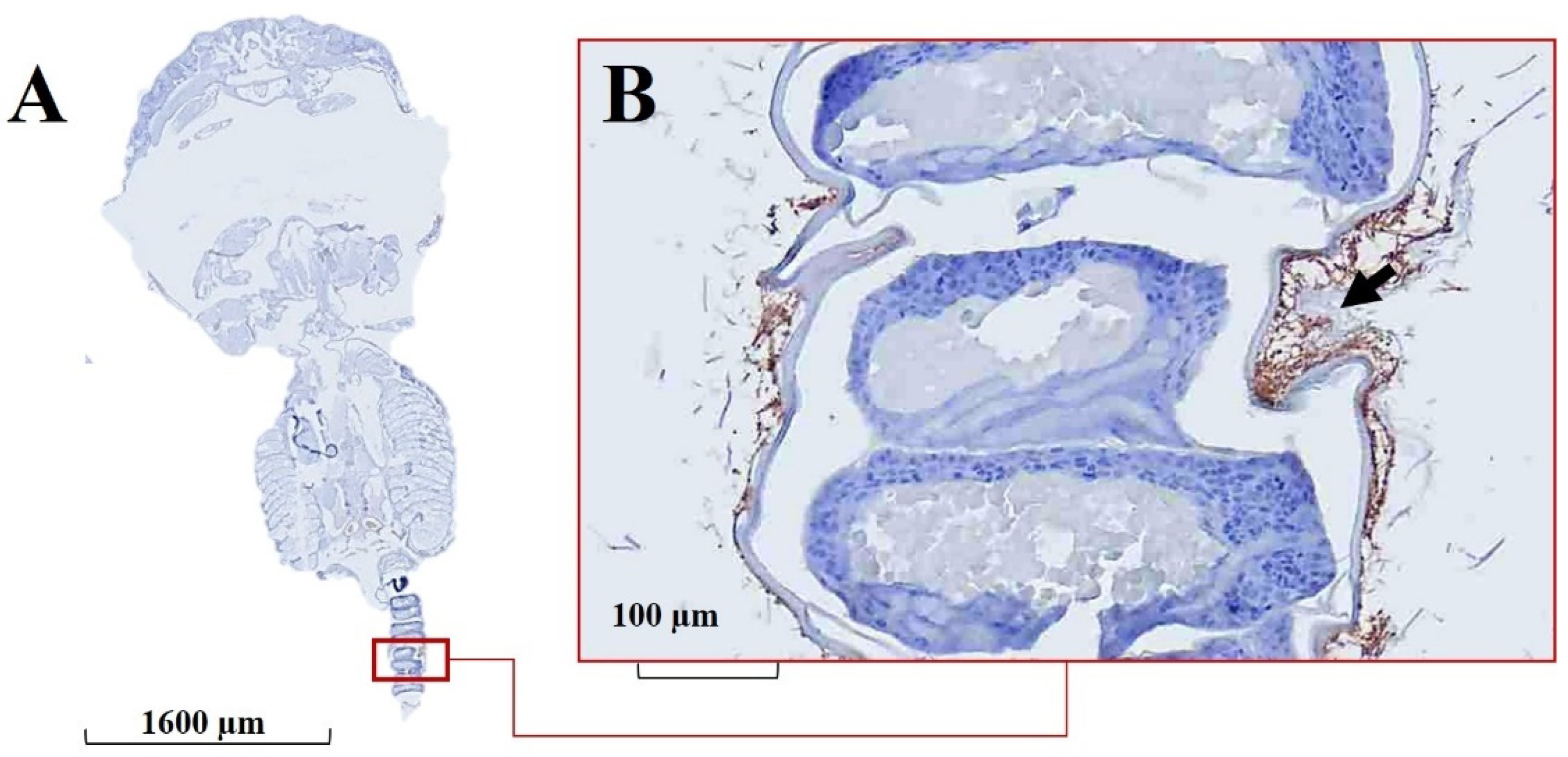
In the case of immunohistochemistry targeting T. dicentrarchi, positive signals were primarily detected in the cuticle of the ectoparasites as well as in the superficial region of the ovigerous sac, where moderate immunoreactivity was consistently observed (Figure 3B).
Figure 3.
Immunohistochemical detection of T. dicentrarchi in Sea Lice (female). (A) Whole sea lice slide, magnification 1×. (B) Magnification 20×; a moderate positive reaction to T. dicentrarchi is observed in the superficial region of the ovigerous sac. The scale bar for each image is indicated in micrometers (µm) in the lower left corner of the corresponding panel. The arrows indicate positive reactions by IHC.
3.2. Specific PCR-Based Detection of Tenacibaculum Species in Caligus rogercresseyi
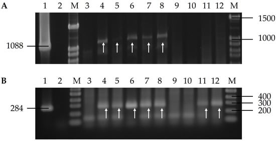
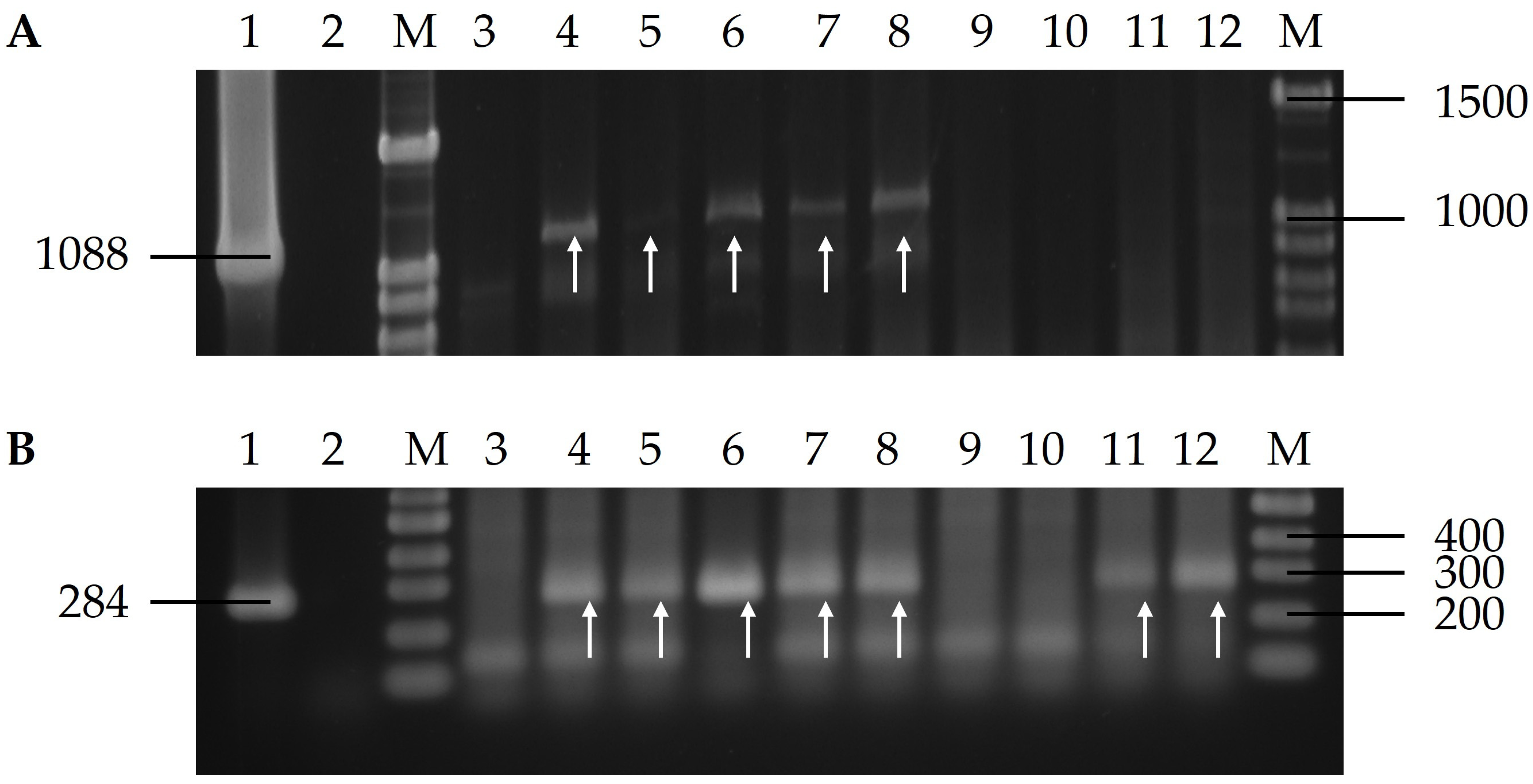
Conventional PCR assays were performed separately on the 10 parasite samples obtained (five males and five ovigerous females), using the species-specific primers described in Table 1 for the detection of T. maritimum and T. dicentrarchi. Five of the ten samples analyzed (5/10) tested positive for T. maritimum which amplified the 1088 bp amplification fragment (Figure 4A). In the case of T. dicentrarchi, seven of the ten samples analyzed (7/10) tested positive for this pathogen, which amplified the 284 bp amplification fragment. Positive controls were consistent with the amplicon sizes generated by the corresponding reference strains used in this study.
Figure 4.
Detection of Tenacibaculum spp. in Caligus rogercresseyi by conventional PCR. (A) Amplification of the 1088 bp fragment specific for T. maritimum using the MAR1/MAR2 primer set. C+ (Lane 1): positive control (T. maritimum NCIMB 2154ᵀ); C− (Lane 2): negative control (no DNA template); M: molecular weight marker. Lanes 3–12 correspond to individual C. rogercresseyi samples. Arrows indicate positive amplification products. Numbers on the right indicate molecular marker sizes (bp), and the number on the left indicates the expected amplicon size (1088 bp). (B) Amplification of the 284 bp fragment specific for T. dicentrarchi using the Tenadi Fw/Rv primer set. C+ (Lane 1): positive control (T. dicentrarchi NCIMB 15498ᵀ); C− (Lane 2): negative control (no DNA template); M: molecular weight marker. Lanes 3–12 correspond to individual C. rogercresseyi samples. Arrows indicate positive amplification products. Numbers on the right indicate molecular marker sizes (bp), and the number on the left indicates the expected amplicon size (284 bp). The arrows indicate positive identificaction by PCR.
4. Discussion
Ectoparasites are increasingly recognized as important drivers in the dissemination of infectious diseases in aquaculture. The presence of sea lice such as C. rogercresseyi may contribute to the spread of emerging pathogens, particularly species of the genus Tenacibaculum (T. maritimum, T. dicentrarchi, T. finnmarkense), which are globally relevant fish pathogens associated with ulcerative skin disorders in salmonids and other species of economic importance [4,17]. In Chile, the primary impacts of C. rogercresseyi infestations on salmon are immunosuppression and mechanical damage to the skin, producing extensive petechial hemorrhages. These lesions provide an entry point for opportunistic bacteria and increase host susceptibility to P. salmonis, the major bacterial pathogen affecting salmon farming in Chile [2].
The skin microbiota of Atlantic salmon plays a central role in shaping the bacterial community associated with C. rogercresseyi [31]. As the parasite feeds on host mucus and epidermal tissues, it acquires not only cellular debris but also bacteria inhabiting the skin surface [31,32]. Opportunistic genera such as Polaribacter, Photobacterium, and Tenacibaculum have been detected in both the salmon skin and the parasite, indicating direct microbial transfer [32]. Similar interactions have been described in other fish–parasite systems, where ectoparasites were shown to modulate host-associated microbial communities, further evidencing the role of parasites as key drivers of microbiota composition and dynamics [33,34]. However, high-throughput sequencing often resolves bacterial identity only to the genus level; thus, Tenacibaculum was identified as a dominant genus in previous studies, but species-level resolution was not possible [31]. In contrast, the present study provides direct evidence of T. maritimum and T. dicentrarchi in C. rogercresseyi, confirmed through species-specific PCR and immunohistochemistry. These findings also validate the reliability of the molecular assays employed, providing robust confirmation of the presence of both Tenacibaculum species in the analyzed C. rogercresseyi specimens.
Histological analysis further demonstrated the tissue-specific localization of T. maritimum within the parasite. Mild to moderate immunohistochemical signals were observed in the superficial cuticle, whereas consistent moderate positive reactions were detected in several regions of the gastrointestinal tract of both male and female lice. Similarly, T. dicentrarchi was detected in the cuticle and superficial ovigerous sac, suggesting that the bacterium may adhere to or colonize parasite surfaces that are in direct contact with the host’s mucus and skin. This localization may favor environmental persistence, while its presence in the ovigerous sac raises the possibility that bacterial cells are associated with reproductive structures, potentially contributing to horizontal and, possibly, vertical transmission pathways. The detection of both pathogens in distinct anatomical structures indicates that they are not restricted to external contamination but may also persist within the parasite, supporting the hypothesis that C. rogercresseyi may share a similar ecological role to L. salmonis as a potential carrier of bacterial pathogens [15,35]. Similar observations have been reported in other ectoparasite systems. Ticks are well-recognized carriers and vectors of a wide range of bacterial, viral, and protozoan pathogens, and interactions among pathogens, microbiota, and arthropod hosts have been shown to influence transmission success [15]. Likewise, aquatic ectoparasites such as L. salmonis have been reported to harbor several putative fish pathogens, including bacterial and viral agents, while other marine invertebrates, such as jellyfish, have been implicated in the transport of fish pathogens [11,12,22]. However, the detection of microorganisms within an ectoparasite does not necessarily demonstrate vector competence, as successful pathogen transmission requires experimental evidence of acquisition, persistence, and transfer to a susceptible host. Previous studies on L. salmonis and C. rogercresseyi have highlighted the importance of distinguishing pathogen carriage from true vectorial capacity [13,14]. At least for T. maritimum, these localization patterns are consistent with a surface-associated lifestyle: its moderately hydrophobic cells rapidly form viable biofilms that may act as transient reservoirs in aquaculture environments, providing a plausible mechanism for persistence on the louse cuticle and other parasite-associated interfaces [36].
Analogous situations have been documented in other host–parasite systems. For instance, the hydromedusa Phialella quadrata was reported to act as a reservoir and vector of T. maritimum, causing gill disease in Atlantic salmon in Scotland [12]. Similarly, L. salmonis in the Northern Hemisphere has been associated with bacterial pathogens such as A. salmonicida, T. maritimum, T. dicentrarchi, P. fluorescens, V. anguillarum and Vibrio spp., as well as viral pathogens including salmonid alphavirus and infectious salmon anemia virus [11,21,26,27]. Experimental studies have also demonstrated that L. salmonis can act as a mechanical rather than biological vector of IHNV, transmitting the virus only after feeding on infected donor fish [13].
Taken together, these findings reinforce the hypothesis that C. rogercresseyi may share a similar ecological status with L. salmonis as a potential carrier of bacterial pathogens. Our study is the first to demonstrate the presence of T. maritimum and T. dicentrarchi in C. rogercresseyi through combined molecular and histological approaches. While the evidence confirms their presence within the parasite, further experimental work is needed to establish whether sea lice actively transmit Tenacibaculosis among salmon within cages or across sites under specific environmental conditions. Considering the persistence of Sea Lice in Chilean salmon farming since the early stages of the industry [37,38], and its potential to harbor pathogens already documented in related ectoparasites, our findings highlight the urgent need for targeted studies to clarify the epidemiological role of C. rogercresseyi in disease dynamics.
5. Conclusions
This work constitutes the first report showing the presence of Tenacibaculum species in Caligus rogercresseyi through the combined use of immunohistochemistry and PCR. While PCR confirmed the presence of T. maritimum and T. dicentrarchi, immunohistochemistry provided evidence of their anatomical localization within the parasite. The results demonstrate that C. rogercresseyi can harbor both pathogens, supporting the potential role of C. rogercresseyi as a carrier of Tenacibaculum spp. in salmon aquaculture. These findings expand current knowledge regarding potential pathogen reservoirs in farming systems; however, they should be interpreted as evidence of pathogen carriage rather than transmission, since no evidence of vector competence or pathogen transmission was generated in the present study. Experimental studies are required to determine whether C. rogercresseyi can act as a mechanical or biological vector of Tenacibaculum spp.
Author Contributions
Conceptualization, P.I. and H.O.; methodology, E.J., S.V. and C.S.; writing—original draft preparation, P.I.; writing—review and editing, J.R.; funding acquisition, J.R. and P.I. All authors have read and agreed to the published version of the manuscript.
Funding
The authors are grateful for the financial support provided by the Grant PI–5061 from CORFO. This work was also supported by the Agencia Nacional de Investigación y Desarrollo de Chile (ANID), through National Doctoral Scholarships No. 2121395 (P.I.) and No. 21212273 (S.V.), and Anillos Regulares de Tecnología ACT240045.
Institutional Review Board Statement
According to our institution’s policies and local regulations, Ethics Committee approval was not required for this work.
Informed Consent Statement
Not applicable.
Data Availability Statement
The data presented in this study are available on request from the corresponding author. Access to the data may be subject to restrictions imposed by collaborating companies to protect confidential and commercially sensitive information.
Acknowledgments
P.I. acknowledges the support of Agencia Nacional de Investigación y Desarrollo de Chile (ANID), Chile, through the National Doctoral Scholarship No. 2121395, Doctorado en Acuicultura, Universidad de Chile, Universidad Católica del Norte, Pontificia Universidad Católica de Valparaíso, Chile. S.V. acknowledges the support of the Agencia Nacional de Investigación y Desarrollo de Chile (ANID), through the National Doctoral Scholarship No. 21212273 and Doctoral Program in Molecular Biotechnology, Universidad de Concepción.
Conflicts of Interest
Authors Pedro Ilardi, Eugenia Jerez, Sara Valdes, Harold Oliva were employed by the company Investigación y Desarrollo Farmacología en Aquacultura Veterinaria FAV S.A. Author Carlos Sandoval was employed by the company Research & Development Veterinary Histopathology Center SpA. The remaining authors declare that the research was conducted in the absence of any commercial or financial relationships that could be construed as a potential conflict of interest.
Abbreviations
The following abbreviations are used in this manuscript:
| ISAV | Infectious Salmon Anemia Virus |
| IHNV | Infectious Hematopoietic Necrosis Virus |
| IHC | Immunohistochemistry |
| NCIMB | National Collection of Industrial, Food and Marine Bacteria |
| PCR | Polymerase Chain Reaction |
References
- Gallardo-Escarate, C.; Valenzuela-Muñoz, V.; Núñez-Acuña, G.; Carrera, G.; Gonçalves, A.T.; Valenzuela-Miranda, D.; Benavente, B.P.; Roberts, S. Catching the complexity of salmon–louse interactions. Fish Shellfish. Immunol. 2019, 90, 199–209. [Google Scholar] [CrossRef] [Scilit] [PubMed]
- Arriagada, G.; Hamilton-West, C.; Nekouei, O.; Foerster, C.; Müller, A.; Lara, M.; Gallardo-Escarate, C. Caligus rogercresseyi infestation is associated with Piscirickettsia salmonis attributed mortalities in farmed salmonids in Chile. Prev. Vet. Med. 2017, 171, 104771. [Google Scholar]
- Lepe-López, M.; Lepe, A.; Farias, C. Assessing the Present and Future Habitat Suitability of Caligus rogercresseyi in Marine Salmon–Farm Areas in Southern Chile. Front. Vet. Sci. 2021, 8, 615039. [Google Scholar] [PubMed]
- Mabrok, M.; Algammal, A.M.; Sivaramasamy, E.; Hetta, H.F.; Atwah, B.; Alghamdi, S.; Fawzy, A.; Avendaño-Herrera, R.; Rodkhum, C. Tenacibaculosis caused by Tenacibaculum maritimum: Update knowledge of this marine bacterial fish pathogen. Front. Cell. Infect. Microbiol. 2023, 12, 1068000. [Google Scholar] [CrossRef] [Scilit] [PubMed]
- Kumanan, K.; Carson, J.; Hunter, R.B.J.; Rolton, A.; von Ammon, U.; Bandaranayake, C.; Angelucci, C.; Morrison, R.N.; Walker, S.P.; Symonds, J.E.; et al. Experimental challenge of Chinook Salmon (Oncorhynchus tshawytscha) with Tenacibaculum maritimum and Tenacibaculum dicentrarchi fulfils Koch’s postulates. J. Fish Dis. 2025, 48, e14088. [Google Scholar] [CrossRef] [Scilit] [PubMed]
- SERNAPESCA. 2026. Available online: https://www.sernapesca.cl/informes-y-datos/resultados-de-gestion/ (accessed on 15 July 2026).
- Ramírez, C.; Romero, J. Unveiling Hidden Allies: In Silico Discovery of Prophages in Tenacibaculum Species. Antibiotics 2024, 13, 1184. [Google Scholar] [CrossRef] [Scilit] [PubMed]
- Valdes, S.; Irgang, R.; Barros, M.C.; Ilardi, P.; Saldarriaga-Córdova, M.; Rivera-Bohle, J.; Madrid, E.; Gajardo-Córdova, J.; Avendaño-Herrera, R. First report and characterization of Tenacibaculum maritimum isolates recovered from rainbow trout (Oncorhynchus mykiss) farmed in Chile. J. Fish Dis. 2021, 44, 1481–1490. [Google Scholar] [CrossRef] [Scilit] [PubMed]
- Escribano, M.P.; Ageitos, L.; Balado, M.; Buedenbender, L.; Rodríguez, J.; Jiménez, C.; Magariños, B.; Lemos, M.L. Genetic and biochemical insights into siderophore biosynthesis in the marine fish pathogen Tenacibaculum maritimum. Sci. Rep. 2025, 15, 26210. [Google Scholar] [CrossRef] [Scilit] [PubMed]
- Gaete-Carrasco, Á.; Rosenfeld, C.; Lara, M.; Mardones, F.O.; Avendaño-Herrera, R. Risk factors associated with Tenacibaculosis-related mortality in farmed salmon in Chile from 2018 to 2021. J. Fish Dis. 2026, e70249. [Google Scholar] [CrossRef] [Scilit] [PubMed]
- Klimesova, B.; Ruane, N.M.; Domingo-Bretón, R.; Moroni, F.; Naya-Catalá, F.; Pérez-Sanchez, J.; O’Dwyer, K.; Lyashevska, O.; Rodger, H.; Talbot, A. Sea Lice (Lepeophtheirus salmonis) harbour putative fish pathogens: Insights from illumina and nanopore sequencing. J. Fish Dis. 2026, 49, e70182. [Google Scholar] [CrossRef] [Scilit] [PubMed]
- Ferguson, H.W.; Delannoy, C.M.J.; Hay, S.; Nicolson, J.; Sutherland, D.; Crumlish, M. Jellyfish as vectors of bacterial disease for farmed salmon (Salmo salar). J. Vet. Diagn. Investig. 2010, 22, 376–382. [Google Scholar] [CrossRef] [Scilit] [PubMed]
- Jakob, E.; Barker, D.E.; Garver, K.A. Vector potential of the salmon louse Lepeophtheirus salmonis in the transmission of infectious haematopoietic necrosis virus (IHNV). Dis. Aquat. Org. 2011, 97, 155–165. [Google Scholar] [CrossRef] [Scilit] [PubMed]
- Labra, A.; Bravo, S.; Marshall, S. Defining the role of Caligus rogercresseyi in transmission and spreading of Piscirickettsia salmonis. Aquaculture 2020, 528, 735489. [Google Scholar] [CrossRef] [Scilit]
- Cabezas-Cruz, A.; Mateos-Hernández, L.; Alberdi, P.; Villar, M.; Riveau, G.; Hermann, E.; Díaz-Sánchez, S.; Estrada-Peña, A.; Hodžić, A.; de la Fuente, J. Effect of vaccines against Tick gut Microbiota in Ixodes ricinus on vector competence for transmission of pathogens. Vaccines 2022, 10, 1063. [Google Scholar]
- Novak, C.W.; Lewis, D.L.; Collicutt, B.; Verkaik, K.; Barker, D.E. Investigations on the role of the salmon louse, Lepeophtheirus salmonis (Caligidae), as a vector in the transmission of Aeromonas salmonicida subsp. salmonicida. J. Fish Dis. 2016, 39, 1165–1178. [Google Scholar] [CrossRef] [Scilit] [PubMed]
- Bravo, S.; Treasurer, J. The Management of the Sea Lice in Chile: A Review. Rev. Aquac. 2023, 15, 1749–1764. [Google Scholar] [CrossRef] [Scilit]
- Nese, L.; Enger, Ø. Isolation of Aeromonas salmonicida from salmon lice Lepeophtheirus salmonis and marine plankton. Dis. Aquat. Org. 1993, 16, 79–81. [Google Scholar] [CrossRef] [Scilit]
- Petterson, E.; Sandberg, M.; Santi, N. Salmonid alphavirus associated with Lepeophtheirus salmonis (Copepoda: Caligidae) from Atlantic salmon, Salmo salar, L. J. Fish Dis. 2009, 32, 477–479. [Google Scholar] [CrossRef] [Scilit] [PubMed]
- Nylund, A.; Bjørknes, B.; Wallace, C. Lepeophtheirus salmonis: A possible vector in the spread of diseases on salmonids. Bull. Eur. Assoc. Fish Pathol. 1991, 11, 213–216. [Google Scholar]
- Nylund, A.; Wallace, C.; Hovland, T. The possible role of Lepeophtheirus salmonis (Kroyer) in the transmission of infectious salmon anaemia. In Pathogens of Wild and Farmed Fish: Sea Lice; Boxshall, G.A., Defaye, D., Eds.; Ellis Horwood: Chichester, UK, 1993; pp. 367–378. [Google Scholar]
- Barker, D.E.; Braden, L.M.; Coombs, M.P.; Boyce, B. Preliminary studies on the isolation of bacteria from sea lice, Lepeophtheirus salmonis, infecting farmed salmon in British Columbia, Canada. Parasitol. Res. 2009, 105, 1173–1177. [Google Scholar] [CrossRef] [Scilit] [PubMed]
- Lhorente, J.P.; Gallardo, J.A.; Villanueva, B.; Carabaño, M.J.; Neira, R. Disease resistance in Atlantic salmon (Salmo salar): Coinfection of the intracellular bacterial pathogen Piscirickettsia salmonis and the sea louse Caligus rogercresseyi. PLoS ONE 2014, 9, e95397. [Google Scholar] [CrossRef] [Scilit] [PubMed]
- Diethelm-Varela, B.; Atero, N.; Córdova-Buhrle, F.; Rezende, E.L.; Gelcich, S.; Sandoval, O.; Navarro, C.; Mardones, F.O. Epidemiology of Salmonid Rickettsial Septicemia (SRS) in farmed salmon: The role of Sea Lice infestations in mortality risk. J. Fish Dis. 2026, 49, e70097. [Google Scholar] [CrossRef] [Scilit] [PubMed]
- Valenzuela-Miranda, D.; Gonçalves, A.T.; Valenzuela-Muñoz, V.; Nuñez-Acuña, G.; Liachko, I.; Nelson, B.; Gallardo-Escarate, C. Proximity ligation strategy for the genomic reconstruction of microbial communities associated with the ectoparasite Caligus rogercresseyi. Sci. Rep. 2022, 12, 783. [Google Scholar] [CrossRef] [Scilit] [PubMed]
- Oelckers, K.; Vike, S.; Duesund, H.; Gonzalez, J.; Wadsworth, S.; Nylund, A. Caligus rogercresseyi as a potential vector for transmission of infectious Salmon Anaemia (ISA) virus in Chile. Aquaculture 2014, 420–421, 126–132. [Google Scholar] [CrossRef] [Scilit]
- Økland, A.L.; Skoge, R.H.; Nylund, A. The complete genome sequence of CrRV–Ch01, a new member of the family Rhabdoviridae in the parasitic copepod Caligus rogercresseyi present on farmed Atlantic salmon (Salmo salar) in Chile. Arch. Virol. 2018, 163, 1657–1661. [Google Scholar] [CrossRef] [Scilit] [PubMed]
- Sandoval, C.; Infante, J.; Abad, J.; Ferguson, H.W.; Paredes, E.; Valdebenito, S.; Yáñez, A.; Ilardi, P.; Avendaño-Herrera, R. Case report: Strawberry disease in farmed Chilean rainbow trout (Oncorhynchus mykiss, Walbaum). J. Aquat. Anim. Health 2016, 28, 1–10. [Google Scholar] [PubMed]
- Toyama, T.; Kita-Tsukamoto, K.; Wakabayashi, H. Identification of Flexibacter maritimus, Flavobacterium branchiophilum and Cytophaga columnaris by PCR targeted 16S Ribosomal DNA. Fish Pathol. 1996, 31, 25–31. [Google Scholar] [CrossRef] [Scilit]
- Avendaño-Herrera, R.; Irgang, R.; Tapia-Cammas, D. PCR procedure for detecting the fish pathogen Tenacibaculum dicentrarchi. J. Fish Dis. 2018, 41, 715–719. [Google Scholar] [CrossRef] [Scilit] [PubMed]
- Llewellyn, M.S.; Leadbeater, S.; Garcia, C.; Sylvain, F.-E.; Custodio, M.; Ang, K.P.; Powell, F.; Carvalho, G.R.; Creer, S.; Elliot, J.; et al. Parasitism perturbs the mucosal microbiome of Atlantic Salmon. Sci. Rep. 2017, 7, 43465. [Google Scholar] [CrossRef] [Scilit] [PubMed]
- Valenzuela-Miranda, D.; Valenzuela-Muñoz, V.; Benavente, B.P.; Muñoz-Troncoso, M.; Nuñez-Acuña, G.; Gallardo-Escárate, C. The Atlantic salmon microbiome infected with the Sea Louse Caligus rogercresseyi reveals tissue–specific functional dysbiosis. Aquaculture 2024, 580, 740328. [Google Scholar] [CrossRef] [Scilit]
- Revault, J.; Desdevises, Y.; Magnanou, É. Link between bacterial communities and contrasted loads in ectoparasitic monogeneans from the external mucus of two wild sparid species (Teleostei). BMC Anim. Microbiome 2024, 6, 42. [Google Scholar] [CrossRef] [Scilit] [PubMed]
- Scheifler, M.; Magnanou, E.; Sanchez-Brosseau, S.; Desdevises, Y. Host–microbiota–parasite interactions in two wild sparid fish species, Diplodus annularis and Oblada melanura (Teleostei, Sparidae) over a year: A pilot study. BMC Microbiol. 2023, 23, 340. [Google Scholar] [CrossRef] [Scilit] [PubMed]
- Mathieu-Begné, E.; Blanchet, S.; Rey, O.; Toulza, E.; Veyssière, C.; Manzi, S.; Lefort, M.; Scelsi, O.; Loot, G.A. Longitudinal survey in the wild reveals major shifts in fish host microbiota after parasite infection. Mol. Ecol. 2023, 32, 3014–3024. [Google Scholar] [CrossRef] [Scilit] [PubMed]
- Levipan, H.A.; Tapia-Cammas, D.; Molina, V.; Irgang, R.; Toranzo, A.E.; Magariños, B.; Avendaño-Herrera, R. Biofilm development and cell viability: An undervalued mechanism in the persistence of the fish pathogen Tenacibaculum maritimum. Aquaculture 2019, 511, 734267. [Google Scholar] [CrossRef] [Scilit]
- Boxshall, G.A.; Bravo, S. On the Identity of Caligus Species (Copepoda: Caligidae) from farmed salmonids in Chile. Contrib. Zool. 2000, 69, 137–146. [Google Scholar] [CrossRef] [Scilit]
- Johnson, S.C.; Bravo, S.; Nagasawa, K.; Kabata, Z.; Hwang, J.; Ho, J.S.; Shih, C.T.A. Review of the impact of parasitic Copepods on marine aquaculture. Zool. Stud. 2004, 43, 229–243. [Google Scholar]

Disclaimer/Publisher’s Note: The statements, opinions and data contained in all publications are solely those of the individual author(s) and contributor(s) and not of MDPI and/or the editor(s). MDPI and/or the editor(s) disclaim responsibility for any injury to people or property resulting from any ideas, methods, instructions or products referred to in the content. |
© 2026 by the authors. Licensee MDPI, Basel, Switzerland. This article is an open access article distributed under the terms and conditions of the Creative Commons Attribution (CC BY) license.