


Abstract
This study assessed the diagnostic reliability of anti-Schistosoma serology in Ghanaian patients with and without concomitant human immunodeficiency virus (HIV) infection. Serological testing was performed on serum samples containing PCR-confirmed Schistosoma spp. DNA using a commercial enzyme-linked immunosorbent assay (ELISA; EUROIMMUN, Lübeck, Germany). Following real-time PCR confirmation of either Schistosoma mansoni or Schistosoma haematobium complex DNA, samples were subjected to anti-Schistosoma IgG and IgM serology in duplicate. Results were qualitatively interpreted as positive, uncertain, or negative; reproducible positive results across test runs were defined as “consistent,” while all other profiles were classified as “inconsistent”. Serological consistency was then correlated with patient demographic and clinical data, including age, gender, HIV status, CD4+ and CD8+ T-lymphocyte counts, CD4+/CD8+ ratios, and HIV viral loads. Of the 41 individuals with PCR-proven schistosomiasis (S. mansoni, n = 36; S. haematobium, n = 5) and no clinical signs of Katayama syndrome, 39 were HIV-positive. Among these 39 coinfected patients, consistently positive serology was recorded in 27 (69.2%) cases for IgG and 31 (79.5%) cases for IgM. Consistent IgG seropositivity was significantly associated with higher CD4+ (293 vs. 58 cells/µL, p = 0.009) and CD8+ (981.5 vs. 306 cells/µL, p = 0.007) T-lymphocyte counts. The association between higher CD4+ cell counts and consistent IgG reactivity remained robust in multivariable analysis (adjusted odds ratio = 2.04 per 100 cells/µL, p = 0.01). A non-significant trend in the same direction was observed for anti-Schistosoma IgM, whereas no other significant associations with epidemiological or medical factors were identified. In conclusion, the state of cellular immunity critically influences the reliability of serological schistosomiasis diagnostics in HIV-positive individuals. For patients with severe cellular immune depletion and suspected schistosomiasis, alternative diagnostic approaches, such as highly sensitive real-time PCR, should be strongly considered.
1. Introduction
Affecting over a quarter of a billion people in tropical endemic regions [1], schistosomiasis ranks among the most prevalent systemic parasitic diseases worldwide. The causative agents are trematodes of the genus Schistosoma spp., with clinical manifestations depending on the infecting species: S. mansoni and S. japonicum complexes typically cause intestinal and hepatosplenic schistosomiasis, whereas the S. haematobium complex leads to urinary schistosomiasis [2]. In travelers and migrants returning from endemic areas who present with eosinophilia, schistosomiasis must be considered a primary differential diagnosis [3].
Transmission occurs through skin contact with parasite-infested freshwater in endemic environments [1]. Acute schistosomiasis, also known as Katayama syndrome, is characterized by flu-like symptoms [2]. However, the clinical presentation of chronic schistosomiasis can vary widely, often featuring mild, intermittent, or nonspecific symptoms [2]. Standard treatment involves single or multiple doses of oral praziquantel to eradicate adult worms [2]. The clinical efficacy of praziquantel has recently been shown to be independent of the presence or absence of acquired immunodeficiency [4,5], yielding an expected cure rate of approximately 80% [6].
Diagnosis relies on a combination of microscopy, molecular biology, antigen testing, and serology. Because each method independently exhibits imperfect diagnostic accuracy, a combined diagnostic approach is often required [2]. Various serological assays utilizing different antigen types are currently in clinical use [7]. However, major limitations of serology include the delayed emergence of anti-Schistosoma antibodies and the inability of these tests to reliably distinguish between acute, chronic, and past infections [8]. Furthermore, a recent review highlighted the highly variable diagnostic accuracy of these antibody assays, reporting sensitivities ranging from 71% to 99% and specificities from 6% to 100% [9]. While the sensitivity of Schistosoma serology is generally considered superior to its specificity, it is hindered by broad confidence intervals, particularly when validated against composite reference standards [10,11]. A lack of assay standardization and broad validation remains a primary driver of this diagnostic uncertainty [9].
The interplay between human immunodeficiency virus (HIV) infection and schistosomiasis has been widely debated, with studies indicating increased odds of HIV positivity among individuals with schistosomiasis [12,13]. Furthermore, coincident schistosomiasis has been linked to higher HIV transmission rates and accelerated disease progression [14]. Although the exact reasons and directionality of these associations remain partly unclear [12], several underlying mechanisms have been identified. Specifically, S. mansoni infection alters anti-HIV immune responses, leading to the reduced detectability of bifunctional (IFNγ+ IL-2- TNF-α+) CD4+ T-cells, heightened CD4+ T-cell activation, and diminished anti-HIV antibody-dependent cellular cytotoxicity (ADCC) titers [15]. Additionally, soluble egg antigen (SEA) from S. mansoni reduces the susceptibility of T-cells to CCR5-tropic HIV-1 [16]. In a Chinese cohort coinfected with HIV and S. japonicium, patients exhibited reduced CD4+ T-cell counts and CD4+/CD8+ ratios, alongside elevated CD8+ T-cell counts [17]. Cross-reactivity has also been documented between antibodies targeting HIV-1 envelope glycoproteins (env) and S. mansoni glycan antigens [18,19,20], as well as between anti-S. mansoni antibodies and the HIV virion infectivity factor (vif) [21,22]. Consequently, immune responses directed against schistosomiasis have been linked to false-positive HIV serology [23]. Notably, egg shedding is often reduced in HIV-coinfected schistosomiasis patients compared to their HIV-negative counterparts [24]. In patients progressing to acquired immunodeficiency syndrome (AIDS), the initiation of antiretroviral therapy (ART) can trigger a schistosomiasis-associated immune reconstitution inflammatory syndrome (IRIS) [25]. Conversely, schistosomiasis has also been shown to negatively impair HIV-specific immune responses [26].
Schistosomiasis inherently induces significant systemic immune alterations [12]. The disease typically elicits a slow adaptive immune response, even in immunocompetent hosts, and despite extensive research, no licensed vaccine is currently available [27]. Inflammation is primarily driven by tissue-deposited trematode eggs rather than the adult worms residing in the host’s vasculature [28]. Embedded within tissue granulomas, Schistosoma spp. eggs provoke a CD4+ T-cell-dependent hypersensitivity reaction, where the balance between T-helper 1 (Th1) and Th2 responses determines the severity of the inflammation and subsequent disease [28]. A dominant Th1 response is associated with intense local inflammation and disease progression, whereas a Th2 response exerts antagonistic, tissue-protective effects [28]. However, this Th2-mediated reduction in host tissue damage represents a double-edged sword, as it simultaneously promotes parasite survival [29]. When considering worm rather than egg antigens, pronounced γδ T-cell responses are elicited, which can be further modulated by co-existing active or latent tuberculosis [30]. Interestingly, unisexual infection models have revealed sex-specific immunological host responses: while male worms provoke strong innate immunity that ultimately reduces worm and tissue egg burdens, female worms suppress the early innate immune response to invading cercariae and upregulate host genes linked to anergy [29]. Schistosomiasis has also been associated with reduced autoimmunity, evidenced by lower antinuclear antibody (ANA) levels in individuals infected with S. haematobium [31]. Although schistosomiasis-induced immunity does not confer general protection against reinfection, elevated eosinophil levels have been identified as a likely protective factor in reinfection-resistant subpopulations [32]. Conversely, HIV coinfection correlates with increased reinfection rates, a phenomenon linked to blunted immune responses following praziquantel treatment [33]. While HIV does not completely abrogate the immune response to schistosomiasis, treated coinfected individuals exhibit only an isolated increase in interleukin-5 (IL-5). In contrast, HIV-negative individuals demonstrate a robust, broad-spectrum response post-treatment, characterized by elevated levels of soluble worm antigen-specific IgG1–Ig4, IL-4, and IL-5 [33].
Given the complex interplay between HIV-induced immunosuppression and schistosomiasis-associated immune responses, we investigated the impact of HIV-related immune parameters on the reliability of antibody-based Schistosoma serodiagnosis. To this end, we screened for Schistosoma spp.-specific antibodies in sera of Ghanaian individuals with PCR-confirmed schistosomiasis—both with and without HIV coinfection—who presented without clinical signs of acute schistosomiasis or Katayama syndrome.
2. Materials and Methods
2.1. Sample Population
Residual serum samples with PCR-confirmed Schistosoma spp. DNA (desoxyribonucleic acid) [11,34] from Ghanaian individuals, both with and without HIV infection, were included in this assessment. As detailed elsewhere [34], these samples represent a subpopulation drawn from an original study of 1095 HIV-positive Ghanaian patients and a comparison group of 107 HIV-negative Ghanaian blood donors. While the presence of Schistosoma DNA was the primary inclusion criterion, samples were excluded if the residual volume was insufficient for the serological testing described below. Clinical metadata available for the serum donors included age, sex, HIV status, and, for the HIV-positive cohort, immunological parameters from a previous assessment, specifically, CD4+ T-lymphocyte counts, CD4+/CD8+ ratios, and HIV viral loads [35,36]. Prior to further investigations, all serum samples had been stored deep-frozen at −80 °C.
2.2. Serological Assessment
Anti-Schistosoma immunoglobulin G (IgG) and immunoglobulin M (IgM) were measured automatically using a commercial enzyme-linked immunosorbent assay (ELISA) provided by EUROIMMUN (EUROIMMUN Medizinische Labordiagnostika AG, Lübeck, Germany) on a EUROIMMUN Analyzer I device. Following the manufacturer’s instructions for in vitro diagnostic use, the results were non-parametrically classified as positive, uncertain, or negative. Beyond the manufacturer’s internal validation, independent diagnostic accuracy assessments have been published for this assay. A recent study in a Brazilian area endemic for S. mansoni reported a sensitivity of 100% and a specificity of 67% when evaluated against a composite reference standard of serological, microscopic, and molecular methods [37]. In our previous work assessing pregnant Madagascan women [38] with confirmed negative HIV status [39], the estimated sensitivity and specificity were 38.3% and 87.6% for the IgM assay, and 77.7% and 90.3% for the IgG assay, respectively. All samples included in the present study were tested in duplicate.
2.3. Interpretation of Serological Test Results and Statistical Analysis
Using the duplicate serological assessments, each sample was categorized into one of six ordinal groups for both anti-Schistosoma IgG and IgM reactivity: 0 = negative in both runs; 1 = uncertain in one run and negative in the other; 2 = uncertain in both runs; 3 = positive in one run and negative in the other; 4 = positive in one run and uncertain in the other; and 5 = positive in both runs. Category 5 was defined as “consistent”, while all other profiles (categories 0–4) were classified as “inconsistent”. Cycle threshold (Ct) values specific to either the S. mansoni or S. haematobium complex were obtained from prior analyses [34].
Categorical variables are presented as absolute frequencies and percentages, whereas continuous variables are summarized as medians with interquartile ranges (IQRs). Group comparisons between consistently positive participants (category 5) and those with inconsistent or negative profiles (categories 0–4) were performed using Fisher’s exact test for categorical variables and the Mann–Whitney U test for continuous variables. To evaluate the relationship between host immune status and parasite burden, Spearman’s rank correlation coefficient was calculated between CD4+ T-lymphocyte counts and PCR Ct values. Multivariable logistic regression models were constructed to identify independent factors associated with consistent IgG and IgM seroreactivity after adjusting for potential confounders. Adjusted odds ratios (aORs) and their corresponding 95% confidence intervals (CIs) are reported. Additional sensitivity analyses incorporated antiretroviral therapy status and HIV viral load. All statistical tests were two-tailed, and a p-value < 0.05 was considered statistically significant. Analyses were conducted using R software (version 4.5.3; R Foundation for Statistical Computing, Vienna, Austria).
2.4. Ethics
The samples were collected and analyzed in accordance with study protocols approved by the Committee on Human Research of the Kwame Nkrumah University of Science and Technology in Kumasi, Ghana (CHRPE/AP/12/11, approved on 8 September 2012), and the Ethics Committee of the Medical Council in Hamburg, Germany (PV3771). All study participants provided written informed consent prior to enrollment. All procedures were conducted in full accordance with the Declaration of Helsinki and its subsequent amendments.
3. Results
3.1. Sample Characteristics
Following the application of the established case definitions and exclusion criteria, a total of 41 serum samples were included in the investigation. As detailed in Appendix A (Table A1), 36 samples tested positive for S. mansoni complex DNA, whereas 5 samples harbored S. haematobium complex DNA. Regarding serological profiling, detectable reactivity—ranging from borderline to strongly positive results—for anti-Schistosoma spp.-specific IgG or IgM was observed across all analyzed samples. Notably, none of the participants presented with clinical symptoms indicative of acute schistosomiasis or Katayama syndrome.
3.2. Characteristics of the Study Cohort
Of the 41 samples included in the study, 39 originated from HIV-positive individuals and two from HIV-negative participants. Within the HIV-positive cohort, qualitative PCR analysis revealed that the vast majority of Schistosoma infections were attributable to S. mansoni (92.3%, n = 36), while S. haematobium accounted for the remaining 7.7% (n = 3, Table 1). Conversely, only S. haematobium complex DNA was detected in the HIV-negative participants.
Table 1.
Baseline demographic, clinical, and immunological characteristics of the HIV-positive study cohort, stratified by serological consistency regarding Schistosoma-specific IgM and IgG.
The median age of the HIV-positive sub-cohort was 35 years (IQR: 32–42), with females comprising 46.2% (n = 18) of the group. Antiretroviral therapy (ART) was actively utilized by 33.3% (n = 13) of these participants. The median CD4+ T-lymphocyte count was 244 cells/µL (IQR: 60–434), and the median HIV viral load was 5.0 log10 copies/mL (IQR: 3.1–5.5). Comprehensive baseline characteristics for this HIV-positive population are presented in Table 1.
3.3. Immunological and Clinical Parameters Stratified by Serological Consistency
Stratification of the cohort by IgG seroreactivity revealed significant immunological discrepancies between participants demonstrating consistently positive results across all serological test runs (n = 27) and those with inconsistent profiles (n = 12). Specifically, participants with consistent IgG positivity exhibited significantly higher median CD4+ T-lymphocyte counts compared to the inconsistent group (293 vs. 58 cells/µL, p = 0.009). Correspondingly, CD8+ T-lymphocyte counts were also significantly elevated in the consistent IgG group (981.5 vs. 306 cells/µL, p = 0.007). Conversely, other parameters, including age, gender distribution, the CD4+/CD8+ T-lymphocyte ratio, HIV viral load, and PCR Ct values, demonstrated no statistically significant differences between the two IgG groups (p > 0.05 for all). When evaluating IgM serological consistency, individuals classified as consistently positive (n = 31) similarly demonstrated a trend toward higher median CD4+ T-lymphocyte counts than their counterparts with inconsistent or negative results (n = 8; 288.5 vs. 67.5 cells/µL); however, this variation approached but did not reach conventional statistical significance (p = 0.055).
3.4. Association Between Parasite Burden and Immune Status
To address potential confounding by infection intensity, specifically, the possibility that higher antibody consistency simply reflected a heavier parasite burden rather than host immune competence, we assessed the correlation between CD4+ T-lymphocyte counts and PCR Ct values. Spearman’s rank correlation analysis revealed no significant relationship between host CD4+ T-lymphocyte counts and parasite Ct values (rho = 0.068, p = 0.686).
3.5. Multivariable Determinants of Stable IgG and IgM Seropositivity
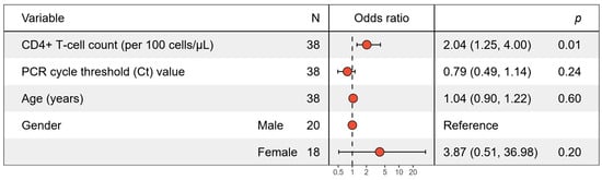
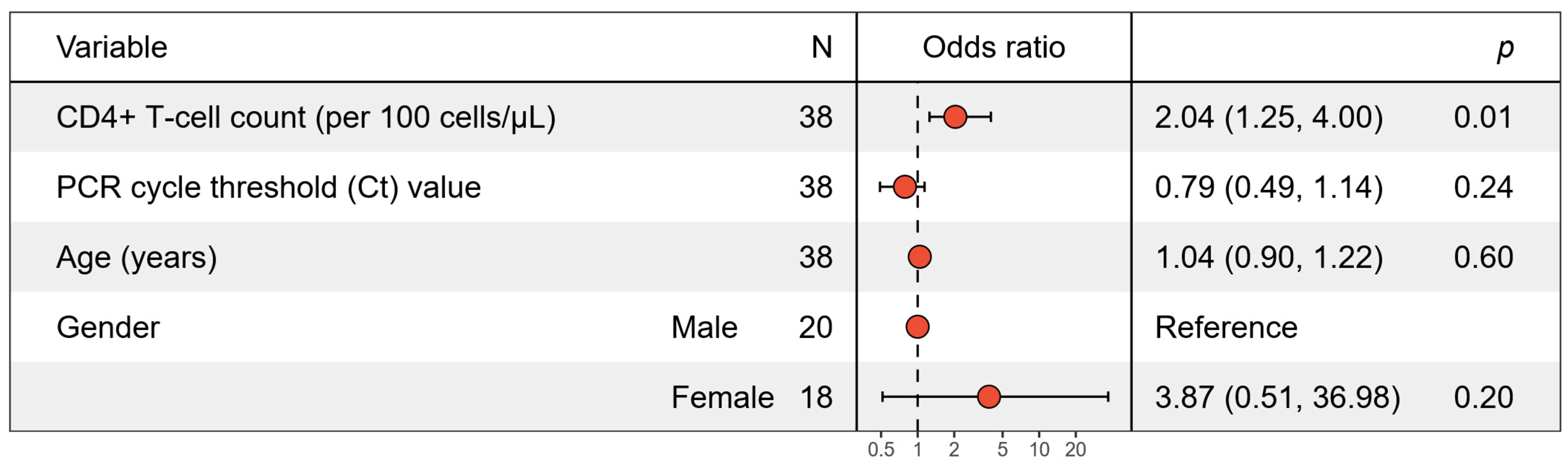
In a multivariable logistic regression model adjusted for PCR Ct value, age, and gender, higher CD4+ T-lymphocyte counts (scaled per 100 cells/µL) emerged as independent predictors of consistent IgG seropositivity (adjusted odds ratio [aOR] = 2.04, 95% CI: 1.25–4.00, p = 0.013, Figure 1). In contrast, PCR Ct values (aOR = 0.79, 95% CI: 0.49–1.14, p = 0.237), age, and gender demonstrated no significant independent associations with consistent IgG reactivity. Sensitivity analyses incorporating ART status or HIV viral load confirmed that the independent association between higher CD4+ T-lymphocyte counts and consistent IgG seroreactivity remained stable and robust. Similarly, multivariable adjustment for consistent IgM seropositivity revealed a parallel trend, with higher CD4+ T-lymphocyte counts showing a positive independent association that approached conventional significance (aOR = 1.74, 95% CI: 1.03–3.63, p = 0.072). Consistent with the IgG models, PCR Ct values (aOR = 0.94, 95% CI: 0.60–1.35, p = 0.741), age (aOR = 1.00, 95% CI: 0.86–1.17, p = 0.963), and gender (aOR = 0.70, 95% CI: 0.08–4.92, p = 0.720) exhibited no significant independent effects on stable IgM seroreactivity.
Figure 1.
Forest plot displaying multivariable logistic regression analyses for factors associated with consistent Schistosoma-specific IgG seroreactivity. Adjusted odds ratios (aOR) and 95% confidence intervals (CI) are shown. Predictors include CD4+ T-lymphocyte counts (scaled per 100 cells/µL), PCR cycle threshold (Ct) values, age, and gender. One patient was excluded from multivariate analysis due to a missing CD4+ T lymphocyte count value.
4. Discussion
This study was conducted to assess the potential effects of HIV-associated immunosuppression on the reliability of the serodiagnosis of schistosomiasis. Both IgG and IgM antibodies were included in the assessment because the humoral responses to Schistosoma spp. egg and worm epitopes have been shown to be equivalent during both acute and chronic stages of the disease [40,41,42]. While HIV positivity is known to exert deleterious effects on the diagnostic sensitivity of serology for visceral leishmaniasis, another systemic parasitic infection [43], the potential impact of HIV-induced immunosuppression on Schistosoma serodiagnosis remains poorly addressed. This gap in the literature is particularly surprising given the extensive body of research demonstrating the complex immunological interactions between schistosomiasis and HIV infection [12,13,14,15,16,17,18,19,20,21,22,23,24,25,26,27,28,29,30,31,32,33].
Our assessment revealed a marked association between severely depleted cellular immunity and the inconsistent serological detection of anti-Schistosoma spp. antibodies in HIV-positive individuals. This phenomenon was more pronounced for the IgG subclass than for IgM. Because the medical histories of the assessed individuals did not suggest acute infections, considerable variability in anti-Schistosoma spp. IgM titers was not unexpected. Focusing specifically on CD4+ T-lymphocytes, the very low cell densities typical of late-presenting HIV infections [44] were strongly associated with inconsistent anti-Schistosoma IgG detection. Given that the START study has firmly established the benefits of early antiretroviral therapy [45], such severe AIDS-associated immune depletion should nowadays be the exception rather than the rule. Nevertheless, it remains a critical clinical consideration in settings where HIV diagnosis is delayed, for instance, due to fear of stigmatization [46].
The robustness of this association in multivariable testing strongly suggests that the observed serological inconsistency in the presence of severe cellular immunodeficiency is not merely coincidental. Notably, such a discrepancy between negative or inconsistent serology and positive PCR results requires careful consideration of both general assay limitations and factors associated with HIV infection. Data from a historical cohort of HIV-negative pregnant Madagascan women [38,39] assessed with identical assays showed that serological inconsistencies in immunocompetent individuals were strongly correlated with low parasite burdens, as evidenced by significantly higher S. mansoni PCR Ct values in the inconsistent IgG group compared to the consistent group (38.0 vs. 35.0; p < 0.001). In contrast, PCR Ct values were not associated with serological consistency in our HIV-positive cohort (aOR = 0.79, p = 0.24); rather, severe CD4+ T-cell depletion emerged as an independent correlate of diagnostic inconsistency. Furthermore, seropositivity in the absence of detectable Schistosoma DNA is a common finding in endemic areas, as serological assays cannot reliably differentiate between active infections and previous, therapeutically resolved schistosomiasis [8].
Hypothetically, very early Schistosoma spp. infections could also explain the detection of parasite DNA in the blood without concomitant seropositivity, given that anti-Schistosoma spp. DNA antibodies emerge relatively late in the course of infection [8,27]. However, neither the historical cohort of pregnant Madagascan women [38] nor the participants in the present study exhibited any clinical signs of Katayama syndrome or other indications of recent infection events. Therefore, while not formally excluded, a very early acute infection is highly unlikely to be the primary driver of the serological inconsistency observed here.
This study has several limitations. First, the retrospective design inherently restricts the analysis to the description of associations, precluding any definitive conclusions regarding causality or the potential recovery of serological consistency following ART-mediated immune reconstitution. Second, the modest sample size of PCR-positive individuals (n = 41) restricts statistical power and broadens the confidence intervals, increasing the likelihood that associations with small to moderate effect sizes may have been overlooked. Third, because our investigation relied exclusively on a single commercial ELISA platform, the results may not fully generalize to assays utilizing alternative antigen preparations, such as soluble egg antigen versus adult worm antigen [8]. Finally, due to the absence of multiple testing corrections, this study is strictly exploratory and hypothesis-generating in nature [47]. Consequently, all reported p-values should be interpreted with appropriate caution.
5. Conclusions
Despite these limitations, this study highlights the vulnerability of serology-based schistosomiasis diagnostics to severe cellular immune depletion in HIV-positive patients. Consequently, negative or inconsistent anti-Schistosoma spp. antibody results must be interpreted with caution in this population, particularly when there is a strong clinical or epidemiological suspicion of schistosomiasis. In such cases of advanced immunosuppression, clinicians should strongly consider alternative diagnostic approaches, such as highly sensitive real-time PCR assays [11], to ensure accurate detection and appropriate patient management.
Author Contributions
Conceptualization, K.A.E. and H.F.; methodology, K.A.E., F.W., M.T.H. and H.F.; software, K.A.E., H.F., F.W. and M.T.H.; validation, H.F., F.W. and M.T.H.; formal analysis, K.A.E., H.F., F.W. and M.T.H.; investigation, K.A.E., H.F., F.W. and M.T.H.; resources, K.A.E., H.F., F.S.S., A.D., S.O.A., B.R.N., R.B., and T.F.; data curation, K.A.E., H.F., F.W. and M.T.H.; writing—original draft preparation, K.A.E. and H.F.; writing—review and editing, K.A.E., H.F., F.S.S., A.D., S.O.A., B.R.N., T.F., M.T.H., R.B., F.W. and T.B.T.; visualization, K.A.E.; supervision, K.A.E. and H.F.; project administration, K.A.E. and H.F.; funding acquisition, K.A.E. All authors have read and agreed to the published version of the manuscript.
Funding
The here provided study was financially supported by the ESTHER Alliance for Global Health Partnerships, the German Federal Ministry of Education and Research (Project No. 01KA1102) as well as by the German Ministry of Defense (grant 36K2-S-45 1922). In addition, we acknowledge the generous support of the Heinz Ansmann Foundation for AIDS Research.
Institutional Review Board Statement
The included samples were collected and analyzed in line with the protocols approved by the Committee on Human Research of the Kwame Nkrumah University of Science and Technology in Kumasi, Ghana: CHRPE/AP/12/11 (approved on 8 September 2012), as well as by the ethics committee of the Medical Council in Hamburg, Germany: PV3771 (13 May 2011). The respective study participants provided written informed consent prior to enrolment. All assessments were performed in full accordance with the Declaration of Helsinki and all its amendments.
Informed Consent Statement
All study participants provided written informed consent prior to enrolment.
Data Availability Statement
All relevant data are presented either in the manuscript text or its appendix tables.
Acknowledgments
We thank the study participants for their valuable contribution. We further acknowledge the staff of the Komfo Anokye Teaching Hospital for their important work. Simone Priesnitz, Annett Michel and Marion Grund are gratefully acknowledged for excellent technical assistance.
Conflicts of Interest
The authors declare no conflicts of interest. The funders had no role in the design of the study; in the collection, analyses, or interpretation of data; in the writing of the manuscript; or in the decision to publish the results.
Abbreviations
The following abbreviations are used in this manuscript:
| ADCC | antibody-dependent cellular cytotoxicity |
| AIDS | acquired immunodeficiency syndrome |
| ANA | antinuclear antibodies |
| aOR | adjusted odds ratio |
| ART | anti-retroviral therapy |
| CI | confidence interval |
| DNA | desoxyribonucleic acid |
| ELISA | enzyme-linke immuno-sorbent assy |
| env | Envelope |
| HIV | human immunodeficiency virus |
| Ig | immune globulin |
| IQR | inter-quartile range |
| IRIS | immune reconstitution inflammatory syndrome |
| vif | virion infectivity factor |
| PCR | polymerase chain reaction |
| Th | T-helper |
Appendix A
Table A1.
Cycle threshold values of Schistosoma mansoni complex-specific as well as Schistosoma haematobium complex-specific real-time PCR as well as qualitative results of automated anti-Schistosoma spp.-IgG-specific and anti-Schistosoma spp.-IgM-specific serological testing. Serological results were obtained in duplicate and non-parametrically assigned as follows: 0 = negative in both runs, 1 = uncertain in one run, negative in the other one, 2 = uncertain in both runs, 3 = positive in one run, negative in the other one, 4 = positive in one run, uncertain in the other one, 5 = positive in both runs. Categories 0–4 were defined as “inconstient”, category 5 as “consistent”.
References
- Utzinger, J.; Becker, S.L.; van Lieshout, L.; van Dam, G.J.; Knopp, S. New diagnostic tools in schistosomiasis. Clin. Microbiol. Infect. 2015, 21, 529–542. [Google Scholar] [CrossRef] [Scilit] [PubMed]
- Gryseels, B. Schistosomiasis. Infect. Dis. Clin. N. Am. 2012, 26, 383–397. [Google Scholar] [CrossRef] [Scilit] [PubMed]
- Checkley, A.M.; Chiodini, P.L.; Dockrell, D.H.; Bates, I.; Thwaites, G.E.; Booth, H.L.; Brown, M.; Wright, S.G.; Grant, A.D.; Mabey, D.C.; et al. Eosinophilia in returning travellers and migrants from the tropics: UK recommendations for investigation and initial management. J. Infect. 2010, 60, 1–20. [Google Scholar] [CrossRef] [Scilit] [PubMed]
- Mazigo, H.D.; Dunne, D.W.; Kinung’hi, S.M.; Nuwaha, F. Praziquantel efficacy against Schistosoma mansoni among HIV-1 infected and uninfected adults living in fishing villages along Lake Victoria, Northwest Tanzania. Infect. Dis. Poverty 2014, 3, 47. [Google Scholar] [CrossRef] [Scilit] [PubMed]
- Karanja, D.M.; Boyer, A.E.; Strand, M.; Colley, D.G.; Nahlen, B.L.; Ouma, J.H.; Secor, W.E. Studies on schistosomiasis in western Kenya: II. Efficacy of praziquantel for treatment of schistosomiasis in persons coinfected with human immunodeficiency virus-1. Am. J. Trop. Med. Hyg. 1998, 59, 307–311. [Google Scholar] [CrossRef] [Scilit] [PubMed]
- Barsoum, R.S.; Esmat, G.; El-Baz, T. Human schistosomiasis: Clinical perspective: Review. J. Adv. Res. 2013, 4, 433–444. [Google Scholar] [CrossRef] [Scilit] [PubMed]
- Ramos, L.M.D.S.; da Silva-Pereira, R.A.; Oliveira, E.; Fonseca, C.T.; Graeff-Teixeira, C. A review of serological tests available in Brazil for intestinal schistosomiasis diagnosis. Mem. Inst. Oswaldo Cruz 2023, 118, e230236. [Google Scholar] [CrossRef] [Scilit] [PubMed]
- Hinz, R.; Schwarz, N.G.; Hahn, A.; Frickmann, H. Serological approaches for the diagnosis of schistosomiasis—A review. Mol. Cell. Probes 2017, 31, 2–21. [Google Scholar] [CrossRef] [Scilit] [PubMed]
- Graeff-Teixeira, C.; Favero, V.; de Souza, R.P.; Pascoal, V.F.; Bittencourt, H.R.; Fukushige, M.; Geiger, S.M.; Negrão-Corrêa, D. Use of Schistosoma mansoni soluble egg antigen (SEA) for antibody detection and diagnosis of schistosomiasis: The need for improved accuracy evaluations of diagnostic tools. Acta Trop. 2021, 215, 105800. [Google Scholar] [CrossRef] [Scilit] [PubMed]
- Martínez-Vallejo, P.; Mediavilla, A.; Silgado, A.; Zarzuela, F.; Goterris, L.; Maturana, C.R.; Serre-Delcor, N.; Oliveira-Souto, I.; Salvador, F.; Joseph-Munne, J.; et al. Evaluation of molecular and serological testing for imported urogenital schistosomiasis screening in a referral tropical medicine centre in Barcelona, Spain. Parasit. Vectors 2025, 18, 196. [Google Scholar] [CrossRef] [Scilit] [PubMed]
- Frickmann, H.; Lunardon, L.M.; Hahn, A.; Loderstädt, U.; Lindner, A.K.; Becker, S.L.; Mockenhaupt, F.P.; Weber, C.; Tannich, E. Evaluation of a duplex real-time PCR in human serum for simultaneous detection and differentiation of Schistosoma mansoni and Schistosoma haematobium infections-cross-sectional study. Travel Med. Infect. Dis. 2021, 41, 102035. [Google Scholar] [CrossRef] [Scilit] [PubMed]
- Yegorov, S.; Galiwango, R.M.; Good, S.V.; Mpendo, J.; Tannich, E.; Boggild, A.K.; Kiwanuka, N.; Bagaya, B.S.; Kaul, R. Schistosoma mansoni infection and socio-behavioural predictors of HIV risk: A cross-sectional study in women from Uganda. BMC Infect. Dis. 2018, 18, 586. [Google Scholar] [CrossRef] [Scilit] [PubMed]
- Patel, P.; Rose, C.E.; Kjetland, E.F.; Downs, J.A.; Mbabazi, P.S.; Sabin, K.; Chege, W.; Watts, D.H.; Secor, W.E. Association of schistosomiasis and HIV infections: A systematic review and meta-analysis. Int. J. Infect. Dis. 2021, 102, 544–553. [Google Scholar] [CrossRef] [Scilit] [PubMed]
- Wall, K.M.; Kilembe, W.; Vwalika, B.; Dinh, C.; Livingston, P.; Lee, Y.M.; Lakhi, S.; Boeras, D.; Naw, H.K.; Brill, I.; et al. Schistosomiasis is associated with incident HIV transmission and death in Zambia. PLoS Negl. Trop. Dis. 2018, 12, e0006902. [Google Scholar] [CrossRef] [Scilit] [PubMed]
- Obuku, A.E.; Lugemwa, J.K.; Abaasa, A.; Joloba, M.; Ding, S.; Pollara, J.; Ferrari, G.; Harari, A.; Pantaleo, G.; Kaleebu, P. HIV specific Th1 responses are altered in Ugandans with HIV and Schistosoma mansoni coinfection. BMC Immunol. 2023, 24, 25. [Google Scholar] [CrossRef] [Scilit] [PubMed]
- Mouser, E.E.; Pollakis, G.; Smits, H.H.; Thomas, J.; Yazdanbakhsh, M.; de Jong, E.C.; Paxton, W.A. Schistosoma mansoni soluble egg antigen (SEA) and recombinant Omega-1 modulate induced CD4+ T-lymphocyte responses and HIV-1 infection in vitro. PLoS Pathog. 2019, 15, e1007924. [Google Scholar] [CrossRef] [Scilit] [PubMed]
- Yang, Y.; Xiao, P.L.; Yang, Y.; Gao, J.C.; Shi, Y.; Cheng, W.T.; Chen, Y.; Song, X.X.; Jiang, Q.W.; Zhou, Y.B. Immune Dysfunction and Coinfection with Human Immunodeficiency Virus and Schistosoma japonicum in Yi People. J. Immunol. Res. 2018, 2018, 6989717. [Google Scholar] [CrossRef] [Scilit] [PubMed]
- Huettner, I.; Krumm, S.A.; Serna, S.; Brzezicka, K.; Monaco, S.; Walpole, S.; van Diepen, A.; Allan, F.; Hicks, T.; Kimuda, S.; et al. Cross-reactivity of glycan-reactive HIV-1 broadly neutralizing antibodies with parasite glycans. Cell Rep. 2022, 38, 110611. [Google Scholar] [CrossRef] [Scilit] [PubMed]
- Deimel, L.P.; Sattentau, Q.J. Shared sugars-parasite glycan homology in HIV-1 vaccine design. Trends Parasitol. 2022, 38, 498–500. [Google Scholar] [CrossRef] [Scilit] [PubMed]
- Mello, M.A.; Mascarenhas, R.E.; Ferraro, G.A.; Harn, D.; Galvão-Castro, M.; Bou-Habib, D.C. Inhibition of HIV-1 infection by monoclonal antibodies to carbohydrates of Schistosoma mansoni. Med. Microbiol. Immunol. 2005, 194, 61–65. [Google Scholar] [CrossRef] [Scilit] [PubMed]
- Khalife, J.; Pierce, R.J.; Godin, C.; Capron, A. Molecular characterization of two Schistosoma mansoni proteins sharing common motifs with the vif protein of HIV-1. Parasitology 1994, 108, 533–542. [Google Scholar] [CrossRef] [Scilit] [PubMed]
- Khalife, J.; Grzych, J.M.; Pierce, R.; Ameisen, J.C.; Schacht, A.M.; Gras-Masse, H.; Tartar, A.; Lecocq, J.P.; Capron, A. Immunological crossreactivity between the human immunodeficiency virus type 1 virion infectivity factor and a 170-kD surface antigen of Schistosoma mansoni. J. Exp. Med. 1990, 172, 1001–1004. [Google Scholar] [CrossRef] [Scilit] [PubMed]
- Everett, D.B.; Baisely, K.J.; McNerney, R.; Hambleton, I.; Chirwa, T.; Ross, D.A.; Changalucha, J.; Watson-Jones, D.; Helmby, H.; Dunne, D.W.; et al. Association of schistosomiasis with false-positive HIV test results in an African adolescent population. J. Clin. Microbiol. 2010, 48, 1570–1577. [Google Scholar] [CrossRef] [Scilit] [PubMed]
- Furch, B.D.; Mwakamui, S.; Sianongo, S.; Zyambo, K.; Heimburger, D.C.; Koethe, J.R.; Kelly, P. Contribution of Schistosoma mansoni to systemic inflammation and microbial translocation among people with HIV in Zambia. Trans. R. Soc. Trop. Med. Hyg. 2022, 116, 179–186. [Google Scholar] [CrossRef] [Scilit] [PubMed]
- Goovaerts, O.; Mwinzi, P.N.M.; Muok, E.M.O.; Ceulemans, A.; Colebunders, R.; Kestens, L. Aberrant plasma MMP and TIMP dynamics in Schistosoma-Immune reconstitution inflammatory syndrome (IRIS). PLoS Negl. Trop. Dis. 2018, 12, e0006710. [Google Scholar] [CrossRef] [Scilit] [PubMed]
- Dzhivhuho, G.A.; Rehrl, S.A.; Ndlovu, H.; Horsnell, W.G.C.; Brombacher, F.; Williamson, A.L.; Chege, G.K. Chronic schistosomiasis suppresses HIV-specific responses to DNA-MVA and MVA-gp140 Env vaccine regimens despite antihelminthic treatment and increases helminth-associated pathology in a mouse model. PLoS Pathog. 2018, 14, e1007182. [Google Scholar] [CrossRef] [Scilit] [PubMed]
- McManus, D.P.; Bergquist, R.; Cai, P.; Ranasinghe, S.; Tebeje, B.M.; You, H. Schistosomiasis-from immunopathology to vaccines. Semin. Immunopathol. 2020, 42, 355–371. [Google Scholar] [CrossRef] [Scilit] [PubMed]
- da Paz, V.R.F.; Sequeira, D.; Pyrrho, A. Infection by Schistosoma mansoni during pregnancy: Effects on offspring immunity. Life Sci. 2017, 185, 46–52. [Google Scholar] [CrossRef] [Scilit] [PubMed]
- Sombetzki, M.; Koslowski, N.; Rabes, A.; Seneberg, S.; Winkelmann, F.; Fritzsche, C.; Loebermann, M.; Reisinger, E.C. Host Defense Versus Immunosuppression: Unisexual Infection with Male or Female Schistosoma mansoni Differentially Impacts the Immune Response Against Invading Cercariae. Front. Immunol. 2018, 9, 861. [Google Scholar] [CrossRef] [Scilit] [PubMed]
- McLaughlin, T.A.; Khayumbi, J.; Ongalo, J.; Matete, D.; Tonui, J.; Muchiri, B.; Sasser, L.E.; Campbell, A.; Allana, S.; Ouma, S.G.; et al. Adults from Kisumu, Kenya have robust γδ T cell responses to Schistosoma mansoni, which are modulated by tuberculosis. PLoS Negl. Trop. Dis. 2020, 14, e0008764. [Google Scholar] [CrossRef] [Scilit] [PubMed]
- Mutapi, F.; Imai, N.; Nausch, N.; Bourke, C.D.; Rujeni, N.; Mitchell, K.M.; Midzi, N.; Woolhouse, M.E.; Maizels, R.M.; Mduluza, T. Schistosome infection intensity is inversely related to auto-reactive antibody levels. PLoS ONE 2011, 6, e19149. [Google Scholar] [CrossRef] [Scilit] [PubMed]
- Ganley-Leal, L.M.; Mwinzi, P.N.; Cetre-Sossah, C.B.; Andove, J.; Hightower, A.W.; Karanja, D.M.; Colley, D.G.; Secor, W.E. Correlation between eosinophils and protection against reinfection with Schistosoma mansoni and the effect of human immunodeficiency virus type 1 coinfection in humans. Infect. Immun. 2006, 74, 2169–2176. [Google Scholar] [CrossRef] [Scilit] [PubMed]
- Joseph, S.; Jones, F.M.; Laidlaw, M.E.; Mohamed, G.; Mawa, P.A.; Namujju, P.B.; Kizza, M.; Watera, C.; Whitworth, J.A.; Dunne, D.W.; et al. Impairment of the Schistosoma mansoni-specific immune responses elicited by treatment with praziquantel in Ugandans with HIV-1 coinfection. J. Infect. Dis. 2004, 190, 613–618. [Google Scholar] [CrossRef] [Scilit] [PubMed]
- Weinreich, F.; Weinreich, F.; Hahn, A.; Hagen, R.M.; Rohde, H.; Sarfo, F.S.; Feldt, T.; Dompreh, A.; Asibey, S.O.; Boateng, R.; et al. Screening for Schistosoma spp. and Leishmania spp. DNA in Serum of Ghanaian Patients with Acquired Immunodeficiency. Pathogens 2022, 11, 760. [Google Scholar] [CrossRef] [Scilit] [PubMed]
- Sarfo, F.S.; Eberhardt, K.A.; Dompreh, A.; Kuffour, E.O.; Soltau, M.; Schachscheider, M.; Drexler, J.F.; Eis-Hübinger, A.M.; Häussinger, D.; Oteng-Seifah, E.E.; et al. Helicobacter pylori Infection Is Associated with Higher CD4 T Cell Counts and Lower HIV-1 Viral Loads in ART-Naïve HIV-Positive Patients in Ghana. PLoS ONE 2015, 10, e0143388. [Google Scholar] [CrossRef] [Scilit] [PubMed]
- Eberhardt, K.A.; Sarfo, F.S.; Dompreh, A.; Kuffour, E.O.; Geldmacher, C.; Soltau, M.; Schachscheider, M.; Drexler, J.F.; Eis-Hübinger, A.M.; Häussinger, D.; et al. Helicobacter pylori Coinfection Is Associated With Decreased Markers of Immune Activation in ART-Naive HIV-Positive and in HIV-Negative Individuals in Ghana. Clin. Infect. Dis. 2015, 61, 1615–1623. [Google Scholar] [CrossRef] [Scilit] [PubMed]
- Mesquita, S.G.; Caldeira, R.L.; Favre, T.C.; Massara, C.L.; Beck, L.C.N.H.; Simões, T.C.; de Carvalho, G.B.F.; Dos Santos Neves, F.G.; de Oliveira, G.; de Souza Barbosa Lacerda, L.; et al. Assessment of the accuracy of 11 different diagnostic tests for the detection of Schistosomiasis mansoni in individuals from a Brazilian area of low endemicity using latent class analysis. Front. Microbiol. 2022, 13, 1048457. [Google Scholar] [CrossRef] [Scilit] [PubMed]
- Hoffmann, T.; Carsjens, I.; Rakotozandrindrainy, R.; Girmann, M.; Randriamampionona, N.; Maïga-Ascofaré, O.; Podbielski, A.; Hahn, A.; Frickmann, H.; Schwarz, N.G. Serology- and Blood-PCR-Based Screening for Schistosomiasis in Pregnant Women in Madagascar-A Cross-Sectional Study and Test Comparison Approach. Pathogens 2021, 10, 722. [Google Scholar] [CrossRef] [Scilit] [PubMed]
- Frickmann, H.; Schwarz, N.G.; Girmann, M.; Hagen, R.M.; Poppert, S.; Crusius, S.; Podbielski, A.; Heriniaina, J.N.; Razafindrabe, T.; Rakotondrainiarivelo, J.P.; et al. Serological survey of HIV and syphilis in pregnant women in Madagascar. Trop. Med. Int. Health 2013, 18, 35–39. [Google Scholar] [CrossRef] [Scilit] [PubMed]
- Kanamura, H.Y.; Hoshino-Shimizu, S.; Camargo, M.E.; da Silva, L.C. Class specific antibodies and fluorescent staining patterns in acute and chronic forms of schistosomiasis mansoni. Am. J. Trop. Med. Hyg. 1979, 28, 242–248. [Google Scholar] [CrossRef] [Scilit] [PubMed]
- Rabello, A.L.; Garcia, M.M.; da Silva, R.A.; Rocha, R.S.; Chaves, A.; Katz, N. Humoral immune responses in acute schistosomiasis mansoni: Relation to morbidity. Clin. Infect. Dis. 1995, 21, 608–615. [Google Scholar] [CrossRef] [Scilit] [PubMed]
- Caldas, I.R.; Campi-Azevedo, A.C.; Oliveira, L.F.; Silveira, A.M.; Oliveira, R.C.; Gazzinelli, G. Human schistosomiasis mansoni: Immune responses during acute and chronic phases of the infection. Acta Trop. 2008, 108, 109–117. [Google Scholar] [CrossRef] [Scilit] [PubMed]
- Cota, G.F.; de Sousa, M.R.; Demarqui, F.N.; Rabello, A. The diagnostic accuracy of serologic and molecular methods for detecting visceral leishmaniasis in HIV infected patients: Meta-analysis. PLoS. Negl. Trop. Dis. 2012, 6, e1665. [Google Scholar] [CrossRef] [Scilit] [PubMed]
- Clemente, T.; Papaioannu-Borjesson, R.; Castagna, A.; Spagnuolo, V. Addressing late HIV presentation: The current knowledge and unmet needs. Expert Rev. Anti Infect. Ther. 2025, 23, 763–779. [Google Scholar] [CrossRef] [Scilit] [PubMed]
- INSIGHT START Study Group; Lundgren, J.D.; Babiker, A.G.; Gordin, F.; Emery, S.; Grund, B.; Sharma, S.; Avihingsanon, A.; Cooper, D.A.; Fätkenheuer, G.; et al. Initiation of Antiretroviral Therapy in Early Asymptomatic HIV Infection. N. Engl. J. Med. 2015, 373, 795–807. [Google Scholar] [CrossRef] [Scilit] [PubMed]
- Lazarus, J.V.; Jürgens, R.; Weait, M.; Phillips, A.; Hows, J.; Gatell, J.; Coenen, T.; Sönnerborg, A.; Raben, D.; Lundgren, J.D. Overcoming obstacles to late presentation for HIV infection in Europe. HIV Med. 2011, 12, 246–249. [Google Scholar] [CrossRef] [Scilit] [PubMed]
- Armstrong, R.A. When to use the Bonferroni correction. Ophthalmic Physiol. Opt. 2014, 34, 502–508. [Google Scholar] [PubMed]

Disclaimer/Publisher’s Note: The statements, opinions and data contained in all publications are solely those of the individual author(s) and contributor(s) and not of MDPI and/or the editor(s). MDPI and/or the editor(s) disclaim responsibility for any injury to people or property resulting from any ideas, methods, instructions or products referred to in the content. |
© 2026 by the authors. Licensee MDPI, Basel, Switzerland. This article is an open access article distributed under the terms and conditions of the Creative Commons Attribution (CC BY) license.