


Abstract
Background: D-amino acids are increasingly recognized as bioactive molecules with diverse physiological and pathological roles in humans, particularly in the gut, kidneys, and nervous system. Advances in analytical techniques have revealed their widespread presence in biological fluids, including plasma, urine, cerebrospinal fluid, amniotic fluid, and saliva, challenging the long-standing assumption that D-amino acids are absent or biologically insignificant in mammals. Scope: This review systematically summarizes the current knowledge on D-amino acid sources, distribution, metabolic regulation, and biological functions, with emphasis on their roles in human physiology and disease. Key findings: Accumulating evidence indicates that major D-amino acids, including D-serine, D-aspartate, and D-alanine, are derived from multiple sources such as diet, intestinal microbiota, and endogenous racemization processes. Rather than being passive metabolic byproducts, D-amino acids are now understood to participate in host–microbe interactions, neurotransmission, and renal physiology. Importantly, a consistent trend across studies is their dual and concentration-dependent nature, exhibiting beneficial effects under physiological conditions but potential cytotoxic effects at elevated levels. Conclusions and perspectives: Overall, D-amino acids represent multifunctional biomolecules with tightly regulated physiological roles and context-dependent pathological implications. However, major gaps remain in understanding their quantitative dynamics, tissue-specific regulation, and microbiota-dependent metabolism. Future studies addressing these mechanisms will be essential for establishing their clinical utility as biomarkers and for developing D-amino acid-based therapeutic and nutritional strategies.
1. Introduction
Amino acids, with the exception of glycine, possess a chiral center and exist as mirror-image L- and D-enantiomers. In biological systems, proteins are predominantly composed of L-amino acids, whereas D-amino acids have long been considered to be restricted to bacterial cell wall components such as peptidoglycans and certain peptide antibiotics. This classical view was challenged in the mid-1980s, when free D-aspartic acid was first identified in mammalian tissues, including the brain and blood of rats and humans. Notably, fetal blood was reported to contain approximately five-fold higher levels of D-aspartic acid compared with adult blood, suggesting the developmental regulation of D-amino acid metabolism [1]. Subsequent studies further questioned the presumed microbial origin of D-amino acids. For example, comparative analyses in germ-free and specific pathogen-free mice revealed no significant differences in total D-amino acid levels across multiple tissues, including serum, the liver, kidneys, and urine [2], indicating that intestinal microbiota alone cannot fully account for their presence in mammals.
Among D-amino acids, D-aspartic acid was initially recognized for its tissue-specific distribution and functional roles in endocrine and neuroendocrine regulation. It has been shown to modulate melatonin synthesis in the pineal gland [3], regulate prolactin secretion in the pituitary gland [4], and stimulate testosterone biosynthesis in the testis [5,6], suggesting that D-amino acids participate in hormonal regulation and reproductive physiology.
A major breakthrough occurred with the discovery of D-serine as a quantitatively significant D-amino acid in the mammalian brain. D-serine accounts for a substantial proportion of total serine in the rat brain and is present at high concentrations in the cerebral cortex [7,8]. It is now established that D-serine is endogenously synthesized from L-serine by serine racemase and functions as a gliotransmitter and co-agonist of N-methyl-D-aspartate (NMDA) receptors in the central nervous system [9,10,11]. These findings fundamentally revised the concept that D-amino acids are absent or biologically irrelevant in eukaryotes.
Collectively, current evidence indicates that D-amino acids are not restricted to prokaryotic systems but are also generated endogenously in archaea and eukaryotes, as well as by eubacteria, through enzymatic racemization and microbial metabolism [12,13]. Importantly, recent studies have expanded their functional relevance beyond the nervous system, demonstrating roles in renal physiology, where D-serine contributes to kidney remodeling [14], and in metabolic regulation, where D-alanine has been implicated in circadian rhythm modulation [15].
Despite these advances, the physiological significance of D-amino acids remains incompletely understood, particularly regarding their systemic distribution, regulatory mechanisms, and concentration-dependent effects, including potential cytotoxicity.
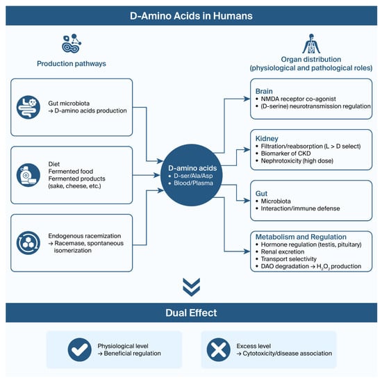
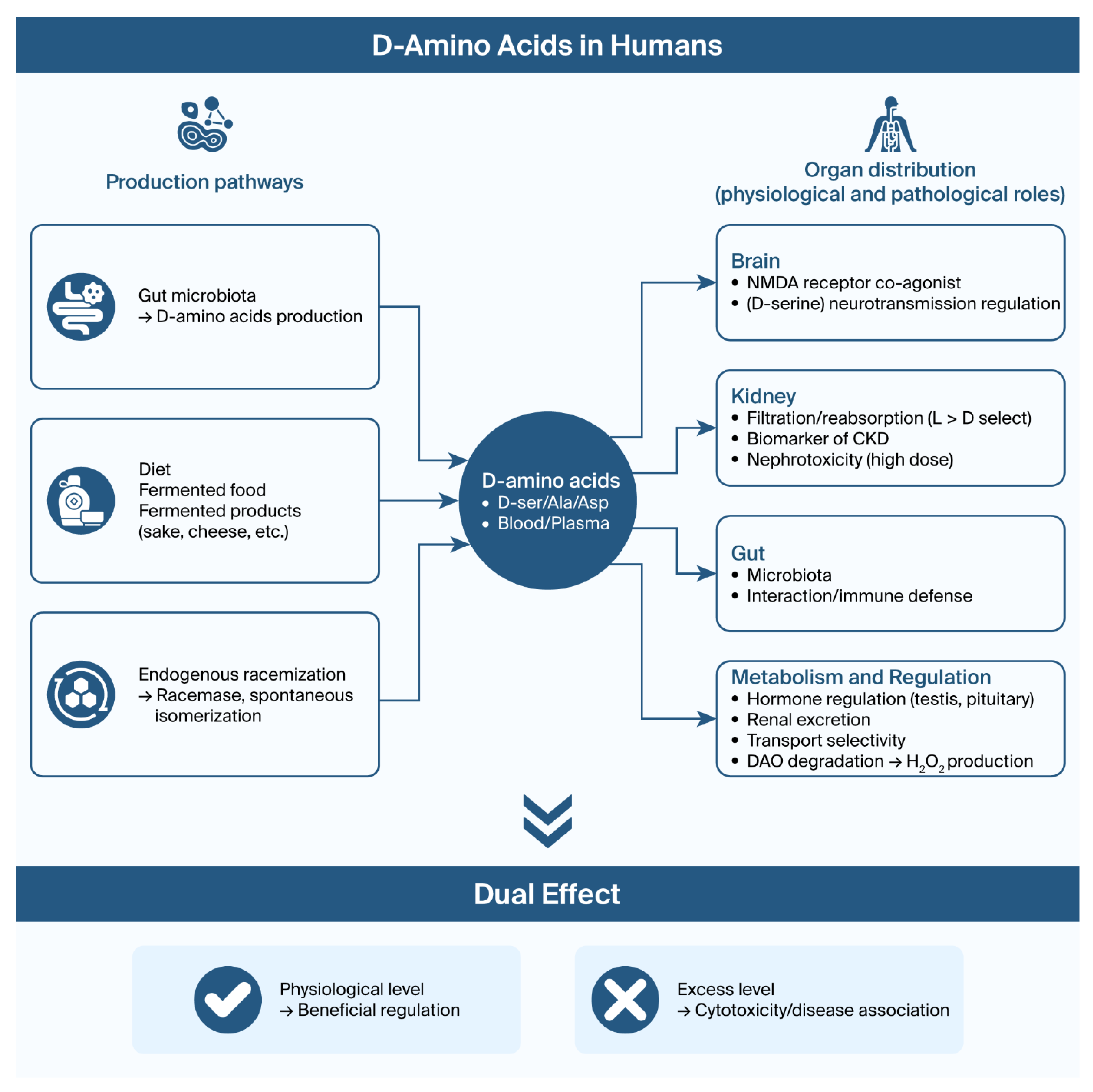
In this review, we summarize current knowledge on the sources, distribution, metabolic regulation, and biological functions of endogenous and exogenous D-amino acids, with particular emphasis on their roles in human body fluids, tissue-specific functions, and physiological and pathological implications. Figure 1 conceptually illustrates that D-amino acids are multifunctional biomolecules with tightly regulated physiological roles and context-dependent pathological implications.
Figure 1.
Systemic metabolism and functional roles of D-amino acids in humans.
A schematic representation is shown of the sources, systemic distribution, metabolic regulation, and tissue-specific functions of D-amino acids in Figure 1. The diagram highlights the central role of circulation as a hub and the dual physiological and pathological effects depending on concentration and tissue context.
2. Exogenous and Endogenous D-Amino Acids in the Mammalian Body
D-amino acids in mammals originate from multiple sources, which can be broadly classified into exogenous inputs, enzymatic production, non-enzymatic isomerization, and rare biosynthetic incorporation pathways.
Exogenous sources, including diet and intestinal microbiota, represent a major contribution to the D-amino acid pool. For example, Konno et al. [16] demonstrated that approximately 94% of D-alanine was excreted in the urine of D-amino acid oxidase (DAO; EC 1.4.3.3)-deficient mice, highlighting the importance of DAO in D-amino acid metabolism. Notably, antibiotic treatment reduced urinary D-alanine levels to one-eighth of the original levels [17], indicating that intestinal bacteria are a primary source of D-alanine rather than dietary intake alone [18]. Consistent with this, it has been estimated that microorganism-derived D-amino acids may account for approximately one-third of the total D-amino acid pool in humans, although this remains inconclusive [19,20]. In addition, various fermented foods, such as wine, beer, vinegar, bread, pickles, cheese, sake, and natto, have been identified as dietary sources of D-amino acids [21,22].
At the microbial level, the production of D-amino acids is largely mediated by racemases with varying substrate specificity. While mammalian racemases typically exhibit strict specificity, several microorganisms, including Vibrio cholerae, Escherichia coli, Bacillus subtilis, and Pseudomonas putida, possess racemases with broader substrate ranges [23,24,25]. Notably, the enzyme identified in V. cholerae can catalyze the racemization of up to 12 amino acids, suggesting a more versatile role of microbial metabolism in shaping the D-amino acid profile. In addition to racemases, D-amino acid transaminases (EC 2.2.1.21) provide an alternative enzymatic pathway by transferring amino groups between D-amino acids and α-keto acids [26,27].
Endogenous enzymatic processes also contribute to the generation of D-amino acids. Tissue-specific racemization catalyzed by amino acid racemases produces free D-amino acids, while post-translational modification via isomerases generates peptide-bound D-amino acids. Such modified peptides have been identified in diverse organisms, including amphibian skin, snail ganglia, crustacean hormones, spider venom, and platypus venom [28,29,30]. Importantly, these D-amino-acid-containing peptides often exhibit enhanced or unique biological activity compared to their L-amino acid counterparts, highlighting their functional significance.
In contrast, non-enzymatic isomerization represents a time-dependent process associated with aging and disease. This pathway is particularly relevant in long-lived proteins located in metabolically stable or avascular environments. D-aspartic acid accumulation has been widely observed in proteins associated with brain white matter [31], β-amyloid in Alzheimer’s disease [32], photoaged skin [33], tooth enamel and dentin proteins [34,35], and a crystalline lens [36,37]. A notable trend across these studies is the progressive increase in D-amino acid content with aging, which has led to its application in age estimation [38,39] and fossil dating [40]. In the case of aspartic acid residues, L-form racemization to the D-form occurs more rapidly in proteins than expected [41]. Furthermore, the recent identification of D-aspartic-acid-containing peptides in human serum proteins highlights its potential relevance in disease pathology and biomarker development [42].
Finally, the ribosomal incorporation of D-amino acids has been proposed but remains highly limited. D-amino acids can occasionally be mis-acylated onto tRNAs [43,44] and participate in translation initiation under experimental conditions [45,46], resulting in the translation initiation apparatus accepting D-amino acids as opposed to only L-methionine as translational initiators. L-amino acids have been shown to have an affinity for D-ribose-containing RNA, whereas D-amino acids prefer L-ribose-containing RNA [47]; however, the chiral preference of amino acylation favors the incorporation of L-amino acids [48]. Ribosomes can inherently discriminate the chirality of amino acids, thereby preventing the incorporation of D-amino acids into nascent proteins by slowing peptide bond formation during protein biosynthesis [49]. In addition, protein quality control systems in the endoplasmic reticulum further eliminate improperly folded proteins. Taken together, these observations indicate that ribosomal incorporation is unlikely to be a significant physiological source of D-amino acids.
Overall, current evidence suggests a clear trend in which exogenous and microbial sources dominate the D-amino acid pool, while endogenous enzymatic and non-enzymatic processes contribute in a context-dependent manner, particularly in aging and disease. However, the relative contribution of each pathway under physiological and pathological conditions remains poorly understood.
3. D-Amino Acid Levels in Human Body Fluids
Recent advances in analytical techniques have established that free D-amino acids are naturally present in a variety of human physiological fluids, including blood, urine, and saliva (Table 1). Early work by Armstrong et al. [50] demonstrated the presence of D-phenylalanine and D-proline in plasma and urine, while only trace levels were detected in cerebrospinal and amniotic fluids, suggesting the fluid-specific regulation of D-amino acid distribution.
Table 1.
The distribution of L- and D-amino acids and their concentration (nmol/mL) and their ratio (%D *1) in serum, urine, and saliva in healthy humans.
Among these fluids, blood and urine have been most extensively studied, revealing distinct distribution patterns and regulatory mechanisms. In serum, several D-amino acids, including D-serine, D-aspartate, D-alanine, and D-proline, are consistently detected. These circulating D-amino acids are thought to originate primarily from dietary intake and intestinal microbiota. However, their concentrations are tightly regulated, largely due to renal handling and enzymatic degradation. In particular, the kidney plays a central role in maintaining low plasma levels through both excretion and metabolism [50,54,55]. DAO, which is highly active in renal proximal tubules, contributes to D-amino acid degradation, while selective tubular transport mechanisms further modulate its reabsorption [56,57,58].
A characteristic feature observed across studies is that urinary D-amino acid levels are substantially higher than those in plasma, often by an order of magnitude or more. This reflects the kidney’s active excretion of D-amino acids, as supported by time-course analyses showing continuous urinary elimination [58]. Notably, renal handling exhibits chiral selectivity; for example, D-serine is reabsorbed far less efficiently than L-serine in the proximal tubules [59]. The concentration of L- and D-form amino acid ratio (%D), calculated as D/(D + L) × 100, of urinary D-serine shows an abnormally high value (Table 1), and a similar value of 42% has been reported in another study as well [60].
Alterations in D-amino acid profiles are particularly evident under pathological conditions, especially in kidney disease. Multiple studies consistently report increased plasma and urinary levels of D-amino acids in patients with impaired renal function. For instance, elevated urinary D-serine excretion and increased plasma D-amino acid levels have been observed in kidney disease and glomerulonephritis [54,61,62,63]. Furthermore, as shown in Table 2, both the absolute concentrations and %D value of specific D-amino acids increase with declining renal function, as demonstrated in chronic kidney disease (CKD) patients [64]. Similar trends have been reported in pediatric CKD, where plasma D-asparagine and the D/L-serine ratio serve as sensitive and specific indicators independent of body size or conventional biochemical markers [64]. Collectively, these findings indicate a clear trend in which D-amino acids accumulate in response to reduced renal clearance, highlighting their potential as early and sensitive biomarkers of kidney dysfunction.
Table 2.
Amounts of L- and D-amino acids their ratio (%D) in the plasma of CKD patients.
Saliva represents a distinct and less well-correlated compartment, where amino acid composition does not directly mirror that of blood. For example, L-serine and L-glutamine levels in saliva are markedly lower than those in plasma, indicating independent regulatory mechanisms. Unlike blood, salivary amino acid levels appear to be minimally influenced by dietary intake or oral microbiota under normal conditions [58]. Instead, endogenous sources such as salivary glands and oral epithelial cells have been proposed as contributors to salivary D-amino acids, particularly D-alanine and D-aspartate. The physiological roles of these two amino acids remain unclear.
However, under pathological conditions such as CKD, this balance may shift. Increased salivary D-alanine levels have been reported in CKD patients, potentially reflecting both systemic alterations and microbial contributions, particularly from oral Streptococcus species [65]. In addition, experimental studies in animals have shown that D-aspartic acid is abundantly present in salivary glands, with smaller amounts of D-serine and D-alanine also detected [66].
Overall, current evidence reveals a fluid-specific distribution of D-amino acids, governed primarily by renal regulation in blood and urine and by local production mechanisms in saliva. Importantly, a consistent trend across studies is the elevation of D-amino acids in renal dysfunction, supporting their emerging role as clinically relevant biomarkers. However, standardized analytical methods and large-scale validation studies are still required to establish their clinical utility.
4. Cytotoxicity for Hosts
D-amino acids play diverse and context-dependent roles in mammalian systems, ranging from host defense to potential cytotoxicity and therapeutic applications. At the host–microbe interface, D-amino acids contribute to innate immune defense mechanisms. The human body can tolerate D-amino acids produced by commensal bacteria, and these molecules may even exert protective effects. For instance, D-amino acids in the intestinal lumen can be oxidized by DAO, generating hydrogen peroxide (H2O2) with bactericidal activity. Early studies demonstrated that DAO in polymorphonuclear leukocytes contributes to microbial killing during phagocytosis via H2O2 production [67]. Similarly, in the intestinal mucosa, DAO-mediated oxidation of bacterial D-amino acids has been shown to protect against pathogenic colonization, such as Vibrio cholerae infection [68]. A notable feature of this system is the selective interaction between the host and microbes: while bacterial D-amino acids attract neutrophils and promote bacterial clearance, symbiotic bacteria can evade this mechanism by actively importing D-amino acids, thereby reducing their susceptibility to DAO-mediated oxidation [69].
Despite these beneficial roles, the biological effects of D-amino acids on mammalian cells are highly concentration- and species-dependent. A broad range of in vitro studies indicates that many D-amino acids are relatively well tolerated at low-to-moderate concentrations [70,71,72], with some even supporting cellular functions such as osteoblast differentiation [73]. However, at higher concentrations, several D-amino acids exhibit cytotoxic effects, including reduced cell viability, proliferation inhibition, and apoptosis induction [70,74,75]. Importantly, these effects are not uniform across amino acids or cell types, and in some cases, L-amino acids display greater toxicity than their D-enantiomers [70]. Collectively, these findings suggest a general trend in which D-amino acids are largely biocompatible within physiological ranges but may become cytotoxic when exceeding threshold concentrations.
Among D-amino acids, D-serine has emerged as a particularly important molecule with both physiological and pathological significance. While low concentrations of D-serine have been shown to exert beneficial effects, such as promoting kidney remodeling [14], higher concentrations can induce cytotoxicity in renal cells in a dose-dependent manner, suggesting its potential role as a uremic toxin [76]. This dual nature highlights the importance of tightly regulating D-serine levels, particularly in the context of renal function.
Beyond its endogenous roles, D-serine has also been extensively investigated as a therapeutic agent, particularly in neuropsychiatric disorders, because D-serine can cross the blood–brain barrier [77,78]. This supports its potential utility as a therapeutic agent. Serum D-serine levels are reduced in patients with schizophrenia, and supplementation has been shown to improve symptoms in multiple clinical studies [77,79,80,81,82,83]. Mechanistically, D-serine acts as a co-agonist at the glycine-binding site of the NMDA receptor, thereby modulating glutamatergic neurotransmission [84]. Clinical trials have demonstrated its efficacy and general safety across a range of doses, including high-dose regimens, with minimal adverse renal effects reported [84,85]. However, its clinical application is limited by rapid degradation by DAO, resulting in low bioavailability and the need for relatively high doses. Furthermore, discrepancies between animal models and human studies—particularly regarding nephrotoxicity—highlight the need for careful dose optimization and further investigation [85,86,87]. In addition to D-serine, other D-amino acids such as D-alanine have shown promising protective effects, particularly in renal and metabolic contexts. Experimental studies indicate that D-alanine can attenuate oxidative stress, improve mitochondrial function, and reduce tissue damage under hypoxic conditions [88,89,90]. Moreover, D-alanine may modulate the gut microbiome and reduce uremic toxin levels, suggesting a potential role in dietary interventions for chronic kidney disease (CKD) [91,92]. Emerging evidence also links D-alanine to regulating gluconeogenesis and circadian rhythm, further expanding its potential therapeutic relevance [15].
Overall, current evidence reveals a unifying trend in which D-amino acids exhibit dual and context-dependent effects: they contribute to host defense and physiological regulation at controlled levels, while exerting cytotoxic or pathological effects at elevated concentrations. This balance is particularly critical in kidney function and neurological regulation, underscoring the importance of precisely controlling D-amino acid metabolism in health and disease. This review not only summarizes existing studies but also identifies key trends, limitations, and future research directions.
5. Conclusions
D-amino acids exert diverse and system-specific physiological effects across the gut, kidneys, and nervous system in both humans and animals. Accumulating evidence highlights their multifaceted roles in host defense, metabolic regulation, and inter-organ communication, particularly through interactions with the gut microbiota and DAO-mediated metabolism.
Among them, D-serine and D-alanine have attracted considerable attention due to their therapeutic potential in neurological and renal disorders. These D-amino acids contribute to key physiological processes, including neurotransmission modulation, oxidative stress regulation, and microbial homeostasis maintenance.
However, a consistent trend across studies is their dual and concentration-dependent nature. While physiological or moderate levels are generally beneficial, elevated concentrations have been associated with cytotoxic effects and renal injury, particularly in the case of D-serine. This highlights the importance of tightly regulated metabolism and carefully considering dosage in clinical applications.
Overall, current evidence supports a model in which D-amino acids function as both regulatory and potentially harmful molecules, depending on their concentration, tissue distribution, and metabolic context.
Future research should focus on elucidating the precise mechanisms underlying these context-dependent effects, as well as the complex interactions between D-amino acids and the gut microbiota. Such insights will be essential for optimizing their safe and effective use in therapeutic strategies and for developing microbiome-based nutritional interventions. In particular, quantitative thresholds that distinguish physiological from pathological effects remain to be defined.
Author Contributions
Conceptualization, M.S.; resources, B.Y.H.; data curation, M.S. and B.Y.H.; writing—original draft preparation, M.S.; writing—review and editing, B.Y.H. All authors have read and agreed to the published version of the manuscript.
Funding
This research received no external funding.
Data Availability Statement
Data is contained within the review.
Acknowledgments
We sincerely thank Noriko Shiohara sun at the library of Matsumoto Dental University for their assistance in collecting the relevant literature.
Conflicts of Interest
The authors declare no conflicts of interest.
Abbreviations
The following abbreviations are used in this manuscript:
| CKD | chronic kidney disease |
| DAO | D-amino acid oxidase |
| eGFR | estimated glomerular filtration rate |
| NMDA receptors | N-methyl-D-aspartate receptors |
References
- Dunlop, D.S.; Neidle, A.; McHale, D.; Dunlop, D.M.; Lajtha, A. The presence of free D-aspartic acid in rodents and man. Biochem. Biophys. Res. Commun. 1986, 141, 27–32. [Google Scholar] [CrossRef] [Scilit]
- Nagata, Y.; Akino, T. D-Amino acids in mouse tissues are not of microbial origin. Experientia 1990, 46, 466–468. [Google Scholar] [CrossRef] [Scilit]
- Ishio, S.; Yamada, H.; Hayashi, M.; Yatsushiro, S.; Noumi, T.; Yamaguchi, A.; Moriyama, Y. D-Aspartate modulates melatonin synthesis in rat pinealocytes. Neurosci. Lett. 1998, 249, 143–146. [Google Scholar] [CrossRef] [Scilit]
- D’Aniello, G.; Tolino, A.; D’Aniello, A.; Errico, F.; Fisher, G.H.; Di Fiore, M.M. The role of D-aspartic acid and N-methyl-D-aspartic acid in the regulation of prolactin release. Endocrinology 2000, 141, 3862–3870. [Google Scholar] [CrossRef]
- Nagata, Y.; Homma, H.; Lee, J.A.; Imai, K. D-Aspartate stimulation of testosterone synthesis in rat Leydig cells. FEBS Lett. 1999, 444, 160–164. [Google Scholar] [CrossRef] [Scilit] [PubMed]
- Di Fiore, M.M.; Santillo, A.; Falvo, S.; Longobardi, S.; Chieffi Baccari, G.C. Molecular mechanisms elicited by D-aspartate in Leydig cells and spermatogonia. Int. J. Mol. Sci. 2016, 17, 1127. [Google Scholar] [CrossRef] [Scilit] [PubMed]
- Hashimoto, A.; Nishikawa, T.; Hayashi, T.; Fujii, N.; Harada, K.; Oka, T.; Takahashi, K. The presence of free D-serine in rat brain. FEBS Lett. 1992, 296, 33–36. [Google Scholar] [CrossRef] [Scilit]
- Nagata, Y.; Horiike, K.; Maeda, T. Distribution of free D-serine in vertebrate brains. Brain Res. 1994, 634, 291–295. [Google Scholar] [CrossRef] [Scilit]
- Wolosker, H.; Sheth, K.N.; Takahashi, M.; Mothet, J.P.; Brady, R.O., Jr.; Ferris, C.D.; Snyder, S.H. Purification of serine racemase: Biosynthesis of the neuromodulator D-serine. Proc. Natl. Acad. Sci. USA 1999, 96, 721–725. [Google Scholar] [CrossRef] [Scilit]
- Basu, A.C.; Tsai, G.E.; Ma, C.L.; Ehmsen, J.T.; Mustafa, A.K.; Han, L.; Jiang, Z.I.; Benneyworth, M.A.; Froimowitz, M.P.; Lange, N.; et al. Targeted disruption of serine racemase affects glutamatergic neurotransmission and behavior. Mol. Psychiatry 2009, 14, 719–727. [Google Scholar] [CrossRef] [Scilit] [PubMed]
- Benneyworth, M.A.; Li, Y.; Basu, A.C.; Bolshakov, V.Y.; Coyle, J.T. Cell selective conditional null mutations of serine racemase demonstrate a predominate localization in cortical glutamatergic neurons. Cell. Mol. Neurobiol. 2012, 32, 613–624. [Google Scholar] [CrossRef] [Scilit]
- Sasabe, J.; Suzuki, M. Distinctive roles of D-amino acids in the homochiral world: Chirality of amino acids modulates mammalian physiology and pathology. Keio J. Med. 2019, 68, 1–16. [Google Scholar] [CrossRef] [Scilit]
- Lam, H.; Oh, D.C.; Cava, F.; Takacs, C.N.; Clardy, J.; de Pedro, M.A.; Waldor, M.K. D-Amino acids govern stationary phase cell wall remodeling in bacteria. Science 2009, 325, 1552–1555. [Google Scholar] [CrossRef] [Scilit] [PubMed]
- Hesaka, A.; Tsukamoto, Y.; Nada, S.; Kawamura, M.; Ichimaru, N.; Sakai, S.; Nakane, M.; Mita, M.; Okuzaki, D.; Okada, M.; et al. D-Serine mediates cellular proliferation for kidney remodeling. Kidney360 2021, 2, 1611–1624. [Google Scholar] [CrossRef] [Scilit]
- Sakai, S.; Tanaka, Y.; Tsukamoto, Y.; Kimura-Ohba, S.; Hesaka, A.; Hamase, K.; Hsieh, C.L.; Kawakami, E.; Ono, H.; Yokote, K.; et al. D-Alanine affects the circadian clock to regulate glucose metabolism in the kidney. Kidney360 2024, 5, 237–251. [Google Scholar] [CrossRef] [Scilit] [PubMed]
- Konno, R.; Nagata, Y.; Niwa, A.; Yasumura, Y. Spontaneous excretion of D-alanine in urine in mutant mice lacking D-amino acid oxidase. Biochem. J. 1989, 261, 285–287. [Google Scholar] [CrossRef] [Scilit] [PubMed]
- Konno, R.; Niwa, A.; Yasumura, Y. Intestinal bacterial origin of D-alanine in urine of mutant mice lacking D-amino-acid oxidase. Biochem. J. 1990, 268, 263–265. [Google Scholar] [CrossRef] [Scilit]
- Genchi, G. An overview on D-amino acids. Amino Acids 2017, 49, 1521–1533. [Google Scholar] [CrossRef] [Scilit] [PubMed]
- Friedman, M. Origin, microbiology, nutrition, and pharmacology of D-amino acids. Chem. Biodivers. 2010, 7, 1491–1530. [Google Scholar] [CrossRef] [Scilit]
- Leuchtenberger, W.; Huthmacher, K.; Drauz, K. Biotechnological production of amino acids and derivatives: Current status and prospects. Appl. Microbiol. Biotechnol. 2005, 69, 1–8. [Google Scholar] [CrossRef] [Scilit]
- Kobayashi, J. d-Amino acids and lactic acid bacteria. Microorganisms 2019, 7, 690. [Google Scholar] [CrossRef] [Scilit]
- Bastings, J.J.A.J.; van Eijk, H.M.; Olde Damink, S.W.O.; Rensen, S.S. D-Amino acids in health and disease: A focus on cancer. Nutrients 2019, 11, 2205. [Google Scholar] [CrossRef] [Scilit] [PubMed]
- Espaillat, A.; Carrasco-López, C.; Bernardo-García, N.; Pietrosemoli, N.; Otero, L.H.; Álvarez, L.; de Pedro, M.A.; Pazos, F.; Davis, B.M.; Waldor, M.K.; et al. Structural basis for the broad specificity of a new family of amino-acid racemases. Acta Crystallogr. D Biol. Crystallogr. 2014, 70, 79–90. [Google Scholar] [CrossRef] [Scilit]
- Miyamoto, T.; Katane, M.; Saitoh, Y.; Sekine, M.; Homma, H. Identification and characterization of novel broad-spectrum amino acid racemases from Escherichia coli and Bacillus subtilis. Amino Acids 2017, 49, 1885–1894. [Google Scholar] [CrossRef] [Scilit]
- Radkov, A.D.; Moe, L.A. A broad spectrum racemase in Pseudomonas putida KT2440 plays a key role in amino acid catabolism. Front. Microbiol. 2018, 9, 1343. [Google Scholar] [CrossRef] [Scilit]
- Thorne, C.B.; Gomez, C.G.; Housewright, R.D. Transamination of D-amino acids by Bacillus subtilis. J. Bacteriol. 1955, 69, 357–362. [Google Scholar] [CrossRef] [Scilit] [PubMed]
- Thorne, C.B.; Molnar, D.M. D-Amino acid transamination in Bacillus anthracis. J. Bacteriol. 1955, 70, 420–426. [Google Scholar] [CrossRef] [Scilit]
- Kreil, G. D-Amino acids in animal peptides. Annu. Rev. Biochem. 1997, 66, 337–345. [Google Scholar] [CrossRef] [Scilit]
- Torres, A.M.; Tsampazi, C.; Geraghty, D.P.; Bansal, P.S.; Alewood, P.F.; Kuchel, P.W. D-Amino acid residue in a defensin-like peptide from platypus venom: Effect on structure and chromatographic properties. Biochem. J. 2005, 391, 215–220. [Google Scholar] [CrossRef] [Scilit]
- Whittington, C.M.; Koh, J.M.S.; Warren, W.C.; Papenfuss, A.T.; Torres, A.M.; Kuchel, P.W.; Belov, K. Understanding and utilising mammalian venom via a platypus venom transcriptome. J. Proteom. 2009, 72, 155–164. [Google Scholar] [CrossRef] [Scilit] [PubMed]
- Man, E.H.; Sandhouse, M.E.; Burg, J.; Fisher, G.H. Accumulation of D-aspartic acid with age in the human brain. Science 1983, 220, 1407–1408. [Google Scholar] [CrossRef] [Scilit]
- Roher, A.E.; Lowenson, J.D.; Clarke, S.; Wolkow, C.; Wang, R.; Cotter, R.J.; Reardon, I.M.; Zürcher-Neely, H.A.; Heinrikson, R.L.; Ball, M.J.; et al. Structural alterations in the peptide backbone of beta-amyloid core protein may account for its deposition and stability in Alzheimer’s disease. J. Biol. Chem. 1993, 268, 3072–3083. [Google Scholar] [CrossRef] [Scilit]
- Fujii, N.; Tajima, S.; Tanaka, N.; Fujimoto, N.; Takata, T.; Shimo-Oka, T. The presence of D-beta-aspartic acid-containing peptides in elastic fibers of sun-damaged skin: A potent marker for ultraviolet-induced skin aging. Biochem. Biophys. Res. Commun. 2002, 294, 1047–1051. [Google Scholar] [CrossRef] [Scilit] [PubMed]
- Helfman, P.M.; Bada, J.L. Aspartic acid racemization in tooth enamel from living humans. Proc. Natl. Acad. Sci. USA 1975, 72, 2891–2894. [Google Scholar] [CrossRef] [Scilit]
- Helfman, P.M.; Bada, J.L. Aspartic acid racemisation in dentine as a measure of ageing. Nature 1976, 262, 279–281. [Google Scholar] [CrossRef] [Scilit]
- Masters, P.M.; Bada, J.L.; Zigler, J.S., Jr. Aspartic acid racemisation in the human lens during ageing and in cataract formation. Nature 1977, 268, 71–73. [Google Scholar] [CrossRef] [Scilit]
- Fujii, N.; Takata, T.; Fujii, N.; Aki, K. Isomerization of aspartyl residues in crystallins and its influence upon cataract. Biochim. Biophys. Acta 2016, 1860, 183–191. [Google Scholar] [CrossRef] [Scilit]
- Ritz, S.; Turzynski, A.; Schütz, H.W. Estimation of age at death based on aspartic acid racemization in noncollagenous bone proteins. Forensic Sci. Int. 1994, 69, 149–159. [Google Scholar] [CrossRef] [Scilit] [PubMed]
- Ohtani, S.; Matsushima, Y.; Kobayashi, Y.; Kishi, K. Evaluation of aspartic acid racemization ratios in the human femur for age estimation. J. Forensic Sci. 1998, 43, 949–953. [Google Scholar] [CrossRef] [Scilit]
- Bada, J.L.; Protsch, R. Racemization reaction of aspartic acid and its use in dating fossil bones. Proc. Natl. Acad. Sci. USA 1973, 70, 1331–1334. [Google Scholar] [CrossRef] [Scilit] [PubMed]
- Fujii, N.; Takata, T.; Fujii, N.; Aki, K.; Sakaue, H. D-Amino acids in protein: The mirror of life as a molecular index of aging. Biochim. Biophys. Acta Proteins Proteom. 2018, 1866, 840–847. [Google Scholar] [CrossRef] [Scilit]
- Ha, S.; Kim, I.; Takata, T.; Kinouchi, T.; Isoyama, M.; Suzuki, M.; Fujii, N. Identification of D-amino acid-containing peptides in human serum. PLoS ONE 2017, 12, e0189972. [Google Scholar] [CrossRef] [Scilit]
- Soutourina, J.; Plateau, P.; Blanquet, S. Metabolism of D-aminoacyl-tRNAs in Escherichia coli and Saccharomyces cerevisiae cells. J. Biol. Chem. 2000, 275, 32535–32542. [Google Scholar] [CrossRef] [Scilit]
- Takayama, T.; Ogawa, T.; Hidaka, M.; Shimizu, Y.; Ueda, T.; Masaki, H. Esterification of Escherichia coli tRNAs with D-histidine and D-lysine by aminoacyl-tRNA synthetases. Biosci. Biotechnol. Biochem. 2005, 69, 1040–1041. [Google Scholar] [CrossRef] [Scilit] [PubMed]
- Goto, Y.; Murakami, H.; Suga, H. Initiating translation with D-amino acids. RNA 2008, 14, 1390–1398. [Google Scholar] [CrossRef] [Scilit]
- Fujino, T.; Goto, Y.; Suga, H.; Murakami, H. Reevaluation of the D-amino acid compatibility with the elongation event in translation. J. Am. Chem. Soc. 2013, 135, 1830–1837. [Google Scholar] [CrossRef] [Scilit] [PubMed]
- Tamura, K.; Schimmel, P. Chiral-selective aminoacylation of an RNA minihelix. Science 2004, 305, 1253. [Google Scholar] [CrossRef] [Scilit]
- Rybak, M.Y.; Rayevsky, A.V.; Gudzera, O.I.; Tukalo, M.A. Stereospecificity control in aminoacyl-tRNA-synthetases: New evidence of d-amino acids activation and editing. Nucleic Acids Res. 2019, 47, 9777–9788. [Google Scholar] [CrossRef] [Scilit]
- Melnikov, S.V.; Khabibullina, N.F.; Mairhofer, E.; Vargas-Rodriguez, O.; Reynolds, N.M.; Micura, R.; Söll, D.; Polikanov, Y.S. Mechanistic insights into the slow peptide bond formation with D-amino acids in the ribosomal active site. Nucleic Acids Res. 2019, 47, 2089–2100. [Google Scholar] [CrossRef] [Scilit] [PubMed]
- Armstrong, D.W.; Gasper, M.; Lee, S.H.; Zukowski, J.; Ercal, N. D-Amino acid levels in human physiological fluids. Chirality 1993, 5, 375–378. [Google Scholar] [CrossRef] [Scilit]
- Liu, M.; Li, M.; He, J.; He, Y.; Yang, J.; Sun, Z. Chiral amino acid profiling in serum reveals potential biomarkers for Alzheimer’s disease. J. Alzheimer’s Dis. 2023, 94, 291–301. [Google Scholar] [CrossRef] [Scilit]
- Deng, W.; Ye, C.; Wang, W.; Huang, R.; Guo, C.; Pan, Y.; Sun, C. LC-MS analysis of chiral amino acids in human urine reveals D-amino acids as potential biomarkers for colorectal cancer. J. Chromatogr. B 2024, 1245, 124270. [Google Scholar] [CrossRef] [Scilit] [PubMed]
- Nagata, Y.; Higashi, M.; Ishii, Y.; Sano, H.; Tanigawa, M.; Nagata, K.; Noguchi, K.; Urade, M. The presence of high concentrations of free D-amino acids in human saliva. Life Sci. 2006, 78, 1677–1681. [Google Scholar] [CrossRef] [Scilit] [PubMed]
- Ishii, C.; Akita, T.; Kimura, T.; Sakai, S.; Mita, M.; Ide, T.; Isaka, Y.; Hamase, K. Evaluation of individual variation of D-amino acids in human plasma by a two-dimensional LC-MS/MS system and application to the early diagnosis of chronic kidney disease. Anal. Chem. 2024, 96, 4876–4883. [Google Scholar] [CrossRef] [Scilit]
- Chan, A.W.; Perry, S.G.; Burch, H.B.; Fagioli, S.; Alvey, T.R.; Lowry, O.H. Distribution of two aminotransferases and D-amino acid oxidase within the nephron of young and adult rats. J. Histochem. Cytochem. 1979, 27, 751–755. [Google Scholar] [CrossRef] [Scilit]
- Nagata, Y.; Konno, R.; Yasumura, Y.; Akino, T. Involvement of D-amino acid oxidase in elimination of free D-amino acids in mice. Biochem. J. 1989, 257, 291–292. [Google Scholar] [CrossRef] [Scilit] [PubMed]
- Angermüller, S.; Fahimi, H.D. Heterogenous staining of D-amino acid oxidase in peroxisomes of rat liver and kidney. A light and electron microscopic study. Histochemistry 1988, 88, 277–285. [Google Scholar] [CrossRef] [Scilit]
- Brückner, H.; Schieber, A. Determination of amino acid enantiomers in human urine and blood serum by gas chromatography-mass spectrometry. Biomed. Chromatogr. 2001, 15, 166–172. [Google Scholar] [CrossRef] [Scilit]
- Kimura, T.; Hesaka, A.; Isaka, Y. D-Amino acids and kidney diseases. Clin. Exp. Nephrol. 2020, 24, 404–410. [Google Scholar] [CrossRef] [Scilit]
- Lorenzo, M.P.; Dudzik, D.; Varas, E.; Gibellini, M.; Skotnicki, M.; Zorawski, M.; Zarzycki, W.; Pellati, F.; García, A. Optimization and validation of a chiral GC-MS method for the determination of free D-amino acids ratio in human urine: Application to a gestational diabetes mellitus study. J. Pharm. Biomed. Anal. 2015, 107, 480–487. [Google Scholar] [CrossRef] [Scilit]
- Nagata, Y.; Akino, T.; Ohno, K.; Kataoka, Y.; Ueda, T.; Sakurai, T.; Shiroshita, K.; Yasuda, T. Free D-amino acids in human plasma in relation to senescence and renal diseases. Clin. Sci. 1987, 73, 105–108. [Google Scholar] [CrossRef] [Scilit]
- Brückner, H.; Hausch, M. Gas chromatographic characterization of free D-amino acids in the blood serum of patients with renal disorders and of healthy volunteers. J. Chromatogr. 1993, 614, 7–17. [Google Scholar] [CrossRef] [Scilit]
- Oyaide, M.; Ishii, C.; Akita, T.; Kimura, T.; Sakai, S.; Mizui, M.; Mita, M.; Ide, T.; Isaka, Y.; Hamase, K. Development of a three-dimensional HPLC system for the determination of serine, threonine and allo-threonine enantiomers in the plasma of patients with chronic kidney disease. J. Chromatogr. A 2024, 1719, 464739. [Google Scholar] [CrossRef] [Scilit]
- Morishita, T.; Nishizaki, N.; Taniguchi, S.; Sakai, S.; Kimura, T.; Mita, M.; Nakagawa, M.; Endo, A.; Ohtomo, Y.; Yasui, M.; et al. Plasma D-asparagine and the D/L-serine ratio reflect chronic kidney diseases in children regardless of physique. Amino Acids 2024, 56, 38. [Google Scholar] [CrossRef] [Scilit] [PubMed]
- Nakade, Y.; Iwata, Y.; Sakai, N.; Mita, M.; Nakane, M.; Hamase, K.; Suda, W.; Toyama, T.; Kitajima, S.; Hara, A.; et al. Increased levels of oral Streptococcus-derived D-alanine in patients with chronic kidney disease and diabetes mellitus. Sci. Rep. 2022, 12, 21773. [Google Scholar] [CrossRef] [Scilit] [PubMed]
- Yoshikawa, M.; Kan, T.; Shirose, K.; Watanabe, M.; Matsuda, M.; Ito, K.; Kawaguchi, M. Free D-amino acids in salivary gland in rat. Biology 2022, 11, 390. [Google Scholar] [CrossRef] [Scilit]
- Robinson, J.M.; Briggs, R.T.; Karnovsky, M.J. Localization of D-amino acid oxidase on the cell surface of human polymorphonuclear leukocytes. J. Cell Biol. 1978, 77, 59–71. [Google Scholar] [CrossRef] [Scilit]
- Sasabe, J.; Miyoshi, Y.; Rakoff-Nahoum, S.; Zhang, T.; Mita, M.; Davis, B.M.; Hamase, K.; Waldor, M.K. Interplay between microbial D-amino acids and host D-amino acid oxidase modifies murine mucosal defense and gut microbiota. Nat. Microbiol. 2016, 1, 16125. [Google Scholar] [CrossRef] [Scilit] [PubMed]
- Tuinema, B.R.; Reid-Yu, S.A.; Coombes, B.K. Salmonella evades D-amino acid oxidase to promote infection in neutrophils. mBio 2014, 5, e01886. [Google Scholar] [CrossRef] [Scilit]
- Ercal, N.; Luo, X.; Matthews, R.H.; Armstrong, D.W. In vitro study of the metabolic effects of D-amino acids. Chirality 1996, 8, 24–29. [Google Scholar] [CrossRef] [Scilit]
- Brandenburg, K.S.; Rodriguez, K.J.; McAnulty, J.F.; Murphy, C.J.; Abbott, N.L.; Schurr, M.J.; Czuprynski, C.J. Tryptophan inhibits biofilm formation by Pseudomonas aeruginosa. Antimicrob. Agents Chemother. 2013, 57, 1921–1925. [Google Scholar] [CrossRef] [Scilit]
- Sanchez, C.J., Jr.; Prieto, E.M.; Krueger, C.A.; Zienkiewicz, K.J.; Romano, D.R.; Ward, C.L.; Akers, K.S.; Guelcher, S.A.; Wenke, J.C. Effects of local delivery of D-amino acids from biofilm-dispersive scaffolds on infection in contaminated rat segmental defects. Biomaterials 2013, 34, 7533–7543. [Google Scholar] [CrossRef] [Scilit]
- Rawson, M.; Haggard, W.; Jennings, J.A. Osteocompatibility of biofilm inhibitors. Open Orthop. J. 2014, 8, 442–449. [Google Scholar] [CrossRef] [Scilit] [PubMed]
- Harmata, A.J.; Ma, Y.; Sanchez, C.J.; Zienkiewicz, K.J.; Elefteriou, F.; Wenke, J.C.; Guelcher, S.A. D-Amino acid inhibits biofilm but not new bone formation in an ovine model. Clin. Orthop. Relat. Res. 2015, 473, 3951–3961. [Google Scholar] [CrossRef] [Scilit]
- Zhang, Z.; Li, B.; Cai, Q.; Qiao, S.; Wang, D.; Wang, H.; Zhang, H.; Yang, Y.; Meng, W. Synergistic effects of D-arginine, D-methionine and D-histidine on the inhibition and eradication of Porphyromonas gingivalis biofilm. Biofouling 2021, 37, 222–234. [Google Scholar] [CrossRef] [PubMed]
- Okada, A.; Nangaku, M.; Jao, T.M.; Maekawa, H.; Ishimono, Y.; Kawakami, T.; Inagi, R. D-Serine, a novel uremic toxin, induces senescence in human renal tubular cells via GCN2 activation. Sci. Rep. 2017, 7, 11168. [Google Scholar] [CrossRef] [Scilit]
- Cho, S.E.; Na, K.S.; Cho, S.J.; Kang, S.G. Low D-serine levels in schizophrenia: A systematic review and meta-analysis. Neurosci. Lett. 2016, 634, 42–51. [Google Scholar] [CrossRef] [Scilit]
- Pernot, P.; Maucler, C.; Tholance, Y.; Vasylieva, N.; Debilly, G.; Pollegioni, L.; Cespuglio, R.; Marinesco, S. D-Serine diffusion through the blood-brain barrier: Effect on D-serine compartmentalization and storage. Neurochem. Int. 2012, 60, 837–845. [Google Scholar] [CrossRef] [Scilit] [PubMed]
- Tsai, G.E.; Yang, P.; Chung, L.C.; Lange, N.; Coyle, J.T. D-Serine added to antipsychotics for the treatment of schizophrenia. Biol. Psychiatry 1998, 44, 1081–1089. [Google Scholar] [CrossRef] [Scilit]
- Tsai, G.E.; Yang, P.; Chung, L.C.; Tsai, I.C.; Tsai, C.W.; Coyle, J.T. D-Serine added to clozapine for the treatment of schizophrenia. Am. J. Psychiatry 1999, 156, 1822–1825. [Google Scholar] [CrossRef] [Scilit]
- Heresco-Levy, U.; Javitt, D.C.; Ebstein, R.; Vass, A.; Lichtenberg, P.; Bar, G.; Catinari, S.; Ermilov, M. D-Serine efficacy as add-on pharmacotherapy to risperidone and olanzapine for treatment-refractory schizophrenia. Biol. Psychiatry 2005, 57, 577–585. [Google Scholar] [CrossRef] [Scilit] [PubMed]
- Lane, H.Y.; Chang, Y.C.; Liu, Y.C.; Chiu, C.C.; Tsai, G.E. Sarcosine or D-serine add-on treatment for acute exacerbation of schizophrenia: A randomized, double-blind, placebo-controlled study. Arch. Gen. Psychiatry 2005, 62, 1196–1204. [Google Scholar] [CrossRef] [Scilit]
- Kantrowitz, J.T.; Malhotra, A.K.; Cornblatt, B.; Silipo, G.; Balla, A.; Suckow, R.F.; D’Souza, C.; Saksa, J.; Woods, S.W.; Javitt, D.C. High dose D-serine in the treatment of schizophrenia. Schizophr. Res. 2010, 121, 125–130. [Google Scholar] [CrossRef] [Scilit]
- Mothet, J.P.; Parent, A.T.; Wolosker, H.; Brady, R.O., Jr.; Linden, D.J.; Ferris, C.D.; Rogawski, M.A.; Snyder, S.H. D-Serine is an endogenous ligand for the glycine site of the N-methyl-D-aspartate receptor. Proc. Natl. Acad. Sci. USA 2000, 97, 4926–4931. [Google Scholar] [CrossRef] [Scilit] [PubMed]
- Meftah, A.; Hasegawa, H.; Kantrowitz, J.T. D-Serine: A cross species review of safety. Front. Psychiatry 2021, 12, 726365. [Google Scholar] [CrossRef] [Scilit]
- Heresco-Levy, U.; Lerer, B. Synergistic psychedelic-NMDAR modulator treatment for neuropsychiatric disorders. Mol. Psychiatry 2024, 29, 146–152. [Google Scholar] [CrossRef] [Scilit]
- Hasegawa, H.; Masuda, N.; Natori, H.; Shinohara, Y.; Ichida, K. Pharmacokinetics and toxicokinetics of d-serine in rats. J. Pharm. Biomed. Anal. 2019, 162, 264–271. [Google Scholar] [CrossRef] [Scilit]
- Nakade, Y.; Iwata, Y.; Toyama, T.; Kobayashi, T.; Mita, M.; Yamamura, Y.; Oshima, M.; Tokumaru, T.; Kitajima, S.; Shimizu, M.; et al. D-Serine and D-alanine supplementation protects against chronic kidney disease. Sci. Rep. 2026, 16, 8740. [Google Scholar] [CrossRef] [Scilit]
- Iwata, Y.; Nakade, Y.; Kitajima, S.; Yoneda-Nakagawa, S.; Oshima, M.; Sakai, N.; Ogura, H.; Sato, K.; Toyama, T.; Yamamura, Y.; et al. Protective effect of D-alanine against acute kidney injury. Am. J. Physiol. Ren. Physiol. 2022, 322, F667–F679. [Google Scholar] [CrossRef] [Scilit]
- Oshima, M.; Toyama, T.; Nakade, Y.; Sakurai, G.; Yuasa, T.; Koshino, A.; Horikoshi, K.; Minami, T.; Sako, K.; Nakagawa, S.; et al. D-Alanine supplementation for chronic kidney disease: A study protocol for a pilot randomized controlled trial. Chem. Biodivers. 2025, 22, e00746. [Google Scholar] [CrossRef] [Scilit] [PubMed]
- Tokumaru, T.; Toyama, T.; Nakade, Y.; Ogura, H.; Oshima, M.; Nakagawa, S.; Furuichi, M.; Kitajima, S.; Sakai, N.; Shimizu, M.; et al. Design and rationale for an open-label, randomized, controlled pilot trial to evaluate the changes in blood uremic toxins in patients with chronic kidney disease by dietary therapy with sake lees. Clin. Exp. Nephrol. 2024, 28, 440–446. [Google Scholar] [CrossRef] [Scilit] [PubMed]
- Tokumaru, T.; Toyama, T.; Nakade, Y.; Ogura, H.; Oshima, M.; Nakagawa, S.; Mita, M.; Miyagawa, T.; Kitajima, S.; Hara, A.; et al. Effects of sake lees intake on fecal uremic toxins, plasma D-alanine, constipation, and gut microbiome in healthy adults: A single-arm clinical trial. PLoS ONE 2025, 20, e0325482. [Google Scholar] [CrossRef] [Scilit] [PubMed]

Disclaimer/Publisher’s Note: The statements, opinions and data contained in all publications are solely those of the individual author(s) and contributor(s) and not of MDPI and/or the editor(s). MDPI and/or the editor(s) disclaim responsibility for any injury to people or property resulting from any ideas, methods, instructions or products referred to in the content. |
© 2026 by the authors. Licensee MDPI, Basel, Switzerland. This article is an open access article distributed under the terms and conditions of the Creative Commons Attribution (CC BY) license.