


Highlights
What are the main findings of the study?
- This study examines the behavior of three microRNA (miR-141-3p, miR-146a-5p, and miR-148b-3p) present in healthy patients and in those with periodontal disease selected randomly among attendees of the Periodontics clinics of the Faculty of Dentistry of the Autonomous University of Sinaloa.
- We feel that the reduction in miR-146a-5p expression is another kind of characteristic consequence of pathogenicity factors (such as gingipain) produced by bacterial metabolism.
What are the limitations and needs for future research?
- Further research is needed to identify molecular mechanisms that allow restoring the expression of miR-146a-5p and determining its therapeutic potential in the control of periodontal inflammation.
Abstract
Background: Periodontal disease is a chronic inflammatory condition characterized by progressive destruction of the supporting tissues of the teeth. MicroRNAs (miRNAs) have emerged as key post-transcriptional regulators of immune and inflammatory responses; however, their coordinated expression patterns in periodontal disease remain incompletely understood. Objective: This study aimed to evaluate the expression of miR-141-3p, miR-146a-5p, and miR-148b-3p in periodontal disease and to explore their potential co-regulation in relation to periodontal inflammation. Methods: Gingival samples were collected from individuals with periodontal disease and periodontally healthy controls. miRNA expression levels were quantified by real-time quantitative PCR using TaqMan assays and normalized to miR-191-5p. Relative expression was calculated using the ΔΔCt method. Statistical analyses included group comparisons and correlation analyses to assess relationships among miRNA expression levels. Results: miR-146a-5p expression was significantly reduced in periodontal disease compared with healthy controls (p < 0.05), while miR-141-3p showed a consistent downward trend without reaching statistical significance. No significant differences were observed for miR-148b-3p expression between groups. Notably, strong positive correlations were identified among the three miRNAs, indicating coordinated expression patterns independent of disease stage or grade. Conclusions: The findings demonstrate altered expression of miR-146a-5p and coordinated regulation of miR-141-3p, miR-146a-5p, and miR-148b-3p in periodontal disease. These results support the concept that miRNAs act within regulatory networks rather than as isolated markers and contribute to a more nuanced understanding of miRNA-mediated regulation in periodontal inflammation.
1. Introduction
Periodontal diseases (PD) are chronic inflammatory conditions initiated by dysbiotic microbial biofilms but largely driven by a dysregulated host immune response. PD encompasses a continuum ranging from reversible gingivitis to destructive periodontitis, which is characterized by progressive loss of periodontal ligament, connective tissue attachment, and alveolar bone. According to recent estimates from the Global Burden of Disease Study 2021, severe periodontitis affects more than one billion individuals worldwide, with prevalence increasing markedly after the fourth decade of life, underscoring its relevance as a global public health problem [1,2] and representing a global age-standardized prevalence of 12.5%. This condition was most prevalent in people over 35, with the highest case number and prevalence rate observed in individuals aged 50–54 years and 55–59 years, respectively [2]. Its systemic implications include associations with cardiovascular disease, diabetes, and rheumatoid arthritis [3]. Periodontitis is considered the most prevalent infectious inflammatory disease and frequently progresses to a chronic state [4]. Previously classified as mild, moderate, severe, and chronic, today the current classification system describes periodontitis based on stages, reflecting disease severity and complexity, and grades, which estimate the rate of progression. In stage I, 1–2 mm of clinical attachment loss (CAL) is observed, accompanied by less than 15% radiographic bone loss, probing depths under 4 mm, and horizontal bone loss. Stage II is characterized by 3–4 mm CAL, 15–33% bone loss, ≤5 mm probing depth, and horizontal bone loss. In stage III, periodontal involvement includes ≥5 mm CAL, bone loss extending to the mid/apical third, ≥3 mm vertical bone loss or furcation involvement, and more than four teeth lost due to PD, with a probing depth bigger than 6 mm. Stage IV presents ≥5 mm CAL with extensive tooth loss (≥5 teeth), masticatory dysfunction, occlusal trauma, bite collapse, or <20 remaining teeth. Grade A is associated with slow progression, grade B with moderate progression, and grade C with rapid progression [5].
Although this framework has improved clinical characterization, it does not fully explain the marked interindividual variability in inflammatory response and tissue destruction observed among patients with comparable microbial and clinical profiles. Increasing evidence indicates that host-related molecular factors play a central role in determining susceptibility to periodontal breakdown and disease persistence [3].
Research in humans with periodontitis has reported the deregulation of some key miRNAs in the host inflammatory response against bacterial pathogens [6]. MicroRNAs (miRNAs) are small non-coding RNA molecules that regulate gene expression at the post-transcriptional level [7] and are now recognized as key modulators of innate and adaptive immunity. Recent studies have demonstrated that miRNAs orchestrate inflammatory signaling, immune cell differentiation, and bone remodeling processes that are directly relevant to periodontal homeostasis and disease [8,9]. Importantly, miRNAs rarely act in isolation; instead, they function within coordinated regulatory networks that fine-tune complex biological responses to microbial and inflammatory stimuli [10].
Among the miRNAs implicated in periodontal inflammation, miR-146a-5p has emerged as a critical negative regulator of pro-inflammatory signaling, primarily through modulation of the NF-κB pathway via the suppression of adaptor molecules such as IRAK1 and TRAF6. Contemporary evidence highlights its role in limiting chronic inflammation and maintaining immune homeostasis across multiple tissues [11,12,13]. A recent study reported that the levels of miR-146a-5p were significantly lower among patients with PD than among healthy individuals [14]. On the other hand, the expression of miR146a-5p is upregulated by inflammatory factors such as interleukin-1 and tumor necrosis factor alpha and downregulates a number of target genes that are involved in toll-like receptor pathways [15]. When analyzing the role of the pathogenicity factors LPS and gingipains of Porphyromonas gingivalis, the causative agent of periodontitis, with respect to the expression of miR-146a-5p, opposing effects were observed. Meanwhile, a study performed using human gingival fibroblasts (HGFs) after Porphyromonas gingivalis (p.g) lipopolysaccharide (LPS) stimulation detected an overexpression of miR-146a-5p [16]. The Gingipains reduced the levels of miR-146a-5p in exosomes, which enhanced osteoclast differentiation through the miR-146a-5p/TRAF6 signaling pathway [17], increasing the production of pro-inflammatory cytokines, such as IL-1β, IL-6 and TNF-α. miR-141-3p, a member of the miR-200 family, has been associated with endothelial inflammation, immune cell adhesion, and osteogenic differentiation, suggesting a potential role in both inflammatory regulation and alveolar bone homeostasis [18,19]. In parallel, miR-148b-3p has been linked to an attenuation of inflammatory cytokine production and promotion of osteogenic differentiation, positioning it at the interface between immune regulation and bone metabolism [20,21,22].
Despite growing evidence supporting the involvement of these miRNAs in inflammation and osteogenesis, their coordinated behavior in PD remains insufficiently characterized. Most available studies have focused on individual miRNAs evaluated in distinct biological compartments or disease contexts, yielding heterogeneous and sometimes conflicting results. This gap highlights the need for integrative analyses that explore not only differential expressions but also the potential co-regulation of functionally related miRNAs within the periodontal microenvironment.
Therefore, the present study aimed to evaluate the expression of miR-141-3p, miR-146a-5p, and miR-148b-3p in gingival samples from individuals with periodontal disease and periodontally healthy controls. By analyzing their relative expression patterns and interrelationships, this study seeks to provide insight into the coordinated regulation of miRNAs involved in periodontal inflammation and tissue homeostasis, contributing to a more refined molecular understanding of periodontal disease.
2. Materials and Methods
2.1. Study Design and Population
This cross-sectional study included individuals recruited from the Periodontics clinic of the Faculty of Dentistry, Culiacán, Sinaloa, Mexico. Participants were classified into two groups: periodontally healthy controls (HC) and patients diagnosed with PD. All procedures were conducted in accordance with the Declaration of Helsinki and were approved by the Institutional Ethics Committee of the Faculty of Dentistry at the Autonomous University of Sinaloa (approval number: P-007-2021). Written informed consent was obtained from all participants prior to enrollment.
2.2. Inclusion and Exclusion Criteria
Participants aged ≥ 18 years were eligible for inclusion. Periodontally healthy individuals were defined as having no sites with a probing depth > 3 mm and no clinical attachment loss. Patients with periodontal disease were diagnosed according to the 2017 World Workshop classification criteria [5].
Exclusion criteria included pregnancy or lactation, systemic inflammatory or autoimmune diseases, antibiotic or anti-inflammatory therapy within the previous three months, history of periodontal treatment within the last six months, and use of medications known to affect periodontal status.
2.3. Clinical Periodontal Examination
- Periodontal health
Periodontal health is defined by the absence of clinically detectable inflammation. Clinical gingival health can be present in an intact periodontium, characterized by the absence of clinical attachment loss and bone loss, as well as in a reduced periodontium. This includes non-periodontitis patients, such as those with gingival recession or who have undergone crown-lengthening procedures, and patients with a history of periodontitis who are currently periodontally stable.
Clinical gingival health in an intact periodontium is characterized by the absence of bleeding on probing, erythema and edema, patient symptoms, and attachment and bone loss. Physiological bone levels vary from 1.0 to 3.0 mm apical to the amelocementary junction [23].
- Gingivitis and Periodontitis
Patients with gingivitis were determined by possessing 10% or more sites with positive BOP (Full Mouth Bleeding on Probing Score ≥ 10%) and no CAL. In contrast, the patients with periodontitis were defined as interdental CAL at two or more non-adjacent teeth, or buccal or oral CAL ≥ 3 mm with pocketing ≥ 3 mm at two or more non-adjacent teeth.
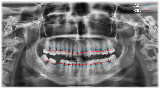
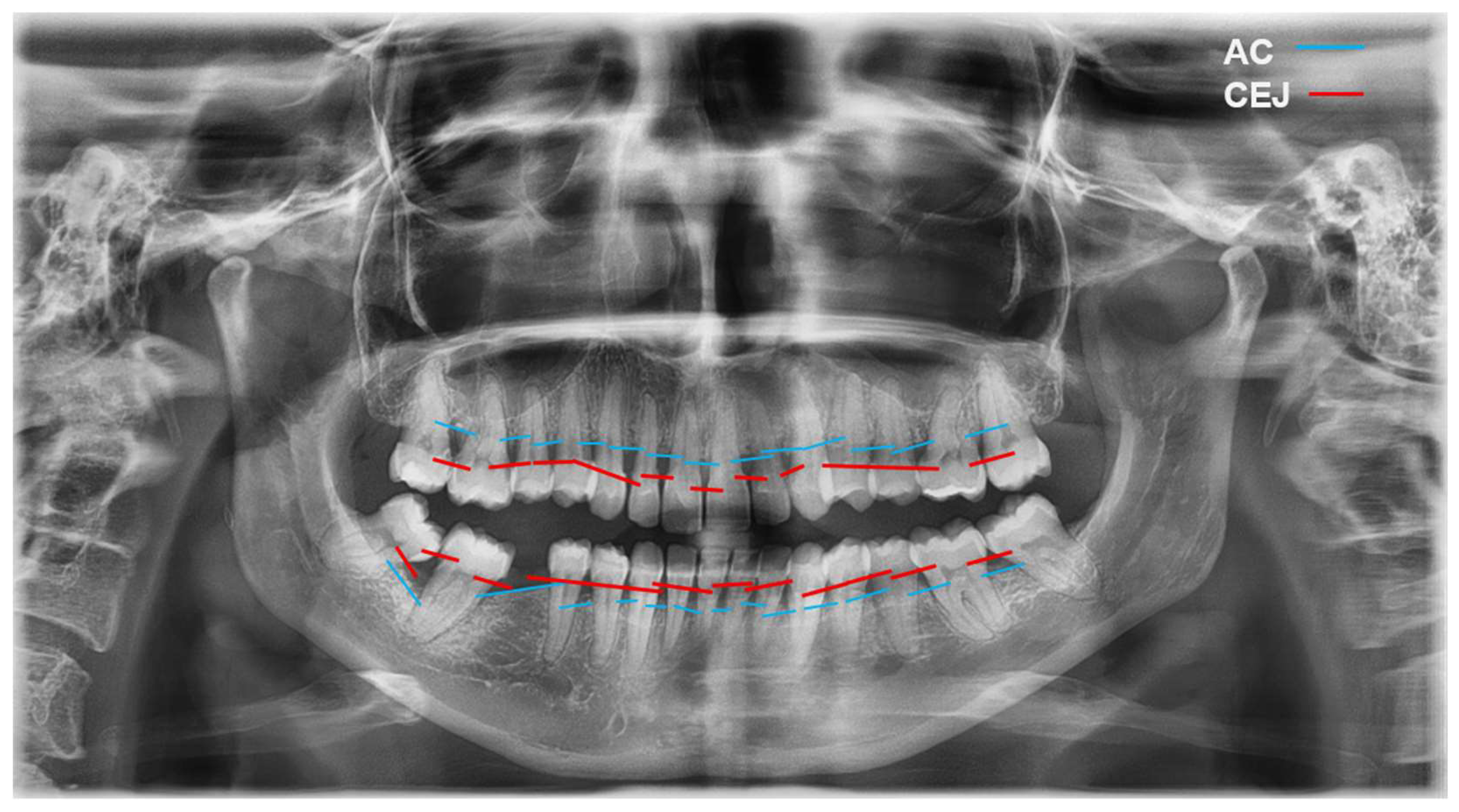
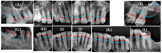
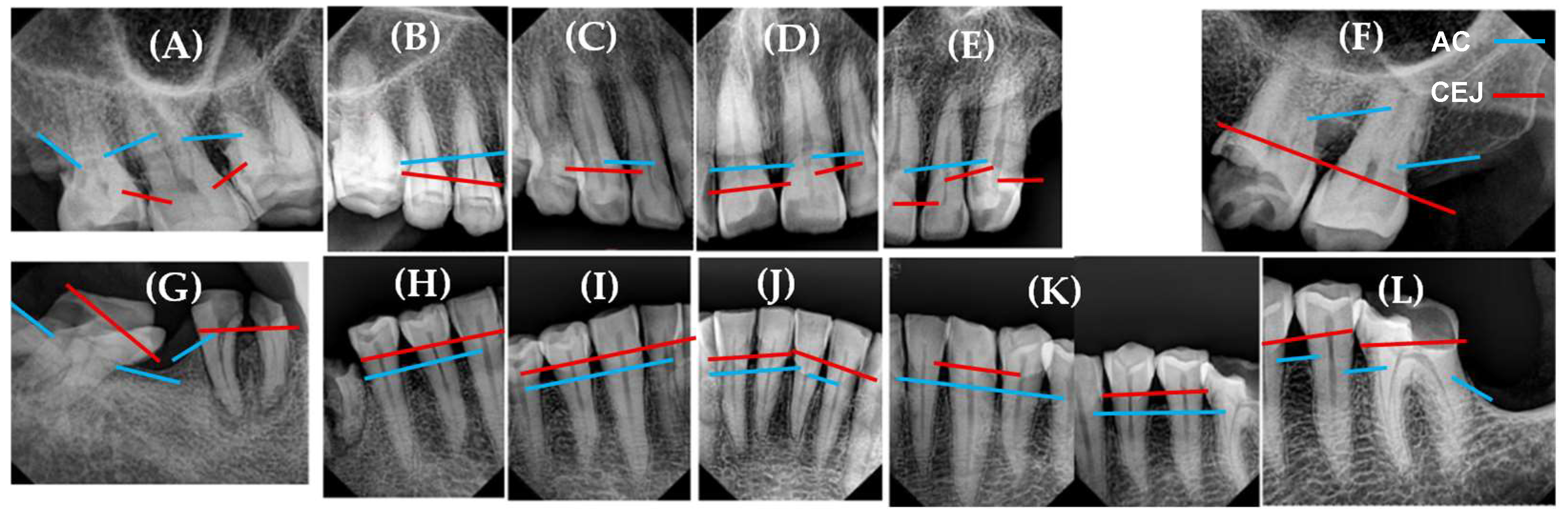
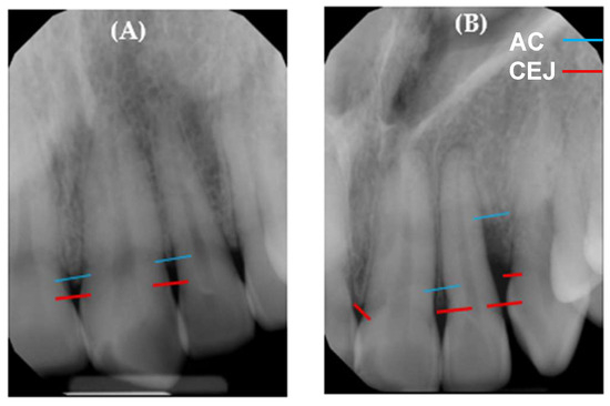
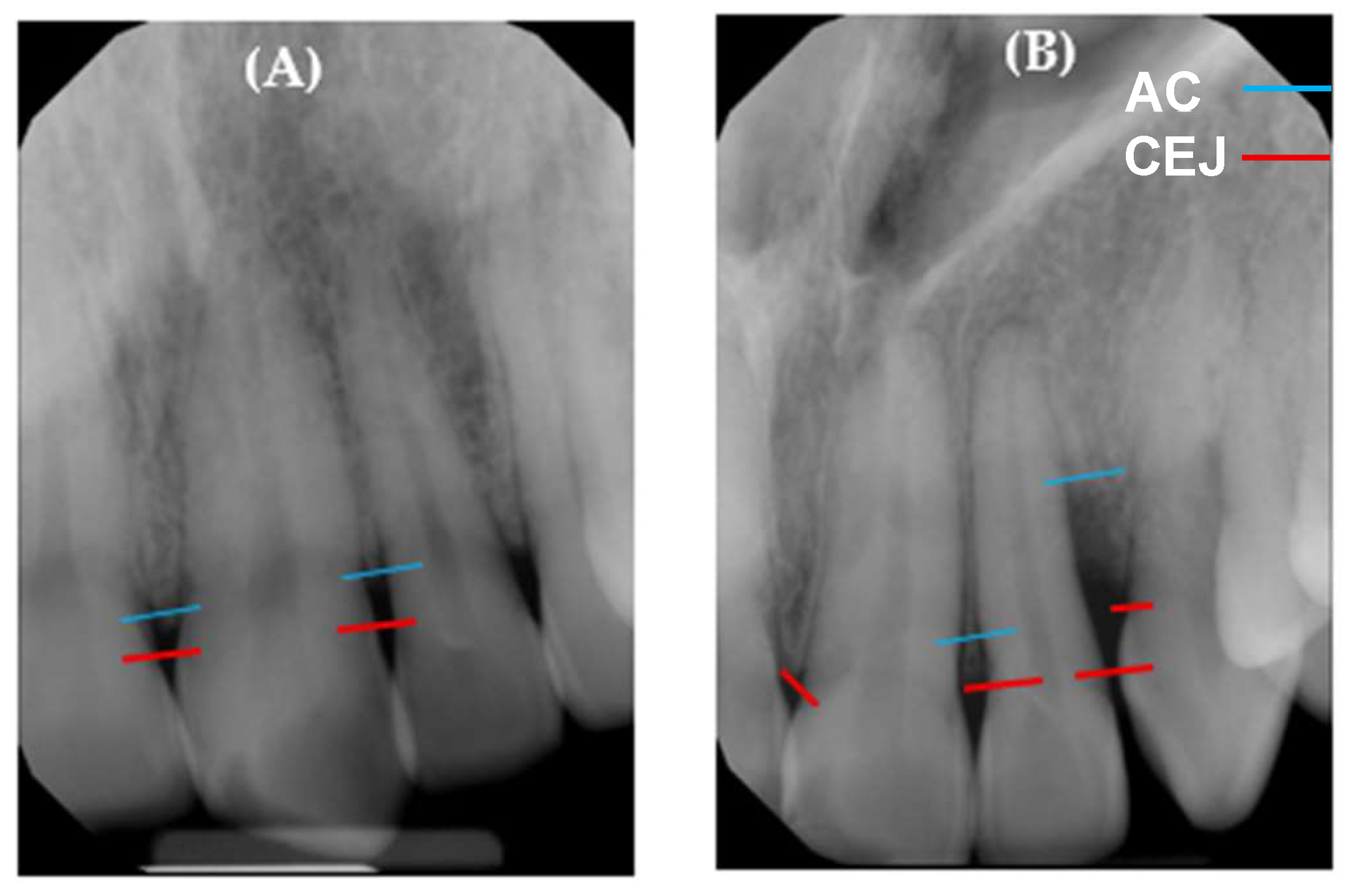
A full-mouth periodontal examination was performed by a calibrated and duly certified periodontist from the Periodontics Clinic of the Faculty of Dentistry of the Autonomous University of Sinaloa. Each patient underwent a clinical evaluation to verify the presence of periodontal pockets in at least 14 teeth. Additionally, a general medical history was conducted, covering identification data, reason for consultation, personal and family medical history, current condition, review of systems, and physical examination, in order to obtain a comprehensive view of the patient’s overall health. As part of the radiographic study, each patient underwent an orthopantomogram (Figure 1) and a series of 14 periapical radiographs (Figure 2) to assess the level of alveolar bone loss (Figure 3).
Figure 1.
Orthopantomographic image of a patient with periodontitis. Showing an average distance of 4–5 mm between the most coronal portion of the alveolar bone crest (AC) and the cementoenamel junction (CEJ), indicative of alveolar bone loss.
Figure 2.
Periapical radiographic series obtained from a single patient with periodontitis. (A) and (F) depict teeth exhibiting 15–33% alveolar bone loss, corresponding to Stage II periodontitis. (B–E) and (H–L) illustrate teeth with <15% alveolar bone loss, consistent with Stage I periodontitis. (G) presents root remnants of the mandibular right molar, associated with extensive caries and a mesially impacted third molar.
Figure 3.
Periapical radiographs. (A) Normal and anatomically with intact periodontium, showing preservation of the lamina dura and an average distance of approximately 2 mm between the most coronal portion of the alveolar bone crest (AC) and the cementoenamel junction (CEJ), which corresponds to the physiological range of 1.0–3.0 mm observed in healthy individuals. (B) Periodontium with alveolar bone loss, characterized by disruption of lamina dura continuity and an increased AC–CEJ distance of 8 mm.
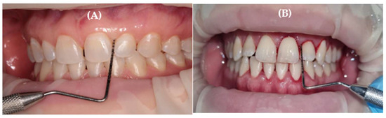
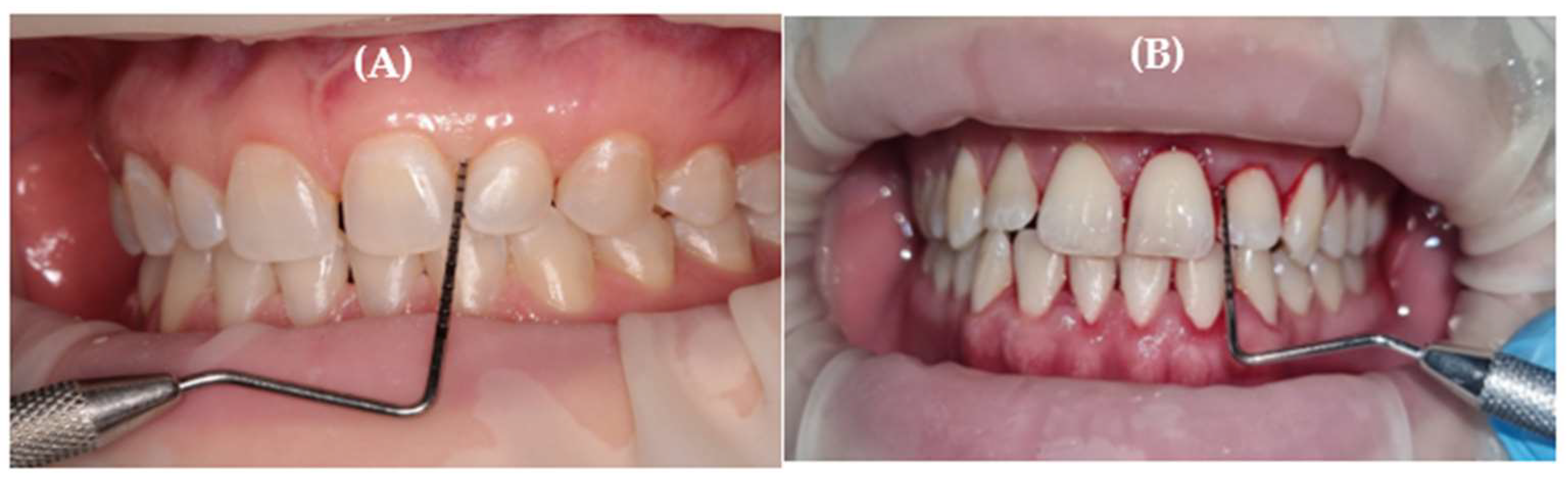
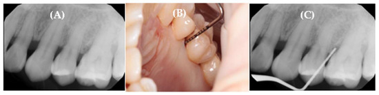
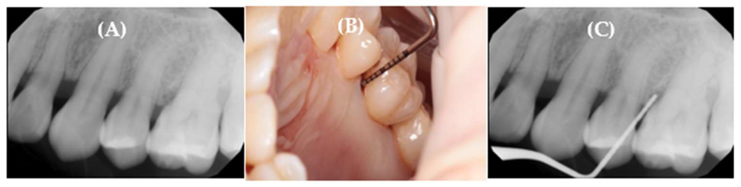
Subsequently, the periodontal examination was performed, recording the data obtained from pocket probing using the University of North Carolina UNC-15 periodontal probe in the gingival sulcus at six sites (mesio-buccal, buccal, disto-buccal, mesio-lingual, lingual, disto-lingual) on all teeth present. During this procedure, the clinical attachment level, the bleeding on probing (BOP), and the distance from the CEJ to the gingival margin were simultaneously evaluated (Figure 4), with the aim of diagnosing the presence of gingival recessions (Figure 5).
Figure 4.
Periodontal probing. (A) Probing depth on the mesiobuccal surface of a lateral incisor of 2 mm from the free gingival margin to the base of the gingival/periodontal sulcus, with no clinical attachment loss. (B) Presence of bleeding on probing (BOP) following periodontal probing.
Figure 5.
Periodontium with alveolar bone loss. (A) Periapical radiograph showing alveolar bone loss. (B) A 5 mm periodontal pocket with BOP at the mesial surface of the maxillary first molar. (C) Periapical radiograph with a periodontal probe inserted to measure the distance from the base of the periodontal pocket to the cementoenamel junction (CEJ).
The periodontogram recorded each one of the patient’s teeth, noting healthy teeth, the presence of fixed prostheses, functional or defective dental restorations, and teeth that are abutments for removable prostheses. With the data collected from medical history and radiographs, the general diagnosis of the patient was performed, as well as for each individual tooth, establishing their respective prognosis.
Following the assessment and diagnosis of the patients, they were categorized into two groups based on the clinical findings. Periodontal disease was classified according to stage and grade criteria according to the 2017 World Workshop on the Classification of Periodontal and Peri-Implant Diseases and Conditions [5,24].
2.4. Gingival Sample Collection
Once the medical records of all the adult patients were assessed, periodontal epithelial cells were collected by brushing the gingival tissue with a cytobrush with a 4-inch handle and an ultra-thin 1.5 mm diameter tip from the brand PREMIUM PLUS. The brush was inserted into the free gingival tissue and gently rotated in a downward direction for approximately 10 s to obtain epithelial cells and gingival crevicular material, thus adequately collecting the necessary sample. Samples were immediately placed in RNase-free tubes containing RNA stabilization solution and stored at −80 °C until further processing. Isolation of the selected teeth for sampling was not necessary due to the nature of the methodology used.
2.5. RNA Extraction and Reverse Transcription (RT) PCR
Total RNA, including miRNAs, was extracted using the miRNeasy RNA extraction kit (Qiagen) according to the manufacturer’s instructions. The integrity and purity of the RNA were evaluated spectrophotometrically with a GENESYS 10S-UV–Vis (Thermo Scientific) (Therermo Scientific™, Waltham, MA, USA). Complementary DNA (cDNA) was synthesized from 10 ng of total RNA using the TaqMan Advanced miRNA cDNA Synthesis Kit (Applied Biosystems, Foster City, CA, USA). Reverse transcription reactions were carried out in a T100 Thermal Cycler (Bio-Rad) (Bio-Rad, Hercules, CA, USA) under conditions specified by the supplier. This workflow ensured the efficient conversion of mature miRNAs into stable cDNA templates for subsequent amplification.
2.6. Quantification of miRNA Expression by Real-Time PCR
Quantification of miRNA expression was performed by real-time PCR using TaqMan MicroRNA Assays (Applied Biosystems, Foster City, CA, USA) specific for miR-141-3p, miR-146a-5p, and miR-148b-3p, following the manufacturer’s recommendations. Amplification reactions were carried out using a StepOnePlus Real-Time PCR System (Applied Biosystems) under optimized cycling conditions provided by the assay manufacturer.
To ensure analytical reliability, amplification efficiency for each miRNA assay was assessed using serial dilutions of cDNA, and only assays demonstrating efficiencies within the acceptable range (90–110%) were included in the analysis. Each sample was analyzed in duplicate, and non-template controls were included in all runs to exclude potential contamination or nonspecific amplification.
For normalization of miRNA expression data, candidate reference miRNAs were evaluated using the RefFinder algorithm, which integrates geNorm, NormFinder, BestKeeper, and the comparative ΔCt method. Based on this comprehensive stability assessment, miR-191-5p was selected as the most stable endogenous control across all samples. Relative miRNA expression levels were calculated using the method proposed by Taylor et al. [25], based on the 2−ΔΔCt algorithm [26]. For statistical analyses, relative normalized expression values were log-transformed to improve data normality and variance homogeneity.
2.7. Statistical Analysis
All statistical analyses were performed using Stata version 14 (StataCorp, College Station, TX, USA). Prior to comparative testing, the distribution of relative normalized miRNA expression values was assessed for normality in each study group using the Shapiro–Wilk test.
For comparisons between two independent groups, the Student’s t-test was applied when data followed a normal distribution, whereas the Mann–Whitney U test was used for non-normally distributed data. For comparisons involving three or more groups, one-way analysis of variance (ANOVA) was performed when normality assumptions were met, and the Kruskal–Wallis test was used otherwise. The level of statistical significance was set at p < 0.05.
Correlations between relative miRNA expression levels and clinicopathological variables, including age, sex, disease stage, and grade, were evaluated using Pearson’s or Spearman’s correlation coefficients, according to data distribution.
3. Results
3.1. Study Population and Clinical Characteristics
A total of 46 participants were included in this study and classified into periodontally healthy controls (HC) and patients with periodontal disease (PD).
The demographic and clinical characteristics of the study population are summarized in Table 1. No statistically significant differences were observed between groups with respect to age or sex distribution. As expected, PD patients exhibited a significantly higher probing depth, clinical attachment loss, and bleeding on probing compared with healthy controls.
Table 1.
Statistical assessment of demographic and clinical characteristics.
3.2. Differential Expression of miRNAs in Periodontal Disease
The relative expression levels of miR-141-3p, miR-146a-5p, and miR-148b-3p were evaluated in gingival samples from HC and PD groups using RT-qPCR. Expression values were normalized to miR-191-5p and are presented as relative normalized expression.
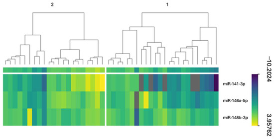
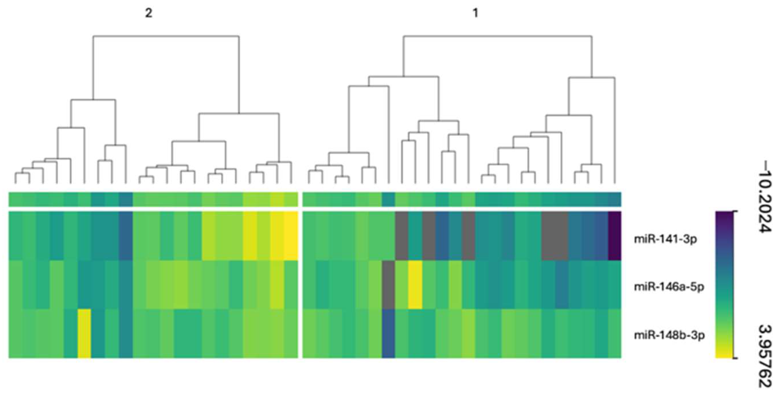
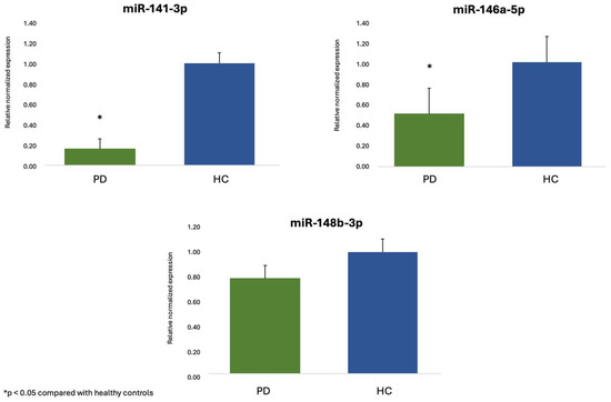
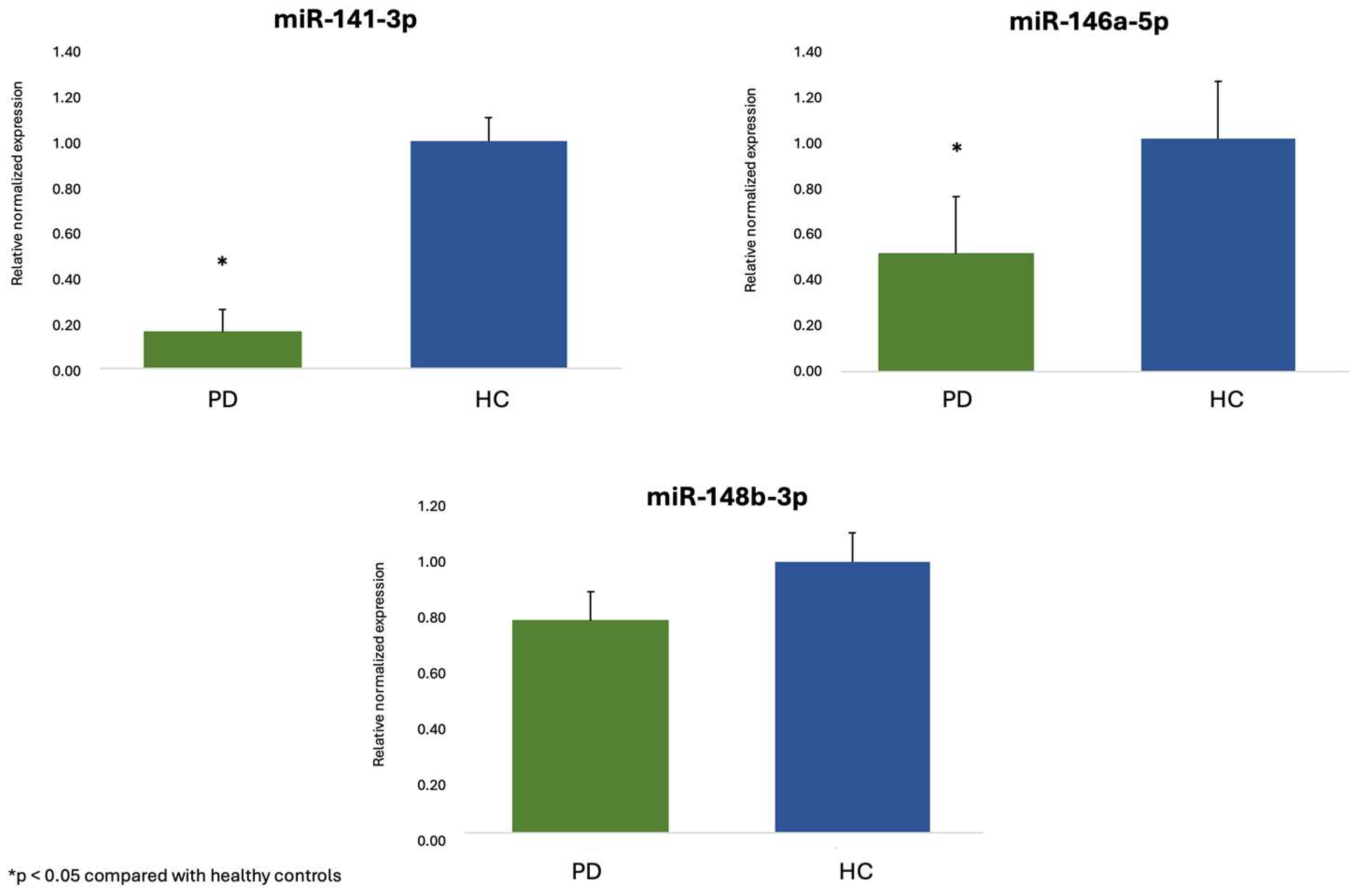
A significant downregulation of miR-141-3p was observed in the PD group compared with healthy controls (p < 0.05, Figure 6). In contrast, miR-146a-5p showed a trend toward reduced expression in PD patients; however, this difference did not reach statistical significance. No significant differences in the expression of miR-148b-3p were detected between the two groups.
Figure 6.
Heatmap represents differential miRNA expressions between PD and HC groups.
3.3. Association Between miRNA Expression and Periodontal Disease Severity
To evaluate whether miRNA expression levels were associated with disease severity, relative expression values were analyzed according to periodontal stage and grade. No statistically significant differences in the expression of miR-141-3p, miR-146a-5p, or miR-148b-3p were observed across different stages or grades of periodontal disease (Figure 7).
Figure 7.
Relative expression of miR-141-3p, miR-146a-5p, and miR-148b-3p in gingival samples from patients with periodontal disease (PD) and periodontally healthy controls (HC); green bars indicate PD and blue bars indicate HC. miRNA expression levels were quantified by RT-qPCR, normalized to miR-191-5p, and expressed as relative normalized expression. Data are presented as mean ± standard error of the mean. * p < 0.05 compared with healthy controls.
3.4. Correlation Analysis of miRNA Expression Levels
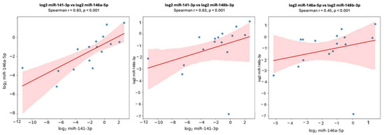
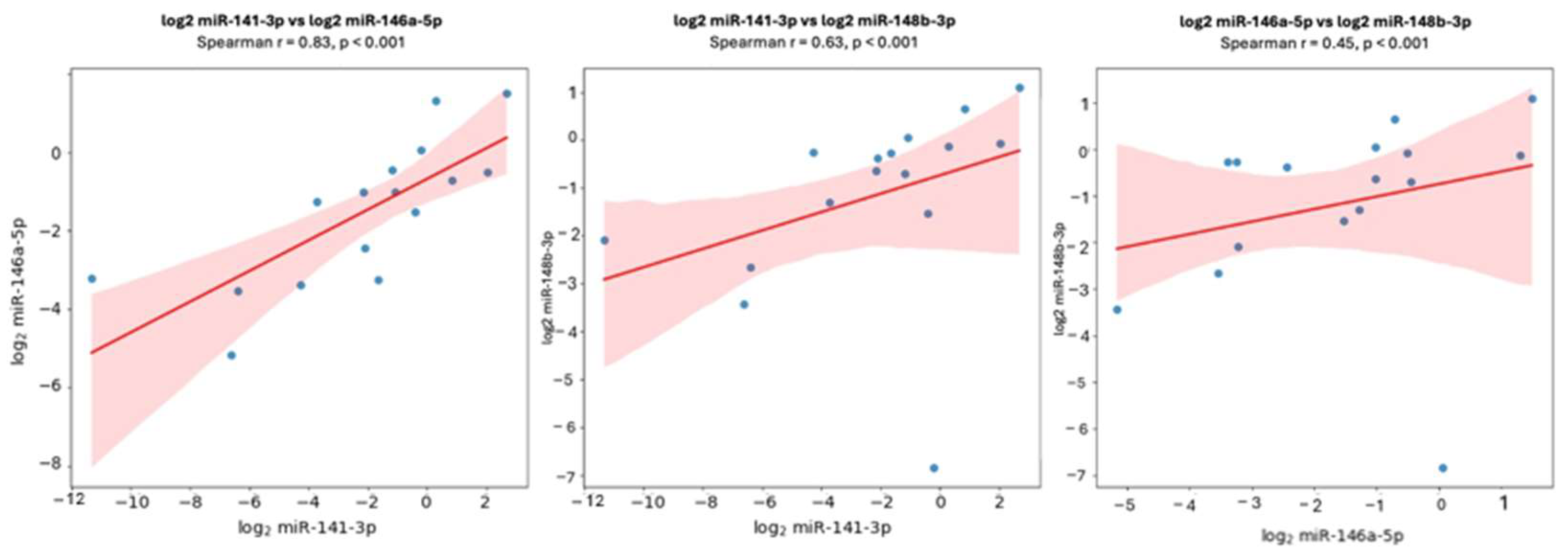
Correlation analyses were performed to explore potential relationships among the three miRNAs evaluated. A strong positive correlation was identified between miR-141-3p and miR-146a-5p expression levels (Spearman’s ρ = 0.83, p < 0.001). Additionally, miR-148b-3p expression showed significant positive correlations with both miR-141-3p (Spearman’s ρ = 0.63, p < 0.001) and miR-146a-5p (Spearman’s ρ = 0.45, p < 0.001) (Figure 8).
Figure 8.
Relative miRNA expression correlation analysis. Blue dots represent individual samples, the red line indicates the fitted linear regression trend, and the shaded area corresponds to the 95% confidence interval.
No significant correlations were observed between miRNA expression levels and clinicopathological variables, including age, sex, periodontal stage, or grade.
4. Discussion
In this study, we evaluated the expression of miR-141-3p, miR-146a-5p, and miR-148b-3p in gingival samples from individuals with periodontal disease and periodontally healthy controls, with the aim of exploring their potential involvement in periodontal inflammation. The most robust finding was the significant downregulation of miR-146a-5p in periodontal disease, accompanied by a consistent trend toward reduced expression of miR-141-3p, while miR-148b-3p did not show significant differential expression. Importantly, correlation analyses revealed strong positive associations among the three miRNAs, suggesting coordinated regulation rather than independent behavior.
Among the miRNAs analyzed, miR-146a-5p is a well-established negative regulator of inflammatory signaling. It modulates the NF-κB pathway through suppression of key adaptor molecules such as IRAK1 and TRAF6, thereby limiting excessive inflammatory responses [11,13]. Recent studies have further demonstrated that reduced miR-146a-5p expression is associated with sustained inflammation and impaired immune homeostasis in chronic inflammatory conditions [11,12,13,15]. In the context of periodontal disease, the observed downregulation of miR-146a-5p may therefore reflect a compromised negative feedback mechanism that favors persistent inflammatory signaling within the periodontal microenvironment. Although the LPS of Porphyromonas gingivalis increases the expression of miR-146a-5p, its effect is counteracted by the action of gingipains; this could be explained since gingipains are cysteine proteases attached to the bacterial surface or secreted into the environment of the periodontal sulcus [16,17].
In contrast, miR-141-3p did not reach conventional statistical significance when comparing periodontal disease and healthy controls, although a clear downward trend was observed. This finding should be interpreted cautiously, as miR-141-3p has been implicated in endothelial inflammation, immune cell adhesion, and regulation of osteogenic differentiation [18,19]. Emerging evidence indicates that modest alterations in the expression of regulatory miRNAs can exert biologically meaningful effects, particularly in complex and heterogeneous tissues such as periodontal samples, where subtle changes may not always be captured by strict statistical thresholds [19]. The expression of miR-148b-3p did not differ significantly between groups. Although miR-148b has been implicated in inflammation-related pathways and osteogenic processes in other biological contexts, its role appears to be highly context dependent and may vary according to tissue type and disease setting [27]. Notably, despite the absence of differential expression, miR-148b-3p exhibited significant correlations with both miR-141-3p and miR-146a-5p. This observation supports the notion that miR-148b-3p may participate in a coordinated regulatory program rather than functioning as an isolated marker of periodontal disease, consistent with recent models of miRNA network regulation [8,9].
The strong positive correlations identified among the three miRNAs represent a central finding of this study. Increasing evidence supports the concept that miRNAs act within regulatory modules, fine-tuning complex biological processes through coordinated expression patterns rather than isolated effects [8,9,10]. In periodontal disease, such co-regulation may reflect shared upstream inflammatory signaling pathways or convergent involvement in immune and bone-related responses, reinforcing the value of network-based approaches for understanding periodontal pathophysiology.
Notably, no significant associations were observed between miRNA expression levels and periodontal disease stage or grade. This finding suggests that the evaluated miRNAs may be more closely related to disease susceptibility or inflammatory responsiveness than to disease severity or progression. Similar observations have been reported in chronic inflammatory conditions, where early molecular dysregulation precedes overt clinical deterioration and may define individual thresholds for inflammatory damage [8,11].
Several limitations of this study should be acknowledged. Gingival samples were collected using a cytobrush, which yields heterogeneous cell populations, including epithelial cells, immune cells, and components of gingival crevicular fluid. While such heterogeneity may attenuate expression differences, it also reflects the complex cellular composition of periodontal tissues. Importantly, the detection of coherent co-expression patterns despite this variability supports the robustness of the observed regulatory relationships [8,9,10]. Additionally, the cross-sectional design limits causal inference, and longitudinal studies will be necessary to clarify the temporal dynamics of miRNA regulation in periodontal disease.
5. Conclusions
This study demonstrates the differential expression and coordinated behavior of selected microRNAs in periodontal disease. Specifically, miR-146a-5p was significantly downregulated in affected individuals, given that the appearance and development of periodontal disease arises due to dysbiosis among microorganisms located in the gingival sulcus, and being as it is reported in various studies that the microorganisms identified as the main etiological agents of PD produces and secretes metabolites such as gingipains, generating a microenvironment that induces inflammation and modifies the immune response. It is highly probable that the variation in the expression of miR-146a-5p in patients with periodontal disease is influenced by the metabolism of the microbiota as a result of infection and inflammation in the periodontal tissue. miR-146a-5p’s reduced expression in periodontitis, which likely reflects a disease-specific dysregulation of inflammatory resolution rather than a universal anti-inflammatory association, considering it is generally upregulated in response to inflammatory stimuli as part of a negative feedback mechanism. Therefore, the regulatory role of miR-146a-5p depends on the context and may be impaired in chronic periodontal inflammation, not necessarily describing a behavior present across different inflammatory conditions. Meanwhile, miR-141-3p showed a consistent trend toward reduced expression. Although miR-148b-3p did not differ significantly between groups, its strong correlation with the other miRNAs supports its participation in a coordinated regulatory network rather than an isolated role. The lack of association with disease stage or grade suggests that these miRNAs may be more closely related to inflammatory susceptibility than to disease severity. Overall, these findings contribute to a deeper molecular understanding of periodontal inflammation and highlight the relevance of miRNA network analysis in periodontal research.
Author Contributions
E.A.M. contributed to the conception and design and drafted and critically revised the manuscript. A.R.O. contributed to the analysis, data interpretation, and drafted and critically revised the manuscript. M.R.Á. contributed to the conception and design and critically revised the manuscript. N.G.M. contributed to the conception, analysis, data interpretation, and critically revised the manuscript. A.Y.T.M. contributed to data analysis and interpretation and critically revised the manuscript. G.E.G.C. contributed to data acquisition and analysis and drafted and critically revised the manuscript. V.I.V. contributed to data acquisition and analysis and drafted the manuscript. J.A.B.L. and A.K.O.G. contributed to data interpretation and analysis and drafted the manuscript. F.L.O. contributed to the conception and design, data interpretation, and critically revised the manuscript. All authors have read and agreed to the published version of the manuscript.
Funding
This work was supported by the program “Programa de Fomento y Apoyo a Proyectos de Investigación” Call for Research Proposals PROFAPI 2022 from the Universidad Autónoma de Sinaloa (Projects PRO_A3_002, PRO_A3_003, PRO_A3_032).
Institutional Review Board Statement
This study was conducted in accordance with the Declaration of Helsinki and reviewed and approved by the Ethics and Research Committee of Faculty of Dentistry at Universidad Autónoma de Sinaloa (study approval number P-007-2021, 26 August 2021).
Informed Consent Statement
Informed consent was obtained from all subjects involved in the study. Written informed consent has been obtained from the patient(s) to publish this paper.
Data Availability Statement
The data presented in this study are available on request from the corresponding author because they respect the confidentiality of the individuals under study who participated in the research.
Acknowledgments
We thank the Polytechnic University of Sinaloa for providing part of the infrastructure necessary for the development of this research.
Conflicts of Interest
The authors declare no conflicts of interest. The funders had no role in the design of the study; in the collection, analyses, or interpretation of data; in the writing of the manuscript; or in the decision to publish the results.
Abbreviations
The following abbreviations are used in this manuscript:
| AC | Alveolar Bone Crest |
| BOP | Bleeding On Probing |
| CAL | Clinical Attachment Loss |
| CEJ | Cementoenamel Junction |
| HC | Healthy Controls |
| ICAM | Intercellular Adhesion Molecule |
| miRNA | MicroRNA |
| mm | Millimeters |
| PD | Periodontal Disease |
References
- Kwon, T.; Lamster, I.B.; Levin, L. Current Concepts in the Management of Periodontitis. Int. Dent. J. 2021, 71, 462–476. [Google Scholar] [CrossRef] [Scilit]
- Wu, L.; Huang, C.-M.; Wang, Q.; Wei, J.; Xie, L.; Hu, C.-Y. Burden of Severe Periodontitis: New Insights Based on a Systematic Analysis from the Global Burden of Disease Study 2021. BMC Oral Health 2025, 25, 861. [Google Scholar] [CrossRef] [Scilit]
- Sojod, B.; Périer, J.-M.; Zalcberg, A.; Bouzegza, S.; Halabi, B.E.; Anagnostou, F. Enfermedad Periodontal y Salud General. EMC-Tratado Med. 2022, 26, 1–8. [Google Scholar] [CrossRef] [Scilit]
- Yildirim, B.G.; Aksit, C.; Mutlu, M.; Ainola, M.; Eklund, K.K.; Leskelä, J.; Pussinen, P.; Beklen, A. Severity and Progression Rate of Periodontitis Are Associated with an Increased Risk of Hypertension of Patients Attending a University Clinic. BMC Oral Health 2022, 22, 627. [Google Scholar] [CrossRef] [Scilit] [PubMed]
- Papapanou, P.N.; Sanz, M.; Buduneli, N.; Dietrich, T.; Feres, M.; Fine, D.H.; Flemmig, T.F.; Garcia, R.; Giannobile, W.V.; Graziani, F. Periodontitis: Consensus Report of Workgroup 2 of the 2017 World Workshop on the Classification of Periodontal and Peri-Implant Diseases and Conditions. J. Periodontol. 2018, 89, S173–S182. [Google Scholar] [CrossRef] [Scilit] [PubMed]
- Luan, X.; Zhou, X.; Naqvi, A.; Francis, M.; Foyle, D.; Nares, S.; Diekwisch, T.G.H. MicroRNAs and Immunity in Periodontal Health and Disease. Int. J. Oral Sci. 2018, 10, 24. [Google Scholar] [CrossRef] [Scilit] [PubMed]
- Rodrigues, L.; Da Cruz Paula, A.; Soares, P.; Vinagre, J. Unraveling the Significance of DGCR8 and miRNAs in Thyroid Carcinoma. Cells 2024, 13, 561. [Google Scholar] [CrossRef] [Scilit]
- Gaál, Z. Role of microRNAs in Immune Regulation with Translational and Clinical Applications. Int. J. Mol. Sci. 2024, 25, 1942. [Google Scholar] [CrossRef] [Scilit]
- Luan, X.; Zhou, X.; Fallah, P.; Pandya, M.; Lyu, H.; Foyle, D.; Burch, D.; Diekwisch, T.G.H. MicroRNAs: Harbingers and Shapers of Periodontal Inflammation. Semin. Cell Dev. Biol. 2022, 124, 85–98. [Google Scholar] [CrossRef] [Scilit]
- Luan, X.; Zhou, X.; Trombetta-eSilva, J.; Francis, M.; Gaharwar, A.; Atsawasuwan, P.; Diekwisch, T. MicroRNAs and Periodontal Homeostasis. J. Dent. Res. 2017, 96, 491–500. [Google Scholar] [CrossRef] [Scilit]
- Gilyazova, I.; Asadullina, D.; Kagirova, E.; Sikka, R.; Mustafin, A.; Ivanova, E.; Bakhtiyarova, K.; Gilyazova, G.; Gupta, S.; Khusnutdinova, E.; et al. MiRNA-146a—A Key Player in Immunity and Diseases. Int. J. Mol. Sci. 2023, 24, 12767. [Google Scholar] [CrossRef] [Scilit]
- Mahmoudi Anzabi, R.; Shenasa, N.; Jafarian, A.; Davodpour, A.H.; Zareanshahraki, M.; Ghasemirad, M.; Rostamzadeh, S. Enhancing Osteogenesis in Dental Implants: ZNF710-AS1 and miR-146a-5p in Smad Signaling Pathway Activation. Int. J. Mol. Cell. Med. 2025, 14, 964–975. [Google Scholar] [CrossRef] [Scilit]
- Zhang, H.; Wang, Y.; Feng, K.; Niu, Q.; Xin, Y.; Xuan, S.; Liu, S. MiR-146a-5p-Enriched Exosomes Inhibit M1 Macrophage Activation and Inflammatory Response by Targeting CD80. Mol. Biol. Rep. 2024, 51, 1133. [Google Scholar] [CrossRef] [Scilit]
- Rovas, A.; Puriene, A.; Snipaitiene, K.; Punceviciene, E.; Buragaite-Staponkiene, B.; Matuleviciute, R.; Butrimiene, I.; Jarmalaite, S. Gingival Crevicular Fluid microRNA Associations with Periodontitis. J. Oral Sci. 2022, 64, 11–16. [Google Scholar] [CrossRef] [Scilit]
- Damodaran, M.; Paul, S.F.D.; Venkatesan, V. Genetic Polymorphisms in miR-146a, miR-196a2 and miR-125a Genes and Its Association in Prostate Cancer. Pathol. Oncol. Res. 2020, 26, 193–200. [Google Scholar] [CrossRef] [Scilit] [PubMed]
- Xie, Y.-F.; Shu, R.; Jiang, S.-Y.; Liu, D.-L.; Ni, J.; Zhang, X.-L. MicroRNA-146 Inhibits pro-Inflammatory Cytokine Secretion through IL-1 Receptor-Associated Kinase 1 in Human Gingival Fibroblasts. J. Inflamm. 2013, 10, 20. [Google Scholar] [CrossRef] [Scilit] [PubMed]
- Dong, J.; Liao, Y.; Sun, M.; Chen, H.; Zhou, K.; Zhang, H.; Zhou, W.; Song, Z. Gingipains Disrupt Bone Homeostasis via Dual Regulation of Osteogenesis and Osteoclastogenesis through Exosomal miR-146a-5p/TRAF6 Signaling. Front. Cell. Infect. Microbiol. 2025, 15, 1614126. [Google Scholar] [CrossRef] [Scilit] [PubMed]
- Vyavahare, S.; Kumar, S.; Smith, K.; Mendhe, B.; Zhong, R.; Cooley, M.A.; Baban, B.; Isales, C.M.; Hamrick, M.; Hill, W.D.; et al. Hill Inhibiting MicroRNA-141-3p Improves Musculoskeletal Health in Aged Mice. Aging Dis. 2023, 14, 2303–2618. [Google Scholar] [CrossRef] [Scilit]
- Yu, S.-L. Diagnostic Potential of miR-200 Family Members in Gingival Crevicular Fluid for Chronic Periodontitis: Correlation with Clinical Parameters and Therapeutic Implications. BMC Oral Health 2023, 23, 532. [Google Scholar] [CrossRef] [Scilit]
- Jurčić, V.; Bolha, L.; Matjašič, A.; Sedej, I.; Dolinar, A.; Grubelnik, G.; Hauptman, N.; Pižem, J.; Jevšinek-Skok, D.; Hočevar, A.; et al. Association between Histopathological Changes and Expression of Selected microRNAs in Skin of Adult Patients with IgA Vasculitis. Histopathology 2019, 75, 683–693. [Google Scholar] [CrossRef] [Scilit]
- Li, W.; Alimujiang, A. METTL3 Promotes Osteogenic Differentiation of Human Periodontal Ligament Stem Cells Under the Inflammatory Microenvironment Through the miR-141-3p/ZEB1 Axis. Cell Biochem. Biophys. 2025, 83, 1771–1783. [Google Scholar] [CrossRef] [Scilit]
- Mollazadeh, S.; Fazly Bazzaz, B.S.; Neshati, V.; de Vries, A.A.F.; Naderi-Meshkin, H.; Mojarad, M.; Mirahmadi, M.; Neshati, Z.; Kerachian, M.A. Overexpression of MicroRNA-148b-3p Stimulates Osteogenesis of Human Bone Marrow-Derived Mesenchymal Stem Cells: The Role of MicroRNA-148b-3p in Osteogenesis. BMC Med. Genet. 2019, 20, 117. [Google Scholar] [CrossRef] [Scilit]
- Chapple, I.L.; Mealey, B.L.; Van Dyke, T.E.; Bartold, P.M.; Dommisch, H.; Eickholz, P.; Geisinger, M.L.; Genco, R.J.; Glogauer, M.; Goldstein, M. Periodontal Health and Gingival Diseases and Conditions on an Intact and a Reduced Periodontium: Consensus Report of Workgroup 1 of the 2017 World Workshop on the Classification of Periodontal and Peri-Implant Diseases and Conditions. J. Periodontol. 2018, 89, S74–S84. [Google Scholar] [CrossRef] [Scilit]
- Heitz-Mayfield, L.J.A. Conventional Diagnostic Criteria for Periodontal Diseases (Plaque-Induced Gingivitis and Periodontitis). Periodontol. 2000 2024, 95, 10–19. [Google Scholar] [CrossRef] [Scilit] [PubMed]
- Taylor, S.C.; Nadeau, K.; Abbasi, M.; Lachance, C.; Nguyen, M.; Fenrich, J. The Ultimate qPCR Experiment: Producing Publication Quality, Reproducible Data the First Time. Trends Biotechnol. 2019, 37, 761–774. [Google Scholar] [CrossRef] [Scilit] [PubMed]
- Livak, K.J.; Schmittgen, T.D. Analysis of Relative Gene Expression Data Using Real-Time Quantitative PCR and the 2−ΔΔCT Method. Methods 2001, 25, 402–408. [Google Scholar] [CrossRef] [Scilit] [PubMed]
- Miscianinov, V.; Martello, A.; Rose, L.; Parish, E.; Cathcart, B.; Mitić, T.; Gray, G.A.; Meloni, M.; Al Haj Zen, A.; Caporali, A. MicroRNA-148b Targets the TGF-β Pathway to Regulate Angiogenesis and Endothelial-to-Mesenchymal Transition during Skin Wound Healing. Mol. Ther. 2018, 26, 1996–2007. [Google Scholar] [CrossRef] [Scilit]

Disclaimer/Publisher’s Note: The statements, opinions and data contained in all publications are solely those of the individual author(s) and contributor(s) and not of MDPI and/or the editor(s). MDPI and/or the editor(s) disclaim responsibility for any injury to people or property resulting from any ideas, methods, instructions or products referred to in the content. |
© 2026 by the authors. Licensee MDPI, Basel, Switzerland. This article is an open access article distributed under the terms and conditions of the Creative Commons Attribution (CC BY) license.