


Abstract
This study aimed to develop bioactive pectin-based films incorporating Deverra tortuosa (D. tortuosa) essential oil (EO) nanoemulsions stabilized by sodium caseinate for potential active food packaging applications. EO nanoemulsions were incorporated into a pectin–maltodextrin matrix, and the resulting films were characterized for their thermal, morphological, antioxidant, and antimicrobial properties. Thermal analyses revealed an additional low-temperature mass loss in EO-loaded films, attributed to volatile oil constituents, while differential scanning calorimetry showed an endothermic transition at 171.9 °C associated with matrix reorganization and volatile release. Scanning electron microscopy (SEM) demonstrated increased surface heterogeneity and the formation of microcavities compared with EO-free films (F−). The incorporation of EO significantly enhanced antioxidant activity, with half-maximal inhibitory concentration (IC50) values of 9.18 µg mL−1 and 1.95 µg mL−1 in 2,2-Diphenyl-1-picrylhydrazyl (DPPH) and 2,2′-azinobis(3-ethylbenzothiazoline-6-sulfonic acid) (ABTS) assays, respectively. In addition, the films exhibited antimicrobial activity against Escherichia coli, Salmonella enterica, and Listeria innocua, with minimum inhibitory concentrations ranging from 1.25 to 2.5 mg mL−1. These results demonstrate that the incorporation of Deverra tortuosa EO nanoemulsions improves the functional properties of pectin-based films and supports their potential use as biodegradable active packaging materials.
1. Introduction
Growing environmental concerns and the need for sustainable materials have accelerated research into biodegradable and bioactive packaging systems. Among natural polymers, pectin has attracted considerable attention due to its availability, biocompatibility, and excellent film-forming ability, making it suitable for edible and active food coatings. However, conventional pectin-based films typically exhibit limited mechanical strength, high moisture sensitivity, and weak barrier performance, which restrict their practical use in food preservation [1].
To overcome these limitations, various functional additives have been incorporated into pectin-based matrices, including natural antioxidants and antimicrobials. In particular, essential oils (EOs) have emerged as promising bioactive agents owing to their strong antimicrobial, antioxidant, and anti-inflammatory properties [2]. The addition of EOs to biopolymer-based films can affect their mechanical and physicochemical characteristics, such as tensile strength, moisture-related characteristics, and water vapor barrier performance, in addition to their bioactive roles. Additionally, since preserving film activity over time is crucial for their potential use as active food packaging materials, subsequent studies have emphasized the significance of evaluating these properties’ evolution during storage [3,4]. Thus, EO inclusion can give bioactivity while concurrently altering the physicochemical and mechanical behavior of biopolymer films, as previous research has shown [5,6]. Nevertheless, their direct addition to hydrophilic polymer matrices remains challenging because of their hydrophobicity, volatility, and susceptibility to oxidation and thermal degradation [2]. These factors often result in poor dispersion and uncontrolled release within the film structure, limiting the reproducibility and functionality of the resulting materials [7].
Nanoemulsion technology provides an effective strategy to stabilize and deliver EO in polymeric systems. By reducing droplet size to the submicron scale and using food-grade emulsifiers, nanoemulsions improve the solubility, retention, and bioavailability of volatile compounds, while promoting homogeneous distribution within biopolymer networks [8]. According to earlier research, adding EO nanoemulsions to biopolymer-based films might enhance EO dispersion and affect the mechanical, barrier, physicochemical, and bioactive characteristics of the resulting materials [9,10]. However, these effects depend strongly on the composition of the polymer matrix, the emulsifier, and the nature of the EO. Sodium caseinate, a milk-derived protein, is very effective as a natural emulsifier due to its amphiphilic nature and strong interfacial activity, allowing it to stabilize oil–water interfaces and form cohesive networks with polysaccharides such as pectin [11]. These interactions can modulate the structural, thermal, and release properties of the composite films.
D. tortuosa (syn. Pituranthos tortuosus), a perennial shrub of the Apiaceae family, is native to arid regions of North Africa and the Middle East. Its EO is rich in bioactive terpenoids and phenylpropanoids, including dillapiole, elemicin, and myristicin, compounds known for their potent antioxidant, antimicrobial, and insecticidal activities [12]. Despite extensive pharmacological and ecological studies on D. tortuosa, its incorporation into biopolymer-based active packaging systems remains poorly explored. Integrating its EO into biodegradable packaging could provide a new route for the development of natural and multifunctional active materials.
Moreover, the influence of sodium-caseinate-stabilized D. tortuosa EO nanoemulsions on the interfacial organization and resulting physicochemical, thermal, structural, and bioactive properties of pectin-based films remains unclear. Understanding these interactions is essential to determine whether essential oil nanoemulsification can enhance bioactivity while preserving the film’s functional properties.
Therefore, the present study aimed to develop and characterize pectin-based films incorporating nanoemulsions of D. tortuosa EO stabilized by sodium caseinate. The films were prepared by solvent casting and evaluated for their physicochemical, thermal, morphological, and biofunctional properties. The work focuses on how the interfacial structuring induced by sodium caseinate influences film morphology, stability, and bioactivity. This approach provides new insights into the design of protein-polysaccharide–lipid systems for sustainable and bioactive food packaging applications.
2. Materials and Methods
2.1. Materials
Aerial parts of D. tortuosa were collected from the Bir Lahmar region (33°10′45.4″ N, 10°24′07.4″ E; Tataouine Governorate, Tunisia) during the winter of 2022. A voucher specimen (Voucher No. AC-BK-2023-09) was deposited in the Regional Herbarium of the Institut des Régions Arides (IRA), Médenine, Tunisia. The EO was extracted by hydrodistillation using a conventional Clevenger-type apparatus consisting of a 1 L flask, a condenser, and a graduated collection tube equipped with a stopcock. In short, 60 g of dried D. tortuosa stems were hydrodistilled for 90 min using 300 mL of distilled water. Before being examined further, the extracted EO samples were gathered in amber vials and kept below 4 °C. The extraction was performed in triplicate. Sodium caseinate, ethanol, glycerol, 2,2-diphenyl-1-picrylhydrazyl (DPPH), 2,2′-azinobis(3-ethylbenzothiazoline-6-sulfonic acid) (ABTS), potassium persulfate, and resazurin were purchased from Sigma-Aldrich (Saint-Quentin-Fallavier, France). Maltodextrin with a dextrose equivalent (DE) of 19 was supplied as a 50% (w/v) aqueous solution. Pectin powder (from citrus peel) was obtained from Roquette Frères (Lestrem, France). The bacterial strains Escherichia coli (DSM 613), Salmonella enterica (DSM 11320), and Listeria innocua (DSM 20649) were obtained from the DSMZ culture collection (Braunschweig, Germany). All aqueous solutions and emulsions were prepared with distilled water (DW).
2.2. Preparation of Nanoemulsions (NE)
Sodium caseinate (0.5%, w/w) was dissolved in DW under magnetic stirring until complete hydration, and the pH was adjusted to 7.0 using 0.1 M HCl or NaOH. For the EO-loaded formulation (F+), D. tortuosa EO (1.0%, w/w) was added to the sodium caseinate solution. For the control (F−), the EO was replaced with an equivalent volume of non-bioactive oil (sunflower oil). Both mixtures were pre-homogenized for 5 min at 12,000 rpm using an Ultra-Turrax PT 4000 (Kinematica, Luzern, Switzerland). The primary emulsions were then subjected to high-pressure microfluidization (LM20 Microfluidizer, Microfluidics International Corporation, Westwood, MA, USA) at 500 bar for three passes. Maltodextrin DE 19 (20%, w/w) was subsequently added to the emulsions under gentle stirring to obtain stable NE systems used for film preparation.
2.3. Preparation of Pectin-Based Films
2.3.1. Film-Forming Solutions
Pectin (9%, w/w) was dissolved in DW under continuous stirring for 4 h at room temperature, followed by addition of glycerol (2%, w/w) as a plasticizer. For active formulations (P/E), the pectin solution was mixed with NE at a pectin-to-emulsion ratio of 3:1 (w/w) and stirred for an additional 30 min to ensure homogeneity. The formulation described by Baghi et al. [13] served as the basis for the selection of the glycerol concentration and the pectin-solution-to-NE ratio, with minor adjustments.
2.3.2. Film Casting
An electrically driven film applicator (UNICOATER 409, ERICHSEN GmbH & Co. KG, Hemer, NW, Germany) with a four-way film applicator (Model 360, ERICHSEN GmbH & Co. KG, Hemer, NW, Germany) was used to spread the film-forming solution onto glass supports (9 × 25 cm) at a fixed wet gap of 1000 µm and dried at 60 °C in a ventilated oven (UNB 400, Memmert GmbH + Co. KG, Schwabach, BY, Germany) until complete solvent evaporation, following the film-drying procedure reported by Baghi et al. [13], with minor modifications. The dry films were conditioned for 48 h at 23 ± 2 °C and 54% relative humidity (saturated Mg(NO3)2 solution) before characterization.
2.4. Physicochemical Characterization of Nanoemulsions
2.4.1. Particle Size Distribution
The mean droplet diameter (D [3,2]) was determined by laser diffraction (Mastersizer 3000, Malvern Instruments Ltd., Malvern, UK). Samples were dispersed in ethanol under vortex agitation before measurement. Each sample was measured in triplicate, and the mean ± standard deviation was used to express the results.
2.4.2. Zeta Potential
Zeta potential (ζ) values were measured using a Zetasizer Nano-ZS90 (Malvern Instruments Ltd., Malvern, UK) at 25 °C to evaluate the electrostatic stability of the nanoemulsion droplets against aggregation and their surface charge. For nanoemulsion, 1 mL was diluted in 10 mL of DW before analysis. The measurements were performed in triplicate and expressed as the average (avg) ± standard deviation (SD).
2.5. Characterization of Films
2.5.1. Thickness
Film thickness was measured using a digital micrometer (accuracy ±1 μm) at five random positions per sample, and the mean ± SD was reported.
2.5.2. Optical Properties
Film opacity and light transmittance were determined with a UV–visible spectrophotometer (UV-3100pc, VWR International, Radnor, PA, USA). Rectangular film strips (9 mm × 40 mm) were scanned between 200 and 800 nm in wavelength, with an empty cell serving as a blank. Two representative wavelengths, 335 and 600 nm, were selected from the recorded spectra for quantitative comparison. Opacity (O, A·mm−1) was calculated as:
2.5.3. Water Activity (aw)
Water activity was measured using a Lab Swift-aw meter (Novasina AG, Lachen, SZ, Switzerland) at 23 ± 1 °C. Samples (2 cm × 2 cm) were analyzed in triplicate.
2.5.4. Moisture Content
The moisture content was determined using a thermogravimetric moisture analyzer (MA 50/1, RADWAG Wagi Elektroniczne, Radom, MZ, Poland) by heating 1 g of sample to 105 °C until constant weight. The percentage of water loss was calculated based on the initial mass.
2.5.5. Mechanical Properties
Tensile strength (TS), elongation at break (EB), and Young’s modulus (YM) were measured using a universal testing machine (EZ-X Series, Shimadzu Corporation, Kyoto, Japan) with a 500 N load cell, following ASTM D882. Films (15 mm × 100 mm) were tested at a crosshead speed of 10 mm min−1 and a gauge length of 40 mm. At least five replicates were analyzed.
2.6. Scanning Electron Microscopy (SEM)
Scanning Electron Microscopy (SEM) was performed with a FEI Quanta 250 FEG microscope (FEI Company, Hillsboro, OR, USA) at the “Centre Technologique des Microstructures” (CTμ) at the University of Lyon (Villeurbanne, France). Films were deposited on a flat steel holder. The samples were coated under vacuum by cathodic sputtering with copper. The samples were observed by SEM under an accelerating voltage of 1.5 kV.
2.7. Fourier Transform Infrared (FTIR) Spectroscopy
The spectra were recorded using a Bomem FTLA 2000-102 spectrometer (ABB Bomem, Quebec City, QC, Canada; ABB Group, Zurich, Switzerland) equipped with an attenuated total reflectance (ATR) accessory (Specac Golden Gate, Specac Ltd., Orpington, KEN, UK). Sixteen scans were accumulated between 4000 and 600 cm−1 at a resolution of 4 cm−1. The spectra were processed with the OMNIC software, version 9.16 Hotfix 3 (Thermo Fisher Scientific, Madison, WI, USA).
2.8. Thermogravimetric Analysis (TGA)
Thermal degradation was analyzed using a Q5000 IR thermogravimetric analyzer (TA Instruments, New Castle, DE, USA). Approximately 10 mg of sample was heated from 20 to 600 °C at 10 °C min−1 under nitrogen. Weight loss and derivative thermogravimetric (DTG) curves were recorded to identify degradation stages.
2.9. Thermal Analysis by Differential Scanning Calorimetry (DSC)
Thermal transitions were determined using a DSC 25 (TA Instruments, New Castle, DE, USA). Approximately 5 mg of sample was sealed in an aluminum pan and heated from 20 to 220 °C at 10 °C min−1 under a nitrogen flow of 50 mL min−1. The glass transition (Tg) and endothermic peaks were evaluated using the TRIOS software v6.0 (TA Instruments, New Castle, DE, USA).
2.10. Antioxidant Activity Assays
2.10.1. 2,2-Diphenyl-1-Picrylhydrazyl (DPPH) Assay
The antioxidant activity of the film extracts was evaluated using DPPH radical-scavenging assay following Sánchez et al. [14] with minor modifications. In short, a stock solution (5 mg mL−1) was obtained by dispersing 50 mg of dry film in 10 mL of DW. To achieve final concentrations of 3.33, 6.67, 10.00, 16.67, 20.00, 26.67, and 33.33 μg mL−1, serial dilutions were made using DW.
In order to achieve an absorbance between 0.5 and 0.7 at 515 nm, the DPPH radical solution was made by dissolving 7 mg of DPPH in 15 mL of methanol and diluting it tenfold before use. Next, 180 μL of the DPPH solution and 20 μL of each film extract were combined in a 96-well microplate, and the mixture was allowed to sit at room temperature in the dark for 30 min. A microplate reader was used to measure the absorbance at 515 nm. The radical-scavenging activity was calculated according to the following equation:
where Ac and As are the absorbances of the control and sample, respectively. Ascorbic acid was used as positive control. The inhibition curve was used to calculate the IC50, which is the concentration of film extract needed to inhibit 50% of DPPH radicals and is represented as μg of film extract mL−1. The results are presented as mean ± standard deviation, and all measures were made in triplicate.
2.10.2. 2,2′-Azinobis(3-Ethylbenzothiazoline-6-Sulfonic Acid) (ABTS) Assay
The antioxidant capacity of the film extracts was evaluated using the ABTS radical cation (ABTS•+) assay according to Dissanayake D. et al. [15], with minor modifications. In short, 50 mg of dry film were diluted in 10 mL of DW to provide a stock solution (5 mg mL−1). To achieve final concentrations of 3.33, 6.67, 10.00, 16.67, 20.00, 26.67, and 33.33 μg mL−1, serial dilutions were made using distilled water.
After dissolving 38.4 mg of ABTS in 10 mL of distilled water, a 7 mm ABTS solution was created (Solution A). 6.6 mg of potassium persulfate (K2S2O8) was dissolved in 10 mL of distilled water to create a 2.45 mm solution (Solution B). To create the ABTS•+ radical solution, equal volumes of Solutions A and B were combined and left to incubate in the dark for 48 h. To achieve an absorbance between 0.5 and 0.7 at 734 nm, the ABTS•+ working solution was diluted with 90% ethanol before using.
A 75 μL of each film extract dilution and 125 μL of the ABTS•+ working solution were combined in a 96-well microplate, and the mixture was let to sit at room temperature in the dark for 25 min. Using a microplate reader, the absorbance was measured at 734 nm.
Equation (2), where Ac and As are the absorbance values of the control and the sample, respectively, was used to compute the radical-scavenging activity. The inhibition curve was used to calculate the IC50 value, which is defined as the amount of film extract needed to inhibit 50% of ABTS radicals and is expressed as μg of film extract mL−1. The mean ± standard deviation is used to express the findings of all measurements, which were done in triplicate.
2.11. Determination of Antimicrobial Activity
2.11.1. Agar Disc Diffusion Assay
The antibacterial activity of the developed films was evaluated against three bacterial strains using the agar disc diffusion assay: one Gram-positive bacterium Listeria innocua (DSM 20649) and two Gram-negative bacteria Escherichia coli (DSM 613) and Salmonella enterica (DSM 11320).
Based on measurements of optical density, the overnight bacterial cultures were suspended in TSB and corrected to a 0.5 McFarland standard. After that, the standardized bacterial suspensions were evenly distributed across the surface of sterile Mueller–Hinton agar (MHA) plates after being diluted in TSB to around 106 colony-forming units (CFU) mL−1. The inoculated agar surface was aseptically covered with 6 mm diameter circular film discs made with a sterile hole punch. The positive control was gentamicin (0.01 mg mL−1), while the negative control was sterile DW.
The plates were incubated for 24 h at 37 °C. A digital caliper was used to measure the inhibition zone diameters (including the disc diameter) in millimeters following incubation. Every experiment was carried out in triplicate.
2.11.2. Minimum Inhibitory Concentration (MIC)
MIC values were determined in 96-well microplates following Balouiri et al. [16], with minor modifications. For the film with D. tortuosa EO, 100 mg of film was dispersed in 5 mL of sterile DW to provide a stock dispersion (20 mg/mL). Mueller–Hinton broth supplemented with 0.15% agar was used to create two-fold serial dilutions ranging from 20 to 0.0195 mg mL−1. 50 µL of the matching film dilution and 50 µL of bacterial suspension at 106 colony-forming units (CFU) mL−1 were added to each well. As a result, the final bacterial inoculum concentration was 5 × 10⁵ CFU mL−1 in a total incubation volume of 100 µL, while the final film concentrations in the wells varied from 10 to roughly 0.0098 mg mL−1.
For the pure EO, a stock solution (5 mg mL−1) was prepared in dimethyl sulfoxide (DMSO) 80% (v/v). To account for any possible antibacterial activity related to the solvent, a DMSO solvent control without EO was produced and tested under the same experimental conditions. The same method used for the film samples was used to prepare two-fold serial dilutions in Mueller–Hinton broth containing 0.15% agar, ranging from 5 to 0.0049 mg mL−1. The final EO concentrations in the wells varied from 2.5 to about 0.0024 mg mL−1 when an equal volume (50 µL) of bacterial culture was added.
After a 24 h incubation period at 37 °C, a final volume of 115 µL per well was obtained by adding 15 µL of 0.015% resazurin solution. Bacterial growth was shown by a color shift from blue to pink, and the MIC was defined as the lowest film concentration that prevented this color shift.
2.11.3. Minimum Bactericidal Concentration (MBC)
Aliquots (3 µL) from non-turbid MIC wells were streaked onto Mueller–Hinton (MH) agar and incubated at 37 °C for 24 h. The lowest concentration showing no visible growth was recorded as the MBC.
2.12. Statistical Analysis
All experiments were conducted in triplicate using freshly prepared samples. The data are expressed as mean ± standard deviation (SD). Statistical analyses were performed using OriginPro 2022 (OriginLab Corporation, Northampton, MA, USA). Differences among samples were evaluated by one-way analysis of variance (ANOVA), followed by Tukey’s post hoc multiple-comparison test. Differences were considered statistically significant at p < 0.05.
3. Results and Discussion
3.1. Properties of Nanoemulsions
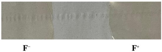
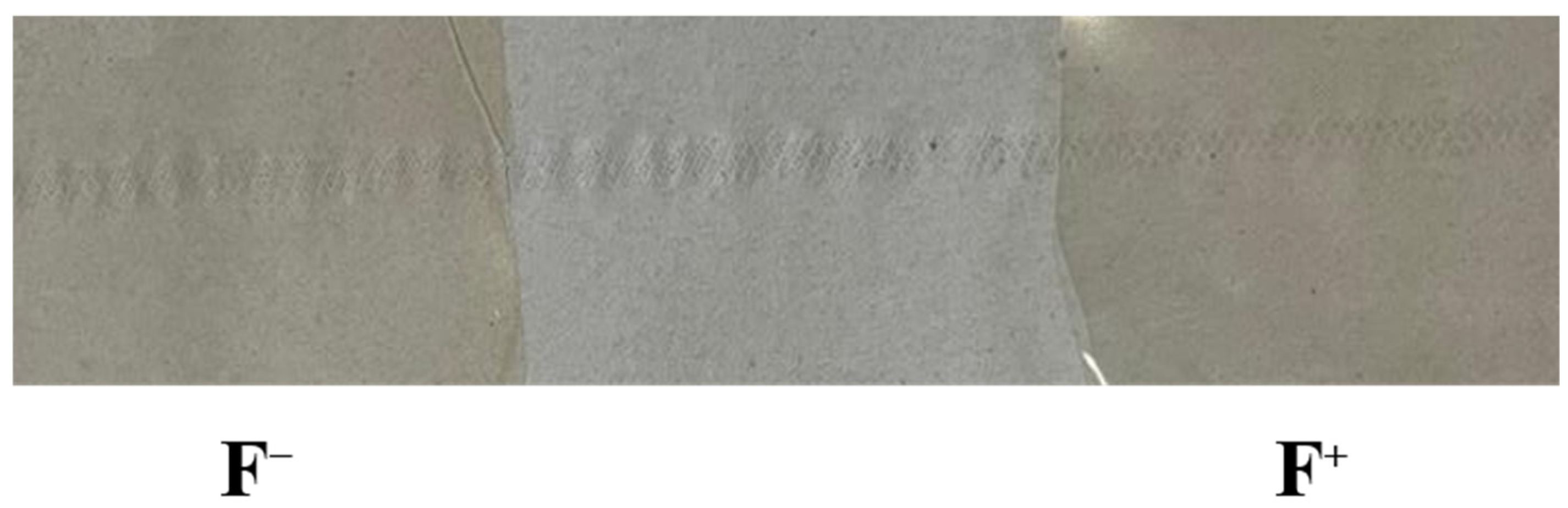
The appearance of the pectin-based films, comprising the films with (F+) and without (F−) D. tortuosa EO, is illustrated in Figure 1, highlighting the visual differences induced by the incorporation of EO.
Figure 1.
Images showing the appearance of pectin-based films without (F−) and with (F+) Deverra tortuosa (D. tortuosa) essential oil (EO).
The droplet size and surface charge of the D. tortuosa EO NE were first evaluated to assess their suitability for film incorporation. The average droplet diameter (D [3,2]) of the EO-loaded NE (F+) was 0.41 ± 0.02 µm, slightly higher than the control (F−, 0.33 ± 0.05 µm). This moderate increase is attributed to the presence of hydrophobic EO constituents, mainly monoterpenes such as α-pinene and sabinene, which tend to coalesce more readily during homogenization. Such droplet sizes, in the submicron range, are generally considered optimal to improve the physical stability of emulsions and to promote homogeneous dispersion inside hydrophilic matrices [17].
Zeta potential values were −37.9 ± 1.2 mV for F+ and −32.6 ± 1.0 mV for F−, both exceeding the ±30 mV stability threshold. The slightly more negative surface charge of F+ may result from phenolic or carboxylic groups of EO molecules adsorbed at the interface, enhancing the electrostatic repulsion between droplets. The magnitude of the potential confirms the efficiency of sodium caseinate as a natural emulsifier able to produce electrostatically stabilized systems [18].
Overall, the microfluidization step at 500 bar yielded uniform, kinetically stable NE. Such stability is a prerequisite to achieve reproducible incorporation into film-forming matrices and to control the subsequent release of volatile bioactives.
3.2. Physical and Optical Properties of Pectin-Based Films
The incorporation of EO NE significantly modified the physicochemical and optical properties of the films (Table 1).
Table 1.
Physicochemical and optical properties of pectin-based films with (F+) and without (F−) Deverra tortuosa (D. tortuosa) essential oil (EO).
The film thickness increased from 41 ± 1 µm (F−) to 57 ± 2 µm (F+). This increase can be attributed to the additional solid matter introduced by the NE, particularly lipids, which raises the overall dry matter content of the film-forming mixture and promotes the formation of a bulkier matrix upon drying. Similar increases in thickness have been reported for pectin- or alginate-based films incorporating emulsified oils or waxes [19,20].
The optical properties also changed significantly. Film opacity increased more than fivefold (from 0.547 ± 0.036 to 2.756 ± 0.025 A·mm−1), while visible light transmittance at 600 nm decreased from 93.1% to 77.1%. Enhanced light scattering linked to scattered lipid droplets and protein clumps created during drying could be the cause of this increased opacity. Additionally, the EO-loaded films had lower transmittance in the UV area, with transmittance at 335 nm dropping from 71.2% to 48.1%, according to the UV–Vis spectra obtained spanning the 200–800 nm range. This result suggests that the EO-loaded films have improved UV-screening capabilities, which may help prevent photooxidation of light-sensitive food ingredients or bioactive substances [21].
For water properties, the water activity (aw) increased slightly (0.456 to 0.549), reflecting higher water sorption at protein-rich interfaces. Sodium caseinate contains polar amino acid residues attracting moisture through hydrogen bonding, while pectin and maltodextrin contribute hydrophilic sites. However, the overall moisture content decreased (12.4% to 10.9%), consistent with the hydrophobic EO phase limiting bulk water retention. This duality, moderate water activity combined with lower moisture, may be related to the composite nature of the films, in which the dispersed lipid phase can reduce moisture transfer while the hydrophilic components of the polymer matrix retain an affinity for water [22].
These results collectively indicate that EO incorporation enhances optical opacity and UV protection, while slightly modifying the water-binding equilibrium. The microstructure of the EO-loaded films thus favors improved barrier behavior and photostability, two crucial features for active food packaging applications.
3.3. Mechanical Properties of Pectin-Based Films
Mechanical analysis revealed the effect of EO incorporation on film integrity (Table 2).
Table 2.
Mechanical properties of pectin-based films with (F+) and without (F−) D. tortuosa EO.
The tensile strength (TS) declined from 30.6 ± 0.2 MPa (F−) to 23.0 ± 0.3 MPa (F+), whereas the Young’s modulus (YM) increased from 122.1 ± 0.6 to 184.2 ± 0.5 MPa. The elongation at break (EB) decreased slightly from 25.8 ± 0.2% to 22.7 ± 0.1%. This combined decrease in TS and EB, but increase in YM, illustrates the classical stiffening–brittleness trade-off induced by dispersed lipid droplets in hydrophilic matrices [23].
In the control film, sodium caseinate interacts strongly with pectin through hydrogen bonding and electrostatic attraction, creating a continuous, cohesive polymer network. When EO is introduced, a fraction of the caseinate migrates to the droplet interface, reducing the number of intermolecular bridges between protein and polysaccharide chains. This redistribution weakens the cohesive strength of the continuous phase, hence the lower TS. Meanwhile, the EO droplets themselves, coated with caseinate, behave as semi-rigid fillers locally restricting polymer mobility, leading to increased modulus. Reduced EB indicates limited chain flexibility and lower capacity for plastic deformation. From an application standpoint, such mechanical behavior remains acceptable for thin coatings or sachets intended for dry or semi-moist foods, although optimization of the glycerol or protein content could further balance flexibility and strength.
3.4. Morphological Analysis (SEM)
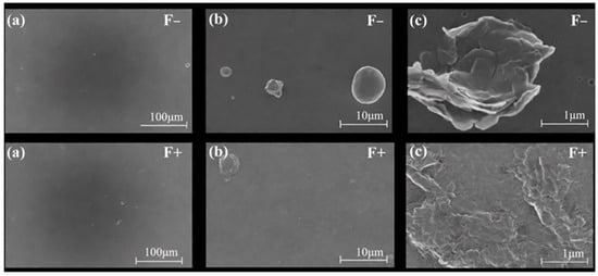
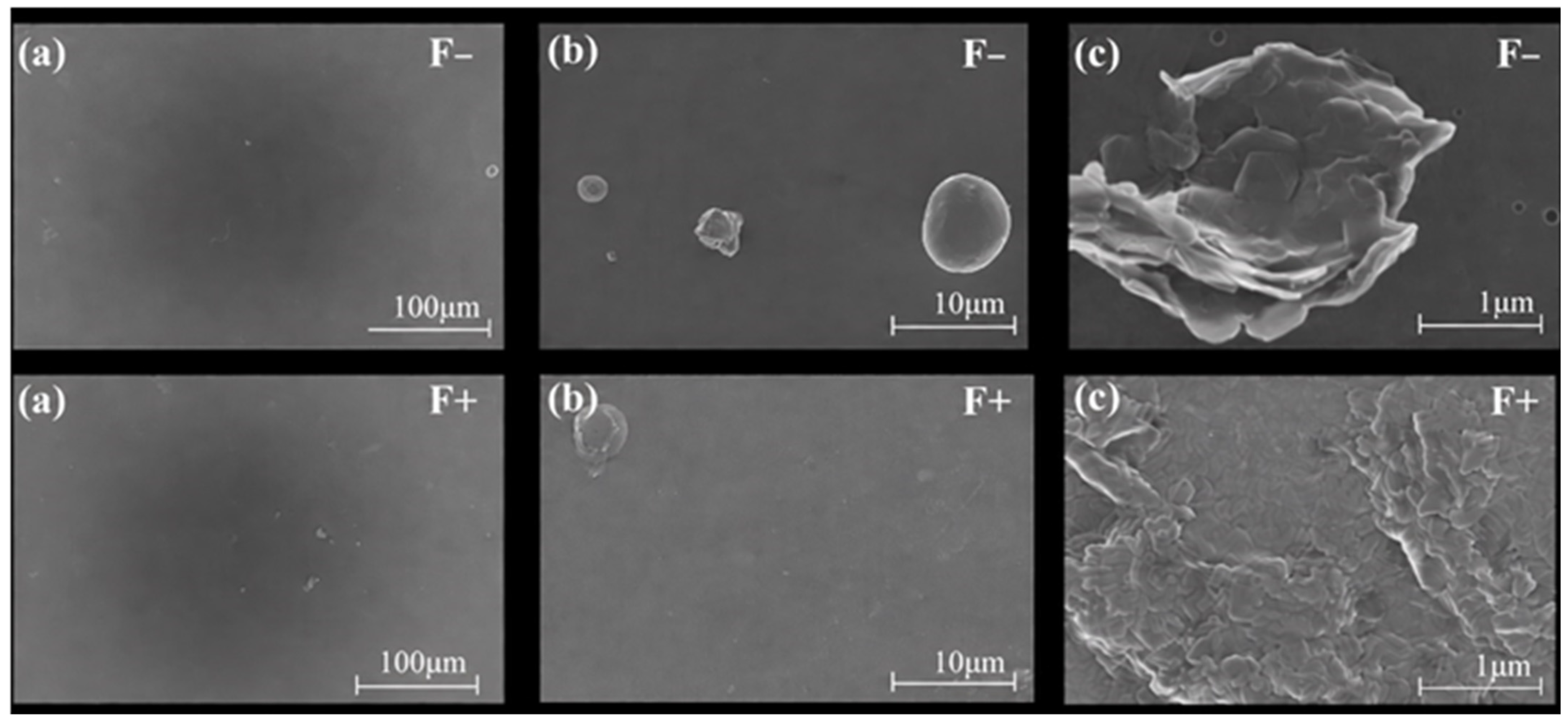
SEM micrographs revealed striking morphological differences between F− and F+ (Figure 2).
Figure 2.
Scanning electron micrographs of pectin-based films without EO (F−, upper row) and with D. tortuosa EO (F+, lower row) at different magnifications: (a) 100×, (b) 1000×, and (c) 10,000×.
At low magnification (100×), the control film (F−) displayed a continuous, compact surface with minimal defects, typical of uniform polysaccharide–protein matrices. In contrast, F+ exhibited a more varied surface shape, where embedded oil droplets are represented by micro-domains.
At 1000×, irregular depressions and microtextures are visible on F+ surfaces, suggesting partial coalescence of EO droplets during drying. These micro-features likely originate from the localized accumulation of caseinate-coated lipids that interfered with polymer packing. Similar morphological heterogeneity was reported in chitosan and starch films containing oregano or clove oil NE [24,25].
At 10,000×, EO-loaded films showed microcavities and cracks absent in F−, consistent with partial volatilization of terpenoid components. Such microvoids can serve as diffusion channels, facilitating controlled release of volatile actives. Despite these discontinuities, no phase separation or large oil pools were observed, confirming that sodium caseinate efficiently stabilized the droplets within the pectin network.
Thus, the morphological evidence supports the dual role of caseinate as an emulsifier and interfacial compatibilizer, ensuring dispersion stability while enabling microstructural heterogeneity beneficial for release kinetics.
3.5. Fourier Transform Infrared (FTIR) Spectroscopy
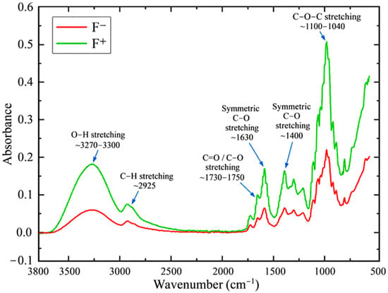
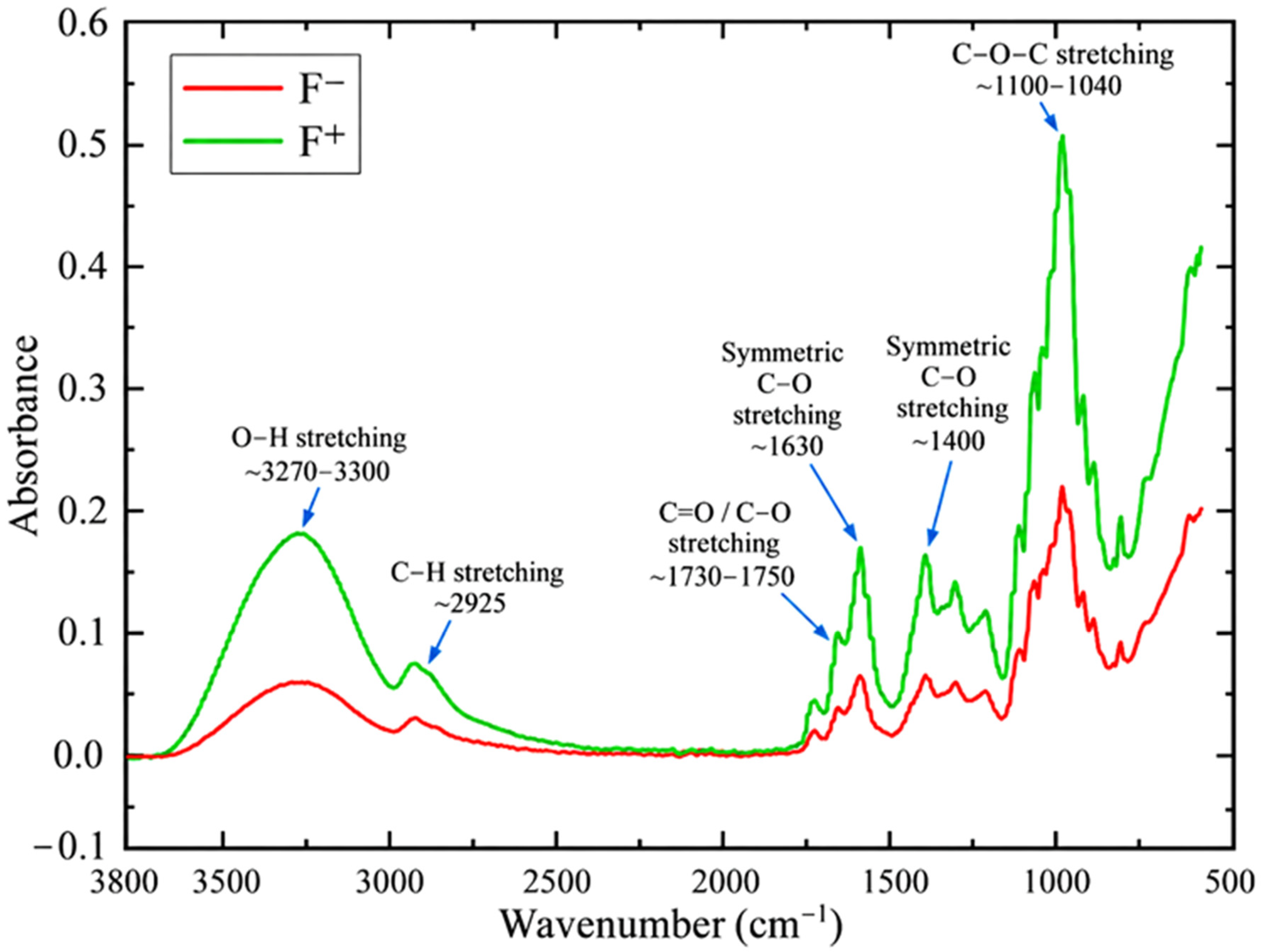
FTIR spectra provided insights into molecular interactions among pectin, sodium caseinate, and EO components (Figure 3).
Figure 3.
FTIR spectra of pectin-based films with (F+) and without (F−) D. tortuosa EO.
Both films exhibited typical pectin bands: O–H stretching (3270–3300 cm−1), C–H stretching (2925 cm−1), C=O stretching of esterified carboxyl groups (1730–1750 cm−1), and COO− asymmetric stretching (1600–1630 cm−1).
In F+, the O–H band intensity decreased slightly and shifted toward lower wavenumbers, indicating hydrogen-bond formation between pectin hydroxyls and amide groups of sodium caseinate. The 1735 cm−1 band shifted slightly to 1728 cm−1, reflecting electrostatic and hydrogen-bond interactions between carboxyl groups of pectin and the protonated amino groups of the protein [26]. A small intensity increase in the band at 2925 cm−1 was also observed in F+, corresponding to the aliphatic C–H vibrations of terpenes and phenylpropanoids from EO [27]. The fingerprint region (1200–900 cm−1), attributed to C–O–C stretching and pyranose ring vibrations, showed minor variations in peak intensity but no structural degradation of pectin. This confirms that EO addition altered mainly the secondary interactions rather than the polysaccharide backbone.
Together, these spectral modifications indicate the formation of a pectin–caseinate complex network modified by EO incorporation. The EO compounds mainly remain confined inside lipid domains, but their presence influences the hydrogen-bonding equilibrium and interfacial organization of the matrix, an effect essential for mechanical and release behavior.
3.6. Thermal Properties
3.6.1. Thermogravimetric and Derivative Thermogravimetric Analyses (TGA/DTG)
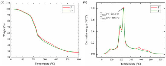
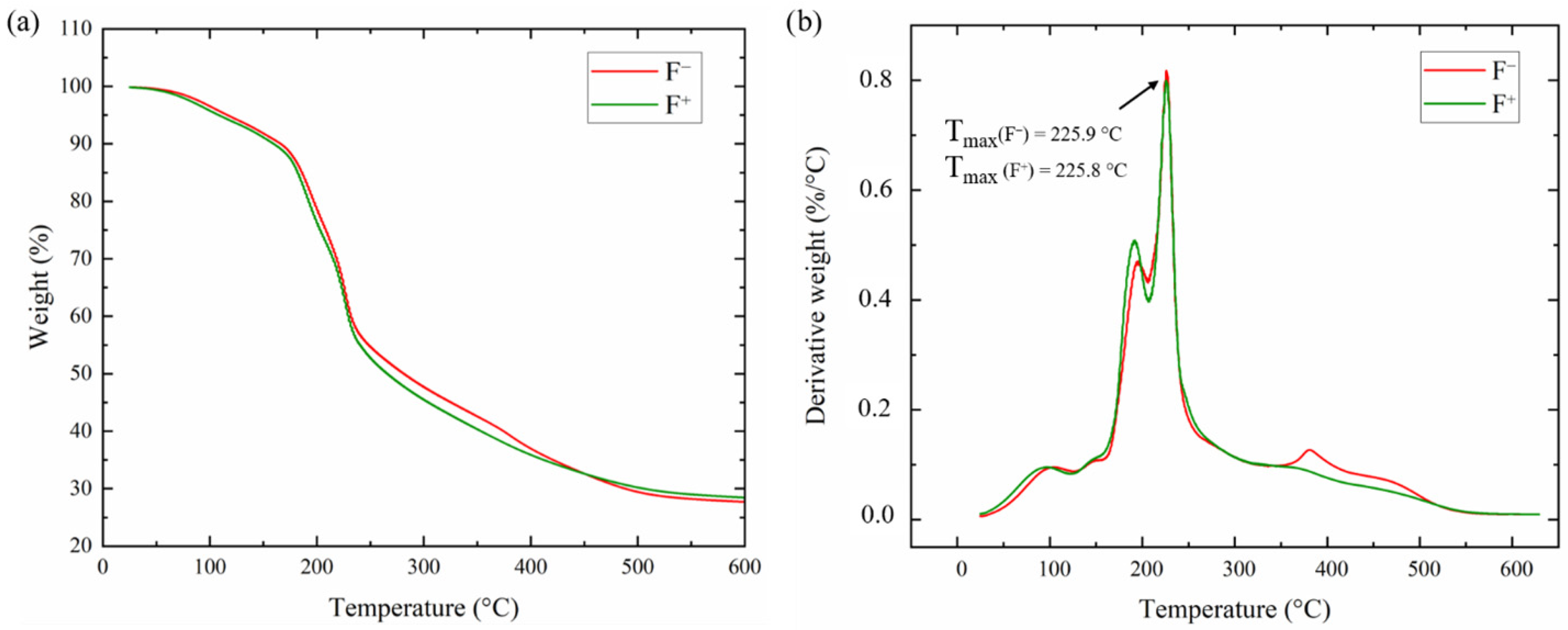
The TGA and DTG curves of F+ and F− films are presented in Figure 4. The TGA profiles exhibited three distinct mass-loss stages, while the DTG curves provided further information on the temperature associated with the main degradation events.
Figure 4.
(a) Thermogravimetric Analysis (TGA) and (b) derivative thermogravimetric (DTG) curves of pectin-based films with (F+) and without (F−) D. tortuosa EO.
The first stage (25–150 °C) corresponds to evaporation of absorbed moisture and low-molecular-weight volatiles. The slightly higher mass loss in F+ (8.9% vs. 8.2%) reflects the volatilization of entrapped EO compounds, especially monoterpenes such as α-pinene and limonene, whose boiling points lie between 150 and 170 °C [28].
The second stage (150–300 °C) represents the main decomposition of the pectin–protein matrix. F+ exhibited a slightly higher weight loss (45.6% vs. 44.1%), likely due to a less cohesive polymer network caused by the presence of lipid droplets. Nonetheless, the degradation onset remained similar, suggesting that caseinate–pectin interactions maintained the overall matrix integrity.
The DTG curves further confirmed the multistep degradation behavior of both films. Two main derivative peaks were observed at approximately 195.3 and 225.9 °C for F− and 192.1 and 225.8 °C for F+, respectively. After the EO nanoemulsion was incorporated, the main degradation temperature (Tmax) remained nearly identical (225.9 °C for F− and 225.8 °C for F+), suggesting that the EO nanoemulsion had little effect on the temperature linked to the primary thermal degradation event. Nonetheless, minor variations in the DTG profiles’ shape and intensity were noted between the two formulations. Similar multistep thermal degradation behavior has been reported for polysaccharide-based films containing essential oils. In these systems, EO incorporation did not substantially alter the primary decomposition pathway of some polymer matrices [29], which is consistent with the nearly unchanged Tmax observed for F− and F+ in the present study. The final stage (300–600 °C) involves decomposition of residual carbonaceous material. F+ showed lower total mass loss (17.1% vs. 20.0%), implying enhanced char formation and thermal resistance of the residue. This result is in accordance with earlier research showing improved thermal stability of protein-polysaccharide composite films following the addition of an ideal essential oil concentration [30].
Overall, EO incorporation induced earlier volatilization but improved the high-temperature stability of the residue, a dual behavior observed in several biopolymer films containing encapsulated terpenoids [13].
3.6.2. Differential Scanning Calorimetry (DSC)
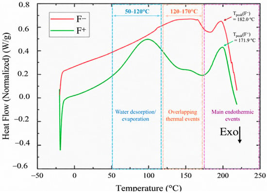
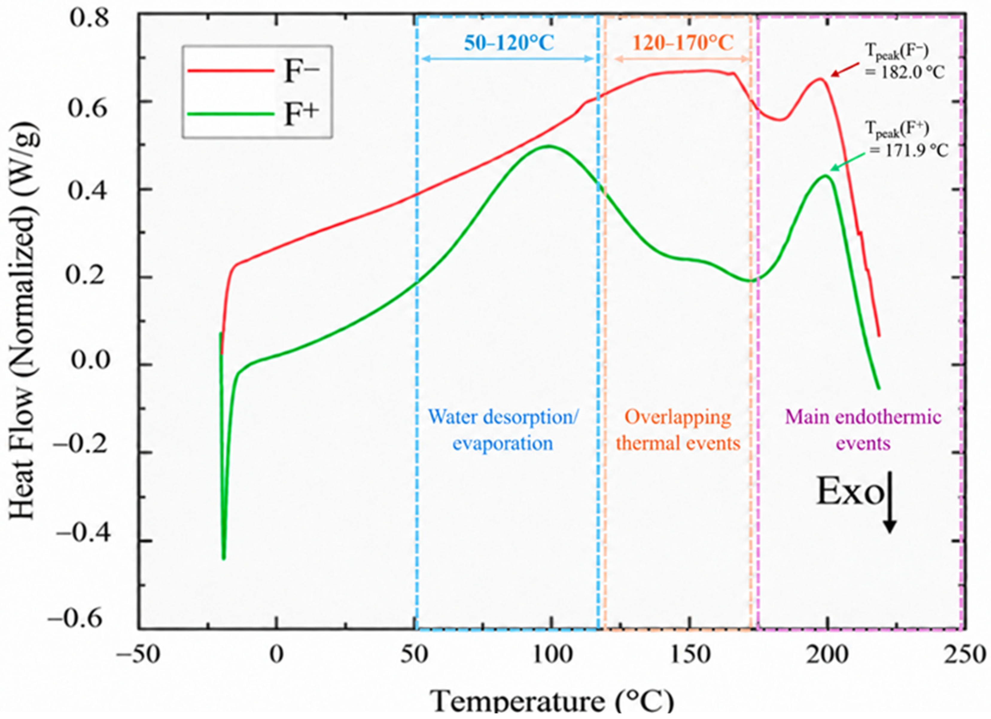
DSC thermograms (Figure 5) revealed a broad endothermic transition in F− centered near 182.0 °C, corresponding to dehydration and structural rearrangement of the pectin–protein network. In F+, this transition became sharper and shifted to 171.9 °C, consistent with the release of entrapped volatile EO constituents and reorganization of interfacial domains. Table 3 summarizes the thermal characteristics of the EO-loaded and control films.
Figure 5.
Differential scanning calorimetry (DSC) thermograms of pectin-based films with (F+) and without (F−) D. tortuosa EO. The main thermal regions and endothermic events are indicated in the thermograms.
Table 3.
Thermal properties of pectin-based films with (F+) and without (F−) D. tortuosa EO.
The DSC thermograms exhibited several overlapping thermal events, reflecting the complex composition of the pectin–caseinate films. The desorption and evaporation of free and bound water from the hydrophilic polymer matrix is primarily responsible for the large endothermic area seen at roughly 50–120 °C [31]. The volatilization of low-molecular-weight EO constituents may also occur in this region for the EO-containing film (F+), which is consistent with the initial mass-loss stage detected by TGA between 25 and 150 °C.
The broad thermal event that was seen between roughly 120 and 170 °C might be explained by thermal transitions of the polymeric matrix as well as overlapping thermal phenomena involving the desorption/evaporation of volatile essential oil components and residual bound water [32]. Since EO NE inclusion modifies interactions between the protein, polysaccharide, and dispersed lipid phase, the sodium caseinate–EO interface may also play a role in alterations in the molecular organization of the pectin matrix.
Both films (F− and F+) exhibited similar glass transition temperatures (69.5 and 70.0 °C, respectively), suggesting that EO NE incorporation did not substantially affect polymer chain mobility in the amorphous regions. For F−, the primary endothermic event happened at 182.0 °C, while for F+, it moved to 171.9 °C. This shift suggests that EO NE incorporation modified the thermal-transition behavior and molecular organization of the pectin–caseinate matrix. However, given DTG analysis revealed essentially comparable principal degradation peaks for F− and F+ (225.9 and 225.8 °C, respectively), it should not be readily construed as a decrease in the main thermal degradation temperature. When combined, the DSC and TGA/DTG results show that EO NE incorporation had no effect on the temperature linked to the primary degradation event, but it had an impact on the thermal-transition behavior and molecular organization of the film matrix.
3.7. Antioxidant Activity
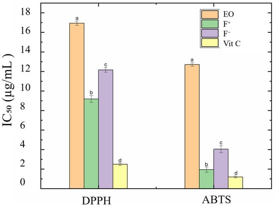
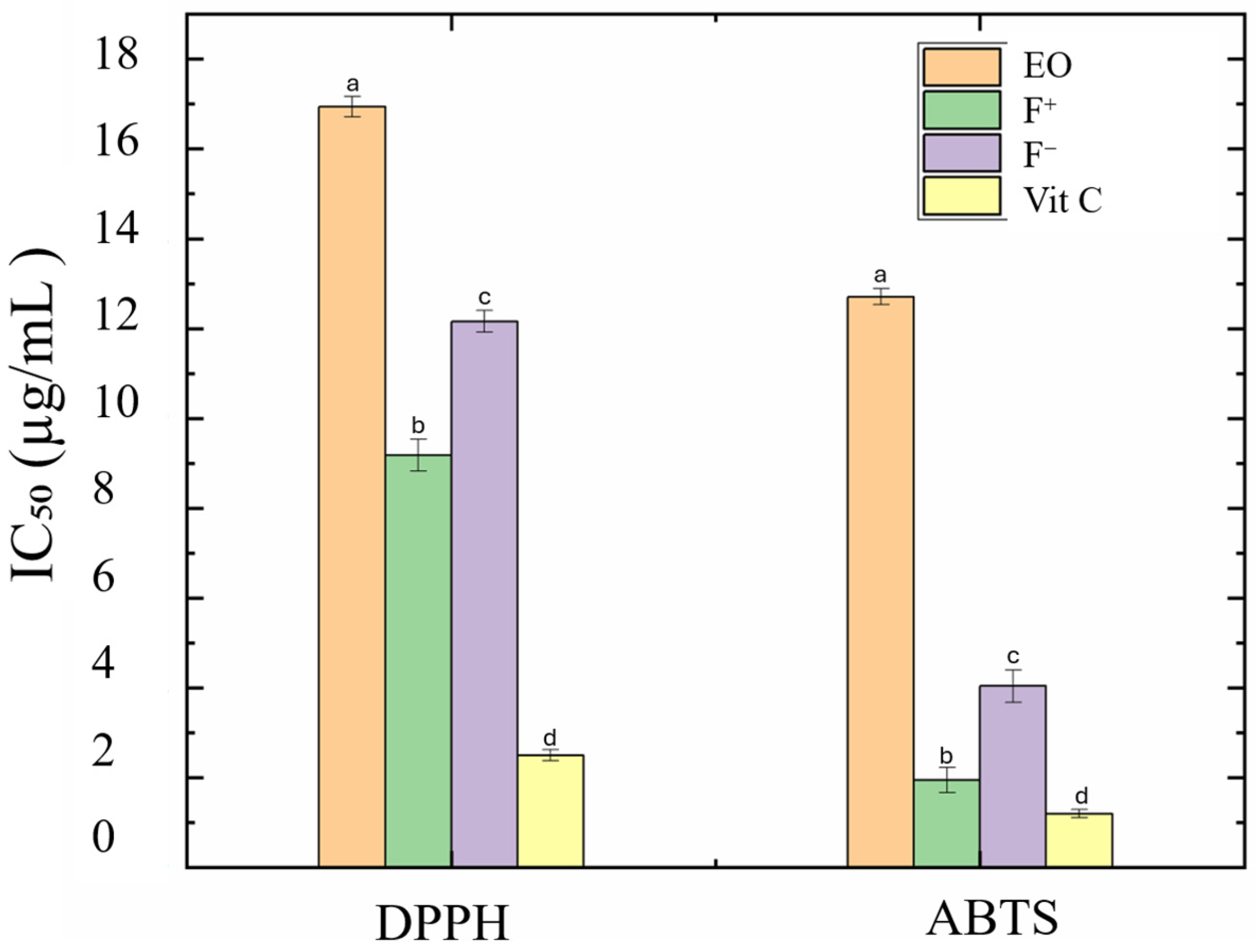
The antioxidant potential of the films was evaluated by DPPH• and ABTS•+ radical scavenging assays (Figure 6).
Figure 6.
Half-maximal inhibitory concentration (IC50) values obtained using the 2,2-diphenyl-1-picrylhydrazyl (DPPH•) and 2,2′-azinobis(3-ethylbenzothiazoline-6-sulfonic acid) (ABTS•+) radical-scavenging assays for D. tortuosa essential oil (EO), EO-loaded films (F+), sunflower-oil control films (F−), and vitamin C. Values are expressed as mean ± SD (n = 3). Values with different superscript lowercase letters are significantly different according to Tukey’s post hoc multiple-comparison test (p < 0.05).
EO-loaded films (F+) showed significantly higher antioxidant activity than the control (F−). For DPPH•, F+ achieved 66.2–78.9% inhibition with a half-maximal inhibitory concentration (IC50) of 9.18 µg mL−1, compared with 12.16 µg mL−1 for F−. For ABTS•+, the improvement was even more pronounced (IC50 1.95 µg mL−1 vs. 4.04 µg mL−1). Vitamin C (used as reference) exhibited IC50 values of 2.43 and 1.12 µg mL−1 for DPPH and ABTS, respectively.
Interestingly, pure EO exhibited higher IC50 values than both F+ and F−, indicating that the antioxidant activity of F+ cannot be attributed exclusively to the EO constituents. In F−, moderate antioxidant activity was observed, which may partly arise from the intrinsic antioxidant properties of sodium caseinate [33]. Therefore, the lower IC50 values observed for F+ may result from the combined contributions of the antioxidant activity of the film matrix and the bioactive constituents of D. tortuosa EO.
The nanoemulsion structure may enhance the accessibility of EO constituents and promote their more uniform distribution throughout the film matrix. Moreover, the protein–lipid interface created by sodium caseinate may facilitate electron transfer between hydrophilic and hydrophobic phases, acting as a molecular bridge [34].
It has been extensively documented that adding essential oils to protein-polysaccharide composite films improves their antioxidant activity. For instance, the antioxidant components of Salvia sclarea essential oil caused a concentration-dependent increase in antioxidant activity in sodium alginate/casein films containing the oil [35], while thyme EO in hawthorn pectin films decreased ABTS IC50 from 6.5 to 2.3 µg mL−1 [36]. These comparisons show that when added to the pectin-caseinate film matrix, D. tortuosa EO can offer significant antioxidant functionality; however, direct quantitative comparisons should be interpreted cautiously due to variations in film composition, EO concentration, and assay conditions across studies.
3.8. Antimicrobial Activity
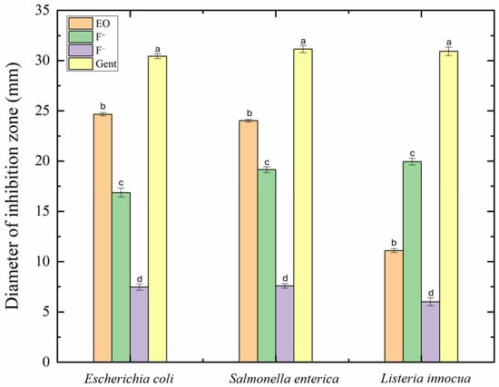
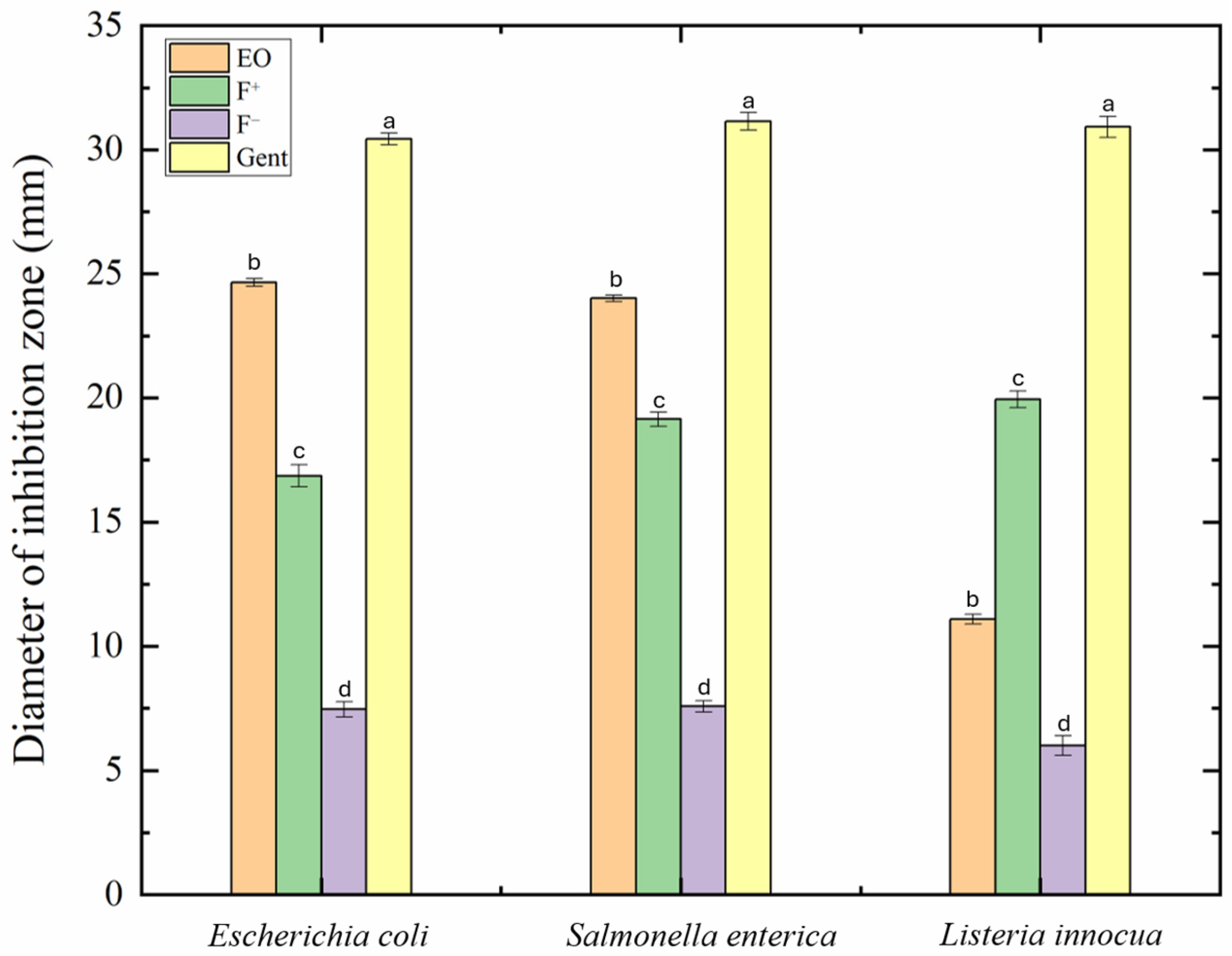
The antimicrobial activity of the films was tested against E. coli, S. enterica (Gram-negative bacteria), and L. innocua (Gram-positive bacteria). As shown in Figure 7, EO-loaded films exhibited clear inhibition zones, whereas EO-free films produced negligible or no zones. The average diameters were 16.86 ± 0.92 mm for E. coli, 19.14 ± 0.71 mm for S. enterica, and 19.94 ± 0.84 mm for L. innocua. These results are in agreement with the general observation that Gram-positive bacteria are more susceptible to EO due to the absence of an outer membrane barrier [12].
Figure 7.
Antimicrobial activity of D. tortuosa essential oil (EO), pectin-based films with (F+) and without (F−) D. tortuosa EO, and gentamicin (positive control), expressed as inhibition zone diameters against selected Gram-positive and Gram-negative bacterial strains. Values are expressed as mean ± SD (n = 3). Values with different superscript letters are significantly different (p < 0.05).
MIC values ranged from 1.25 mg mL−1 (E. coli, S. enterica) to 2.5 mg mL−1 (L. innocua), while all MBC values exceeded 5 mg mL−1, indicating predominantly bacteriostatic effects (Table 4). Similar MIC ranges have been reported for pectin or starch films containing thymol, eugenol, or lemongrass oils [24,25].
Table 4.
Minimum inhibitory concentration (MIC) and minimum bactericidal concentration (MBC) values of D. tortuosa essential oil (EO) and EO-loaded pectin-based films (F+).
Interestingly, although F+ produced a larger inhibition zone against L. innocua than pure EO (19.94 ± 0.84 vs. 11.0 ± 0.4 mm), its MIC was higher (2.50 vs. 1.25 mg mL−1). This apparent discrepancy can be explained by the different principles and experimental conditions of the two antimicrobial assays. In agar diffusion assays, the inhibition-zone diameter is influenced not only by antimicrobial potency but also by the release and diffusion of active compounds through the agar medium. In contrast, the MIC assay determines the minimum concentration required to inhibit bacterial growth in liquid medium. Therefore, inhibition-zone diameters should not be directly correlated with MIC values. The results suggest that F+ exhibited favorable antimicrobial activity against L. innocua under agar-diffusion conditions, whereas pure EO showed greater inhibitory effectiveness under the broth conditions used for MIC determination.
The antimicrobial mechanism likely involves several complementary actions such as:
- Diffusion of volatile EO molecules through the polymer matrix, disrupting bacterial membranes and increasing permeability.
- Interfacial adsorption of EO components on the droplet surface could potentially modulate EO release.
The strong activity of D. tortuosa EO is consistent with its major constituents, dillapiole, elemicin, and myristicin, which have documented bacteriostatic and antifungal actions via membrane destabilization and enzyme inhibition [12].
The incorporation of EO into NE stabilized by sodium caseinate provides both protection against volatilization and may facilitate the diffusion of volatile active compounds, thereby prolonging antimicrobial efficiency.
Although the films were less efficient than the antibiotic control gentamicin (30 mm zones), their natural composition and biodegradability make them promising candidates for food applications where moderate, sustained inhibition is preferred over complete sterilization.
Such materials can inhibit surface contamination and slow microbial proliferation during refrigerated storage, thereby extending product shelf life without synthetic preservatives.
3.9. Correlations Between Film Properties
Integrating the mechanical, thermal, and bioactive results provides a deeper understanding of the structure–function relationships in these pectin–protein–lipid systems.
The slight weakening of mechanical strength (Table 2) is consistent with the structural heterogeneity observed by SEM (Figure 2) and the interfacial interactions revealed by FTIR (Figure 3). In parallel, the slightly higher low-temperature mass loss observed in F+ (Figure 4) can be attributed to the volatilization of EO constituents. The increased antioxidant and antibacterial activities observed in F+ may result from the combined effects of EO bioactive constituents, their dispersion within the film matrix and the ways in which the lipid, sodium caseinate, and pectin phases interact. Caseinate plays a pivotal role in mediating these interfacial interactions due to its amphiphilic properties, decreasing the interfacial tension between EO droplets and the hydrophilic matrix, enabling its adsorption at the oil–water interface and encouraging emulsion stabilization [37]. This could affect the final film microstructure by encouraging a more uniform dispersion of the EO-containing phase inside the film matrix. The resulting materials exhibit balanced functionality: adequate mechanical integrity, sufficient thermal stability, and high bioactivity.
Such cross-correlations have been documented in other protein–polysaccharide composites [26,28], confirming that the interactions observed here follow general physicochemical principles. However, the unique composition of D. tortuosa EO, especially its phenylpropanoid content, may also contribute to the observed radical-scavenging and bacteriostatic activities.
Overall, these findings suggest that the incorporation of D. tortuosa EO nanoemulsion modifies the structure–function relationships of the pectin–caseinate matrix, allowing for improved bioactivity while preserving appropriate mechanical and thermal characteristics.
3.10. Functional and Practical Implications
From a technological perspective, these findings highlight the potential of D. tortuosa EO NE films as multifunctional active packaging materials. Their strong antioxidant capacity can slow lipid oxidation in high-fat foods, while their moderate antimicrobial activity could reduce surface spoilage and pathogen growth. Furthermore, the natural origin and edibility of all components (pectin, maltodextrin, sodium caseinate, and EO) make the films fully biodegradable and potentially compostable.
Importantly, the NE-based design provides flexibility to modulate film functionality. Controlling droplet size, protein-to-oil ratio, or pectin concentration can tailor the release rate and barrier properties. The film’s opacity and UV-blocking ability also make it suitable for light-sensitive foods such as dairy desserts or fruit-based products.
On a fundamental level, this work contributes to understanding the interfacial synergy between proteins and polysaccharides in bioactive materials. By coupling structural analysis (FTIR, SEM, DSC) with functional evaluation (antioxidant, antimicrobial), it elucidates how microstructural heterogeneity can be exploited to optimize performance without chemical modification.
This insight is valuable for future development of bioinspired composite films, where functionality arises not from additives, but from controlled self-assembly of natural biopolymers.
However, the present study did not address migration, cytotoxicity, or food-contact safety. Therefore, the suitability of these films for direct food-contact applications cannot yet be established. Appropriate migration studies using food simulants and toxicological assessments will be required before practical application.
4. Conclusions
This study demonstrates the successful design of bioactive pectin-based films incorporating D. tortuosa essential oil nanoemulsions stabilized by sodium caseinate. The formation of submicron droplets with high electrostatic stability ensured a homogeneous dispersion of the hydrophobic phase within the pectin–maltodextrin matrix, enabling effective integration of bioactive compounds. The incorporation of nanoemulsions induced significant modifications in the films’ physicochemical properties, including increased thickness, opacity, and UV-barrier capacity, alongside moderate reductions in tensile strength and flexibility. These changes are attributed to the formation of a heterogeneous microstructure governed by protein-mediated interfacial organization. Spectroscopic and thermal analyses confirmed the establishment of strong intermolecular interactions between pectin, caseinate, and essential oil constituents, leading to partial reorganization of the polymer network and improved thermal residue stability. Importantly, these structural features translated into enhanced functional performance, with a marked increase in antioxidant activity and a sustained antimicrobial effect against both Gram-positive and Gram-negative bacteria. This behavior highlights the central role of interfacial structuring in controlling the stability, distribution, and bioactivity of encapsulated compounds.
Overall, this work establishes interfacial engineering as a key design principle for developing multifunctional, biodegradable films for active food packaging. This study provides a simple and scalable strategy to design bioactive films through interfacial engineering. Despite these promising results, some limitations of the present study should be acknowledged. The performance of the developed films was assessed in a controlled laboratory setting and has not yet been verified in actual food systems. Furthermore, the current study did not examine food-contact safety or the release and migration behavior of the essential oil from the film matrix. These aspects are essential for assessing the practical applicability of the developed films as active food packaging. Therefore, future research should concentrate on the EO release and migration kinetics using suitable food simulants, food-contact safety evaluations, including cytotoxicity studies, film performance validation in actual food systems, and scale-up considerations to support possible industrial implementation.
Author Contributions
Conceptualization, A.G., M.R. and D.R.; methodology, T.Z., E.D. and A.G.; software, T.Z.; validation, A.G., M.R., D.R. and E.D.; formal analysis, T.Z., E.D., G.A. and A.G.; investigation, T.Z.; resources, A.G., D.R. and G.A.; data curation, T.Z., E.D., G.A. and A.G.; writing—original draft preparation, T.Z. and A.G.; writing—review and editing, A.G., M.R. and D.R.; visualization, T.Z. and A.G.; supervision, A.G., M.R., E.D. and D.R.; project administration, D.R., M.R. and A.G. All authors have read and agreed to the published version of the manuscript.
Funding
T.Z. received a PhD scholarship from the Tunisian Ministry of Higher Education and Scientific Research.
Institutional Review Board Statement
Not applicable.
Informed Consent Statement
Not applicable.
Data Availability Statement
The original contributions presented in this study are included in the article. Further inquiries can be directed to the corresponding author.
Acknowledgments
The authors gratefully acknowledge the LAGEPP Laboratory (Université Claude Bernard Lyon 1, France) for providing access to research facilities, technical resources, and valuable scientific support during the doctoral student’s research stay in France. The laboratory’s stimulating research environment and guidance significantly contributed to the successful completion of this work. We acknowledge the microscopy facility “Centre Technologique des Microstructures CTµ” of Lyon 1 Claude Bernard University, member of the national infrastructure France-BioImaging (https://ror.org/01y7vt929, accessed on 31 August 2026) supported by the French National Research Agency (ANR-24-INBS-0005 FBI BIOGEN).
Conflicts of Interest
A.G. is a member of the Editorial Board of Macromol. A.G. was not involved in the editorial processing or decision-making regarding this manuscript. The other authors declare no conflicts of interest.
Abbreviations
The following abbreviations are used in this manuscript:
| ABTS | 2,2′-azinobis(3-ethylbenzothiazoline-6-sulfonic acid) |
| ATR | Attenuated total reflectance |
| CFU | Colony-forming units |
| DE | Dextrose equivalent |
| DMSO | Dimethyl sulfoxide |
| DSC | Differential Scanning Calorimetry |
| DPPH | 2,2-Diphenyl-1-picrylhydrazyl |
| D. tortuosa | Deverra tortuosa |
| DW | Distilled Water |
| EB | Elongation at Break |
| EO | Essential Oil |
| F+ | Pectin-based Films with EO |
| F− | Pectin-based Films without EO |
| FTIR | Fourier Transform Infrared Spectroscopy |
| IC50 | Half-maximal inhibitory concentration |
| MBC | Minimum Bactericidal Concentration |
| MH | Mueller–Hinton |
| MIC | Minimum Inhibitory Concentration |
| NE | Nanoemulsion |
| SEM | Scanning Electron Microscopy |
| SD | Standard deviation |
| TGA | Thermogravimetric Analysis |
| TS | Tensile Strength |
| YM | Young’s Modulus |
References
- Said, N.S.; Olawuyi, I.F.; Lee, W.Y. Tailoring Pectin-PLA Bilayer Film for Optimal Properties as a Food Pouch Material. Polymers 2024, 16, 712. [Google Scholar] [CrossRef] [Scilit]
- Mouhoub, A.; Guendouz, A.; El Alaoui-Talibi, Z.; Ibnsouda Koraichi, S.; El Modafar, C. Effect of Storage Time on Major Physical Properties and Microbial Growth and Adhesion Inhibitory Performance of Chitosan-Based Film Enriched with Cinnamon and Clove Essential Oils. Eur. Food Res. Technol. 2026, 252, 3. [Google Scholar] [CrossRef] [Scilit]
- Mouhoub, A.; Guendouz, A.; El Alaoui-Talibi, Z.; Ibnsouda Koraichi, S.; El Modafar, C. Optimization of Chitosan-Based Film Performance by the Incorporation of Cinnamomum Zeylanicum Essential Oil. Food Biophys. 2025, 20, 48. [Google Scholar] [CrossRef] [Scilit]
- Lopes, A.I.; Melo, A.; Afonso, T.B.; Silva, S.; Barros, L.; Tavaria, F.K.; Pintado, M. Alginate Edible Films Containing Essential Oils: Characterization and Bioactive Potential. Polymers 2025, 17, 1188. [Google Scholar] [CrossRef] [Scilit]
- Guzmán-Pincheira, C.; Moeini, A.; Oliveira, P.E.; Abril, D.; Paredes-Padilla, Y.A.; Benavides-Valenzuela, S. Development of Alginate-Chitosan Bioactive Films Containing Essential Oils for Use in Food Packaging. Foods 2025, 14, 256. [Google Scholar] [CrossRef] [Scilit]
- Stoleru, E.; Brebu, M. Stabilization Techniques of Essential Oils by Incorporation into Biodegradable Polymeric Materials for Food Packaging. Molecules 2021, 26, 6307. [Google Scholar] [CrossRef] [Scilit]
- Ebrahimzadeh, S.; Biswas, D.; Roy, S.; McClements, D.J. Incorporation of Essential Oils in Edible Seaweed-Based Films: A Comprehensive Review. Trends Food Sci. Technol. 2023, 135, 43–56. [Google Scholar] [CrossRef] [Scilit]
- Lazăr, A.R.; Pușcaș, A.; Tanislav, A.E.; Mureșan, V. Bioactive Compounds Delivery and Bioavailability in Structured Edible Oils Systems. Compr. Rev. Food Sci. Food Saf. 2024, 23, e70020. [Google Scholar] [CrossRef] [Scilit]
- Acevedo-Fani, A.; Salvia-Trujillo, L.; Rojas-Graü, M.A.; Martín-Belloso, O. Edible Films from Essential-Oil-Loaded Nanoemulsions: Physicochemical Characterization and Antimicrobial Properties. Food Hydrocoll. 2015, 47, 168–177. [Google Scholar] [CrossRef] [Scilit]
- Kong, I.; Degraeve, P.; Pui, L.P. Polysaccharide-Based Edible Films Incorporated with Essential Oil Nanoemulsions: Physico-Chemical, Mechanical Properties and Its Application in Food Preservation—A Review. Foods 2022, 11, 555. [Google Scholar] [CrossRef] [Scilit]
- Liao, W.; Elaissari, A.; Ghnimi, S.; Dumas, E. Effect of Pectin on the Properties of Nanoemulsions Stabilized by Sodium Caseinate at Neutral PH. Int. J. Biol. Macromol. 2022, 209, 1858–1866. [Google Scholar] [CrossRef] [Scilit]
- Khammassi, M.; Polito, F.; Kochti, O.; Kouki, H.; Souihi, M.; Khedhri, S.; Hamrouni, L.; Mabrouk, Y.; Amri, I.; De Feo, V. Investigation on Chemical Composition, Antioxidant, Antifungal and Herbicidal Activities of Volatile Constituents from Deverra Tortuosa (Desf.). Plants 2023, 12, 2556. [Google Scholar] [CrossRef] [Scilit]
- Baghi, F.; Gharsallaoui, A.; Dumas, E.; Agusti, G.; Ghnimi, S. Characterization of Antimicrobial Multilayer Film Based on Ethylcellulose-Pectin Incorporated with Nanoemulsions of Trans-Cinnamaldehyde Essential Oil. Food Chem. X 2024, 22, 101261. [Google Scholar] [CrossRef] [Scilit]
- Sánchez, E.; Dávila-Aviña, J.; Castillo, S.L.; Heredia, N.; Vázquez-Alvarado, R.; García, S. Antibacterial and Antioxidant Activities in Extracts of Fully Grown Cladodes of 8 Cultivars of Cactus Pear. J. Food Sci. 2014, 79, M659–M664. [Google Scholar] [CrossRef] [Scilit]
- Dissanayake, D.P.A.; Sivaganesh, S.; Tissera, M.H.A.; Handunnetti, S.M.; Arawwawala, L.D.A.M. Comparison of Antioxidant Properties of Cyathula Prostrata Linn and Achyranthes Aspera Linn Grown in Sri Lanka. Res. Rev. Insights 2018, 2, 1–3. [Google Scholar] [CrossRef] [Scilit]
- Balouiri, M.; Sadiki, M.; Ibnsouda, S.K. Methods for In Vitro Evaluating Antimicrobial Activity: A Review. J. Pharm. Anal. 2016, 6, 71–79. [Google Scholar] [CrossRef] [Scilit]
- Jacob, S.; Kather, F.S.; Boddu, S.H.S.; Shah, J.; Nair, A.B. Innovations in Nanoemulsion Technology: Enhancing Drug Delivery for Oral, Parenteral, and Ophthalmic Applications. Pharmaceutics 2024, 16, 1333. [Google Scholar] [CrossRef] [Scilit]
- Liao, W.; Gharsallaoui, A.; Dumas, E.; Elaissari, A. Understanding of the Key Factors Influencing the Properties of Emulsions Stabilized by Sodium Caseinate. Compr. Rev. Food Sci. Food Saf. 2022, 21, 5291–5317. [Google Scholar] [CrossRef] [Scilit]
- Valentino, M.; Volpe, S.; Di Giuseppe, F.A.; Cavella, S.; Torrieri, E. Active Biopolymer Coating Based on Sodium Caseinate: Physical Characterization and Antioxidant Activity. Coatings 2020, 10, 706. [Google Scholar] [CrossRef] [Scilit]
- Bhatia, S.; Al-Harrasi, A.; Shah, Y.A.; Saif Alrasbi, A.N.; Jawad, M.; Koca, E.; Aydemir, L.Y.; Alamoudi, J.A.; Almoshari, Y.; Mohan, S. Structural, Mechanical, Barrier and Antioxidant Properties of Pectin and Xanthan Gum Edible Films Loaded with Grapefruit Essential Oil. Heliyon 2024, 10, e25501. [Google Scholar] [CrossRef] [Scilit]
- Basumatary, I.B.; Mukherjee, A.; Kumar, S. Chitosan-Based Composite Films Containing Eugenol Nanoemulsion, ZnO Nanoparticles and Aloe Vera Gel for Active Food Packaging. Int. J. Biol. Macromol. 2023, 242, 124826. [Google Scholar] [CrossRef] [Scilit]
- Shen, G.; Yu, G.; Wu, H.; Li, S.; Hou, X.; Li, M.; Li, Q.; Liu, X.; Zhou, M.; Chen, A.; et al. Incorporation of Lipids into Wheat Bran Cellulose/Wheat Gluten Composite Film Improves Its Water Resistance Properties. Membranes 2022, 12, 18. [Google Scholar] [CrossRef] [Scilit]
- Cheng, L.; Ye, A.; Yang, Z.; Hemar, Y.; Singh, H. Formation and Properties of Highly Concentrated Oil-in-Water Emulsions Stabilised by Emulsion Droplets. Food Hydrocoll. 2023, 145, 109059. [Google Scholar] [CrossRef] [Scilit]
- Baghi, F.; Ghnimi, S.; Agusti, G.; Dumas, E.; Gharsallaoui, A. Development and Characterization of Pectin-Based Antimicrobial Packaging Films Containing Nanoemulsified Trans-Cinnamaldehyde. Appl. Sci. 2024, 14, 2256. [Google Scholar] [CrossRef] [Scilit]
- Moghimi, R.; Aliahmadi, A.; Rafati, H. Antibacterial Hydroxypropyl Methyl Cellulose Edible Films Containing Nanoemulsions of Thymus Daenensis Essential Oil for Food Packaging. Carbohydr. Polym. 2017, 175, 241–248. [Google Scholar] [CrossRef] [Scilit]
- Ren, J.N.; Hou, Y.Y.; Fan, G.; Zhang, L.L.; Li, X.; Yin, K.; Pan, S.Y. Extraction of Orange Pectin Based on the Interaction between Sodium Caseinate and Pectin. Food Chem. 2019, 283, 265–274. [Google Scholar] [CrossRef] [Scilit]
- Pasini Cabello, S.D.; Takara, E.A.; Marchese, J.; Ochoa, N.A. Influence of Plasticizers in Pectin Films: Microstructural Changes. Mater. Chem. Phys. 2015, 162, 491–497. [Google Scholar] [CrossRef] [Scilit]
- Niu, J.; Zhu, J. Thermal Reaction Products and Formation Pathways of Two Monoterpenes Under In Situ Thermal Desorption Conditions That Mimic Vaping Coil Temperatures. Sci. Rep. 2023, 13, 21650, Correction in Sci. Rep. 2024, 14, 5301. https://doi.org/10.1038/s41598-024-54633-5. [Google Scholar] [CrossRef] [Scilit]
- Nahas, E.O.; Furtado, G.F.; Lopes, M.S.; Silva, E.K. From Emulsions to Films: The Role of Polysaccharide Matrices in Essential Oil Retention Within Active Packaging Films. Foods 2025, 14, 1501. [Google Scholar] [CrossRef] [Scilit]
- Bhatia, S.; Al-Harrasi, A.; Al-Azri, M.S.; Ullah, S.; Bekhit, A.E.D.A.; Pratap-Singh, A.; Chatli, M.K.; Anwer, M.K.; Aldawsari, M.F. Preparation and Physiochemical Characterization of Bitter Orange Oil Loaded Sodium Alginate and Casein Based Edible Films. Polymers 2022, 14, 3855. [Google Scholar] [CrossRef] [Scilit]
- Horn, M.M.; Martins, V.C.A.; Plepis, A.M.G. Effect of Amylopectin Content on Mechanical, Barrier and Thermal Properties of Plasticized Starch/Chitosan Films. Polysaccharides 2023, 4, 208–218. [Google Scholar] [CrossRef] [Scilit]
- Fahim, H.; Bagheri, H.; Motamedzadegan, A.; Mirarab Razi, S.; Rashidinejad, A. Fabrication and Characterization of Active Gelatin-Based Films Integrated with Nanocellulose-Stabilized Pickering Emulsion Containing Oliveria Decumbens Vent. Essential Oil. LWT 2024, 209, 116725. [Google Scholar] [CrossRef] [Scilit]
- Jiang, J.; Watowita, P.S.M.S.L.; Chen, R.; Shi, Y.; Geng, J.T.; Takahashi, K.; Li, L.; Osako, K. Multilayer Gelatin/Myofibrillar Films Containing Clove Essential Oil: Properties, Protein-Phenolic Interactions, and Migration of Active Compounds. Food Packag. Shelf Life 2022, 32, 100842. [Google Scholar] [CrossRef] [Scilit]
- Delmonte, A.; Visentini, F.F.; Fernández, J.L.; Santiago, L.G.; Perez, A.A. Development, Characterization, and Evaluation of the Antioxidant Activity of Nanocarriers Based on Surfactant Swollen Micelles That Encapsulate Essential Oils. Colloids Surf. B Biointerfaces 2024, 235, 113783. [Google Scholar] [CrossRef] [Scilit]
- Bhatia, S.; Al-Harrasi, A.; Shah, Y.A.; Jawad, M.; Al-Azri, M.S.; Ullah, S.; Anwer, M.K.; Aldawsari, M.F.; Koca, E.; Aydemir, L.Y. The Effect of Sage (Salvia sclarea) Essential Oil on the Physiochemical and Antioxidant Properties of Sodium Alginate and Casein-Based Composite Edible Films. Gels 2023, 9, 233. [Google Scholar] [CrossRef] [Scilit]
- Qin, C.; Li, Z.; Zhang, J.; Meng, H.; Zhu, C. Preparation, Physicochemical Properties, Antioxidant, and Antibacterial Activities of Quaternized Hawthorn Pectin Films Incorporated with Thyme Essential Oil. Food Packag. Shelf Life 2024, 41, 101235. [Google Scholar] [CrossRef] [Scilit]
- Liao, W.; Elaissari, A.; Dumas, E.; Gharsallaoui, A. Effect of Trans-Cinnamaldehyde or Citral on Sodium Caseinate: Interfacial Rheology and Fluorescence Quenching Properties. Food Chem. 2023, 400, 134044. [Google Scholar] [CrossRef] [Scilit]

Disclaimer/Publisher’s Note: The statements, opinions and data contained in all publications are solely those of the individual author(s) and contributor(s) and not of MDPI and/or the editor(s). MDPI and/or the editor(s) disclaim responsibility for any injury to people or property resulting from any ideas, methods, instructions or products referred to in the content. |
© 2026 by the authors. Licensee MDPI, Basel, Switzerland. This article is an open access article distributed under the terms and conditions of the Creative Commons Attribution (CC BY) license.