


Abstract
This study proposes a non-invasive dual respiratory monitoring system for intensive care units (ICU), based on a triaxial MEMS accelerometer ADXL345 attached to the thoracic cage and a nasal NTC thermistor, interfaced with an Arduino Mega 2560 microcontroller. The accelerometer signal is filtered through a second-order Butterworth low-pass filter (fc = 0.6 Hz) and double-integrated to obtain thoracic displacement ΔD, while the thermistor detects respiratory cycles through the thermal variation in nasal airflow. The system detects respiratory rate in the range of 4–60 breaths/minute, with amplitudes ΔD of 2–5 mm, providing superior sensor redundancy over individual methods.
1. Introduction
Respiratory rate (RR) is a parameter used in intensive care medicine, recognized as a sensitive indicator of clinical deterioration in critically ill patients [1]. Multiple studies have demonstrated that an increase in RR frequently precedes admission to intensive care units, cardiac arrest, and death [2]. Despite this recognized importance, RR is paradoxically the vital sign most frequently measured with poor accuracy, with substantial inter-observer variability and systematic underreporting documented in clinical practice [3].
The proposed dual system is of clinical relevance for patients with partially or totally obstructed upper airways—a situation encountered in cases of cervical spinal cord injuries, laryngeal or pharyngeal tumors, or post-intubation upper-airway trauma [4]. In these cases, nasal airflow may constitute the only patent respiratory pathway, making thermistor-based monitoring essential for the timely detection of apnea or hypopnea events.
The original contributions of this work are as follows: (i) a dual-modality acquisition architecture combining triaxial accelerometry and nasal thermal sensing on a single low-cost microcontroller platform; (ii) a real-time fusion rule (|RR_acc − RR_therm| < 4 breaths/min) that automatically detects motion-corrupted accelerometric estimates and falls back on the thermal channel, providing graceful degradation in the presence of artifacts; (iii) a quantitative repeatability characterization across eupneic, tachypneic and apneic regimes under ISO 5725-2 conditions [5].
1.1. Conventional Methods for Measuring Respiratory Rate
Conventional methods for respiratory rate monitoring include oronasal thermal sensors, capnography, nasal cannulas connected to pressure transducers, and respiratory inductance plethysmography (RIP) [6,7]. Each approach presents specific advantages and limitations, and understanding the physical and mathematical principles governing them is essential for designing a high-performance monitoring system.
Although individually validated, each conventional modality presents specific failure modes that limit its applicability in the ICU. Capnography requires invasive integration with the ventilation circuit and becomes unreliable in spontaneously breathing non-intubated patients. Nasal pressure transducers and oronasal thermistors are sensitive to mouth breathing, sensor displacement, and oxygen-therapy interference [6,7]. Respiratory inductance plethysmography (RIP) provides volumetric information but depends on properly tensioned thoracic and abdominal bands whose calibration drifts during prolonged use and patient repositioning. Single-axis accelerometers, while non-invasive, are highly susceptible to motion artifacts that cannot be distinguished from genuine respiratory cycles without an independent reference [8,9]. To our knowledge, no low-cost ICU-oriented system has previously combined a triaxial MEMS accelerometer with a nasal thermistor on a single Arduino-class platform with explicit cross-channel validation logic—an architectural gap that the present work directly addresses.
1.1.1. The Nasal Thermistor
The thermistor measures temperature changes between inspired (cool) and expired (warm) air, and is considered the gold standard for apnea detection according to the American Academy of Sleep Medicine [10]. Expired air reaches a temperature of approximately 34–37 °C due to thermal exchange with the richly vascularized network of the upper airways and pulmonary alveoli, where blood circulating at body temperature (~37 °C) transfers heat to air in transit through convection and evaporation from the moist mucosal surface. The operating principle is based on the variation in electrical resistance of a semiconductor material with a negative temperature coefficient (NTC). The relationship between thermistor resistance and temperature is described by the Steinhart–Hart equation [11]:
where T is the absolute temperature (K), R is the thermistor resistance (Ω), and A, B, C are the Steinhart–Hart constants specific to the material. For small temperature variations, a simplified approximation uses the β-parameter model:
where R0 is the resistance at reference temperature T0 (typically 25 °C = 298.15 K), and β is the material constant (typically 3000–5000 K for NTC thermistors). The signal generated by the thermistor is proportional to the temperature difference ΔT between expired air (~34–37 °C) and inspired air (~20–25 °C).
1/T = A + B·ln(R) + C·[ln(R)]3,
R(T) = R0 · exp[β · (1/T − 1/T0)],
1.1.2. MEMS Accelerometer
MEMS (Micro-Electro-Mechanical Systems) accelerometers have emerged as a promising alternative for non-invasive respiratory monitoring. The operating principle is based on the fact that gravity is the predominant component of the detected acceleration; as the thoracic cage expands and changes shape, the angle relative to the gravitational vector changes [8,9]. The acceleration component on the z-axis, perpendicular to the thoracic surface, is:
where g = 9.81 m/s2 is gravitational acceleration, θ0 is the initial angle of the sensor relative to the vertical, and Δθ(t) is the angular variation caused by expansion of the thoracic cage during inspiration. For small angles Δθ, a first-order Taylor expansion allows linearization:
a_z(t) = g · cos(θ0 + Δθ(t)),
Δa_z(t) ≈ −g · sin(θ0) · Δθ(t).
The thoracic displacement ΔD is obtained by double numerical integration of acceleration, an approach previously validated for extracting respiratory rate and flow waveforms from tri-axial accelerometer data [12]. The first integration yields velocity and the second yields displacement:
v(t) = ∫ a_z,filt(t) dt,
ΔD(t) = ∫ v(t) dt.
1.1.3. Tidal Volume Estimation
Tidal volume (VT) estimation via wearable sensors represents a growing field. The simplified relationship between thoracic displacement and tidal volume is [13,14]:
where k is an individualized calibration coefficient (in mL/mm), ideally determined by comparison with spirometry. Accelerometer-based approaches have also been used to jointly estimate tidal volume variability and respiratory rate [15]. In the absence of individual spirometric calibration, a nominal value can be derived from aggregated physiological data: for a healthy adult at rest, tidal volume is approximately 500 mL [13,14], and the antero-posterior displacement of the thoracic cage has been measured at 3–5 mm by optoelectronic analysis [16,17], yielding a plausible range of k ≈ 100–165 mL/mm. The linear principle VT ∝ ΔD has been shown valid with an error below 10% as long as posture is maintained [17]. Recent studies have reported a mean percent error of approximately 10% for quasi-static activities [14].
V_T ≈ k · ΔD_max,
2. Methods and Materials
2.1. System Architecture
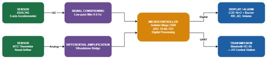
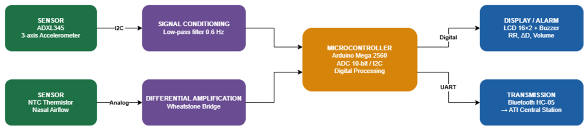
The proposed system consists of five main subsystems: (1) the sensor subsystem (ADXL345 accelerometer and NTC thermistor), (2) the signal conditioning subsystem, (3) the processing unit (Arduino Mega 2560, Arduino S.r.l., Monza, Italy), (4) the display and alarm subsystem, and (5) the wireless communication module (Bluetooth HC-05, Guangzhou HC Information Technology Co., Ltd., Guangzhou, China). All components were sourced from a commercial electronics distributor (Optimus Digital, Bucharest, Romania) The system block diagram is presented in Figure 1.
Figure 1.
Block diagram of the respiratory monitoring system with dual-sensor architecture and signal processing flow.
2.2. ADXL345 Accelerometer
The ADXL345 is a high-resolution (13-bit) triaxial digital MEMS accelerometer manufactured by Analog Devices, Inc. (Wilmington, MA, USA) [18], capable of measuring accelerations in the range of ±19.62 m/s2 and ±156.96 m/s2 [18]. For the respiratory monitoring application, the sensor was configured in ±19.62 m/s2 mode, providing a sensitivity of 3.9 mg/LSB, sufficient for detecting the subtle accelerations generated by thoracic cage expansion during breathing [19].
The sensor communicates with the microcontroller via the I2C protocol at address 0 × 53, using the dedicated hardware pins SDA (pin 20) and SCL (pin 21) of the Arduino Mega 2560 platform.
2.3. Nasal NTC Thermistor
The NTC thermistor used has a reference resistance of R0 = 10 kΩ at T0 = 25 °C, positioned at the nasal entrance, 0–4 mm from the nostril, in accordance with recommendations from the literature [20]. The conditioning circuit uses a Wheatstone bridge with three fixed 10 kΩ resistors, followed by an LM358 operational amplifier in differential configuration with a gain of 100. The amplifier output is connected to analog input A0, where the 10-bit ADC (resolution 4.88 mV) digitizes the signal [21]. A reference thermistor exposed to ambient temperature enables differential amplification, eliminating the influence of ambient temperature.
2.4. Electrical Schematic
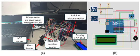
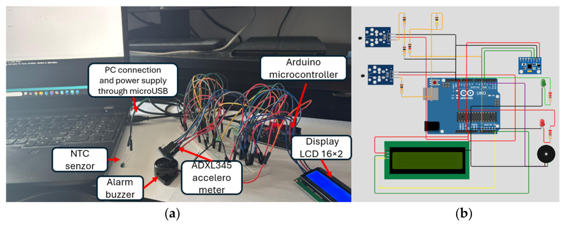
The complete electrical schematic is presented in Figure 2. The main connections include: ADXL345 via I2C (SDA → pin 20, SCL → pin 21), thermistor via ADC (Vout → A0), Bluetooth HC-05 module via UART (TX1/RX1), 16 × 2 LCD via digital pins D4–D9, alarm buzzer on D3, and status LEDs on D10 (green) and D11 (red).
Figure 2.
Dual respiratory monitoring system: (a) physical laboratory prototype with nasal NTC sensor, ADXL345 accelerometer, alarm buzzer, and Arduino Mega 2560 microcontroller powered via USB; (b) wiring schematic in Tinkercad Circuits (Autodesk, Inc., San Francisco, CA, USA), showing the ADXL345 module (I2C), the Wheatstone–LM358 thermistor circuit (A0), the Bluetooth HC-05 module (UART), 16 × 2 LCD, and alarm indicators.
2.5. Digital Signal Processing
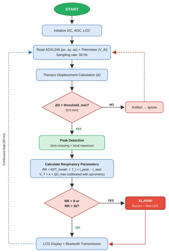
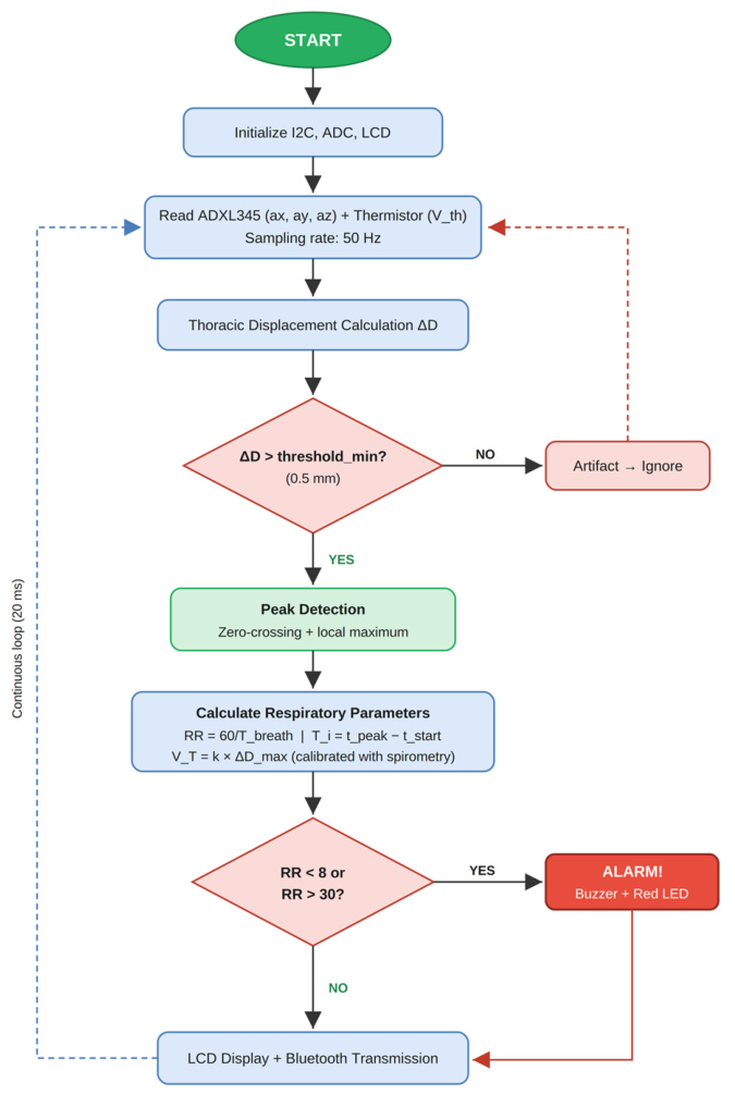
The processing algorithm, presented in Figure 3, proceeds through the following stages:
Figure 3.
Logical diagram of the respiratory parameter extraction algorithm.
Stage 1—Acquisition: Acceleration signals (ax, ay, az) are read from the ADXL345 via I2C, simultaneously with the thermistor voltage reading via ADC.
Stage 2—Filtering: The second-order Butterworth low-pass filter described in Equation (8) is applied, with cutoff frequency fc = 0.6 Hz and coefficients calculated for fs = 50 Hz: b0 = 0.000554, b1 = 0.001109, b2 = 0.000554, a1 = −1.9556, a2 = 0.9567, resulting in the following:
y[n] = b0·x[n] + b1·x[n−1] + b2·x[n−2] − a1·y[n−1] − a2·y[n−2].
Stage 3—ΔD Calculation: Displacement is obtained by double numerical integration according to Equations (5) and (6), with periodic reset to eliminate drift.
Stage 4—Cycle Detection: A peak detection algorithm combined with zero-crossing is applied. The minimum threshold ΔD = 0.5 mm eliminates non-respiratory fluctuations.
Stage 5—Parameter Calculation: Respiratory rate RR = 60/Tbreath (breaths/minute), inspiratory time Ti, expiratory time Te, and estimated tidal volume according to Equation (7).
3. Results and Discussion
3.1. Operational Parameters of the System
The proposed system was tested under laboratory conditions simulating ICU clinical scenarios. The key operational parameters are summarized in Table 1.
Table 1.
Operational parameters of the dual respiratory monitoring system.
3.2. Measurement Repeatability Under Eupneic Conditions
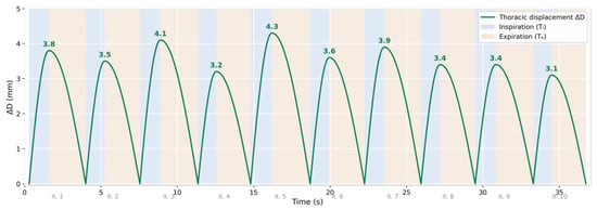
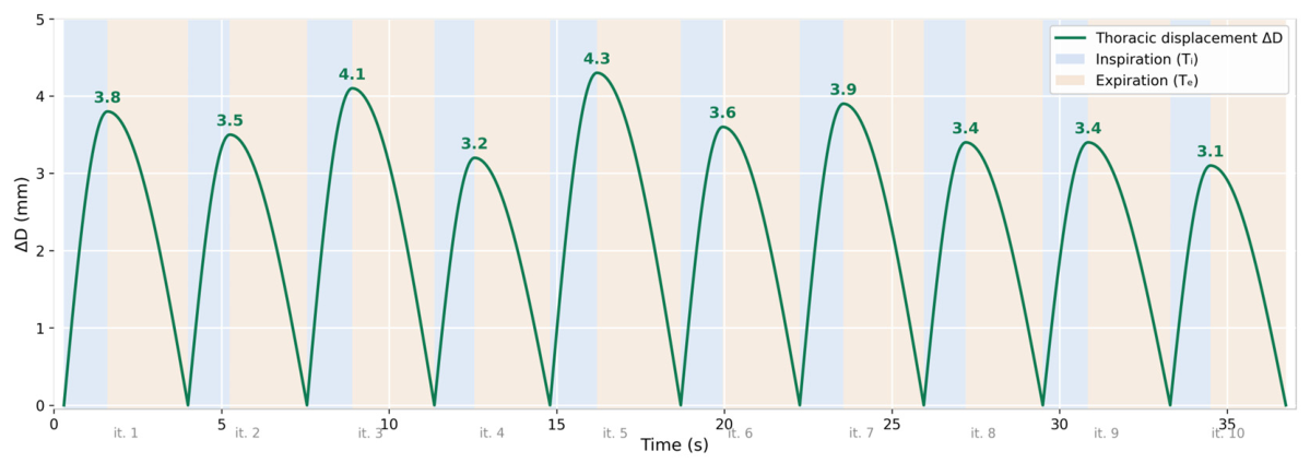
The filtered accelerometer signal demonstrated a clear periodic waveform, with amplitudes corresponding to thoracic cage displacements of 2.8–4.5 mm during normal breathing, consistent with values reported in previous studies [12,22]. The results of 10 measurement iterations under eupneic conditions are presented in Table 2. The reconstructed thoracic displacement waveform over ten consecutive respiratory cycles is shown in Figure 4.
Table 2.
Results of 10 measurement iterations under eupneic conditions (60 s per iteration).
Figure 4.
Periodic thoracic displacement waveform ΔD reconstructed from Table 2 data, over 10 respiratory cycles. Blue areas represent the inspiratory phase (Ti); orange areas represent the expiratory phase (Te). Peak ΔD values (mm) are annotated above each cycle.
The mean absolute error (MAE) was 0.44 breaths/min for the accelerometer and 0.0 breaths/min for the thermistor under eupneic conditions, both below the clinical significance threshold of ±2 breaths/min. The coefficient of variation for RR_acc measurements (CV = 6.4%) and RR_therm (CV = 6.0%) primarily reflects the natural physiological variability of spontaneous respiratory rate between iterations, rather than measurement error of the system. The higher variability in ΔD (CV = 10.8%) is correlated with the amplitude dependence on the individual depth of each inspiration.
3.3. Results in Tachypnea, Bradypnea, and Apnea Scenarios
The use of a metronome eliminates physiological variability, enabling direct evaluation of instrumental precision.
The thermistor, although incapable of estimating tidal volume, provides independent confirmation of the presence of nasal respiratory airflow. However, the limitations identified in various studies [6] remain relevant, underscoring the need for the dual approach proposed in the present experiment.
Data fusion from both measurement modalities enabled cross-validation: respiratory cycles detected via ΔD corresponded with nasal thermal cycles, providing measurement redundancy. In cases where motion artifacts compromised the accelerometer signal, the thermistor signal continued to provide valid RR measurements. As an example, a continuous 60 s monitoring session was performed. At t = 20.0 s, the subject raised their right arm over the chest to simulate repositioning (effective movement duration: 3.0 s). Analysis windows are 10 s long and the fusion rule is defined in Section 3.5: |RR_acc − RR_therm| < 4 breaths/min for accelerometric channel validation. The behavior of both sensor channels across the six consecutive 10 s analysis windows of this session is detailed in Table 3.
Table 3.
Behavior of the two sensor channels during a voluntary arm movement event (independent test, N = 1 session × 60 s; baseline RR = 12.0 breaths/min, baseline ΔD = 4.2 mm). RR_acc = respiratory rate estimated from the accelerometric channel; RR_therm = respiratory rate estimated from the thermal channel; ΔD_max = maximum thoracic displacement amplitude in window; |ΔRR| = absolute difference between the two channel estimates.
System performance in the three controlled scenarios (with metronome-imposed frequency) is summarized in Table 4.
Table 4.
System performance in controlled respiratory scenarios (mean ± SD, N = 10 iterations per scenario).
In the tachypnea scenario, accelerometer error increased (MAE = 1.2 breaths/min) compared to eupnea, explicable by reduced displacement amplitudes (ΔD = 1.4 ± 0.3 mm) approaching the minimum detection threshold of 0.5 mm. The thermistor maintained superior performance (MAE = 0.5 breaths/min) due to the persistent thermal contrast between inspired and expired air. Of the 300 respiratory cycles in the tachypnea scenario (30 cycles × 10 iterations), 12 cycles were not detected by the accelerometer, all corresponding to very shallow breaths with ΔD < 0.5 mm. The low coefficient of variation (CV_acc = 4.1%, CV_therm = 2.0%) confirms good measurement repeatability even at high frequency.
In the bradypnea scenario, both sensors showed excellent performance, with large ΔD amplitudes (5.8 ± 0.6 mm) facilitating reliable detection. All 80 respiratory cycles were correctly detected by both sensors in all 10 iterations.
The alarm system functioned correctly in all iterations. The tachypnea alarm triggered at 4.2 + 0.8 s after exceeding the threshold of 30 breaths/min, consistent with approximately two respiratory cycles of ~2 s each (at 30 breaths/min) required by the algorithm to confirm the increased frequency and avoid false alarms generated by transient variations. The bradypnea alarm triggered at 8.1 + 1.2 s after dropping below 8 breaths/min, an interval corresponding approximately to the duration of one complete respiratory cycle at 8 breaths/min (7.5 s), plus a small confirmation margin—necessary to distinguish true bradypnea from physiological but prolonged expiratory pauses. The apnea alarm triggered at 12.3 + 1.8 s after the last detected respiratory cycle, a value respecting the clinical threshold of 10 s recommended by the American Academy of Sleep Medicine [10] for defining apnea, to which approximately 2 s of margin are added to reduce false positives generated by normal respiratory pauses, sighs, or speech, while maintaining sufficiently early detection for clinical intervention.
3.4. Thermistor Signal Analysis
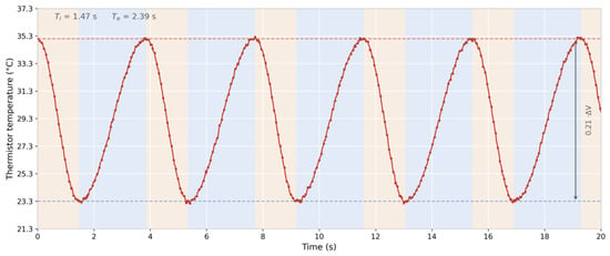
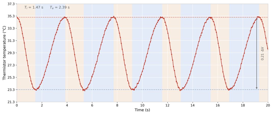
The output voltage values from Figure 5 result from the signal conditioning chain described in Section 2.3. The 10 kΩ NTC thermistor at 25 °C is connected in a Wheatstone bridge with three fixed 10 kΩ resistors. During the inspiration phase, ambient air (~22–25 °C) maintains the thermistor near its nominal resistance value (10 kΩ), and the bridge is nearly balanced, generating minimal differential voltage. The LM358 operational amplifier, configured with differential gain Ad = Rf/Rin = 100 kΩ/1 kΩ = 100, produces an output voltage of approximately 1.2 V. During the expiration phase, warm air (~34–37 °C) reduces the thermistor resistance according to Equation (2) to approximately 5–6 kΩ, unbalancing the bridge. The amplified differential voltage increases to approximately 3.8 V, resulting in a total output voltage variation of approximately 2.6 V between the inspiratory and expiratory phases, effectively utilizing the 0–5 V ADC range of the microcontroller.
Figure 5.
NTC nasal thermistor temperature variation recorded over a 20 s period under eupneic conditions (iteration 1, RR_ref = 15 breaths/min). The upper dotted line marks the temperature of expired air (~35.3 °C), and the lower dotted line the temperature of inspired air (~23.3 °C), resulting in a thermal excursion ΔT ≈ 12 °C. Alternating colored areas indicate the inspiratory (blue, Ti ≈ 1.47 s) and expiratory (orange, Te ≈ 2.39 s) phases of the respiratory cycle.
The respiratory rate of ~15.6 breaths/minute corresponds to the mean of iterations from Table 2 (RR_ref = 15.6 ± 0.97 breaths/min), and the Ti/Te ratio of 1.47/2.39 s reflects the normal physiological pattern, in which expiration is a slower passive process than active inspiration.
3.5. Global Performance and Statistical Analysis
The negligible mean bias for both sensors (−0.08 breaths/min for the accelerometer, +0.03 breaths/min for the thermistor) indicates the absence of a significant systematic error. The 95% limits of agreement were −2.72 to +2.56 breaths/min for the accelerometer, comparable to the precision reported by other studies (MAE = 2.67 breaths/min) [22] and superior to the ±3 breaths/min reported for a comparable wireless accelerometric monitor [23]. The global performance metrics of both sensing channels, aggregated across the three scenarios, are summarized in Table 5.
Table 5.
Global performance of the dual system (3 scenarios × 10 iterations, N = 30 intervals of 60 s).
The mean inter-iteration coefficient of variation (CV = 5.1% for the accelerometer, 3.5% for the thermistor) demonstrates good system repeatability, below the 10% threshold considered acceptable for respiratory monitoring devices [3]. The higher variability of the accelerometer is due to sensitivity to the exact placement of the sensor on the thoracic cage, confirming observations from the specialized literature [8].
Agreement within ±2 breaths/min was 90.0% for the accelerometer and 96.7% for the thermistor. The combined use of both sensors (decision rule: RR valid if |RR_acc − RR_therm| < 4 breaths/min) allowed identification and exclusion of all erroneous measurements, increasing the effective reliability of the system.
3.6. Limitations of the System
Despite the encouraging laboratory performance, several limitations warrant explicit discussion. The accelerometric channel is sensitive to sensor placement: other research [8] reported that deviations of ±2 cm from the xiphoid process can alter ΔD by up to 30% and introduce a parasitic cardiogenic component. In addition, cross-channel artifact rejection was validated only through a single controlled arm movement session (Table 3); involuntary movements typical of the ICU such as coughing, sleep myoclonus, shivering, or repositioning by clinical staff all exhibit different spatio-temporal signatures and have not yet been tested against the fusion rule.
The tidal volume coefficient k = 130 mL/mm is a population-average value [13,14,16] that may vary by ±25–40% between individuals depending on BMI, sex, age, and lung compliance [14]; consequently, V_T values in Table 2 should be interpreted as population-level estimates, and an individualized calibration against a known-volume spirometric reference is recommended before clinical deployment.
Finally, the thermal excursion ΔT ≈ 12 °C reported in Figure 5 was obtained at 22–25 °C ambient temperature. Warmer environments, oxygen therapy via nasal cannulas, and elevated humidity may attenuate the thermal signal or modify the thermistor time constant. Interactions that were not quantified here and represent a priority for future clinical validation.
4. Conclusions
This study presented the design and implementation of a non-invasive dual respiratory monitoring system intended for intensive care wards. The combination of the ADXL345 MEMS accelerometer with a nasal NTC thermistor provides a solution with sensor redundancy, capable of measuring respiratory rate, thoracic cage displacement, and estimating tidal volume.
The main contributions of the work consist of designing an integrated circuit with dual sensor acquisition on a single Arduino platform and implementing a real-time processing algorithm that combines Butterworth filtering with double numerical integration for calculating thoracic displacement ΔD. The dual architecture enabled cross-validation between the two measurement modalities, increasing the reliability of estimates in the presence of artifacts, and the system was completed with a clinical alarm module with software-configurable thresholds for bradypnea, tachypnea, and apnea.
Future research directions aim, first and foremost, at clinical validation of the system on a group of patients admitted to intensive care units, under real conditions of continuous monitoring. On the signal processing side, implementation of machine learning algorithms for automatic classification of abnormal respiratory patterns [9] is envisioned, capable of identifying specific types of dysfunction beyond simple frequency estimation. In parallel, integration with medical IoT protocols such as MQTT and HL7 FHIR would allow inclusion of the system in the hospital’s telemedicine infrastructure, while miniaturization of components into a wearable patch-type device would open the possibility of prolonged ambulatory monitoring, eliminating dependence on USB power.
Author Contributions
Conceptualization, A.V.-A. and M.M.; methodology, M.M.; software, A.V.-A.; validation, M.M. and V.M.; writing—review and editing, A.V.-A. and M.M.; supervision, V.M. All authors have read and agreed to the published version of the manuscript.
Funding
This research received no external funding.
Institutional Review Board Statement
Not applicable.
Informed Consent Statement
Informed consent was obtained from all subjects involved in the study.
Data Availability Statement
All data supporting the reported results are contained within the article.
Conflicts of Interest
The authors declare no conflict of interest.
Abbreviations
The following abbreviations are used in this manuscript:
| ADC | Analog-to-Digital Converter |
| AASM | American Academy of Sleep Medicine |
| BPM | Breaths Per Minute |
| CV | Coefficient of Variation |
| SD | Standard Deviation |
| HC-05 | Bluetooth Serial Communication Module (HC-05) |
| I2C | Inter-Integrated Circuit (serial communication protocol) |
| IIR | Infinite Impulse Response (digital filter type) |
| IoT | Internet of Things |
| LCD | Liquid Crystal Display |
| MAE | Mean Absolute Error |
| MEMS | Micro-Electro-Mechanical Systems |
| NTC | Negative Temperature Coefficient (thermistor type) |
| RIP | Respiratory Inductance Plethysmography |
| RMSE | Root Mean Square Error |
| RR | Respiratory Rate (breaths/min) |
| SCL | Serial Clock Line (I2C) |
| SDA | Serial Data Line (I2C) |
| UART | Universal Asynchronous Receiver-Transmitter |
| USB | Universal Serial Bus |
References
- Rolfe, S. The importance of respiratory rate monitoring. Br. J. Nurs. 2019, 28, 504–508. [Google Scholar] [CrossRef] [Scilit] [PubMed]
- Hodgetts, T.J.; Kenward, G.; Vlackonikolis, I.; Payne, S.; Castle, N.; Crouch, R.; Ineson, N.; Shaikh, L. Incidence, location and reasons for avoidable in-hospital cardiac arrest in a district general hospital. Resuscitation 2002, 54, 115–123. [Google Scholar] [CrossRef] [Scilit] [PubMed]
- Granholm, A.; Pedersen, N.E.; Lippert, A.; Petersen, L.F.; Rasmussen, L.S. Respiratory rates measured by a standardised clinical approach, ward staff, and a wireless device. Acta Anaesthesiol. Scand. 2016, 60, 1444–1452. [Google Scholar] [CrossRef] [Scilit] [PubMed]
- Brodsky, M.B.; Levy, M.J.; Jedlanek, E.; Pandian, V.P.; Blackford, B.M.; Price, C.M.; Cole, G.D.; Hillel, A.T.; Best, S.R.; Akst, L.M. Laryngeal injury and upper airway symptoms after oral endotracheal intubation with mechanical ventilation during critical care: A systematic review. Crit. Care Med. 2018, 46, 2010–2017. [Google Scholar] [CrossRef] [Scilit] [PubMed]
- ISO 5725-2; Accuracy (Trueness and Precision) of Measurement Methods and Results—Part 2: Basic Method for the Determination of Repeatability and Reproducibility of a Standard Measurement Method. International Organization for Standardization: Geneva, Switzerland, 2025.
- Norman, R.G.; Ahmed, M.M.; Walsleben, J.A.; Rapoport, D.M. Detection of respiratory events during NPSG: Nasal cannula/pressure sensor versus thermistor. Sleep 1997, 20, 1175–1184. [Google Scholar] [CrossRef] [Scilit]
- Sabil, A.; Glos, M.; Günther, A.; Schöbel, C.; Veauthier, C.; Fietze, I.; Penzel, T. Comparison of apnea detection using oronasal thermal airflow sensor, nasal pressure transducer, respiratory inductance plethysmography and tracheal sound sensor. J. Clin. Sleep Med. 2019, 15, 285–292. [Google Scholar] [CrossRef] [Scilit] [PubMed]
- Hughes, S.; Liu, H.; Zheng, D. Influences of sensor placement site and subject posture on measurement of respiratory frequency using triaxial accelerometers. Front. Physiol. 2020, 11, 823. [Google Scholar] [CrossRef] [Scilit] [PubMed]
- Drummond, G.B.; Fischer, D.; Lees, M.; Bates, A.; Mann, J.; Arvind, D.K. Classifying signals from a wearable accelerometer device to measure respiratory rate. ERJ Open Res. 2021, 7, 00681–02020. [Google Scholar] [CrossRef] [Scilit] [PubMed]
- Berry, R.B.; Budhiraja, R.; Gottlieb, D.J.; Gozal, D.; Iber, C.; Kapur, V.K.; Marcus, C.L.; Mehra, R.; Parthasarathy, S.; Quan, S.F.; et al. Rules for scoring respiratory events in sleep: Update of the 2007 AASM Manual. J. Clin. Sleep Med. 2012, 8, 597–619. [Google Scholar] [CrossRef] [Scilit] [PubMed]
- Steinhart, J.S.; Hart, S.R. Calibration curves for thermistors. Deep Sea Res. 1968, 15, 497–503. [Google Scholar] [CrossRef] [Scilit]
- Bates, A.; Ling, M.J.; Mann, J.; Arvind, D.K. Respiratory rate and flow waveform estimation from tri-axial accelerometer data. In Proceedings of the International Conference on Body Sensor Networks, Singapore, 7–9 June 2010; IEEE: Piscataway, NJ, USA, 2010; pp. 144–150. [Google Scholar]
- Monaco, V.; Stefanini, C. Assessing the tidal volume through wearables: A scoping review. Sensors 2021, 21, 4124. [Google Scholar] [CrossRef] [Scilit] [PubMed]
- Jayarathna, T.; Gargiulo, G.D.; Breen, P.P. Respiration rate and volume measurements using wearable strain sensors. npj Digit. Med. 2019, 2, 8. [Google Scholar] [CrossRef] [Scilit] [PubMed]
- Yoon, J.W.; Noh, Y.S.; Kwon, Y.S.; Kim, W.K.; Yoon, H.R. Tidal volume variability and respiration rate estimation using a wearable accelerometer sensor. In Proceedings of the 36th Annual International Conference of the IEEE Engineering in Medicine and Biology Society (EMBC), Chicago, IL, USA, 26–30 August 2014; IEEE: Piscataway, NJ, USA, 2014; pp. 6233–6236. [Google Scholar]
- De Groote, A.; Wantier, M.; Cheron, G.; Estenne, M.; Paiva, M. Chest wall motion during tidal breathing. J. Appl. Physiol. 1997, 83, 1531–1537. [Google Scholar] [CrossRef] [Scilit] [PubMed]
- Konno, K.; Mead, J. Measurement of the separate volume changes of rib cage and abdomen during breathing. J. Appl. Physiol. 1967, 22, 407–422. [Google Scholar] [CrossRef] [Scilit] [PubMed]
- Analog Devices. ADXL345 Digital Accelerometer Datasheet Rev. G; Analog Devices, Inc.: Wilmington, MA, USA, 2015. [Google Scholar]
- Soliman, M.M.; Ganti, V.G.; Inan, O.T. Towards wearable estimation of tidal volume via electrocardiogram and seismocardiogram signals. IEEE Sens. J. 2022, 22, 18093–18103. [Google Scholar] [CrossRef] [Scilit] [PubMed]
- Marks, M.K.; South, M.; Carter, B.G. Measurement of respiratory rate and timing using a nasal thermocouple. J. Clin. Monit. Comput. 1995, 11, 159–164. [Google Scholar] [CrossRef] [Scilit] [PubMed]
- Arduino LLC. Arduino Mega 2560 Rev3 Documentation. Available online: https://docs.arduino.cc/hardware/mega-2560 (accessed on 19 April 2026).
- Cesareo, A.; Biffi, E.; Cuesta-Frau, D.; D’Angelo, M.G.; Aliverti, A. A differential inertial wearable device for breathing parameter detection. Sensors 2022, 22, 9953. [Google Scholar] [CrossRef] [Scilit] [PubMed]
- Jacobs, F.; Scheerhoorn, J.; Mestrom, E.; van der Stam, J.; Bouwman, R.A.; Nienhuijs, S. Reliability of heart rate and respiration rate measurements with a wireless accelerometer in postbariatric recovery. PLoS ONE 2021, 16, e0247903. [Google Scholar] [CrossRef] [Scilit] [PubMed]

Disclaimer/Publisher’s Note: The statements, opinions and data contained in all publications are solely those of the individual author(s) and contributor(s) and not of MDPI and/or the editor(s). MDPI and/or the editor(s) disclaim responsibility for any injury to people or property resulting from any ideas, methods, instructions or products referred to in the content. |
© 2026 by the authors. Licensee MDPI, Basel, Switzerland. This article is an open access article distributed under the terms and conditions of the Creative Commons Attribution (CC BY) license.