


Abstract
Background/Objectives: Metabolic-dysfunction-associated steatotic liver disease (MASLD) and its progressive inflammatory/fibrotic form, metabolic-dysfunction-associated steatohepatitis (MASH), represent a growing global health burden. This progression is driven by complex mechanisms involving metabolic dysregulation, chronic inflammation, oxidative stress, and progressive fibrosis. To date, effective pharmacological therapies remain limited. Pentacyclic triterpenes have attracted increasing attention due to their broad biological activities and ability to modulate multiple molecular pathways implicated in chronic liver disease. This review aims to provide a mechanistic overview of the potential role of pentacyclic triterpenes in MASLD and MASH. Methods: A literature review was conducted using major scientific databases (PubMed and Web of Science) to identify experimental studies investigating pentacyclic triterpenes in metabolic liver diseases. Selected studies were analyzed according to triterpene structural classification, reported bioactivities, molecular targets, and experimental evidence from in vitro and in vivo models of MASLD/MASH or related pathogenic processes. Results: Pentacyclic triterpenes, especially ursolic acid, oleanolic acid, and glycyrrhizin, exhibit hepatoprotective effects including regulation of lipid metabolism, attenuation of oxidative and endoplasmic reticulum stress, suppression of pro-inflammatory signaling, inhibition of inflammasome activation, and reduction in hepatic stellate cell activation and extracellular matrix deposition. These effects involve modulation of signaling pathways, including AMPK, NF-κB, NLRP3, TGF-β, FXR, and MAPK. Preclinical evidence demonstrates improvements in steatosis, inflammation, and fibrosis in experimental models. Conclusions: Pentacyclic triterpenes emerge as multitarget modulators of MASH pathophysiology. However, translating preclinical evidence into well-designed clinical trials is necessary to validate their safety and efficacy in humans.
1. Introduction
Metabolic-dysfunction-associated steatotic liver disease (MASLD) has emerged as one of the most prevalent chronic liver diseases worldwide and represents an important and growing public health challenge. The disease is strongly associated with the global increase in obesity, insulin resistance, and type 2 diabetes mellitus (T2DM). MASLD encompasses a broad spectrum of liver abnormalities ranging from simple hepatic steatosis to the progressive inflammatory condition known as metabolic-dysfunction-associated steatohepatitis (MASH) [1]. In more advanced stages, persistent hepatocellular injury may result in fibrosis, cirrhosis, and hepatocellular carcinoma [2,3,4]. Because of its increasing incidence and clinical impact, MASLD and MASH have become major contributors to chronic liver disease and liver-related morbidity and mortality [1].
The pathogenesis of MASLD/MASH is complex and involves interactions among multiple metabolic, inflammatory, and fibrogenic processes. According to the widely accepted “multiple-hit” hypothesis, disease progression results from the simultaneous action of several pathogenic mechanisms, including insulin resistance, dysregulated lipid metabolism, oxidative stress, mitochondrial dysfunction, and chronic inflammatory signaling [5]. These processes promote hepatocyte injury, hepatic stellate cell activation, and progressive extracellular matrix deposition, ultimately leading to liver fibrosis [6]. Despite important advances in understanding the molecular mechanisms underlying MASLD/MASH, effective pharmacological therapies remain limited. However, the therapeutic landscape is evolving rapidly, with the development of several pharmacological strategies targeting key metabolic and inflammatory pathways involved in disease progression. Recent advances include the approval of the thyroid hormone receptor-β agonist resmetirom [7], as well as the clinical investigation of farnesoid X receptor (FXR) agonists, glucagon-like peptide-1 (GLP-1) receptor agonists, especially semaglutide, and other metabolic modulators that aim to improve hepatic steatosis, inflammation, and fibrosis [1,8]. Nevertheless, most of these therapeutic candidates primarily target specific molecular pathways, which may not fully address the multifactorial and interconnected mechanisms underlying MASLD/MASH pathogenesis.
Natural products have historically played an important role in the discovery of pharmacologically active molecules for the treatment of metabolic and inflammatory diseases. Among these compounds, pentacyclic triterpenes represent a structurally diverse class of plant-derived secondary metabolites widely distributed in medicinal plants and dietary sources [9,10]. These compounds are characterized by a thirty-carbon skeleton organized into five fused rings and are commonly classified into three main structural groups: oleananes, ursanes, and lupanes [11]. Increasing evidence indicates that pentacyclic triterpenes exhibit a wide range of biological activities, including anti-inflammatory, antioxidant, antiadipogenic, antifibrotic, and hepatoprotective effects [12,13]. Importantly, several triterpenes have been shown to modulate key molecular pathways involved in MASLD/MASH pathogenesis, including adenosine monophosphate-activated protein kinase (AMPK) signaling, nuclear factor kappa-light-chain-enhancer of activated B cells (NF-κB)-mediated inflammatory responses, oxidative stress pathways, inflammasome activation, and hepatic stellate cell activation [14,15,16,17,18,19,20,21,22,23,24,25].
In this context, natural triterpenes may offer complementary mechanistic advantages due to their multitarget pharmacological profile, which allows the simultaneous modulation of metabolic, inflammatory, oxidative, and fibrogenic pathways implicated in MASLD/MASH progression. Such pleiotropic effects may offer a potential advantage over single-target pharmacological strategies, although important limitations remain, including low oral bioavailability, pharmacokinetic variability, and limited availability of robust clinical data.
Although numerous experimental studies have investigated individual triterpenes in the context of metabolic liver diseases, the available literature remains fragmented across different experimental models and mechanistic approaches. This review therefore provides an integrative mechanistic perspective linking the structural diversity of pentacyclic triterpenes with their ability to modulate multiple molecular pathways involved in MASLD/MASH progression. Specifically, this review is organized as follows: first, the structural characteristics and biosynthesis of pentacyclic triterpenes are described; second, the major biological activities of triterpenes are discussed, including anti-inflammatory, antioxidant, metabolic, and antifibrotic effects; third, the molecular mechanisms involved in MASLD/MASH pathogenesis are summarized; and fourth, current experimental evidence regarding the application of triterpenes in MASLD and MASH is presented. In addition, a section summarizing current clinical evidence for triterpenes is included, followed by a discussion of toxicological considerations and safety aspects, and finally, strategies aimed at improving triterpene bioavailability and therapeutic efficacy.
2. Methods
A narrative literature review was conducted using the PubMed and Web of Science databases to identify experimental studies investigating the biological activities of pentacyclic triterpenes and their potential relevance to MASLD and MASH. The initial search strategy combined the terms “triterpene” OR “triterpenes” with keywords related to major pathogenic processes involved in MASLD/MASH progression, including steatosis, inflammation, oxidative stress, endoplasmic reticulum stress, fibrosis, and metabolic dysregulation. In addition, previous nomenclature for the disease was also included in the search strategy, such as “non-alcoholic fatty liver disease (NAFLD)” and “non-alcoholic steatohepatitis (NASH)”, which were widely used prior to the recent terminology standardization.
In the initial screening phase, studies published from 2010 onward were preferentially considered. Earlier studies were included when their abstracts indicated strong mechanistic relevance or when they provided foundational experimental evidence supporting more recent findings. Titles and abstracts were screened to identify studies investigating the mechanistic effects or therapeutic efficacy of triterpenes. Review articles, conference abstracts, and studies not directly related to mechanistic investigation were excluded. Studies focused on complex or poorly characterized plant extracts containing multiple classes of bioactive compounds were also excluded to avoid confounding interpretation of triterpene-specific effects. When extracts were included, only those chemically standardized and predominantly composed of triterpenes were considered.
Following this initial screening, the triterpenes with the most consistent experimental evidence were identified. A second targeted search was then performed using the specific names of each triterpene, combined with disease-related terms (MASLD, MASH, NASH, or NAFLD), to retrieve studies evaluating their roles in metabolic liver disease. In this stage, articles were reviewed from the earliest available reports to the present, allowing the reconstruction of the experimental development of each compound. Review articles, conference abstracts, studies not directly investigating triterpene activity in MASLD/MASH-related mechanisms, or studies involving complex matrices were excluded.
3. Results
3.1. Mechanisms of Metabolic-Dysfunction-Associated Steatotic Liver Disease (MASLD) and Metabolic-Dysfunction-Associated Steatohepatitis (MASH)
Chronic liver diseases (CLDs) and cirrhosis represent major global health challenges, accounting for substantial morbidity and mortality worldwide [26]. MASLD and its progressive form, MASH, have emerged as leading contributors to the global burden of liver disease, driving an increasing incidence of cirrhosis and hepatocellular carcinoma (HCC) [27].
MASLD is a multisystemic disorder in which insulin resistance and metabolic dysfunction play central roles in disease initiation and progression [28]. The diagnosis is established when more than 5% of hepatocytes exhibit steatosis in the presence of defined metabolic abnormalities, including obesity, T2DM, or dyslipidemia [29]. Clinically, MASLD encompasses a broad spectrum ranging from isolated triglyceride accumulation in hepatocytes—termed metabolic-dysfunction-associated steatotic liver (MASL)—to MASH, which is characterized by lobular or portal inflammation and hepatocellular ballooning [30,31]. In advanced stages, persistent injury may culminate in fibrosis, cirrhosis, and ultimately HCC [30].
MASLD/MASH pathogenesis is increasingly recognized as a complex network of interconnected metabolic, inflammatory, and fibrogenic processes rather than a series of isolated pathways [28]. To account for this complexity, the “multiple-hit” hypothesis was proposed (Figure 1), postulating that disease progression results from the simultaneous action of several pathogenic stimuli. These include genetic susceptibility, dietary factors, obesity, insulin resistance, altered lipid metabolism, lipotoxicity, oxidative stress, mitochondrial and endoplasmic reticulum dysfunction, intestinal dysbiosis, and dysregulated cytokine and adipokine production [6]. Collectively, these factors promote activation of pro-inflammatory cascades and inflammasomes, particularly NLR family pyrin domain containing 3 (NLRP3), leading to hepatocellular injury, cell death, and fibrogenesis—hallmarks of MASH. Concomitantly, activation of hepatic stellate cells (HSCs) and immune cells enhances extracellular matrix (ECM) deposition, accelerating fibrosis progression [27,28,32].
Figure 1.
Main mechanisms involved in the development and progression of MASLD/MASH and their complications. Genetic predisposition, dietary factors, and insulin resistance contribute to hepatic steatosis and the development of metabolic-dysfunction-associated steatotic liver disease (MASLD). Lipotoxicity, oxidative stress, mitochondrial dysfunction, and endoplasmic reticulum (ER) stress promote hepatocellular injury and inflammation, leading to metabolic-dysfunction-associated steatohepatitis (MASH). Activation of inflammatory pathways (e.g., TLR4–MyD88/NF-κB signaling), Kupffer cells (KCs), and inflammasomes, together with gut-derived factors such as lipopolysaccharide (LPS), further sustain chronic inflammation. Persistent injury promotes hepatic stellate cell (HSC) activation and extracellular matrix (ECM) deposition via profibrogenic mediators such as transforming growth factor beta (TGF-β), contributing to fibrosis progression and, ultimately, cirrhosis. Abbreviations: COL-1, collagen type I; DAMPs, damage-associated molecular patterns; FAs, fatty acids; IL-1β, interleukin-1 beta; IL-6, interleukin-6; MMP, matrix metalloproteinases; MyD88, myeloid differentiation primary response 88; NF-κB, nuclear factor kappa B; TIMP, tissue inhibitors of metalloproteinases; TLR4, Toll-like receptor 4; TNF-α, tumor necrosis factor alpha.
Within this context, dysregulated fatty acid (FA) metabolism has been closely linked to cellular stress and lipotoxicity, triggering inflammation, immune cell recruitment, and hepatocyte death—key drivers of the transition from MASLD to MASH and more advanced liver disease stages [33]. Elevated levels of free FA and cholesterol exacerbate insulin resistance, promote adipocyte dysfunction, and alter gut microbiota composition, thereby increasing intestinal permeability and enhancing the translocation of lipopolysaccharides (LPS). These events further amplify hepatic inflammation via the gut–liver axis [5].
In the liver, excessive lipid influx and de novo lipogenesis induce oxidative stress, mitochondrial dysfunction, and inflammasome activation, culminating in hepatocyte injury and apoptosis [2]. Lipotoxicity activates pro-inflammatory signaling through Toll-like receptors (TLRs), resulting in increased production of cytokines such as tumor necrosis factor alpha (TNF-α), interleukin (IL)-6, and IL-1β. These mediators amplify inflammatory responses and drive the transdifferentiation of HSCs into myofibroblasts, thereby promoting fibrosis [33]. TLR4, abundantly expressed in Kupffer cells (KCs) and HSCs, is activated by bacterial LPS and FA, triggering the MyD88/NF-κB pathway and sustaining chronic inflammation.
In parallel, oxidative stress arising from mitochondrial and endoplasmic reticulum dysfunction activates the NLRP3 inflammasome, further intensifying inflammatory and profibrotic signaling [27]. Persistent activation of the inhibitor κB kinase (IKK)/NF-κB and c-Jun N-terminal kinase (JNK) pathways in HSCs, along with TLR5-mediated NF-κB and MAPK signaling, contributes to chronic inflammation, tissue remodeling, and fibrosis progression [33].
More recently, pyroptosis has been recognized as an additional driver of hepatic inflammation and fibrogenesis. Pyroptosis is a pro-inflammatory form of programmed cell death mediated by caspase activation and gasdermin D (GSDMD)–dependent pore formation in the plasma membrane, resulting in the release of inflammatory cytokines, lactate dehydrogenase (LDH), osmotic swelling, and cell lysis. This process occurs through canonical (caspase-1–dependent) and non-canonical (caspase-4/5/11–dependent) pathways and has been shown to promote macrophage activation and fibrosis progression [23,34,35].
Activation of these interconnected pathways compromises hepatic regenerative capacity and promotes excessive ECM deposition, ultimately leading to progressive fibrosis [31]. Regardless of etiology, liver fibrosis shares common molecular mechanisms, including hepatocyte death, chronic inflammation, HSC activation, and disruption of epithelial and endothelial barriers, all of which are closely associated with liver-related mortality [36].
Under physiological conditions, HSCs remain quiescent, storing vitamin A and producing type IV collagen. However, chronic liver injury induces a phenotypic switch characterized by increased proliferation, contractility, and synthesis of type I and III collagen, typical of cirrhotic tissue [2]. This transition is driven by sustained inflammation, whereby hepatocyte death releases damage-associated molecular patterns (DAMPs) and apoptotic bodies that further activate HSCs [27].
Transforming growth factor beta (TGF-β) signaling plays a pivotal role in hepatic fibrogenesis by activating the SMAD pathway and inducing the expression of genes encoding type I collagen, fibronectin, and proteoglycans—key components of the ECM [27,28]. Additional pathways, including platelet-derived growth factor (PDGF) and vascular endothelial growth factor (VEGF), contribute to HSC proliferation, migration, survival, and ECM production, while also enhancing TGF-β signaling [37]. NF-κB activation by inflammatory stimuli and inflammasome signaling further reinforces the profibrotic response, perpetuating excessive ECM accumulation [27,28].
The hepatic ECM undergoes dynamic remodeling during fibrogenesis and plays a critical role in CLD progression [38]. Its proteome, comprising structural proteins, proteases, and protease inhibitors, evolves with disease severity, influencing cell–cell communication and inflammatory signaling [37]. Maintenance of ECM homeostasis depends on the balance between matrix metalloproteinases (MMPs) and their tissue inhibitors (TIMPs). Disruption of this balance—through increased TIMP activity or reduced MMP function—results in excessive collagen deposition, increased liver stiffness, and fibrosis progression [39].
As fibrosis advances, ECM deposition predominates due to elevated TIMP-1 expression, which also protects activated HSCs from apoptosis, thereby sustaining fibrogenesis. Activated macrophages and persistent MAPK and NF-κB signaling further exacerbate ECM dysregulation, aggravating liver injury [37,39]. Ultimately, prolonged fibrotic remodeling leads to cirrhosis, an advanced and often irreversible stage of CLDs characterized by extensive ECM accumulation and regenerative nodule formation, resulting in architectural distortion and functional impairment of the liver [4,40].
Cirrhosis arises from continuous cycles of hepatocyte necrosis and regeneration, leading to sinusoidal capillarization, reduced functional parenchyma, altered hepatic blood flow, and portosystemic shunting. These changes underpin the development of portal hypertension and its complications, including ascites, hepatic encephalopathy, and pulmonary, renal, and cardiac dysfunction, as well as hyponatremia [41].
MASLD is also closely linked to alterations in gut microbiota composition and intestinal barrier integrity, positioning the gut–liver axis as a central component of disease pathophysiology. Metabolic and dietary factors promote intestinal dysbiosis, characterized by depletion of commensal bacteria and expansion of pro-inflammatory Gram-negative species. Inflammatory and hypoxic conditions disrupt tight junction integrity, increasing intestinal permeability and facilitating bacterial translocation and passage of microbial products such as LPS into the portal circulation. These molecules activate KCs via pattern recognition receptors, particularly TLRs, triggering inflammatory cascades mediated by cytokines and ROS [42].
Nicotinamide adenine dinucleotide phosphate (NADPH) oxidase 2 (NOX2), a major source of ROS in phagocytes, and the NLRP3 inflammasome are activated by signals derived from the dysbiotic microbiota and impaired epithelial barrier, modulating hepatic inflammation and fibrosis progression [43]. Clinically, cirrhosis presents as a heterogeneous condition with two major prognostic stages: a compensated phase, characterized by preserved liver function, and a decompensated phase marked by complications such as ascites, variceal bleeding, infections, and hepatic encephalopathy [44]. Disease progression may culminate in severe outcomes, including hepatorenal syndrome, acute-on-chronic liver failure (ACLF), and multiple organ failure [45].
The progression from MASH to HCC is complex and not yet fully understood. Chronic hepatic inflammation promotes the generation of multiple pro-tumorigenic factors, which may induce DNA damage and activate aberrant intracellular signaling cascades that contribute to carcinogenesis. Several immune-mediated mechanisms have been implicated in this process. Increased activity of CD8+ T cells and natural killer T (NKT) cells has been associated with the release of pro-inflammatory cytokines that sustain hepatic injury. Meanwhile, activation of Kupffer cells (KCs) further amplifies inflammatory signaling and cytokine production within the hepatic microenvironment. During the progression of MASLD and MASH, multiple signaling pathways closely associated with tumor development become dysregulated, including Wnt/β-catenin signaling, phosphatidylinositol-3-kinase/protein kinase B/mammalian target of rapamycin (PI3K/Akt/mTOR) signaling, Hedgehog signaling, and mesenchymal–epithelial transition (MET) pathways involved in cell migration and metastasis. Because the primary focus of this review is on MASLD and MASH, tumorigenesis will not be extensively discussed. A more detailed overview of the mechanisms linking metabolic liver disease to hepatocellular carcinoma can be found in the review by Phoolchund and Khakoo [3].
Given the multifactorial nature of MASLD and MASH, therapeutic strategies that modulate multiple pathogenic pathways simultaneously are of particular interest. Natural products have emerged as valuable sources of bioactive molecules with pleiotropic biological activities that can target complex metabolic and inflammatory disorders. Among these compounds, pentacyclic triterpenes have attracted increasing attention due to their broad pharmacological properties, including anti-inflammatory, antioxidant, metabolic, and antifibrotic effects. These characteristics make triterpenes promising candidates for modulating key mechanisms involved in MASLD/MASH progression [12,13]. To better understand their biological potential, it is important to first examine their structural characteristics, classification, and biosynthetic origin, which are discussed in the following section.
3.2. Classification and Structural Characteristics of Pentacyclic Triterpenes
Triterpenes constitute a vast class of bioactive compounds characterized by a thirty-carbon skeleton. Synthesized by a wide range of organisms, including bacteria, fungi, and animals, they reach their greatest structural diversity within the plant kingdom, with over 20,000 identified variants [9,46]. Characterized as complex molecules, triterpenes can be found either (i) in their free form, as non-volatile lipophilic substances soluble in organic solvents, acting as components of cellular membranes or as signaling molecules, or (ii) in oxygenated, esterified, or glycosylated derivative forms, which may confer protection to plants against pathogens and pests [10,47].
The fundamental terpene architecture arises from the sequential conjugation of isoprene units CH2=C(CH3)-CH=CH2. Terpenes are classified according to the number of isoprene units they contain, as follows: hemiterpenes (one isoprene unit), monoterpenes (two isoprene units), sesquiterpenes (three isoprene units), diterpenes (four isoprene units), sesterpenes (five isoprene units), triterpenes (six isoprene units), tetraterpenes (eight isoprene units), and polyterpenoids (more than eight isoprene units) [48]. Among these, the pentacyclic triterpenes—characterized by five fused rings—stand out for their remarkable pharmacological potential in modulating metabolic and inflammatory pathways. Representative structures of these groups are illustrated in Figure 2.
Figure 2.
Representative structures of each group of terpenes.
The biosynthetic precursors of terpenes are generated through two independent metabolic pathways: the mevalonic acid (MVA) pathway, which involves the condensation of acetyl-CoA units, and the 1-deoxy-D-xylulose-5-phosphate (DXP) pathway, which proceeds through the condensation of pyruvate and glyceraldehyde-3-phosphate. Regardless of the pathway, both routes yield two isomeric C5 building blocks: isopentenyl pyrophosphate (IPP) and dimethylallyl pyrophosphate (DMAPP) (Figure 3) [49,50,51,52].
Figure 3.
Schematic representation of the synthesis of the direct precursors isopentenyl diphosphate (IPP) and dimethylallyl diphosphate (DMAPP) involved in terpenoid biosynthesis. The mevalonate (MVA) pathway occurs in the cytosol. It provides precursors for the production of sesquiterpenes and triterpenes, whereas the plastid-localized 1-deoxy-D-xylulose-5-phosphate (DXP) pathway supports the biosynthesis of mono-, di-, and tetraterpenes. Abbreviations: GDPS, geranyl diphosphate synthase; FDPS, farnesyl diphosphate synthase; GGDPS, geranylgeranyl diphosphate synthase; SQS, squalene synthase.
Although both pathways produce identical C5 units, they are compartmentalized and contribute differently to terpene biosynthesis. The MVA pathway occurs predominantly in the cytosol. It is mainly responsible for the formation of sesquiterpenes and triterpenes, whereas the plastid-localized DXP pathway primarily supports the biosynthesis of mono-, di-, and tetraterpenes [53].
Terpene biosynthesis is driven by a series of sequential condensation reactions. The process initiates with the condensation of DMAPP and one IPP unit to form geranyl pyrophosphate (GPP), the fundamental precursor of monoterpenes, in a reaction catalyzed by geranyl diphosphate synthase (GDPS). GPP subsequently undergoes condensation with an additional IPP unit to generate farnesyl pyrophosphate (FPP), the C15 precursor of sesquiterpenes and triterpenes, mediated by farnesyl diphosphate synthase (FDPS). Through the action of geranylgeranyl diphosphate synthase, FPP may further condense with IPP to form geranylgeranyl diphosphate (GGPP), the C20 precursor of di- and tetraterpenes.
A critical branch point occurs when two FPP molecules undergo a reductive head-to-head condensation, catalyzed by squalene synthase (SQS), to produce squalene. This hydrocarbon is subsequently epoxidized into 2,3-oxidosqualene, the primary substrate for oxidosqualene cyclases (OSCs), which orchestrate the complex cyclization reactions that define the diverse triterpene skeletons. The stereochemical outcome is dictated by the folding conformation of the substrate: while the chair–boat–chair (CBC) conformation leads to sterol biosynthesis, the chair–chair–chair (CCC) conformation generates the dammarenyl cation. This key intermediate undergoes further ring expansion and rearrangements to yield the characteristic five-fused-ring architecture that defines the pentacyclic triterpene family (Figure 4) [9,51,53,54,55,56].
Figure 4.
Oxidosqualene cyclase (OSC)-catalyzed 2,3-oxidosqualene cyclization pathways leading to sterols and triterpene compounds through protosteryl and dammarenyl carbocation intermediates, respectively. Abbreviations: OSC, oxidosqualene cyclase; SQE, squalene epoxidase.
Structurally, these pentacyclic triterpenes consist of thirty carbon atoms arranged into five fused rings (A–E), a configuration that allows for immense chemical diversity through functionalization (Figure 5). Based on their core carbocyclic skeletons, these compounds are primarily categorized into three dominant groups: oleananes, ursanes, and lupanes [11]. The distinction between oleanane and ursane types lies in the specific arrangement of methyl substituents on ring E: oleananes feature a gem-dimethyl group at the C20 position, whereas ursanes possess one methyl group at C19 and another at C20. In contrast, lupane-type triterpenes are structurally unique, characterized by a five-membered E ring and a trans-fused D/E ring junction [57,58,59].
Figure 5.
General structure of pentacyclic triterpenes classified according to their carbon skeletons into oleananes, ursanes, and lupanes.
Due to their ubiquitous presence in the plant kingdom and their remarkably broad spectrum of biological activities, pentacyclic triterpenes have emerged as pivotal bioactive natural products. Beyond their ecological roles, these compounds are increasingly recognized as promising lead scaffolds for the development of novel therapeutic agents, particularly for chronic metabolic and inflammatory diseases [12,13].
4. Bioactivity of Triterpenes
4.1. Anti-Inflammatory Action
The therapeutic application of plants rich in pentacyclic triterpenes is deeply rooted in traditional medicine, particularly for managing inflammatory conditions [60,61]. Modern pharmacological research has validated these traditional uses, identifying triterpenoids as potent modulators of the inflammatory cascade. Among these, glycyrrhizin—a triterpenoid saponin abundantly found in licorice root (Glycyrrhiza spp.)—stands out due to its broad anti-inflammatory properties. Upon oral administration, glycyrrhizin undergoes microbial transformation in the gut into glycyrrhetinic acid, an oleanane-type aglycone that serves as the primary effector molecule [62,63]. Glycyrrhizin exerts anti-inflammatory effects through a sophisticated, multi-target network. It directly inhibits key pro-inflammatory enzymes, including 5-lipoxygenase and cyclooxygenase-2 (COX-2), thereby reducing the biosynthesis of leukotrienes and prostaglandins [64,65,66]. A hallmark of its activity is the suppression of the NF-κB and MAPK signaling axes, which orchestrate the expression of inducible nitric oxide synthase (iNOS) and critical cytokines such as TNF-α, IL-1β, and IL-6. Notably, glycyrrhizin possesses a unique ability to bind and neutralize high-mobility group box 1 (HMGB1), a potent damage-associated molecular pattern (DAMP) that drives sterile inflammation by antagonizing HMGB1 activity and modulating Toll-like receptor (TLR) signaling. Glycyrrhizin effectively limits neutrophil activation, reduces reactive oxygen species (ROS) production, and downregulates adhesion molecules. These pleiotropic actions collectively attenuate leukocyte infiltration and tissue damage, positioning glycyrrhizin as a formidable candidate for mitigating the chronic inflammatory milieu characteristic of MASH and other metabolic liver disorders [20,67,68].
Similarly, celastrol, a bioactive triterpenoid isolated from Tripterygium wilfordii, exhibits potent anti-inflammatory and immunomodulatory properties by targeting multiple regulatory nodes. Its mechanism of action is uniquely characterized by the K63-linked deubiquitination of the NLRP3 inflammasome [69]. Furthermore, celastrol effectively suppresses the HMGB1/NF-κB axis, dampening the propagation of pro-inflammatory signals [19].
Advanced systems pharmacology and multi-omics analyses have elucidated that celastrol targets key pro-inflammatory mediators, including TNF-α and IL-6, while regulating pathways such as phosphoinositide 3-kinase (PI3K)/protein kinase B (Akt), MAPK, TGFβ, and IL signaling. By attenuating immune cell activation, fibroblast invasiveness, cytokine production, and synovial inflammation, celastrol demonstrates a broad anti-inflammatory profile [70]. Beyond its anti-inflammatory profile, celastrol exerts significant anti-proliferative and pro-apoptotic effects. It potentiates TNF-induced apoptosis by downregulating a suite of survival genes, such as IAP1/2, Bcl-2, and c-FLIP, while simultaneously inhibiting mediators of invasion and angiogenesis, including MMP-9 and VEGF [71], By attenuating cytokine production, fibroblast activation, and oxidative stress, celastrol emerges as a robust therapeutic candidate for chronic inflammatory disorders, particularly those where inflammation and fibrogenesis are closely intertwined, such as MASH.
A pivotal anti-inflammatory mechanism of pentacyclic triterpenes involves the activation of the AMPK pathway, a master regulator of energy homeostasis and immune signaling. A diverse array of compounds, including oleanolic acid (OA), ursolic acid (UA), betulinic acid (BA), glycyrrhetinic acid, and asiatic acid (AA), has been identified as potent AMPK activators, underscoring their therapeutic potential in metabolism-driven inflammation [61].
Specifically, recent investigations into Δ-OA have highlighted its efficacy as a robust AMPK activator. Comparative assays using 5-aminoimidazole-4-carboxamide ribonucleotide (AICAR)—a gold-standard AMPK agonist—demonstrated that Δ-OA achieves similar pharmacological outcomes in modulating inflammatory responses. This equivalence to established activators positions Δ-OA and related triterpenoids as promising drug candidates for conditions where AMPK dysregulation is central, such as the chronic inflammatory milieu of MASH [61].
Recent studies have highlighted the anti-inflammatory activity of ursane-type triterpenes, particularly UA, with potential applications in the treatment of neuroinflammation. UA has been shown to exert immunomodulatory effects and to promote neuronal repair by directly remyelinating during the chronic-progressive stage of multiple sclerosis [72]. Additionally, UA has demonstrated anti-inflammatory activity in models of Parkinson’s disease [73], arthritis [74], type 2 diabetes mellitus (T2DM) [75], and myasthenia gravis [76]. Administration of UA prior to disease onset also prevented encephalopathy development by inhibiting Th17 cell differentiation in a mouse model of the disease [77]. Several mechanisms have been associated with its anti-inflammatory effects, including reductions in pro-inflammatory cytokines IL-1β, IL-6, IL-8, and TNF-α levels, and malondialdehyde (MDA) level, one of the products of lipid peroxidation and a biomarker of liver oxidative damage, along with increases in superoxide dismutase (SOD) and glutathione (GSH) levels, that are key components of the cellular antioxidant system [78].
Another ursane-type triterpene under investigation is boswellic acid. Ammon demonstrated that boswellic-type triterpenes inhibit the synthesis of the pro-inflammatory enzyme 5-lipoxygenase and its products, including 5-hydroxyeicosatetraenoic acid and leukotriene B4 [79]. This compound also modulates several pathways associated with obesity and T2DM, which are discussed in later sections.
A lupane-type triterpene that has attracted attention due to its anti-inflammatory, antioxidant, and antifibrotic activities is ganoderic acid A, the major triterpenoid extracted from Ganoderma mushrooms. In a study conducted by Ma et al., this compound demonstrated significant biological activity [80].
Despite extensive investigation, it remains unclear whether the carbon skeleton of triterpenes significantly influences their anti-inflammatory efficacy. While some studies suggest that oleanane-type structures may exhibit higher activity than ursane-type compounds [81], other reports indicate comparable effects among oleanane-, lupane-, and ursane-type skeletons [82,83].
4.2. Antioxidant Activity
Oxidative stress is a central driver in the progression of MASLD to MASH, contributing significantly to hepatocyte injury and fibrogenesis. Several studies have demonstrated that pentacyclic triterpenes possess significant antioxidant potential, which directly contributes to their hepatoprotective profile.
In early investigations, Allouche et al. evaluated the antioxidant and antithrombotic activities of uvaol, erythrodiol, OA, and maslinic acid added to isolated human low-density lipoprotein (LDL), aiming to assess their cardiovascular protective effects. The results indicated that maslinic acid, uvaol, and erythrodiol were evaluated for their ability to protect isolated human LDL from oxidation [84].
The antioxidant effect of maslinic acid had been previously reported [21,85]. Wang et al. demonstrated that maslinic acid strongly inhibited copper-induced LDL oxidation in vitro, reducing the formation of conjugated dienes by approximately 21% compared with the control group [85]. According to Allouche et al., this effect was significant at a concentration of approximately 100 μM, while no effect was initially detected at 10 μM. However, the same study later reported that even 10 μM maslinic acid exhibited antioxidant properties [84].
The relevance of this mechanism to hepatic disease was highlighted by Lim et al., who investigated the protective effects of four triterpenes—AA, asiaticoside, madecassic acid, and madecassoside—in different cell culture models, including Hs68 (human skin fibroblasts), HepG2 (human hepatocytes), RAW 264.7 (murine macrophages), and EA.hy926 (human umbilical vein endothelial cells). Hs68 cells were exposed to UVB radiation, while HepG2 and EA.hy926 cells were challenged with tert-butyl hydroperoxide (TBHP) and hydrogen peroxide, respectively. RAW 264.7 macrophages were stimulated with LPS. Pretreatment with triterpenes attenuated UVB-induced ROS and MDA generation and preserved GSH levels in skin fibroblasts, while increasing intracellular collagen content. In HepG2 cells, pretreatment significantly improved cell viability and reduced TBHP-induced ROS levels, as well as alanine aminotransferase (ALT) and aspartate aminotransferase (AST) levels. In EA.hy926 cells, triterpenes reduced ROS levels and decreased MDA and GSH expression. In RAW 264.7 macrophages, treatment significantly reduced nitric oxide, TNF-α, and IL-6 production [21].
In another study, Mamadalieva et al. evaluated the antioxidant activity of fifteen cycloartane-type triterpenes using cell-free assays, including phosphomolybdenum, 1,1-diphenyl-2-picrylhydrazyl (DPPH•), 2,2′-azinobis(3-ethylbenzothiazoline)-6-sulfonic acid (ABTS•+), cupric ion-reducing antioxidant capacity (CUPRAC), ferric-reducing antioxidant power (FRAP), and metal-chelating activity assays. Most of the compounds exhibited antioxidant activity, despite lacking typical phenolic or catecholic moieties commonly associated with antioxidant effects in natural products such as flavonoids [86].
Ostapiuk et al. investigated the antioxidant mechanisms of betulin and BA, two lupane-type triterpenes, using triterpene-rich extracts. The results demonstrated a significant reduction in hydrogen peroxide and superoxide anion formation, as well as decreased lipid peroxidation [22].
As observed for anti-inflammatory effects, no clear structure–activity relationship has been established for triterpene antioxidant activity. Nevertheless, most compounds in this class exhibit antioxidant activity to varying degrees, underscoring their therapeutic potential for diseases and conditions associated with oxidative stress.
4.3. Antiadipogenic Activity and T2DM Control
Antiadipogenic activity refers to the ability of a compound to inhibit adipocyte differentiation and lipid accumulation in adipose tissue. This effect is typically mediated through the modulation of transcription factors involved in adipogenesis, such as peroxisome proliferator–activated receptor gamma (PPARγ) and CCAAT/enhancer-binding proteins (C/EBPs) [15,87]. Because excessive adipogenesis contributes to obesity, insulin resistance, and metabolic dysfunction, antiadipogenic compounds may indirectly improve metabolic disorders, including T2DM and MASLD [88].
Triterpenes have demonstrated considerable potential as antiadipogenic agents. Peng et al. evaluated the antiadipogenic effects of lanostane-type triterpenoids by testing forty compounds for their ability to inhibit the differentiation of 3T3-L1 preadipocytes into adipocytes. Several compounds significantly reduced adipocyte differentiation, particularly those containing an A-seco-23 → 26 lactone skeleton [89].
Mohsen et al. demonstrated that BA effectively reduced lipid accumulation in 3T3-L1 cells by downregulating the expression of PPARγ and liver X receptor alpha (LXRα). The study also showed enhanced interaction of steroid receptor coactivator-1 (SRC-1) with PPARγ and reduced SRC-3 levels [90]. These findings were supported by Melo et al., who investigated the effects of BA extracted from Clusia nemorosa L. (Clusiaceae) in a mouse model of obesity. The treatment resulted in significant reductions in body weight, abdominal fat accumulation, blood glucose levels, plasma triglycerides, and total cholesterol, primarily through inhibition of pancreatic lipase [87].
Betancur et al. demonstrated that an extract composed of a mixture of UA, OA, and UA lactone reduced lipid accumulation in 3T3-L1 cells, accompanied by decreased leptin expression [91]. These findings were corroborated by Acín et al., who showed that C57BL/6J mice fed a high-fat diet and treated with this extract exhibited improved glucose tolerance, reduced body weight, decreased hepatic and adipose fat content, lower plasma lipid levels, and reduced hepatic mRNA expression of PPARα, CPT1A, and sterol regulatory element-binding protein (SREBP)-1 [92].
In in vitro studies using HepG2 cells treated with free fatty acids, UA reduced lipid accumulation and modulated the expression of genes involved in lipid synthesis and degradation, concomitant with increased AMPK phosphorylation. These effects were reversed by the addition of an AMPK inhibitor, confirming that AMPK activation is essential for UA activity. Molecular docking studies further demonstrated binding of UA to AMPK at the 4CFH site, similarly to the agonist Pound–Thirsk compound 1 (PT1) [14].
Bermúdez-Contreras et al. demonstrated that extracts of Agave durangensis, containing approximately 30% triterpenes, reduced triglyceride levels by 59.28% and very-low-density lipoprotein (VLDL) cholesterol by 60.27% in a mouse model of T2DM [93]. Liou et al. investigated maslinic acid extracted from olive peels and reported reductions in obesity in mice treated with doses between 10 and 20 mg/kg, along with improvements in hepatic steatosis. These effects were associated with suppression of TNF-α gene expression in the liver and epididymal adipose tissue, reduced serum TNF-α levels, downregulation of SREBP-1c, PPARγ, CCAAT/Enhancer-binding protein (C/EBP)β, and fatty acid synthase (FAS), and increased expression of adipose triglyceride lipase (ATGL), Sirtuin (Sirt)1, and AMPK phosphorylation [15].
Zaitone et al. demonstrated that oral administration of boswellic acids reduced food intake and body weight gain and improved energy expenditure. These effects were associated with reduced pro-inflammatory mediators, decreased systemic insulin resistance, and attenuation of hepatic lipid accumulation, leading to improved liver function [94].
Han et al. treated human skin fibroblasts with β-boswellic acid, an ursane-type triterpene, and demonstrated safety at concentrations up to 40 μM. Treatment reduced high-glucose–induced ferroptosis and lipid peroxidation while improving cell migration. Western blot analysis revealed increased levels of phosphorylated signal transducer and activator of transcription (STAT)3. In vivo, β-boswellic acid (10 mg/kg, subcutaneous) accelerated wound closure, reduced inflammatory cell infiltration, improved granulation tissue organization, and increased collagen deposition in diabetic rat wounds [95].
Given the strong association between obesity and T2DM, these findings collectively highlight the promising potential of triterpenes for treating metabolic disorders.
4.4. Antifibrotic Activity
Fibrosis is a pathological process resulting from dysregulated tissue repair following persistent or severe injury, most commonly associated with chronic inflammatory conditions. It is characterized by excessive accumulation of extracellular matrix (ECM) components, including collagen and fibronectin [96]. In cases of repetitive injury, continuous ECM deposition leads to progressive disruption of tissue architecture, organ dysfunction, and ultimately organ failure [97,98].
Among the most extensively studied antifibrotic triterpenes is AA, an ursane-type compound isolated from Centella asiatica, widely used in traditional African, Ayurvedic, and Chinese medicine. AA exhibits anti-inflammatory, antioxidant, antitumor, and wound-healing properties [99]. In vitro studies have shown that AA protects hepatocytes and Kupffer cells from injury by modulating mitochondrial stress and enhancing cellular antioxidant defenses [100].
Tang et al. demonstrated in vitro that AA effectively blocks TGF-β1-induced myofibroblast differentiation and collagen I expression in hepatic stellate cells (HSCs), suggesting direct antagonism of profibrotic signaling [101]. These findings align with earlier evidence indicating the involvement of TGF-β pathways in hepatic fibrosis [25]. The antifibrotic potential of AA was further confirmed in vivo in a rat model of carbon tetrachloride–induced hepatic fibrosis, where treatment significantly attenuated liver fibrosis and preserved hepatic function [102].
UA has also been extensively investigated for its antifibrotic properties. Wang et al. demonstrated in vitro that UA selectively induces apoptosis in activated HSCs, as evidenced by increased caspase-3 cleavage, while sparing hepatocytes [24]. Additionally, UA undergoes in vivo biotransformation into an epoxy derivative capable of covalently interacting with Cys-163 in caspase-3, leading to enzyme inactivation [103]. UA treatment was also associated with increased mitochondrial membrane permeability and inhibition of NF-κB activation, reinforcing its dual pro-apoptotic and anti-inflammatory mechanisms [24].
Synthetic triterpenoids have also demonstrated strong antifibrotic activity. Wei et al. evaluated the synthetic oleanane-type triterpenoid (2-cyano-3,12-dioxooleana-1,9-dien-28-oic acid) CDDO using in vivo fibrosis models [104]. In a bleomycin-induced skin fibrosis model mimicking the inflammatory phase of systemic sclerosis, CDDO treatment significantly attenuated dermal fibrosis and inflammatory cell infiltration [105,106].
Given the anti-inflammatory properties of CDDO, these effects may result from immunosuppression, inhibition of TGF-β signaling, or both. To isolate its direct antifibrotic action, a complementary inflammation-independent, TGF-β–driven scleroderma model was employed. In this model, CDDO abolished dermal thickening and myofibroblast accumulation. Consistently, in vitro experiments using explanted skin fibroblasts showed that CDDO directly antagonized TGF-β–induced fibrotic responses [107].
Additional evidence for the antifibrotic activity of lupane-type triterpenes was provided by Chang et al., who evaluated an enriched extract from Momordica charantia leaves containing kuguacin R and 3β,7β,25-trihydroxycucurbita-5,23-dien-19-al. Oral administration of the extract to ICR mice, concomitantly treated with carbon tetrachloride, significantly suppressed hepatic necroinflammation and collagen deposition. Treatment also reduced α-smooth muscle actin (α-SMA) expression, TGF-β1 levels, pro-inflammatory cytokines (IL-6 and TNF-α), TLR4 expression, and intrahepatic infiltration of Ly6C+monocytes [108].
Idiopathic pulmonary fibrosis (IPF) is a progressive and fatal lung disease of unknown etiology, influenced by genetic susceptibility, epigenetic modifications, aging, and environmental factors [109]. Multiple signaling pathways, including TGF-β, PPARγ, mTOR, and Wnt, are implicated in IPF progression.
BA, a lupane-type triterpene, has demonstrated antifibrotic activity in pulmonary fibrosis models. BA inhibits Wnt3a-induced fibroblast activation by suppressing Wnt/β-catenin signaling. In vivo, BA treatment was beneficial in a bleomycin-induced pulmonary fibrosis mouse model and exhibited superior pharmacodynamic effects compared with pirfenidone. These findings suggest that BA and its derivatives represent promising candidates for antifibrotic drug development targeting pulmonary fibrosis [110].
4.5. Antitumor Activity
Pentacyclic triterpenes have been extensively investigated for their antitumor properties, largely due to their ability to modulate key signaling pathways involved in cell proliferation, apoptosis, angiogenesis, and metastasis. In vitro studies have consistently demonstrated that triterpenes such as OA, UA, and BA exert cytotoxic effects against a broad spectrum of cancer cell lines, including hepatocellular carcinoma, breast, prostate, colon, and lung cancer cells [111,112,113]. These effects are frequently associated with cell cycle arrest and induction of apoptosis through both intrinsic and extrinsic pathways.
Mechanistically, in vitro evidence indicates that UA and BA induce mitochondrial dysfunction, characterized by loss of mitochondrial membrane potential, cytochrome c release, and activation of caspase-9 and caspase-3 [114,115]. These compounds also downregulate anti-apoptotic proteins such as Bcl-2 and Bcl-xL, while upregulating pro-apoptotic mediators such as Bax and Bad. Additionally, triterpenes have been shown to suppress pro-survival signaling cascades, notably NF-κB, PI3K/Akt, and STAT3 pathways, thereby sensitizing tumor cells to apoptotic stimuli [116].
Beyond apoptosis, triterpenes interfere with tumor cell proliferation by modulating cell cycle regulators. In vitro studies have demonstrated that BA induces G0/G1 or G2/M cell cycle arrest, depending on the tumor cell type, through downregulation of cyclins (cyclin D1, cyclin B1) and cyclin-dependent kinases, accompanied by increased expression of cyclin-dependent kinase (CDK) inhibitors such as p21 and p27 [117]. Moreover, triterpenes inhibit angiogenesis-related processes by suppressing VEGF expression and endothelial cell migration, further limiting tumor growth and metastatic potential [118].
The antitumor efficacy of triterpenes has been corroborated in multiple in vivo animal models. BA treatment significantly reduced tumor growth in xenograft mouse models of melanoma, breast cancer, and hepatocellular carcinoma, with concomitant increases in tumor cell apoptosis and reductions in microvessel density [119,120,121]. Similarly, UA administration inhibited tumor progression in chemically induced and xenograft cancer models, effects that were associated with decreased inflammatory signaling and oxidative stress within the tumor microenvironment [122].
Synthetic triterpenoids have also shown enhanced antitumor potency in vivo. Compounds such as CDDO and its derivatives (CDDO-Me and CDDO-Im) markedly suppressed tumor growth and metastasis in preclinical models of breast, lung, and pancreatic cancer [26]. These effects were attributed to simultaneous inhibition of NF-κB signaling, activation of Nrf2-mediated cytoprotective responses, and induction of apoptosis in malignant cells, highlighting the therapeutic potential of structural modification of naturally occurring triterpenes.
Although robust in vitro and in vivo data support the antitumor activity of pentacyclic triterpenes, clinical evidence remains limited. Early-phase clinical studies evaluating synthetic triterpenoids, particularly CDDO derivatives, have demonstrated acceptable safety profiles but modest efficacy, underscoring the need for improved bioavailability, optimized dosing strategies, and combination approaches to enhance translational success [27].
Collectively, the bioactivity of triterpenes highlights the substantial therapeutic potential of this class of compounds for the treatment of a wide range of diseases. Among these, metabolic dysfunction-associated fatty liver disease (MASLD) and metabolic-dysfunction-associated steatohepatitis (MASH) are particularly relevant, as their pathogenesis encompasses several of the biological processes modulated by triterpenes. These processes operate at different stages of disease development, acting as predisposing factors—such as obesity and T2DM—as integral components of disease progression, including hepatic steatosis, chronic inflammation, oxidative stress, and fibrogenesis, or as long-term complications, such as hepatocellular carcinoma. In the following section, we review the experimental evidence regarding the application of selected triterpenes in MASLD and MASH, tracing the evolution of research from the earliest reports to the most recent studies.
5. Current Applications of Triterpenes for MASLD and MASH
Accumulating preclinical evidence supports pentacyclic triterpenes as promising multitarget modulators of metabolic-dysfunction-associated steatotic liver disease (MASLD) and metabolic-dysfunction-associated steatohepatitis (MASH). Although these compounds differ in chemical structure and primary molecular targets, they consistently converge on key pathogenic processes, including dysregulated lipid metabolism, oxidative and endoplasmic reticulum stress, chronic inflammation, inflammasome activation, and hepatic fibrogenesis. The multitarget mechanisms by which pentacyclic triterpenes modulate MASLD/MASH pathophysiology are summarized in Figure 6.
Figure 6.
Multitarget mechanisms of pentacyclic triterpenes in MASLD and MASH. Pentacyclic triterpenes, including ursolic acid (UA), betulinic acid (BA), asiatic acid (AA), oleanolic acid (OA), celastrol, and glycyrrhizin/glycyrrhetinic acid, exert hepatoprotective effects through modulation of multiple pathways involved in metabolic dysfunction-associated steatotic liver disease (MASLD) and metabolic dysfunction-associated steatohepatitis (MASH). These compounds regulate metabolic pathways (AMPK, FXR, and SREBP-1c), reduce oxidative and endoplasmic reticulum stress, suppress inflammatory signaling (TLR4–NF-κB, NLRP3 inflammasome, and pro-inflammatory cytokines), and inhibit fibrogenic pathways (TGF-β/SMAD and hepatic stellate cell activation). Collectively, these effects contribute to reduced hepatic steatosis and inflammation, attenuation of fibrosis progression, improved insulin sensitivity, and overall improvement of liver function.
Pentacyclic triterpenes, including UA, BA, AA, OA, celastrol, and glycyrrhizin/glycyrrhetinic acid, modulate key pathogenic pathways involved in MASLD/MASH progression. These compounds converge on metabolic regulation (AMPK, FXR, lipogenesis), inflammatory signaling (TLR–NF-κB, NLRP3 inflammasome), oxidative and endoplasmic reticulum stress, and hepatic fibrogenesis (TGF-β/SMAD, HSC activation), resulting in reduced steatosis, inflammation, and fibrosis and improved hepatic function.
A comparative overview of the main triterpenes investigated to date, including their molecular mechanisms, experimental models, therapeutic outcomes, and key supporting references, is provided in Table 1. The following subsections focus on compound-specific mechanistic features and translational insights that extend beyond the comparative framework summarized in the table.
Table 1.
Summary of pentacyclic triterpenes in MASLD/MASH models.
5.1. Ursolic Acid (UA)
Beyond its well-established hepatoprotective effects summarized in Table 1, UA exhibits a distinctive capacity to simultaneously modulate metabolic reprogramming and fibrogenic signaling.
UA has long been recognized for its hepatoprotective and antifibrotic properties, with early evidence of its therapeutic potential in acute liver injury models dating back to the 1980s [138]. In a seminal study, Martín-Aragón et al. demonstrated that pretreatment with UA in a rat model of CCl4-induced acute liver injury significantly reduced lipid peroxidation and serum transaminase levels, while enhancing hepatic antioxidant defenses, including superoxide dismutase, catalase, GSH reductase, GSH peroxidase, and total GSH content [123].
Subsequent studies extended these findings to chronic injury and metabolic contexts. Saravanan et al. reported that UA administration attenuated ethanol-induced liver injury in rats, reducing serum AST, ALT, and bilirubin levels, lowering lipid peroxidation markers, and restoring circulating antioxidants, with efficacy comparable to silymarin [124]. In diet-induced steatosis models, oral UA administration mitigated hepatic inflammation and fibrosis, in part by increasing decorin expression, a key regulator of extracellular matrix homeostasis, thereby promoting matrix repair and remodeling [139]. Consistently, UA has been shown to reduce hepatic lipid accumulation, adipocyte hypertrophy, and circulating lipid levels by activating AMPK signaling, highlighting its metabolic regulatory capacity [14].
The prophylactic potential of UA against MASLD has also been explored in the context of excessive fructose intake. Using a developmental “multiple-hit” model, Mukonowenzou et al. demonstrated that early-life UA supplementation significantly attenuated fructose-induced hepatic lipid accumulation and long-term metabolic dysfunction, suggesting a role for UA in metabolic programming and disease prevention [140].
Strong antifibrotic effects of UA have been consistently demonstrated in chemically induced fibrosis models. In thioacetamide (TAA)-induced liver fibrosis, UA treatment reduced fibrotic area, hepatic hydroxyproline content, and expression of α-SMA, Col1a2, and TIMP1, while inducing apoptosis of activated hepatic stellate cells, as evidenced by reduced TUNEL and α-SMA staining [24]. These findings were corroborated by Li et al., who showed that UA attenuated TAA-induced fibrosis by inhibiting TGF-β1 signaling, thereby reducing HSC activation and collagen deposition [12].
Similarly, in CCl4-induced fibrosis models, UA administration consistently reduced fibrosis scores, collagen accumulation, and serum transaminases, while modulating gut microbiota composition toward a profile resembling that of healthy controls, particularly increasing Firmicutes and Bacteroidetes populations [141]. Comparable antifibrotic effects were observed in a methionine- and choline-deficient (MCD) diet model [43]. Additional studies demonstrated that UA suppresses fibrogenesis by restoring the balance between MMPs and their inhibitors, reducing pyroptosis, inhibiting caspase-1 and gasdermin D activation, and attenuating inflammatory cytokine release [23].
In experimental MASH models, UA exhibited both preventive and therapeutic efficacy, reducing oxidative stress, inflammation, fibrosis, and hepatic dysfunction. These effects were accompanied by downregulation of NADPH oxidase (NOX) isoforms and suppression of NLRP3 inflammasome activation, reinforcing the role of UA in targeting key oxidative and inflammatory pathways involved in disease progression [142]. Collectively, these findings support UA as a potent modulator of oxidative stress, inflammation, and fibrogenesis, with significant therapeutic potential for MASLD and MASH.
5.2. Betulinic Acid (BA)
While the comparative metabolic and antifibrotic outcomes of betulinic acid (BA) are outlined in Table 1, mechanistic studies highlight BA as a potent regulator of lipid synthesis and bile acid signaling.
The first evidence supporting the therapeutic potential of BA in hepatic steatosis was provided at the beginning of the 2010s. Quan et al. demonstrated that BA reduced lipid accumulation in HepG2 cells by inhibiting SREBP1 maturation and nuclear translocation, leading to downregulation of lipogenic genes and upregulation of lipid oxidation–related pathways. These effects were mediated by activation of the CAMKK–AMPK axis, leading to the subsequent suppression of the mTOR/S6K pathway, and were both dose- and time-dependent [16]. These molecular mechanisms were later confirmed in vivo in high-fat diet (HFD)-fed mice, validating the relevance of AMPK signaling in BA-mediated metabolic regulation.
In another study, Zhao et al. demonstrated that BA treatment of TGF-β1–induced HSCs reduced cell activation, as evidenced by decreased Col1a1 and α-SMA expression, and impaired cell proliferation and migration, as shown by reduced wound closure in a scratch assay. These effects were associated with suppression of Lck expression and activity, a Src-family tyrosine kinase that promotes hepatic stellate cell activation and profibrotic JAK/STAT signaling, as well as increased expression of SOCS1, a key negative regulator of cytokine signaling that limits JAK/STAT-driven inflammation and fibrogenesis [143].
Kim et al. showed that BA administration attenuated HFD-induced obesity, improved insulin sensitivity, and enhanced energy expenditure in mice. These systemic metabolic benefits were associated with AMPK activation across multiple metabolic tissues, including liver, adipose tissue, and skeletal muscle, resulting in suppression of lipogenic pathways and improved energy balance [144].
The relevance of BA in MASLD has been further substantiated by Gu et al., who demonstrated that BA significantly attenuated hepatic steatosis in murine models induced by either a HFD or an MCD diet. These effects were mechanistically linked to activation of the farnesoid X receptor (FXR) and subsequent suppression of endoplasmic reticulum stress pathways, including protein kinase RNA-like ER kinase (PERK)/eukaryotic initiation factor (eIF)2α/activating transcription factor (ATF)4/C/EBP homologous Protein (CHOP) signaling. Importantly, the protective effects of BA were largely abolished in FXR-deficient mice, underscoring the central role of FXR in mediating BA activity [125].
BA has also shown robust antifibrotic activity in experimental models of liver fibrosis. In TAA-induced fibrosis models, BA treatment markedly improved liver histology and function, reducing collagen deposition and suppressing profibrotic mediators such as TGF-β1, Col1α1, and α-SMA, while restoring antioxidant enzyme activity, thereby highlighting its hepatoprotective and antifibrotic profile [145]. These results were corroborated in an experimental model of CCl4-induced fibrosis in mice, in which BA treatment reduced ALT and AST levels and fibrotic index, associated with decreases in α-SMA and Col1α1 levels [143]. Mu et al. also demonstrated that BA markedly protected against hepatic steatosis, inflammation, and fibrosis in both HFD- and MCD-induced MASLD/MASH models. The hepatoprotective effects were attributed to inhibition of de novo lipogenesis via suppression of the YY1/FAS signaling axis, thereby limiting triglyceride synthesis and lipid accumulation in hepatocytes [126].
More recently, Zhang et al. performed the rational design of BA derivatives as intestine-selective antagonists of the FXR for the treatment of MASH. Through structure–activity relationship optimization, a new compound was identified as a potent FXR antagonist with preferential intestinal accumulation and minimal systemic exposure. This compound selectively inhibits intestinal FXR signaling, leading to compensatory activation of hepatic FXR, increased bile acid synthesis, reduced hepatic cholesterol levels, and suppression of NLRP3 inflammasome activation. In two mouse models of diet-induced MASH (Gubra Amylin NASH [GAN] diet or high-fat methionine- and choline-deficient [HFMCD] diet), this compound reduced hepatic steatosis, inflammation, fibrosis, and improved glucose homeostasis and insulin sensitivity, outperforming the FXR antagonist glycoursodeoxycholic acid (GUDCA). Collectively, these findings establish intestinal FXR antagonism as a viable therapeutic strategy for MASH and highlight BA-derivatives as promising gut-restricted FXR modulators [146].
Taken together, these studies consistently demonstrate that BA exerts hepatoprotective, antifibrotic, and metabolic regulatory effects through modulation of AMPK, FXR, lipid metabolism, oxidative stress, and inflammatory signaling pathways, supporting its potential as a therapeutic candidate for MASLD and MASH.
5.3. Asiatic Acid (AA)
In contrast to other pentacyclic triterpenes, AA displays moderate but consistent efficacy in experimental MASH models, with mechanistic actions primarily linked to oxidative stress and ER stress modulation.
Similar to BA, the potential use of AA to attenuate MASH development has been reported since the early 2010s. Yan et al. demonstrated that AA treatment (10 or 20 mg/kg/day, oral gavage, for 10 weeks) in HFD-fed mice reduced body weight, water and feed intake, epididymal fat, and plasma and hepatic triglyceride levels, which was associated with downregulation of key lipogenic enzymes (coenzyme A carboxylase [ACC1], FAS, and stearoyl CoA desaturase [SCD-1]) and the transcription factor SREBP-1c. In parallel, AA attenuated hepatic steatosis, inflammation, and oxidative stress by suppressing pro-inflammatory cytokine production (IL-1β, IL-6, TNF-α) and NF-κB signaling while increasing catalase and GSH-peroxidase activities and GSH level, and improving insulin sensitivity [128]. Concordant findings were reported by Wang et al., in which HFD-fed mice were treated with AA (4 or 8 mg/Kg/day, oral gavage for 10 weeks). In addition to attenuating hepatic steatosis and liver injury, the authors demonstrated reductions in myeloperoxidase (MPO) and MDA levels, while increasing SOD, GSH-peroxidase, and GSH-reductase activities. It was also demonstrated that treatment with AA reduced glucose-regulated protein 78 (GRP78), PERK, eIF2α, and CHOP expression in liver tissue; all of these proteins are correlated with endoplasmic reticulum stress [127].
In a rat model of CCl4-induced MASH, Wei et al. demonstrated that AA treatment (5 or 15 mg/kg/day, oral gavage, for six weeks) resulted in modest but consistent hepatoprotective effects, including reductions in serum AST, ALT, and total bilirubin levels, as well as decreases in fibrosis index and circulating fibrosis markers—hyaluronic acid (HA), laminin (LN), collagen type IV (IV-C), and procollagen III N-terminal peptide (PIIINP)—and downregulation of Col1a1 and Col1a3 mRNA expression. In line with findings from HFD-fed mouse models, AA treatment dose-dependently reduced hepatic malondialdehyde (MDA) levels and pro-inflammatory mediators, including TGF-β, COX-2, TNF-α, IL-6, and IL-1β, while enhancing antioxidant defenses through increased SOD and GSH-peroxidase activities. AA also attenuated CCl4-induced hepatocyte apoptosis, as evidenced by reduced caspase-3 levels, and inhibited hepatic stellate cell activation, reflected by decreased α-SMA expression and lower levels of TIMP-1 and TIMP-2. Mechanistically, these protective effects were associated with inhibition of the PI3K/AKT/mTOR signaling pathway [129].
Although studies with AA consistently demonstrate beneficial effects in experimental MASH, the magnitude of improvement does not appear to have been sufficient to justify more in-depth investigations of this compound for the treatment of MASLD and MASH. One plausible explanation is the poor aqueous solubility of AA, reported to be approximately 0.1 mg/mL [147], which may limit its oral bioavailability and, consequently, its therapeutic efficacy. While several studies have explored structural modifications of AA to enhance its water solubility [148,149,150,151], to the best of our knowledge, no investigations have yet evaluated these derivatives in the context of MASLD or MASH, but these are potential molecules for this use.
5.4. Oleanolic Acid (OA)
OA is an oleanane-type pentacyclic triterpene widely investigated for its effects on lipid metabolism, insulin sensitivity, oxidative stress, and inflammatory responses, particularly in experimental models of metabolic-associated steatotic liver disease, obesity, and liver injury.
In vitro evidence demonstrates that OA directly modulates hepatocellular lipid accumulation under metabolic stress conditions. OA significantly reduced intracellular triglyceride and total cholesterol levels and attenuated lipid droplet formation in hepatocytes exposed to free fatty acids, indicating a cell-autonomous anti-steatotic effect [152]. Lin et al. further showed that, in hepatic and intestinal cell models treated with OA for 24 h or up to 14 days, OA suppressed ligand-induced lipogenesis by inhibiting LXRα- and PXR-mediated transcriptional activity. Specifically, OA reduced the expression of lipogenic markers, including SREBP-1c, FAS, SCD-1, ATP-citrate lyase (ACLY), ACC1, and Fatty acid elongase (FAE) in hepatic cells, while preserving or enhancing ATP-binding cassette transporter (ABC)A1 and ABCG1 expression in intestinal cells. These effects were accompanied by increased AMPK phosphorylation in hepatic cells and differential recruitment of nuclear receptor coregulators, characterized by decreased SRC-1 and increased small heterodimer partner–interacting leucine zipper protein (SMILE) binding to the SREBP-1c promoter, as assessed by reporter assays, qRT-PCR, Western blotting, and DNA affinity precipitation analyses [130].
Consistent with these cellular findings, Liu et al. reported that oral administration of OA (25 mg/kg/day, oral gavage, once daily for 10 weeks) markedly attenuated liquid fructose–induced hepatic triglyceride accumulation in rats. This was accompanied by reductions in hepatocyte ballooning, hepatic collagen deposition, and serum insulin levels, and was mechanistically associated with downregulation of hepatic SREBP-1/1c mRNA and nuclear protein levels, as well as decreased expression of FAS, SCD1, and ACC1. In contrast, Carbohydrate response element–binding protein (ChREBP) expression and PPARα and PPARγ signaling were not altered, indicating selective suppression of de novo lipogenesis [131].
Djeziri et al. further demonstrated that OA administration (15–60 mg/kg/day, oral gavage, once daily for four weeks) significantly attenuated hepatic steatosis in HFD–fed rodents, reducing liver weight and hepatic lipid content and improving histopathological features. These effects were associated with improvements in serum lipid parameters, including reductions in total cholesterol, triglycerides, and LDL, and activation of hepatic AMPK signaling [153].
Nyakudya et al. provided evidence that early-life exposure to OA exerts long-term metabolic effects. In this developmental programming model, neonatal oral administration of OA (60 mg/kg/day, once daily for 7 days) significantly attenuated the later development of high-fructose diet–induced MASLD in adult rats. Early OA treatment prevented hepatic lipid accumulation, steatosis, low-grade inflammation, fibrosis, increased MASLD activity scores, visceral and epididymal adiposity, impaired glucose metabolism, and excessive body weight gain, demonstrating sustained metabolic modulation originating from neonatal exposure [152].
Wang et al. reported that OA improves hepatic insulin resistance in leptin receptor–deficient Lep^db/db mice. In this model, OA administration (20 mg/kg/day, intraperitoneal injection, once daily for two weeks) restored body, liver, and fat weights and normalized liver morphology and function. OA lowered fasting blood glucose, improved glucose and insulin tolerance, enhanced hepatic insulin signaling, and suppressed gluconeogenesis. These effects were accompanied by reductions in serum triglycerides, total cholesterol, LDL, free fatty acids, and pro-inflammatory cytokines (IL-1β, IL-6, and TNF-α), increased HDL levels, attenuation of hepatic lipid accumulation, and decreased mitochondrial ROS production in the liver [154].
Gamede et al. similarly demonstrated that OA administration in a diet-induced pre-diabetes rat model (80 mg/kg, oral administration, every third day for 12 weeks following 20 weeks of high-fat high-carbohydrate feeding) significantly reduced the liver-to-body weight ratio, plasma triglycerides, and VLDL cholesterol. OA treatment lowered plasma SREBP levels, intrahepatic triglyceride content, ALT and AST activities, and malondialdehyde levels, while increasing superoxide dismutase and glutathione peroxidase activities. Histological analyses confirmed attenuation of hepatic lipid accumulation and structural abnormalities [155].
Yao et al. investigated OA in fructose-induced models of hepatic steatosis. OA administration in rats (25 mg/kg/day, oral gavage, once daily for 5 weeks) and in mice (75 mg/kg/day, once daily for 7–11 days) significantly reduced hepatic triglyceride accumulation and lipid droplet deposition across multiple genotypes, including SREBP1c+/+, SREBP1c−/−, SCD1+/+, and SCD1−/− mice. OA consistently suppressed SCD1 mRNA and protein expression, while SREBP1c and ChREBP expression remained unchanged under fasting conditions. In fructose plus oleic acid–supplemented models, OA additionally reduced SREBP1c, FAS, and ACC1 expression and altered hepatic fatty acid composition, decreasing oleic acid levels and the C18:1/C18:0 ratio. OA also increased AMPK phosphorylation and CPT1α expression in SCD1−/− mice, indicating modulation of fatty acid oxidation pathways under specific metabolic contexts [156].
Xue et al. demonstrated that OA targets the gut–liver axis in high-fat diet–induced obesity and NAFLD. OA administration (25–100 mg/kg/day, oral gavage, once daily for 8 weeks following 12 weeks of HFD feeding) reduced hepatic lipid accumulation, improved serum lipid profiles, and decreased ALT and AST levels. OA suppressed hepatic expression of LXRα, SREBP-1c, FAS, and ACCα, while increasing PPARα, CPT1, and AMPK phosphorylation. Additionally, OA reduced hepatic and systemic levels of TNF-α, IL-1β, and IL-6 and inhibited TLR4/NF-κB signaling. In the intestine, OA enhanced barrier integrity by increasing occludin, claudin-1, and ZO-1 expression, reducing circulating LPS levels, and modulating gut microbiota composition, including reductions in the Firmicutes/Bacteroidetes ratio [132].
Pi et al. reported that OA alleviates ischemia–reperfusion injury in a rat model of severe steatotic liver IRI. OA was administered orally by gavage at 21 mg/kg/day for 7 consecutive days prior to surgery, followed by 80 min of ischemia and 24 h of reperfusion. OA reduced Suzuki histological scores, serum ALT and AST levels, and circulating IL-6, TNF-α, and IL-1β. OA also decreased hepatic Fe2+, MDA, 4-hydroxynonenal, and TLR4 levels, while increasing glutathione content and GPX4 expression. These effects were associated with activation of the KEAP1/NRF2/ARE signaling pathway, including increased nuclear NRF2 and elevated NQO1 and GCLC expression [157].
In a very recent study published in 2026, Pi et al. demonstrated that OA attenuates intestinal injury secondary to hepatic ischemia–reperfusion under steatotic conditions. Male Sprague–Dawley rats fed a high-fat diet for 20 weeks received OA (21 mg/kg/day, oral gavage, once daily for 7 days) prior to ischemia–reperfusion. OA reduced liver and intestinal injury scores, serum ALT and AST levels, and circulating IL-6, TNF-α, and IL-1β. OA preserved intestinal barrier integrity by increasing ZO-1 and occludin expression and reducing D-lactate, diamine oxidase, and LPS levels. Mechanistically, OA activated PPARG signaling and promoted macrophage polarization toward an M2 phenotype, evidenced by increased CD206 and ARG1 and reduced iNOS and CD86 expression. Pharmacological inhibition of PPARG with GW9662 reversed these effects, confirming PPARG-dependent signaling in this recently described model [158].
Collectively, these findings demonstrate the substantial potential of oleanolic acid for the prevention, control, and attenuation of disease progression in MASLD and MASH.
5.5. Celastrol
Early in vitro studies demonstrate that celastrol directly modulates hepatocellular pathways involved in lipid accumulation, bile acid metabolism, inflammation, and insulin resistance, corresponding to initial stages of MASLD development.
Hu et al. employed HepG2 hepatocytes to validate targets identified by multiomic and network pharmacology analyses, showing that celastrol-induced regulation of bile acid metabolism was dependent on CYP7B1, as siRNA-mediated knockdown of CYP7B1 abolished these effects. Celastrol increased CYP7B1 expression and modulated FXR and LXR signaling, indicating activation of the alternative bile acid synthetic pathway; markers analyzed included CYP7B1, CYP27A1, FXR, LXR, and bile acid–related metabolic signatures assessed by RT-qPCR and Western blotting [133]. Complementarily, Li et al. demonstrated that celastrol reduced lipid accumulation in palmitic-acid–treated AML-12 cells and primary murine hepatocytes while downregulating Notch2 and osteopontin (OPN) expression, thereby suppressing hepatocyte-driven activation of hepatic stellate cells in co-culture systems [159]. In oleic-acid–induced steatosis models, celastrol reduced lipid droplet accumulation and triglyceride content in HepG2 cells, with effects reversed by the PPARγ agonist rosiglitazone, implicating PPARγ-dependent signaling [160]. Additional HepG2 experiments showed that celastrol (0.1–0.4 µg/mL, 24 h) decreased intracellular triglycerides and cholesterol while increasing FGF21, phosphorylated AMPK, PGC-1α, PPARγ, and SIRT3, linking celastrol to mitochondrial and energy-sensing pathways relevant to steatosis control [17].
Inflammatory signaling and insulin resistance—key drivers of MASLD progression—were also addressed in vitro. Han et al. established a steatotic HepG2 model using free fatty acids (palmitate/oleate) and demonstrated that celastrol reduced triglyceride accumulation and suppressed TLR4/MyD88/NF-κB signaling, with decreased IL-1β and TNF-α protein levels; TLR4 siRNA further potentiated these effects [161]. Zhang et al. modeled lipid-induced insulin resistance by treating HepG2 cells with palmitic acid and adding celastrol, restoring insulin-stimulated glucose uptake and implicating the TLR4/MD2 axis in the regulation of insulin signaling [134]. Although performed in renal cells, Kim et al. further demonstrated that celastrol suppressed fatty acid– and high-glucose–induced inflammatory responses via inhibition of TLR4/MyD88/NF-κB, pathways that are central to inflammation-driven insulin resistance in MASLD [162]. Additionally, Ma et al. showed that celastrol activated HSF1, leading to increased PGC-1α transcription and enhanced mitochondrial and oxidative metabolism gene expression, establishing a mechanistic link to lipid oxidation and systemic insulin sensitivity [163].
Consistent with these cellular findings, extensive in vivo evidence demonstrates that celastrol attenuates disease progression from hepatic steatosis to steatohepatitis and fibrosis. Hu et al. induced MASH in mice using Western diet or HFD feeding and administered celastrol (100–300 µg/kg/day, i.p. for 4 weeks or 0.75–1.5 mg/kg/day, oral gavage for 28 days), resulting in reduced hepatic steatosis, liver fibrosis index, serum ALT, AST, triglycerides, cholesterol, and total bile acids, alongside correction of the 12α-hydroxylated/non-12-hydroxylated bile acid ratio; these effects were associated with restoration of CYP7B1 expression and modulation of FXR/LXR signaling [133]. Canová et al. further showed that celastrol (200 µg/kg i.p., every second day for 4 weeks) administered during either early or late stages of WD/fructose-induced disease attenuated MASLD-to-MASH progression, reducing hepatic triglycerides, lipid peroxidation, liver weight, and improving glucose tolerance, with regulation of SREBP-dependent lipogenic markers and hepatic GalR2 expression [164]. Benmouna et al. reported that celastrol (100 µg/kg/day i.p. for 4 weeks) reduced hepatic SREBP-1c, FAS, and ACC1 expression and lowered liver triglyceride and cholesterol content, alongside decreased IL-6 and TNF-α, supporting its role in early MASLD [165].
Progression to steatohepatitis and fibrosis was addressed in more severe models. Li et al. demonstrated that in a Western diet plus low-dose CCl4–induced NASH model, celastrol (100 or 250 µg/kg/day i.p. for 2 weeks) reduced hepatic triglycerides, collagen deposition, α-SMA expression, and Sirius Red–positive fibrosis through suppression of Notch2/OPN signaling, with hepatocyte-specific Notch2 overexpression abolishing these effects [159]. Xue et al. showed that celastrol (1–5 mg/kg/day by gavage for 12 weeks) attenuated HFD-induced MASLD, reducing hepatic steatosis, ALT, AST, triglycerides, and inflammatory cytokines while improving mitochondrial ultrastructure, ATP content, and antioxidant status via activation of the FGF21/AMPK/PGC-1α pathway [17]. In a combined T2DM–MASLD model, Sun et al. demonstrated that celastrol (10–100 mg/kg/day orally for 6 weeks) reduced hepatic lipid accumulation, oxidative stress (reduced MDA and increased SOD), hepatocyte apoptosis, and improved liver histology through activation of the Nrf2/HO-1 pathway [166]. Finally, systemic insulin resistance and its hepatic consequences were corroborated in vivo by Zhang et al., who showed that celastrol reduced hepatic steatosis, suppressed SREBP-1c, activated AMPK/LKB1, and inhibited NF-κB signaling in HFD-fed mice [134,167], and by Kim et al., who demonstrated that celastrol (1 mg/kg/day i.p. for 2 months) improved HOMA-IR, fasting glucose, HbA1c, and reduced hepatic lipid accumulation and inflammatory cytokine expression in db/db mice [162].
Collectively, these in vitro and in vivo findings demonstrate that celastrol consistently targets key pathogenic mechanisms across the MASLD–MASH spectrum—attenuating hepatocellular lipid accumulation, bile acid dysregulation, inflammatory and insulin-resistance signaling, mitochondrial dysfunction, and fibrogenic activation—thereby limiting disease initiation and progression under diverse metabolic stress conditions.
5.6. Glycyrrhizin and Glycyrrhetinic Acid
Glycyrrhizin and its active metabolite, glycyrrhetinic acid, have been investigated in experimental settings relevant to MASLD and MASH, with early evidence suggesting direct effects on hepatic lipid handling and bile acid–related pathways.
Initial in vitro observations by Yamamoto et al. demonstrated that direct exposure of rat liver slices to glycyrrhizin increased the incorporation of acetate and mevalonate into cholesterol, suggesting stimulation of hepatic cholesterol synthesis. In parallel in vivo experiments, intramuscular pretreatment with glycyrrhizin (10–100 mg/kg/day for 5 days) accelerated biliary and fecal excretion of cholesterol and bile acids. It reduced plasma cholesterol and triglyceride levels in cholesterol-fed rats, indicating a coordinated effect on hepatic cholesterol turnover and bile acid elimination rather than lipid accumulation per se [135]. Complementarily, Negishi et al. identified a specific, high-affinity binding site for glycyrrhetinic acid in rat liver membrane fractions, supporting a direct liver-targeted mechanism of action for glycyrrhizin. However, this study did not evaluate steatosis, inflammation, or fibrotic endpoints [168].
Subsequent in vivo studies addressed early metabolic alterations that precede MASLD development, focusing on systemic and hepatic lipid homeostasis. Lim et al. reported that short-term oral administration of glycyrrhizin (50 mg/kg/day, oral gavage, for 7 days) increased lipoprotein lipase (LPL) expression in multiple tissues, including the liver, and improved circulating lipid profiles, as evidenced by reductions in serum triglycerides, total cholesterol, and LDL, alongside increased HDL levels. These changes were accompanied by reduced lipid deposition in peripheral tissues assessed by Oil Red O staining. However, hepatic lipid reduction did not reach statistical significance within the short treatment window [169]. These findings were corroborated by studies of Eu et al. that demonstrated that longer glycyrrhizin treatment (100 mg/kg/day, oral gavage, for 28 days) in HFD–induced obese rats significantly improved insulin sensitivity, reflected by reduced fasting glucose and HOMA-IR, while also lowering serum triglycerides, total cholesterol, LDL, and free fatty acids. These metabolic improvements were accompanied by reduced lipid deposition in several tissues, including the liver, and tissue-specific regulation of LPL expression, interpreted in the context of altered PPAR-associated lipid metabolism [170].
Additional in vivo evidence under combined dietary and stress-related metabolic challenge was provided by Yaw et al. In this study, glycyrrhizin (100 mg/kg/day, oral gavage for 14 or 28 days) administered to rats fed a high-calorie diet and exposed to short- or long-term stress selectively modulated the expression of lipid metabolism–related genes, including PPARα, PPARγ, and LPL, as well as fatty acid elongases and desaturases (ELOVL5 and ELOVL6), in hepatic and extrahepatic tissues. While stress exposure markedly influenced lipid metabolic pathways, glycyrrhizin did not significantly alter hepatic elongase or desaturase expression, suggesting that its hepatic effects under these conditions were primarily related to lipid uptake and handling rather than de novo fatty acid synthesis or modification. These molecular findings were supported by analyses of serum lipid profiles and tissue fatty acid composition [171].
Shi et al. employed in vitro and in vivo approaches centered on retinoid metabolism. In HepG2 cells, glycyrrhizin directly inhibited the activity of aldo–keto reductase family 1 member B10 (AKR1B10), a key enzyme involved in vitamin A metabolism, as demonstrated by enzymatic kinetics, molecular docking, and loss-of-function experiments using AKR1B10-knockout hepatocytes. Glycyrrhizin treatment restored intracellular retinoic acid levels, whereas glycyrrhetinic acid exerted comparable effects only after conversion to glycyrrhizinic acid. Markers analyzed included AKR1B10 expression and activity, retinol and retinoic acid species, and retinoid-related metabolic genes assessed by RNA sequencing, RT-qPCR, and HPLC. In vivo, oral administration of GL (15, 30, or 60 mg/kg/day, once daily for 4 weeks) significantly reduced hepatic lipid accumulation and inflammatory lesions in both HFD–induced MASLD and MCD diet–induced MASH models. These effects were accompanied by normalization of hepatic retinoid metabolism, reduced serum ALT and AST activities, and decreased hepatic triglyceride and cholesterol levels, without evidence of classical PPARα or PPARγ agonist activation [136].
Finally, recent studies have linked glycyrrhizin to the regulation of inflammatory steatosis through modulation of the hepatic ACE2 axis. Zhou et al. demonstrated in vitro that glycyrrhizin attenuated lipid accumulation and inflammatory injury in HepG2 hepatocytes exposed to free fatty acids (palmitic acid/oleic acid) combined with LPS. Glycyrrhizin reduced intracellular triglyceride content, lipid droplet accumulation, and ALT and AST release into the culture medium. Molecular analyses showed restoration of ACE2 and Mas receptor expression, suppression of Ang II and SREBP-1c overexpression, increased phosphorylation of AMPKα, and reduced total and phosphorylated MDM2 levels. Silencing of ACE2 abolished glycyrrhizin-induced reductions in lipid accumulation and enzyme release, indicating ACE2-dependent signaling [18]. Consistently, in vivo experiments by Zhou et al. showed that oral glycyrrhizin administration (5 or 30 mg/kg/day, once daily for 3 days) significantly attenuated hepatic injury in an LPS/D-galactosamine–induced steatohepatitis mouse model. Glycyrrhizin reduced serum ALT and AST activities, hepatic triglyceride accumulation, and histopathological liver damage, while restoring hepatic ACE2/Ang-(1–7)/Mas signaling and modulating AMPK and MDM2 expression in liver tissue [137].
These studies indicate that glycyrrhizin and glycyrrhetinic acid exert consistent hepatoprotective effects across early and intermediate stages of MASLD and MASH. The available evidence supports a mechanistic framework in which these compounds modulate hepatic lipid and bile acid metabolism, improve insulin sensitivity, and suppress metabolic inflammation through defined molecular pathways, including regulation of LPL and PPAR-associated lipid handling, AKR1B10-mediated retinoid metabolism, and ACE2/Ang-(1–7)/Mas signaling coupled to AMPK activation and MDM2 inhibition.
Taken together, the sections above highlight that pentacyclic triterpenes can reproducibly attenuate key pathogenic steps along the MASLD–MASH spectrum—ranging from early hepatocellular lipid accumulation and insulin resistance to inflammatory amplification, oxidative and endoplasmic reticulum stress, and fibrogenic activation—through pleiotropic yet convergent mechanisms across multiple experimental models. Nevertheless, despite the breadth of preclinical efficacy and the mechanistic depth now available for several triterpene scaffolds, translation into clinical practice remains challenging. In the following section, we briefly examine the current clinical status of pentacyclic triterpenes, highlighting compounds that have reached clinical use and those currently under early-stage clinical investigation.
6. Current Clinical Status of Triterpenes
Despite considerable pharmaceutical interest in triterpenoid compounds, their progression toward clinical use has remained limited to date. In the context of liver diseases or related metabolic conditions, only two compounds have been approved for human use, both in East Asia. In Japan, Stronger Neo-Minophagen C (SNMC) (Minophagen Pharmaceutical Co., Tokyo, Japan), a preparation containing glycyrrhizin combined with glycine and L-cysteine, has been used for the treatment of chronic liver diseases due to its hepatoprotective and anti-inflammatory properties [172,173,174].
In China, OA was approved in the 1980s for the treatment of liver disorders, including protection against hepatotoxins and both acute and chronic viral hepatitis [175]. Although its primary indication is related to hepatic protection, both traditional Chinese medicine and clinical studies have explored its potential use in metabolic syndrome and diabetes, as it may contribute to glycemic control and improvement of insulin resistance [176,177,178] (NCT05049304). Other terpenoid compounds are gradually progressing through early stages of translational investigation (Table 2; www.clinicaltrials.gov (accessed on 12 February 2026)).
Table 2.
Summary of pentacyclic triterpenes in clinical trials.
UA has been investigated in phase I/II clinical trials, with preliminary results suggesting improvements in insulin sensitivity, reductions in adiposity, and potential benefits in hepatic steatosis [180]. The triterpene celastrol is currently under early clinical safety evaluation (phase I), with potential applications for metabolic disorders, although robust clinical studies in liver diseases are still lacking. Asiatic acid has limited clinical evidence, restricted to small clinical trials in type 2 diabetes and systemic inflammation [181,182,183]. Similarly, maslinic acid has been evaluated in a small number of clinical studies, mainly pilot trials. Although some reports indicate improvements in metabolic markers and lipid profiles, these studies are limited by small sample sizes and highlight the need for more robust data regarding safety and efficacy [185,186] (NCT06484543). In contrast, BA remains largely in the preclinical stage, with early clinical trials conducted primarily in oncology, particularly for the treatment of dysplastic nevi, but not specifically for HCC (NCT00346502).
Although available clinical data suggest promising therapeutic potential for several triterpenes, the number of well-designed clinical studies remains limited. Furthermore, as with many plant-derived bioactive compounds, the clinical development of triterpenes requires careful evaluation of their safety profile, pharmacokinetics, and potential adverse effects. A comprehensive understanding of toxicological aspects is therefore essential to determine their feasibility as therapeutic agents. In the following section, we discuss the current knowledge regarding the toxicological properties and safety considerations associated with pentacyclic triterpenes.
7. Toxicological Considerations of Triterpenes
Given the growing pharmacological interest in triterpenes, a critical evaluation of their toxicological profiles and potential adverse effects is essential to properly assess their translational potential as plant-derived bioactive compounds.
Among triterpenes, glycyrrhizin is one of the best-characterized compounds from a toxicological perspective. In humans, intake through licorice (dried roots or stolons of Glycyrrhiza glabra or G. uralensis), herbal preparations, or dietary supplements has been associated with the development of pseudoaldosteronism (PsA), as documented in clinical studies and case reports. The European Medicines Agency (EMA) monograph for Glycyrrhiza spp., Radix, highlights the risk of fluid retention, hypokalemia, hypertension, and cardiac arrhythmias, particularly when used concomitantly with drugs that affect electrolyte balance. After oral ingestion, glycyrrhizin is hydrolyzed by intestinal microbiota into glycyrrhetinic acid, which is subsequently converted into glycyrrhetinic acid-3-O-sulfate. This metabolite inhibits 11β-hydroxysteroid dehydrogenase type 2 (11β-HSD2) in the distal nephron, reducing the conversion of cortisol into cortisone. As a consequence, cortisol can activate the mineralocorticoid receptor, leading to sodium retention, potassium loss, and suppression of plasma renin. This condition mimics primary hyperaldosteronism and may progress to severe arrhythmias [187,188]. The World Health Organization suggests a toxic threshold of approximately 100 mg/day for glycyrrhizin intake, although considerable interindividual variability has been reported.
Evidence supporting these effects was summarized in a systematic review with meta-analysis and Trial Sequential Analysis (TSA) conducted by Luís et al. The authors included 26 clinical trials comprising 985 participants to evaluate metabolic effects associated with licorice consumption. Although licorice intake was associated with reductions in body weight and body mass index, a dose-dependent increase in blood pressure was also observed, accompanied by hypernatremia and reduced plasma potassium levels. These findings reinforce the link between glycyrrhizin exposure and disturbances in the renin–angiotensin–aldosterone axis, which may contribute to increased cardiovascular risk [189].
In contrast, available evidence does not support a significant hepatotoxic potential for glycyrrhizin. The LiverTox database (Clinical and Research Information on Drug-Induced Liver Injury, NIDDK/NIH) does not identify licorice-derived compounds as agents associated with clinically apparent liver injury, nor does it report consistent elevations in ALT or AST at usual doses [190]. Thus, glycyrrhizin toxicity appears to be primarily related to endocrine and electrolyte disturbances rather than direct hepatotoxic effects. Importantly, this toxicological profile should not be generalized to other triterpenes, given the considerable structural and pharmacodynamic diversity within this class.
Celastrol represents an example of a triterpene with a broader and more complex toxicological profile. Reports describe potential hepatic, renal, cholangiocellular, cardiovascular, reproductive, auditory, and immunological toxicity, particularly at higher doses or under specific pathological conditions [191].
In a murine model of lipopolysaccharide (LPS)-induced sepsis, Wu et al. demonstrated that pretreatment with celastrol (1.5 mg/kg/day, intraperitoneally, for two days) did not cause detectable toxicity in healthy animals but significantly aggravated inflammatory responses following LPS challenge. Treated animals showed increased mortality, elevated levels of pro-inflammatory cytokines (IL-6, IL-1β, and TNF-α), higher ALT, AST, and LDH concentrations, and more pronounced hepatic necrosis. Renal injury markers, including urea, creatinine, KIM-1, and NGAL, were also increased, accompanied by structural alterations consistent with tubular damage. These findings suggest that celastrol toxicity may depend not only on dose but also on the inflammatory status of the host [192].
Further insights into celastrol immunotoxicity were provided by Geng et al. using an integrative approach combining network toxicology, molecular docking, and experimental validation. Computational analysis identified the PI3K–Akt signaling pathway as a central mediator of celastrol-associated immunotoxicity, supported by predicted interactions with targets such as EGFR, PI3K, AKT, mTOR, and PTEN. In vivo administration of high doses (10 mg/kg/day for seven days) in mice induced body weight loss and significant histopathological alterations in the thymus and spleen. These effects were associated with activation of the PI3K–Akt–mTOR signaling axis and reduced PTEN expression, indicating hyperactivation of mTORC1. This process promoted differentiation of pro-inflammatory T cells and polarization of macrophages toward the M1 phenotype. In RAW264.7 macrophages, celastrol exhibited a concentration-dependent biphasic effect, activating the pathway at higher concentrations while suppressing signaling at lower doses through modulation of mTOR [193].
In contrast to celastrol, UA appears to exhibit a more favorable safety profile in preclinical models. Subchronic toxicity studies in Han–Wistar rats conducted by Geerlofs et al. demonstrated that oral administration for 90 days at doses of 100, 300, and 1000 mg/kg/day produced no significant changes in body weight, food consumption, clinical condition, hematology, coagulation parameters, blood biochemistry, or macroscopic and microscopic pathology. The no-observed-adverse-effect level (NOAEL) was estimated to be greater than 1000 mg/kg/day, indicating a wide safety margin compared with the recommended human intake of ursolic acid as a dietary supplement (150–300 mg/day, approximately 2.4–4.8 mg/kg/day for a 62 kg adult) [194].
Clinical data also suggest a favorable safety profile for UA formulations. In a phase I open-label clinical trial conducted in China, Qian et al. evaluated the safety and preliminary antitumor activity of a liposomal formulation of ursolic acid (UAL) in patients with advanced solid tumors. Twenty-one participants received daily infusions for 14 consecutive days in 21-day cycles at doses of 56, 74, or 98 mg/m2. The treatment was generally well tolerated, with no grade ≥ 3 adverse events according to the National Cancer Institute Common Toxicity Criteria (NCI-CTC). Mild and transient increases in liver enzymes (ALT, AST, and GGT) were observed, but no clinically relevant hepatotoxicity or drug accumulation was detected. Among the five patients who completed two treatment cycles, three achieved stable disease, and the dose of 98 mg/m2 was recommended for subsequent phase II studies [195].
Beyond toxicological considerations, another critical challenge in the therapeutic development of pentacyclic triterpenes is related to their physicochemical and pharmacokinetic properties. Many compounds within this class exhibit low aqueous solubility and limited oral bioavailability, which may restrict their clinical efficacy despite promising biological activity. Consequently, significant research efforts have focused on the development of strategies aimed at improving the pharmacokinetic profile and therapeutic performance of these molecules. In the following section, we discuss current approaches designed to enhance triterpene bioavailability and overall therapeutic efficacy.
8. Strategies for Enhancing Triterpenes Bioavailability and Efficacy
Across pentacyclic triterpenes, one major route to improve drug-like exposure has been chemical derivatization, typically aimed at increasing polarity, tuning ionization, and/or strengthening target engagement to ensure that efficacious dosing becomes feasible despite intrinsically low aqueous solubility. For OA, examples include simple scaffold modification such as 3-acetylation (3-acetyl-oleanolic acid) and target-oriented derivatives (e.g., HA-20) designed to enhance functional activity and thereby support practical administration in vivo, as well as more systematic structure-optimization programs that introduce amino acid–type conjugation (e.g., 12β-O-γ-glutamyl OA derivatives) to obtain FXR antagonist/modulator profiles for MASH-oriented applications. In parallel, broader surveys of OA derivatives emphasize the strategy of tailoring the OA scaffold to modulate metabolic nuclear receptors, reflecting a medicinal-chemistry approach in which optimized potency and physicochemical properties can jointly improve the likelihood of sufficient exposure at tolerable doses [196,197,198,199].
A second, widely used strategy focuses on formulation-driven solubility and dissolution enhancement, which can markedly improve oral performance for triterpenes that are otherwise poorly absorbed. Multiple studies illustrate “drug–carrier” approaches that keep the parent triterpene unchanged while improving its apparent solubility and release: solid dispersions (e.g., UA in Gelucire 50/13) and self-micelle solid-dispersion systems (e.g., based on inositol hexanicotinate) are used to increase drug dispersion at the intestinal interface and facilitate uptake; cyclodextrin complexation (e.g., hydroxypropyl-γ-cyclodextrin complexes with OA/UA) provides host–guest inclusion to improve aqueous compatibility; and more recently, OA has been incorporated into cyclodextrin metal–organic frameworks (MOFs) via co-crystallization, representing an emerging encapsulation platform intended to improve handling and delivery of highly lipophilic triterpenes. Collectively, these approaches primarily address the “front-end” barrier of poor dissolution/solubilization that limits oral absorption [200,201,202,203].
Beyond solubilization, a substantial body of work has applied nanotechnology-based carriers to enhance triterpene delivery by improving dispersion stability, protecting the payload, and enabling more controllable biodistribution—features that can translate into higher effective exposure and more reliable administration. For UA, reported platforms include phospholipid nanoparticles (including freeze-dried preparations to support handling and dosing), polymeric nanoparticles (including block-copolymer systems), liposomes engineered for extended circulation and pH sensitivity, chitosan-based nanoparticles (including ligand-directed variants such as folate-functionalized systems), PLGA nanoparticles, and nanocrystals where particle-size control is used to increase dissolution rate and thereby improve bioavailability. Related engineering efforts extend to prodrug-like dendrimeric constructs for targeted delivery and to nanoemulsions or other nanoplatforms designed for controlled release in specialized settings. Although several of these examples were developed for non-hepatic indications, they collectively exemplify generalizable delivery principles for triterpenes: increasing apparent solubility, improving stability, and enabling administration via formulations that overcome absorption and distribution constraints [204,205,206,207,208,209,210,211,212,213].
Newer studies illustrate how targeted or biomimetic delivery systems can be leveraged for particularly challenging triterpenes (notably celastrol) where oral dosing and tissue selectivity are limiting, and how route-of-administration engineering can help when absorption is inherently poor (as in glycyrrhizin). Celastrol has been formulated into albumin-based nanoparticles (including biodegradable and ligand-decorated versions), polysaccharide-derived hybrid micelles (e.g., chondroitin sulfate–based), and co-assembly systems where hyaluronic acid functions as a self-assembly “chaperone,” all aiming to improve dispersibility, reduce nonspecific exposure, and support effective dosing. A particularly translationally oriented approach is the design of oral celastrol micelles that form a HDL corona to promote hepatocyte targeting, directly addressing both oral delivery and liver selectivity. Separately, for glycyrrhizin—classically recognized as poorly absorbed—systematic evaluation of non-vascular administration routes has been explored to enhance systemic availability when oral absorption is insufficient, providing a complementary “delivery-by-route” solution distinct from chemical derivatization or nanocarriers [212,213,214,215,216,217,218].
9. Conclusions and Future Perspectives
Pentacyclic triterpenes emerge from the available evidence as a coherent class of multitarget modulators capable of intercepting key pathogenic processes across the MASLD–MASH continuum. By converging on central nodes of disease progression—namely dysregulated lipid metabolism, oxidative and endoplasmic reticulum stress, chronic inflammation, inflammasome activation, and hepatic stellate cell–driven fibrogenesis—compounds such as ursolic acid, oleanolic acid, betulinic acid, asiatic acid, celastrol, and glycyrrhizin consistently attenuate steatosis, inflammatory signaling, and fibrotic remodeling in diverse experimental models. Despite structural diversity, these triterpenes share common mechanistic themes, including activation of energy-sensing pathways (e.g., AMPK), suppression of lipogenic transcriptional programs (e.g., SREBP-1c), inhibition of pro-inflammatory cascades (e.g., TLR–NF-κB and NLRP3 inflammasome signaling), and modulation of profibrotic axes (e.g., TGF-β/SMAD). Collectively, these observations support the concept that multitarget pharmacological modulation may be particularly well suited to the multifactorial pathophysiology of MASLD and MASH.
Within the current therapeutic landscape of MASLD/MASH, which includes emerging agents such as thyroid hormone receptor-β agonists, FXR agonists, and GLP-1 receptor agonists, pentacyclic triterpenes may offer complementary pharmacological advantages due to their multitarget mechanisms of action. While many current drug candidates primarily modulate specific metabolic pathways, triterpenes have been shown to simultaneously influence metabolic regulation, inflammatory signaling, oxidative stress, and fibrogenesis. This broader mechanistic profile may represent a potential advantage in the context of the complex and interconnected biology of MASLD/MASH and suggests that these compounds could play complementary roles within future therapeutic strategies.
However, translation of these promising experimental findings into effective clinical interventions remains limited. Most of the currently available evidence derives from in vitro studies and animal models, while well-designed clinical investigations remain scarce. Future progress will therefore require studies aimed at strengthening the translational bridge between experimental evidence and clinical application. In particular, mechanism-informed clinical trials and improved characterization of molecular targets will be essential to better establish the therapeutic relevance and safety of triterpenes in metabolic liver diseases. In addition, considering the physicochemical characteristics commonly observed in this class of compounds, the development of advanced formulation and drug-delivery strategies may represent an important avenue to enhance their therapeutic applicability. Within this framework, pentacyclic triterpenes should be viewed not only as individual bioactive compounds but also as promising molecular scaffolds for the development of multitarget therapeutic strategies aligned with the complex biology of MASLD and MASH.
Author Contributions
Conceptualization: D.d.S.F.; Methodology: D.d.S.F. and A.E.M.d.F.M.O.; Writing—Original Draft Preparation: D.d.S.F., A.E.M.d.F.M.O., I.M.S., J.d.A.L. and I.d.C.S.; Writing—Figures Preparation: J.d.A.L. and I.d.C.S.; Writing—Reviewing and Editing: D.d.S.F., A.E.M.d.F.M.O., J.d.A.L. and I.d.C.S.; Supervision: D.d.S.F. All authors have read and agreed to the published version of the manuscript.
Funding
This work was supported by the Fundação de Amparo à Pesquisa do Estado de Minas Gerais (FAPEMIG; grant no. APQ-01209-22) and the Conselho Nacional de Desenvolvimento Científico e Tecnológico (CNPq; grant no. 436526/2018-2). I.C.Z. and J.A.L. received fellowships from FAPEMIG (PROBIC) and Coordenação de Aperfeiçoamento de Pessoal de Nível Superior (CAPES, DS), respectively.
Institutional Review Board Statement
Not applicable.
Informed Consent Statement
Not applicable.
Data Availability Statement
No new data were created or analyzed in this study. Data sharing is not applicable to this article.
Conflicts of Interest
The authors declare no conflicts of interest.
References
- European Association for the Study of the Liver (EASL); European Association for the Study of Diabetes (EASD); European Association for the Study of Obesity (EASO); Tacke, F.; Horn, P.; Wai-Sun Wong, V.; Ratziu, V.; Bugianesi, E.; Francque, S.; Zelber-Sagi, S.; et al. EASL–EASD–EASO Clinical Practice Guidelines on the management of metabolic dysfunction-associated steatotic liver disease (MASLD). J. Hepatol. 2024, 81, 492–542. [Google Scholar] [CrossRef] [Scilit] [PubMed]
- Bansal, S.K.; Bansal, M.B. Pathogenesis of MASLD and MASH—Role of insulin resistance and lipotoxicity. Aliment. Pharmacol. Ther. 2024, 59, S10–S22. [Google Scholar] [CrossRef] [Scilit]
- Phoolchund, A.G.S.; Khakoo, S.I. MASLD and the Development of HCC: Pathogenesis and Therapeutic Challenges. Cancers 2024, 16, 259. [Google Scholar] [CrossRef] [Scilit] [PubMed]
- Jalan, R.; D’Amico, G.; Trebicka, J. New clinical and pathophysiological perspectives defining the trajectory of cirrhosis. J. Hepatol. 2021, 75, S14–S26. [Google Scholar] [CrossRef] [Scilit] [PubMed]
- Buzzetti, E.; Pinzani, M.; Tsochatzis, E.A. The multiple-hit pathogenesis of non-alcoholic fatty liver disease. Metabolism 2016, 65, 1038–1048. [Google Scholar] [CrossRef] [Scilit]
- Caligiuri, A.; Gentilini, A.; Marra, F. Molecular Pathogenesis of NASH. Int. J. Mol. Sci. 2016, 17, 1575. [Google Scholar] [CrossRef] [Scilit]
- Keam, S.J. Resmetirom: First Approval. Drugs 2024, 84, 729–735. [Google Scholar] [CrossRef] [Scilit] [PubMed]
- Sanyal, A.J.; Newsome, P.N.; Kliers, I.; Østergaard, L.H.; Long, M.T.; Kjær, M.S.; Cali, A.M.G.; Bugianesi, E.; Rinella, M.E.; Roden, M.; et al. Phase 3 Trial of Semaglutide in Metabolic Dysfunction–Associated Steatohepatitis. N. Engl. J. Med. 2025, 392, 2089–2099. [Google Scholar] [CrossRef] [Scilit]
- Thimmappa, R.; Geisler, K.; Louveau, T.; O’Maille, P.; Osbourn, A. Triterpene Biosynthesis in Plants. Annu. Rev. Plant Biol. 2014, 65, 225–257. [Google Scholar] [CrossRef] [Scilit] [PubMed]
- Bishop, G.J.; Koncz, C. Brassinosteroids and Plant Steroid Hormone Signaling. Plant Cell 2002, 14, S97–S110. [Google Scholar] [CrossRef] [Scilit]
- Dzhemileva, L.U.; Tuktarova, R.A.; Dzhemilev, U.M.; D’yakonov, V.A. Pentacyclic Triterpenoids-Based Ionic Compounds: Synthesis, Study of Structure–Antitumor Activity Relationship, Effects on Mitochondria and Activation of Signaling Pathways of Proliferation, Genome Reparation and Early Apoptosis. Cancers 2023, 15, 756. [Google Scholar] [CrossRef] [Scilit]
- Li, Y.; Wang, J.; Li, L.; Song, W.; Li, M.; Hua, X.; Wang, Y.; Yuan, J.; Xue, Z. Natural products of pentacyclic triterpenoids: From discovery to heterologous biosynthesis. Nat. Prod. Rep. 2023, 40, 1303–1353. [Google Scholar] [CrossRef] [Scilit]
- Hodon, J.; Borkova, L.; Pokorny, J.; Kazakova, A.; Urban, M. Design and synthesis of pentacyclic triterpene conjugates and their use in medicinal research. Eur. J. Med. Chem. 2019, 182, 111653. [Google Scholar] [CrossRef] [Scilit] [PubMed]
- Cheng, J.; Liu, Y.; Liu, Y.; Liu, D.; Liu, Y.; Guo, Y.; Wu, Z.; Li, H.; Wang, H. Ursolic acid alleviates lipid accumulation by activating the AMPK signaling pathway in vivo and in vitro. J. Food Sci. 2020, 85, 3998–4008. [Google Scholar] [CrossRef] [Scilit]
- Liou, C.-J.; Dai, Y.-W.; Wang, C.-L.; Fang, L.-W.; Huang, W.-C. Maslinic acid protects against obesity-induced nonalcoholic fatty liver disease in mice through regulation of the Sirt1/AMPK signaling pathway. FASEB J. 2019, 33, 11791–11803. [Google Scholar] [CrossRef] [Scilit] [PubMed]
- Quan, H.Y.; Kim, D.Y.; Kim, S.J.; Jo, H.K.; Kim, G.W.; Chung, S.H. Betulinic acid alleviates non-alcoholic fatty liver by inhibiting SREBP1 activity via the AMPK–mTOR–SREBP signaling pathway. Biochem. Pharmacol. 2013, 85, 1330–1340. [Google Scholar] [CrossRef] [Scilit]
- Xue, J.; Liu, Y.; Liu, B. Celastrus orbiculatus extracts and celastrol alleviate NAFLD by preserving mitochondrial function. Front. Pharmacol. 2024, 15, 1444117. [Google Scholar] [CrossRef] [Scilit] [PubMed]
- Zhou, L.; Yi, Y.; Lin, B. Glycyrrhizic acid mitigates hepatocyte steatosis and inflammation via AMPK activation. Eur. J. Pharmacol. 2025, 1002, 177817. [Google Scholar] [CrossRef] [Scilit]
- Zhang, X.; Zhao, W.; Liu, X.; Huang, Z.; Shan, R.; Huang, C. Celastrol ameliorates inflammatory pain and modulates HMGB1/NF-κB signaling pathway in dorsal root ganglion. Neurosci. Lett. 2019, 692, 83–89. [Google Scholar] [CrossRef] [Scilit]
- Fatima, I.; Sahar, A.; Tariq, A.; Naz, T.; Usman, M. Exploring the Role of Licorice and Its Derivatives in Cell Signaling Pathway NF-κB and MAPK. J. Nutr. Metab. 2024, 2024, 9988167. [Google Scholar] [CrossRef] [Scilit]
- Lim, J.; Lee, H.; Hong, S.; Lee, J.; Kim, Y. Comparison of the Antioxidant Potency of Four Triterpenes of Centella asiatica Against Oxidative Stress. Antioxidants 2024, 13, 483. [Google Scholar] [CrossRef] [Scilit]
- Ostapiuk, A.; Kurach, Ł.; Strzemski, M.; Kurzepa, J.; Hordyjewska, A. Evaluation of Antioxidative Mechanisms In Vitro and Triterpenes Composition of Extracts from Silver Birch (Betula pendula Roth) and Black Birch (Betula obscura Kotula) Barks by FT-IR and HPLC-PDA. Molecules 2021, 26, 4633. [Google Scholar] [CrossRef] [Scilit] [PubMed]
- Wan, Y.; Zhang, W.; Huang, C. Ursolic acid alleviates Kupffer cells pyroptosis in liver fibrosis by the NOX2/NLRP3 inflammasome signaling pathway. Int. Immunopharmacol. 2022, 113, 109321. [Google Scholar] [CrossRef] [Scilit]
- Wang, X.; Ikejima, K.; Kon, K.; Arai, K. Ursolic Acid Ameliorates Hepatic Fibrosis in the Rat by Specific Induction of Apoptosis in Hepatic Stellate Cells. J. Hepatol. 2011, 55, 379–387. [Google Scholar] [CrossRef] [Scilit] [PubMed]
- Dong, M.-S.; Jung, S.-H.; Kim, H.; Kim, J.-R.; Zhao, L.-X.; Lee, E.-S.; Lee, E.-J.; Yi, J.B.; Lee, N.; Cho, Y.-B.; et al. Structure-related cytotoxicity and anti-hepatofibric effect of asiatic acid derivatives in rat hepatic stellate cell-line, HSC-T6. Arch. Pharmacal Res. 2004, 27, 512–517. [Google Scholar] [CrossRef] [Scilit]
- Xu, J.-H.; Yu, Y.-Y.; Xu, X.-Y. Management of Chronic Liver Diseases and Cirrhosis: Current Status and Future Directions. Chin. Med. J. 2020, 133, 2647–2649. [Google Scholar] [CrossRef] [Scilit]
- Roehlen, N.; Crouchet, E.; Baumert, T.F. Liver Fibrosis: Mechanistic Concepts and Therapeutic Perspectives. Cells 2020, 9, 875. [Google Scholar] [CrossRef] [Scilit]
- Targher, G.; Byrne, C.D.; Tilg, H. MASLD: A systemic metabolic disorder with cardiovascular and malignant complications. Gut 2024, 73, 691–702. [Google Scholar] [CrossRef] [Scilit]
- Mladenić, K.; Lenartić, M.; Marinović, S. The Domino Effect in MASLD: The Inflammatory Cascade of Steatohepatitis. Eur. J. Immunol. 2024, 54, e2149641. [Google Scholar] [CrossRef] [Scilit]
- Lekakis, V.; Papatheodoridis, G.V. Natural History of Metabolic Dysfunction-Associated Steatotic Liver Disease. Eur. J. Intern. Med. 2024, 122, 3–10. [Google Scholar] [CrossRef] [Scilit] [PubMed]
- Israelsen, M.; Johansen, S.; Torp, N. Steatotic Liver Disease. Lancet 2024, 404, 1761–1778. [Google Scholar] [CrossRef] [Scilit]
- Bessone, F.; Razori, M.V.; Roma, M.G. Molecular pathways of nonalcoholic fatty liver disease development and progression. Cell. Mol. Life Sci. 2019, 76, 99–128. [Google Scholar] [CrossRef] [Scilit]
- Tourkochristou, E.; Assimakopoulos, S.F.; Thomopoulos, K. NAFLD and HBV interplay—Related mechanisms underlying liver disease progression. Front. Immunol. 2022, 13, 965548. [Google Scholar] [CrossRef] [Scilit]
- Shu, B.; Zhou, Y.-X.; Li, H.; Zhang, R.-Z.; He, C.; Yang, X. The METTL3/MALAT1/PTBP1/USP8/TAK1 axis promotes pyroptosis and M1 polarization of macrophages and contributes to liver fibrosis. Cell Death Discov. 2021, 7, 368. [Google Scholar] [CrossRef] [Scilit] [PubMed]
- Zhang, K.; Shi, Z.; Zhang, M.; Dong, X.; Zheng, L.; Li, G.; Han, X.; Yao, Z.; Han, T.; Hong, W. Silencing lncRNA Lfar1 alleviates the classical activation and pyroptosis of macrophage in hepatic fibrosis. Cell Death Dis. 2020, 11, 132. [Google Scholar] [CrossRef] [Scilit] [PubMed]
- Berumen, J.; Baglieri, J.; Kisseleva, T.; Mekeel, K. Liver fibrosis: Pathophysiology and clinical implications. WIREs Mech. Dis. 2021, 13, e1499. [Google Scholar] [CrossRef] [Scilit]
- Hammerich, L.; Tacke, F. Hepatic inflammatory responses in liver fibrosis. Nat. Rev. Gastroenterol. Hepatol. 2023, 20, 633–646. [Google Scholar] [CrossRef] [Scilit] [PubMed]
- Kurzepa, J.; Mądro, A.; Czechowska, G.; Kurzepa, J.; Celiński, K.; Kazmierak, W.; Słomka, M. Role of MMP-2 and MMP-9 and their natural inhibitors in liver fibrosis, chronic pancreatitis and non-specific inflammatory bowel diseases. Hepatobiliary Pancreat. Dis. Int. 2014, 13, 570–579. [Google Scholar] [CrossRef] [Scilit]
- Sabir, U.; Gu, H.M.; Zhang, D.W. Extracellular matrix turnover: Phytochemicals target and modulate MMPs in liver fibrosis. Phytother. Res. 2023, 37, 4932–4962. [Google Scholar] [CrossRef] [Scilit]
- Sharma, B.; John, S. Hepatic Cirrhosis; StatPearls Publishing: Treasure Island, FL, USA, 2022. Available online: https://www.ncbi.nlm.nih.gov/books/NBK482419/ (accessed on 28 December 2025).
- Yoshiji, H.; Nagoshi, S.; Akahane, T.; Asaoka, Y.; Ueno, Y.; Ogawa, K.; Kawaguchi, T.; Kurosaki, M.; Sakaida, I.; Shimizu, M.; et al. Evidence-based clinical practice guidelines for Liver Cirrhosis 2020. J. Gastroenterol. 2021, 56, 593–619. [Google Scholar] [CrossRef] [Scilit]
- Benedé-Ubieto, R.; Cubero, F.J.; Nevzorova, Y.A. Breaking the barriers: The role of gut homeostasis in Metabolic-Associated Steatotic Liver Disease (MASLD). Gut Microbes 2024, 16, 2331460. [Google Scholar] [CrossRef] [Scilit]
- Liu, Q.; Liu, L.-X.; Li, B.-M.; Zhang, W.; Zhang, Y.; Chen, P.; Huang, C.-K.; Nie, Y.; Zhu, X. Exploring the mechanism of ursolic acid in preventing liver fibrosis and improving intestinal microbiota based on NOX2/NLRP3 inflammasome signaling pathway. Chem.-Biol. Interact. 2025, 405, 111305. [Google Scholar] [CrossRef] [Scilit] [PubMed]
- Fortea, J.I.; Crespo, J.; Puente, Á. Cirrhosis, a Global and Challenging Disease. J. Clin. Med. 2022, 11, 6512. [Google Scholar] [CrossRef] [Scilit] [PubMed]
- Engelmann, C.; Clària, J.; Szabo, G. Pathophysiology of Decompensated Cirrhosis. J. Hepatol. 2021, 75, S49–S66. [Google Scholar] [CrossRef] [Scilit]
- Goddard, Z.R.; Searcey, M.; Osbourn, A. Advances in triterpene drug discovery. Trends Pharmacol. Sci. 2024, 45, 964–968. [Google Scholar] [CrossRef] [Scilit]
- Tarasova, E.V.; Luchnikova, N.A.; Grishko, V.V.; Ivshina, I.B. Actinomycetes as Producers of Biologically Active Terpenoids: Current Trends and Patents. Pharmaceuticals 2023, 16, 872. [Google Scholar] [CrossRef] [Scilit] [PubMed]
- Krivoruchko, A.; Nielsen, J. Production of natural products through metabolic engineering of Saccharomyces cerevisiae. Curr. Opin. Biotechnol. 2015, 35, 7–15. [Google Scholar] [CrossRef] [Scilit]
- Silva, F.C.; Duarte, L.P.; Filho, S.A.V. Celastraceae Family: Source of Pentacyclic Triterpenes with Potential Biological Activity. Rev. Virtual Quím. 2014, 6, 1205–1220. [Google Scholar] [CrossRef] [Scilit]
- Sato, T. Unique Biosynthesis of Sesquarterpenes (C35 Terpenes). Biosci. Biotechnol. Biochem. 2013, 77, 1155–1159. [Google Scholar] [CrossRef] [Scilit]
- Ninkuu, V.; Zhang, L.; Yan, J.; Fu, Z.; Yang, T.; Zeng, H. Biochemistry of Terpenes and Recent Advances in Plant Protection. Int. J. Mol. Sci. 2021, 22, 5710. [Google Scholar] [CrossRef] [Scilit]
- Silva, F.C.O.; Ferreira, M.K.A.; Silva, A.W.d.; Matos, M.G.C.; Magalhães, F.E.A.; Silva, P.T.d.; Bandeira, P.N.; de Menezes, J.E.S.A.; Santos, H.S. Bioativities of plant-isolated Triterpenes: A brief review. Rev. Virtual Quím. 2020, 12, 234–247. [Google Scholar] [CrossRef] [Scilit]
- Kirby, J.; Keasling, J.D. Biosynthesis of Plant Isoprenoids: Perspectives for Microbial Engineering. Annu. Rev. Plant Biol. 2009, 60, 335–355. [Google Scholar] [CrossRef] [Scilit]
- Thibodeaux, C.J.; Liu, H. The type II isopentenyl Diphosphate:Dimethylallyl diphosphate isomerase (IDI-2): A model for acid/base chemistry in flavoenzyme catalysis. Arch. Biochem. Biophys. 2017, 632, 47–58. [Google Scholar] [CrossRef] [Scilit]
- Phillips, D.R.; Rasbery, J.M.; Bartel, B.; Matsuda, S.P.T. Biosynthetic diversity in plant triterpene cyclization. Curr. Opin. Plant Biol. 2006, 9, 305–314. [Google Scholar] [CrossRef] [Scilit] [PubMed]
- Parveen, S.; Misra, R.; Sahoo, S.K. Nanoparticles: A boon to drug delivery, therapeutics, diagnostics and imaging. Nanomed. Nanotechnol. Biol. Med. 2012, 8, 147–166. [Google Scholar] [CrossRef] [Scilit] [PubMed]
- Ames, T.R.; Halsall, T.G.; Jones, E.R.H. 98. The chemistry of the triterpenes. Part VII. An interrelationship between the lupeol and the β-amyrin series. Elucidation of the structure of lupeol. J. Chem. Soc. 1951, 450–457. [Google Scholar] [CrossRef] [Scilit]
- Halsall, T.G.; Aplin, R.T. A Pattern of Development in the Chemistry of Pentacyclic Triterpenes. In Fortschritte der Chemie Organischer Naturstoffe/Progress in the Chemistry of Organic Natural Products/Progrès dans la Chimie des Substances Organiques Naturelles; Springer: Berlin/Heidelberg, Germany, 1964; pp. 153–202. [Google Scholar] [CrossRef] [Scilit]
- Kimble, B.J.; Maxwell, J.R.; Philp, R.P.; Eglinton, G.; Albrecht, P.; Ensminger, A.; Arpino, P.; Ourisson, G. Tri- and tetraterpenoid hydrocarbons in the Messel oil shale. Geochim. Cosmochim. Acta 1974, 38, 1165–1181. [Google Scholar] [CrossRef] [Scilit]
- Safayhi, H.; Sailer, E.-R. Anti-Inflammatory Actions of Pentacyclic Triterpenes. Planta Med. 1997, 63, 487–493. [Google Scholar] [CrossRef] [Scilit] [PubMed]
- Liu, L.; Li, H.; Hu, K.; Xu, Q.; Wen, X.; Cheng, K.; Chen, C.; Yuan, H.; Dai, L.; Sun, H. Synthesis and anti-inflammatory activity of saponin derivatives of δ-oleanolic acid. Eur. J. Med. Chem. 2021, 209, 112932. [Google Scholar] [CrossRef] [Scilit]
- Yu, J.-Y.; Ha, J.; Kim, K.-M.; Jung, Y.-S.; Jung, J.-C.; Oh, S. Anti-Inflammatory Activities of Licorice Extract and Its Active Compounds, Glycyrrhizic Acid, Liquiritin and Liquiritigenin, in BV2 Cells and Mice Liver. Molecules 2015, 20, 13041–13054. [Google Scholar] [CrossRef] [Scilit]
- Pastorino, G.; Cornara, L.; Soares, S.; Rodrigues, F.; Oliveira, M.B.P.P. Liquorice (Glycyrrhiza glabra): A phytochemical and pharmacological review. Phytother. Res. 2018, 32, 2323–2339. [Google Scholar] [CrossRef] [Scilit] [PubMed]
- Bisht, D.; Rashid, M.; Arya, R.K.K.; Kumar, D.; Chaudhary, S.K.; Rana, V.S.; Sethiya, N.K. Revisiting liquorice (Glycyrrhiza glabra L.) as anti-inflammatory, antivirals and immunomodulators: Potential pharmacological applications with mechanistic insight. Phytomedicine Plus 2022, 2, 100206. [Google Scholar] [CrossRef] [Scilit] [PubMed]
- Bhattacharjee, S.; Bhattacharjee, A.; Majumder, S.; Majumdar, S.B.; Majumdar, S. Glycyrrhizic acid suppresses Cox-2-mediated anti-inflammatory responses during Leishmania donovani infection. J. Antimicrob. Chemother. 2012, 67, 1905–1914. [Google Scholar] [CrossRef] [Scilit]
- Houssen, M.E.; Ragab, A.; Mesbah, A.; El-Samanoudy, A.Z.; Othman, G.; F.Moustafa, A.; Badria, F.A. Natural anti-inflammatory products and leukotriene inhibitors as complementary therapy for bronchial asthma. Clin. Biochem. 2010, 43, 887–890. [Google Scholar] [CrossRef] [Scilit]
- Račková, L.; Jančinová, V.; Petríková, M.; Drábiková, K.; Nosáľ, R.; Štefek, M.; Košťálová, D.; Prónayová, N.; Kováčová, M. Mechanism of anti-inflammatory action of liquorice extract and glycyrrhizin. Nat. Prod. Res. 2007, 21, 1234–1241. [Google Scholar] [CrossRef] [Scilit]
- Oh, H.; Choi, A.; Seo, N.; Lim, J.S.; You, J.S.; Chung, Y.E. Protective effect of glycyrrhizin, a direct HMGB1 inhibitor, on post-contrast acute kidney injury. Sci. Rep. 2021, 11, 15625. [Google Scholar] [CrossRef] [Scilit]
- Yan, C.-Y.; Ouyang, S.-H.; Wang, X.; Wu, Y.-P.; Sun, W.-Y.; Duan, W.-J.; Liang, L.; Luo, X.; Kurihara, H.; Li, Y.-F.; et al. Celastrol ameliorates Propionibacterium acnes/LPS-induced liver damage and MSU-induced gouty arthritis via inhibiting K63 deubiquitination of NLRP3. Phytomedicine 2021, 80, 153398. [Google Scholar] [CrossRef] [Scilit]
- Zeng, L.; Yu, G.; Yang, K.; He, Q.; Hao, W.; Xiang, W.; Long, Z.; Chen, H.; Tang, X.; Sun, L. Exploring the mechanism of Celastrol in the treatment of rheumatoid arthritis based on systems pharmacology and multi-omics. Sci. Rep. 2024, 14, 1604. [Google Scholar] [CrossRef] [Scilit]
- Sethi, G.; Ahn, K.S.; Pandey, M.K.; Aggarwal, B.B. Celastrol, a novel triterpene, potentiates TNF-induced apoptosis and suppresses invasion of tumor cells by inhibiting NF-κB–regulated gene products and TAK1-mediated NF-κB activation. Blood 2007, 109, 2727–2735. [Google Scholar] [CrossRef] [Scilit]
- Zhang, Y.; Li, X.; Ciric, B.; Curtis, M.T.; Chen, W.-J.; Rostami, A.; Zhang, G.-X. A dual effect of ursolic acid to the treatment of multiple sclerosis through both immunomodulation and direct remyelination. Proc. Natl. Acad. Sci. USA 2020, 117, 9082–9093. [Google Scholar] [CrossRef] [Scilit] [PubMed]
- Mortiboys, H.; Aasly, J.; Bandmann, O. Ursocholanic acid rescues mitochondrial function in common forms of familial Parkinson’s disease. Brain 2013, 136, 3038–3050. [Google Scholar] [CrossRef] [Scilit] [PubMed]
- Kim, E.Y.; Sudini, K.; Singh, A.K.; Haque, M.; Leaman, D.; Khuder, S.; Ahmed, S. Ursolic acid facilitates apoptosis in rheumatoid arthritis synovial fibroblasts by inducing SP1-mediated Noxa expression and proteasomal degradation of Mcl-1. FASEB J. 2018, 32, 6174–6185. [Google Scholar] [CrossRef] [Scilit] [PubMed]
- Alqahtani, A.; Hamid, K.; Kam, A.; Wong, K.H.; Abdelhak, Z.; Razmovski-Naumovski, V.; Chan, K.; Li, K.M.; Groundwater, P.W.; Li, G.Q. The Pentacyclic Triterpenoids in Herbal Medicines and Their Pharmacological Activities in Diabetes and Diabetic Complications. Curr. Med. Chem. 2013, 20, 908–931. [Google Scholar] [CrossRef] [Scilit]
- Xu, H.; Zhang, M.; Li, X.-L.; Li, H.; Yue, L.-T.; Zhang, X.-X.; Wang, C.-C.; Wang, S.; Duan, R.-S. Low and high doses of ursolic acid ameliorate experimental autoimmune myasthenia gravis through different pathways. J. Neuroimmunol. 2015, 281, 61–67. [Google Scholar] [CrossRef] [Scilit] [PubMed]
- Xu, T.; Wang, X.; Zhong, B.; Nurieva, R.I.; Ding, S.; Dong, C. Ursolic Acid Suppresses Interleukin-17 (IL-17) Production by Selectively Antagonizing the Function of RORγt Protein. J. Biol. Chem. 2011, 286, 22707–22710. [Google Scholar] [CrossRef] [Scilit]
- Zhao, M.; Wu, F.; Tang, Z.; Yang, X.; Liu, Y.; Wang, F.; Chen, B. Anti-inflammatory and antioxidant activity of ursolic acid: A systematic review and meta-analysis. Front. Pharmacol. 2023, 14, 1256946. [Google Scholar] [CrossRef] [Scilit]
- Ammon, H. Boswellic Acids in Chronic Inflammatory Diseases. Planta Med. 2006, 72, 1100–1116. [Google Scholar] [CrossRef] [Scilit]
- Ma, J.-Q.; Zhang, Y.-J.; Tian, Z.-K. Anti-oxidant, anti-inflammatory and anti-fibrosis effects of ganoderic acid A on carbon tetrachloride induced nephrotoxicity by regulating the Trx/TrxR and JAK/ROCK pathway. Chem.-Biol. Interact. 2021, 344, 109529. [Google Scholar] [CrossRef] [Scilit]
- Figueroa-Suárez, M.Z.; Christen, J.G.; Cardoso-Taketa, A.T.; Villafuerte, M.d.C.G.; Rodríguez-López, V. Anti-inflammatory and antihistaminic activity of triterpenoids isolated from Bursera cuneata (Schldl.) Engl. J. Ethnopharmacol. 2019, 238, 111786. [Google Scholar] [CrossRef] [Scilit]
- Li, M.-M.; Su, X.-Q.; Sun, J.; Gu, Y.-F.; Huang, Z.; Zeng, K.-W.; Zhang, Q.; Zhao, Y.-F.; Ferreira, D.; Zjawiony, J.K.; et al. Anti-inflammatory Ursane- and Oleanane-Type Triterpenoids from Vitex negundo var. cannabifolia. J. Nat. Prod. 2014, 77, 2248–2254. [Google Scholar] [CrossRef] [Scilit]
- Recio, M.d.C.; Giner, R.; Máñez, S.; Ríos, J. Structural Requirements for the Anti-Inflammatory Activity of Natural Triterpenoids. Planta Med. 1995, 61, 182–185. [Google Scholar] [CrossRef] [Scilit] [PubMed]
- Allouche, Y.; Beltrán, G.; Gaforio, J.J.; Uceda, M.; Mesa, M.D. Antioxidant and antiatherogenic activities of pentacyclic triterpenic diols and acids. Food Chem. Toxicol. 2010, 48, 2885–2890. [Google Scholar] [CrossRef] [Scilit]
- Wang, R.; Wang, W.; Wang, L.; Liu, R.; Ding, Y.; Du, L. Constituents of the flowers of Punica granatum. Fitoterapia 2006, 77, 534–537. [Google Scholar] [CrossRef] [Scilit]
- Mamadalieva, N.Z.; Youssef, F.S.; Hussain, H.; Zengin, G.; Mollica, A.; Musayeib, N.M.A.; Ashour, M.L.; Westermann, B.; Wessjohann, L.A. Validation of the Antioxidant and Enzyme Inhibitory Potential of Selected Triterpenes Using In Vitro and In Silico Studies, and the Evaluation of Their ADMET Properties. Molecules 2021, 26, 6331. [Google Scholar] [CrossRef] [Scilit]
- Melo, C.L.d.; Queiroz, M.G.R.; Filho, A.C.V.A.; Rodrigues, A.M.; de Sousa, D.F.; Almeida, J.G.L.; Pessoa, O.D.L.; Silveira, E.R.; Menezes, D.B.; Melo, T.S.; et al. Betulinic Acid, a Natural Pentacyclic Triterpenoid, Prevents Abdominal Fat Accumulation in Mice Fed a High-Fat Diet. J. Agric. Food Chem. 2009, 57, 8776–8781. [Google Scholar] [CrossRef] [Scilit]
- Sarjeant, K.; Stephens, J.M. Adipogenesis. Cold Spring Harb. Perspect. Biol. 2012, 4, a00841. [Google Scholar] [CrossRef] [Scilit]
- Peng, X.-R.; Wang, Q.; Su, H.-G.; Zhou, L.; Xiong, W.-Y.; Qiu, M.-H. Anti-Adipogenic Lanostane-Type Triterpenoids from the Edible and Medicinal Mushroom Ganoderma applanatum. J. Fungi 2022, 8, 331. [Google Scholar] [CrossRef] [Scilit] [PubMed]
- Mohsen G, A.-M.; Abu-Taweel, G.M.; Rajagopal, R.; Sun-Ju, K.; Kim, H.-J.; Kim, Y.O.; Mothana, R.A.; Kadaikunnan, S.; Khaled, J.M.; Siddiqui, N.A.; et al. Betulinic acid lowers lipid accumulation in adipocytes through enhanced NCoA1–PPARγ interaction. J. Infect. Public Health 2019, 12, 726–732. [Google Scholar] [CrossRef] [Scilit] [PubMed]
- Betancur, L.I.; Muñoz, D.L.; Guillen, A.; Echeverri, L.F.; Balcazar, N.; Acín, S. Major triterpenoids from Eucalyptus tereticornis have enhanced beneficial effects in cellular models when mixed with minor compounds present in raw extract. An. Acad. Bras. Ciênc. 2021, 93. [Google Scholar] [CrossRef] [Scilit]
- Acín, S.; Muñoz, D.L.; Guillen, A.; Soscue, D.; Castaño, A.; Echeverri, F.; Balcazar, N. Triterpene-enriched fractions from Eucalyptus tereticornis ameliorate metabolic alterations in a mouse model of diet-induced obesity. J. Ethnopharmacol. 2021, 265, 113298. [Google Scholar] [CrossRef] [Scilit]
- Bermudes-Contreras, J.D.; Gutiérrez-Velázquez, M.V.; Delgado-Alvarado, E.A.; Torres-Ricario, R.; Cornejo-Garrido, J. Hypoglycemic and Hypolipidemic Effects of Triterpenoid Standardized Extract of Agave durangensis Gentry. Plants 2025, 14, 894. [Google Scholar] [CrossRef] [Scilit]
- Zaitone, S.A.; Barakat, B.M.; Bilasy, S.E.; Fawzy, M.S.; Abdelaziz, E.Z.; Farag, N.E. Protective effect of boswellic acids versus pioglitazone in a rat model of diet-induced non-alcoholic fatty liver disease: Influence on insulin resistance and energy expenditure. Naunyn-Schmiedeberg’s Arch. Pharmacol. 2015, 388, 587–600. [Google Scholar] [CrossRef] [Scilit] [PubMed]
- Han, Z.; Wu, W.; Bai, Z.; Xiu, Y.; Zhou, D. Beta-boswellic Acid Facilitates Diabetic Wound Healing by Targeting STAT3 and Inhibiting Ferroptosis in Fibroblasts. Front. Pharmacol. 2025, 16, 1578625. [Google Scholar] [CrossRef] [Scilit]
- Reish, R.G.; Eriksson, E.; Galiano, R.D.; Dobryansky, M.; Levine, J.P.; Gurtner, G.C. Scars: A Review of Emerging and Currently Available Therapies. Plast. Reconstr. Surg. 2008, 122, 1068–1078. [Google Scholar] [CrossRef] [Scilit] [PubMed]
- Richmond, N.A.; Maderal, A.D.; Vivas, A.C. US-National Institutes of Health-Funded Research for Cutaneous Wounds in 2012. Wound Repair Regen. 2013, 21, 789–792. [Google Scholar] [CrossRef] [Scilit] [PubMed]
- Allen, R.J.; Porte, J.; Braybrooke, R.; Flores, C.; Fingerlin, T.E. Genetic Variants Associated with Susceptibility to Idiopathic Pulmonary Fibrosis in People of European Ancestry. Lancet Respir. Med. 2017, 5, 869–880. [Google Scholar] [CrossRef] [Scilit]
- Friedman, S.L. Hepatic stellate cells: Protean, multifunctional, and enigmatic cells of the liver. Physiol. Rev. 2008, 88, 125–172. [Google Scholar] [CrossRef] [Scilit]
- Inagaki, Y.; Okazaki, I.; Sanjo, A. Emerging Insights into Transforming Growth Factor β/Smad Signal in Hepatic Fibrogenesis. Gut 2007, 56, 284–292. [Google Scholar] [CrossRef] [Scilit]
- Tang, L.X.; Wu, W.J.; Chen, X.H.; Li, Q. Asiatic Acid Inhibits Liver Fibrosis by Blocking TGF-β/Smad Signaling. PLoS ONE 2012, 7, e31350. [Google Scholar] [CrossRef] [Scilit]
- Ueberham, E.; Löw, R.; Ueberham, U.; Schönig, K.; Bujard, H.; Gebhardt, R. Conditional Tetracycline–Regulated Expression of Tgf–β1 in Liver of Transgenic Mice Leads to Reversible Intermediary Fibrosis. Hepatology 2003, 37, 1067–1078. [Google Scholar] [CrossRef] [Scilit]
- Ma, X.; Zhang, M.; Fang, G.; Cheng, C.; Wang, M.; Han, Y.; Hou, X.; Hao, E.; Hou, Y.; Bai, G. Ursolic acid reduces hepatocellular apoptosis and alleviates alcohol-induced liver injury via irreversible inhibition of CASP3 in vivo. Acta Pharmacol. Sin. 2021, 42, 1101–1110. [Google Scholar] [CrossRef] [Scilit]
- Wei, J.; Zhu, H.; Komura, K.; Lord, G.; Tomcik, M.; Wang, W.; Doniparthi, S.; Tamaki, Z.; Hinchcliff, M.; Distler, J.H.W.; et al. A synthetic PPAR-γ agonist triterpenoid ameliorates experimental fibrosis: PPAR-γ-independent suppression of fibrotic responses. Ann. Rheum. Dis. 2014, 73, 446–454. [Google Scholar] [CrossRef] [Scilit] [PubMed]
- Batteux, F.; Kavian, N.; Servettaz, A. New insights on chemically induced animal models of systemic sclerosis. Curr. Opin. Rheumatol. 2011, 23, 511–518. [Google Scholar] [CrossRef] [Scilit]
- Liao, D.; Liu, Z.; Wrasidlo, W.J.; Luo, Y.; Nguyen, G.; Chen, T.; Xiang, R.; Reisfeld, R.A. Targeted Therapeutic Remodeling of the Tumor Microenvironment Improves an HER-2 DNA Vaccine and Prevents Recurrence in a Murine Breast Cancer Model. Cancer Res. 2011, 71, 5688–5696. [Google Scholar] [CrossRef] [Scilit] [PubMed]
- Akhmetshina, A.; Palumbo, K.; Dees, C.; Bergmann, C.; Venalis, P.; Zerr, P.; Horn, A.; Kireva, T.; Beyer, C.; Zwerina, J.; et al. Activation of canonical Wnt signalling is required for TGF-β-mediated fibrosis. Nat. Commun. 2012, 3, 735. [Google Scholar] [CrossRef] [Scilit]
- Chang, M.-L.; Lin, Y.-T.; Kung, H.-N.; Hou, Y.-C.; Liu, J.-J.; Pan, M.-H.; Chen, H.-L.; Yu, C.-H.; Tsai, P.-J. A Triterpenoid-Enriched Extract of Bitter Melon Leaves Alleviates Hepatic Fibrosis by Inhibiting Inflammatory Responses in Carbon Tetrachloride-Treated Mice. Food Funct. 2021, 12, 7805–7815. [Google Scholar] [CrossRef] [Scilit]
- Thannickal, V.J.; Zhou, Y.; Gaggar, A. Fibrosis: Ultimate and proximate causes. J. Clin. Investig. 2014, 124, 4673–4677. [Google Scholar] [CrossRef] [Scilit]
- Li, X.; Wang, Y.; Zhang, Y.; Chen, J.; Li, H.; Sun, Q.; Zhao, Y.; Wang, X.; Zhang, L. Betulinic Acid Attenuated Bleomycin-Induced Pulmonary Fibrosis by Effectively Intervening Wnt/β-Catenin Signaling. Phytomedicine 2021, 81, 153428. [Google Scholar] [CrossRef] [Scilit]
- Hanahan, D.; Weinberg, R.A. Hallmarks of cancer: The next generation. Cell 2011, 144, 646–674. [Google Scholar] [CrossRef] [Scilit] [PubMed]
- Razboršek, M.; Voncina, D.; Doleček, V.; Voncina, E. Determination of Oleanolic, Betulinic and Ursolic Acid in Lamiaceae and Mass Spectral Fragmentation of Their Trimethylsilylated Derivatives. Chromatographia 2008, 67, 433–440. [Google Scholar] [CrossRef] [Scilit]
- Cantley, L.C.; Baselga, J. The Era of Cancer Discovery. Cancer Discov. 2011, 1, 1–4. [Google Scholar] [CrossRef] [Scilit] [PubMed]
- Yasui, W. Future Perspectives of Gastric Cancer Treatment—From Bench to Bedside. Pathobiology 2011, 78, 293–294. [Google Scholar] [CrossRef] [Scilit]
- Muffler, K.; Leipold, D.; Scheller, M.C.; Haans, C.; Steingroewer, J.; Bley, T.; Neuhaus, H.E.; Mirata, M.A.; Schrader, J.; Ulber, R. Biotransformation of Triterpenes. Process Biochem. 2011, 46, 1–15. [Google Scholar] [CrossRef] [Scilit]
- Huang, L.; Chen, T.; Ye, Z.; Chen, G. Use of Liquid Chromatography–Atmospheric Pressure Chemical Ionization–Ion Trap Mass Spectrometry for Identification of Oleanolic Acid and Ursolic Acid in Anoectochilus roxburghii. J. Mass Spectrom. 2007, 42, 910–917. [Google Scholar] [CrossRef] [Scilit]
- Shang, J.-H.; Sun, W.-J.; Zhu, H.-T.; Wang, D.; Yang, C.-R.; Zhang, Y.-J. New Hydroperoxylated and 20,24-Epoxylated Dammarane Triterpenes from the Rot Roots of Panax notoginseng. J. Ginseng Res. 2020, 44, 405–412. [Google Scholar] [CrossRef] [Scilit]
- Xie, J. Anti-inflammatory and Analgesic Effects and the Mechanism of Actions of Ginsenoside Rg1. Chin. J. Hosp. Pharm. 2013, 33, 1592. [Google Scholar]
- Ahn, S.; Siddiqi, M.H.; Noh, H.Y.; Kim, Y.J.; Jin, C.G.; Yang, D.C. Anti-inflammatory Activity of Ginsenosides in LPS-Stimulated RAW264.7 Cells. Sci. Bull. 2015, 60, 773–784. [Google Scholar] [CrossRef] [Scilit]
- He, W.; Li, X.; Xia, S. Lupeol Triterpene Exhibits Potent Antitumor Effects in A427 Human Lung Carcinoma Cells. J. BUON 2018, 23, 635–640. [Google Scholar] [PubMed]
- Ali, S.; Khan, M.A.; Khan, M.R.; Anwar, F.; Khan, A.; Shah, N.; Ullah, R.; Rahman, A. Evaluation of the Cytotoxic Potential of a New Pentacyclic Triterpene from Rhododendron arboreum Stem Bark. Pharm. Biol. 2017, 55, 1927–1930. [Google Scholar] [CrossRef] [Scilit]
- Hsu, Y.-L.; Kuo, P.-L.; Lin, L.-T.; Lin, C.-C. Proliferative Inhibition, Cell-Cycle Dysregulation, and Induction of Apoptosis by Ursolic Acid in Human Non-Small Cell Lung Cancer A549 Cells. Life Sci. 2004, 75, 2303–2316. [Google Scholar] [CrossRef] [Scilit]
- Martin-Aragón, S.; de las Heras, B.; Sanchez-Reus, M.I.; Benedi, J. Pharmacological Modification of Endogenous Antioxidant Enzymes by Ursolic Acid on Tetrachloride-Induced Liver Damage in Rats and Primary Cultures of Rat Hepatocytes. Exp. Toxicol. Pathol. 2001, 53, 199–206. [Google Scholar] [CrossRef] [Scilit]
- Saravanan, R.; Viswanathan, P.; Pugalendi, K.V. Protective Effect of Ursolic Acid on Ethanol-Mediated Experimental Liver Damage in Rats. Life Sci. 2006, 78, 713–718. [Google Scholar] [CrossRef] [Scilit]
- Gu, M.; Zhao, P.; Zhang, S.; Fan, S. Betulinic acid alleviates ER stress-mediated NAFLD through activation of farnesoid X receptors. Br. J. Pharmacol. 2019, 176, 847–863. [Google Scholar] [CrossRef] [Scilit]
- Mu, Q.; Wang, H.; Tong, L.; Fang, Q. Betulinic acid improves NAFLD through YY1/FAS signaling pathway. FASEB J. 2020, 34, 13033–13048. [Google Scholar] [CrossRef] [Scilit]
- Wang, D.; Lao, L.; Pang, X.; Qiao, Q. Asiatic acid alleviates non-alcoholic fatty liver by regulating ER stress and lipid metabolism. Int. Immunopharmacol. 2018, 65, 256–267. [Google Scholar] [CrossRef] [Scilit]
- Yan, S.L.; Yang, H.T.; Lee, Y.J.; Lin, C.C.; Chang, M.H.; Yin, M.C. Asiatic acid ameliorates hepatic lipid accumulation and insulin resistance in mice consuming a high-fat diet. J. Agric. Food Chem. 2014, 62, 4625–4631. [Google Scholar] [CrossRef] [Scilit]
- Wei, L.; Chen, Q.; Guo, A.; Fan, J. Asiatic acid attenuates CCl4-induced liver fibrosis in rats via PI3K/AKT/mTOR signaling. Int. Immunopharmacol. 2018, 60, 1–8. [Google Scholar] [CrossRef] [Scilit] [PubMed]
- Lin, Y.N.; Chang, H.Y.; Wang, C.C.N. Oleanolic acid inhibits liver X receptor alpha and pregnane X receptor. J. Agric. Food Chem. 2018, 66, 10964–10976. [Google Scholar] [CrossRef] [Scilit] [PubMed]
- Liu, Q.; Liu, L.X.; Li, B.M. Oleanolic acid diminishes fructose-induced fatty liver in rats. Evid.-Based Complement. Altern. Med. 2013, 2013, 534084. [Google Scholar] [CrossRef] [Scilit]
- Xue, C.; Li, Y.; Lv, H. Oleanolic acid targets the gut–liver axis to alleviate metabolic disorders. J. Agric. Food Chem. 2021, 69, 7884–7897. [Google Scholar] [CrossRef] [Scilit]
- Hu, M.; Yang, J.; Chen, Y.; Zhang, Y. Celastrol ameliorates metabolic dysfunction-associated steatohepatitis by regulating the CYP7B1-mediated bile acid pathway. Phytomedicine 2025, 147, 157172. [Google Scholar] [CrossRef] [Scilit]
- Zhang, X.; Wang, Y.; Ge, H.Y. Celastrol reverses palmitic acid-induced insulin resistance via disrupting MD2 signaling. J. Cell. Physiol. 2018, 233, 6814–6824. [Google Scholar] [CrossRef] [Scilit] [PubMed]
- Yamamoto, M.; Takeuchi, N.; Kotani, S.; Kumagai, A. Effects of glycyrrhizin and cortisone on cholesterol metabolism in the rat. Endocrinol. Jpn. 1970, 17, 339–348. [Google Scholar] [CrossRef] [Scilit]
- Shi, L.; Guo, S.; Zhang, S.; Gao, X.; Liu, A.; Wang, Q.; Zhang, T.; Zhang, Y.; Wen, A. Glycyrrhetinic acid attenuates disturbed vitamin A metabolism in non-alcoholic fatty liver disease through AKR1B10. Eur. J. Pharmacol. 2020, 883, 173167. [Google Scholar] [CrossRef] [Scilit]
- Zhou, L.; Chen, S.; Wei, Y. Glycyrrhizic acid restores hepatic ACE2 signaling in steatohepatitis. Eur. J. Pharmacol. 2024, 967, 176365. [Google Scholar] [CrossRef] [Scilit] [PubMed]
- Ma, X.H.; Zhao, Y.C.; Yin, L.; Xu, R.L.; Han, D.W.; Wang, M.S. Studies on the preventive and therapeutic effects of ursolic acid (UA) on acute hepatic injury in rats. Yao Xue Xue Bao 1986, 21, 332–335. [Google Scholar]
- Zheng, Y.; Huang, C.; Zhao, L.; Chen, Y.; Liu, F. Regulation of Decorin by Ursolic Acid Protects against Non-Alcoholic Steatohepatitis. Biomed. Pharmacother. 2021, 143, 112166. [Google Scholar] [CrossRef] [Scilit] [PubMed]
- Mukonowenzou, N.C.; Dangarembizi, R.; Chivandi, E.; Nkomozepi, P.; Erlwanger, K.H. Administration of ursolic acid to new-born pups prevents dietary fructose-induced non-alcoholic fatty liver disease in Sprague Dawley rats. J. Dev. Orig. Health Dis. 2021, 12, 101–112. [Google Scholar] [CrossRef] [Scilit]
- Wan, S.; Huang, C.; Wang, A.; Zhu, X. Ursolic Acid Improves the Bacterial Community Mapping of the Intestinal Tract in Liver Fibrosis Mice. PeerJ 2020, 8, e9050. [Google Scholar] [CrossRef] [Scilit]
- Gan, D.; Zhang, W.; Huang, C.; Chen, J.; He, W.; Wang, A.; Li, B.; Zhu, X. Ursolic Acid Ameliorates CCl4-Induced Liver Fibrosis through the NOXs/ROS Pathway. J. Cell. Physiol. 2018, 233, 6799–6813. [Google Scholar] [CrossRef] [Scilit]
- Zhao, H.; Wu, L.; Zhang, Y.; Feng, S.; Ding, Y. Betulinic acid prevents liver fibrosis by binding Lck and suppressing HSC activation. J. Ethnopharmacol. 2022, 296, 115459. [Google Scholar] [CrossRef] [Scilit] [PubMed]
- Kim, K.D.; Jung, H.Y.; Ryu, H.G.; Kim, B. Betulinic acid inhibits high-fat diet-induced obesity and improves energy balance by activating AMPK. Nutr. Metab. Cardiovasc. Dis. 2019, 29, 409–420. [Google Scholar] [CrossRef] [Scilit] [PubMed]
- Wan, Y.; Wu, Y.L.; Lian, L.H.; Xie, W.X.; Li, X.; OuYang, B.Q.; Bai, T.; Li, Q.; Yang, N.; Nan, J.X. The anti-fibrotic effect of betulinic acid is mediated through the inhibition of NF-κB nuclear protein translocation. Chem.-Biol. Interact. 2012, 195, 215–223. [Google Scholar] [CrossRef] [Scilit]
- Zhang, C.; Liu, Y.; Wang, Y.; Ge, X.; Jiao, T.; Yin, J.; Wang, K.; Li, C.; Guo, S.; Xie, X.; et al. Discovery of betulinic acid derivatives as potent intestinal farnesoid X receptor antagonists to ameliorate nonalcoholic steatohepatitis. J. Med. Chem. 2022, 65, 13452–13472. [Google Scholar] [CrossRef] [Scilit]
- Kim, C.K.; Kim, J.H.; Park, K.M.; Oh, K.H.; Oh, U.; Hwang, S.J. Preparation and evaluation of a titrated extract of Centella asiatica injection in the form of an extemporaneous micellar solution. Int. J. Pharm. 1997, 146, 63–70. [Google Scholar] [CrossRef] [Scilit]
- Lu, Y.H.; Chen, M.C.; Liu, F.; Xu, Z.; Tian, X.T.; Xie, Y.; Huang, C.G. Synthesis and cytotoxic activity of novel C-23-modified asiatic acid derivatives. Molecules 2020, 25, 3709. [Google Scholar] [CrossRef] [Scilit]
- Li, J.F.; Huang, R.Z.; Yao, G.Y.; Ye, M.Y.; Wang, H.S.; Pan, Y.M.; Xiao, J.T. Synthesis and biological evaluation of novel aniline-derived asiatic acid derivatives as potential anticancer agents. Eur. J. Med. Chem. 2014, 86, 175–188. [Google Scholar] [CrossRef] [Scilit]
- Jing, Y.; Wang, G.; Ge, Y.; Xu, M.; Gong, Z. Synthesis, anti-tumor and anti-angiogenic activity evaluations of asiatic acid amino acid derivatives. Molecules 2015, 20, 7309–7324. [Google Scholar] [CrossRef] [Scilit]
- Jew, S.S.; Yoo, C.H.; Lim, D.Y.; Kim, H.; Mook-Jung, I.; Jung, M.W.; Choi, H.; Jung, Y.H.; Kim, H.; Park, H.G. Structure-activity relationship study of asiatic acid derivatives against beta amyloid (Aβ)-induced neurotoxicity. Bioorg. Med. Chem. Lett. 2000, 10, 119–121. [Google Scholar] [CrossRef] [Scilit]
- Nyakudya, T.T.; Mukwevho, E.; Nkomozepi, P.; Erlwanger, K.H. Neonatal intake of oleanolic acid attenuates the development of fructose-induced NAFLD in rats. J. Dev. Orig. Health Dis. 2018, 9, 500–510. [Google Scholar] [CrossRef] [Scilit] [PubMed]
- Djeziri, F.Z.; Belarbi, M.; Murtaza, B.; Hichami, A.; Benammar, C.; Khan, N.A. Oleanolic acid improves diet-induced obesity by modulating fat preference and inflammation in mice. Biochimie 2018, 152, 110–120. [Google Scholar] [CrossRef] [Scilit]
- Wang, X.; Liu, R.; Zhang, W. Oleanolic acid improves hepatic insulin resistance via antioxidant and anti-inflammatory effects. Mol. Cell. Endocrinol. 2013, 376, 70–80. [Google Scholar] [CrossRef] [Scilit] [PubMed]
- Gamede, M.; Mabuza, L.; Ngubane, P. Plant-derived oleanolic acid ameliorates markers associated with NAFLD. Diabetes Metab. Syndr. Obes. 2019, 12, 1953–1962. [Google Scholar] [CrossRef] [Scilit]
- Yao, L.; Wang, M.; Zhang, J. Oleanolic acid inhibits SCD1 gene expression to ameliorate fructose-induced hepatosteatosis. Mol. Nutr. Food Res. 2023, 67, e2200533. [Google Scholar] [CrossRef] [Scilit] [PubMed]
- Pi, X.; Zuo, H.; Wang, Y. Oleanolic acid alleviates ischemia–reperfusion injury via KEAP1/NRF2/ARE signaling. Int. Immunopharmacol. 2024, 138, 112617. [Google Scholar] [CrossRef] [Scilit]
- Pi, Y.; Wang, Y.; Guo, Q.; Zheng, W.; Zhou, H.; Deng, L.; Xu, N.; Song, H. Oleanolic acid alleviates intestinal injury after hepatic ischemia–reperfusion under steatosis via PPARG-dependent M2 macrophage polarization. Int. Immunopharmacol. 2026, 171, 116162. [Google Scholar] [CrossRef] [Scilit]
- Li, D.; Chen, J.; Ye, C.; Lin, B. Celastrol ameliorates fibrosis in Western diet-induced NASH by suppressing Notch signaling. Phytomedicine 2025, 137, 156369. [Google Scholar] [CrossRef] [Scilit]
- Luo, M.; Wang, Y.; Ma, Y. Celastrol stabilizes glycolipid metabolism in hepatic steatosis via PPARγ signaling. Metabolites 2024, 14, 64. [Google Scholar] [CrossRef] [Scilit]
- Han, L.P.; Sun, B.; Li, C.J. Effect of celastrol on toll-like receptor 4 mediated inflammatory response in HepG2 cells. Int. J. Mol. Med. 2018, 42, 2053–2061. [Google Scholar] [CrossRef] [Scilit]
- Kim, J.E.; Lee, M.H.; Nam, D.H. Celastrol improves insulin resistance and attenuates renal injury in db/db mice. PLoS ONE 2013, 8, e62068. [Google Scholar] [CrossRef] [Scilit] [PubMed]
- Ma, X.; Xu, L.; Alberobello, A.T. Celastrol protects against obesity and metabolic dysfunction through HSF1–PGC1α activation. Cell Metab. 2015, 22, 695–708. [Google Scholar] [CrossRef] [Scilit]
- Canová, N.; Šípková, J.; Arora, M. Effects of celastrol on liver metabolic dysfunction in a mouse model of MASLD. Front. Pharmacol. 2025, 16, 1476994. [Google Scholar] [CrossRef] [Scilit]
- Benmouna, M.; Benammar, C.; Khan, A.S. Celastrol improves fatty acid preference and inflammation in obese mice. Nutrients 2025, 17, 1308. [Google Scholar] [CrossRef] [Scilit] [PubMed]
- Sun, J.; Wang, H.J.; Yu, J. Protective effect of celastrol on type 2 diabetes with NAFLD in mice. Food Sci. Nutr. 2020, 8, 6207–6216. [Google Scholar] [CrossRef] [Scilit] [PubMed]
- Zhang, Y.; Geng, C.; Liu, X. Celastrol ameliorates liver metabolic damage through Sirt1. Mol. Metab. 2017, 6, 138–147. [Google Scholar] [CrossRef] [Scilit] [PubMed]
- Negishi, M.; Irie, A.; Nagata, N.; Ichikawa, A. Specific binding of glycyrrhetinic acid to the rat liver membrane. Biochim. Biophys. Acta 1991, 1066, 77–82. [Google Scholar] [CrossRef] [Scilit]
- Lim, W.Y.; Chia, Y.Y.; Liong, S.Y.; Ton, S.H.; Kadir, K.A.; Husain, S.N. Lipoprotein lipase expression, serum lipid and tissue lipid deposition in orally administered glycyrrhizic acid-treated rats. Lipids Health Dis. 2009, 8, 31. [Google Scholar] [CrossRef] [Scilit]
- Eu, C.H.; Lim, W.Y.; Ton, S.H.; Abdul Kadir, K. Glycyrrhizic acid improved lipoprotein lipase expression, insulin sensitivity, serum lipid and lipid deposition in high-fat diet-induced obese rats. Lipids Health Dis. 2010, 9, 81. [Google Scholar] [CrossRef] [Scilit]
- Yaw, H.P.; Ton, S.H.; Chin, H.F.; Karim, M.K.; Fernando, H.A.; Kadir, K.A. Modulation of lipid metabolism in glycyrrhizic acid-treated rats fed on a high-calorie diet and exposed to short- or long-term stress. Int. J. Physiol. Pathophysiol. Pharmacol. 2015, 7, 61–75. [Google Scholar]
- Yao, P.; Liu, Y. Terpenoids: Natural Compounds for Non-Alcoholic Fatty Liver Disease (NAFLD) Therapy. Molecules 2022, 28, 272. [Google Scholar] [CrossRef] [Scilit]
- Sato, H.; Goto, W.; Yamamura, J.; Kurokawa, M.; Kageyama, S.; Takahara, T.; Watanabe, A.; Shiraki, K. Therapeutic basis of glycyrrhizin on chronic hepatitis B. Antivir. Res. 1996, 30, 171–177. [Google Scholar] [CrossRef] [Scilit] [PubMed]
- Rehman, M.U.; Farooq, A.; Ali, R.; Bashir, S.; Bashir, N.; Majeed, S.; Taifa, S.; Ahmad, S.B.; Arafah, A.; Sameer, A.S.; et al. Preclinical Evidence for the Pharmacological Actions of Glycyrrhizic Acid: A Comprehensive Review. Curr. Drug Metab. 2020, 21, 436–465. [Google Scholar] [CrossRef] [Scilit]
- Liu, J.; Lu, Y.; Wu, Q.; Xu, S.; Shi, F.; Klaassen, C.D. Oleanolic acid reprograms the liver to protect against hepatotoxicants, but is hepatotoxic at high doses. Liver Int. 2019, 39, 427–439. [Google Scholar] [CrossRef] [Scilit]
- Fernández-Aparicio, Á.; Schmidt-RioValle, J.; Perona, J.S.; Correa-Rodríguez, M.; Castellano, J.M.; González-Jiménez, E. Potential Protective Effect of Oleanolic Acid on the Components of Metabolic Syndrome: A Systematic Review. J. Clin. Med. 2019, 8, 1294. [Google Scholar] [CrossRef] [Scilit]
- Oboh, M.; Govender, L.; Siwela, M.; Mkhwanazi, B.N. Anti-Diabetic Potential of Plant-Based Pentacyclic Triterpene Derivatives: Progress Made to Improve Efficacy and Bioavailability. Molecules 2021, 26, 7243. [Google Scholar] [CrossRef] [Scilit]
- Santos-Lozano, J.M.; Rada, M.; Lapetra, J.; Guinda, Á.; Jiménez-Rodríguez, M.C.; Cayuela, J.A.; Ángel-Lugo, A.; Vilches-Arenas, Á.; Gómez-Martín, A.M.; Ortega-Calvo, M.; et al. Prevention of type 2 diabetes in prediabetic patients by using functional olive oil enriched in oleanolic acid: The PREDIABOLE study, a randomized controlled trial. Diabetes Obes. Metab. 2019, 21, 2526–2534. [Google Scholar] [CrossRef] [Scilit]
- Luo, H.Q.; Shen, J.; Chen, C.P.; Ma, X.; Lin, C.; Ouyang, Q.; Xuan, C.-X.; Liu, J.; Sun, H.-B.; Liu, J. Lipid-lowering effects of oleanolic acid in hyperlipidemic patients. Chin. J. Nat. Med. 2018, 16, 339–346. [Google Scholar] [CrossRef] [Scilit]
- Ramírez-Rodríguez, A.M.; González-Ortiz, M.; Martínez-Abundis, E.; Acuña Ortega, N. Effect of Ursolic Acid on Metabolic Syndrome, Insulin Sensitivity, and Inflammation. J. Med. Food 2017, 20, 882–886. [Google Scholar] [CrossRef] [Scilit]
- Chong, N.J.; Aziz, Z. A Systematic Review of the Efficacy of Centella asiatica for Improvement of the Signs and Symptoms of Chronic Venous Insufficiency. Evid.-Based Complement. Altern. Med. 2013, 2013, 627182. [Google Scholar]
- Lou, J.S.; Dimitrova, D.M.; Murchison, C.; Arnold, G.C.; Belding, H.; Seifer, N.; Le, N.; Andrea, S.B.; Gray, N.E.; Wright, K.M.; et al. Centella asiatica triterpenes for diabetic neuropathy: A randomized, double-blind, placebo-controlled, pilot clinical study. Esper. Dermatol. 2018, 20, 12–22. [Google Scholar] [CrossRef] [Scilit]
- Tawanwongsri, W.; Mordmuang, A.; Phenwan, T.; Siri-archawawat, D. Efficacy and safety of Centella asiatica (L.) Urb. [Apiaceae] as a dietary supplement for glycemic control and lipid regulation in patients with type 2 diabetes: A randomized controlled trial in Thailand. Front. Pharmacol. 2025, 16, 1680647. [Google Scholar] [CrossRef] [Scilit]
- Yamauchi, Y.; Kinoshita, T.; Fukumitsu, S.; Aida, K.; Maruyama, K.; Saito, I.; Yamamoto, N. Pharmacokinetics and effect of maslinic acid with physical exercise on grip strength and trunk muscle mass in healthy Japanese individuals. J. Clin. Biochem. Nutr. 2023, 72, 23–25. [Google Scholar] [CrossRef] [Scilit]
- Shirai, T.; Myoenzono, K.; Kawai, E.; Yamauchi, Y.; Suzuki, K.; Maeda, S.; Takagi, H.; Takemasa, T. Effects of maslinic acid supplementation on exercise-induced inflammation and oxidative stress in water polo athletes: A randomized, double-blind, crossover, and placebo-controlled trial. J. Int. Soc. Sports Nutr. 2023, 20, 2239196. [Google Scholar] [CrossRef] [Scilit] [PubMed]
- Nagai, N.; Yagyu, S.; Hata, A.; Nirengi, S.; Kotani, K.; Moritani, T.; Sakane, N. Maslinic acid derived from olive fruit in combination with resistance training improves muscle mass and mobility functions in the elderly. J. Clin. Biochem. Nutr. 2019, 64, 224–230. [Google Scholar] [CrossRef] [Scilit]
- Yoshino, T.; Shimada, S.; Homma, M.; Makino, T.; Mimura, M.; Watanabe, K. Clinical risk factors of licorice-induced pseudoaldosteronism based on glycyrrhizin-metabolite concentrations: A narrative review. Front. Nutr. 2021, 8, 719197. [Google Scholar] [CrossRef] [Scilit]
- Ceccuzzi, G.; Rapino, A.; Perna, B.; Costanzini, A.; Farinelli, A.; Fiorica, I.; Marziani, B.; Cianci, A.; Rossin, F.; Cesaro, A.E.; et al. Liquorice toxicity: A comprehensive narrative review. Nutrients 2023, 15, 3866. [Google Scholar] [CrossRef] [Scilit]
- Luís, Â.; Domingues, F.; Pereira, L. Metabolic changes after licorice consumption: A systematic review with meta-analysis and trial sequential analysis of clinical trials. Phytomedicine 2018, 39, 17–24. [Google Scholar] [CrossRef] [Scilit]
- National Institute of Diabetes and Digestive and Kidney Diseases Licorice. LiverTox: Clinical and Research Information on Drug-Induced Liver Injury. 2023. Available online: https://www.ncbi.nlm.nih.gov/books/NBK590484/ (accessed on 11 February 2026).
- Song, J.; He, G.N.; Dai, L. A comprehensive review on celastrol, triptolide and triptonide: Insights on their pharmacological activity, toxicity, combination therapy, new dosage form and novel drug delivery routes. Biomed. Pharmacother. 2023, 162, 114705. [Google Scholar] [CrossRef] [Scilit] [PubMed]
- Wu, M.; Chen, W.; Yu, X.; Ding, D.; Zhang, W.; Hua, H.; Xu, M.; Meng, X.; Zhang, X.; Zhang, Y.; et al. Celastrol aggravates LPS-induced inflammation and injuries of liver and kidney in mice. Am. J. Transl. Res. 2018, 10, 2078–2088. [Google Scholar]
- Geng, S.; Wen, J.; Shen, C.; Liu, L.; Jiang, Y.; Fu, J.; Guan, Y.; Ye, Z.; Wu, Y.; Li, C.; et al. Dose-dependent immunotoxic mechanisms of celastrol via modulation of the PI3K–Akt signaling pathway. Front. Pharmacol. 2025, 16, 1567193. [Google Scholar] [CrossRef] [Scilit] [PubMed]
- Geerlofs, L.; He, Z.; Xiao, S.; Xiao, Z.C. Repeated dose (90 days) oral toxicity study of ursolic acid in Han-Wistar rats. Toxicol. Rep. 2020, 7, 610–618. [Google Scholar] [CrossRef] [Scilit]
- Qian, Z.; Wang, X.; Song, Z.; Zhang, H.; Zhou, S.; Zhao, J.; Wang, H. A phase i trial to evaluate the multiple-dose safety and antitumor activity of ursolic acid liposomes in subjects with advanced solid tumors. BioMed Res. Int. 2015, 2015, 809714. [Google Scholar] [CrossRef] [Scilit] [PubMed]
- Ou-Yang, Q.; Xuan, C.X.; Wang, X.; Luo, H.Q.; Liu, J.E.; Wang, L.L.; Li, T.T.; Chen, Y.P.; Liu, J. 3-Acetyl-oleanolic acid ameliorates non-alcoholic fatty liver disease in high fat diet-treated rats by activating AMPK-related pathways. Acta Pharmacol. Sin. 2018, 39, 1284–1293. [Google Scholar] [CrossRef] [Scilit]
- Wang, J.; Zhang, Y.; Shen, Q.; Wu, J.; Li, J.X. Oleanolic acid derivative HA-20 inhibits adipogenesis in a manner involving PPARγ-FABP4/aP2 pathway. J. Mol. Endocrinol. 2021, 66, 245–258. [Google Scholar] [CrossRef] [Scilit]
- Ma, H.; Bao, Y.; Niu, S.; Wang, S.; Li, Y.; He, H.; Zhang, N.; Fang, W. Structure optimization of 12β-O-γ-glutamyl oleanolic acid derivatives resulting in potent FXR antagonist/modulator for NASH therapy. Pharmaceuticals 2023, 16, 758. [Google Scholar] [CrossRef] [Scilit]
- Radwan, M.O.; Kadasah, S.F.; Aljubiri, S.M.; Alrefaei, A.F.; El-Maghrabey, M.H.; El Hamd, M.A.; Tateishi, H.; Otsuka, M.; Fujita, M. Harnessing oleanolic acid and its derivatives as modulators of metabolic nuclear receptors. Biomolecules 2023, 13, 1465. [Google Scholar] [CrossRef] [Scilit]
- de Oliveira Eloy, J.; Saraiva, J.; de Albuquerque, S.; Marchetti, J.M. Solid dispersion of ursolic acid in Gelucire 50/13: A strategy to enhance drug release and trypanocidal activity. AAPS PharmSciTech 2012, 13, 1436–1445. [Google Scholar] [CrossRef] [Scilit] [PubMed]
- Zheng, L.; Sun, C.; Zhu, X.; Xu, W.; Yu, J.; Zhang, Q.; Dushkin, A.V.; Su, W. Inositol hexanicotinate self-micelle solid dispersion is an efficient drug delivery system in the mouse model of non-alcoholic fatty liver disease. Int. J. Pharm. 2021, 602, 120576. [Google Scholar] [CrossRef] [Scilit]
- Soica, C.; Oprean, C.; Borcan, F.; Danciu, C.; Trandafirescu, C.; Coricovac, D.; Crăiniceanu, Z.; Dehelean, C.A.; Munteanu, M. The synergistic biologic activity of oleanolic and ursolic acids in complex with hydroxypropyl-γ-cyclodextrin. Molecules 2014, 19, 4924–4940. [Google Scholar] [CrossRef] [Scilit] [PubMed]
- Li, M.; Hu, D.; Zhang, D.; Xu, G.; Wu, D.; Gao, C.; Meng, L.; Feng, X.; Cheng, W.; Wang, Z.; et al. Encapsulation of oleanolic acid into cyclodextrin metal-organic frameworks by co-crystallization. Food Res. Int. 2025, 204, 115936. [Google Scholar] [CrossRef] [Scilit] [PubMed]
- Zhou, X.J.; Hu, X.M.; Yi, Y.M.; Wan, J. Preparation and body distribution of freeze-dried powder of ursolic acid phospholipid nanoparticles. Drug Dev. Ind. Pharm. 2009, 35, 305–310. [Google Scholar] [CrossRef] [Scilit]
- Zhang, H.; Li, X.; Ding, J.; Xu, H.; Dai, X.; Hou, Z.; Zhang, K.; Sun, K.; Sun, W. Delivery of ursolic acid in polymeric nanoparticles promotes apoptosis of gastric cancer cells. Int. J. Pharm. 2013, 441, 261–268. [Google Scholar] [CrossRef] [Scilit]
- Lopes, S.C.d.A.; Novais, M.V.M.; Teixeira, C.S.; Honorato-Sampaio, K.; Pereira, M.T.; Ferreira, L.A.M.; Braga, F.C.; Oliveira, M.C. Preparation, physicochemical characterization, and cell viability evaluation of long-circulating and pH-sensitive liposomes containing ursolic acid. BioMed Res. Int. 2013, 2013, 467147. [Google Scholar] [CrossRef] [Scilit] [PubMed]
- Dong, H.; Yang, X.; Xie, J.; Xiang, L.; Li, Y.; Ou, M.; Chi, T.; Liu, Z.; Yu, S.; Gao, Y.; et al. UP12, a novel ursolic acid derivative with potential for targeting multiple signaling pathways in hepatocellular carcinoma. Biochem. Pharmacol. 2015, 93, 151–162. [Google Scholar] [CrossRef] [Scilit]
- Gao, Y.; Li, Z.; Xie, X.; Wang, C.; You, J.; Mo, F.; Jin, B.; Chen, J.; Shao, J.; Chen, H.; et al. Dendrimeric anticancer prodrugs for targeted delivery of ursolic acid to folate receptor-expressing cancer cells. Eur. J. Pharm. Sci. 2015, 70, 55–63. [Google Scholar] [CrossRef] [Scilit]
- Alvarado, H.L.; Abrego, G.; Garduño-Ramirez, M.L.; Clares, B.; Calpena, A.C.; García, M.L. Design and optimization of oleanolic/ursolic acid-loaded nanoplatforms for ocular anti-inflammatory applications. Nanomedicine 2015, 11, 521–530. [Google Scholar] [CrossRef] [Scilit] [PubMed]
- Zhang, H.; Zheng, D.; Ding, J.; Xu, H.; Li, X.; Sun, W. Efficient delivery of ursolic acid by poly(N-vinylpyrrolidone)-block-poly(ε-caprolactone) nanoparticles for inhibiting the growth of hepatocellular carcinoma in vitro and in vivo. Int. J. Nanomed. 2015, 10, 1909–1920. [Google Scholar] [CrossRef] [Scilit]
- Jin, H.; Pi, J.; Yang, F.; Wu, C.; Cheng, X.; Bai, H.; Huang, D.; Jiang, J.; Cai, J.; Chen, Z.W. Ursolic acid-loaded chitosan nanoparticles induce anti-angiogenesis in tumor. Appl. Microbiol. Biotechnol. 2016, 100, 6643–6652. [Google Scholar] [CrossRef] [Scilit] [PubMed]
- Pi, J.; Liu, Z.; Wang, H.; Gu, X.; Wang, S.; Zhang, B.; Luan, H.; Zhu, Z. Ursolic acid nanocrystals for dissolution rate and bioavailability enhancement. Curr. Drug Deliv. 2016, 13, 1358–1366. [Google Scholar] [CrossRef] [Scilit]
- Baishya, R.; Nayak, D.K.; Kumar, D.; Sinha, S.; Gupta, A.; Ganguly, S.; Debnath, M.C. Ursolic acid loaded PLGA nanoparticles for tumor targeting. Pharm. Res. 2016, 33, 2691–2703. [Google Scholar] [CrossRef] [Scilit]
- Fan, N.; Zhao, J.; Zhao, W.; Shen, Y.; Song, Q.; Shum, H.C.; Wang, Y.; Rong, J. Biodegradable celastrol-loaded albumin nanoparticles ameliorate inflammation and lipid accumulation. Biomater. Sci. 2022, 10, 984–996. [Google Scholar] [CrossRef] [Scilit]
- Zheng, T.; Gan, X.; Luo, J.; Shi, Z.; Zhao, Y.; Wang, Y.; Chen, J.; Yu, C. Hyaluronic acid act as drug self-assembly chaperone and co-assembled with celastrol for ameliorating non-alcoholic steatohepatitis. Int. J. Biol. Macromol. 2024, 282, 137289. [Google Scholar] [CrossRef] [Scilit]
- Pan, Y.; Zhang, Y.; Ouyang, H.; Gong, T.; Zhang, Z.; Cao, X.; Fu, Y. Targeted delivery of celastrol via hybrid micelles for alleviating NAFLD. ACS Appl. Bio Mater. 2023, 6, 4877–4893. [Google Scholar] [CrossRef] [Scilit] [PubMed]
- Xu, C.; Zhu, H.; Wang, K.; Hu, H.; Wei, X.; Xu, D.; Zhang, J.; Liu, Y.; Chen, J.; Shen, Y.; et al. Oral celastrol micelles targeting hepatocytes for MASLD treatment. Adv. Sci. 2025, 12, e00854. [Google Scholar] [CrossRef] [Scilit] [PubMed]
- Sasaki, K.; Yonebayashi, S.; Yoshida, M.; Shimizu, K.; Aotsuka, T.; Takayama, K. Improvement in the bioavailability of glycyrrhizin via alternative administration routes. Int. J. Pharm. 2003, 265, 95–102. [Google Scholar] [CrossRef] [Scilit] [PubMed]
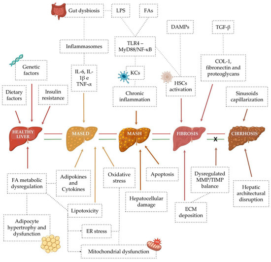
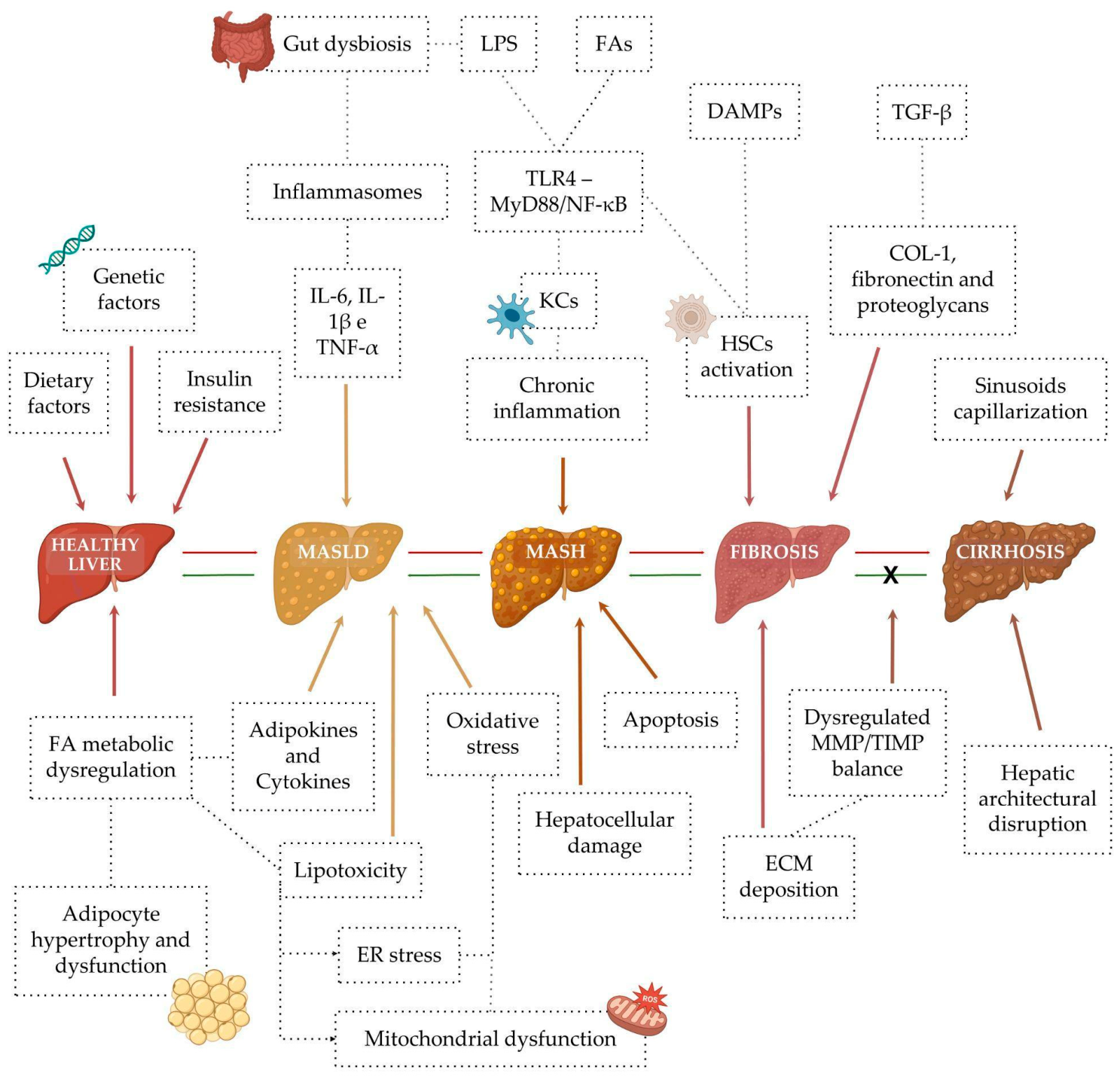
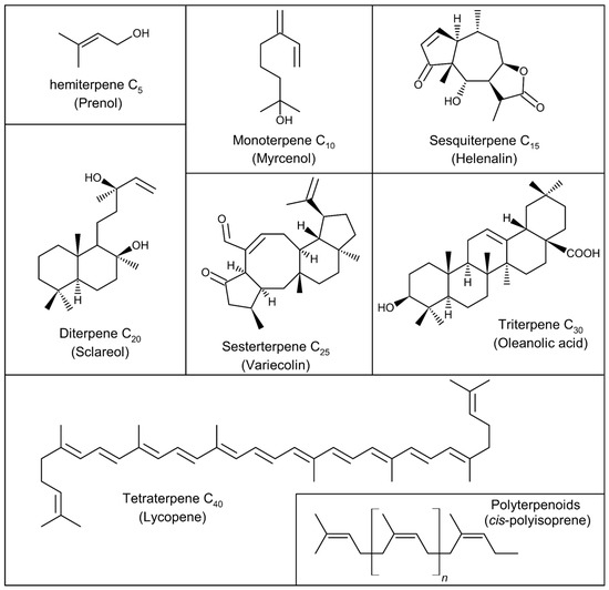
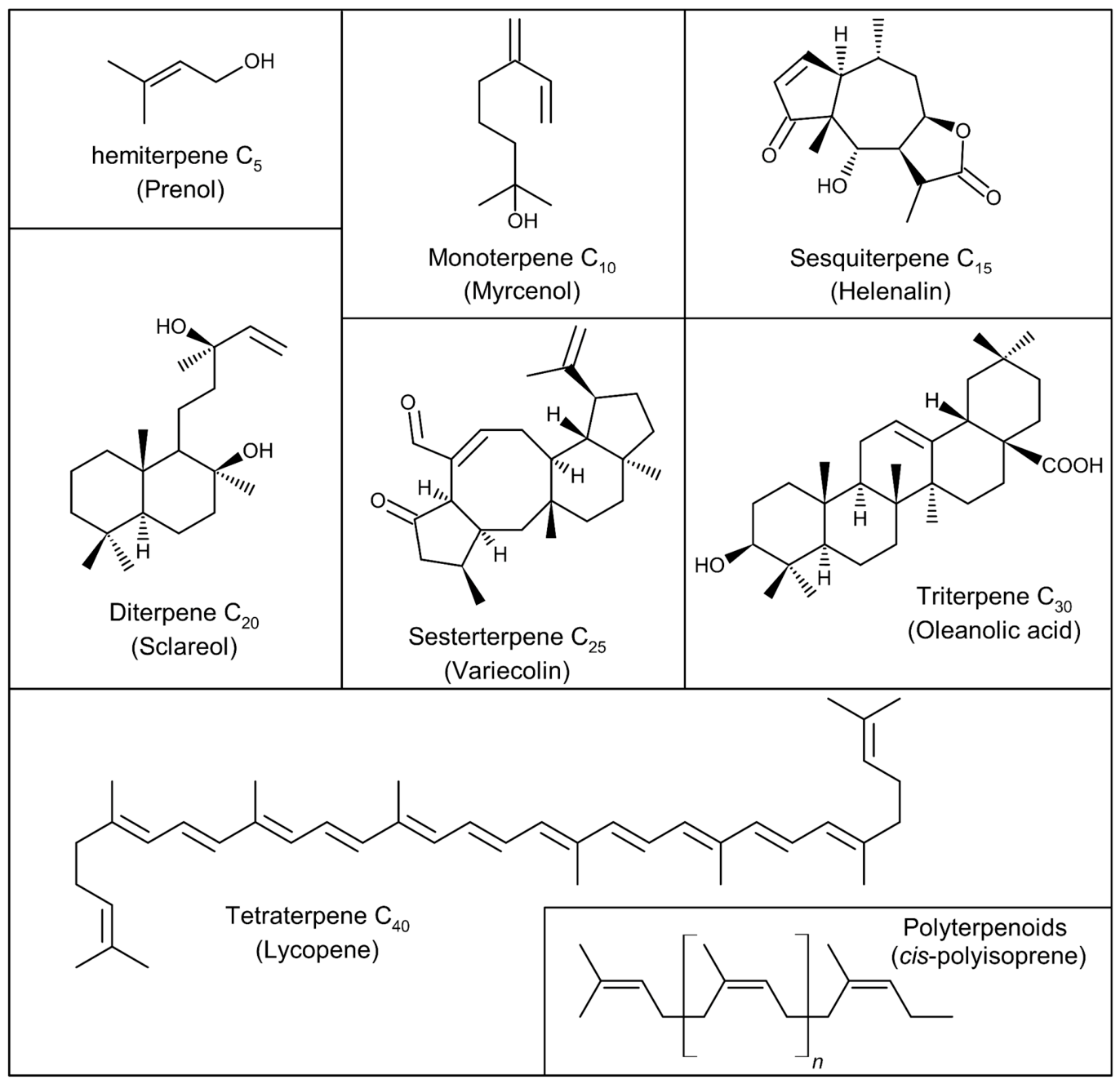
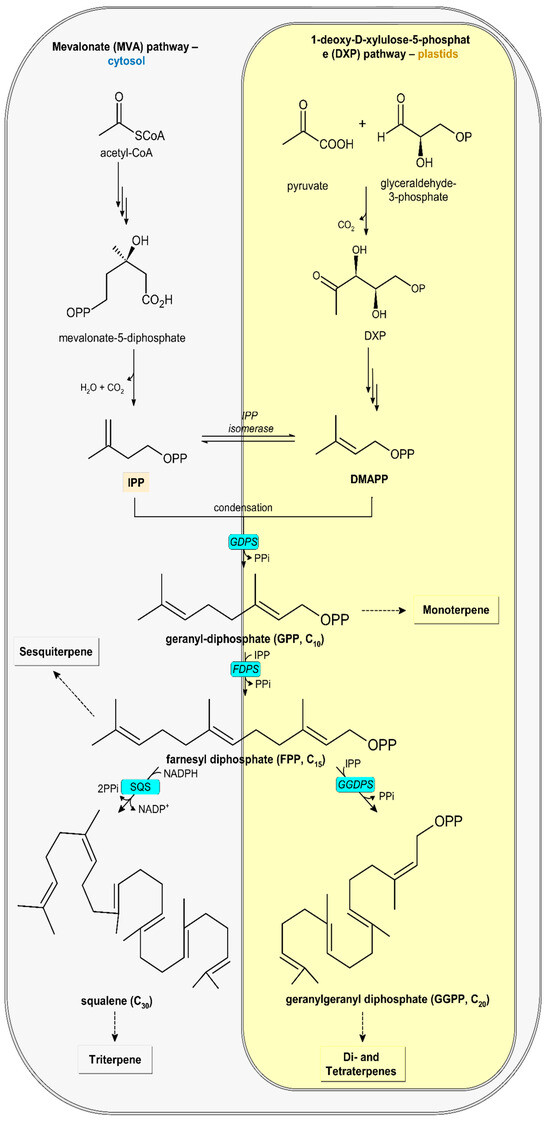
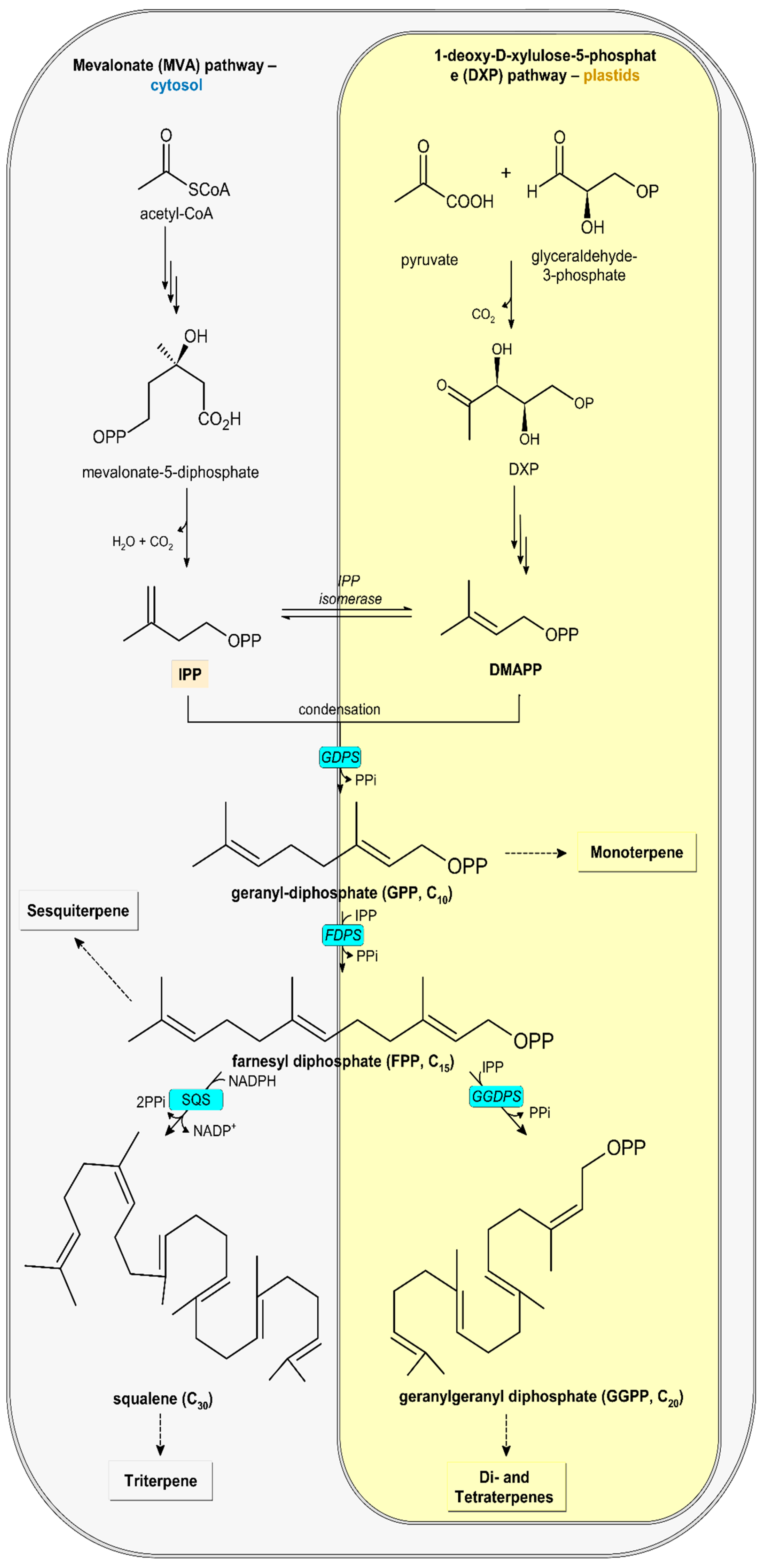
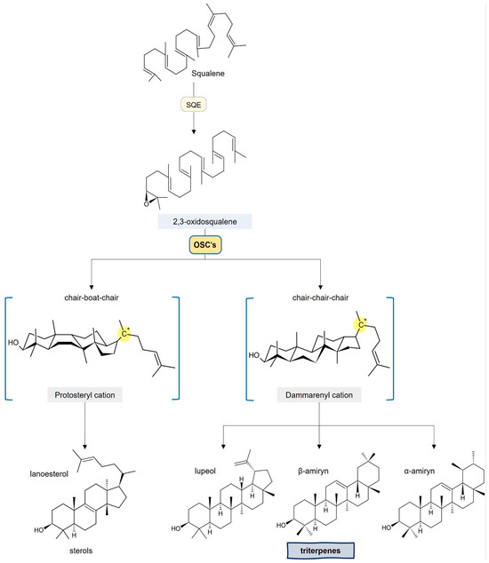
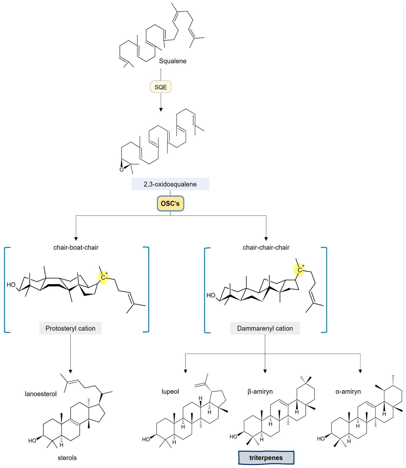
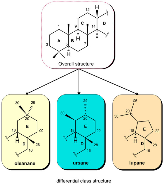
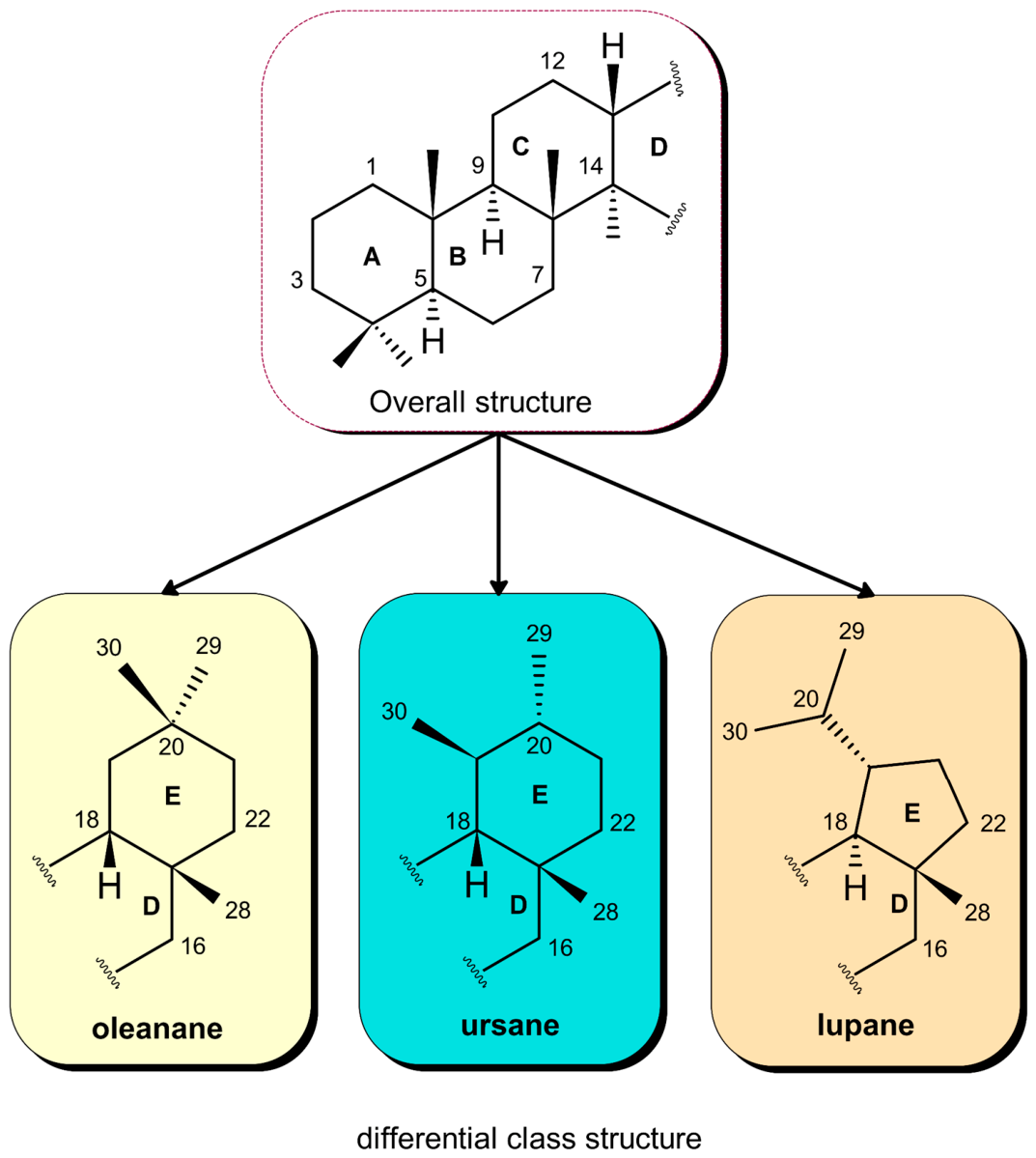
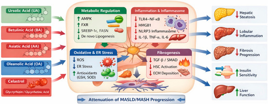
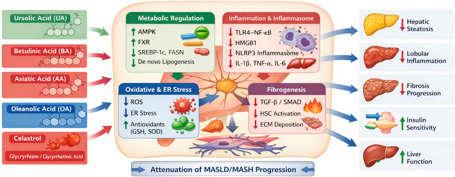

Disclaimer/Publisher’s Note: The statements, opinions and data contained in all publications are solely those of the individual author(s) and contributor(s) and not of MDPI and/or the editor(s). MDPI and/or the editor(s) disclaim responsibility for any injury to people or property resulting from any ideas, methods, instructions or products referred to in the content. |
© 2026 by the authors. Licensee MDPI, Basel, Switzerland. This article is an open access article distributed under the terms and conditions of the Creative Commons Attribution (CC BY) license.