Systematic Review

Surgical Decision-Making and Clinical Characteristics in Porcelain Gallbladder: A Systematic Review of Case Reports


by
Osvaldo Huchim-Mendez
1,2,
Ruben R. Lozano-Salazar
1,*,
Alejandro D. Munive-Ramirez
1,
Jorge E. Mendicuti-Carrillo
1,
Erick Rochel-Perez
1 and
Nina Isabel Mendez Dominguez
1,*
1
Hospital Regional de Alta Especialidad de la Peninsula de Yucatan, Servicios de Salud del Instituto Mexicano del Seguro Social para el Bienestar IMSS-BIENESTAR, Merida 97130, Mexico
2
School of Medicine, Universidad Anahuac, Merida 97310, Mexico
*
Authors to whom correspondence should be addressed.
Livers 2026, 6(3), 47; https://doi.org/10.3390/livers6030047
Submission received: 6 April 2026 / Revised: 8 May 2026 / Accepted: 28 May 2026 / Published: 2 June 2026
(This article belongs to the Special Issue Latest Progress in Hepatobiliary Surgery Research)

Abstract

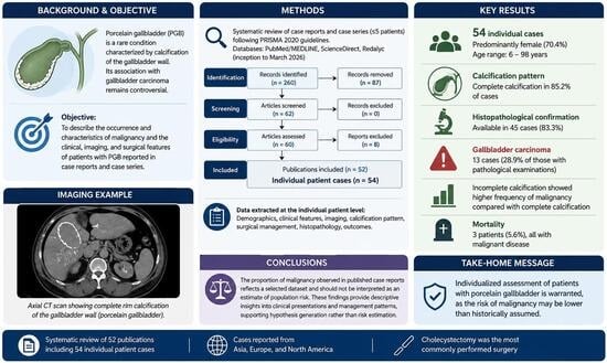
Porcelain gallbladder (PGB) has historically been considered a premalignant condition, although recent evidence questions the strength of its association with gallbladder carcinoma. Objective: To describe the occurrence and characteristics of malignancy in patients with porcelain gallbladder, as well as clinical, imaging, and surgical characteristics reported in clinal cases. Methods: A systematic review of case reports and case series (≤5 patients) was conducted following PRISMA 2020 guidelines. PubMed/MEDLINE, ScienceDirect, and Redalyc from database inception through March 2026. Individual patient data were extracted including demographic characteristics, imaging findings, calcification pattern, surgical management, histopathology, and outcomes. Descriptive statistics were performed. Results: Fifty-four individual cases of porcelain gallbladder were identified. Patients were predominantly female (70.4%), with ages ranging from 6 to 98 years. Complete calcification was reported in 85.2% of cases. Histopathological confirmation was available in 45 cases (83.3%). Thirteen patients had gallbladder carcinoma associated with porcelain gallbladder, corresponding to 28.9% of those with pathological examinations. Incomplete calcification showed higher frequency of malignancy compared with complete calcification. Mortality was reported in three patients (5.6%), all with malignant disease. Conclusions: The proportion of malignancy observed in published case reports reflects a selected dataset and should not be interpreted as an estimate of population risk. These findings provide descriptive insights into clinical presentations and management patterns, supporting hypothesis generation rather than risk estimation.

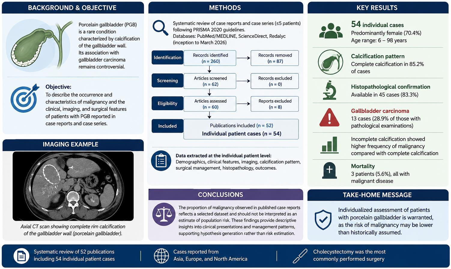
Graphical Abstract

1. Introduction

Porcelain gallbladder (PGB) is a rare condition characterized by calcification of the gallbladder wall, producing a brittle and bluish appearance that resembles porcelain. It is estimated to occur in fewer than 1% of gallbladder specimens and has traditionally been associated with chronic gallbladder inflammation and gallstone disease [1,2,3]. Although frequently discovered incidentally during imaging studies, symptomatic patients may present with clinical features similar to chronic cholecystitis, including right upper-quadrant pain, nausea, and intolerance to fatty foods [3].
Gallbladder adenocarcinoma is an uncommon malignancy associated with high mortality, and most cases are diagnosed incidentally during surgical exploration for cholelithiasis, where malignancy may be found in approximately 1–2% of procedures [4]. Historically, PGB was considered a premalignant condition strongly associated with gallbladder carcinoma (GBC). Early reports suggested a high coexistence rate, leading many clinicians to recommend prophylactic cholecystectomy when the condition was identified.
However, subsequent studies have challenged this assumption. For example, Towfigh et al. reported no cases of GBC among patients with PGB in a large cholecystectomy series, questioning the strength of this association [5]. Similarly, Schnelldorfer suggested that the estimated incidence of malignancy in patients with gallbladder calcification may be considerably lower than previously believed [6].
Gallbladder cancer remains the most common malignancy of the biliary tract and one of the most aggressive gastrointestinal tumors [7]. Because laparoscopic cholecystectomy is widely performed for benign gallbladder disease [8], incidental gallbladder carcinoma is often detected during postoperative histopathological examination [9]. Determining whether PGB represents a true premalignant condition or simply a coincidental finding is therefore clinically important for surgical decision-making.
Recent systematic reviews have further questioned the magnitude of the association between PGB and GBC. It has been previously reported that the prevalence of malignancy among patients with gallbladder calcification may be substantially lower than historically reported, emphasizing the need for individualized management strategies rather than routine prophylactic surgery [10].
Gallbladder cancer often presents with nonspecific symptoms such as abdominal pain, nausea, vomiting, anorexia, and weight loss, which frequently leads to delayed diagnosis and poor prognosis [11]. In addition, Khan et al. noted that gallbladder wall calcification may lose predictive significance for malignancy when other factors and imaging findings are considered [12].
Despite these advances, important uncertainties remain. Most existing studies are based on large surgical or pathological series that may not capture the detailed clinical presentation and disease progression observed in individual patients. Case reports and small case series provide granular clinical information that may help clarify the relationship between PGB and malignancy at the patient level.
The aim of the present study was therefore to systematically review published clinical case reports describing the occurrence of malignancy, as well as the clinical, imaging, and surgical characteristic reported in these cases.

2. Materials and Methods

We conducted a systematic review with individual patient-level analysis of published clinical case reports following the Preferred Reporting Items for Systematic Reviews and Meta-Analyses (PRISMA) guidelines [13]. The study protocol was prospectively performed under PROSPERO registration: CRD420250654236.
A structured literature search was performed in PubMed, ScienceDirect and Redalyc to identify published case reports describing patients diagnosed with porcelain gallbladder. The search strategy combined terms related to porcelain gallbladder and malignancy. The following keywords and MeSH terms were used: “porcelain gallbladder” OR “gallbladder calcification” OR “calcified gallbladder” combined with “cancer” OR “malignancy” OR “carcinoma”
Searches were restricted to case reports published between January 2000 through March 2026 with an abstract available in English. In ScienceDirect, equivalent keyword combinations were applied to titles, abstracts, and keywords. The complete search results from each database were exported into a spreadsheet for further screening.
Records retrieved from PubMed and ScienceDirect and Redalyc were merged into a single dataset. Duplicate records were identified and removed. Titles and abstracts were screened to identify potentially eligible publications. Full texts were subsequently assessed for eligibility.
Studies included reported individual clinical cases of porcelain gallbladder, provided sufficient patient-level information and were published as case reports or case series. Studies were excluded if unrelated to porcelain gallbladder that was misclassified or incorrectly indexed, or if they did not provide individual patient information or were unavailable in full text at the time of review.
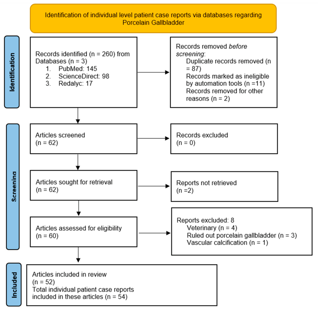
The initial search provided 260 results; after restricting results by inclusion criteria, a CSV dataset was created and shared among coauthors. O.H-M., A.D.M-R., J.E.M-C., and E.R-P. performed the initial screening and removal of duplicate and non-eligible articles. In total, 11 were identified as ineligible using abstract screening. Ineligibility was also assessed by R.R.L-Z and N.M-D, who also verified the complete dataset. A total of 62 articles were assessed after the elimination of 10 full-text articles; 52 articles were included in the present review. Out of the 52 articles, 54 single-patient cases were reviewed. A flowchart of each stage is presented in Figure 1; the Prima checklist is included as Supplementary Material Table S1.
Screening and data extraction were performed independently by two reviewers, with disagreements resolved by consensus with a third reviewer. No automation tools were used for eligibility assessment beyond duplicate detection.
Data were extracted at the individual patient level from each eligible publication. When articles reported more than one patient, each case was recorded separately. Variables were extracted manually while reading each case at the individual patient level and included demographic characteristics (age, sex, pediatric status), clinical features (presence of gallstones, cholecystitis), imaging modalities, surgical management, histopathological diagnosis, and clinical outcomes. Calcification pattern was classified as complete or incomplete according to the description provided in each report or inferred from imaging findings when necessary. Malignancy was defined based on histopathological confirmation of cancer.
Descriptive statistics were used to summarize patient characteristics and clinical findings. Exploratory analyses were performed using Fisher’s exact test. Given the small sample size, results are presented descriptively with corresponding contingency tables and should be interpreted cautiously. Confidence intervals and p-values are reported in Supplementary Table S2. Statistical analyses were conducted using Stata 19.5.

3. Results

The final dataset included 52 publications reporting 54 individual patient cases of porcelain gallbladder (PGB). When multiple patients were described within the same publication, cases were recorded separately using suffix identifiers (e.g., 11a, 11b). Regarding the type of articles, full case reports were the most common, as presented in Table 1.
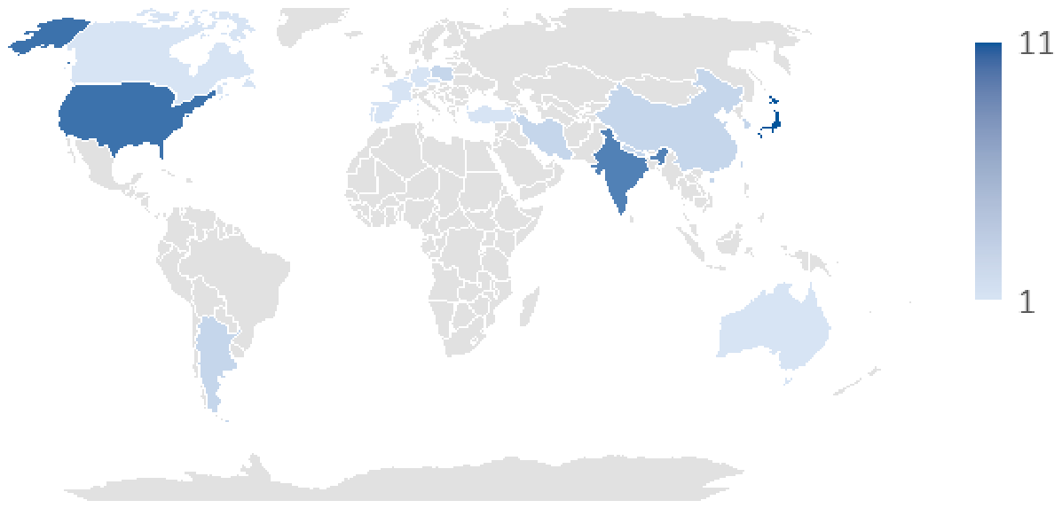
The geographic distribution of reports was heterogeneous, with cases originating from Asia, Europe, and North America (Figure 2). Japan reported 11 cases, followed by the United States with 9, and India with 8; the remaining countries reported 1 to 2.
The temporal distribution of reported cases shows a gradual accumulation of evidence over time, with sporadic case reports published between 2001 and 2025. Peaks in reporting occurred in 2002, 2011, 2012, and 2019, although most years contributed to only one or two cases. The cumulative curve illustrates the slow growth in the published evidence regarding porcelain gallbladder, reflecting the rarity of the condition and the predominance of isolated case reports in the literature.
Characteristics of the included cases and patients are summarized in Table 2. Among the 54 patients, 38 (70.4%) were female and 16 (29.6%) were male, corresponding to a female-to-male ratio of approximately 2.4:1. Age ranged from 6 to 98 years, with most patients being middle-aged or elderly. Two cases (3.7%) corresponded to pediatric patients.
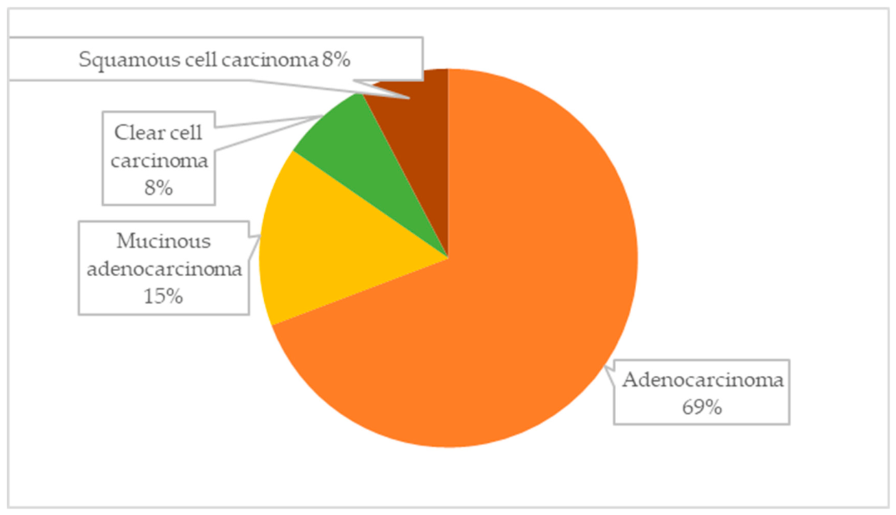
Gallstones were reported in 18 patients (33.3%), while findings compatible with chronic cholecystitis were described in 20 patients (37.0%). Histopathological confirmation of gallbladder tissue was available in 45 of 54 cases (83.3%). Overall, 13 patients (24.1%) were reported to have gallbladder carcinoma associated with porcelain gallbladder, while 41 cases (75.9%) were classified as non-malignant. When restricting this to cases with histopathological confirmation, the proportion of malignancy was 13/45 (28.9%). The most frequently reported histological subtype was adenocarcinoma, followed by mucinous adenocarcinoma, clear cell carcinoma, and squamous cell carcinoma, as shown in Figure 3.
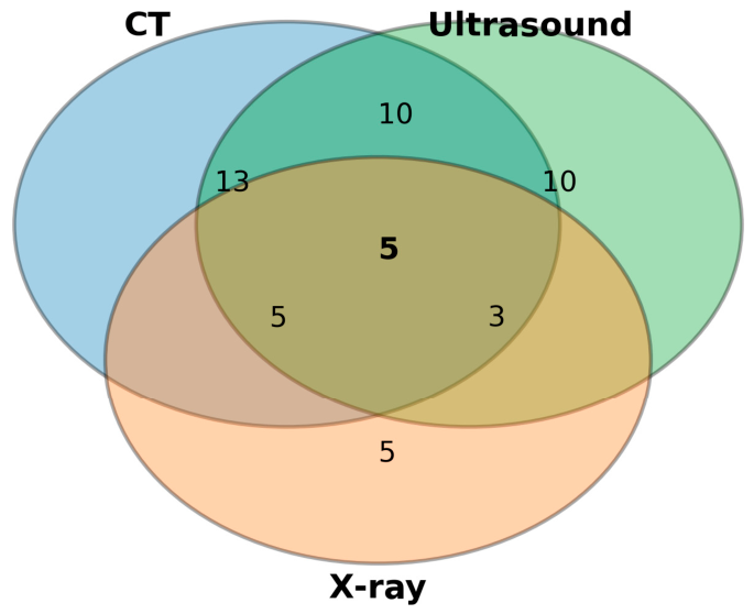
Porcelain gallbladder was identified using several imaging modalities. The most commonly reported modalities included Ultrasound and Computed Tomography (CT). Magnetic resonance imaging (MRI) and endoscopic retrograde cholangiopancreatography (ERCP) were less frequently used but occasionally provided additional evaluation of biliary anatomy or associated complications.
In several cases, multiple imaging modalities were used to confirm the diagnosis (Figure 4). Additionally, in one case, biliary stent placement was reported; in two more, endoscopic ultrasound (EUS) and upper endoscopy were performed to further characterize biliary anatomy or investigate suspected complications, such as ulceration or gallbladder gangrene.
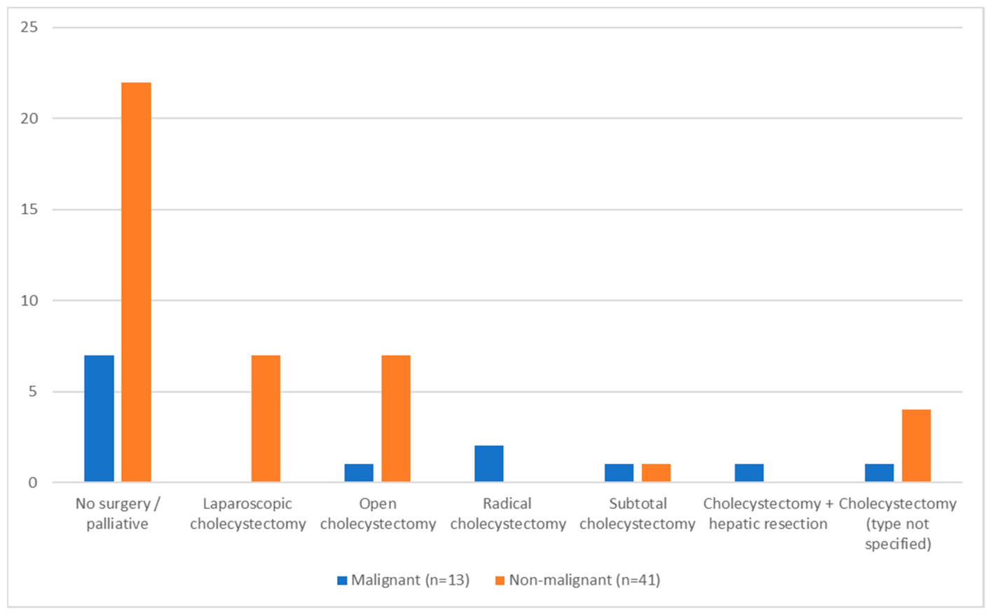
Surgical treatment was reported in most cases. Management strategies varied according to the presence or absence of malignancy, as shown in Figure 5. Extended surgical procedures were more frequently performed in patients with malignancy.
Exploratory analyses were conducted to examine the potential associations between selected clinical variables and the presence of malignancy. Variables explored included sex, age, calcification pattern, the presence of gallstones, and cholecystitis. Incomplete calcification showed a higher frequency of malignancy compared with complete calcification, although this exploratory association was not statistically significant.
Reported complications were relatively infrequent and heterogeneous across cases. The most commonly described events included peritonitis associated with colonic necrosis (two cases, 3.7%), choledocholithiasis (two cases, 3.7%), and hepatic abscess (two cases, 3.7%). Less frequent complications included cholecystoduodenal fistula (one case, 1.9%), gallbladder perforation (one case, 1.9%), and acute abdominal visceral perforation (one case, 1.9%). In addition, metastatic complications were reported in two patients (3.7%). Overall, complications were reported in 10 of the 54 patients (18.5%).
Follow-up information was inconsistently reported across the included case reports. In several patients, additional diagnostic procedures were performed during evaluation or postoperative monitoring. When reported, follow-up duration varied widely, ranging from short clinical monitoring to longer radiological surveillance, including 5-month, 18-month, and 52-month follow-up assessments.
In one case, fine-needle aspiration cytology (FNAC) from a liver metastasis confirmed adenocarcinoma, while another report documented negative lymph nodes and absence of metastatic disease with stable follow-up at five months. Mortality information was available for most cases; three deaths were reported in the dataset, all occurring in patients with advanced gallbladder carcinoma associated with porcelain gallbladder. No deaths were reported among patients with benign disease.

4. Discussion

The present study synthesized the approach to and characteristics of porcelain gallbladder, showing it to be the rare entity it is. The systematically reviewed published case reports permitted an individual-level descriptive analysis, but given the paucity of these reports and the diagnostic improvements along the studied period and across reporting countries, extending the timeframe further backward would likely increase heterogeneity related to diagnostic methods and reporting quality by including publications from before our studied period to evaluate the coexistence of malignancy and characterize clinical, imaging, and surgical features.
Among the 54 patients, identified across 52 publications, most cases occurred in women and in middle-aged or elderly individuals, consistent with the demographic patterns previously described for gallbladder disease [1,2,3], but we also reviewed two pediatric cases [18,32] meaning that, although infrequently, children may also present with PG.
Histopathological confirmation was available in most reports; however, the proportion of malignancy observed in this review should be interpreted with extreme caution. Because the dataset is derived exclusively from case reports and small case series, it is highly susceptible to publication bias. Malignant or unusual cases are more likely to be reported, while benign cases may be underrepresented.
Imaging-centered publications in our review [31,46,48,50,52,53,56,57] reported no definitive pathological findings regarding benign or malignant nature, and future studies could further explore the concordance of calcification type and oncological nature. These findings are consistent with previously published discussions, although direct comparisons should be avoided given the descriptive nature and inherent bias of case report data.
Historically, porcelain gallbladder has been considered a potential premalignant condition; however, the present review was not designed to determine malignant potential or establish causal associations between gallbladder calcification and carcinoma.
Because the analyzed dataset derives exclusively from published case reports and small case series, it is strongly influenced by publication and selection bias; therefore, the present study should be interpreted as exploratory and hypothesis-generating rather than confirmatory. Our findings should not be interpreted as evidence supporting or refuting prophylactic cholecystectomy. Instead, the reviewed cases illustrate the heterogeneity of clinical presentations and management strategies reported in the literature, reinforcing the importance of individualized clinical assessment based on imaging findings, symptoms, comorbidities, and surgical risk.
In the cases analyzed in this review, surgical treatment was reported in the majority of patients, with laparoscopic cholecystectomy being the most frequently described approach, while extended or open procedures were more often performed when malignancy was suspected or confirmed [22,27,28,34,42,59,61,62]. These reports illustrate that management decisions described in the literature frequently incorporated radiological findings, clinical presentation, and patient characteristics [12].
Although porcelain gallbladder is frequently described as an incidental radiological finding, several reports included in this review documented clinically significant complications [16,17,21,22,23,26,28,34,37,39]; the spectrum of complications observed in the analyzed cases included peritonitis, gallbladder perforation, hepatic abscess, biliary obstruction, and fistula formation, reflecting the potential consequences of chronic gallbladder inflammation and advanced disease. Metastatic disease was also reported in a small number of patients [28,37], emphasizing that gallbladder carcinoma associated with porcelain gallbladder may already be locally advanced or disseminated at the time of diagnosis.
Mortality in the present dataset was relatively low, with three deaths reported among the 54 cases, all occurring in patients with cancer [27,28,37]. This observation is consistent with the known aggressive behavior of gallbladder carcinoma, which often presents at advanced stages and carries a poor prognosis compared with other gastrointestinal malignancies [7,11,65]. Additionally, Morimoto, Matsuo and Mori [66] from their review conclude the importance of considering symptoms or complications, calcification pattern, and age of the patient for defining treatment. Taken together, our findings highlight that porcelain gallbladder may present with heterogeneous clinical scenarios, including benign disease and associated malignancy, with inflammatory or obstructive complications reported in isolated cases. However, the present descriptive review cannot determine the true frequency or predictive value of these outcomes.
Regarding follow-up, few authors reported mid-term outcomes and some reported postoperative monitorization [24,26,31,32,33,51,58,59] but timing and endpoints were inconsistent; future communications in single clinical case reports may consider adhering to CARE guidelines for transparent reporting. This could facilitate a wider comprehension of the case, the outcomes, prognosis and even the patients’ perspective [67].
Several limitations should be acknowledged. Case reports are subject to publication bias and may preferentially describe unusual or complicated presentations. Histopathological findings were not reported in all cases, particularly in imaging-focused publications.
Additionally, variations in diagnostic methods, reporting quality, and follow-up duration limit direct comparison between reports. Despite these limitations, individual patient analysis provides valuable insights into clinical patterns that are not always captured in large surgical series. Lastly, because case reports do not provide an appropriate denominator population, the present review cannot estimate incidence, prevalence, relative risk, or malignant potential associated with porcelain gallbladder.

5. Conclusions

This systematic review of published case reports provides a descriptive overview of clinical presentations, imaging findings, histopathological characteristics, and management approaches reported in patients with porcelain gallbladder.
The coexistence of malignancy observed in the reviewed literature reflects a highly selected dataset and should not be interpreted as an estimate of malignant risk or evidence of premalignant behavior. Because the present study is exploratory and hypothesis-generating in nature, its findings should not be used to support definitive surgical recommendations.
Clinical management should remain individualized and guided by contemporary evidence, imaging findings, patient characteristics, and multidisciplinary clinical judgment. Larger prospective and observational studies are required to better clarify the clinical significance of gallbladder wall calcification.

Supplementary Materials

The following supporting information can be downloaded at: https://www.mdpi.com/article/10.3390/livers6030047/s1, Table S1: Prisma Template; Table S2: Exploratory associations with malignancy in porcelain gallbladder.

Author Contributions

Conceptualization, O.H.-M. and R.R.L.-S.; methodology, N.I.M.D. and A.D.M.-R.; data curation, O.H.-M., A.D.M.-R., J.E.M.-C. and E.R.-P.; formal analysis, N.I.M.D.; investigation, O.H.-M., A.D.M.-R., J.E.M.-C. and E.R.-P.; visualization, O.H.-M.; writing—original draft preparation, O.H.-M., J.E.M.-C., E.R.-P. and A.D.M.-R.; writing—review and editing, N.I.M.D. and R.R.L.-S.; supervision, R.R.L.-S.; project administration, R.R.L.-S.; funding acquisition, N.I.M.D. All authors have read and agreed to the published version of the manuscript.

Funding

This research was funded by IMSS-BIENESTAR PP Q08.

Institutional Review Board Statement

Not applicable.

Informed Consent Statement

Not applicable.

Data Availability Statement

No new data was created for the present manuscript.

Conflicts of Interest

The authors declare no conflicts of interest.

Abbreviations

The following abbreviations are used in this manuscript:
AbbreviationDefinition
PGBPorcelain gallbladder
GBCGallbladder carcinoma
CTComputed tomography
MRIMagnetic resonance imaging
MRCPMagnetic resonance cholangiopancreatography
ERCPEndoscopic retrograde cholangiopancreatography
EUSEndoscopic ultrasound
FNACFine-needle aspiration cytology
USUltrasound
PRISMAPreferred Reporting Items for Systematic Reviews and Meta-Analyses
PROSPEROInternational Prospective Register of Systematic Reviews
OROdds ratio

References

  1. Margetich, L. Pathogenesis, epidemiology, and prognosis of gallbladder cancer. In Hepato-Pancreato-Biliary Malignancies; Doria, C., Rogart, J.N., Eds.; Springer: Cham, Switzerland, 2022; Chapter 21. [Google Scholar]
  2. Elwood, D.R. Cholecystitis. Surg. Clin. N. Am. 2008, 88, 1241–1252. [Google Scholar] [CrossRef]
  3. Lam, R.; Zakko, A.; Petrov, J.C.; Kumar, P.; Duffy, A.J.; Muniraj, T. Gallbladder Disorders: A Comprehensive Review. Dis. Mon. 2021, 67, 101130. [Google Scholar] [CrossRef]
  4. Fernandes, D.V.V.; Oliveros, A.J.; Durán Aguilarte, B.M.; Bravo, S.P.; Vassallo, M. Adenocarcinoma de vesícula biliar: Una entidad poco frecuente y mortal. A propósito de un caso. Rev. Venez. Cirugía 2023, 76, 142–145. [Google Scholar] [CrossRef]
  5. Towfigh, S.; McFadden, D.W.; Cortina, G.R.; Thompson, J.E., Jr.; Tompkins, R.K.; Chandler, C.; Hines, O.J. Porcelain gallbladder is not associated with gallbladder carcinoma. Am. Surg. 2001, 67, 7–10. [Google Scholar] [CrossRef]
  6. Schnelldorfer, T. Porcelain gallbladder: A benign process or concern for malignancy? J. Gastrointest. Surg. 2013, 17, 1161–1168. [Google Scholar] [CrossRef] [PubMed]
  7. Hickman, L.; Contreras, C. Gallbladder Cancer: Diagnosis, Surgical Management, and Adjuvant Therapies. Surg. Clin. N. Am. 2019, 99, 337–355. [Google Scholar] [CrossRef]
  8. Overby, D.W.; Apelgren, K.N.; Richardson, W.; Fanelli, R.; Society of American Gastrointestinal and Endoscopic Surgeons. SAGES guidelines for the clinical application of laparoscopic biliary tract surgery. Surg. Endosc. 2010, 24, 2368–2386. [Google Scholar] [CrossRef] [PubMed]
  9. Brunt, L.M.; Deziel, D.J.; Telem, D.A.; Strasberg, S.M.; Aggarwal, R.; Asbun, H.; Bonjer, J.; McDonald, M.; Alseidi, A.; Ujiki, M.; et al. Safe Cholecystectomy Multi-society Practice Guideline and State of the Art Consensus Conference on Prevention of Bile Duct Injury During Cholecystectomy. Ann. Surg. 2020, 272, 3–23. [Google Scholar] [CrossRef]
  10. Kellil, T.; Chaouch, M.A.; Aloui, E.; Tormane, M.A.; Taieb, S.K.; Noomen, F.; Zouari, K. Incidence and Preoperative Predictor Factors of Gallbladder Cancer Before Laparoscopic Cholecystectomy: A Systematic Review. J. Gastrointest. Cancer 2021, 52, 68–72. [Google Scholar] [CrossRef]
  11. Søreide, K.; Dopazo, C.; Berrevoet, F.; Carrion-Alvarez, L.; Diaz-Nieto, R.; Andersson, B.; Stättner, S. Joint ESSO-EAHPBA-UEMS Core Curriculum Working Group. Biliary tract cancer. Eur. J. Surg. Oncol. 2025, 51, 108489. [Google Scholar] [CrossRef]
  12. Khan, Z.S.; Livingston, E.H.; Huerta, S. Reassessing the need for prophylactic surgery in patients with porcelain gallbladder: Case series and systematic review of the literature. Arch. Surg. 2011, 146, 1143–1147. [Google Scholar] [CrossRef]
  13. Page, M.J.; McKenzie, J.E.; Bossuyt, P.M.; Boutron, I.; Hoffmann, T.C.; Mulrow, C.D.; Shamseer, L.; Tetzlaff, J.M.; Akl, E.A.; Brennan, S.E.; et al. The PRISMA 2020 statement: An updated guideline for reporting systematic reviews. Syst. Rev. 2021, 10, 89. [Google Scholar] [CrossRef] [PubMed]
  14. Ansari, S. Porcelain gallbladder. Indian J. Gastroenterol. Off. J. Indian Soc. Gastroenterol. 2014, 33, 399. [Google Scholar] [CrossRef]
  15. Albergaria, D.; Carvalho, N.; Borges, F.; Folgado, A.; Corte Real, J. Porcelain gallbladder. Rev. Esp. Enfermedades Dig. 2016, 108, 738. [Google Scholar]
  16. Young, K.J.; Johnson, J. Porcelain gallbladder. J. Manip. Physiol. Ther. 2002, 25, 534–543. [Google Scholar] [CrossRef] [PubMed]
  17. Pan, X.-F.; Aizezi, B.; Jia, Z. Porcelain gallbladder secondary to chronic cholelithiasis with filled gallstones: A case report and a minimal literature review. Medicine 2025, 104, E45951. [Google Scholar] [CrossRef]
  18. Kim, S.C. Six-Year-Old Girl with Porcelain Gallbladder. J. Korean Med. Sci. 2019, 34, e152. [Google Scholar] [CrossRef]
  19. Débat Zoguéreh, D.; Abdou, M.; N’Tarundenga, U. Des douleurs intermittentes de l’hypochondre droit. [Porcelain gallbladder]. Rev. Med. Interne 2004, 25, 936–937. [Google Scholar] [CrossRef]
  20. Kidogawa, H.; Ootubo, K.; Noguchi, J.; Yamayoshi, T.; Okamoto, K. A Case of Porcelain Gallbladder Showing High Levels of CA19-9. Cureus 2024, 16, e72583. [Google Scholar] [CrossRef]
  21. Vera, V.; Sharma, A.; Townson, A.T.; Azeem, Q.; Malam, Y.; Mathews, J. Emphysematous cholecystitis in a patient with porcelain gallbladder. J. Surg. Case Rep. 2023, 2023, rjac600. [Google Scholar] [CrossRef]
  22. Matsumoto, Y.; Fujimoto, K.; Mitsuoka, E.; Senda, E.; Shio, S.; Ichikawa, K.; Yamada, H. Cholecystoduodenal fistula caused by aggressive mucinous gallbladder carcinoma with a porcelain gallbladder. Clin. J. Gastroenterol. 2019, 12, 460–465. [Google Scholar] [CrossRef]
  23. Coskun, S.; Soylu, L.; Kilicaslan, I.; Atalay, F. Is it safe to be calcified? Porcelain gallbladder perforation and review of literature. J. Pak. Med. Assoc. 2014, 64, 1310–1312. [Google Scholar]
  24. Sarangi, S.; Rao, M.; Vishnoi, J.R.; Kumar Garg, P. Primary clear cell carcinoma of gallbladder arising in porcelain gallbladder. BMJ Case Rep. 2021, 14, e240319. [Google Scholar] [CrossRef]
  25. Pipal, D.K.; Vardhan, V.; Biswas, P.; Pipal, V.R.; Jatoliya, H. Laparoscopic Cholecystectomy for Entirely Calcified Porcelain Gallbladder: Challenges, Management, and Literature Review. J. West Afr. Coll. Surg. 2024, 14, 440–444. [Google Scholar] [CrossRef] [PubMed]
  26. Takeda, K.; Sekido, H.; Sugita, M.; Tanaka, K.; Endo, I.; Togo, S.; Shimada, H. Porcelain gallbladder complicated with pancreas divisum. J. Hepato-Biliary-Pancreat. Surg. 2006, 13, 580–583. [Google Scholar] [CrossRef] [PubMed]
  27. Joo, Y.E.; Kim, H.S.; Choi, S.K.; Rew, J.S.; Kim, H.J.; Kang, H.K.; Juhng, S.W.; Kim, S.J. Case of mucinous adenocarcinoma with porcelain gallbladder. J. Gastroenterol. Hepatol. 2003, 18, 995–998. [Google Scholar] [CrossRef]
  28. Gómez-López, J.R.; De Andrés-Asenjo, B.; Ortega-Loubon, C. A porcelain gallbladder and a rapid tumor dissemination. Ann. Med. Surg. 2012, 3, 119–122. [Google Scholar] [CrossRef]
  29. Thakrar, R.; Monib, S.; Pakdemirli, E.; Thomson, S. Calcified gallbladder cancer: Is it preventable? J. Surg. Case Rep. 2019, 2019, rjz069. [Google Scholar] [CrossRef]
  30. Tsai, C.J. Porcelain gallbladder and cholangiocarcinoma with anomalous pancreaticobiliary union. Dig. Dis. Sci. 2001, 46, 773–775. [Google Scholar] [CrossRef] [PubMed]
  31. Shelton, J.; Terry, S. CT Imaging Findings of Porcelain Gallbladder and Epiploic Appendagitis: Two Rare Abdominal Pathologies in an Asymptomatic Patient. Cureus 2021, 13, e15578. [Google Scholar] [CrossRef]
  32. Snajdauf, J.; Petrů, O.; Pýcha, K.; Rygl, M.; Kalousová, J.; Keil, R.; Kodet, R. Porcelain gallbladder with extrahepatic bile duct obstruction in a child. Pediatr. Surg. Int. 2006, 22, 293–296. [Google Scholar] [CrossRef]
  33. Aliniagerdroudbari, E.; Ershadinia, N.; Bagherpor, G.; Babaniamansour, S. A Rare Case of Simultaneous Intrahepatic and Porcelain Gallbladder: Case Report. Int. Med. Case Rep. J. 2020, 13, 271–274. [Google Scholar] [CrossRef]
  34. Delpierre, I.; Tack, D.; Moisse, R.; Boudaka, W.; Delcour, C. Cholecystoduodenal fistula in a porcelain gallbladder. Eur. Radiol. 2002, 12, 2284–2286. [Google Scholar] [CrossRef]
  35. Igami, T.; Usui, H.; Ebata, T.; Yokoyama, Y.; Sugawara, G.; Takahashi, Y.; Nagino, M. Single-incision laparoscopic cholecystectomy for porcelain gallbladder: A case report. Asian J. Endosc. Surg. 2013, 6, 52–54. [Google Scholar] [CrossRef] [PubMed]
  36. Karki, S.; Bohara, S.; Upadhaya Regmi, B.; Bhat, P.S.; Malla, S.; Mainali, G.; Khatri, S.; Rawal, S.B. Cholecystopathia chronica calcarea (Porcelain gall bladder): A case report from Nepal. Ann. Med. Surg. 2022, 84, 104947. [Google Scholar] [CrossRef]
  37. Tian, Y.; Luu, C.; Carpenter, D.H.; Montenegro, G. A Unique Presentation of Metastatic Gallbladder Carcinoma. Case Rep. Surg. 2021, 2021, 6455540. [Google Scholar] [CrossRef]
  38. Gupta, S.; Misra, S. Rare presentation of malignant porcelain gallbladder with intrahepatic ductal calcification and surgical obstructive jaundice. Indian J. Surg. Oncol. 2012, 3, 44–46. [Google Scholar] [CrossRef][Green Version]
  39. Fukui, T.; Nakayama, S.; Shimatani, M.; Uchida, K.; Matsushita, M.; Nishio, A.; Seki, T.; Okazaki, K. Endoscopic biliary plastic stenting and successful intentional stent retrieval in a benign biliary stricture with mural spherical calcification and porcelain gallbladder. Intern. Med. 2009, 48, 809–813. [Google Scholar] [CrossRef] [PubMed][Green Version]
  40. Gupta, R.K.; Patton, K.T. Hyalinizing cholecystitis with features of immunoglobulin G4-related disease-coincidence or an unrecognized association? A case report. Hum. Pathol. 2015, 46, 625–628. [Google Scholar] [CrossRef]
  41. Bachmeier, C.A.E.; Morton, A. Man with epigastric pain and persistently elevated serum lipase. BMJ Case Rep. CP 2019, 12, E229208. [Google Scholar] [CrossRef] [PubMed]
  42. Gupta, S.; Jauhari, R.K. Rare presentation of porcelain gall bladder: Carcinoma gall bladder with a large intra-abdominal cystic swelling. Case Rep. 2012, 2012, Bcr2012006894. [Google Scholar] [CrossRef] [PubMed]
  43. Palermo, M.; Núñez, M.; Duza, G.E.; Giménez Dixon, M.; Bruno, M.O.; Tarsitano, F.J. Vesícula de porcelana. Caso clínico y revisión de la literatura. Cirugía Española 2011, 89, 213–217. [Google Scholar] [CrossRef]
  44. Araujo-López, A.; Peña-Muñoz, C.A.; Canseco-González, V. Abordaje quirúrgico efectivo en cáncer de vesícula biliar; variante carcinoma epidermoide. Reporte de caso. El Resid. 2018, 13, 68–71. [Google Scholar]
  45. Iqbal, S.; Ahmad, S.; Saeed, U.; Al-Dabbagh, M. Porcelain Gallbladder: Often an Overlooked Entity. Surg. J. 2017, 3, e145–e147. [Google Scholar] [CrossRef][Green Version]
  46. Opatrny, L. Porcelain gallbladder. CMAJ Can. Med. Assoc. J. 2002, 166, 933. [Google Scholar]
  47. Cox, M.; Patel, M.; Joneja, U.; Shaw, C.; O’Kane, P. Focal disruption in the wall of the porcelain gallbladder: A sign of gallbladder carcinoma. Intern. Emerg. Med. 2017, 12, 891–893. [Google Scholar] [CrossRef][Green Version]
  48. Rickes, S.; Ocran, K. Images in clinical medicine. Porcelain gallbladder. N. Engl. J. Med. 2002, 346, E4. [Google Scholar] [CrossRef]
  49. Smith, P.W.; Farrar, R.A.; Evangelista, R.S. Intrahepatic porcelain gallbladder: Coexistence of pathologies. Am. Surg. 2012, 78, E414–E415. [Google Scholar] [CrossRef]
  50. Liu, K.; Katelaris, P. Education and imaging. Gastrointestinal: Porcelain gallbladder. J. Gastroenterol. Hepatol. 2015, 30, 648. [Google Scholar] [CrossRef] [PubMed]
  51. Buxbaum, J.L.; Eloubeidi, M.A. A unique perspective: Endoscopic sonogram of a porcelain gallbladder. Gastrointest. Endosc. 2011, 73, 361–362. [Google Scholar] [CrossRef]
  52. Liang, H.P.; Cheung, W.K.; Su, F.H.; Chu, F.Y. Porcelain gallbladder. J. Am. Geriatr. Soc. 2008, 56, 960–961. [Google Scholar] [CrossRef]
  53. Gupta, V.; Rao, C.; Rana, S.S.; Bhasin, D.K. Porcelain gallbladder and thalassaemia intermedia: Association or coincidence? Dig. Liver Dis. Off. J. Ital. Soc. Gastroenterol. Ital. Assoc. Study Liver 2012, 44, E22. [Google Scholar] [CrossRef]
  54. Lee, T.C.; Liu, K.L.; Lai, I.R.; Wang, H.P. Diagnosing porcelain gallbladder. Am. J. Med. 2005, 118, 1171–1172. [Google Scholar] [CrossRef] [PubMed]
  55. Sen, K.K.; Upadhyaya, A.; Pimpalwar, Y.; D’Souza, J. Cancer in porcelain gallbladder-rare imaging trait. Indian J. Cancer 2011, 48, 132–134. [Google Scholar] [CrossRef] [PubMed]
  56. Matsuura, H.; Matsushita, H.; Imamura, R. Porcelain Gallbladder. Clin. Gastroenterol. Hepatol. 2021, 19, A30. [Google Scholar] [CrossRef]
  57. Vimalachandran, P.; Paknikar, J. Recurrent right upper quadrant abdominal pain. J. Fam. Pract. 2016, 65, 723. [Google Scholar]
  58. Nakashima, H.; Tanaka, Y. Hepatobiliary and pancreatic: Porcelain gallbladder. J. Gastroenterol. Hepatol. 2002, 17, 1228. [Google Scholar] [CrossRef]
  59. Sadiq, K.O.; Belavadi, R. S3489 Porcelain Gallbladder Stable After 52 Months of Conservative Management. Off. J. Am. Coll. Gastroenterol. ACG 2025, 120, S750. [Google Scholar] [CrossRef]
  60. Pielaciński, K.; Bosak, A.; Pielaciński, J.; Paluszkiewicz, P. Porcelanowy pęcherzyk żółciowy z podejrzeniem raka pęcherzyka żółciowego. Opis przypadku i przegląd literatury [Porcelain gallbladder with suspected gallbladder cancer. Case report and review of the literature]. Pol. Merkur. Lek. Organ Pol. Tow. Lek. 2022, 50, 384–387. [Google Scholar]
  61. Chowdhury, M.M.; Khan, A.S.; Karim, R.; Zeba, I.J.; Mohammed, S.; Haque, M.M.; Farmidi, A.A. Porcelain Gallbladder: A Case Report. Mymensingh Med. J. MMJ 2019, 28, 694–698. [Google Scholar]
  62. Târcoveanu, E.; Vasilescu, A.; Georgescu, S.; Filip, V.; Vlad, N. Vezicula de porţelan--abord laparoscopic [The porcelain gallbladder-laparoscopic approach]. Chirurgia 2012, 107, 246–251. [Google Scholar] [PubMed]
  63. Suliman, E.; Palade, R.; Voiculescu, D.; Simion, G. Vezicula de porţelan--particularităţi diagnostice şi terapeutice [Porcelain gallbladder--diagnostic and therapeutic features]. Chirurgia 2010, 105, 431–434. [Google Scholar]
  64. de Jong, V.M.; Schnater, J.M. Diagnose in beeld (193). Een man met een palpabele zwelling in de bovenbuik. Porseleinen galblaas. Ned. Tijdschr. Voor Geneeskd. 2004, 148, 1198. [Google Scholar]
  65. Wroński, K. Porcelain gallbladder case report. New Med. 2014. Available online: https://www.czytelniamedyczna.pl/4892,porcelain-gallbladder-case-report.html (accessed on 5 April 2026).
  66. Morimoto, M.; Matsuo, T.; Mori, N. Management of Porcelain Gallbladder, Its Risk Factors, and Complications: A Review. Diagnostics 2021, 11, 1073. [Google Scholar] [CrossRef] [PubMed]
  67. Riley, D.S.; Barber, M.S.; Kienle, G.S.; Aronson, J.K.; von Schoen-Angerer, T.; Tugwell, P.; Kiene, H.; Helfand, M.; Altman, D.G.; Sox, H.; et al. CARE guidelines for case reports: Explanation and elaboration document. J. Clin. Epidemiol. 2017, 89, 218–235. [Google Scholar] [CrossRef]
Figure 1. Flowchart indicates the registries retrieved from three datasets to the final dataset of reviewed articles, and the corresponding individual patient case reports.
Livers 06 00047 g001
Figure 2. World map indicating the number of cases included in the present review according to their country of origin. Deeper shades of blue indicate more cases. Gray shapes indicate absence of cases.
Livers 06 00047 g002
Figure 3. Histopathological type in patients with coexistence of porcelain gallbladder and cancer. Histopathological type was reported in 13 cases; percentage by type is presented.
Livers 06 00047 g003
Figure 4. Imaging diagnosis studies performed in patients with porcelain gallbladder. Venn diagram illustrates the total count of cases included in the present review. CT: Computed tomography.
Livers 06 00047 g004
Figure 5. Surgical approaches reported in the 54 patient-level cases reviewed.
Livers 06 00047 g005
Table 1. Article types including case reports of patients with porcelain gallbladder.
Publication TypePublished by (First Author, Year) and Citation Number
Single case reportAnsari, 2014 [14]; Albergaria, 2016 [15]; Young, 2002 [16]; Pan, 2025 [17]; Kim, 2019 [18]; Débat Zoguéreh, 2004 [19]; Kidogawa, 2024 [20]; Vera, 2023 [21]; Matsumoto, 2019 [22]; Coskun, 2014 [23]; Sarangi, 2021 [24]; Pipal, 2024 [25]; Takeda, 2006 [26]; Joo, 2003 [27]; Gómez-López, 2012 [28]; Thakrar, 2019 [29]; Tsai, 2001 [30]; Shelton, 2021 [31]; Snajdauf, 2006 [32]; Aliniagerdroudbari, 2020 [33]; Delpierre, 2002 [34]; Igami, 2013 [35]; Karki, 2022 [36]; Tian, 2021 [37]; Gupta, 2012 [38]; Fukui, 2009 [39]; Gupta, 2015 [40]; Bachmeier, 2019 [41]; Gupta, 2012 [42]; Palermo, 2011 [43];
Case seriesAraujo-López, 2018 [44]; Iqbal, 2017 [45]
Brief communicationOpatrny, 2002 [46]; Cox, 2017 [47]; Rickes, 2002 [48]; Smith, 2012 [49]; Liu, 2015 [50]; Buxbaum, 2011 [51];
LetterLiang, 2008 [52]; Gupta, 2012 [53]; Lee, 2005 [54]; Sen, 2011 [55]
Image reportMatsuura, 2021 [56]; Vimalachandran, 2016 [57]
Diagnostic
challenge
Nakashima [58]
Conference abstractSadiq, 2025 [59]
UnspecifiedPielaciński, 2022 [60]; Chowdhury, 2019 [61]; Târcoveanu, 2012 [62]; Suliman, 2010 [63]; de Jong, 2004 [64]; Wronski, 2014 [65]
Published article types of case reports included in the present review.
Table 2. Characteristics and porcelain gallbladder presentation in patients from the included case reports.
CharacteristicValue
Total cases54
Sex
Female38 (70.4%)
Male16 (29.6%)
Age
Range6–98 years
Pediatric cases (<18 years)2 (3.7%)
Adult cases52 (96.3%)
Calcification pattern
Complete calcification46 (85.2%)
Incomplete calcification8 (14.8%)
Gallstones (lithiasis)
Present18 (33.3%)
Absent/not reported36 (66.7%)
Cholecystitis
Reported20 (37.0%)
Not reported34 (63.0%)
Histopathological confirmation available
Reported45 (83.3%)
No pathology reported9 (16.7%)
Malignancy among cases with pathology
Malignant13 (28.9%)
Non-malignant32 (71.1%)
Disclaimer/Publisher’s Note: The statements, opinions and data contained in all publications are solely those of the individual author(s) and contributor(s) and not of MDPI and/or the editor(s). MDPI and/or the editor(s) disclaim responsibility for any injury to people or property resulting from any ideas, methods, instructions or products referred to in the content.