


Abstract
The pearlitic microstructure, comprising alternating lamellae of ferrite and cementite, provides a favorable combination of strength, toughness, and wear resistance. Consequently, pearlitic steels have been widely utilized in pipeline systems due to their advantageous mechanical properties and cost-effectiveness. These characteristics also render pearlitic steel pipelines promising candidates for hydrogen transport infrastructure, particularly in the context of repurposing existing natural gas networks. However, interactions between hydrogen and the pearlitic microstructure raise significant concerns regarding hydrogen embrittlement, a phenomenon that can substantially degrade mechanical performance and compromise long-term structural integrity. Experimental observations indicate that pearlitic microstructures are particularly susceptible to hydrogen embrittlement, largely due to the high density of ferrite–cementite interfaces, which act as effective hydrogen trapping sites. These detrimental effects motivate the present study, which aims to develop a deeper understanding of nanoscale mechanisms of hydrogen-assisted crack initiation and propagation in pearlitic microstructures. In this work, molecular dynamics simulations are employed to investigate the initiation and propagation of hydrogen-affected cracks in pearlitic microstructures, considering lamellar orientations both parallel and perpendicular to the applied tensile loading direction. The analysis focuses on the synergistic interaction between hydrogen-enhanced decohesion (HEDE), which promotes interfacial separation due to hydrogen segregation, and hydrogen-enhanced localized plasticity (HELP).
1. Introduction
The pearlitic microstructure, composed of alternating lamellae of ferrite and cementite, provides a favorable combination of strength, toughness, and wear resistance [1,2]. The morphology, interlamellar spacing, and orientation of the ferrite–cementite structure strongly influence the mechanical response of the material [3]. Owing to this heterogeneous lamellar architecture, pearlitic steels are particularly susceptible to crack initiation and propagation under mechanical loading, especially in hydrogen-containing environments. The pronounced mechanical mismatch between the ductile ferrite and the brittle cementite phases leads to the development of high local stresses within the cementite lamellae and at ferrite–cementite interfaces, where interfacial decohesion and microcrack initiation may occur [4,5].
Colonies in which the cementite lamellae are oriented perpendicular to the tensile loading direction are more prone to crack initiation. In this orientation, normal stresses acting across the lamellae facilitate interfacial decohesion and promote crack propagation parallel to the ferrite–cementite interfaces [6]. By contrast, cracks advancing transverse to the lamellae must repeatedly traverse ferrite–cementite interfaces, fracture the cementite plates, and induce substantial plastic deformation within the ferritic matrix. These mechanisms increase the resistance to crack propagation and thus impede crack growth [7,8].
Pearlitic steels have been widely employed in pipeline systems owing to their advantageous combination of strength, toughness, weldability, and cost-effectiveness [9,10]. These attributes also make pearlitic steel pipes promising candidates for hydrogen transport infrastructure, particularly in efforts to repurpose existing natural gas pipelines. However, interactions between hydrogen and the pearlitic microstructure give rise to significant concerns regarding hydrogen embrittlement, which can markedly degrade mechanical properties and threaten long-term structural integrity [11,12,13,14].
Experimental observations indicate that pearlitic microstructures are susceptible to hydrogen embrittlement (HE), largely due to the high density of ferrite–cementite interfaces, which act as effective hydrogen trapping sites [15,16]. A detailed understanding of the nanoscale mechanisms governing hydrogen-assisted crack initiation and propagation within the pearlitic microstructure is essential for accurately predicting embrittlement susceptibility and for developing effective mitigation strategies for pipeline steels operating in hydrogen-rich environments. Despite extensive research, significant debate persists regarding the fundamental mechanisms of hydrogen embrittlement, with the two most widely discussed models being hydrogen-enhanced decohesion (HEDE) and hydrogen-enhanced localized plasticity (HELP). The HELP mechanism [17,18,19] proposes that hydrogen embrittlement arises from enhanced dislocation mobility and reduced elastic interactions between dislocations and obstacles [20,21]. These effects promote highly localized plastic deformation, thereby facilitating crack initiation at lower applied stresses compared to hydrogen-free conditions. In contrast, the HEDE mechanism attributes hydrogen embrittlement to a reduction in atomic bond strength caused by hydrogen segregation at grain boundaries (GBs) and phase interfaces [22,23]. Such segregation diminishes the cohesive strength of these interfaces, rendering them more susceptible to fracture. When the applied stress exceeds the reduced cohesive strength, cracks may initiate and propagate along these weakened boundaries. It is widely recognized that multiple hydrogen embrittlement (HE) mechanisms may operate concurrently during hydrogen-induced crack formation in pearlitic microstructures [24]. Cracks propagating parallel to ferrite–cementite interfaces are promoted by interfacial separation resulting from hydrogen segregation (HEDE), while hydrogen simultaneously increases dislocation mobility in ferrite via the hydrogen-enhanced localized plasticity (HELP) mechanism. Unlike HELP, direct experimental evidence supporting HEDE remains limited. Experimental observation is particularly challenging due to the high diffusivity of hydrogen and the rapid kinetics of hydrogen-assisted failure. Consequently, atomistic simulations and modeling approaches are widely employed to advance the mechanistic understanding of HEDE [25,26,27]. The literature is much richer in atomistic simulations of crack formation and hydrogen embrittlement for pure BCC Fe than for full ferrite–cementite pearlite structures. Direct molecular dynamics (MD) simulations of mode-I crack propagation, crack tip evolution, and cleavage behavior in BCC Fe can be found in [27]. Recently, MD simulations have been conducted to investigate the effects of cementite thickness and plasticity-induced damage caused by dislocation pile-ups on crack initiation in the cementite phase. In these studies, the crystallographic orientation relationship between ferrite and cementite was defined according to the Bagaryatsky orientation relationship [28].
The detrimental effects of hydrogen motivate the present study, which aims to achieve a deeper understanding of the role of hydrogen in crack initiation and propagation within pearlitic microstructures. In this study, molecular dynamics (MD) simulations are employed to investigate the initiation and evolution of hydrogen-affected cracks in pearlitic microstructures, considering lamellar orientations both parallel and perpendicular to the applied tensile loading direction. The crystallographic orientation relationship between ferrite and cementite is defined according to the Isaichev orientation relationship. A recently developed Fe–C–H bond-order potential (BOP) [29] is used to model interatomic interactions. For crack formation and ferrite–cementite interface mechanics in Fe–C–H systems, modern BOPs generally describe the underlying physics more accurately than the classical modified embedded-atom method (MEAM) used in [28], particularly when bond breaking and hydrogen effects are dominant. Molecular dynamics simulations provide fundamental insights into crack initiation and propagation in pearlitic microstructures by explicitly modeling ferrite–cementite interfaces and enabling detailed analysis of atomic-scale deformation mechanisms, interfacial cohesion, stress localization, and phase interactions under applied mechanical loading. The findings of this study contribute to a more comprehensive understanding of hydrogen-assisted damage in pearlitic steels.
2. Molecular Dynamics Model Setup for Ferrite/Cementite Bilayer
Molecular dynamics simulations are performed on flat ferrite–cementite bilayers with the Isaichev orientation relationship (IS OR), which exhibits the lowest interface formation energy [30]. In this study, we align the -axis parallel to the interface normal and impose periodic boundary conditions (PBC) in the and directions. To satisfy the IS OR, the crystallographic orientation is defined as , , and for the ferrite and cementite blocks. The subscripts f and c denote the ferrite and cementite phases, respectively. To construct the simulation cell with periodic boundaries along the interface, the sizes of the initial ferrite and cementite layers are determined by minimizing the misfit strain in the and directions while maintaining an affordable computational cost.
To investigate the effect of hydrogen on crack formation and propagation in pearlitic microstructures, a large number of atomistic calculations must be performed in supercells comprising tens of thousands of atoms. Density functional theory is generally too computationally expensive for relaxing supercells of this size. To enable large-scale parallel calculations, we employ the ternary Fe–C–H bond-order potential (BOP) developed by Zhou et al. [29], implemented in the LAMMPS MD code [31]. This potential allows stable molecular dynamics simulations of –Fe, –Fe, and cementite (), and provides a reliable description of hydrogen-related effects within these phases. Biaxial strain is iteratively applied to the ferrite and cementite blocks separately until geometric compatibility is achieved. The strained blocks are subsequently assembled into a ferrite–cementite bilayer. Structural relaxation is then performed via energy minimization at 0 K using the conjugate gradient method, followed by MD simulated tempering to 300 K. The final dimensions of the ferrite–cementite bilayer are = 335.52 Å, = 45.59 Å, = 39.92 Å, and = 32.26 Å. The misfit strains in the and directions are = and = , respectively. The ferrite–cementite interface contains interfacial dislocations characterized by the Burgers vector, line orientation, and spacing given by = [−2.51, 0.00, 0.00] Å, = [0.00, 0.00, 1.00], and = 83.92 Å, respectively ([32]). The details of the procedure are provided elsewhere [30,33]. Due to the limited timescales of MD simulations, hydrogen cannot diffuse to segregation regions where its local concentration significantly exceeds the average hydrogen concentration in the material. For this reason, higher hydrogen concentrations in the vicinity of defects such as dislocations and grain boundaries are considered in MD simulations. During MD simulations of crack propagation in BCC Fe in the presence of hydrogen reported in [27], the number of bulk hydrogen atoms introduced into the simulation cell per unit length along the crack line ranged from 35 to 410 . The hydrogen concentrations used in the present MD simulations correspond to 2.9 and 14.9 hydrogen atoms per unit length along the crack line for tensile loading perpendicular to the interface (11.7 and 58.4 for tensile loading parallel to the interface).
After constructing the ferrite–cementite bilayer, hydrogen atoms are randomly introduced into the supercell at concentrations of 0.1% and 0.5%. Due to the heterogeneous nature of pearlite, hydrogen interacts differently with ferrite and cementite [34]. Hydrogen atoms readily diffuse through the ferritic phase, which exhibits high hydrogen mobility, and tend to accumulate at microstructural traps such as ferrite–cementite interfaces and cementite lamellae. The presence of hydrogen at these sites reduces cohesive strength, promoting complex damage mechanisms that degrade mechanical performance and compromise structural integrity.
Determining the equilibrium distribution of hydrogen over time due to diffusion in a pearlite microstructure is too slow to be accomplished using molecular dynamics alone. Therefore, the advanced sampling method, time-stamped force-bias Monte Carlo (TFBMC) [35], was employed to efficiently capture hydrogen distributions over timescales that are not accessible with MD simulations. TFBMC does not simulate real-time motion directly, but it captures temperature-dependent behavior, which is essential for describing hydrogen diffusion. To determine the equilibrium positions of hydrogen atoms in the pearlitic bilayer, we performed TFBMC simulations for 2,000,000 Monte Carlo steps at a temperature of 300 K. TFBMC simulations were subsequently followed by molecular dynamics simulations of hydrogen diffusion conducted over a duration of 5 nanoseconds, after which tensile stresses were applied in directions perpendicular and parallel to the ferrite–cementite interface. Tensile loading is incrementally applied at the boundaries of the simulation cell at a rate of 1 m/s, with full relaxation of the simulation cell performed after each step. One of the major limitations of MD simulations under tensile loading is the extremely high strain rate required by the short simulation timescales. Such high strain rates can significantly influence the observed deformation and fracture mechanisms. Under rapid loading, atoms have insufficient time to undergo thermally activated relaxation processes such as hydrogen redistribution, dislocation nucleation, or dislocation motion. As a result, the material response obtained from MD simulations may differ from the experimentally observed behavior under quasistatic loading conditions. The MD simulations are performed in an NVT ensemble using a Nose–Hoover thermostat [36] and the velocity-Verlet algorithm [37], with an integration time step of fs. During the simulations, the software OVITO [38] is used to analyze particle coordinates and to identify the atomistic interface structure, as well as newly formed lattice structures and dislocations.
3. Results and Discusion
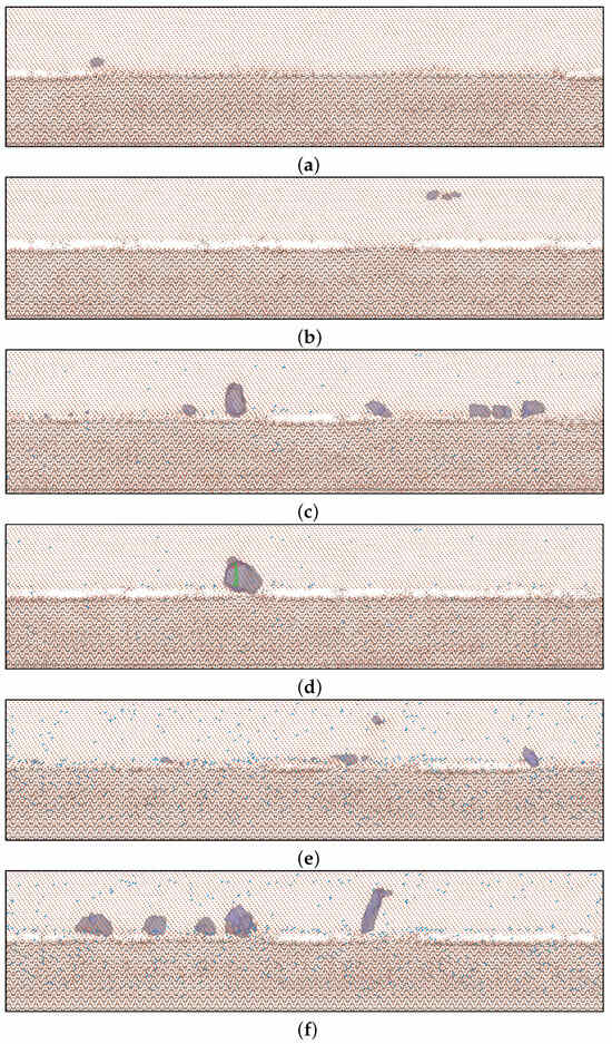
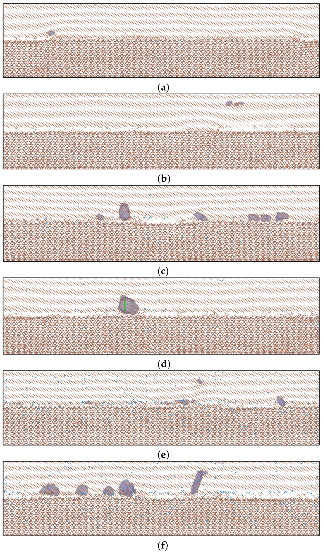
Snapshots illustrating crack initiation and propagation, obtained from MD simulations with incrementally increasing tensile strain applied perpendicular to the ferrite–cementite interface, are presented in Figure 1. The simulations, performed both without hydrogen (Figure 1a,b) and with hydrogen concentrations of 0.1% (Figure 1c,d) and 0.5% (Figure 1e,f), demonstrate that crack initiation and propagation occur predominantly along the interface. Increasing hydrogen concentration results in a pronounced reduction in atomic bond strength, primarily due to hydrogen segregation at ferrite–cementite interfaces. This segregation lowers interfacial cohesion and promotes interfacial separation, consistent with the hydrogen-enhanced decohesion (HEDE) mechanism.
Figure 1.
Snapshots illustrating crack propagation in a ferrite–cementite bicrystal under tensile loading perpendicular to the interface in a hydrogen-free system (a,b) and at hydrogen concentrations of 0.1% (c,d) and 0.5% (e,f) are shown. Local FCC phases in ferrite are highlighted in magenta, while dislocations are visualized as colored lines, with each color representing a distinct dislocation type. H and C atoms are colored blue and grey, respectively.
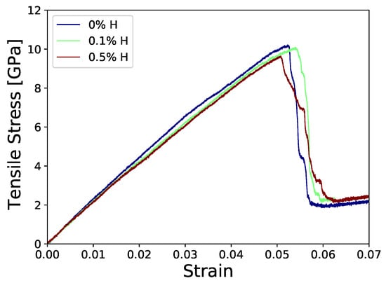
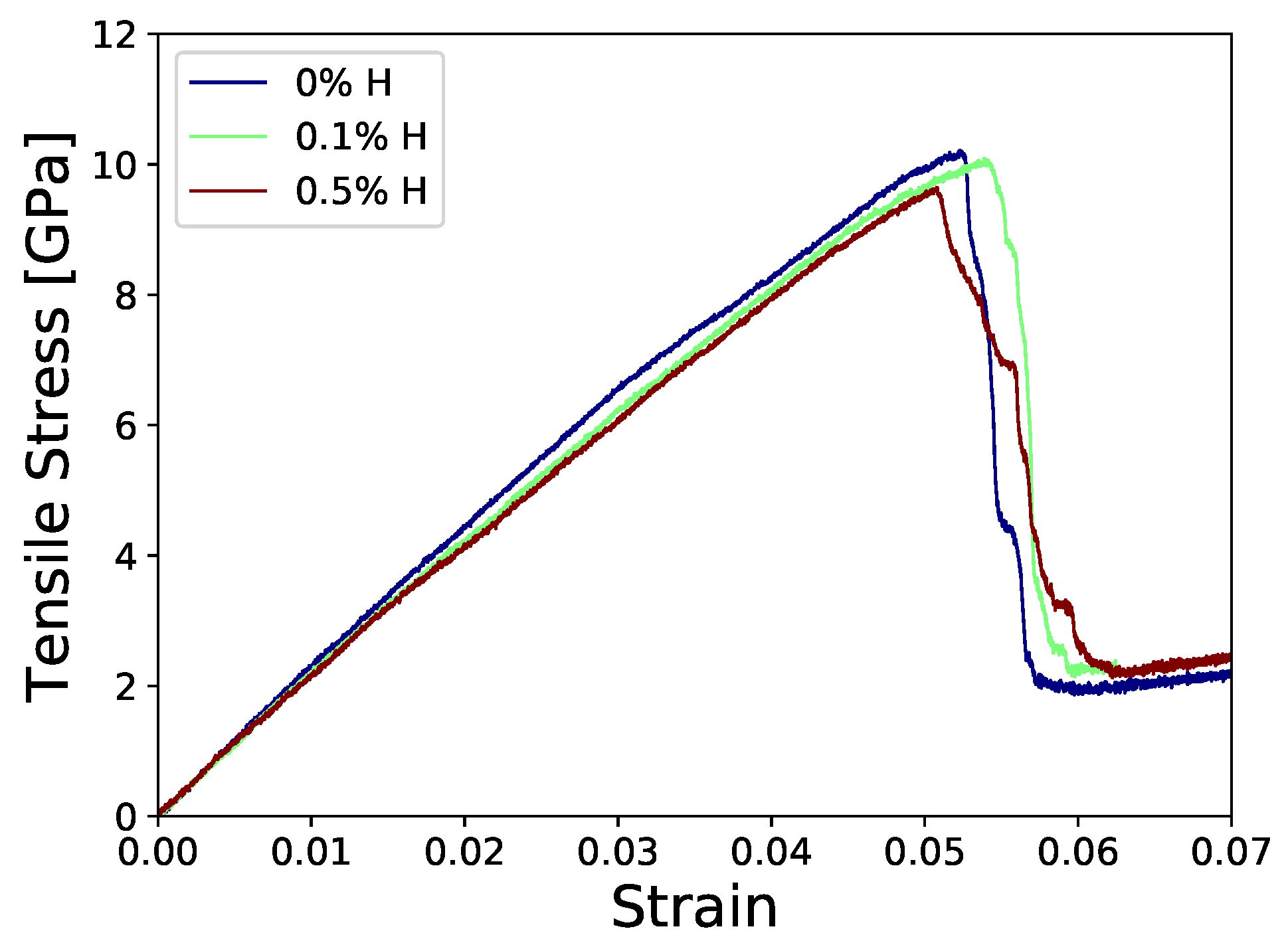
The cohesive strength of the ferrite–cementite interface is determined from the peak value of the normal stress in the stress–strain curve obtained during tensile loading perpendicular to the interface. The cohesive strength for the hydrogen-free bicrystal is 10.18 GPa, while for hydrogen concentrations of 0.1% and 0.5%, the corresponding values are 10.07 and 9.64 GPa, respectively. The work of separation determined from the MD-generated stress–strain curves for the three cases is , and J/, respectively. As a consequence, the critical stress required for crack initiation is reduced, leading to earlier onset of interfacial failure. This trend is clearly reflected in the stress–strain response (Figure 2), where increasing hydrogen content is associated with a progressive decrease in peak stress. Atomic stresses are calculated using the standard virial expression [39].
Figure 2.
Stress—strain response of a ferrite–cementite bicrystal under loading applied perpendicular to the interface at varying hydrogen concentrations.
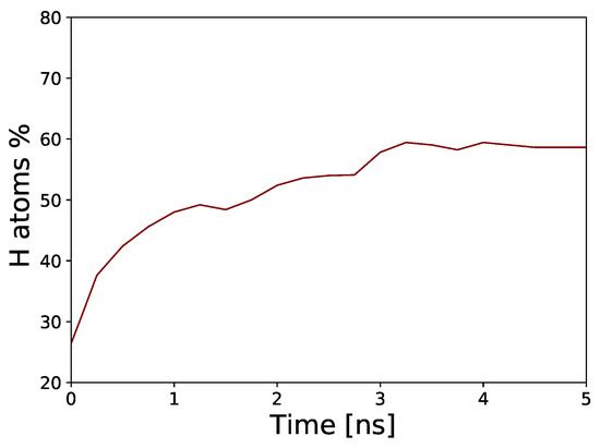
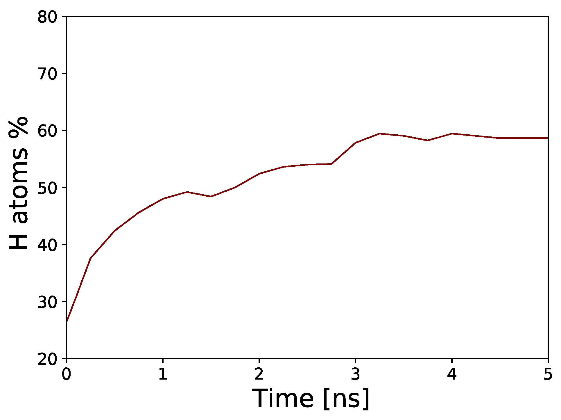
After determining the equilibrium hydrogen distribution in the pearlitic microstructure via time-stamped force-bias Monte Carlo (TFBMC), and subsequently performing molecular dynamics (MD) simulations of hydrogen diffusion over 5 ns, the initial hydrogen concentration at the ferrite–cementite interface remains relatively low. Figure 3 shows the evolution of the fraction of hydrogen atoms segregated within the ferritic phase up to 3 Å from the ferrite–cementite interface following the application of tensile load. Tensile loading perpendicular to the ferrite–cementite interface enhances stress–driven diffusion in ferrite and increases hydrogen trapping. The resulting hydrostatic tensile stress, defect generation, and interfacial stress concentration, arising from the mismatch in elastic and plastic properties between ferrite and cementite, promote hydrogen segregation at the interface. As the applied load increases, the combined effects of stress-driven diffusion, increased trap density, and interfacial decohesion lead to a progressive increase in hydrogen concentration at the ferrite–side interface (Figure 2). This localized hydrogen enrichment reduces interfacial cohesion, thereby facilitating crack initiation and promoting crack propagation along the ferrite–cementite boundary.
Figure 3.
The evolution of the fraction of hydrogen atoms segregated within the ferritic phase up to 3 Å from the ferrite–cementite interface.
In addition to interfacial weakening, hydrogen promotes phase transformations and the formation of FCC- and HCP-like atomic configurations, together with increased dislocation activity in the ferritic phase. Local FCC transformations in the ferrite are highlighted in magenta in Figure 1, while dislocations are visualized as colored lines, with each color representing a distinct dislocation type. The FCC regions arise from local atomic rearrangements associated with dislocation activity and shear deformation. These deformation-induced transformations are associated with energy dissipation and contribute to enhanced plasticity of the material. These processes facilitate local stress relaxation in the vicinity of the crack tip, thereby contributing to a partial retardation of crack propagation. Nevertheless, the coupling between hydrogen-enhanced localized plasticity in ferrite and reduced interfacial cohesion promotes crack initiation along ferrite–cementite interfaces under applied tensile loading. The interplay between HEDE and HELP, together with hydrogen-induced structural transformations, thus governs the observed fracture behavior. These findings underscore the critical role of ferrite–cementite interfaces as preferential sites for hydrogen accumulation and damage evolution, ultimately controlling the macroscopic mechanical response of pearlitic microstructures under hydrogen exposure.
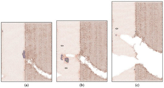
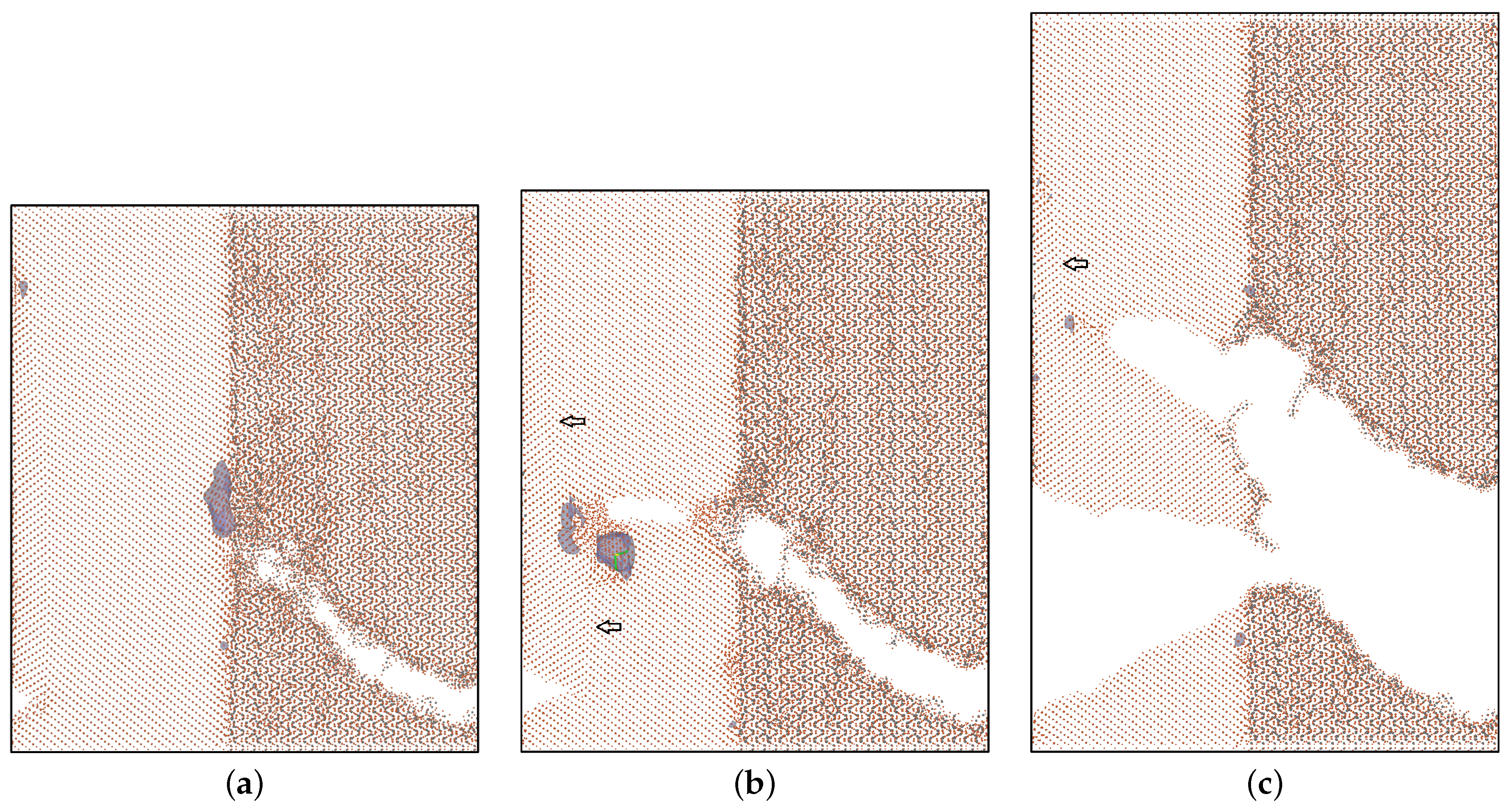
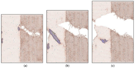
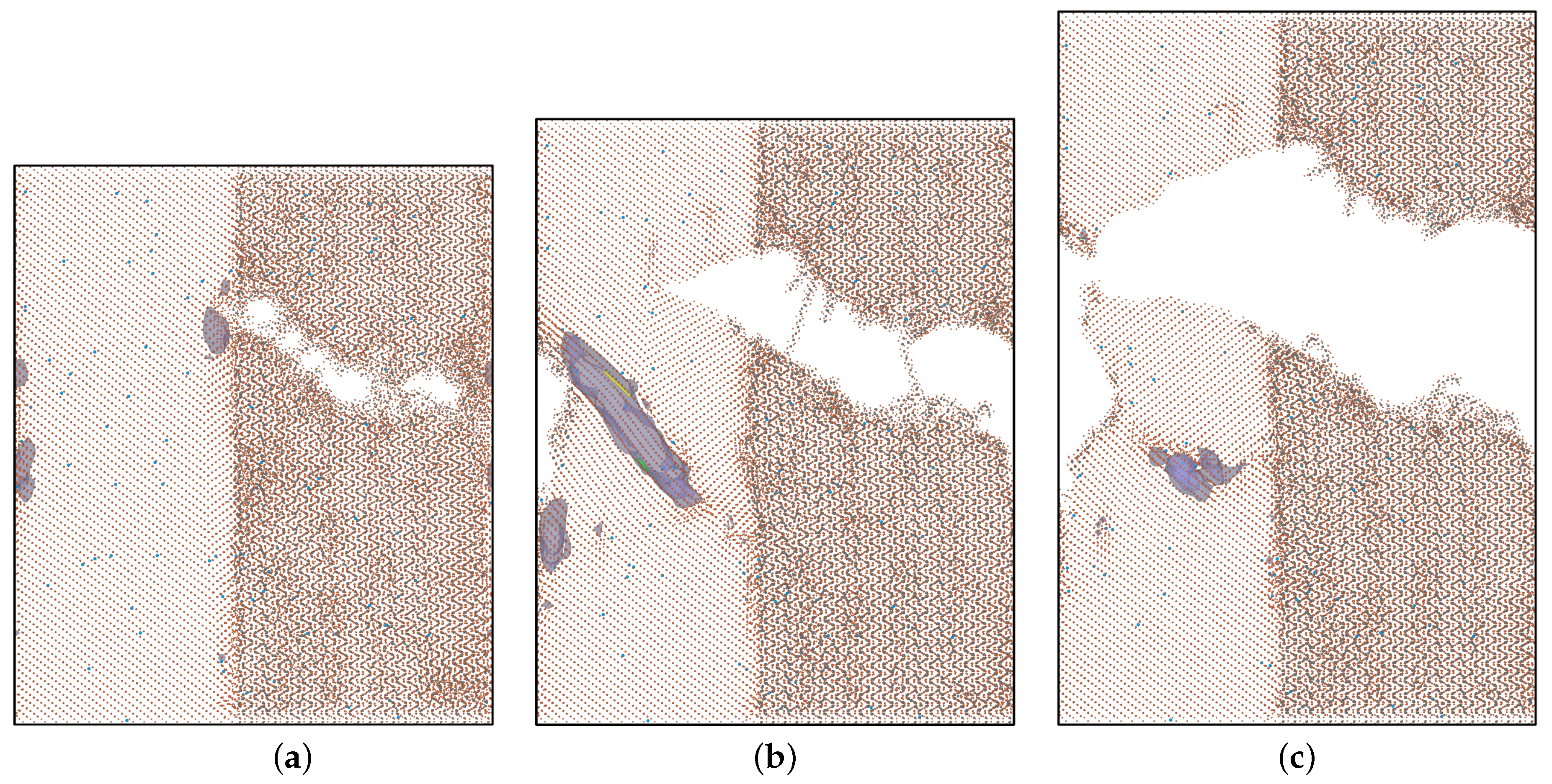
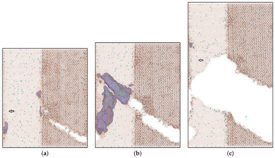
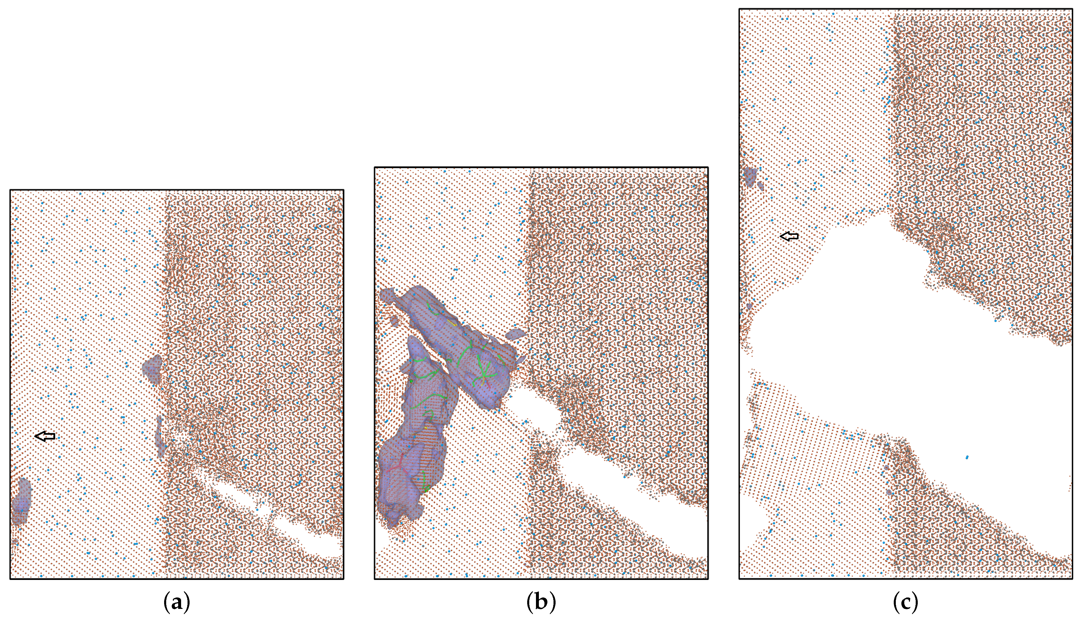
To investigate the effect of hydrogen on the mechanism of crack formation and propagation transverse to the ferrite–cementite interfaces, we apply incrementally increasing tensile strain parallel to the ferrite–cementite interface. The pearlitic microstructure is modeled using molecular dynamics simulations of a ferrite–cementite bicrystal, with periodic boundary conditions (PBC) imposed in the and directions. These periodic boundary conditions effectively generate an infinite lamellar arrangement, allowing the study of a pearlitic lamellar structure using a single ferrite–cementite bicrystal. Snapshots illustrating crack initiation and propagation in the pearlitic bicrystal without hydrogen and at hydrogen concentrations of 0.1% and 0.5% are presented in Figure 4, Figure 5 and Figure 6, respectively. The results reveal a pronounced influence of hydrogen on crack propagation behavior.
Figure 4.
Snapshots illustrating crack initiation (a) and propagation (b,c) in a ferrite–cementite bicrystal under tensile loading parallel to the interface in a hydrogen-free system are shown. Local FCC phases in ferrite are highlighted in magenta, while dislocations are visualized as colored lines, with each color representing a distinct dislocation type. C atoms are colored grey. The arrows show the positions of the twin boundaries.
Figure 5.
Snapshots illustrating crack initiation (a) and propagation (b,c) in a ferrite–cementite bicrystal under tensile loading parallel to the interface at hydrogen concentration of 0.1% are shown. Local FCC phases in ferrite are highlighted in magenta, while dislocations are visualized as colored lines, with each color representing a distinct dislocation type. H and C atoms are colored blue and grey, respectively.
Figure 6.
Snapshots illustrating crack initiation (a) and propagation (b,c) in a ferrite–cementite bicrystal under tensile loading parallel to the interface at hydrogen concentrations of 0.5% are shown. Local FCC phases in ferrite are highlighted in magenta, while dislocations are visualized as colored lines, with each color representing a distinct dislocation type. H and C atoms are colored blue and grey, respectively. The arrows show the positions of the twin boundaries.
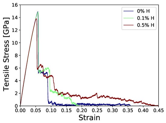
In atomistic simulations and idealized theoretical analyses, the cohesive strength of a material is associated with the peak tensile stress reached during loading. In our MD simulations, the cohesive strength of the considered pearlitic structure under tensile loading parallel to the ferrite–cementite interface is 14.9 GPa for the hydrogen-free bicrystal, and 14.82 GPa and 13.81 GPa for hydrogen concentrations of 0.1% and 0.5%, respectively. The work of separation for the three cases is , , and J/, respectively. Both the cohesive strength and the work of separation under tensile loading parallel to the boundary between ferrite and cementite lamellae are higher than the corresponding values for the ferrite–cementite interface. In all three cases, cracks initiate within the cementite lamellae close to the ferrite–cementite interface due to the mismatch in elastic and plastic properties between ferrite and cementite. The crack rapidly propagates within the cementite lamellae due to the inherently brittle nature of cementite. At the considered hydrogen concentrations, hydrogen promotes the initiation and propagation of brittle microcracks in cementite lamellae, with increasing concentration leading to crack initiation at lower applied tensile stresses.
This behavior is clearly reflected in the first peak of the stress–strain response (Figure 7), which corresponds to the initiation and propagation of cracks in the cementite lamellae. Once microcracks form in cementite, they propagate across the lamellar interfaces. In the hydrogen-free case, crack propagation in ferrite occurs readily across the interface near the crack initiation site and is temporarily arrested at the opposite interface.
Figure 7.
Stress–-strain response of a ferrite–cementite bicrystal under loading applied parallel to the interface at varying hydrogen concentrations.
At this location, the mechanical mismatch between ferrite and cementite leads to the formation of voids in ferrite near the lamellar boundary (Figure 4b). With increasing applied stress, the crack crosses the ferrite–cementite interface and coalesces with the microcrack in the ferrite. Deformation twins, FCC- and HCP-like atomic configurations form in ferrite ahead of the crack tip, where they contribute to energy dissipation. The formation of deformation twins generally modifies crack propagation in two competing ways: it can both impede and assist crack growth depending on the local stress state and microstructural conditions. Twinning is a localized plastic deformation mechanism that reduces stress concentration at the crack tip and locally delays crack advance until critical stress levels are reached. Twin boundaries are crystallographic interfaces capable of temporarily arresting crack propagation and reducing the crack growth rate. Twin boundaries also act as local barriers to dislocation motion and contribute to heterogeneous plastic deformation, thereby enhancing energy dissipation in the vicinity of the crack tip. However, twin boundaries may become energetically favorable sites for damage accumulation under continued loading. Their crystallographic nature and reduced cohesive strength relative to the surrounding matrix can promote preferential crack propagation paths, reflecting a competition between plasticity-induced stress relaxation and interfacial failure.
Ab initio calculations [40] for ferrite–cementite interfaces with the Isaichev orientation relationship showed that the hydrogen trapping energy at cementite interstitial sites is approximately 0.26 eV, whereas the trapping energy at ferrite–cementite interfaces reaches approximately 0.30–0.39 eV. These values indicate that ferrite–cementite interfaces and cementite act as stronger hydrogen trapping regions than bulk ferrite. The present MD simulations also suggest that ferrite primarily acts as a fast diffusion pathway for hydrogen transport, whereas cementite and ferrite–cementite interfaces serve as preferential trapping regions. Consequently, hydrogen can redistribute rapidly through ferritic lamellae and accumulate near interfaces (Figure 1c–f and Figure 3), dislocations and crack tips (Figure 5 and Figure 6), where local hydrogen concentrations may substantially exceed the average bulk concentration. Because cementite possesses a complex orthorhombic structure and limited intrinsic plasticity, dislocation motion within cementite is strongly restricted. Our MD simulations do not indicate a direct effect of hydrogen on dislocation activity within cementite. Therefore, hydrogen-induced changes in cementite dislocation mobility are expected to play a secondary role. At the same time, the MD simulations suggest that hydrogen indirectly influences dislocation processes near cementite. Increased dislocation activity in ferrite through the HELP mechanism can produce local stress concentrations at ferrite–cementite interfaces, thereby promoting crack initiation within cementite or along the interface (Figure 6b).
Hydrogen significantly enhances a range of microstructural processes in ferrite lamellae, including FCC- and HCP-like local atomic configurations, deformation twinning, and the formation and motion of dislocations (Figure 5 and Figure 6). The presence of hydrogen reduces the barriers for dislocation nucleation and mobility, thereby facilitating localized plastic deformation. FCC- and HCP-like local atomic structures emerge as transient configurations associated with enhanced dislocation activity. Their increased occurrence under hydrogen charging reflects intensified localized plastic deformation consistent with the hydrogen-enhanced localized plasticity (HELP) mechanism, contributing to stress redistribution and crack tip shielding. The MD-predicted hydrogen-assisted formation of dislocations in the vicinity of crack tips or ferrite–cementite interfaces is consistent with the adsorption-induced dislocation emission (AIDE) mechanism, which promotes dislocation emission near crack tips and interfacial regions. In pearlitic microstructures, HELP is expected to operate primarily in ferrite, whereas AIDE may contribute locally near crack tips and ferrite–cementite interfaces. This hydrogen-induced localized stress relaxation promotes crack tip blunting, as the initially sharp crack front becomes rounded due to plastic deformation (Figure 5 and Figure 6). Consequently, the driving force for crack propagation is reduced, leading to a partial retardation of crack growth despite the presence of embrittling hydrogen. However, the increased occurrence of phase transformations and the formation of deformation twins lead to a more complex and heterogeneous microstructure, characterized by a high density of crystallographic interfaces. These boundaries act as preferential sites for strain localization and stress concentration due to incompatibility of deformation between adjacent structures. Simultaneously, hydrogen segregation at ferrite–cementite interfaces reduces interfacial cohesive strength, thereby promoting microcrack initiation at lamellar boundaries (Figure 6b). As a result, crystallographic interfaces provide energetically favorable pathways for crack initiation and subsequent propagation. The reduced cohesive strength at these interfaces, potentially further weakened by hydrogen segregation, facilitates crack advance along these boundaries, thereby promoting anisotropic crack growth and accelerating overall crack propagation. The increase in the mechanical work required for separation of the pearlitic structure under tensile loading parallel to the ferrite–cementite interface in the presence of hydrogen may be attributed to stress redistribution and hydrogen-enhanced plasticity, which promote additional plastic dissipation and delay interfacial decohesion. Moreover, the limited diffusion timescales inherent to MD simulations may suppress strong hydrogen segregation to crack tip weakening sites, thereby increasing the calculated work of separation.
The MD prediction that cracks initiate within cementite under tensile loading parallel to the ferrite–cementite interface is qualitatively consistent with experimental observations in hydrogen-charged pearlitic steels, where brittle fracture of cementite lamellae and ferrite–cementite interfacial decohesion are frequently reported. Experimental studies indicate that hydrogen accumulates at ferrite–cementite interfaces and defect-rich regions [41], while dislocation pile-ups in ferrite generate local stress concentrations that promote brittle microcrack nucleation in cementite [42]. A single ferrite–cementite bilayer, while computationally necessary, represents a significant simplification of real pearlite. Consequently, the reported crack-orientation dependence is derived from two idealized loading configurations applied to a bicrystal model and may not fully capture the complexity of fracture processes in real pearlitic microstructures. Nevertheless, although the simplified MD bicrystal model does not fully reproduce the complexity of real pearlitic microstructures, it provides important atomistic insight into the early stages of hydrogen-assisted crack initiation.
The present results highlight the complex role of hydrogen in governing crack initiation and propagation in pearlitic microstructures. Hydrogen enhances localized plastic deformation in ferrite through the hydrogen-enhanced localized plasticity (HELP) mechanism, promotes processes that contribute to stress relaxation and crack blunting, thereby locally delaying crack advance. In parallel, hydrogen segregation at ferrite–cementite interfaces significantly reduces interfacial cohesion by weakening atomic bonding across phase boundaries. Furthermore, hydrogen accumulation within cementite lamellae promotes intrinsic embrittlement and reduces fracture resistance. This behavior is consistent with the hydrogen-enhanced decohesion (HEDE) mechanism. Consequently, interfacial weakening and cementite embrittlement act synergistically to facilitate microcrack nucleation and accelerate crack propagation along lamellar interfaces and within cementite regions. The concurrent processes that both promote and suppress crack propagation in the pearlite structure give rise to a complex interplay between HELP- and HEDE-controlled mechanisms, which ultimately governs the overall fracture behavior under hydrogen charging.
4. Conclusions
In conclusion, the molecular dynamics simulations provide detailed insight into the mechanisms governing crack initiation and propagation in pearlitic structures, particularly in relation to the orientation of cracks with respect to the ferrite–cementite interfaces. MD simulations reveal that hydrogen has a pronounced and orientation-dependent effect on crack initiation and propagation in pearlitic structures. The presence of hydrogen reduces the cohesive strength of the ferrite–cementite interfaces and enhances local lattice decohesion, thereby promoting earlier crack nucleation compared to hydrogen-free conditions. At the same time, hydrogen promotes phase transformations and formation of dislocations in the ferritic phase which facilitate local stress relaxation in the vicinity of the crack tip, thereby contributing to a partial retardation of crack propagation. Cracks oriented transverse to the interfaces initiate within the cementite lamellae close to the ferrite–cementite interface and rapidly propagate within the cementite lamellae. On reaching cementite–ferrite interface cracks encounter significant resistance as they interact with the alternating lamellar structure. The simulations also highlight the importance of local stress concentrations, dislocation activity, and phase contrast in controlling crack paths. The present results highlight the complex role of hydrogen in governing crack initiation and propagation in pearlitic microstructures. Hydrogen enhances localized plastic deformation in ferrite via the HELP mechanism, promoting twinning, dislocation activity and formation of FCC- and HCP-like configurations ahead of the crack tip. These processes contribute to stress redistribution and crack tip blunting, thereby locally delaying crack advance. In parallel, hydrogen segregation at cementite, deformation twins and ferrite–cementite interfaces reduces atomic bonding and interfacial cohesion, consistent with the HEDE mechanism. This facilitates crack initiation and propagation within cementite lamellae and along weakened interfaces once critical stress levels are reached. The concurrent processes that both promote and suppress crack propagation in cementite and ferrite lamellae therefore give rise to a complex interplay between HELP- and HEDE-controlled mechanisms, ultimately governing the overall fracture behavior under hydrogen charging. These findings provide atomistic-level insight into hydrogen embrittlement in pearlitic steels and highlight the critical role of lamellar architecture of pearlite as a key factor in its fracture resistance, with crack orientation relative to interfaces being a decisive parameter. Due to the temporal and spatial limitations of molecular dynamics simulations, the present results are obtained at relatively high strain rates. Furthermore, because of these limitations, the influence of pearlitic lamellae size on crack development is not considered in this study. In addition, the short timescales inherent to MD simulations likely lead to an underestimation of hydrogen segregation at lamellar interfaces and cementite lamellae, as well as its effect on the cohesive strength of ferrite–cementite interfaces.
Funding
This research is financially supported by the Bulgarian National Science Fund, Project KP–06–N67/9 “Mechanism of Hydrogen Embrittlement in Lath Martensite Steel: Model Simulations and Experimental Study”.
Data Availability Statement
Dataset available on request from the author.
Acknowledgments
The work on the publication was performed using equipment funded by project BG16RFPR002-1.014-0006 “National Center for Mechatronics and Clean Technologies”.
Conflicts of Interest
The author declares no conflicts of interest.
References
- Honeycombe, R.W.K. Steels: Microstructure and Properties, 3rd ed.; Butterworth-Heinemann: Oxford, UK, 2006. [Google Scholar]
- Bhadeshia, H.K.D.H. Worked Examples in the Geometry of Crystals; Institute of Materials: London, UK, 2001. [Google Scholar]
- Gladman, T. The Physical Metallurgy of Microalloyed Steels; Maney Publishing: London, UK, 1997. [Google Scholar]
- Zhang, Y.; Li, J.; Wang, Z. Atomistic simulation of deformation and fracture behavior in nanocrystalline materials. Mater. Sci. Eng. A 2010, 527, 7050–7056. [Google Scholar]
- Lynch, S.P. Hydrogen embrittlement phenomena and mechanisms. Corros. Rev. 2012, 30, 105–123. [Google Scholar] [CrossRef] [Scilit]
- Thompson, A.W.; Bernstein, I.M. The role of hydrogen in the deformation and fracture of iron single crystals. Metall. Trans. A 1982, 13, 1609–1619. [Google Scholar]
- Wang, Z.G.; Zhang, Z.F. The role of hydrogen in fatigue crack growth of high-strength steels. Acta Mater. 2006, 54, 3847–3860. [Google Scholar]
- Miller, K.J. Fatigue of Materials; Cambridge University Press: Cambridge, UK, 1999. [Google Scholar]
- Glover, A.G. Pipeline design and construction using high strength steels. In Proceedings of the International Seminar on Welding of High Strength Pipeline Steels (CBMM/TMS); Companhia Brasileira de Metalurgia e Mineração (CBMM): Araxá, Brazil, 2013; pp. 65–83. [Google Scholar]
- Nagumo, M. Fundamentals of hydrogen embrittlement. ISIJ Int. 2001, 41, 590–598. [Google Scholar] [CrossRef] [Scilit]
- Oriani, R.A. Hydrogen embrittlement of steels. Annu. Rev. Mater. Sci. 1978, 8, 327–357. [Google Scholar] [CrossRef] [Scilit]
- Gangloff, R.P.; Somerday, B.P. Gaseous Hydrogen Embrittlement of Materials in Energy Technologies; Woodhead Publishing: Cambridge, UK, 2012. [Google Scholar]
- Zhang, L.; Wen, M.; Imade, M.; Fukuyama, S.; Yokogawa, K. Hydrogen trapping at ferrite–cementite interfaces in pearlitic steel. Acta Mater. 2016, 121, 403–415. [Google Scholar]
- Koyama, M.; Akiyama, E.; Tsuzaki, K.; Raabe, D. Hydrogen-assisted crack initiation in pearlitic steel. Acta Mater. 2013, 61, 4607–4618. [Google Scholar] [CrossRef] [Scilit]
- Cho, L.; Bradley, P.E.; Lauria, D.S.; Martin, M.L.; Connolly, M.J.; Benzing, J.T.; Seo, E.J.; Findley, K.O.; Speer, J.G.; Slifka, A.J. Characteristics and mechanisms of hydrogen-induced quasi-cleavage fracture of lath martensitic steel. Acta Mater. 2021, 206, 116635. [Google Scholar] [CrossRef] [Scilit]
- Yan, C.; Zhang, S.; Zhou, L.; Tian, Z.; Shen, M.; Liu, X. Effect of Quenching Temperature on Microstructure and Hydrogen-Induced Cracking Susceptibility in S355 Steel. Materials 2025, 18, 1161. [Google Scholar] [CrossRef] [Scilit]
- Birnbaum, H.K.; Sofronis, P. Hydrogen-enhanced localized plasticity—A mechanism for hydrogen-related fracture. Mater. Sci. Eng. A Struct. Mater. Prop. Microstruct. Process. 1994, 176, 191–202. [Google Scholar] [CrossRef] [Scilit]
- Robertson, I.M. The effect of hydrogen on dislocation dynamics. Eng. Fract. Mech. 2001, 68, 671–692. [Google Scholar] [CrossRef] [Scilit]
- Ferreira, P.J.; Robertson, I.M.; Birnbaum, H.K. Hydrogen effects on the interaction between dislocations. Acta Mater. 1998, 46, 1749–1757. [Google Scholar] [CrossRef] [Scilit]
- Somerday, B.; Sofronis, P.; Jones, R. Effects of hydrogen on materials. In Proceedings of the 2008 International Hydrogen Conference, Jackson Lake Lodge, WY, USA, 7–10 September 2008. [Google Scholar]
- Abou Ali Modad, O.; Shehadeh, M.A. Modeling dislocation interactions with grain boundaries in lath martensitic steels. J. Mater. Sci. 2024, 59, 4829–4851. [Google Scholar] [CrossRef] [Scilit]
- Troiano, A.R. The Role of Hydrogen and Other Interstitials in the Mechanical Behavior of Metals. Trans. ASM 1960, 52, 54–81. [Google Scholar] [CrossRef] [Scilit]
- Oriani, R.A.; Josephic, P.H. Equilibrium aspects of hydrogen-induced cracking of steels. Acta Metall. 1974, 22, 1065–1074. [Google Scholar] [CrossRef] [Scilit]
- Djukic, M.B.; Bakic, G.M.; Zeravcic, V.S.; Sedmak, A.; Rajicic, B. The synergistic action and interplay of hydrogen embrittlement mechanisms in steels and iron: Localized plasticity and decohesion. Eng. Fract. Mech. 2019, 216, 106528. [Google Scholar] [CrossRef] [Scilit]
- Azocar Guzman, A.; Janisch, R. Effects of mechanical stress, chemical potential, and coverage on hydrogen solubility during hydrogen-enhanced decohesion of ferritic steel grain boundaries: A first-principles study. Phys. Rev. Mater. 2024, 8, 073601. [Google Scholar] [CrossRef] [Scilit]
- Matsubara, K.; Takagi, S.; Meng, F.S.; Ogata, S.; Ishikawa, N. Atomic-scale modelling of boundaries characterized lath martensite microstructure for hydrogen-induced crack initiation: A machine learning interatomic potentials study. In Proceedings of the 7th International Symposium on Steel Science, Kyoto, Japan, 11–14 November 2024. [Google Scholar]
- Song, J.; Curtin, W.A. A nanoscale mechanism of hydrogen embrittlement in metals. Nat. Mater. 2013, 12, 145–151. [Google Scholar] [CrossRef] [Scilit] [PubMed]
- Shimokawa, T.; Kikugawa, K. From Plasticity to Fracture in Pearlitic Microstructures: Atomistic Study of Cementite Thickness and Deformation Localization. ISIJ Int. 2025, 66, 452–459. [Google Scholar] [CrossRef] [Scilit]
- Zhou, X.; Foster, M.; Ronevich, J.; San Marchi, C. Review and construction of interatomic potentials for molecular dynamics studies of hydrogen embrittlement in Fe–C based steels. J. Comput. Chem. 2020, 41, 1299–1309. [Google Scholar] [CrossRef] [Scilit] [PubMed]
- Kim, J.; Kang, K.; Ryu, S. Characterization of the misfit dislocations at the ferrite/cementite interface in pearlitic steel: An atomistic simulation study. Int. J. Plast. 2016, 83, 302–312. [Google Scholar] [CrossRef] [Scilit]
- Thompson, A.P.; Aktulga, H.M.; Berger, R.; Bolintineanu, D.S.; Brown, W.M.; Crozier, P.S.; In’t Veld, P.J.; Kohlmeyer, A.; Moore, S.G.; Nguyen, T.D.; et al. LAMMPS—A flexible simulation tool for particle-based materials modeling at the atomic, meso, and continuum scales. Comput. Phys. Commun. 2022, 271, 108171. [Google Scholar] [CrossRef] [Scilit]
- Kim, J.; Ghaffarian, H.; Kang, K. The lattice dislocation trapping mechanism at the ferrite/cementite interface in the Isaichev orientation relationship. Sci. Rep. 2021, 11, 9324. [Google Scholar] [CrossRef] [Scilit]
- Demkowicz, M.J.; Thilly, L. Structure, shear resistance and interaction with point defects of interfaces in Cu–Nb nanocomposites synthesized by severe plastic deformation. Acta Mater. 2011, 59, 7744–7756. [Google Scholar] [CrossRef] [Scilit]
- Dyakova, V.; Yanachkov, B.; Valuiska, K.; Mourdjeva, Y.; Krastev, R.; Simeonova, T.; Kolev, K.; Lazarova, R.; Katzarov, I. The Impact of Hydrogen Charging Time on Microstructural Alterations in Pipeline Low-Carbon Ferrite-Pearlite Steel. Metals 2025, 15, 1079. [Google Scholar] [CrossRef] [Scilit]
- Frenkel, D.; Smit, B. Understanding Molecular Simulation: From Algorithms to Applications, 2nd ed.; Academic Press: Cambridge, MA, USA, 2002. [Google Scholar]
- Nose, S. A unified formulation of the constant temperature molecular dynamics methods. J. Chem. Phys. 1984, 81, 511–519. [Google Scholar] [CrossRef] [Scilit]
- Swope, W.C.; Andersen, H.C.; Berens, P.H.; Wilson, K.R. A computer simulation method for the calculation of equilibrium constants for the formation of physical clusters of molecules: Application to small water clusters. J. Chem. Phys. 1982, 76, 637–649. [Google Scholar] [CrossRef] [Scilit]
- Stukowski, A. Visualization and analysis of atomistic simulation data with OVITO—The Open Visualization Tool. Model. Simul. Mater. Sci. Eng. 2010, 18, 015012. [Google Scholar] [CrossRef] [Scilit]
- Landau, L.D.; Lifschitz, E.M. Course of Theoretical Physics: Statistical Physics (Part 1), 3rd ed.; Elsevier: Amsterdam, The Netherlands, 1980. [Google Scholar]
- Mirzoev, A.A.; Verkhovykh, A.V.; Okishev, K.Y.; Mirzaev, D.A. Hydrogen interaction with ferrite/cementite interface: Ab initio calculations and thermodynamics. Mol. Phys. 2018, 116, 482–490. [Google Scholar] [CrossRef] [Scilit]
- Barton, D.J.; Nguyen, D.T.; Perea, D.E.; Stoerzinger, K.A.; Lumagui, R.M.; Lambeets, S.V.; Wirth, M.G.; Devaraj, A. Towards quantitative analysis of deuterium absorption in ferrite and austenite during electrochemical charging by comparing cyclic voltammetry and cryogenic transfer atom probe tomography. Int. J. Hydrogen Energy 2024, 50, 30–40. [Google Scholar] [CrossRef] [Scilit]
- Dollar, M.; Bernstein, I.M.; Thompson, A.W. Influence of deformation substructure on flow and fracture of fully pearlitic steel. Acta Metall. 1988, 36, 311–320. [Google Scholar] [CrossRef] [Scilit]
Disclaimer/Publisher’s Note: The statements, opinions and data contained in all publications are solely those of the individual author(s) and contributor(s) and not of MDPI and/or the editor(s). MDPI and/or the editor(s) disclaim responsibility for any injury to people or property resulting from any ideas, methods, instructions or products referred to in the content. |
© 2026 by the author. Licensee MDPI, Basel, Switzerland. This article is an open access article distributed under the terms and conditions of the Creative Commons Attribution (CC BY) license.