


Abstract
Streptococcus pneumoniae is the major causative agent of community-acquired pneumonia, one of the main infectious diseases that causes inflammation in the alveoli and leads to significant morbidity and mortality across various age groups. Current treatments are challenged by multidrug-resistant strains of S. pneumoniae, which has led to the reemergence of pneumonia in recent years; therefore, it is pivotal to identify new drug targets. The enzyme chorismate synthase (CS), involved in the shikimate pathway of S. pneumoniae, aids in the synthesis of vital aromatic amino acids and other metabolites required for bacterial viability. The present study identifies natural compounds that can inhibit CS using an in silico approach, including pharmacophore modeling, virtual screening, molecular docking, ADME analysis, and molecular dynamics (MD) simulations. Our results suggest that the identified compounds can bind effectively to the active site of S. pneumoniae CS (SpCS), exhibiting affinities better than the known inhibitor 1-benzofuran-3-one and close to the enzyme’s natural substrate, 5-enolpyruvylshikimate-3-phosphate (EPSP). This study suggests lead natural compounds as promising candidates for further investigation as potential inhibitors for the treatment of pneumonia, offering a novel strategy to combat this resilient pathogen.
1. Introduction
Pneumonia is one of the most common acute lower respiratory infections (LRIs), characterized by inflammation in the alveoli, affecting a wide population (high morbidity) and accounting for increased mortality rates among various age groups across the globe [1,2]. Over 80% of community-acquired bacterial pneumonia (infection acquired outside the hospital environment) is caused by Streptococcus pneumoniae, a Gram-positive, facultative anaerobic diplococcus that primarily infects the elderly and immunocompromised individuals [3]. Clinical symptoms of pneumonia include persistent cough, fatigue, dyspnea, pleuritic chest pain, and sputum production [4]. It is an airborne, communicable disease and was a leading public health concern from the 1930s to the early 1940s. However, the advent of sulfapyridine, the first anti-pneumococcal sulfonamide, in early 1939, and penicillin reduced the severity and morbidity of the disease. Later, in the 1990s, the emergence of multidrug-resistant strains of pneumococcus led to the reemergence of pneumonia as a public health concern [5,6,7]. Over the past decade, excluding COVID-19, the global annual cost of diagnosing, treating, and managing pneumonia has been close to US$17 billion [8], and India contributes to 23% of global pneumonia cases, with case fatality rates between 14% and 30%, where S. pneumoniae is the major causative organism [9].
The emergence of antimicrobial-resistant strains creates a need to identify new drugs that inhibit metabolic pathways specific to bacteria without affecting human metabolism. One such pathway is the shikimate pathway, which is specific to bacteria, fungi, and plants. This pathway aids in the biosynthesis of aromatic amino acids (L-tryptophan, L-phenylalanine, and L-tyrosine), which are essential for protein synthesis [10]. The shikimate pathway is absent in vertebrates, making it an attractive target for antibacterial agents. Enzymes of this pathway constitute excellent targets for the design of new antibacterial drugs [11,12]. Chorismate, the precursor for the biosynthesis of aromatic amino acids and aromatic compounds such as vitamin B9, vitamin K1, and tetrahydrofolate, is synthesized from phosphoenolpyruvate and erythrose-4-phosphate through a series of seven metabolic steps [13,14]. The final step of the shikimate pathway is catalyzed by chorismate synthase (CS), which synthesizes chorismate from EPSP (5-enolpyruvylshikimate-3-phosphate) using flavin mononucleotide (FMN) as a cofactor [15]. Because aromatic amino acids and their metabolites play crucial roles in biological functions such as electron transport, UV protection, plant defense mechanisms, and cell signaling [16], inhibiting CS would prevent the synthesis of these essential compounds, making it a valuable target for rational antimicrobial design.
The structure of CS has been investigated in various microorganisms, including Mycobacterium tuberculosis, Saccharomyces cerevisiae, Pseudomonas aeruginosa, Bacillus subtilis, and Shigella flexneri [17,18,19,20]. Recent studies have shown that azo-dye-based compounds act as inhibitors by binding to the active site of CS in the pathogenic fungus Paracoccidioides brasiliensis [21]. Other studies report that benzofuran derivatives and phenolic compounds such as caffeic acid, gallic acid, and o-coumaric acid inhibit the substrate-binding site of CS from Moraxella catarrhalis and S. pneumoniae, exhibiting potent antibacterial activity [22,23]. Similarly, the rapid growth of M. tuberculosis has been inhibited by identifying drug targets against CS [24]. In silico and in vitro studies of Listeria monocytogenes CS (LmCS) identified the bioactive ingredient α-longipinene from Nigella sativa as capable of restricting biofilm formation in L. monocytogenes by inhibiting LmCS [25]. Additionally, putative inhibitors of P. falciparum CS have been docked with the dimeric CS structure, resulting in novel drug targets and contributing to antimalarial drug development [26]. These findings suggest that the substrate-binding site of CS is a promising target for structure-based drug discovery of specific inhibitors, making CS a novel target for in silico drug discovery [27].
Computational tools provide in silico drug design techniques that facilitate the rapid identification of new drug targets in biologically active macromolecules compared with conventional pharmacological procedures. Computer-aided drug design (CADD) enables high-throughput virtual screening (VS) of chemical libraries to identify putative antimicrobial drugs in a cost-effective and resource-efficient manner [28,29]. Structure-based pharmacophore modeling is one such CADD approach that identifies key chemical functionalities and spatial arrangements within the active site of a macromolecule–ligand complex [30]. The binding affinity of a chemical compound is evaluated using molecular docking techniques by probing possible interaction sites between ligands and the target macromolecule [31,32]. Pharmacokinetic properties, including absorption, distribution, metabolism, and excretion (ADME), can also be predicted using CADD [33]. In this study, we focus on CADD techniques including molecular docking, VS, pharmacophore modeling, ADME analysis, and molecular dynamics (MD) simulations to identify potential inhibitors of SpCS for the treatment of pneumonia.
2. Materials and Methods
2.1. Structure Retrieval and Interactions Studies
The three-dimensional structure of SpCS was retrieved from the RCSB_PDB (https://www.rcsb.org/) (PDB ID: 1QXO). The monomeric chains constituting the tetrameric assembly were validated using the ProSA web server (https://prosa.services.came.sbg.ac.at/prosa.php (accessed on 10 January 2026)) [34] based on their corresponding z-scores. SpCS was co-crystallized with its substrate and cofactor, and the intermolecular interactions were visualized using BIOVIA Discovery Studio Visualizer, a molecular graphics visualizing tool (https://discover.3ds.com/discovery-studio-visualizer-download (accessed on 10 January 2026)) [35]. Additionally, the binding pocket of SpCS was visualized using PyMOL software version 3.1 (https://pymol.org/2/ (accessed on 10 January 2026)).
2.2. Prediction of Ligand-Binding Sites in SpCS
The PrankWeb (P2Rank) server (https://prankweb.cz/ (accessed on 12 January 2026)) [36] was employed to identify potential binding sites on SpCS for the ligands FMN and EPSP. The dimeric chain was uploaded in PDB format into the entry field, and the binding pocket search was restricted to chain A. The predicted binding sites were summarized in a tabular format, including parameters such as rank, score, probability, number of residues, and average conservation.
2.3. Structure-Based Pharmacophore Modeling and Virtual Screening
Pharmacophore models of the protein–ligand complexes were generated using the pharmit server (https://pharmit.csb.pitt.edu/) [37] with default parameters. For creating a pharmacophore model, the chain A and B dimer of the SpCS in complex with one of the two bound ligands was submitted to the server. The resulting pharmacophore models were subsequently employed to screen the ZINC_Nat compound library in order to identify potential structural analogs with inhibitory activity against the SpCS. Putative structural analogs corresponding to each ligand were retrieved from the ZINC database (https://zinc15.docking.org/substances/home/ (accessed on 15 January 2026)) based on their pharmacophore fit.
2.4. Docking of Potential Ligands with SpCS
Molecular docking studies of the SpCS protein with its native ligands and the predicted structural analogs were performed using the PyRx—Python Prescription VS Tool, version 0.8 which employs Autodock Vina 1.1.2 for docking (https://sourceforge.net/projects/pyrx/ (accessed on 15 January 2026)) [35,38]. Due to the fact that FMN interacted with 4 residues of chain B along with several residues from chain A, protein dimer was used as the receptor, and the identified binding pocket residues of the dimer were defined prior to docking (Center: X: −8.2005, Y: 11.4063, Z: 17.4079. Size: X: 22.4208 Å, Y: 25.0000 Å, Z: 19.0786 Å). The predicted structural analogs were then docked into the selected binding pocket of the protein dimer to evaluate their binding interactions and affinities.
2.5. In Silico Pharmacokinetics and Drug-Likeness Prediction
The drug-likeness and preliminary ADME-related properties of the putative structural analogs were assessed using the Molsoft L.L.C. (San Diego, CA, USA): drug-likeness and molecular property prediction server (https://molsoft.com/mprop/ (accessed on 20 January 2026)) [39,40,41,42]. Physicochemical parameters relevant to oral bioavailability, including molecular weight, octanol/water partition coefficient (LogP), number of hydrogen bond acceptors (HBAs) and donors (HBDs), and molecular polar surface area (MolPSA), were computed. Compounds were subsequently assessed for compliance with Lipinski’s Rule of Five [43], and drug-likeness scores were used to prioritize candidates for molecular docking and binding interaction analysis.
2.6. Docking of Potential Ligands Using Swiss Dock
Additional docking studies were performed using the Swiss Dock server to further validate the binding modes of the shortlisted structural analogs (https://www.swissdock.ch/ (accessed on 25 January 2026)) [44]. Binding pocket coordinates for the protein dimer (Center: X: −8.2005, Y: 11.4063, Z: 17.4079. Size: X: 22.4208 Å, Y: 25.0000 Å, Z: 19.0786 Å) were pre-determined from a previous docking step. Protein receptor preparation and ligands were subsequently subjected to docking using the Autodock Vina docking engine on the Swiss Dock server (https://www.swissdock.ch/ (accessed on 25 January 2026)). The protein dimer, prepared analogs, and corresponding binding site coordinates were uploaded, and the exhaustiveness parameter was set to 8. Docking was conducted for all shortlisted analogs, and the resulting docking scores and protein–ligand interaction profiles were analyzed in detail.
In parallel, the ligands were redocked using the Attracting Cavities 2.0 docking engine on the Swiss Dock server accessed on 25 January 2026. Similar to Autodock Vina, the protein dimer, prepared analogs, and corresponding binding site coordinates for the protein dimer (Center: X: −8.2005, Y: 11.4063, Z: 17.4079. Size: X: 22.4208 Å, Y: 25.0000 Å, Z: 19.0786 Å) were uploaded. The number of RIC (Random Initial Conditions) parameter was set to 1, the sampling exhaustivity parameter was set to “medium”, and cavity prioritization was set to the “buried” option. The resulting docking scores were compared with the scores obtained using the Autodock Vina engine of the Swiss Dock server to see whether the ligands display similar binding energetics across various docking engines.
2.7. MD Simulation Studies
The top-ranked docking pose of each ligand, including the reference ligands EPSP and 1-benzofuran-3-one, bound to the FMN-containing dimeric SpCS enzyme, was further evaluated by molecular dynamics (MD) simulations following our previously described protocol [45,46,47,48]. Each protein–cofactor–ligand complex was prepared using the QuickPrep and Dynamics modules implemented in MOE® (version 2024.06, Chemical Computing Group, Montreal, QC, Canada). Protein preparation, energy minimization, protonation state assignment, and parameterization of the ligands and the FMN cofactor were performed using the AmberEHT force field [49]. AmberEHT combines the Amber19 biomolecular force field with MOE®’s Extended Hückel Theory (EHT)-based parameterization for ligands and other non-standard molecular entities, providing a consistent framework for preparing protein–ligand systems for MD simulations. Protonation states of ionizable amino acid residues were assigned using the Protonate3D module at physiological pH.
MD simulations were performed in triplicate using NAMD [50] with the AmberEHT-parameterized systems. Long-range electrostatic interactions were treated using the Particle Mesh Ewald (PME) method with a real-space cutoff of 10 Å, while van der Waals interactions were evaluated using a switching function between 8 and 10 Å. Each complex was solvated in an orthorhombic TIP3P water box with a 10 Å solvent margin around the protein. The final box dimensions were 117.09 × 84.90 × 77.54 Å3, and Na+ or Cl− counterions were added to neutralize the system. Following energy minimization, the systems were equilibrated for 100 ps under the NVT ensemble and 200 ps under the NPT ensemble. Production simulations were subsequently carried out for 200 ns at 310 K and 1 bar without positional restraints. Binding free energies were estimated from the MD trajectories using the GBVI/WSA ΔG scoring function developed by Labute [51]. Trajectories were analyzed using the Database Viewer (DBV) module in MOE® and VMD [52], and graphs were generated using GraphPad Prism 11.0.2(100)®.
3. Results
3.1. Structure Retrieval and Interaction Studies
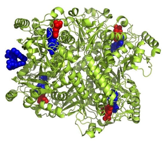
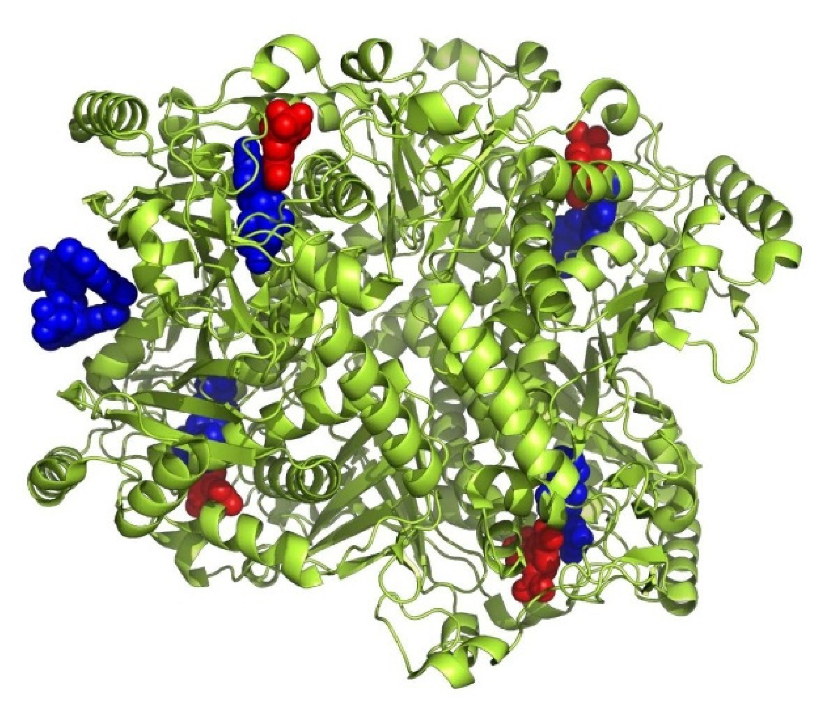
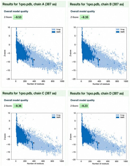
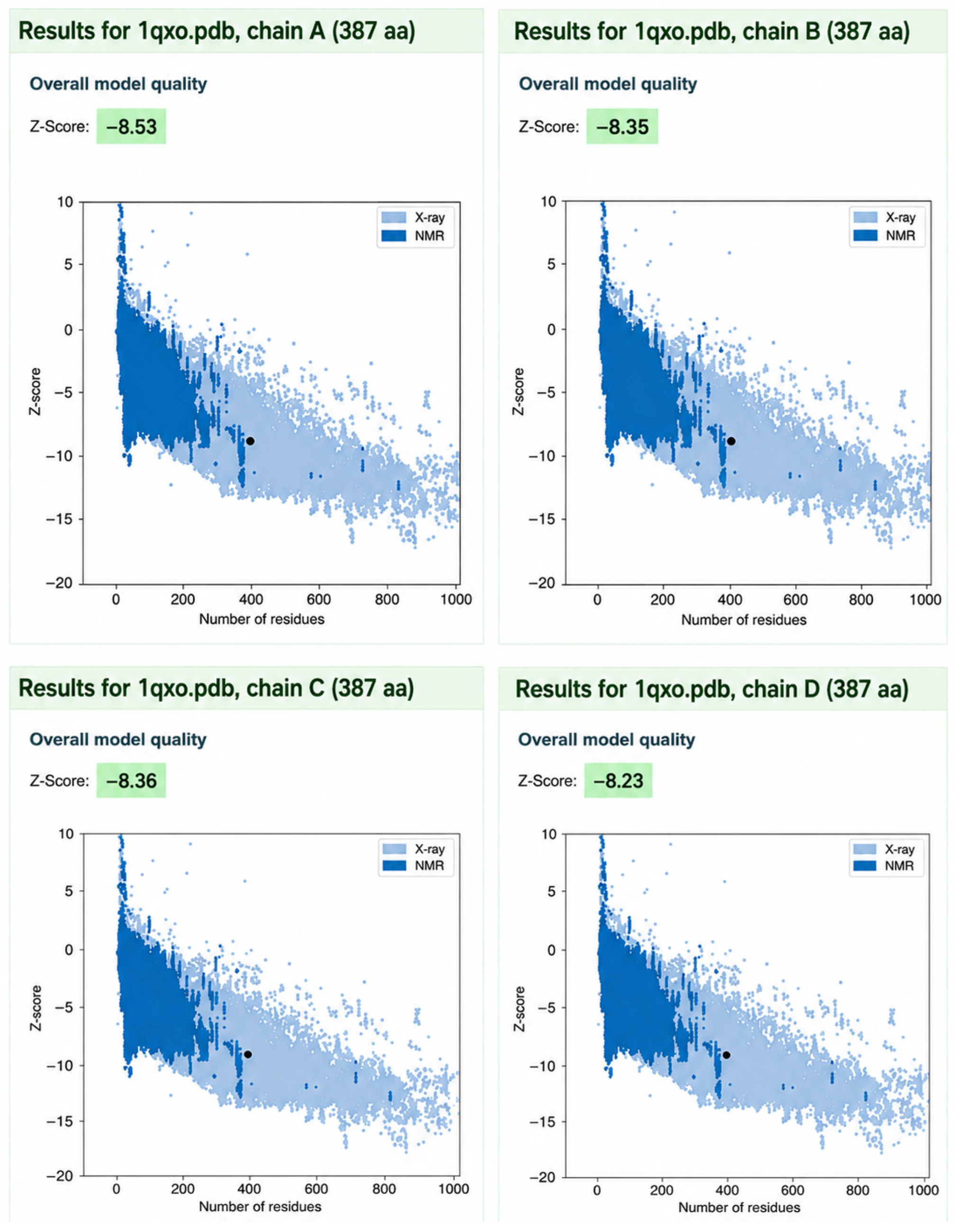
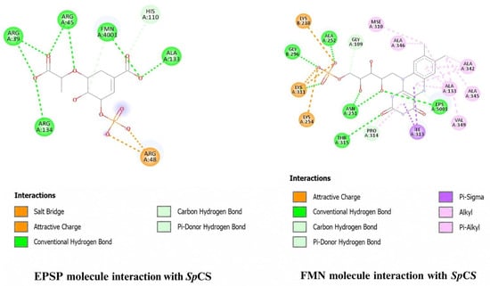
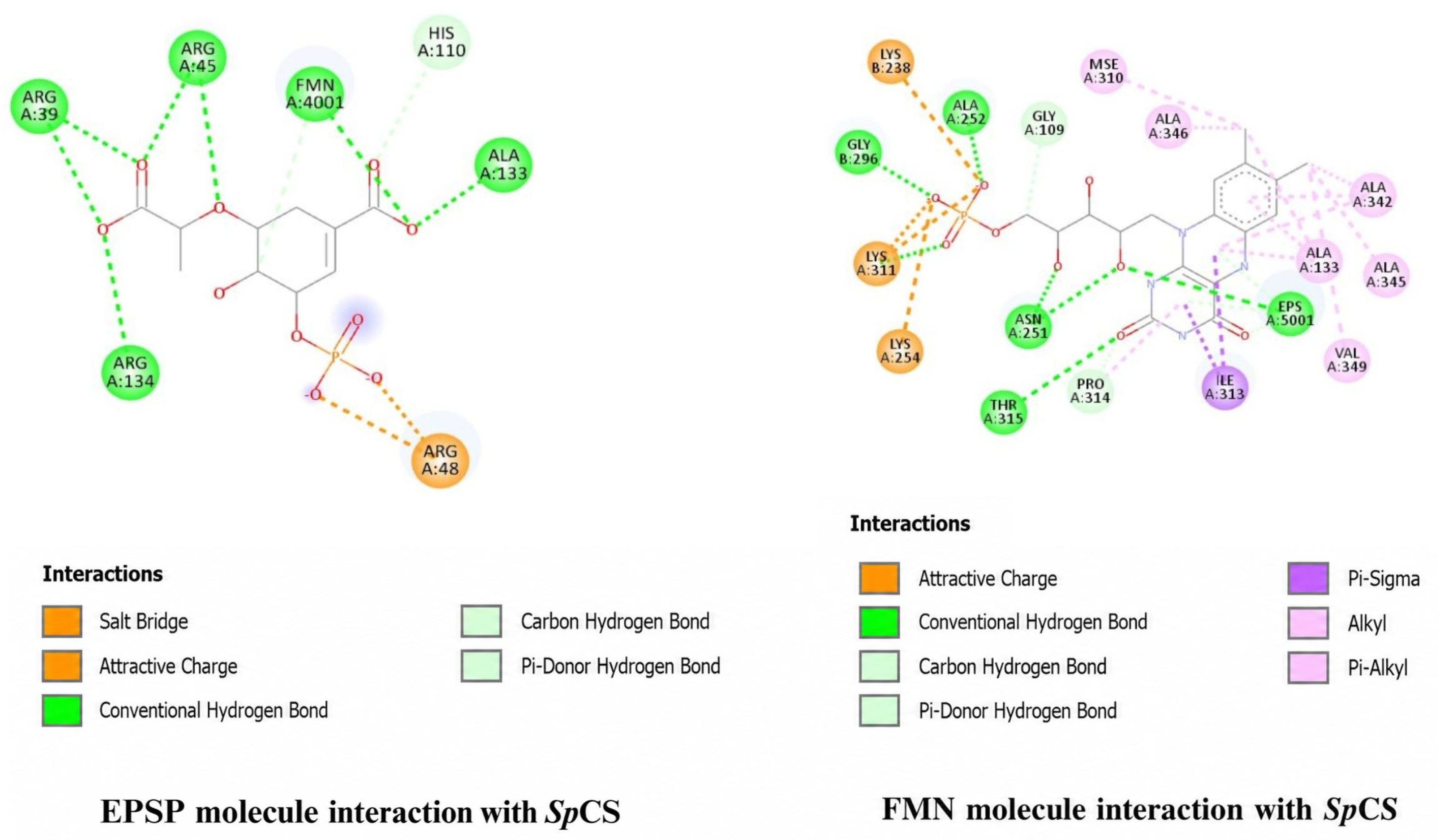
The crystal structure of SpCS, a homotetrameric protein (PDB ID: 1QXO) with 388 residues per chain, was retrieved from the RCSB Protein Data Bank (https://www.rcsb.org/) and determined in complex with substrate 5-enolpyruvylshikimate-3-phosphate (EPSP) and cofactor flavin mononucleotide (FMN) (Figure 1). Structural assessment using z-scores from the ProSA web server (https://prosa.services.came.sbg.ac.at/prosa.php (accessed on 10 January 2026)) confirmed high model reliability across all chains, with Chain A exhibiting a z-score of −8.53 followed by chains B, C, and D displaying z-scores of −8.35, −8.36, −8.23 respectively (Figure 2). Interactions of FMN and EPSP with chain A and chain B of SpCS using the BIOVIA Discovery Studio Visualizer (https://www.3ds.com/products/biovia/discovery-studio/visualization (accessed on 10 January 2026)) revealed that both FMN and EPSP occupy a common pocket within chain A along with a few residues of chain B. FMN interacts with 18 key active-site residues (14 from chain A and 4 from chain B), whereas EPSP engages a broader network of 30 residues, highlighting distinct yet overlapping binding interactions (Figure 3, Table 1).
Figure 1.
Tetrameric structure of SpCS. FMN is depicted as red spheres, and EPSP is depicted in blue spheres.
Figure 2.
Analysis of the individual chains of SpCS by the ProSA web server. The dark blue region represents amino acid residues with z-score distribution by NMR spectroscopy and the light blue region represents amino acid residues with z-score distribution by X-ray crystallography. The black dot represents the z-score of the input structures.
Figure 3.
Interaction analysis of FMN and EPSP with active-site residues in their respective binding pockets: the analysis was carried out via BIOVIA Discovery Studio Visualiser, from the co-crystallized structure of SpCS with FMN and EPSP (PDB ID: 1QXO).
Table 1.
Binding pockets and their respective amino acid residues analyzed via PrankWeb (https://prankweb.cz/ (accessed on 12 January 2026)) and BIOVIA Discovery Studio Visualizer (https://www.3ds.com/products/biovia/discovery-studio/visualization (accessed on 10 January 2026)).
3.2. Prediction of Ligand-Binding Sites in SpCS
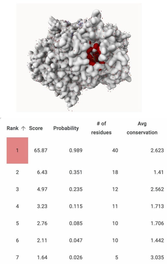
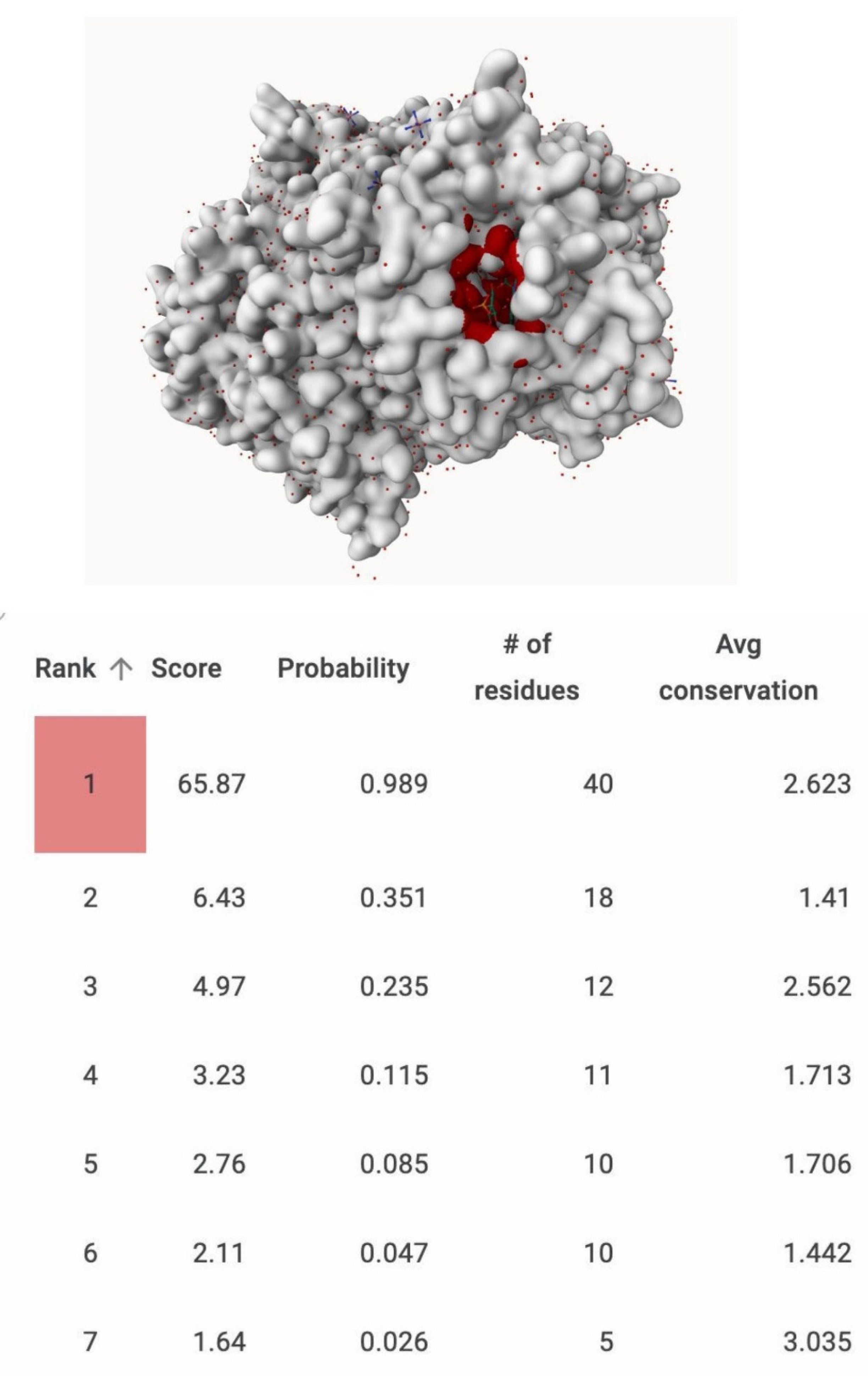
To identify the ligand-binding site of the SpCS, predictions were carried out using the PrankWeb server (https://prankweb.cz/ (accessed on 12 January 2026)), a machine-learning-based platform for binding pocket detection [54]. Seven putative binding pockets were identified in chain A (Figure 4). Among these, Pocket 1 was prioritized due to its comparatively high pocket score (65.87), probability value (0.989), and average conservation score (2.623). This pocket comprised 40 amino acid residues.
Figure 4.
Predicted binding sites on Chain A of SpCS. Binding sites predicted by PrankWeb (https://prankweb.cz/ (accessed on 12 January 2026)) sorted according to rank, score, probability, number of residues and average conservation. The arrow adjacent to the Rank column indicates increasing rank order from bottom to top. The binding pocket highlighted in red represents the selected pocket, which corresponds to the red-highlighted region in the three-dimensional (3D) structure.
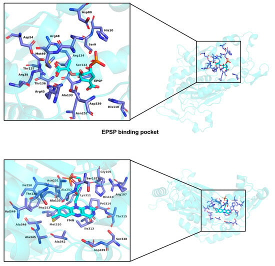
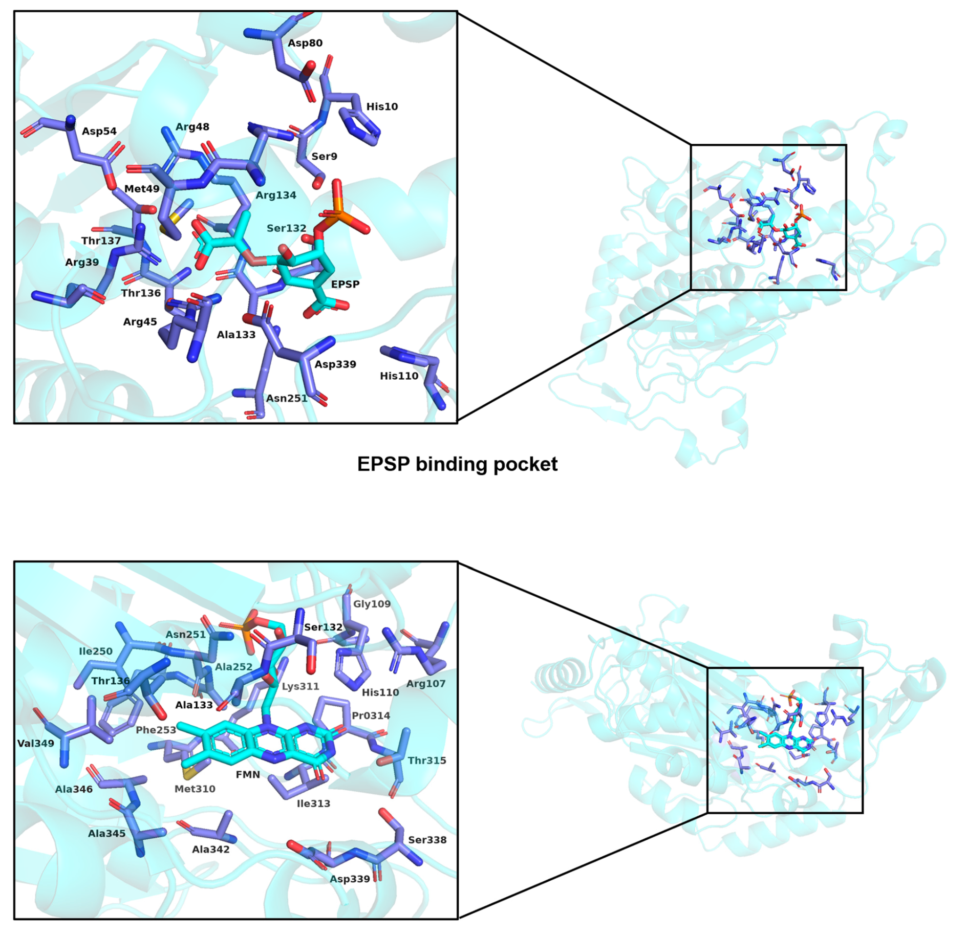
Subsequent structural analysis using BIOVIA Discovery Studio of SpCS in complex with FMN and EPSP indicated that these ligands occupied a region containing many of the same residues identified in the predicted pocket (Figure 5). Importantly, these residues are consistent with those previously reported as key components of the EPSP binding site [55]. The overlap between predicted pocket residues, ligand interaction sites, and literature-reported binding residues supports the reliability of the binding site identified by the PrankWeb server (https://prankweb.cz/ (accessed on 12 January 2026)).
Figure 5.
Visualization of EPSP and FMN binding pockets of SpCS through PyMOL.
3.3. Structure-Based Pharmacophore Modeling and Virtual Screening of Ligands
A pharmacophore represents the spatial arrangement of key electronic and structural features required for effective ligand–receptor interactions leading to a biological response [56]. In the present study, pharmacophore-based virtual screening was performed using SpCS in complex with ligands FMN and EPSP through the Zinc_Nat library via the pharmit server (https://pharmit.csb.pitt.edu/). Interactions between each ligand and the active-site residues were analyzed to identify essential binding features.
For both ligands, five conserved pharmacophore features were defined from atoms of the ligand showing direct interactions with active-site residues, comprising one hydrogen bond donor, three hydrogen bond acceptors, and one hydrophobic feature. These pharmacophore models were subsequently used to screen the Zinc_Nat library, a user-contributed compound repository, for molecules exhibiting similar feature arrangements. The pharmit server (https://pharmit.csb.pitt.edu/) enables open-access VS and supports community-submitted libraries, facilitating flexible compound selection [37]. Using this approach, a total of 346 structural analogs of FMN and 616 analogs of EPSP were identified from the Zinc_Nat library (Supplementary Materials Tables S1 and S2: EPSP and FMN analogs).
3.4. Molecular Docking of Ligands Using PyRx
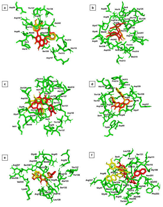
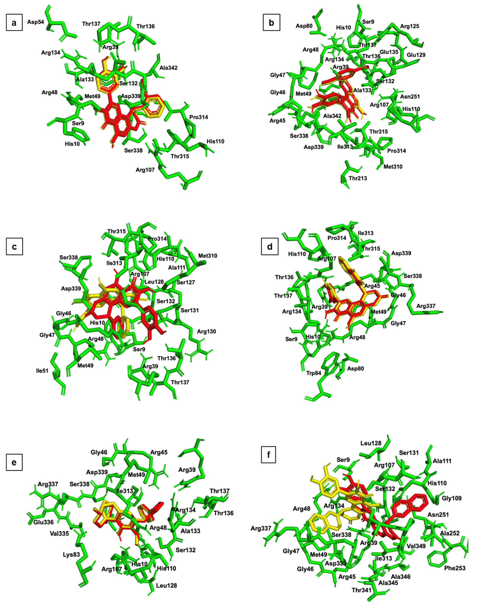
Molecular docking was employed to predict the best binding poses and interaction strengths of ligand analogs with the SpCS dimer. A total of 616 and 346 structural analogs sharing pharmacophore features with EPSP and FMN, respectively, were docked into their corresponding binding sites of the SpCS using the PyRx tool integrated with Autodock Vina software 1.1.2 [38]. Based on docking scores, the top six binding poses were selected for subsequent MD simulations (Figure 6).
Figure 6.
Comparison of binding pockets of 6 potential inhibitors of SpCS: (a) ZINC1780 docked with SpCS, (b) ZINC2684 docked with SpCS, (c) ZINC1317 docked with SpCS, (d) ZINC2116 docked with SpCS, (e) ZINC6435 docked with SpCS, and (f) ZINC2009 docked with SpCS. Ligands in yellow are docked via PyRx and ligands in red docked via Swiss dock (Autodock Vina). The binding pocket is depicted as green color sticks with labeled amino acid residues.
Docking results were evaluated primarily on the basis of binding affinity. For EPSP, 115 compounds were shortlisted from the initial set of 616 analogs due to their favorable binding energies (Supplementary Materials Table S3). Detailed interaction analysis using BIOVIA Discovery Studio Visualizer (https://www.3ds.com/products/biovia/discovery-studio/visualization (accessed on 25 January 2026)) confirmed that these shortlisted compounds interacted with key active-site residues (Ser9, His 10, Arg39, Arg45, Arg48, His110, Ser132, Ala133, Arg134, Arg337) of the EPSP binding pocket, along with additional pocket residues, which likely contributed to their enhanced binding affinities [57] (Supplementary Materials Table S3). The binding energies of these compounds ranged from −8.0 to −10.3 Kcal·mol−1, compared with −6.5 Kcal·mol−1 observed for EPSP. Among them, compound ZINC000218062200 showed the strongest binding affinity (−10.3 Kcal·mol−1).
These results suggest that the selected analogs bind more strongly to SpCS at the EPSP binding site than the native substrate. In contrast, 1-benzofuran-3-one, a known inhibitor of chorismate synthase (22), exhibited a lower binding affinity (−4.7 Kcal·mol−1) when docked at the same site. Docking of the 346 analogs of FMN did not yield compounds with comparable binding affinities or stable interactions at the FMN binding site; therefore, these ligands were not considered for further analysis.
3.5. In Silico Pharmacokinetics and Drug-Likeness Prediction
Drug-likeness and pharmacokinetic suitability of the screened compounds were evaluated using Lipinski’s Rule of Five, a widely accepted framework for predicting absorption, distribution, metabolism, and excretion (ADME) behavior [58]. Given the critical role of ADME properties in determining a drug’s biological fate following administration and its therapeutic efficacy [59], Lipinski’s criteria were applied as an initial filter to identify molecules with favorable membrane permeability and oral bioavailability (logP ≤ 5, molecular weight ≤ 500 Da, hydrogen bond acceptors (HBAs) ≤ 10, hydrogen bond donors (HBDs) ≤ 5, and polar surface area ≤ 140 Å2).
Of the 115 compounds screened, 30 met all Lipinski criteria, indicating favorable pharmacokinetic potential (Supplementary Materials Table S4). These compounds exhibited logP values between 0.21 and 4.9, molecular weights ranging from 257.11 to 499.09 Da, HBA counts of 3–10, HBD counts of 0–5, and PSA values between 41.95 and 136.67 Å2, all within the defined limits. Drug-likeness scoring further refined the selection, identifying 11 compounds with favorable (positive) scores, indicative of higher pharmacological potential. Compounds with zero or negative scores were excluded as non-drug-like candidates [57,60]. Consequently, these 11 compounds were shortlisted for further docking studies with Swiss Dock, as summarized in Table 2.
Table 2.
ADME properties of 11 shortlisted analogs of EPSP analyzed by Molsoft L.L.C (https://molsoft.com/mprop/ (accessed on 20 January 2026)).
3.6. Docking Studies with Swiss Dock
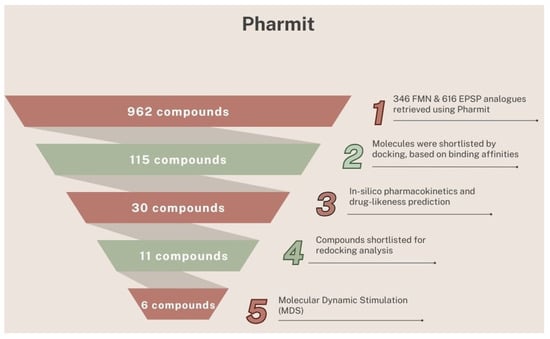
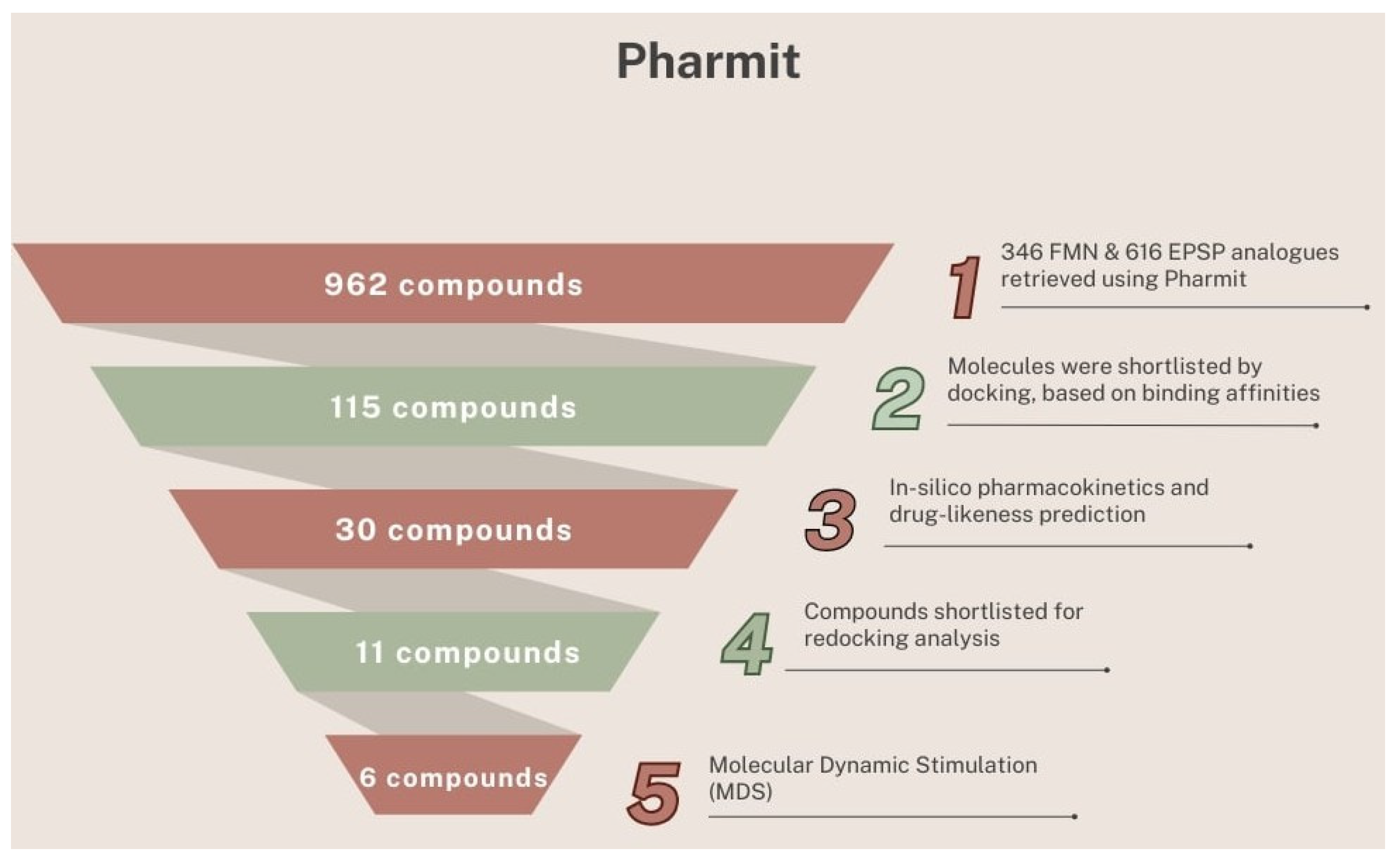
The 11 compounds were docked at the same binding pocket using the Autodock Vina algorithm of the Swiss Dock server (https://www.swissdock.ch/ (accessed on 25 January 2026)), resulting in six molecules shortlisted based on higher binding affinities and favorable orientations compared to the initial docking via PyRx [38] (Figure 6). Additionally, the six ligands along with EPSP and 1-benzofuran-3-one were docked with SpCS to verify the binding pattern of the ligands via a different docking algorithm [61,62,63]. Notably, all three docking analyses showed that these six compounds engaged the SpCS active-site residues at the same binding site as EPSP. Their binding affinities and residue interactions were further compared with EPSP and 1-benzofuran-3-one (Table 3 and Table 4). A detailed workflow illustrating each step and the number of compounds retained at each stage is presented in Figure 7, providing a systematic overview.
Table 3.
Binding affinity of 6 shortlisted analogs of EPSP along with EPSP and 1-benzofuran-3-one calculated via re-docking studies carried out by Swiss Dock (Autodock Vina).
Table 4.
Binding affinity of 6 shortlisted analogs of EPSP along with EPSP and 1-benzofuran-3-one calculated via re-docking studies carried out by Swiss Dock (Attracting Cavities 2.0).
Figure 7.
Funnel approach depicting the gradual steps of dislodging compounds following each parameter.
3.7. MD Simulation Studies
Figure 8 and Figure 9 present the plots of Root Mean Square Deviation (RMSD), hydrogen bonds (H-bonds), and binding energy obtained during the MD simulations. The corresponding plots of Root Mean Square Fluctuation (RMSF) and prevalence of H-bonds are shown in Supplementary Materials Figures S1 and S2.
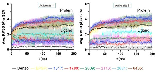
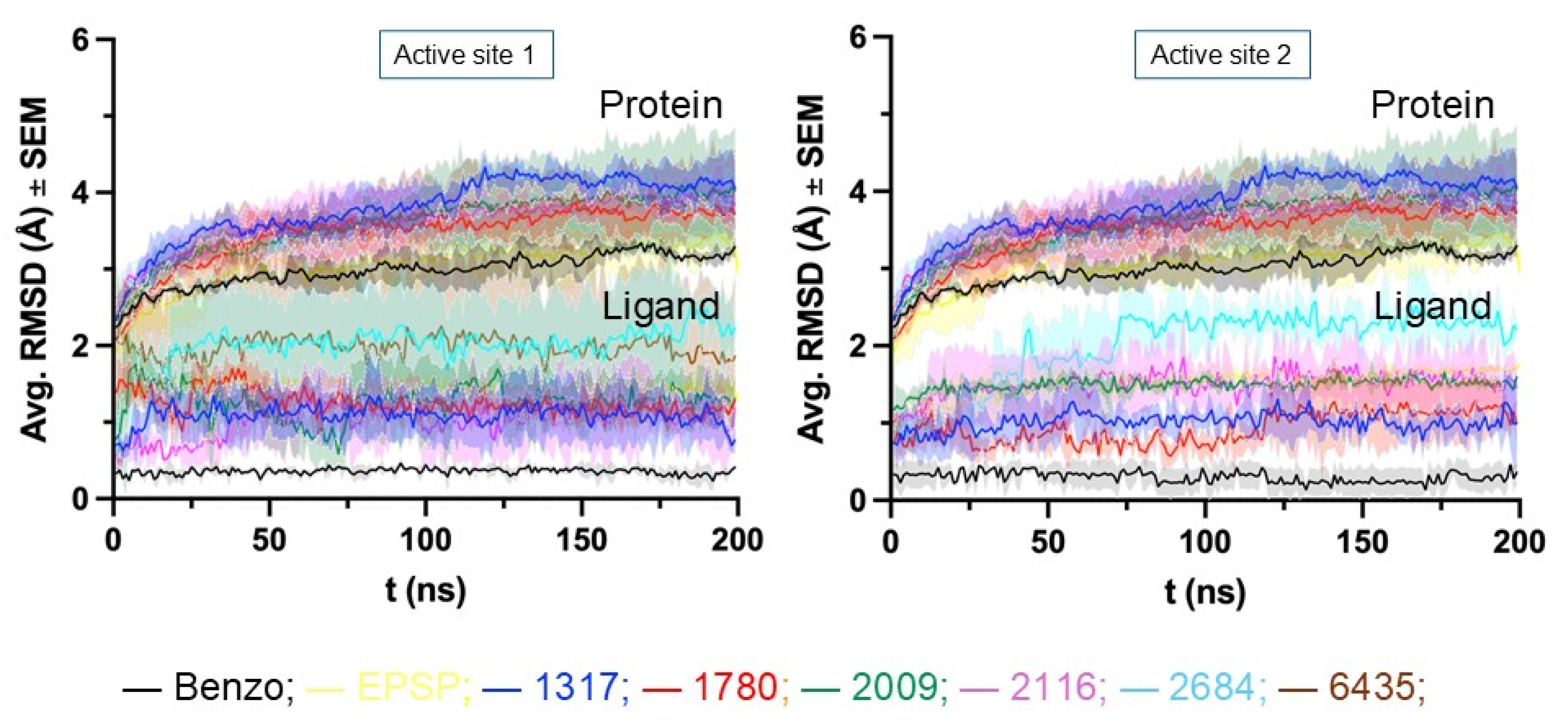
Figure 8.
Variation in RMSD, for the ligands in both active sites over 200 ns of MD simulations. Values represent the averages obtained from three independent simulations. The lighter color above and below each line represents the standard error of the mean (SEM).
Figure 9.
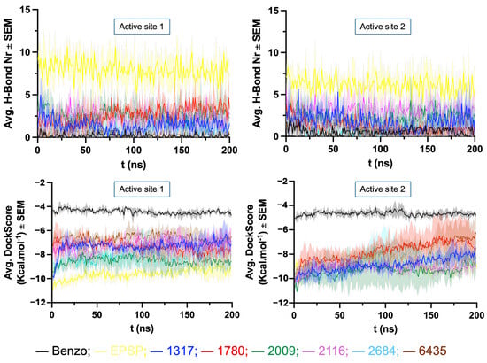
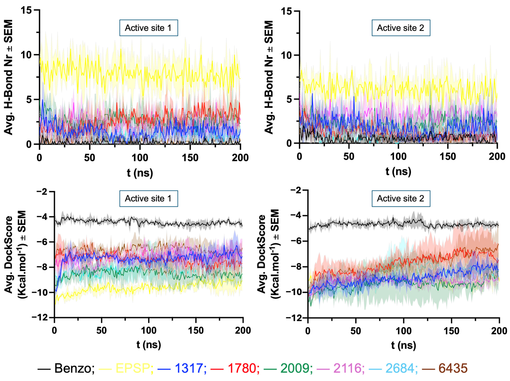
Plots of variation in the number of H-bonds protein–ligand and docking score in both SpCS active sites during each MD simulation. Values represent the averages obtained from three independent simulations. The lighter color above and below each line represents the standard error of the mean (SEM).
The protein RMSD (Figure 8) increased during the initial equilibration phase and subsequently reached a stable plateau at approximately 4.0 Å, remaining essentially constant throughout the 200 ns production simulations. Although the magnitude of the RMSD is relatively high, the absence of progressive structural drift indicates that the systems reached conformational equilibrium. Considering that SpCS is a dimeric enzyme and that the RMSD was calculated for the backbone atoms of the complete dimer, collective inter-subunit motions and flexibility of surface-exposed regions are expected to contribute to the overall RMSD. Consistent with this interpretation, RMSF analysis (Supplementary Materials Figure S1) revealed that the largest fluctuations were localized to flexible regions, whereas the protein core and ligand-binding residues remained comparatively stable throughout the simulations.
The ligand RMSD plots (Figure 8) indicate the stabilization and consistent dynamical behavior, with maximal fluctuations below 2.5 Å and fluctuations between frames no higher than 0.5 Å. 1-benzofuran-3-one demonstrated the most stable behavior, with fluctuations around 0.1 Å between frames. The highest fluctuations were observed around 2.0 Å for ZINC2684 in both active sites and ZINC6435 in active site 1.
Among the ligands, EPSP formed the highest average number of H-bonds inside the SpCS active sites during the MD simulations (7.9 and 6.1 respectively), followed by ZINC2009 (3.00 and 2.29), ZINC2116 (2.00 and 3.09), ZINC1780 (3.0 and 1.2), ZINC317 (1.4 and 1.8), ZINC2684 (1.0 and 1.23), and ZINC6435 (1.00 and 1.14). 1-benzofuran-3-one exhibited the fewest average H-bonds (0.20 and 0.60) (Supplementary Materials Figure S2). This trend is quite consistent with the binding energy data (Figure 9), where the ligands ranked as follows: EPSP (−9.62 and −8.88 kcal/mol), ZINC2009 (−8.43 and −9.48 kcal/mol), ZINC2116 (−7.30 and −9.14 kcal/mol), ZINC1780 (−7.37 and −7.79 kcal/mol), ZINC2317 (−7.30 and −8.88 kcal/mol), ZINC2684 (−8.12 and −8.51 kcal/mol) and ZINC6435 (−6.61 and −7.85). 1-benzofuran-3-one had the highest (least favorable) average binding energies at −4.43 and −4.68 kcal/mol.
Analysis of the MD simulation trajectories also allowed identification of key residues involved in hydrogen bonding with the ligands for more than 8% of the simulation time. As shown in Supplementary Materials Figure S2, these residues include Arg39, Arg45, Arg48, Leu128, Glu129, Ser132, Ala133, Glu336, Arg337 and Asp339. EPSP was the ligand showing the highest prevalence of H-bonds in the SpCS active sites, followed by ZINC2009, ZINC2116, ZINC2684, ZINC1317, ZINC1780, and ZINC6435. As expected, 1-benzofuran-3-one was the one showing the lowest prevalence.
Overall, the MD simulation results suggest that all ZINC ligands, despite not showing the same performance as EPSP, are potential binders to SpCS, forming stronger interactions within the active site compared to 1-benzofuran-3-one. Among them, ZINC2009 and ZINC2116 emerged as the most promising candidates, followed by ZINC2684, ZINC1317, ZINC1780, and ZINC6435.
4. Discussion
S. pneumoniae continues to challenge existing therapeutic strategies due to its increasing resistance to conventional antibiotics, underscoring the need for novel drug targets and inhibitory scaffolds [64]. Metabolic pathways that are essential for bacterial survival yet absent in humans offer a rational framework for selective drug discovery [65,66]. Among these enzymes, CS catalyzes the final and irreversible step of the shikimate pathway, making it crucial for the biosynthesis of aromatic amino acids and downstream metabolites, which support bacterial growth and pathogenicity. Specifically, CS catalyzes the conversion of EPSP to chorismate, using reduced FMN as a cofactor, thereby completing the terminal step of the shikimate pathway [64]. As a result, CS functions as a critical metabolic control point and an attractive but underexplored target in S. pneumoniae. In the present study, we systematically explored the inhibitory potential of multiple natural substrate analogs with substantially higher binding affinities toward CS than the native substrate using an integrated in silico screening strategy.
The structure of SpCS was retrieved from RCSB-PDB (https://www.rcsb.org/) and characterized as a homotetramer with ligands FMN and EPSP binding to the same pocket. The 616 EPSP structural analogs, obtained via the Pharmit web server (https://pharmit.csb.pitt.edu/) were docked to the binding pocket of SpCS using PyRx tool incorporated with Autodock Vina software [38]. Among the screened compounds, a total of 115 compounds structurally similar to EPSP and exhibiting higher binding affinities compared to the substrate were shortlisted. The screened compounds were subjected to ADME analysis, and out of these, 30 compounds were selected based on Lipinski’s rules. Amongst the 30 compounds, 11 compounds exhibiting high drug-likeness scores were further considered for additional docking studies. The docking was carried out through the Swiss Dock server (https://www.swissdock.ch/ (accessed on 25 January 2026)), to the same binding site of CS. Further, the stability of these shortlisted compounds bound to CS was analyzed through MD simulations.
These six finalized molecules, identified by their ZINC IDs—ZINC000096221317 (ZINC1317), ZINC000096221780 (ZINC1780), ZINC000096222009 (ZINC2009), ZINC000096222684 (ZINC2684), ZINC000096222116 (ZINC2116) and ZINC000096316435 (ZINC6435)—displayed binding affinities in the range of −8.0 to −9.5 Kcal·mol−1 when docked to SpCS using the PyRx tool incorporated with Autodock Vina software, which exhibited a higher binding affinity than its substrate EPSP recorded as −6.5 Kcal·mol−1. Among these, the compound ZINC009 displayed the highest binding affinity of −9.5 Kcal·mol−1. Further, the structural interaction analysis indicated that all six compounds established multiple interactions with SpCS, with several residues overlapping those involved in EPSP binding. ZINC1317 interacted with 10 residues, where 4 residues were known to interact with EPSP; ZINC1780 interacted with 9 residues, of which 5 residues were known to interact with EPSP; ZINC2009 interacted with 8 residues, among which 3 residues were known to interact with EPSP; ZINC2684 interacted with 12 residues, including 5 residues identified to interact with EPSP; ZINC2116 interacted with 10 residues, among which 4 residues were associated with EPSP interactions; and ZINC6435 interacted with 7 residues, among which 4 residues were known to interact with EPSP. These observations further suggest the compounds’ binding affinity for CS. The ADME properties of the compounds were evaluated using Lipinski’s Rule of Five, assessing their molecular weight, the number of hydrogen bond acceptors (HBAs), the number of hydrogen bond donors (HBDs), LogP, and Polar Surface Area (PSA). Drug-likeness scores were also predicted, and all these evaluated parameters are presented in Table 2. The Swiss Dock docking studies revealed that the screened compounds bind to the same binding pocket as the substrate and the inhibitor 1-benzofuran-3-one, while exhibiting higher binding affinities than both. The docking analysis performed using PyRx VS software and the Swiss Dock server showed similar binding patterns among the compounds, indicating consistency across platforms. This indicates the potential of these compounds to serve as inhibitors of SpCS. The six finalized docked enzyme–ligand complexes were subjected to the MDS studies to assess their stability and interactions. The SpCS bound to 1-benzofuran-3-one was used as a reference complex for comparison with the selected six compounds.
Similar to the docking results, compound ZINC2009 emerged as the most promising candidate, followed by ZINC2116 and ZINC2684, which formed the most hydrogen bonds. These compounds—ZINC2009 (10-(2H-chromen-3-yl)-2-(3,4-dihydroxyphenyl)-3,5-dihydroxy-4H,8H,9H,10H-pyrano[2,3-h]chromene-4,8-dione), ZINC2116 (2-(3,4-dihydroxyphenyl)-3,5-dihydroxy-10-(pyridin-3-yl)-4H,8H,9H,10H-pyrano[2,3-h]chromene-4,8-dione) and ZINC2684 (2-(3,4-dihydroxyphenyl)-10-(2,5-dimethoxyphenyl)-3,5-dihydroxy-4H, 8H, 9H, 10H-pyrano[2,3-h]chromene-4,8-dione)—are phenyl derivatives. Studies reveal that the phenol scaffold has numerous well-documented profiles, including antiviral, antibacterial, antimutagenic, and anti-inflammatory properties [66]. Moreover, phenyl derivatives as inhibitors have shown favorable inhibitory activity as they can effectively engage the CS active site [21]. The presence of a phenyl moiety possibly enhances binding affinity through aromatic and hydrophobic interactions within the active site [67].
These results point to the potential of these compounds as candidate inhibitors for SpCS, exhibiting favorable binding, stability, and drug-like properties through integrated docking, ADME profiling, toxicity prediction, and MD simulations.
5. Conclusions
In conclusion, this study identifies three natural compounds ZINC2009, ZINC2116, and ZINC2684 as candidate inhibitors for SpCS, requiring experimental validation. A comprehensive in silico analysis, including molecular docking, ADME profiling, and MD simulations, suggests that these compounds might exhibit strong binding affinities and maintain stable interactions within the SpCS active site, outperforming the natural substrate EPSP in binding efficiency. Among these, ZINC2009 and ZINC2116 demonstrated a significant number of H-bonds, indicating strong ligand substrate affinity and positioning them as strong candidates for further development as potential therapeutics against pneumonia.
Collectively, these findings establish SpCS as a viable antimicrobial target and highlight the identified compounds as potential promising leads warranting experimental validation for the development of next-generation therapeutics against drug-resistant S. pneumoniae.
Supplementary Materials
The following supporting information can be downloaded at https://www.mdpi.com/article/10.3390/biophysica6040069/s1, Supplementary Table S1: FMN analogues extracted from Pharmit—Zinc_NAT Database. Supplementary Table S2: EPSP Analogues extracted from Pharmit- Zinc_NAT Database. Supplementary Table S3: Docked EPSP shortlisted analogues based on their estimated Binding Affinities. Supplementary Table S4: Shortlisted EPSP molecules and their ADME as well as drug likeness properties. Supplementary Figure S1: RMSF plots of SpCS dimer. The RMSF graph depicting chain A and chain B of dimer bound to FMN, EPSP and EPSP analogues. Supplementary Figure S2: H-bond prevalence plot of SpCS dimer. H-bond plots of SpCS dimer bound to FMN, EPSP and EPSP analogues.
Author Contributions
Writing—original draft preparation, methodology, data curation, writing—review and editing, resources, D.M.A. and S.B.; writing—review and editing, methodology, resources, D.M.A., S.B., G.M.U. and R.A.; writing—review and editing, software, formal analysis, D.M.A., S.B., R.A., N.M., T.C.C.F. and S.R.L.; Conceptualization, writing—review and editing, investigation, validation, N.M., O.Z., T.C.C.F. and S.R.L. All authors have read and agreed to the published version of the manuscript.
Funding
This research received no external funding.
Institutional Review Board Statement
Not applicable.
Informed Consent Statement
Not applicable.
Data Availability Statement
The original contributions presented in this study are included in the article/Supplementary Materials. Further inquiries can be directed to the corresponding authors.
Acknowledgments
The authors of this article extend their gratitude to Department of Botany; St. Joseph’s University, Bengaluru for its constant support and assistance, which helped the authors in the successful completion of this research work. The authors gratefully acknowledge the California University of Science and Medicine for its support of this publication. T.C.C.F. thanks the Excellence project PřF UHK 2216/2023-2024 and the CNPq agency, Brazil (Process No. 305432/2022-2) for research support.
Conflicts of Interest
The authors declare that there are no conflicts of interest.
Abbreviations
The following abbreviations are used in this manuscript:
| ADME | absorption, distribution, metabolism, excretion |
| CADD | computer-aided drug design |
| CS | chorismate synthase |
| EPSP | 5-enolpyruvylshikimate-3-phosphate |
| FMN | flavin mononucleotide |
| HBA | hydrogen bond acceptors |
| HBD | hydrogen bond donors |
| MDS | molecular dynamics simulation |
| MolPSA | molecular polar surface area |
| PDB | protein data bank |
| RMSD | Root Mean Square Deviation |
| RMSF | Root Mean Square Fluctuation |
| VS | virtual screening |
References
- Borsa, N.; Di Pasquale, M.; Restrepo, M.I. Animal models of pneumococcal pneumonia. Int. J. Mol. Sci. 2019, 20, 4220. [Google Scholar] [CrossRef] [Scilit]
- Musher, D.M.; Thorner, A.R. Community-acquired pneumonia. N. Engl. J. Med. 2014, 371, 1619–1628. [Google Scholar] [CrossRef] [Scilit] [PubMed]
- Biscevic-Tokic, J.; Tokic, N.; Musanovic, A. Pneumonia as the most common lower respiratory tract infection. Med. Arch. 2013, 67, 442–445. [Google Scholar] [CrossRef] [Scilit] [PubMed]
- Hoare, Z.; Lim, W.S. Pneumonia: Update on diagnosis and management. BMJ 2006, 332, 1077–1079. [Google Scholar] [CrossRef] [Scilit] [PubMed]
- Podolsky, S.H. The changing fate of pneumonia as a public health concern in 20th-century America and beyond. Am. J. Public Health 2005, 95, 2144–2154. [Google Scholar] [CrossRef] [Scilit] [PubMed]
- Allen, K.D. Penicillin-resistant pneumococci. J. Hosp. Infect. 1991, 17, 3–13. [Google Scholar] [CrossRef] [Scilit] [PubMed]
- Sharew, B.; Moges, F.; Yismaw, G.; Abebe, W.; Fentaw, S.; Vestrheim, D.; Tessema, B. Antimicrobial resistance profile and multidrug resistance patterns of Streptococcus pneumoniae isolates from patients suspected of pneumococcal infections in Ethiopia. Ann. Clin. Microbiol. Antimicrob. 2021, 20, 26. [Google Scholar] [CrossRef] [Scilit] [PubMed]
- Siddiqi, R.; Javaid, S. Deep learning for pneumonia detection in chest X-ray images: A comprehensive survey. J. Imaging 2024, 10, 176. [Google Scholar] [CrossRef] [Scilit] [PubMed]
- Eshwara, V.K.; Mukhopadhyay, C.; Rello, J. Community-acquired bacterial pneumonia in adults: An update. Indian J. Med. Res. 2020, 151, 287–302. [Google Scholar] [CrossRef] [Scilit] [PubMed]
- Wu, S.; Chen, W.; Lu, S.; Zhang, H.; Yin, L. Metabolic engineering of shikimic acid biosynthesis pathway for the production of shikimic acid and its branched products in microorganisms: Advances and prospects. Molecules 2022, 27, 4779. [Google Scholar] [CrossRef] [Scilit] [PubMed]
- Park, H.; Hilsenbeck, J.L.; Kim, H.J.; Shuttleworth, W.A.; Park, Y.H.; Evans, J.N.; Kang, C. Structural studies of Streptococcus pneumoniae EPSP synthase in unliganded state, tetrahedral intermediate-bound state and S3P-GLP-bound state. Mol. Microbiol. 2004, 51, 963–971. [Google Scholar] [CrossRef] [Scilit] [PubMed]
- Amer, F.A.; El-Behedy, E.M.; Mohtady, H.A. New targets for antibacterial agents. Biotechnol. Mol. Biol. Rev. 2008, 3, 46–57. [Google Scholar]
- Herrmann, K.M.; Weaver, L.M. The shikimate pathway. Annu. Rev. Plant Biol. 1999, 50, 473–503. [Google Scholar] [CrossRef] [Scilit] [PubMed]
- Tzin, V.; Galili, G.; Aharoni, A. Shikimate pathway and aromatic amino acid biosynthesis. eLS 2012, 8, 32. [Google Scholar] [CrossRef] [Scilit]
- Dosselaere, F.; Vanderleyden, J. A metabolic node in action: Chorismate-utilizing enzymes in microorganisms. Crit. Rev. Microbiol. 2001, 27, 75–131. [Google Scholar] [CrossRef] [Scilit] [PubMed]
- Han, Q.; Phillips, R.S.; Li, J. Editorial: Aromatic amino acid metabolism. Front. Mol. Biosci. 2019, 6, 22. [Google Scholar] [CrossRef] [Scilit] [PubMed]
- Dias, M.V.; Borges, J.C.; Ely, F.; Pereira, J.H.; Canduri, F.; Ramos, C.H.; Frazzon, J.; Palma, M.S.; Basso, L.A.; Santos, D.S.; et al. Structure of chorismate synthase from Mycobacterium tuberculosis. J. Struct. Biol. 2006, 154, 130–143. [Google Scholar] [CrossRef] [Scilit] [PubMed]
- Chook, Y.M.; Ke, H.; Lipscomb, W.N. Crystal structures of the monofunctional chorismate mutase from Bacillus subtilis and its complex with a transition state analog. Proc. Natl. Acad. Sci. USA 1993, 90, 8600–8603. [Google Scholar] [CrossRef] [Scilit] [PubMed]
- Quevillon-Cheruel, S.; Leulliot, N.; Meyer, P.; Graille, M.; Bremang, M.; Blondeau, K.; Sorel, I.; Poupon, A.; Janin, J.; van Tilbeurgh, H. Crystal structure of the bifunctional chorismate synthase from Saccharomyces cerevisiae. J. Biol. Chem. 2004, 279, 619–625. [Google Scholar] [CrossRef] [Scilit] [PubMed]
- Zhou, H.; Singh, N.J.; Kim, K.S. Homology modeling and molecular dynamics study of chorismate synthase from Shigella flexneri. J. Mol. Graph. Model. 2006, 25, 434–441. [Google Scholar] [CrossRef] [Scilit] [PubMed]
- Fuchs, K.; Totaro, M.G.; Toplak, M.; Bijelic, A.; Macheroux, P. Investigation of the inhibitory properties of azo-dyes on chorismate synthase from Paracoccidioides brasiliensis. J. Enzym. Inhib. Med. Chem. 2024, 39, 2427175. [Google Scholar] [CrossRef] [Scilit] [PubMed]
- Thomas, M.G.; Lawson, C.; Allanson, N.M.; Leslie, B.W.; Bottomley, J.R.; McBride, A.; Olusanya, O.A. A series of 2(Z)-2-benzylidene-6,7-dihydroxybenzofuran-3[2H]-ones as inhibitors of chorismate synthase. Bioorg. Med. Chem. Lett. 2003, 13, 423–426. [Google Scholar] [CrossRef] [Scilit] [PubMed]
- Neetu, N.; Sharma, M.; Mahto, J.K.; Kumar, P. Biophysical and in-silico studies of phytochemicals targeting chorismate synthase from drug-resistant Moraxella catarrhalis. Protein J. 2020, 39, 449–460. [Google Scholar] [CrossRef] [Scilit] [PubMed]
- Hanif, M.; Khan, S.; Farooq, U.; Nouroz, F.; Sarwar, R. Unraveling the possible inhibitors for chorismate synthase to combat tuberculosis using in silico approach. J. Biomol. Struct. Dyn. 2023, 41, 2823–2830. [Google Scholar] [CrossRef] [Scilit] [PubMed]
- Vanajothi, R.; Bhavaniramya, S.; Vijayakumar, R.; Alothaim, A.S.; Alqurashi, Y.E.; Vishnupriya, S.; Vaseeharan, B.; Umadevi, M. In silico and in vitro analysis of Nigella sativa bioactives against chorismate synthase of Listeria monocytogenes: A target protein for biofilm inhibition. Appl. Biochem. Biotechnol. 2023, 195, 519–533. [Google Scholar] [CrossRef] [Scilit] [PubMed]
- Tapas, S.; Kumar, A.; Dhindwal, S.; Kumar, P. Structural analysis of chorismate synthase from Plasmodium falciparum: A novel target for antimalaria drug discovery. Int. J. Biol. Macromol. 2011, 49, 767–777. [Google Scholar] [CrossRef] [Scilit] [PubMed]
- Dias, M.V.B.; Ely, F.; Palma, M.S.; de Azevedo, W.F.; Basso, L.A.; Santos, D.S. Chorismate synthase: An attractive target for drug development against orphan diseases. Curr. Drug Targets 2007, 8, 437–444. [Google Scholar] [CrossRef] [Scilit] [PubMed]
- Subhaswaraj, P.; Siddhardha, B. Molecular docking and molecular dynamic simulation approaches for drug development and repurposing of drugs for severe acute respiratory syndrome-coronavirus-2. In Computational Approaches for Novel Therapeutic and Diagnostic Designing to Mitigate SARS-CoV-2 Infection; Elsevier: Amsterdam, The Netherlands, 2022; pp. 207–246. [Google Scholar] [CrossRef] [Scilit]
- Oselusi, S.O.; Dube, P.; Odugbemi, A.I.; Akinyede, K.A.; Ilori, T.L.; Egieyeh, E.; Sibuyi, N.R.S.; Meyer, M.; Madiehe, A.M.; Wyckoff, G.J.; et al. The role and potential of computer-aided drug discovery strategies in the discovery of novel antimicrobials. Comput. Biol. Med. 2024, 169, 107927. [Google Scholar] [CrossRef] [Scilit] [PubMed]
- Giordano, D.; Biancaniello, C.; Argenio, M.A.; Facchiano, A. Drug design by pharmacophore and virtual screening approach. Pharmaceuticals 2022, 15, 646. [Google Scholar] [CrossRef] [Scilit] [PubMed]
- Zhu, H.; Zhou, R.; Cao, D.; Tang, J.; Li, M. A pharmacophore-guided deep learning approach for bioactive molecular generation. Nat. Commun. 2023, 14, 6234. [Google Scholar] [CrossRef] [Scilit] [PubMed]
- Agu, P.C.; Afiukwa, C.A.; Orji, O.U.; Ezeh, E.M.; Ofoke, I.H.; Ogbu, C.O.; Ugwuja, E.I.; Aja, P.M. Molecular docking as a tool for the discovery of molecular targets of nutraceuticals in diseases management. Sci. Rep. 2023, 13, 13398. [Google Scholar] [CrossRef] [Scilit] [PubMed]
- Kapetanovic, I. Computer-aided drug discovery and development (CADDD): In silico-chemico-biological approach. Chem. Biol. Interact. 2008, 171, 165–176. [Google Scholar] [CrossRef] [Scilit] [PubMed]
- Wiederstein, M.; Sippl, M.J. ProSA-web: Interactive web service for the recognition of errors in three-dimensional structures of proteins. Nucleic Acids Res. 2007, 35, W407–W410. [Google Scholar] [CrossRef] [Scilit] [PubMed]
- Ayodele, P.F.; Bamigbade, A.; Bamigbade, O.O.; Adeniyi, I.A.; Tachin, E.S.; Seweje, A.J.; Farohunbi, S.T. Illustrated procedure to perform molecular docking using PyRx and Biovia Discovery Studio Visualizer: A case study of 10KT with atropine. Prog. Drug Discov. Biomed. Sci. 2023, 6, 1–31. [Google Scholar] [CrossRef] [Scilit]
- Jendele, L.; Krivak, R.; Skoda, P.; Novotny, M.; Hoksza, D. PrankWeb: A web server for ligand binding site prediction and visualization. Nucleic Acids Res. 2019, 47, W345–W349. [Google Scholar] [CrossRef] [Scilit] [PubMed]
- Sunseri, J.; Koes, D.R. Pharmit: Interactive exploration of chemical space. Nucleic Acids Res. 2016, 44, W442–W448. [Google Scholar] [CrossRef] [Scilit] [PubMed]
- Trott, O.; Olson, A.J. AutoDock Vina: Improving the speed and accuracy of docking with a new scoring function, efficient optimization, and multithreading. J. Comput. Chem. 2010, 31, 455–461. [Google Scholar] [CrossRef] [Scilit] [PubMed]
- Brüstle, M.; Beck, B.; Schindler, T.; King, W.; Mitchell, T.; Clark, T. Descriptors, physical properties, and drug-likeness. J. Med. Chem. 2002, 45, 3345–3355. [Google Scholar] [CrossRef] [Scilit] [PubMed]
- Clark, D.E.; Pickett, S.D. Computational methods for the prediction of ‘drug-likeness’. Drug Discov. Today 2000, 5, 49–58. [Google Scholar] [CrossRef] [Scilit] [PubMed]
- Egan, W.J.; Walters, W.P.; Murcko, M.A. Guiding molecules towards drug-likeness. Curr. Opin. Drug Discov. Dev. 2002, 5, 540–549. [Google Scholar]
- Müller, K.R.; Rätsch, G.; Sonnenburg, S.; Mika, S.; Grimm, M.; Heinrich, N. Classifying ‘drug-likeness’ with kernel-based learning methods. J. Chem. Inf. Model. 2005, 45, 249–253. [Google Scholar] [CrossRef] [Scilit] [PubMed]
- Lipinski, C.A. Drug-like properties and the causes of poor solubility and poor permeability. J. Pharmacol. Toxicol. Methods 2000, 44, 235–249. [Google Scholar] [CrossRef] [Scilit] [PubMed]
- Eberhardt, J.; Santos-Martins, D.; Tillack, A.F.; Forli, S. AutoDock Vina 1.2.0: New docking methods, expanded force field, and Python bindings. J. Chem. Inf. Model. 2021, 61, 3891–3898. [Google Scholar] [CrossRef] [Scilit] [PubMed]
- Ali, Z.; Cardoza, J.V.; Basak, S.; Narsaria, U.; Singh, V.P.; Isaac, S.P.; França, T.C.C.; LaPlante, S.R.; George, S.S. Computational design of candidate multi-epitope vaccine against SARS-CoV-2 targeting structural (S and N) and non-structural (NSP3 and NSP12) proteins. J. Biomol. Struct. Dyn. 2023, 41, 13348–13367. [Google Scholar] [CrossRef] [Scilit] [PubMed]
- França, T.C.C.; Goncalves, A.D.S.; Berube, C.; Voyer, N.; Aubry, N.; LaPlante, S.R. Determining the predominant conformations of Mortiamides A–D in solution using NMR data and molecular modeling tools. ACS Omega 2023, 8, 25832–25838. [Google Scholar] [CrossRef] [Scilit] [PubMed]
- França, T.C.C.; Saïdi, F.; Ajamian, A.; Islam, S.T.; LaPlante, S.R. Molecular dynamics of outer membrane-embedded polysaccharide secretion porins reveals closed resting-state surface gates targetable by virtual fragment screening for drug hotspot identification. ACS Omega 2024, 9, 13217–13226. [Google Scholar] [CrossRef] [Scilit] [PubMed]
- França, T.C.C.; Maddalena, M.; Kouidmi, I.; Ayotte, Y.; Islam, S.T.; LaPlante, S.R. SI/II pocket of Ras: An opportunity for a once “undruggable” target. ACS Omega 2025, 10, 9463–9473. [Google Scholar] [CrossRef] [Scilit] [PubMed]
- Chemical Computing Group ULC. Molecular Operating Environment (MOE); Version 2024.06 [Computer software]; Chemical Computing Group ULC: Montreal, QC, Canada, 2024. [Google Scholar]
- Nelson, M.T.; Humphrey, W.; Gursoy, A.; Dalke, A.; Kalé, L.V.; Skeel, R.D.; Schulten, K. NAMD: A parallel, object-oriented molecular dynamics program. Int. J. Supercomput. Appl. High Perform. Comput. 1996, 10, 251–268. [Google Scholar] [CrossRef] [Scilit]
- Labute, P. The generalized Born/volume integral implicit solvent model: Estimation of the free energy of hydration using London dispersion instead of atomic surface area. J. Comput. Chem. 2008, 29, 1693–1698. [Google Scholar] [CrossRef] [Scilit] [PubMed]
- Humphrey, W.; Dalke, A.; Schulten, K. VMD: Visual molecular dynamics. J. Mol. Graph. 1996, 14, 33–38. [Google Scholar] [CrossRef] [Scilit] [PubMed]
- Maclean, J.; Ali, S. The structure of chorismate synthase reveals a novel flavin binding site fundamental to a unique chemical reaction. Structure 2003, 11, 1499–1511. [Google Scholar] [CrossRef] [Scilit] [PubMed]
- Krivák, R.; Hoksza, D. P2Rank: Machine learning based tool for rapid and accurate prediction of ligand binding sites from protein structure. J. Cheminform. 2018, 10, 39. [Google Scholar] [CrossRef] [Scilit] [PubMed]
- Kumar, B.K.; Faheem, N.; Sekhar, K.V.G.C.; Ojha, R.; Prajapati, V.K.; Pai, A.; Murugesan, S. Pharmacophore based virtual screening, molecular docking, molecular dynamics and MM-GBSA approach for identification of prospective SARS-CoV-2 inhibitor from natural product databases. J. Biomol. Struct. Dyn. 2022, 40, 1363–1386. [Google Scholar] [CrossRef] [Scilit] [PubMed]
- Bhattacharjee, S.; Gouda, T.; Anusha, D.M.; Bauldas, S.; Franca, T.C.C.; LaPlante, S.R.; Shen, G.; Mishra, N. Molecular docking and molecular dynamic simulation studies to identify potential isoquinoline alkaloid derivatives against chorismate synthase of Listeria monocytogenes. Comput. Struct. Biotechnol. Rep. 2025, 2, 100075. [Google Scholar] [CrossRef] [Scilit]
- Karami, T.K.; Hailu, S.; Feng, S.; Graham, R.; Gukasyan, H.J. Eyes on Lipinski’s rule of five: A new “rule of thumb” for physicochemical design space of ophthalmic drugs. J. Ocul. Pharmacol. Ther. 2022, 38, 43–55. [Google Scholar] [CrossRef] [Scilit] [PubMed]
- Celik, I.; Erol, M.; Temiz Arpaci, O.; Sezer Senol, F.; Erdogan Orhan, I. Evaluation of activity of some 2, 5-disubstituted benzoxazole derivatives against acetylcholinesterase, butyrylcholinesterase and tyrosinase: ADME prediction, DFT and comparative molecular docking studies. Polycycl. Aromat. Compd. 2022, 42, 412–423. [Google Scholar] [CrossRef] [Scilit]
- Cheng, F.; Li, W.; Zhou, Y.; Shen, J.; Wu, Z.; Liu, G.; Lee, P.W.; Tang, Y. AdmetSAR: A comprehensive source and free tool for assessment of chemical ADMET properties. J. Chem. Inf. Model. 2012, 52, 3099–3105. [Google Scholar] [CrossRef] [Scilit] [PubMed]
- Bugnon, M.; Röhrig, U.F.; Goullieux, M.; Perez, M.A.S.; Daina, A.; Michielin, O.; Zoete, V. SwissDock 2024: Major enhancements for small-molecule docking with Attracting Cavities and AutoDock Vina. Nucleic Acids Res. 2024, 52, W324–W332. [Google Scholar] [CrossRef] [Scilit] [PubMed]
- Röhrig, U.F.; Goullieux, M.; Bugnon, M.; Zoete, V. Attracting cavities 2.0: Improving the flexibility and robustness for small-molecule docking. J. Chem. Inf. Model. 2023, 63, 3925–3940. [Google Scholar] [CrossRef] [Scilit] [PubMed]
- Zoete, V.; Schuepbach, T.; Bovigny, C.; Chaskar, P.; Daina, A.; Röhrig, U.F.; Michielin, O. Attracting cavities for docking: Replacing the rough energy landscape of the protein by a smooth attracting landscape. J. Comput. Chem. 2016, 37, 437–447. [Google Scholar] [CrossRef] [Scilit] [PubMed]
- Kolawole, O.C. Design and docking studies of inhibitors for the chorismate synthase from Streptococcus pneumoniae using 5-enol pyruvylshikimate 3-phosphate (EPSP) analogues. J. Pharm. Bioresour. 2015, 12, 61–76. [Google Scholar] [CrossRef] [Scilit]
- Du, W.; Wallis, N.G.; Payne, D.J. The kinetic mechanism of 5-enolpyruvylshikimate-3-phosphate synthase from a Gram-positive pathogen Streptococcus pneumoniae. J. Enzym. Inhib. 2000, 15, 571–581. [Google Scholar] [CrossRef] [Scilit] [PubMed]
- Du, W.; Wallis, N.G.; Mazzulla, M.J.; Chalker, A.F.; Zhang, L.; Liu, W.S.; Kallender, H.; Payne, D.J. Characterization of Streptococcus pneumoniae 5-enolpyruvylshikimate 3-phosphate synthase and its activation by univalent cations. Eur. J. Biochem. 2000, 267, 222–227. [Google Scholar] [CrossRef] [Scilit] [PubMed]
- Dunker, C.; Schlegel, K.; Junker, A. Phenol (bio)isosteres in drug design and development. Arch. Pharm. 2025, 358, e2400700. [Google Scholar] [CrossRef] [Scilit] [PubMed]
- Brylinski, M. Aromatic interactions at the ligand-protein interface: Implications for the development of docking scoring functions. Chem. Biol. Drug Des. 2018, 91, 380–390. [Google Scholar] [CrossRef] [Scilit] [PubMed]

Disclaimer/Publisher’s Note: The statements, opinions and data contained in all publications are solely those of the individual author(s) and contributor(s) and not of MDPI and/or the editor(s). MDPI and/or the editor(s) disclaim responsibility for any injury to people or property resulting from any ideas, methods, instructions or products referred to in the content. |
© 2026 by the authors. Licensee MDPI, Basel, Switzerland. This article is an open access article distributed under the terms and conditions of the Creative Commons Attribution (CC BY) license.