


Abstract
Metastatic spinal cord compression (MSCC) requires urgent radiotherapy. However, selecting the optimal technique that balances dosimetric precision with treatment efficiency remains clinically challenging. This study compares four radiotherapy modalities for MSCC—AP-PA with field-in-field, 3D-CRT, 5-field IMRT, and 7-field IMRT—evaluating their precision–efficiency trade-offs and proposing an ALARA-based clinical framework. In this observational dosimetry study, we analyzed treatment plans from 60 consecutive MSCC patients. Four plans were generated per patient (30 Gy/10 fractions). We assessed target coverage (D95%), conformity (Paddick CI), homogeneity (HI), organ-at-risk sparing, and treatment efficiency. The statistical analysis used a repeated-measures ANOVA with Bonferroni correction. IMRT-7 achieved superior dosimetric outcomes, with a target coverage of 95.8% ± 1.5% and a spinal cord dose of 7.4 ± 1.2 Gy—a 70.6% reduction versus AP-PA—but required prolonged sessions (16.9 ± 1.4 min). 3D-CRT provided acceptable coverage (91.2% ± 2.3%) and cord dose (22.8 ± 1.5 Gy) with significantly shorter delivery times (8.7 ± 1.3 min). IMRT-5 offered an intermediate option (cord dose: 8.2 ± 1.3 Gy; session time: 13.4 ± 0.8 min). IMRT benefits were most pronounced in lumbar lesions (68% cord dose reduction) and least in thoracic segments. IMRT offers superior dosimetry but distributes low-dose radiation to larger healthy tissue volumes. Although these doses remain within tolerance, they may conflict with the ALARA principle, whereas 3D-CRT with field-in-field achieves similar coverage and organ sparing in less time. Thus, 3D-CRT is preferred in most palliative settings. However, IMRT or arc therapy becomes essential for re-irradiation or when prior treatment to adjacent levels demands maximal cord sparing. Technique selection should integrate dosimetric goals with clinical context, patient status, and institutional resources.
1. Introduction
Metastatic spinal cord compression (MSCC) is an oncologic emergency requiring prompt intervention to prevent permanent neurologic damage, affecting ~10% of cancer patients and up to 19% of cases of breast, prostate, or lung malignancies [1,2,3]. While radiotherapy remains the primary palliative modality, selecting the optimal technique that balances dose precision, organ sparing, and treatment efficiency remains challenging—particularly when incorporating the ALARA principle and patient-specific factors like performance status and immobilization tolerance [4,5].
Previous studies have examined spinal radiotherapy techniques: Rades et al. [6] and Lutz et al. [7] demonstrated 3D-CRT’s rapid delivery with adequate coverage for palliative settings, while Ding et al. [8] and Lam et al. [9] validated IMRT’s superior precision and cord sparing despite longer delivery times. Multicenter comparisons [10] confirm that IMRT/VMAT achieve better conformity than 3D-CRT, though randomized trials suggest that clinical outcomes are more nuanced than dosimetric parameters alone [11]. However, these studies lacked within-patient comparative designs controlling for inter-patient variability, and few have translated technical comparisons into actionable clinical algorithms incorporating patient-specific factors and resources [7,12].
This study addresses this gap through the systematic dosimetric comparison of four techniques (AP-PA with field-in-field, 3D-CRT, 5-field IMRT, and 7-field IMRT) in 60 MSCC patients, evaluating target coverage, conformity, homogeneity, OAR sparing, and efficiency to establish an evidence-based, ALARA-informed framework for personalized technique selection.
Novel Contributions
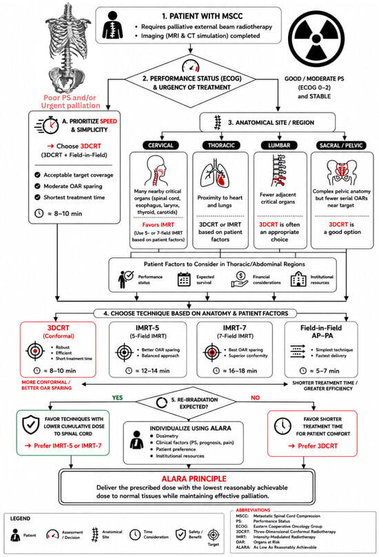
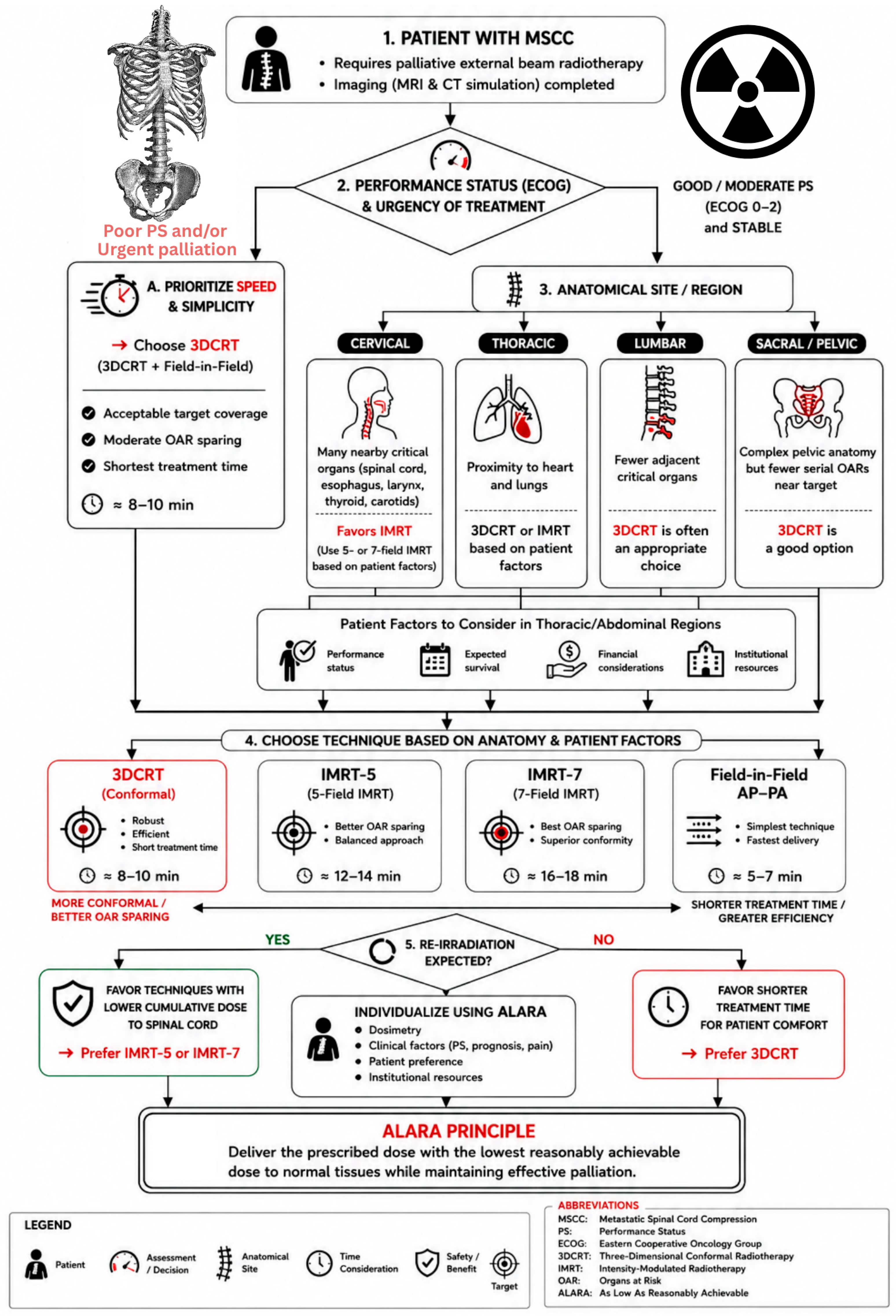
This study advances beyond the existing literature through three distinctive contributions. First, the within-patient design—four plans per 60 identical anatomies—eliminates inter-patient variability, enabling true technique-specific comparison. Second, spatiotemporal integration examines performance across spinal levels, revealing anatomical determinants of optimal technique selection that were previously overlooked. Third, by integrating the ALARA principle, this study transforms technical comparisons into clinically actionable recommendations that account for patient-specific factors and institutional realities. By integrating patient-specific factors with dosimetric performance, we propose a streamlined decision-support framework (Figure 1) to guide clinicians in selecting the most appropriate radiotherapy modality for metastatic spinal cord compression.
Figure 1.
ALARA-based decision tree for radiotherapy technique selection in metastatic spinal cord compression (MSCC). The algorithm integrates patient performance status (ECOG), anatomical site (cervical, thoracic, or lumbar/pelvic), and re-irradiation history to guide the choice among AP-PA, 3DCRT, 5-field IMRT, and 7-field IMRT, with corresponding treatment times ranging from ~5–7 min (AP-PA) to ~16–18 min (IMRT-7). The framework explicitly balances the trade-off between dosimetric conformity/organ-at-risk (OAR) sparing and treatment efficiency, while incorporating patient-specific factors (prognosis, institutional resources). The final pathway emphasizes the ALARA principle, ensuring the prescribed palliative dose is delivered with the lowest reasonably achievable dose to healthy tissues, tailored to clinical context and patient status.
2. Materials and Methods
2.1. Study Design and Patient Selection
This observational dosimetric study analyzed treatment plans from 60 consecutive patients with metastatic spinal cord compression (MSCC) who received palliative radiotherapy at El-Hussein University Hospital (Cairo, Egypt) between January and December 2023. The institutional review board approved the protocol (ZU-IRB#2023-45), and all patients provided written informed consent.
Inclusion criteria were: histologically confirmed metastatic disease, MRI-confirmed cord compression, age ≥ 18 years, no prior radiotherapy to the same level within 12 months, the ability to undergo CT simulation, and informed consent [1,2]. Exclusion criteria included: prior radiotherapy to the same region within 12 months, an inability to tolerate positioning, pregnancy, contraindications to CT/MRI, and spinal instability requiring surgery (Spinal Instability Neoplastic Score > 7) [13]. Patient demographics and tumor characteristics are summarized in Section 3.1. The cohort demonstrated a broad diversity of primary tumor types, spinal level involvement, and age distribution, consistent with epidemiological patterns reported in large registry studies [5,6].
2.2. Treatment Planning and Simulation
All planning was performed using Eclipse™ (v15.6, Varian) with the Acuros XB algorithm (1 cm grid) [14]. CT simulation (2 mm slices) utilized site-specific immobilization: thermoplastic masks for cervical, wing-boards for thoracic, and knee–feet fixation for lumbar cases [15]. MRI fusion (T1, T2, STIR) optimized target delineation [16]. Plans were designed for 6 MV delivery on a Varian Unique linac. IMRT delivery times were inherently longer due to sequential field delivery, a key factor in our efficiency analysis [17].
2.3. Target Volumes and OAR Delineation
Target volumes were contoured on CT-MRI fused images by a radiation oncologist and verified by a second senior oncologist. GTV included all radiologically evident tumors. CTV extended one full vertebral body superiorly and inferiorly beyond GTV, following the International Spine Radiosurgery Consortium guidelines [18]. PTV applied a uniform 5 mm isotropic margin based on institutional setup verification data [15]. OARs (spinal cord, esophagus, lungs, heart, kidneys, liver, bowel, and bladder) were contoured per the RTOG guidelines [19].
2.4. Planning Objectives and Dose Constraints
All plans prescribed 30 Gy in 10 fractions (3 Gy/fraction) per ASTRO palliative guidelines [7]. The mandated objectives were: PTV V95% ≥ 95%, D2% ≤ 107%, conformity index optimization [20], and homogeneity index minimization per ICRU 83 [21]. OAR constraints followed QUANTEC and Emami guidelines [22,23]: spinal cord Dmax < 45 Gy (cumulative BED < 98 Gy for re-irradiation scenarios [24]), esophagus mean < 34 Gy, lungs mean < 20 Gy (V20 < 30%), heart mean < 26 Gy, kidneys mean < 18 Gy, liver mean < 30 Gy, and bowel Dmax < 45 Gy.
2.5. Treatment Plan Specifications
For each patient, four plans were generated using identical optimization objectives, consistent with comparative dosimetric methodology [10]: AP-PA with two opposing fields and field-in-field modulation (2–3 subfields) [25]; 3D-CRT with 3–4 coplanar fields (posterior and bilateral obliques; anterior fields added for lumbosacral cases) [26]; IMRT-5 with five equally spaced beams (0°, 72°, 144°, 216°, 288°) [8]; and IMRT-7 with seven equally spaced beams (0°, 51°, 102°, 153°, 204°, 255°, 306°) [9]. All IMRT plans underwent standard pre-treatment QA (gamma 3%/3 mm, 95% passing) per established protocols [27].
2.6. Plan Evaluation Parameters
Treatment plan evaluation encompassed multiple dosimetric and efficiency parameters, as summarized in Table 1 with their definitions and corresponding references.
Table 1.
Evaluation parameters and definitions.
2.7. Anatomical Subgroup Analysis
To evaluate regional variations, patients were stratified by spinal level (cervical, thoracic, lumbar). For each level, the spinal cord maximum doses and esophageal mean doses were analyzed separately for all four techniques, with a particular focus on IMRT-7 and 3D-CRT performance.
2.8. ALARA-Based Assessment
We evaluated each technique against the ALARA principle using a framework integrating: (1) absolute dosimetric performance; (2) clinical benefit (myelopathy risk reduction and re-irradiation potential); (3) resource efficiency (treatment time relative to patient tolerance and departmental throughput); and (4) patient-specific factors (performance status, pain, prognosis, and immobilization tolerance) [4,5].
2.9. Statistical Analysis
The analyses used SPSS v26.0 [30]. Differences across techniques were assessed via a repeated-measures ANOVA or the Friedman test, with post hoc Bonferroni correction (p < 0.0083) [31]. Subgroup analyses by spinal level employed one-way ANOVA. Significance was set at p < 0.05. With 60 patients, the power exceeded 99% to detect clinically meaningful differences.
3. Results
3.1. Patient Cohort Characteristics
Patient demographics and tumor characteristics for the 60 included patients are presented in Table 2.
Table 2.
Patient and tumor characteristics.
The predominance of breast cancer in our cohort (32%) reflects its status as one of the most common primary tumors associated with metastatic spinal cord compression in clinical practice [3].
3.1.1. Sample Cases
Cervical Spine Case
The representative cervical vertebral metastasis in Figure 2 highlights the anatomical challenges of this region, where the spinal cord, esophagus, and thyroid lie in close proximity to the target. In this case, 3D-CRT using three fields achieved acceptable target coverage (D95: 91%) and cord dose (22.1 Gy) within 8.7 min, making it suitable for patients with limited tolerance. IMRT-7 significantly improved conformality and reduced the cord dose to 8.1 Gy (a 63% reduction) but required 16.9 min—an important trade-off when considering patient comfort and re-irradiation potential. The choice hinges on performance status and whether maximal cord protection outweighs session duration.
Figure 2.
Representative cervical spine 3DCRT plan (C5–C6 metastasis) using three coplanar fields (posterior and bilateral obliques). The color-wash dose distribution illustrates the spatial dose gradient: red represents the highest dose region (>104% of the prescribed dose, i.e., the “hot spot”), orange-to-yellow indicates the 95–104% isodose range (the nominal prescription dose coverage), green denotes the 50–95% intermediate-dose fall-off. In this plan, the PTV D95% was 91% with a spinal cord Dmax of 22.1 Gy, achieved within a total session time of 8.7 minutes. The close proximity of the spinal cord, esophagus, and thyroid to the target volume underscores the anatomical complexity of cervical lesions, where beam-weight optimization is critical to balance target coverage against OAR sparing.
Thoracic and Lumbar Spine Cases
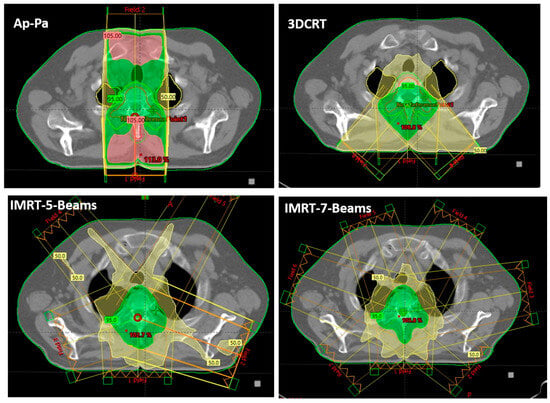
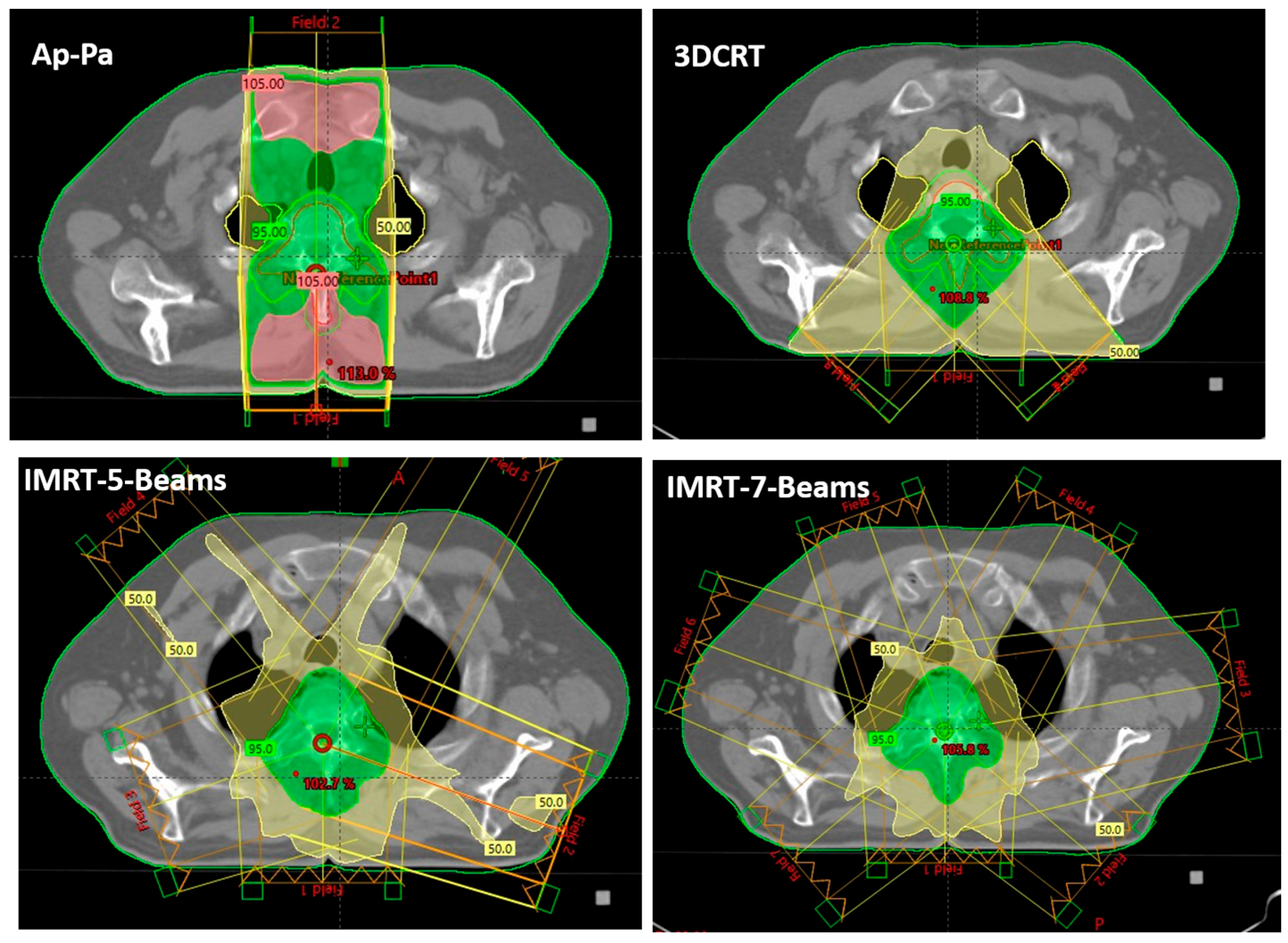
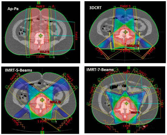
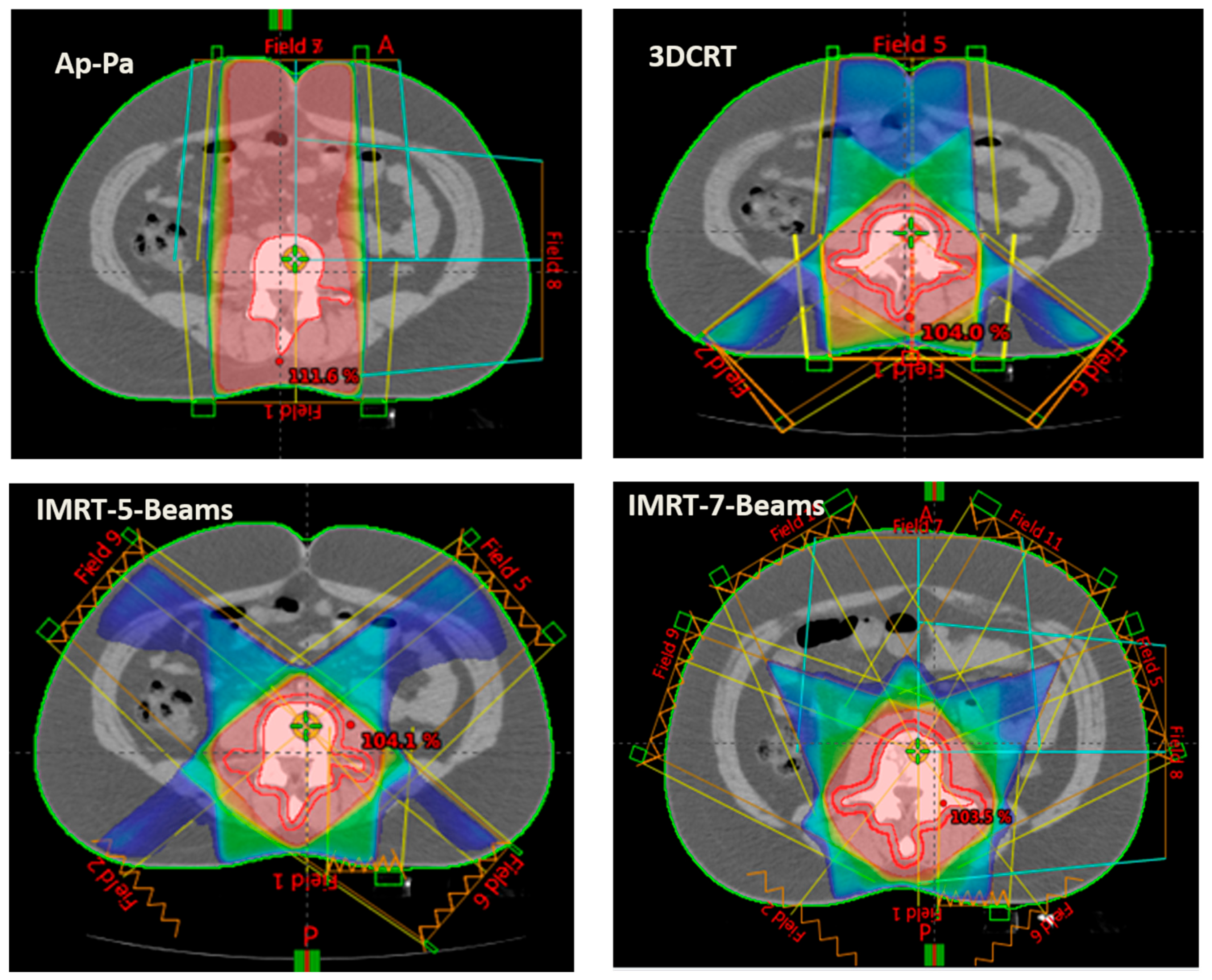
For thoracic lesions, represented in Figure 3, respiratory motion and proximity to the heart and lungs partially attenuate IMRT advantages, with IMRT-7 reducing the cord dose to 8.1 Gy versus 23.4 Gy with 3D-CRT. However, motion uncertainties may compromise precision, making 3D-CRT a pragmatic choice. In contrast, lumbar lesions derive the greatest benefit from IMRT-7, achieving cord doses as low as 5.8 Gy (74% reduction vs. 3D-CRT) due to favorable anatomy and the absence of respiratory motion. Here, IMRT-7 is preferred for patients with good performance status, while 3D-CRT remains a faster alternative for palliative cases.
Figure 3.
Comparative axial dose distributions for a thoracolumbar spinal metastasis using AP-PA, 3D-CRT, IMRT-5, and IMRT-7. The color-wash scale: red-orange (>95% of prescribed dose, target coverage), yellow-green (50–95%, intermediate fall-off), and blue-violet (<50%, low-dose spillage). AP-PA shows poor conformity with the highest cord dose (25.2 Gy). IMRT-7 achieves the best conformity and cord sparing (7.4 Gy) but produces a broader low-dose bath (blue regions) to healthy tissues—a key ALARA trade-off. Thoracic lesions partially attenuate IMRT advantages due to respiratory motion, while lumbar lesions derive maximal benefit.
3.2. Dosimetric Outcomes
Significant differences were observed across all four techniques (p < 0.001), as summarized in Table 3. IMRT-7 achieved the highest target coverage (D95: 95.8%) and best conformity (Paddick CI: 0.86), with the lowest spinal cord dose (7.4 Gy)—a 70.6% reduction versus AP-PA. 3D-CRT provided acceptable coverage (91.2%) and cord dose (22.8 Gy) with intermediate performance. IMRT-5 offered a balanced option (cord dose: 8.2 Gy). Progressive improvements in homogeneity and OAR sparing (esophagus, heart, and lung) were also noted with increasing technique complexity.
Table 3.
Dosimetric comparison of radiotherapy techniques.
Comprehensive dosimetric evaluation revealed statistically significant differences among all four techniques (p < 0.001 for primary endpoints). Table 1 summarizes the key dosimetric parameters.
3.3. Main Differences Across Techniques and Efficiency
Significant efficiency differences were observed across techniques (Table 4). AP-PA was the fastest (5.6 min total session), followed by 3D-CRT (8.7 min), IMRT-5 (13.4 min), and IMRT-7 (16.9 min) (p < 0.001). Beam-on time and monitor units increased proportionally from AP-PA to IMRT-7.
Table 4.
Treatment delivery efficiency parameters.
3.4. Anatomical Subgroup Analysis
Regional variations significantly influenced technique efficacy (Table 5). IMRT-7 achieved optimal spinal cord sparing in lumbar lesions (5.8 ± 0.9 Gy), with progressively higher doses in thoracic (8.1 ± 1.1 Gy) and cervical (8.9 ± 1.3 Gy) regions (p < 0.001). Esophageal sparing with IMRT-7 was also superior in lumbar lesions.
Table 5.
Anatomical subgroup analysis.
These findings confirm that anatomical location is a key determinant of achievable organ sparing, with lumbar metastases deriving the greatest benefit from advanced techniques due to favorable anatomy and the absence of respiratory motion.
4. Discussion
This dosimetric analysis of 60 patients with metastatic spinal cord compression reveals a consistent and statistically significant hierarchy among the four radiotherapy techniques evaluated. IMRT-7 achieved the most favorable dosimetric outcomes, with superior target coverage (95.8% ± 1.5%), optimal conformity (Paddick CI = 0.86 ± 0.04), and maximal spinal cord sparing (7.4 ± 1.2 Gy)—representing a 70.6% reduction in cord dose compared to AP-PA and a 67.5% reduction compared to 3D-CRT. IMRT-5 demonstrated intermediate performance (94.6% coverage, 8.2 Gy cord dose), while 3D-CRT provided clinically acceptable coverage (91.2%) with moderate OAR sparing. However, these dosimetric advantages came at the cost of substantially prolonged treatment sessions: IMRT-7 required 16.9 ± 1.4 min total session time compared to 8.7 ± 1.3 min for 3D-CRT and merely 5.6 ± 1.1 min for AP-PA. The novel Dosimetric Gain per Minute (DGM) metric quantified this trade-off, with IMRT-5 demonstrating greater efficiency (3.11 Gy/min) than IMRT-7 (1.88 Gy/min) in cord sparing per minute of added treatment time. Anatomical stratification revealed that lumbar lesions derived the greatest benefit from IMRT-7 (cord dose 5.8 ± 0.9 Gy), while thoracic lesions presented greater technical challenges with higher esophageal doses across all techniques.
Our findings are consistent with recent dosimetric comparisons in spinal radiotherapy. A multicenter European study by Seravalli et al. comparing five craniospinal irradiation techniques demonstrated that modern modalities (IMRT, VMAT, and TomoTherapy) achieved superior conformity and homogeneity indices compared to 3D-CRT, particularly in the spinal target volume (CI: 0.6 vs. 0.3; HI: 0.1 vs. 0.2), with corresponding dose reductions to the thyroid, heart, esophagus, and pancreas exceeding 10 Gy in some cases [32]. This aligns with our observed improvements in conformity (0.86 vs. 0.74) and homogeneity (0.11 vs. 0.17) with IMRT-7 compared to 3D-CRT. Similarly, a comprehensive Turkish multicenter study reported that modern approaches achieved excellent PTV coverage (>98%), with tomotherapy excelling in conformity and homogeneity, while IMRT yielded lower doses to organs at risk including the heart, kidneys, and esophagus [33,34]. The convergence of evidence across independent investigations using diverse patient populations strengthens the generalizability of these findings and supports the technical superiority of IMRT for spinal radiotherapy when dosimetric precision is prioritized.
Despite the broad agreement on IMRT’s dosimetric advantages, several studies highlight important contextual considerations. A phase III trial investigating esophagus-sparing radiotherapy for spinal metastases found that inter-fraction motion led to dose constraint violations in 20 of 23 simulated plans, with small esophageal volumes inadvertently entering high-dose regions despite strict adherence to planning constraints [35]. This underscores a critical limitation: the dosimetric advantages predicted by planning systems may not fully translate to delivered dose distributions in the presence of inter-fraction motion—particularly relevant for thoracic lesions, where respiratory motion is pronounced. Our finding that IMRT advantages may be partially attenuated in thoracic targets aligns with this observation and reinforces the need for motion management strategies when considering advanced techniques for mobile targets.
Furthermore, a randomized trial by Sprave et al. comparing IMRT versus 3D-CRT (30 Gy in 10 fractions) for vertebral metastases found no statistically significant differences in bone density at 3 months (p = 0.723) or 6 months (p = 0.341), and comparable pain response rates at both time points (p = 0.395 and p = 0.732, respectively) [11]. While IMRT showed improved VAS scores at 3 months (p = 0.037), this advantage did not persist at 6 months (p = 0.430). This randomized evidence suggests that the translational benefit of IMRT’s dosimetric superiority to clinically meaningful outcomes may be more nuanced than physical parameters alone predict. The disparity between dosimetric predictions and observed clinical outcomes may reflect several factors: the dominant influence of tumor biology and systemic disease on patient-reported outcomes; the challenges of maintaining precise setup in frail populations; and the possibility that dosimetric differences, while statistically significant, may exceed the threshold required for clinical benefit in some patients. These considerations reinforce the ALARA principle’s emphasis on “reasonably achievable” rather than “technically possible,” and support our stratified approach, which integrates patient-specific factors including performance status, prognosis, and institutional resources.
Building on these findings, several priority areas warrant investigation. First, prospective clinical validation through multi-institutional trials is needed to establish correlations between technique selection and patient-reported outcomes, pain control, and neurological function [11,34]. Second, adaptive radiotherapy strategies incorporating mid-course plan adaptation could potentially combine 3D-CRT efficiency with IMRT precision [35]. Third, artificial intelligence optimization offers promise for integrating patient-specific factors to recommend optimal techniques balancing precision with deliverability [36]. Fourth, advanced motion management incorporating 4D planning is essential to determine whether IMRT advantages persist in moving thoracic targets. Fifth, dedicated re-irradiation protocols with radiobiological modeling are needed for the growing population requiring repeat treatment [37]. Finally, cost-effectiveness analyses across diverse resource settings must recognize that optimal techniques may differ between academic and community hospitals [34,38]. Crucially, all these threads converge on the importance of systematic clinical follow-up to validate whether technique selection translates to improved neurological outcomes and quality of life.
5. Conclusions
This systematic comparison of four radiotherapy techniques for metastatic spinal cord compression quantifies the fundamental trade-off between dosimetric precision and treatment efficiency—a balance best guided by the ALARA principle. IMRT-7 offers superior target coverage (95.8%) and spinal cord sparing (7.4 Gy, a 70.6% reduction compared to AP-PA), but requires prolonged sessions (16.9 min) that may be impractical for frail patients. Conversely, 3D-CRT delivers acceptable coverage (91.2%) in nearly half the time (8.7 min), making it a more suitable choice when efficiency is critical.
Based on these findings, the following considerations are proposed for technique selection:
- IMRT-7 is best suited for patients with good performance status (ECOG 0–1), favorable anatomy such as lumbar lesions, or those who may require re-irradiation.
- 3D-CRT remains a preferred option for urgent palliative cases, patients with poor performance status (ECOG 3–4), or thoracic lesions where respiratory motion limits the precision of IMRT.
- IMRT-5 offers a balanced alternative for intermediate cases, providing meaningful cord sparing (8.2 Gy) with moderate session times (13.4 min).
- 3D-CRT with field-in-field is a practical alternative in resource-limited settings, while AP-PA should be used only, when necessary, with an awareness of its higher cord doses.
Ultimately, technique selection should integrate dosimetric goals with clinical context and patient status, ensuring that the ALARA principle is meaningfully applied in palliative radiotherapy. Future studies are planned to extend this dosimetric comparison to include advanced rotational techniques such as VMAT and SBRT, aiming to further validate the proposed ALARA-based framework across a broader spectrum of modern treatment modalities.
Author Contributions
Conceptualization, M.E., M.S. and A.M.S.; methodology, M.E. and A.M.S.; software, B.H.; validation, M.E.; formal analysis, B.H.; investigation, B.H.; resources, M.E.; data curation, B.H. (with initial tabulation support from M.E.); writing—original draft preparation, B.H.; writing—review and editing, M.E., M.S. and A.M.S.; visualization, M.E.; supervision, A.M.S. (clinical supervision at El-Hussein Hospital) and M.S. (academic supervision); project administration, M.E. All authors have read and agreed to the published version of the manuscript.
Funding
This research received no external funding.
Institutional Review Board Statement
The study was conducted in accordance with the Declaration of Helsinki and approved by the Institutional Review Board of El-Hussein University Hospital, Al-Azhar University, Cairo, Egypt (protocol code ZU-IRB#2023-45, approved on 15 January 2023).
Informed Consent Statement
Written informed consent was obtained from patients for the publication of this paper.
Data Availability Statement
The data presented in this study are available on request from the corresponding author.
Conflicts of Interest
The authors declare no conflict of interest.
References
- Schmidt, M.H.; Klimo, P.; Vrionis, F.D. Metastatic spinal cord compression. J. Natl. Compr. Cancer Netw. 2005, 3, 711–719. [Google Scholar] [CrossRef] [Scilit] [PubMed]
- Loblaw, D.A.; Laperriere, N.J. Emergency treatment of malignant extradural spinal cord compression. J. Clin. Oncol. 1998, 16, 1613–1624. [Google Scholar] [CrossRef] [Scilit] [PubMed]
- Prasad, D.; Schiff, D. Malignant spinal-cord compression. Lancet Oncol. 2005, 6, 15–24. [Google Scholar] [CrossRef] [Scilit] [PubMed]
- Peyraga, G.; Ducassou, A.; Arnaud, F.X.; Lizée, T.; Pouédras, J.; Moyal, É. Radiotherapy and spinal toxicity: News and perspectives. Cancer/Radiothérapie 2021, 25, 55–61. [Google Scholar] [CrossRef] [Scilit] [PubMed]
- Alcorn, S.; Cortés, Á.A.; Bradfield, L.; Brennan, M.; Dennis, K.; Diaz, D.A.; Doung, Y.C.; Elmore, S.; Hertan, L.; Johnstone, C.; et al. External beam radiation therapy for palliation of symptomatic bone metastases: An ASTRO clinical practice guideline. Pract. Radiat. Oncol. 2024, 14, 377–397. [Google Scholar] [CrossRef] [Scilit] [PubMed]
- Rades, D.; Abrahm, J.L. The Role of Radiotherapy for Metastatic Epidural Spinal Cord Compression. Nat. Rev. Clin. Oncol. 2010, 7, 590–598. [Google Scholar] [CrossRef] [Scilit] [PubMed]
- Lutz, S.; Balboni, T.; Jones, J.; Lo, S.; Petit, J.; Rich, S.E.; Wong, R.; Hahn, C. Palliative radiation therapy for bone metastases: Update of an ASTRO evidence-based guideline. Pract. Radiat. Oncol. 2017, 7, 4–12. [Google Scholar] [CrossRef] [Scilit] [PubMed]
- Ding, M.; Newman, F.; Chen, C.; Stuhr, K.; Gaspar, L.E. Dosimetric comparison between 3D-CRT and IMRT using different multileaf collimators in the treatment of brain tumors. Med. Dosim. 2009, 34, 1–8. [Google Scholar] [CrossRef] [Scilit] [PubMed]
- Lam, F.C.; Guru, S.; AbuReesh, D.; Hori, Y.S.; Chuang, C.; Liu, L.; Wang, L.; Gu, X.; Szalkowski, G.A.; Wang, Z.; et al. Use of Carbon Fiber Implants to Improve the Safety and Efficacy of Radiation Therapy for Spine Tumor Patients. Brain Sci. 2025, 15, 199. [Google Scholar] [CrossRef] [Scilit] [PubMed]
- Dosimetric comparison of 3D-CRT, IMRT, h-IMRT and VMAT for spinal metastases. PLoS ONE 2023, 18, e0279133. [PubMed]
- Sprave, T.; Verma, V.; Förster, R.; Schlampp, I.; Hees, K.; Bruckner, T.; Bostel, T.; El Shafie, R.A.; Welzel, T.; Nicolay, N.H.; et al. Bone density and pain response following intensity-modulated radiotherapy versus three-dimensional conformal radiotherapy for vertebral metastases. Radiat. Oncol. 2023, 18, 67. [Google Scholar]
- Pathak, R.S.; Tibdewal, A.; Kinhikar, R.; Wakeham, K.; Akbarov, K.; Cordero, L.; Khandavalli, A.; Agarwal, J.P. Practice Patterns and Perspectives on Stereotactic Body Radiation Therapy for the Metastatic Spine From Lower- and Middle-Income Countries. JCO Glob. Oncol. 2022, 8, e2200167. [Google Scholar] [CrossRef] [Scilit] [PubMed]
- Fisher, C.G.; DiPaola, C.P.; Ryken, T.C.; Bilsky, M.H.; Shaffrey, C.I.; Berven, S.H.; Harrop, J.S.; Fehlings, M.G.; Boriani, S.; Chou, D.; et al. A Novel Classification System for Spinal Instability in Neoplastic Disease: An Evidence-Based Approach and Expert Consensus from the Spine Oncology Study Group. Spine 2010, 35, E1221–E1229. [Google Scholar] [CrossRef] [Scilit] [PubMed]
- Fogliata, A.; Vanetti, E.; Albers, D.; Brink, C.; Clivio, A.; Knöös, T.; Nicolini, G.; Cozzi, L. On the dosimetric behaviour of photon dose calculation algorithms. Phys. Med. Biol. 2012, 57, 2997–3017. [Google Scholar]
- Hyde, E.; Hardy, M. Delivering patient centred care: A qualitative study. Radiography 2020, 26, e145–e151. [Google Scholar]
- Cox, B.W.; Spratt, D.E.; Lovelock, M.; Bilsky, M.H.; Lis, E.; Ryu, S.; Sheehan, J.; Gerszten, P.C.; Chang, E.; Gibbs, I.; et al. International Spine Radiosurgery Consortium Consensus Guidelines for Target Volume Definition in Spinal Stereotactic Radiosurgery. Int. J. Radiat. Oncol. Biol. Phys. 2012, 83, e597–e605. [Google Scholar] [CrossRef] [Scilit] [PubMed]
- Stieler, F.; Wolff, D.; Bauer, L.; Wertz, H.J.; Wenz, F.; Lohr, F. Reirradiation of spinal column metastases: Comparison of treatment techniques. Strahlenther. Onkol. 2011, 187, 406–415. [Google Scholar] [CrossRef] [Scilit] [PubMed]
- Redmond, K.J.; Robertson, S.; Lo, S.S.; Soltys, S.G.; Ryu, S.; McNutt, T.; Chao, S.T.; Yamada, Y.; Ghia, A.; Chang, E.L.; et al. Consensus Contouring Guidelines for Postoperative Stereotactic Body Radiation Therapy for Metastatic Solid Tumor Malignancies to the Spine. Int. J. Radiat. Oncol. Biol. Phys. 2017, 97, 64–74. [Google Scholar] [CrossRef] [Scilit] [PubMed]
- Kong, F.M.; Ritter, T.; Quint, D.J.; Senan, S.; Gaspar, L.E.; Komaki, R.U.; Hurkmans, C.W.; Timmerman, R.; Bezjak, A.; Bradley, J.D.; et al. Consideration of Dose Limits for Organs at Risk of Thoracic Radiotherapy: Atlas for Lung, Proximal Bronchial Tree, Esophagus, Spinal Cord, Ribs, and Brachial Plexus. Int. J. Radiat. Oncol. Biol. Phys. 2011, 81, 1442–1458. [Google Scholar] [CrossRef] [Scilit] [PubMed]
- Paddick, I. A simple scoring ratio to index conformity of radiosurgical plans. J. Neurosurg. 2000, 93, 219–222. [Google Scholar] [CrossRef] [Scilit] [PubMed]
- International Commission on Radiation Units and Measurements. Prescribing, Recording, and Reporting Photon-Beam Intensity-Modulated Radiation Therapy (IMRT). ICRU Report 83. J. ICRU 2010, 10, 1–106. [Google Scholar] [CrossRef] [Scilit]
- Marks, L.B.; Yorke, E.D.; Jackson, A.; Ten Haken, R.K.; Constine, L.S.; Eisbruch, A.; Bentzen, S.M.; Nam, J.; Deasy, J.O. Use of normal tissue complication probability models. Int. J. Radiat. Oncol. Biol. Phys. 2010, 76, S10–S19. [Google Scholar] [CrossRef] [Scilit] [PubMed]
- Emami, B. Tolerance of normal tissue to therapeutic radiation. Rep. Radiother. Oncol. 2013, 1, 123–127. [Google Scholar]
- Nieder, C.; Grosu, A.L.; Andratschke, N.H.; Molls, M. Update of human spinal cord reirradiation tolerance. Int. J. Radiat. Oncol. Biol. Phys. 2006, 66, 1446–1449. [Google Scholar] [CrossRef] [Scilit] [PubMed]
- Purdy, J.A. 3D treatment planning and conformal therapy. Semin. Radiat. Oncol. 1999, 9, 1–2. [Google Scholar]
- Galvin, J.M.; Ezzell, G.; Eisbrauch, A.; Yu, C.; Butler, B.E.; Xiao, Y.; Rosen, I.I.; Rosenman, J.G.; Sharpe, M.B.; Xing, L.; et al. Implementing IMRT in Clinical Practice: A Joint Document of the American Society for Therapeutic Radiology and Oncology and the American Association of Physicists in Medicine. Int. J. Radiat. Oncol. Biol. Phys. 2004, 58, 1616–1634. [Google Scholar] [CrossRef] [Scilit]
- Ezzell, G.A.; Burmeister, J.W.; Dogan, N.; LoSasso, T.J.; Mechalakos, J.G.; Mihailidis, D.; Molineu, A.; Palta, J.R.; Ramsey, C.R.; Salter, B.J.; et al. IMRT Commissioning: Multiple Institution Planning and Dosimetry Comparisons, a Report from AAPM Task Group 119. Med. Phys. 2009, 36, 5359–5373. [Google Scholar] [CrossRef] [Scilit] [PubMed]
- Feuvret, L.; Noël, G.; Mazeron, J.-J.; Bey, P. Conformity index: A review. Int. J. Radiat. Oncol. Biol. Phys. 2006, 64, 333–342. [Google Scholar] [CrossRef] [Scilit] [PubMed]
- Brown, C.; Burck, A.; Neep, M.J. Workload as predictor of radiographer accuracy. J. Med. Radiat. Sci. 2024, 71, 547–554. [Google Scholar] [CrossRef] [Scilit] [PubMed]
- IBM SPSS Statistics for Windows, Version 26.0; IBM Corp.: Armonk, NY, USA, 2019.
- Altman, D.G. Practical Statistics for Medical Research; Chapman & Hall: London, UK, 1991. [Google Scholar]
- Seravalli, E.; Bosman, M.; Lassen-Ramshad, Y.; Vestergaard, A.; Oldenburger, F.; Visser, J.; Koutsouveli, E.; Paraskevopoulou, C.; Horan, G.; Ajithkumar, T.; et al. Dosimetric comparison of five different techniques for craniospinal irradiation across 15 European centers. Acta Oncol. 2018, 57, 1240–1249. [Google Scholar] [CrossRef] [Scilit] [PubMed]
- Şenkesen, Ö.; Tezcanlı, E.; Alkaya, F.; İspir, B.; Çatlı, S.; Yeşil, A.; Bezirganoglu, E.; Turan, S.; Köksal, C.; Güray, G.; et al. Current Practices of Craniospinal Irradiation Techniques in Turkey: A Comprehensive Dosimetric Analysis. Radiat. Oncol. 2024, 19, 49. [Google Scholar] [CrossRef] [Scilit] [PubMed]
- Kim, M.J.; Marianayagam, N.J.; Chandra, A.; Ranalli, C.; Schonfeld, E.; Avila-Madrigal, J.P.; Flusche, A.M.E.; Schoeffler, K.; Alomari, S.; Rao, N.B.; et al. Integrated management of metastatic spinal tumors: Current status and future directions. Med. Oncol. 2025, 42, 178. [Google Scholar] [CrossRef] [Scilit]
- Nielsen, A.M.; Rechner, L.A.; Krog, S.M.; Gram, V.; Suppli, M.H.; Sibolt, P.; Vogelius, I.R.; Behrens, C.P.; Persson, G.F. Dosimetric impact of esophageal motion in esophagus-sparing radiotherapy of spinal metastases. Acta Oncol. 2025, 64, 1276–1284. [Google Scholar] [CrossRef] [Scilit]
- Jain, H.; Ahluwalia, R.; Laufer, I.; Zuckerman, S.L. Advances in Metastatic Disease Spinal Oncology: Novel Technology Without Forgetting the Fundamentals of Surgical Treatment. Neurospine 2025, 22, 829. [Google Scholar] [CrossRef] [Scilit] [PubMed]
- Mansi, A.; Rezaii, E.G.; Wang, S.; Zhang, C.; Katzir, M. Comparison of dosimetric variability in the cervical spine between carbon fiber and titanium instrumentation. J. Neurosurg. Spine 2025, 42, 615–623. [Google Scholar] [CrossRef] [Scilit] [PubMed]
- Xiao, H.; Guo, F.; Qu, A.; Zhang, N.; Wei, S.; Han, Q.; Deng, X.; Li, C.; Wang, J.; Jiang, P. Efficacy and Safety of Simultaneous Integrated Boost Followed by Selective Sequential Boost Radiotherapy for Spinal Metastases: A Single-Arm Retrospective Study. Technol. Cancer Res. Treat. 2025, 24, 15330338251363694. [Google Scholar] [CrossRef] [Scilit] [PubMed]

Disclaimer/Publisher’s Note: The statements, opinions and data contained in all publications are solely those of the individual author(s) and contributor(s) and not of MDPI and/or the editor(s). MDPI and/or the editor(s) disclaim responsibility for any injury to people or property resulting from any ideas, methods, instructions or products referred to in the content. |
© 2026 by the authors. Licensee MDPI, Basel, Switzerland. This article is an open access article distributed under the terms and conditions of the Creative Commons Attribution (CC BY) license.