


Abstract
We herein report the anticancer activity and molecular docking studies of a series of amide derivatives of two nonsteroidal anti-inflammatory drugs (mefenamic acid and ibuprofen). The hypothesis of drug repurposing has been successfully employed to explore the promising anticancer activity of analogs of known anti-inflammatory agents. The compounds have been tested for their inhibitory potential against cervical cancer cell lines by MTT assay using 5-fluorouracil as the reference standard. Among the compounds screened, 3aa [2-(2,3-dimethylamino)phenyl)(1H-indol-1-yl)methanone] and 3ad [2-(2,3-dimethylphenylamino)phenyl)(9H-carbazol-9-yl)methanone] displayed good potency of less than 25 µg/mL half-maximal inhibitory concentration (IC50). The docking analysis has confirmed that molecule 3aa effectively binds to the active site of the target protein CDK2, with a docking score of −9.21 Kcal/mol and a binding energy of −46.44 Kcal/mol, involving a hydrogen bond with Ile 10. The molecule 3ad also exhibited a good glide score of −6.78 Kcal/mol with the binding energy of −45.90 Kcal/mol. As many of the tested compounds displayed promising potency against cervical cancer cell lines, our investigation revealed the importance of drug repurposing in the development of lead molecules in medicinal chemistry.
Keywords:
mefenamic acid; ibuprofen; cytotoxic activity; cell lines; molecular docking; binding site; SAR 1. Introduction
Cancer has continued to be a menace to humanity for a very long time due to its continually rising morbidity and mortality rates [1]. It is a disease characterized by the proliferation of abnormal cells that spread throughout the body [2]. This malignancy arises from gene mutations that can lead to tumor formation through cell assembly. Another type of tumor is a benign tumor that does not metastasize and is not detrimental [3]. Generally, the definitive metabolic pathway in cancer cells makes it difficult to treat or cure completely [4]. The Warburg effect, a key phenomenon in cancer, is the elevated utilization and uptake of glucose by cancerous cells [5]. Chemotherapy and radiation are the common treatments available for cancer; however, they are sometimes replaced by targeted therapy nowadays. This mainly targets the abnormal cells while leaving the normal cells intact [6]. This modified therapy is now being employed more in the bladder, kidney, lung, breast and cervical cancers [7,8,9,10]. Cervical cancer is one of the most prevalent malignant tumors in women [11]. The compounds with a high level of potency can induce apoptosis and are being used for cancer treatment. Cervical cancer is caused by the Human Papilloma Virus (HPV) and induces blisters in the genital area [12]. It can adversely affect the urinary bladder. It can also cause kidney damage and vaginal bleeding that can spread to other organs. In short, cervical cancer can be life-threatening. However, it can be cured if detected at an early stage. Screening of cervical cells can be done in a Pap test or Pap smear, which will help to identify the abnormalities. The HPV test can be done after detecting the abnormality in the Pap test [13]. Paclitaxel, cisplatin [14], 5-fluorouracil [15] and topotecan [16] are some of the medications used for the treatment of cervical cancer nowadays. With the help of computational studies, CDK2 was reported recently as a main apoptotic gene to forecast cervical cancer [17]. This observation has led us to take CDK2 as the target protein for our in silico studies.
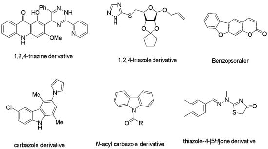
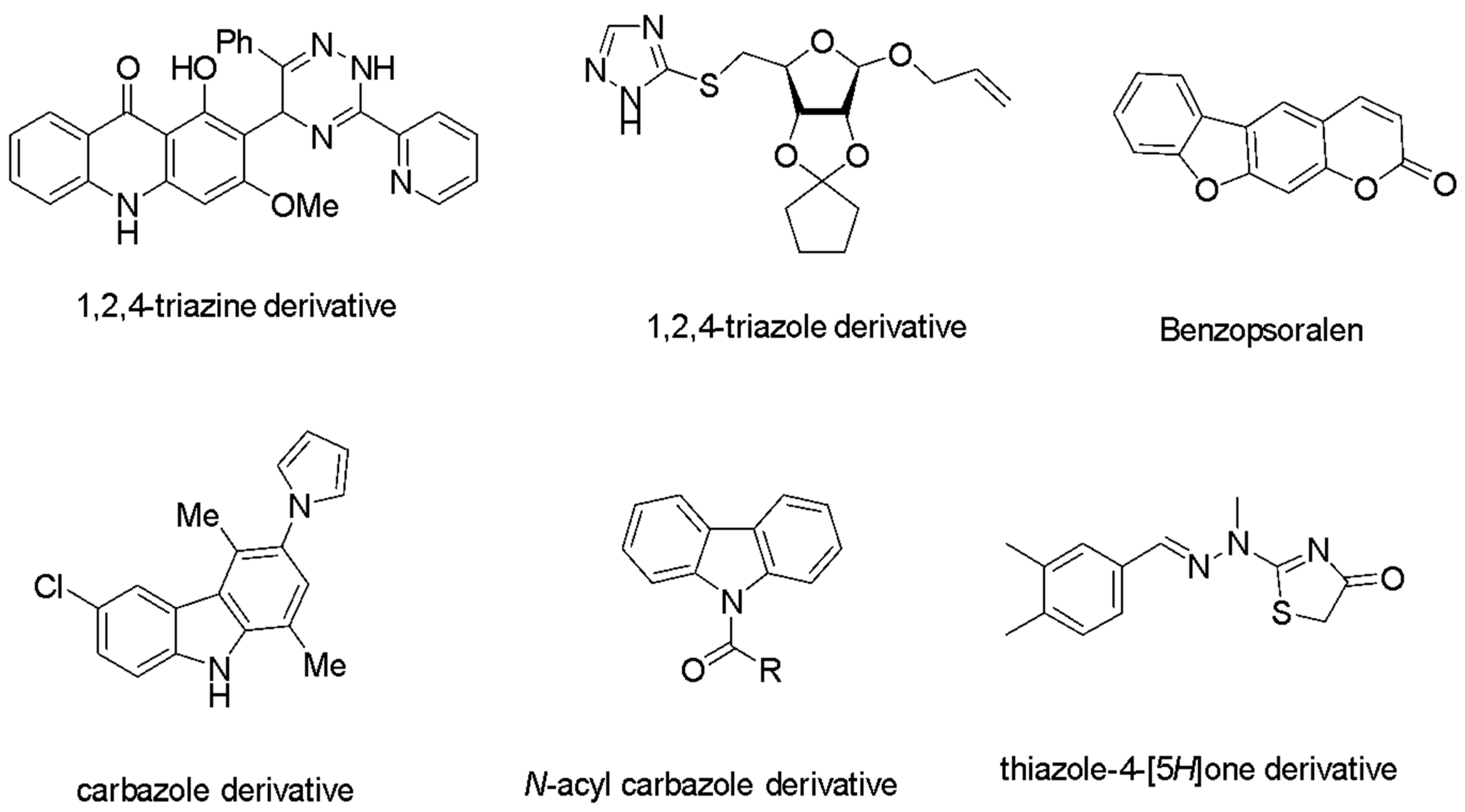
Heterocyclic compounds containing one or more heteroatoms are mainly utilized for various useful applications nowadays. Many heterocycles are important constituents of proteins, essential amino acids and pigments like chlorophyll [18]. Nitrogen-based heterocycles are part of thiamine, riboflavin, nicotinamide, etc., and drugs such as barbituric acid, antipyrine, chlorpromazine, and captopril also contain nitrogen heterocycles. Nitrogen atoms can be quickly bonded with DNA through hydrogen bonding, which makes them find applications in anticancer studies [19,20,21,22,23]. It is reported that the new acridone derivatives [24], isosteviol fused pyrazoline, ursolic acid-linked triazole, or D-ribose, display anticancer activity in the micromolar to nanomolar range [25]. Some heterocyclic compounds with profound anticancer activity have been detailed in Scheme 1. The carbazole-based compounds have recently been reported as effective agents for improving anticancer therapy [26]. In 2025, a bis-carbazole derivative was reported to exhibit excellent activity against the breast cancer cell lines [27]. Derivatives of indole were also tested for anti-breast cancer activity against the MCF-7 cell line (a breast cancer cell line) [28]. Hussain Basha and coworkers reported 2-amino-3-cyanopyridine derivatives with better binding affinity against the EGFR (epidermal growth factor receptor) protein by computational studies [29]. Thiazole hybrids have been reported to show a wide spectrum of activity against different cancers [30]. Benzimidazole–thiazole hybrids have been reported with better results in apoptosis induction and cell cycle arrest recently [31]. Newly synthesized benzyl-4-(4-chlorophenyl) thiazole-2-carboxylate was recently reported for its potent binding capacity with the target protein 6FS1 [32]. Methylated thienopyridine-indole derivatives have recently been reported for their enhanced antiproliferative effect [33].
Scheme 1.
Some heterocycles with anticancer activity.
Nonsteroidal anti-inflammatory drugs (NSAIDs) are widely used for the treatment of inflammation, which usually works by blocking the cyclooxygenase enzyme [34]. It also shows good analgesic and antipyretic properties. Some of the typical examples of NSAIDs available in the market are mefenamic acid (2-[(2,3-dimethylphenyl)amino]benzoic acid) and ibuprofen (2-[4-(2-methylpropyl)phenyl]propanoic acid) [35]. We recently reported the synthesis of some mefenamic acid and ibuprofen derivatives containing amide functionality and evaluated their anti-inflammatory, antimicrobial, and antioxidant potential [36]. Drug repurposing has now emerged as an important step in the field of medicinal chemistry. This technology has now been explored by various medicinal chemists throughout the world and has marked a breakthrough in cancer therapy [37]. Being an antioxidant, ascorbic acid has recently been reported for its anticancer activities, which displayed promising wound healing and tissue repair properties [38]. The ibuprofen-loaded polymer nanoparticles have already been reported for their ability to reduce cellular proliferation compared to the reference drug [39]. In 2017, mefenamic acid was reported for its ability to enhance anticancer drug sensitivity [40]. A critical review on the application of NSAIDs for anticancer activity evaluation has been reported by R.E. Harris and coworkers [41]. These observations prompted us to test the anticancer activity of our previously synthesized amide derivatives of mefenamic acid and ibuprofen, which have shown good antioxidant/anti-inflammatory activities. Different experimental studies have reported anticancer effects of NSAIDs on animal models and cell lines [42]. The aforementioned observations also enlightened us to synthesize and evaluate the anticancer properties of some new amide derivatives of mefenamic acid and ibuprofen.
As a continuation of our ongoing research on the study of the biological activity of mefenamic acid and ibuprofen derivatives, we herein report their synthesis, anticancer activity, molecular docking and structure–activity relationship (SAR) studies.
2. Results and Discussion
2.1. Chemistry
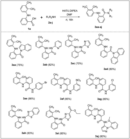
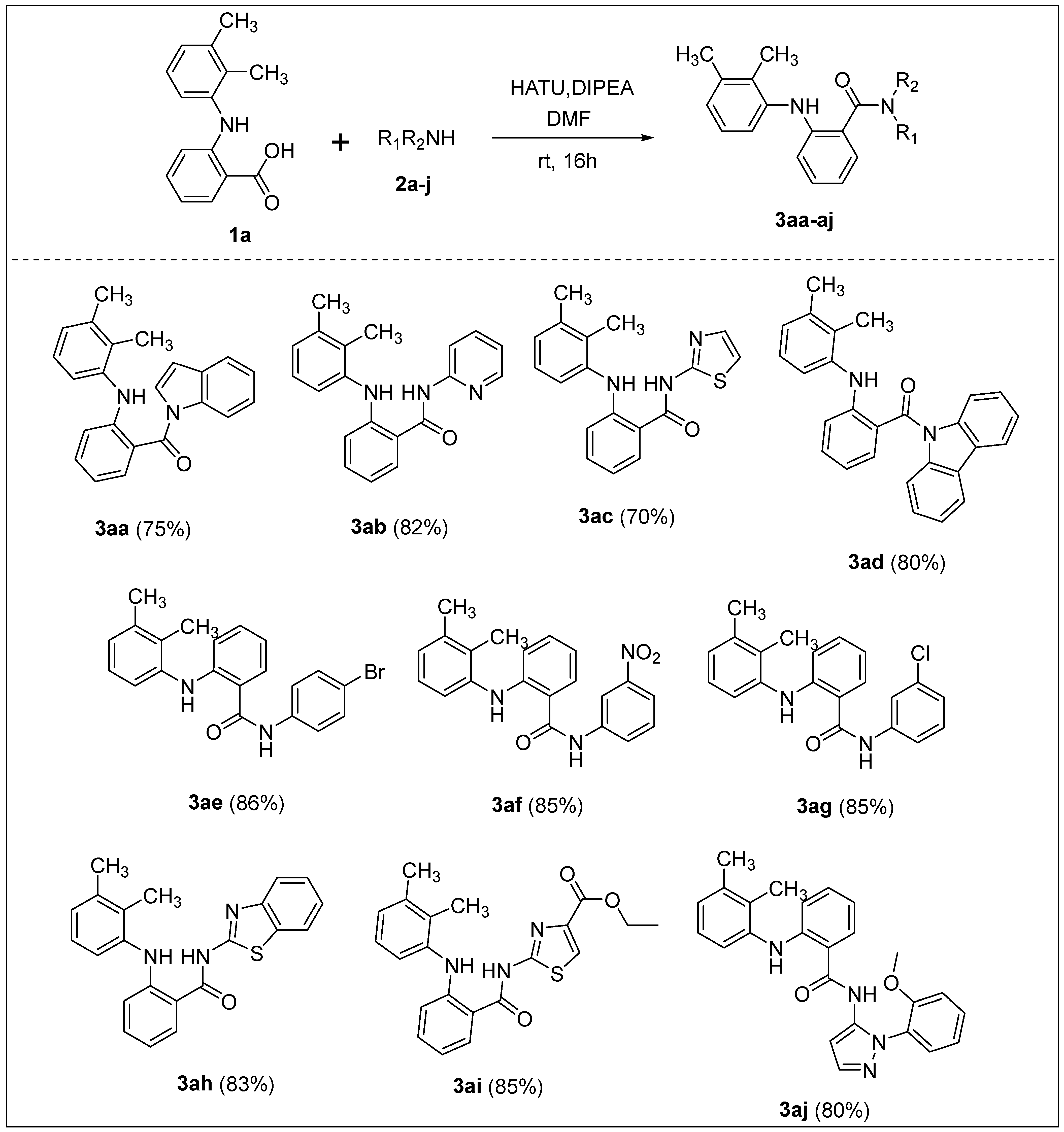
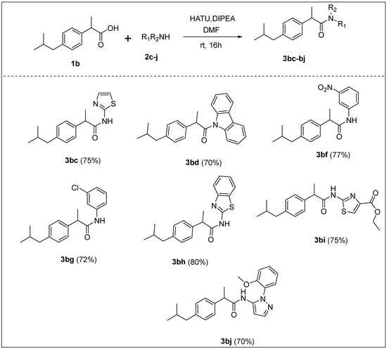
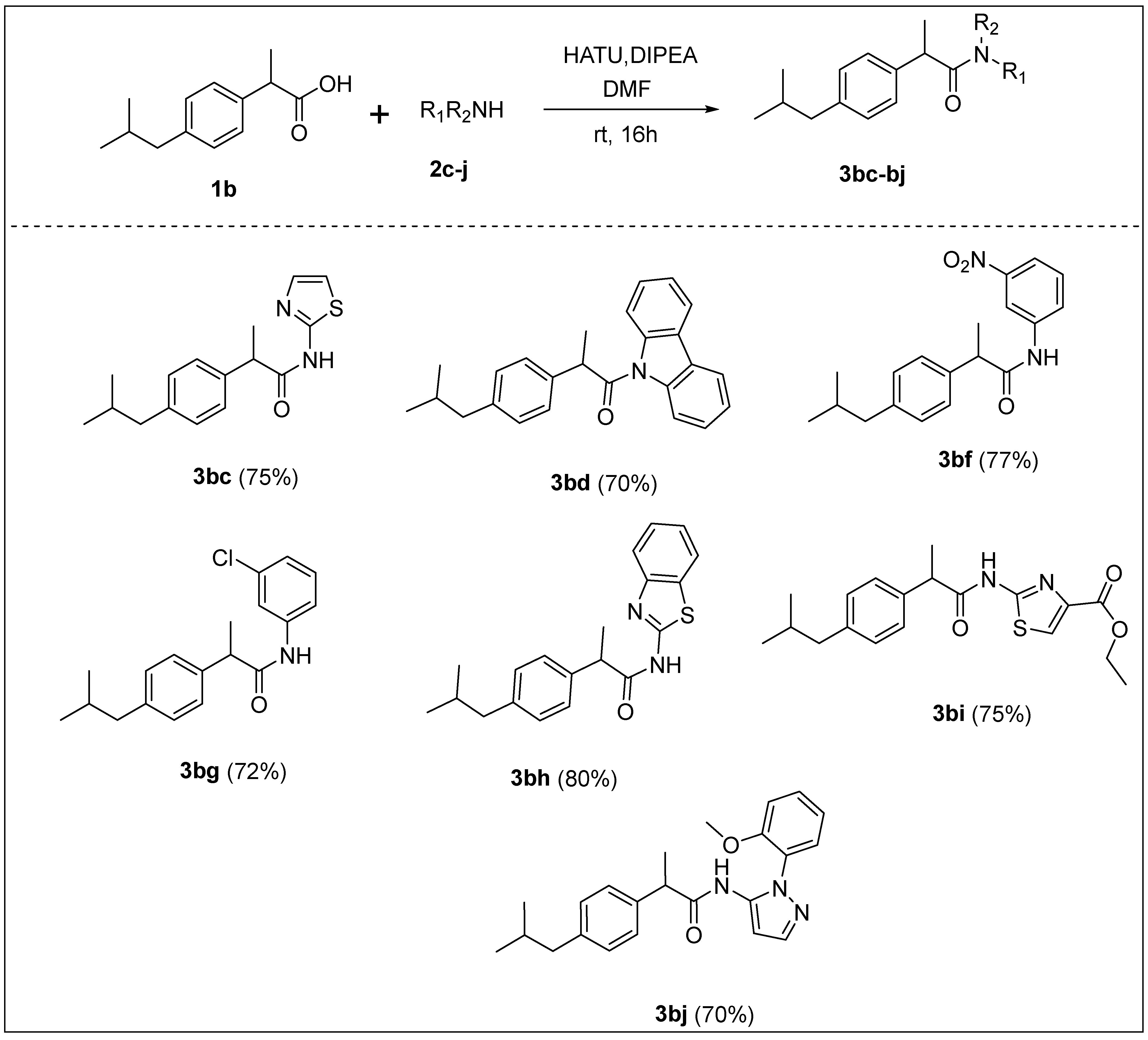
The synthesis of mefenamic acid and ibuprofen derivatives, 3aa–3af, 3ai, 3aj, 3bd and 3bg, was carried out by the coupling of the acid 1a and 1b with different amines 2 according to the procedure previously reported by us [36]. As detailed in Scheme 2 and Scheme 3, we have optimized the reaction conditions using HATU as the coupling agent [43], and DIPEA [44] as a base in DMF solvent. Apart from the previously synthesized compounds, nine new derivatives of mefenamic acid and ibuprofen were also synthesized using this optimized methodology.
Scheme 2.
Substrate scope for the synthesis of amides from mefenamic acid; isolated yield in parenthesis.
Scheme 3.
Synthesis of amides from ibuprofen; isolated yield in parenthesis.
Later, the cytotoxic assay [45] was carried out to evaluate the anticancer activity of our synthesized compounds using 96-well plates. HeLa cell lines were used for in vitro studies due to their durability and rapid proliferation. They were derived from cervical cancer cells. As the concentration of the tested compounds increased, the number of adhering cells gradually declined. The synthesized compounds exhibited good anti-inflammatory and antioxidant activities. Compounds with antioxidant/anti-inflammatory properties generally exhibit anti-tumor activity, for example, Triptolide, Epigallocatechin 3-gallate, and Quercetin. The combination of repurposed therapeutic drugs and approved drugs for cancer can achieve more efficacy and safety. Therefore, drug repurposing has a great impact in the field of pharmaceutical chemistry, where researchers have heightened their awareness nowadays.
2.2. Biological Activity
2.2.1. In Vitro Anticancer Studies
In Vitro Antiproliferative Effect on HeLa Cell Lines: Determination by MTT Assay
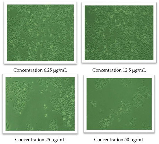
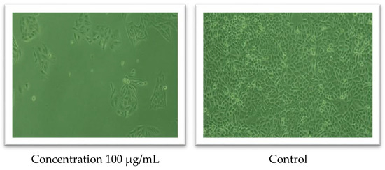
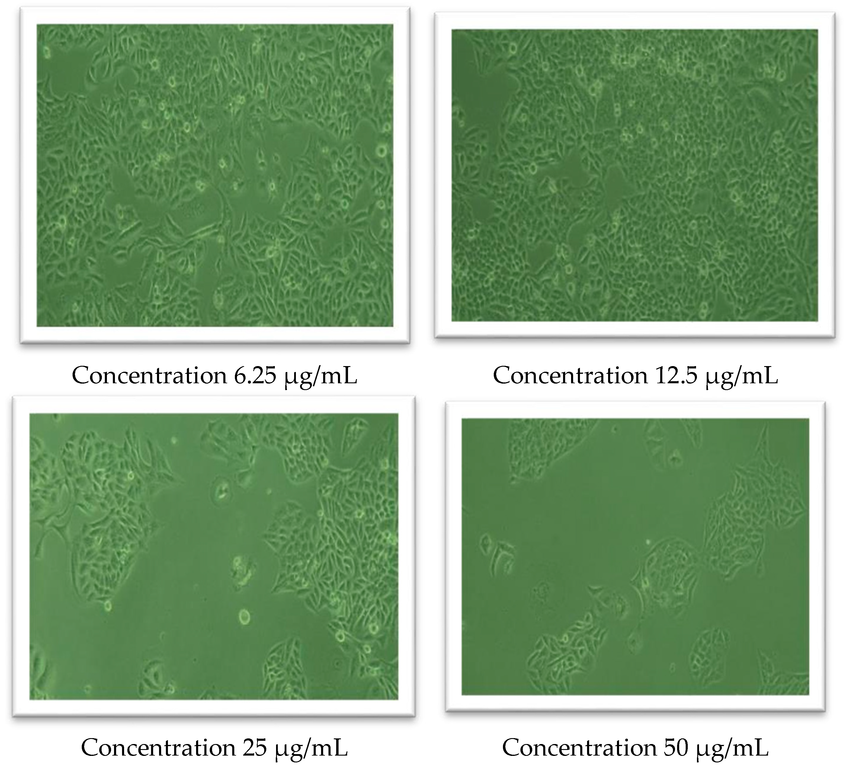
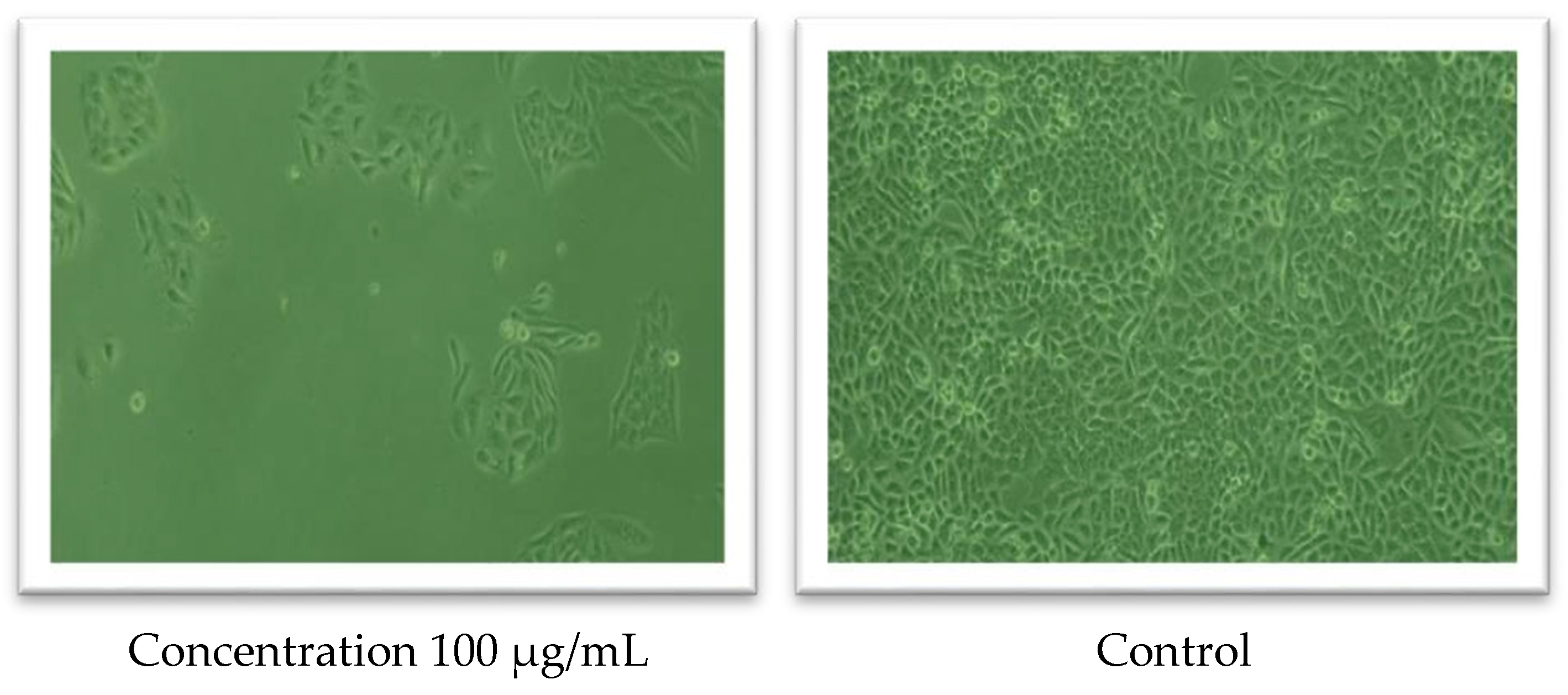
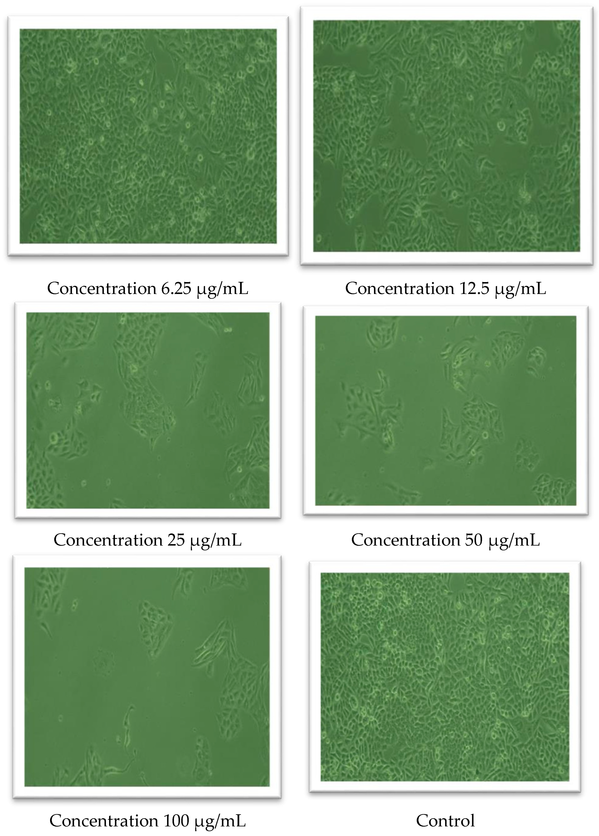
The percentage inhibition of each tested compound was calculated using Equation (1) (see Materials and Methods), and concentration versus percentage absorbance graphs were plotted. From the graph, IC50 values were determined and the results are tabulated in Table 1. 5-Fluorouracil (5-FU) was utilized as the reference standard in our studies. From our studies, the compounds 3aa and 3ad were found to be the most active ones since they exhibited lower IC50 values. The images of the HeLa cell line after adding these two compounds at various concentrations were taken to gain further insights into their profound anticancer activity (Figure 1 and Figure 2).
Table 1.
IC50 values of the compounds obtained in the MTT assay a.
Figure 1.
Observed morphological changes in the HeLa cell line on adding compound 3aa.
Figure 2.
Observed morphological changes in the HeLa cell line on adding compound 3ad.
MTT assay has revealed that the compounds 3aa and 3ad were exhibiting low IC50 values compared to the parent compounds mefenamic acid and ibuprofen. The control drug 5-fluorouracil showed an IC50 value of 20.80 µg/mL. The IC50 value of the control and the compounds 3aa and 3ad came within the same range, which takes into account their effectiveness at lower concentrations. This observation encouraged us to analyze the cellular images (HeLa) of our synthesized compounds 3aa and 3ad (Figure 1 and Figure 2). The cellular image of control represents the cells without the application of the compound.
The significant morphological changes in treated cell lines have revealed the dose-dependent cytotoxic activity. There is a decrease in cellular density, which can be understood from the microscopic images compared to the control (untreated cell line). The number of viable cells reduced when the concentration of the compounds (3aa and 3ad) increased. Hence, the images indicate that the selected compounds 3aa and 3ad exhibit greater cytotoxicity against the HeLa cell line.
These two compounds were then selected to assess cytotoxicity in normal cell lines (Chang cells), and the results are summarized in Table 2 and Table 3.
Table 2.
IC50 values of 3aa for Chang cell line a.
Table 3.
IC50 values of 3ad for Chang cell line a.
The samples 3aa and 3ad exhibited dose-dependent cell cytotoxicity on the Chang cell line. Inhibition has been achieved with IC50 values of 72.51 µg/mL (3aa) and 73.22 µg/mL (3ad), respectively. By analyzing dose-dependent activity and IC50 values, 3aa showed a stronger cytotoxic effect on the Chang cell line. The pharmacokinetic parameters of the synthesized derivatives were also evaluated using the QikProp program, and all compounds demonstrated favorable ADMET profiles. Based on these results, molecular docking was conducted to further assess their binding affinities, providing insight into their potential as effective CDK2 inhibitors.
2.2.2. Molecular Docking Studies
The docking protocol was validated using the redocking method. The in silico docking of the co-crystalized ligand X36 to the CDK2 binding site yielded a complex that matches well with the crystal structure. After superimposing, the RMSD value between the docked complex and the crystal structure was found to be 0.24 Å only. This result supports the reliability of the molecular docking protocol adopted in this study. The X36 showed a glide score of −11.81 kcal/mol. The X36 formed three hydrogen bonds with Glu81 and Leu83.
CDK2 has the typical bilobal kinase fold, with a small N-terminal and a large C-terminal lobe. The N-terminal lobe is a β-sheet, and the C-terminal lobe is an α-sheet. The active site is located between these two lobes, where the adenine ring of ATP is bound. Near the active site, there is a THR 160 located in the T loop structure (residues 146–166) [46]. Phosphorylation of this site is required for the activation of the protein, and this is catalyzed by a heterologous kinase, CAK (CDK-activating kinase) [47]. The ATP γ-phosphate is directed towards the active site, where it binds, and the phosphate group is then transferred. Mutation of the phosphoacceptor, Thr 160, inhibits CDK2 activity; that is, CDK2 is inactive in an unphosphorylated state. The optimization of the ATP alignment and the stabilization of substrate binding are the principal roles of phosphorylation at THR 160.
Cook et al. deduced that in CDK2, the shift in the T loop upon phosphorylation creates a pocket that can accommodate the substrate prolyl side chain and the substrate basic group (at the P + 3 position), allowing them to make contact with the pThr160 (phosphothreonine 160) phosphate [48]. The serine of the peptide substrate is hydrogen-bonded to the ATP γ-phosphate oxygen, to the catalytic aspartate D127, and to the conserved lysine K129. Close to the activation segment (residues 11–18) is a glycine-rich loop (G loop) because its primary sequence includes three highly conserved glycine residues. The G loop helps position ATP and the substrate for phosphate transfer [49]. The G loop includes two sites, Thr 14 and Tyr 15, phosphorylated by human Wee1 (a protein kinase), preventing ATP binding and inhibiting CDK2 activity. Phosphorylation of Thr 14 and Tyr 15 can prevent the binding of the substrate to Thr 160. Both the residues interact with the substrate and prevent Thr 160–substrate interaction [50].
The binding of the protein with a regulatory subunit, cyclin, is also necessary for activating CDK2. The binding of cyclin exposes the activation cavity by inducing a conformational change in the activation loop [51]. In the T loop, the phosphate group is at the C-terminal. This is surrounded by the PSTAIRE helix (residues 45–51) above the phosphate, cyclin A on the right, and the loop of the catalytic base on the left. Hydrogen bonds form with the phosphate group, and the arginine residues Arg 50, Arg 150, and Arg 126 stabilize the phosphate group of CDK2. The structural changes during CDK2 activation are stabilized by Lys 33, Gly 51, and Asp 145, which, in turn, stabilize ATP [52]. The unphosphorylated structure does not make any hydrogen bonds. However, after phosphorylation, Thr 160 is moved away from the catalytic domain toward the C-terminus. The conformational changes in the T loop begin with Gly 153 directing the T loop towards cyclin A and the repositioning of conserved residues Val 154 and Pro 155. Here, Van der Waals interactions are made by the amino acid residues Val 154 and Pro 155 of CDK2. The T loop–cyclin A interaction is formed by Glu 268 of cyclin A and Arg 157 of CDK2.
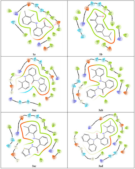
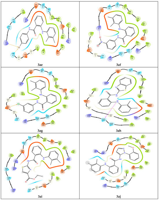
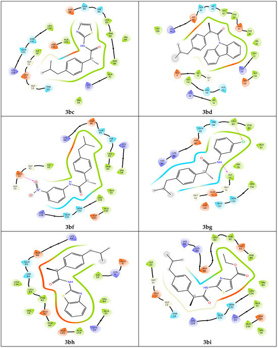
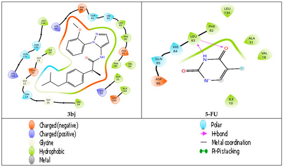
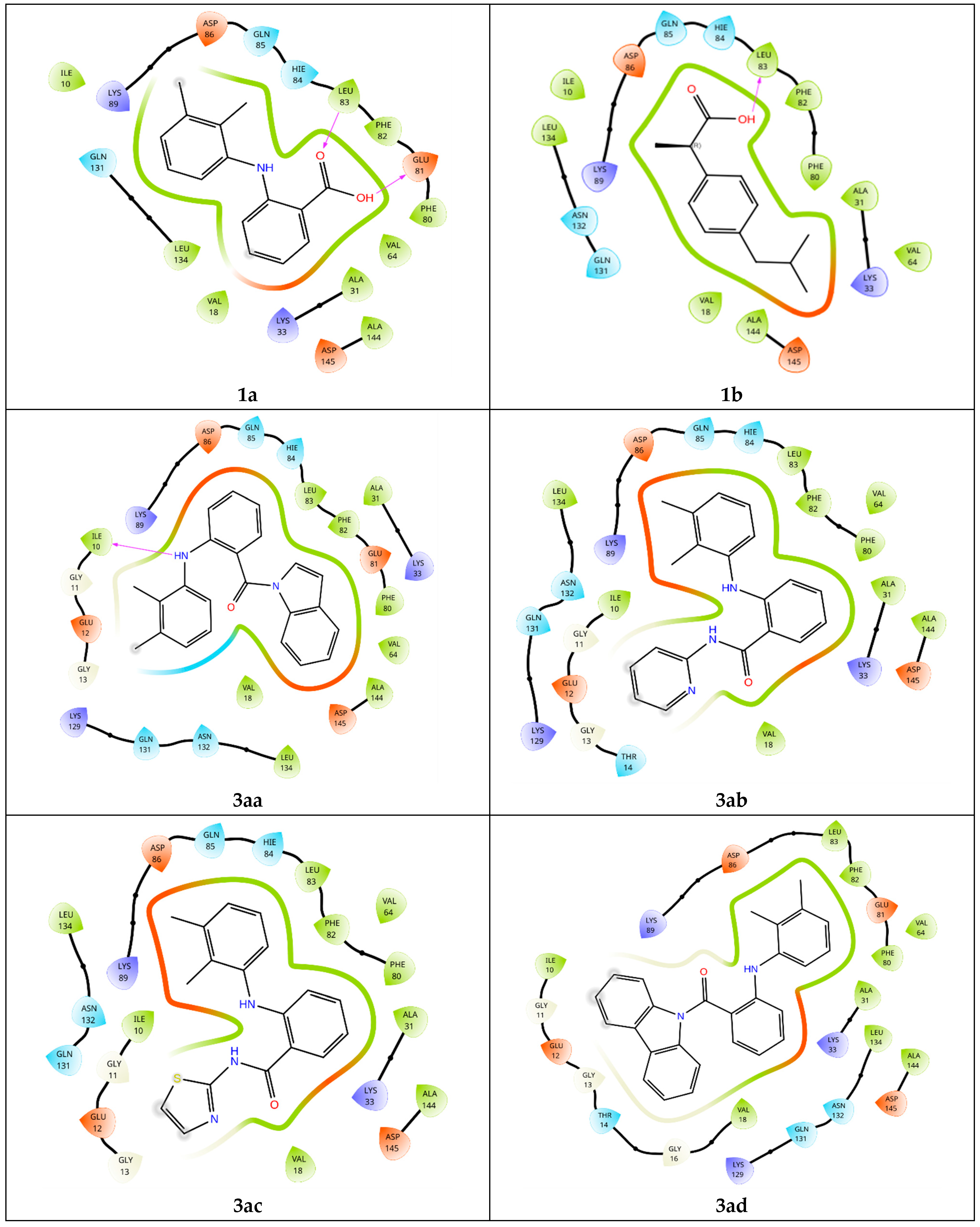
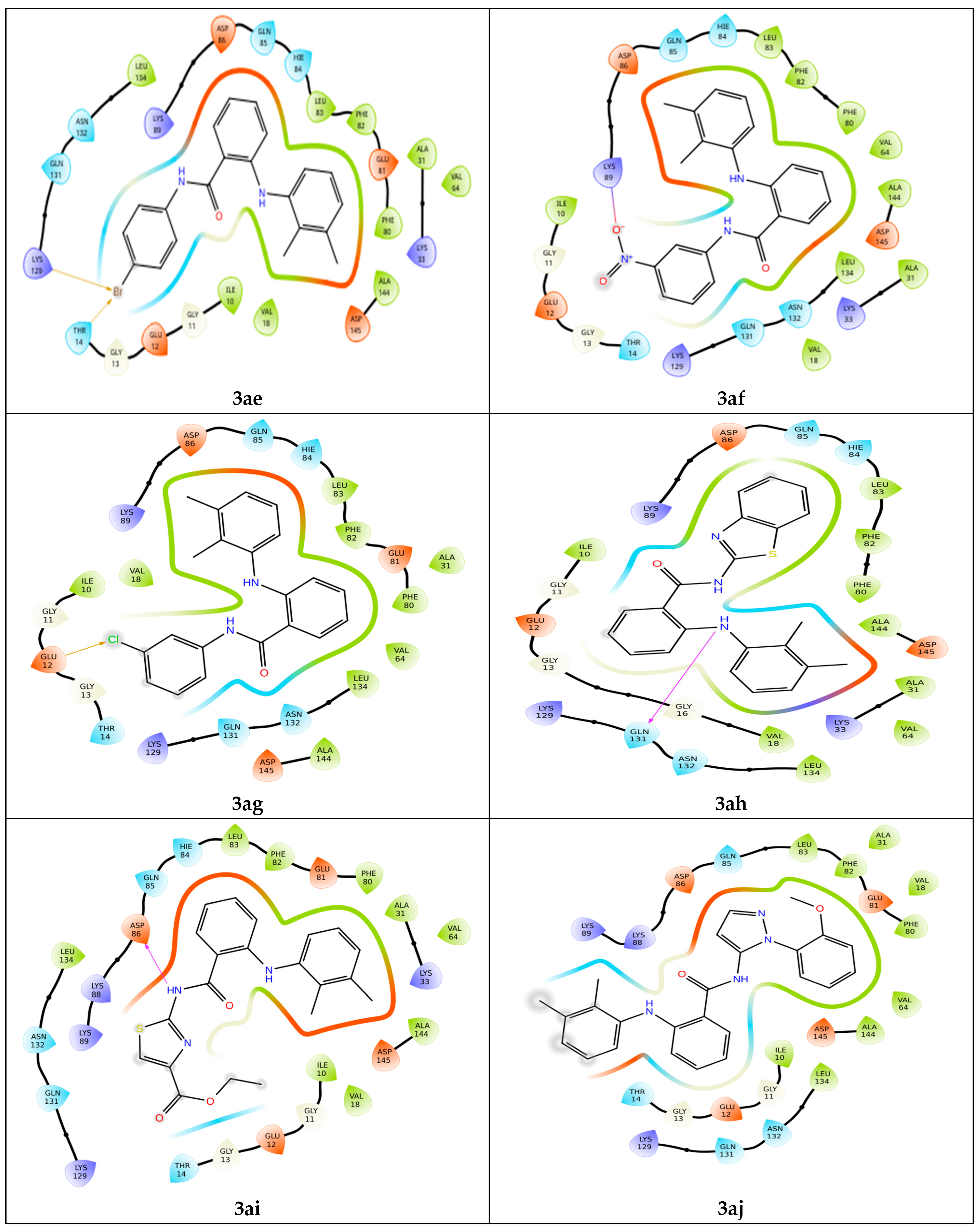
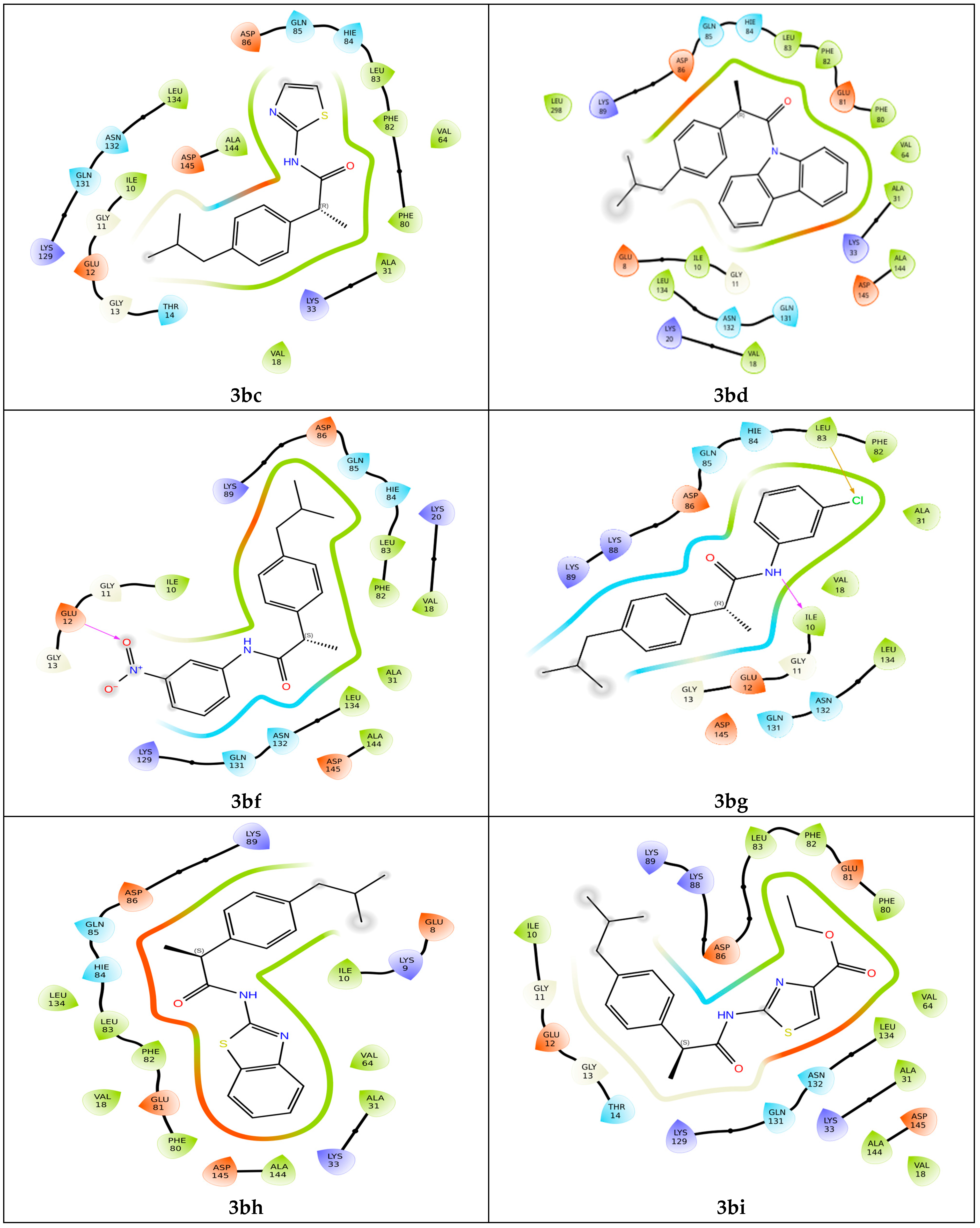
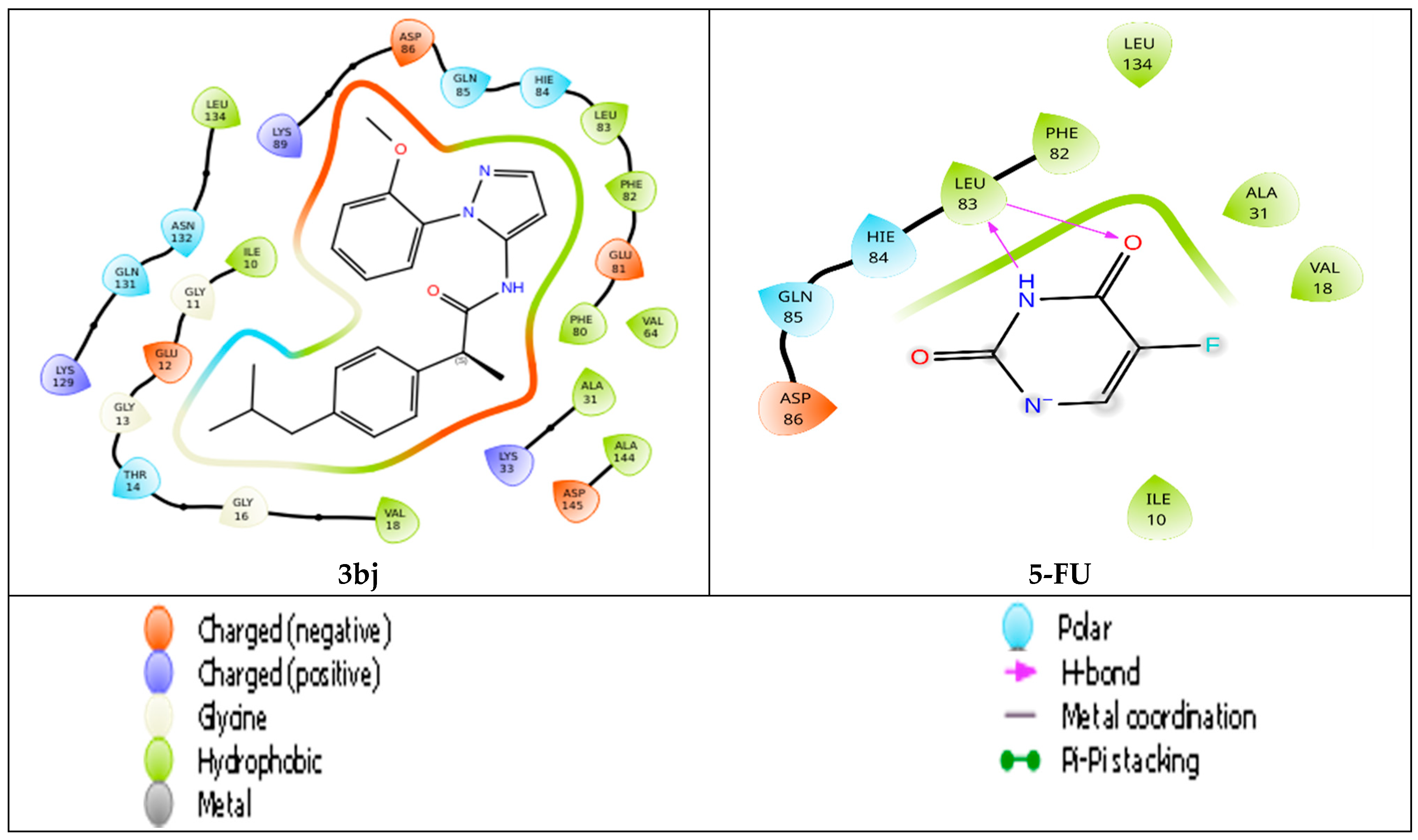
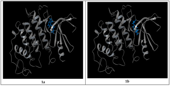
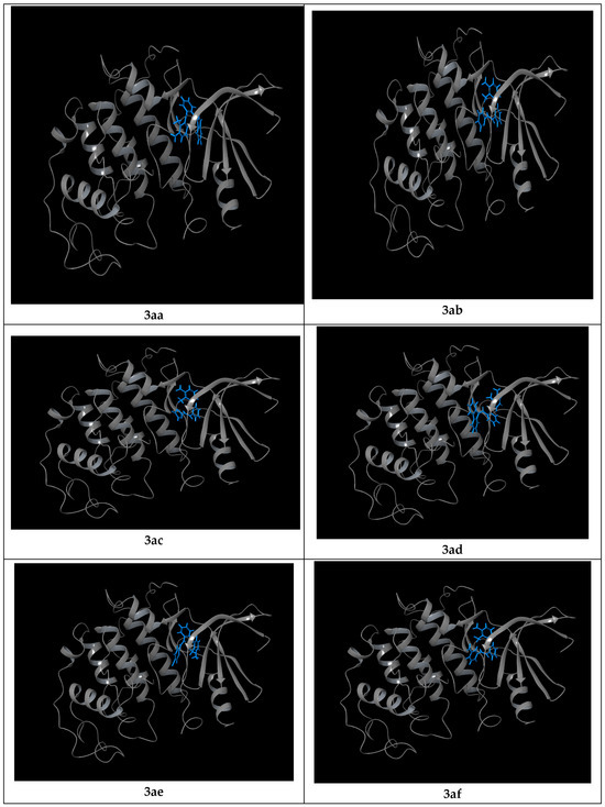
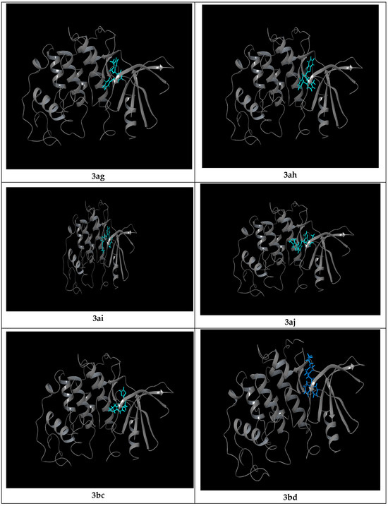
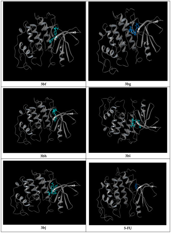
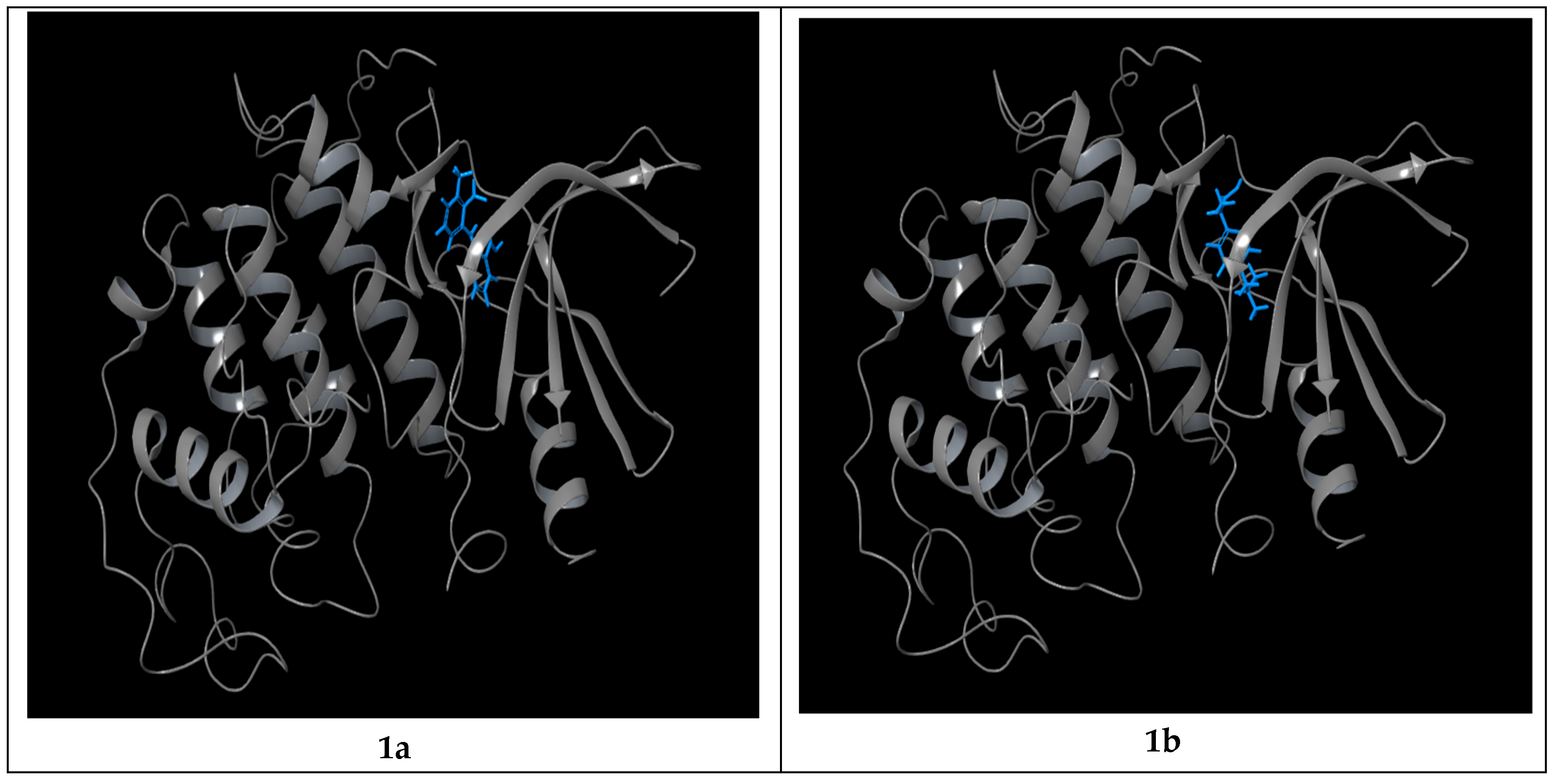
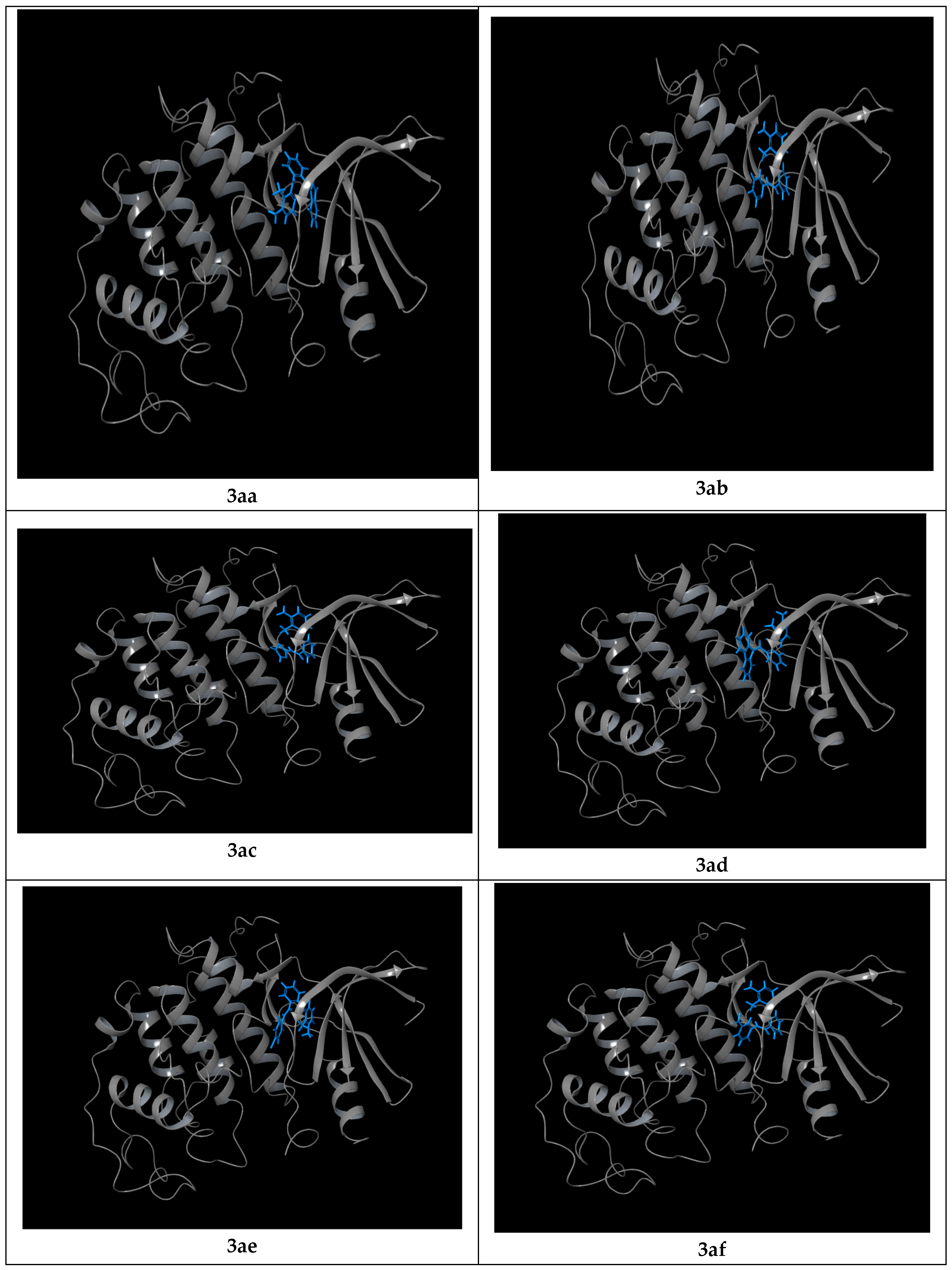
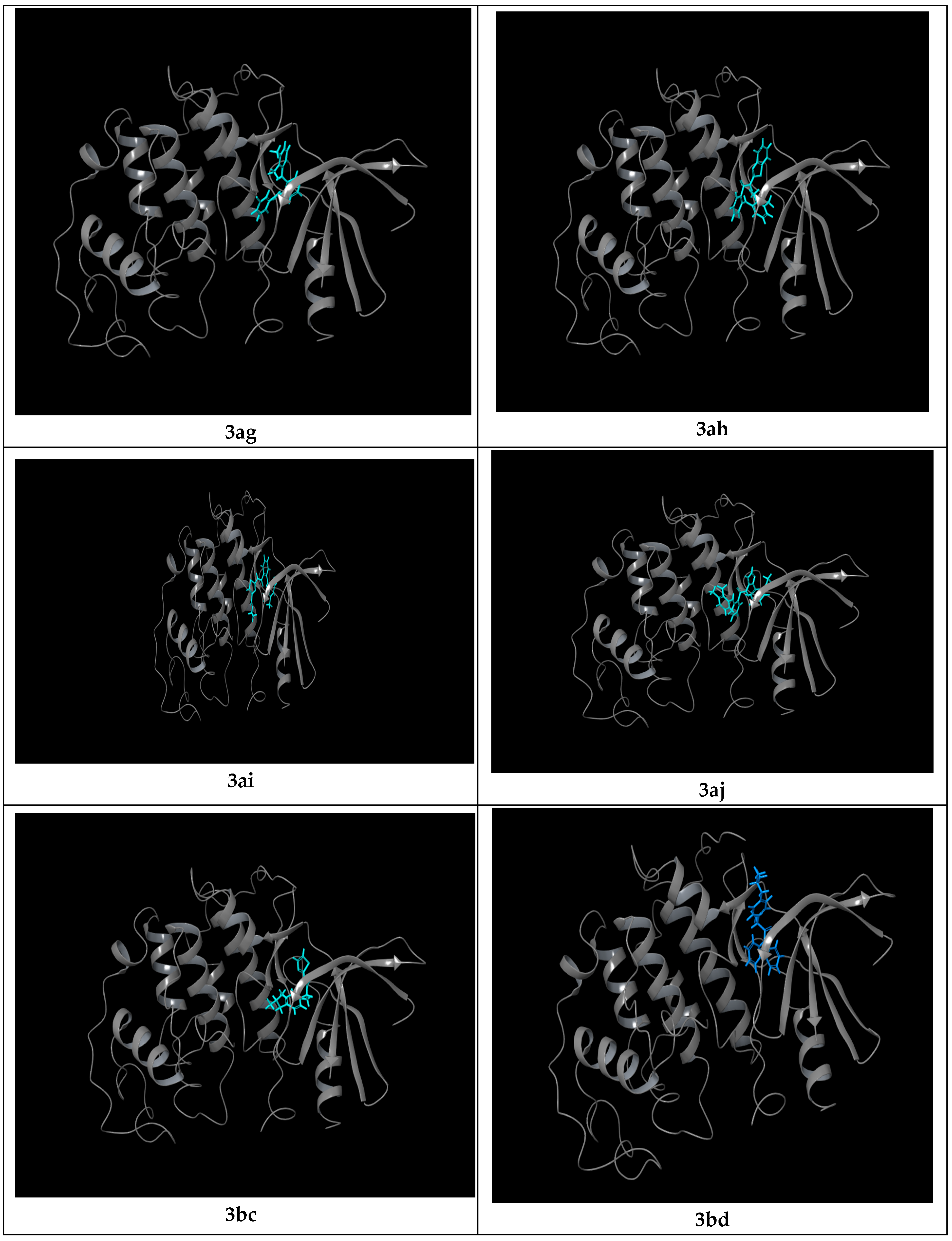
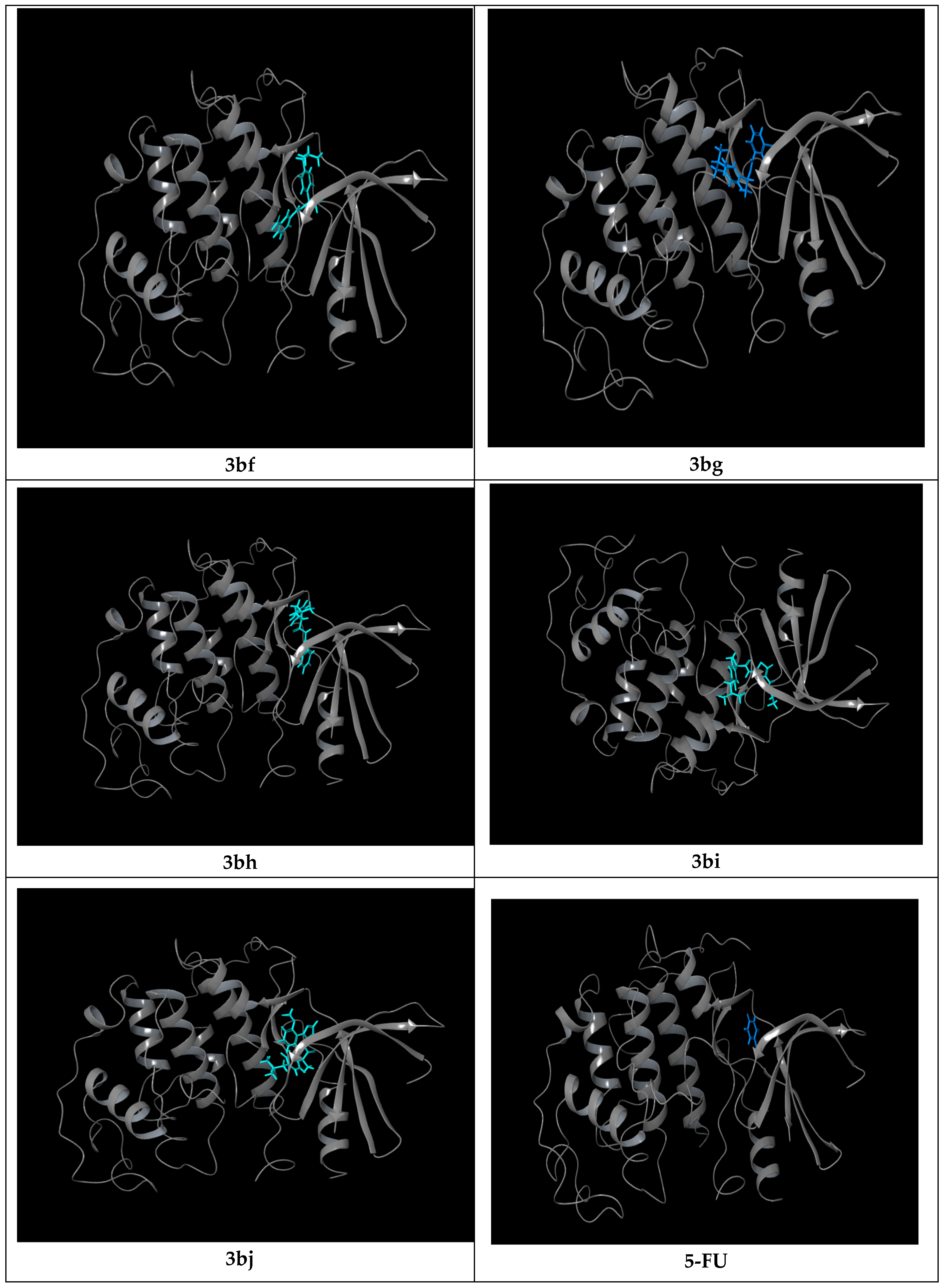
All the synthesized compounds showed significant binding towards the CDK2 active site (Figure 3 and Figure 4). Mefenamic acid and ibuprofen showed a docking score of −8.23 and −6.76 Kcal/mol, respectively, along with a binding energy of −55.21 and −55.81 Kcal/mol. The anticancer drug 5-fluorouracil has displayed a docking score of −6.67 Kcal/mol with a binding energy of −28.62 Kcal/mol. Except for 3bj, all other derivatives possessed a potential docking score. Among them, 3aa scored top with a docking score of −9.21 Kcal/mol and a binding energy of −46.44 Kcal/mol. These scores are stabilized with one H-bond with Ile 10 and hydrophobic interactions with residues Leu 83, Phe 82, Glu 81, Phe 80, and Val 18 and one charge (−) with Lys 33. Val 18 is part of the G loop, which facilitates phosphate group transfer. Lys 33 is a residue that stabilizes the conformational change after activation. 3ac showed a docking score of −7.78 Kcal/mol and binding energy of −69.81 Kcal/mol contributed by hydrophobic interactions with Hie 84, Leu 83, Phe 82, Phe 80, Ala 31, Val 18, and Ile 10. Val 18 and Ile 10 are found inside the G loop. 3ae has a docking score of −7.63 Kcal and a binding energy value of −51.69 Kcal/mol, which is influenced by hydrophobic interaction, negatively charged interaction and polar interaction with residues Leu 83, Phe 82, Glu 81, Phe 80, Val 18, Ile 10, Gln 35, Hie 84, Ala 145 and Ala 164. It also forms two halogen bonds with Lys 129 and Thr 14. Lys 129 is a conserved lysine that forms an H-bond with the serine of the peptide substrate. The interaction of the derivative prevents the formation of a strong H-bond by forming a weak halogen bond. Thr 14 prevents the positioning of ATP in the G loop. 3bd has a docking score of −7.17 Kcal/mol and a binding energy of −72.48 Kcal/mol with hydrophobic interactions and negatively charged interactions with the following residues: Gln 85, Hie 84, Leu 83, Phe 82, Glu 81, Phe 80, Val 64, Ala 31, Gly 11, Asp 86, and Lys 33. Gly 11 resides in the G loop. 3af has a docking score of −6.86 Kcal/mol with a binding energy of −73.14 Kcal/mol. This is contributed by hydrophobic, negatively charged, and polar interactions with various residues Hie 84, Leu 83, Phe 82, Phe 80, Val 64, Ala 144, Asn 132, Asp 86, Gln 85, Lys 89, Leu 134, and Gln 131. 3ad has a docking score of −6.78 Kcal/mol and a binding energy of −45.90 Kcal/mol, with hydrophobic interactions and negatively charged interactions with residues Asp 86, Leu 83, Phe 82, Glu 81, Phe 80, Ala 31, Val 18, and Lys 33. Here, Val 18 resides in the G loop. 3ab possessed a docking score of −6.77 Kcal/mol and a binding energy value of −64.21 Kcal/mol, which is contributed by hydrophobic interaction with residues Hie 84, Leu 83, Phe 82, Phe 80, Ala 31, Val 18, Ile 10 and negatively charged interactions with Asp 86, Lys 89, Lys 33. Lys 33 is a residue that helps to stabilize the conformational change after activation. 3bg has a glide score of −5.41 kcal/mol with a binding energy of −61.60 Kcal/mol with hydrophobic interaction, H-bond interaction, polar interaction, and halogen interactions with residues Asp 86, Gln 85, Hie 84, Leu 83, Phe 82, Val 18, Ile 10 (hydrogen bond), Lys 89, Lys 88, Asp 86, Gly 11, Glu 12, Gly 13, and Leu 83. Gly 11, Glu 12, and Gly 13 are seen in the G loop. These results are tabulated in Table 4 and Table 5. Among the compounds, 3bj displayed a lower docking score and exhibited hydrophobic interaction with Glu 81, Phe 82, Leu 83, and Hie 84.
Figure 3.
The ‘2D’ interaction diagram of the synthesized molecules with CDK2.
Figure 4.
The ‘3D’ interaction diagram of the synthesized molecules with CDK2.
Table 4.
Docking scores of the synthesized compounds.
Table 5.
Docking interaction between the ligand molecule and the protein CDK2 *.
Most of the derivatives showed interactions with sites in the G loop, which ensure proper alignment of the ATP and the interaction of its phosphate group. Therefore, binding of derivatives to this site prevents activation. 3ae interacts with Asp 145, i.e., near the active site, and with Ala 164, which is in the T loop. It also forms an interaction with Thr 14, an inhibitory site in the G loop. 3ae also forms a halogen bond and prevents the formation of a strong H-bond with Lys 129. Ibuprofen derivatives 3bf and 3bg form hydrogen bonds with the protein. All the residues can be classified as type 1 inhibitors of CDK2. Since the in vitro assays were conducted using 5-FU as the reference inhibitor, molecular docking was performed to determine its binding conformation and affinity for CDK2. The docking results indicate that the derivatives 3aa, 3ac, 3ae, 3ah, 3ai, and 3bd exhibit stronger binding affinities than 5-FU, suggesting higher potency. Therefore, these compounds hold promise as potential lead candidates for the development of targeted therapies against CDK2-overexpressing cervical cancer.
2.2.3. ADMET Properties
Absorption, distribution, metabolism, excretion, and toxicity (ADMET) were analyzed computationally using the Quikprop program in the Schrodinger suite. A comparative ADMET evaluation was performed on the tested compounds to assess physicochemical properties, absorption, distribution, metabolism, excretion, and toxicity. The parameters such as Caco-2 permeability, aqueous solubility, brain/blood partition coefficient (BBB), human intestinal absorption activity (HIA), plasma protein binding rate (PPBR), volume of distribution at steady state (VDss), CYP2C9 inhibition, CYP2D6 inhibition, CYP3A4 inhibition, hepatic intrinsic clearance (CL hepa) and intrinsic clearance measured in liver microsomes (CL micro) were evaluated for all the compounds (Table 6 and Table 7). The toxicity parameters were inhibition of the human ether-a-go-go-related gene (hERG blocker), drug-induced liver injury (DILI), Ames test (Ames), and median lethal dose (LD50) (Table 8).
Table 6.
ADMET properties (absorption and distribution).
Table 7.
ADMET properties (metabolism and excretion).
Table 8.
Toxicity prediction.
Overall, the compounds showed acceptable human intestinal absorption, moderate solubility, and reasonably predicted oral bioavailability. Within this set, compound 3aa displayed a particularly balanced profile, with good HIA (~74.5%), favorable oral bioavailability (52%), and moderate lipophilicity (logD 2.16), which together supported efficient absorption and oral exposure. Its distribution parameters (BBB and VDss) were within a comfortable range, and its metabolic and toxicity predictions showed no extreme liabilities, indicating that 3aa could achieve adequate systemic levels without safety red flags. Toxicity predictions identified significant divergence among the candidates. The compound 3bg maintained acceptable hERG inhibition and DILI risk levels, consistent with manageable safety expectations in early discovery.
Among the derivatives evaluated, compound 3ad demonstrated favorable pharmacokinetic properties. It exhibited a long-predicted half-life (~100 h), indicating the potential for prolonged systemic exposure, and retained good human intestinal absorption (~74.7%) along with moderate oral bioavailability (~49.7%). Although 3ad is more lipophilic (higher logKOW) and shows stronger CYP interactions compared to other analogs, these traits can still be advantageous for improving target engagement and sustaining pharmacological effects when properly optimized. Its distribution profile, combined with its strong exposure parameters, highlights 3ad as a promising lead scaffold. Overall, the ADMET data point to 3aa and 3ad as priority candidates for further molecular dynamics simulations to assess their binding stability in physiological conditions.
2.2.4. Structure–Activity Relationship (SAR) Studies
A detailed evaluation of the anticancer effects of mefenamic acid and ibuprofen derivatives incorporated into heterocyclic and non-heterocyclic compounds has been carried out in this study. Cytotoxicity of the compounds was tested, and morphological changes were monitored under an optical microscope. The compounds 3aa and 3ad have shown better results when compared with the other tested derivatives.
Among the compounds analyzed, the mefenamic acid derivatives showed better activity than the ibuprofen derivatives. The incorporation of indole and carbazole rings into the mefenamic acid moiety further enhanced their potency. Encouraged by these results, we introduced different types of substituents on mefenamic acid for further study. For this, we have taken a five-membered ring with sulfur and nitrogen (3ac, 3bc). Then, we looked for a six-membered ring with nitrogen. We also tried fused rings containing five- and six-membered rings as well as other heterocyclic rings. All these compounds are linked with mefenamic acid and ibuprofen through an amide linkage. In the case of the first two compounds, we used an aliphatic amide linkage, whereas the fused rings, such as indole, were also employed. Then we tried with a non-heterocyclic aromatic ring with different substituents at different places (related to 3ab). For this, we have taken para-bromo-substituted and meta-nitro-substituted compounds. We also tried a tricyclic fused ring with nitrogen similar to that of 3ad and 3bd. It was vital for us to study the effect of the mefenamic acid core with that of ibuprofen.
The impact of ring size on mefenamic acid with five-membered, six-membered, bicyclic, and tricyclic heterocycles was also investigated. The compounds 3aa and 3ad showed better activity, presumably because of the delocalization of electrons. The direct amide linkage from the fused ring can enhance the activity. The electronegative nature of sulfur, which is incorporated in the five-membered ring as part of the compound 3ac, is a possible reason for the moderate activity of that molecule. In general, amide derivatives of mefenamic acid obtained from indole and carbazole were found to be the most active compounds, presumably because of the presence of a nitrogen atom and subsequent delocalization of electrons.
When associating SAR studies with ADMET evaluation of the tested compounds, ibuprofen derivative 3bg exhibited the best absorption properties. The electron-withdrawing nature of the chlorine atom could have enhanced its absorption properties. The thiazole moiety in compound 3ac may impart a non-polar character, as evidenced by its good ADME profile. Compounds 3aa and 3ad displayed the best ADMET profile with mefenamic acid as the basic scaffold. The larger pi-conjugated system in the compound 3ad may be the reason for better ADMET profiling. The compound 3aa, being a bi-ring fused to the mefenamic acid core, also displayed a good ADMET score.
3. Materials and Methods
3.1. Chemistry
3.1.1. General Considerations
All reagents and chemicals were purchased from commercial suppliers and used as delivered unless otherwise noted. 1H NMR and 13C NMR spectra were recorded at 400 and 100 MHz respectively. The chemical shifts were reported in parts per million (ppm) and coupling constants in Hertz (Hz). Tetramethylsilane (TMS) (δ = 0.00 ppm) or the residual solvent peak in DMSO-d6 (δ = 2.50 ppm) and CDCl3 (δ = 7.26 ppm) served as internal standard for recording [54]. The molecular weights of unknown compounds were determined by GCMS-QP2010 Ultra gas chromatograph operating at an ionization potential of 70 eV (EI) or Thermo Finnigan LCQ Advantage apparatus (ESI). Microanalyses were performed on PerkinElmer Series II CHNS/O 2400 elemental analyzer and the melting points were determined using a Stuart SMP 3 apparatus. Thin-layer chromatography (TLC) was performed using Merck silica gel 60 F254 TLC plates.
3.1.2. General Procedure for the Synthesis of Amides 3
The synthesis of compounds 3aa–3af, 3ai, 3aj, 3bd and 3bg was carried out according to our previously reported protocol [36]. The synthesis of new derivatives 3ag, 3ah, 3ai, 3aj, 3bc, 3bf, 3bh, 3bi and 3bj was performed using the same methodology. In a round bottomed flask, acid 1a or 1b (1 mmol, 1 equiv.) was taken in 3 mL DMF and various substituted amines, 2a–j (1 mmol, 1 equiv.), were added. To this solution, N,N-diisopropylethylamine (DIPEA) (2 mmol, 2 equiv.) and HATU (2 mmol, 2 equiv.) were added. The reaction mixture was stirred at room temperature for 16 h. After the completion of the reaction as monitored by thin-layer chromatography, ethyl acetate and saturated aqueous sodium bicarbonate were added to the reaction mixture. The organic and aqueous layers were separated. Ethyl acetate was subsequently used to extract the aqueous layer twice and the combined organic fractions were dried in MgSO4 to obtain the crude product. The resultant crude was then purified by crystallization in ethanol to obtain a series of amides 3 in varying yields.
N-(3-Chlorophenyl)-2-((2,3-dimethylphenyl)amino)benzamide (3ag)
Yield: 85%; off white solid; m.p: 204–207 °C.
1H NMR (400 MHz, DMSO-d6): δ = 2.11 (s, 3H, CH3), 2.29 (s, 3H, CH3), 6.52 (s, 1H, NH), 7.20–7.26 (m, 5H, ArH), 7.34 (s, 1H, ArH), 7.42–7.46 (m, 3H, ArH), 7.68–7.71 (m, 2H, ArH), 8.04 (s, 1H, NH).
13C NMR (100 MHz, DMSO-d6): δ = 14.1, 20.2, 109.3, 111.3, 114.5, 117.3, 119.5, 120.3, 122.2, 125.3, 126.4, 127.1, 129.3, 131.2, 133.2, 135.6, 148.2, 149.6, 151.2, 156.0, 169.7.
MS (EI): m/z (%) = 350 (30) [M]+, 126 (100).
Anal. Calcd for C21H19ClN2O: C, 71.89; H, 5.46; Cl, 10.10; N, 7.98; Found: C, 72.12; H, 5.58; Cl, 10.41; N, 8.07.
N-(Benzo[d]thiazol-2-yl)-2-((2,3-dimethylphenyl)amino)benzamide (3ah)
Yield: 83%; light brown solid; m.p: 226–228 °C.
1H NMR (400 MHz, DMSO-d6): δ = 2.10 (s, 3H, CH3), 2.28 (s, 3H, CH3), 6.51 (s, 1H, NH), 7.18–7.26 (m, 3H, ArH), 7.39–7.44 (m, 5H, ArH), 7.78–7.81 (m, 3H, ArH), 8.26 (s, 1H, NH).
13C NMR (100 MHz, DMSO-d6): δ = 14.0, 20.1, 109.4, 111.3, 114.5, 117.2, 119.6, 120.2, 122.3, 123.8, 125.2, 126.4, 127.0, 129.4, 131.1, 133.3, 135.5, 148.1, 149.7, 151.3, 156.2, 169.9.
MS (EI): m/z (%) = 373 (54) [M]+, 134 (100).
Anal. Calcd for C22H19N3OS: C, 70.75; H, 5.13; N, 11.25; S, 8.58. Found: C, 70.59; H, 5.43; N, 11.20; S, 8.50.
Ethyl-2-(2-((2,3-Dimethylphenyl)amino)benzamido)thiazole-4-carboxylate (3ai)
Yield: 85%; light yellow solid; m.p: 211–214 °C.
1H NMR (400 MHz, DMSO-d6): δ = 1.34 (t, J = 6.8 Hz, 3H, CH3), 2.12 (s, 3H, CH3), 2.27 (s, 3H, CH3), 4.34 (q, J = 6.8 Hz, 2H, CH2), 6.50 (s, 1H, NH), 7.01 (s, 1H, ArH), 7.14–7.22 (m, 3H, ArH), 7.36–7.41 (m, 2H, ArH), 7.78–7.81 (m, 2H, ArH), 8.41 (s, 1H, NH).
13C NMR (100 MHz, DMSO-d6): δ = 14.1, 14.3, 20.2, 60.7, 111.2, 114.7, 117.1, 119.5, 122.4, 123.9, 125.4, 127.2, 129.2, 133.2, 135.4, 148.2, 149.8, 151.4, 156.4, 166.3, 172.1.
MS (EI): m/z (%) = 395 (41) [M]+, 156 (100).
Anal. Calcd for C21H21N3O3S: C, 63.78; H, 5.35; N, 10.63; S, 8.11. Found: C, 63.74; H, 5.58; N, 10.26; S, 8.36.
2-((2,3-Dimethylphenyl)amino)-N-(1-(2-methoxyphenyl)-1H-pyrazol-5-yl)benzamide (3aj)
Yield: 80%; off white solid; m.p: 224–227 °C.
1H NMR (400 MHz, DMSO-d6): δ = 2.10 (s, 3H, CH3), 2.29 (s, 3H, CH3), 3.40 (s, 3H, OCH3), 6.52 (s, 1H, NH), 7.02 (d, J = 7.2 Hz, 1H, ArH), 7.07 (d, J = 7.2 Hz, 1H, ArH), 7.18–7.22 (m, 3H, ArH), 7.41–7.47 (m, 4H, ArH), 7.72–7.75 (m, 2H, ArH), 8.35 (s, 1H, NH).
13C NMR (100 MHz, DMSO-d6): δ = 14.1, 20.2, 55.7, 109.6, 111.2, 114.6, 117.1, 118.3, 119.8, 121.0, 122.5, 123.7, 125.1, 126.2, 127.1, 128.4, 129.3, 131.0, 133.2, 135.3, 148.0, 149.6, 151.2, 156.1, 169.8.
MS (EI): m/z (%) = 412 (100) [M]+, 412 (100).
Anal. Calcd for C25H24N4O2: C, 72.80; H, 5.86; N, 13.58. Found: C, 73.17; H, 6.07; N, 13.89.
2-(4-Isobutylphenyl)-N-(thiazol-2-yl)propanamide (3bc)
Yield: 75%; light brown solid; m.p: 180–183 °C.
1H NMR (400 MHz, CDCl3): δ = 0.92 (d, J = 6.8 Hz, 6H, 2CH3), 1.62 (d, J = 7.2 Hz, 3H, CH3), 1.83–1.94 (m, 1H, CH), 2.51 (d, J = 6.8 Hz, 2H, CH2), 3.73 (q, J = 6.8 Hz, 1H, CH), 7.09 (d, J = 6.4 Hz, 1H), 7.20 (d, J = 6 Hz, 1H), 7.43 (d, J = 8.4 Hz, 2H, ArH), 7.66 (d, J = 8.4 Hz, 2H, ArH), 8.13 (s, 1H, NH).
13C NMR (100 MHz, CDCl3): δ = 18.4, 22.4, 30.1, 45.0, 47.7, 114.5, 118.9, 125.4, 127.4, 129.6, 130.2, 133.5, 141.7, 148.8, 172.9.
MS (EI): m/z (%) = 288 (65) [M]+, 99 (100).
Anal. Calcd for C16H20N2OS: C, 66.63; H, 6.99; N, 9.71; S, 11.12. Found: C, 66.95; H, 6.85; N, 10.03; S, 10.98.
2-(4-Isobutylphenyl)-N-(3-nitrophenyl)propanamide (3bf)
Yield: 77%; light yellow solid; m.p: 154–157 °C.
1H NMR (400 MHz, CDCl3): δ = 0.93 (d, J = 6.4 Hz, 6H, 2CH3), 1.62 (d, J = 7.2 Hz, 3H, CH3), 1.82–1.94 (m, 1H, CH), 2.50 (d, J = 7.2 Hz, 2H, CH2), 3.74 (q, J = 7.2 Hz, 1H, CH), 7.18–7.28 (m, 5H, ArH), 7.44 (t, J = 8.0 Hz, 1H, ArH), 7.85–7.93 (m, 2H, ArH), 8.25 (s, 1H, NH).
13C NMR (100 MHz, CDCl3): δ = 18.4, 22.4, 30.2, 45.0, 47.8, 114.4, 118.8, 125.3, 127.3, 127.4, 129.4, 129.8, 130.1, 137.4, 139.0, 141.6, 148.5, 173.0.
MS (EI): m/z (%) = 326 (65) [M]+, 137 (100).
Anal. Calcd for C19H22N2O3: C, 69.92; H, 6.79; N, 8.58. Found: C, 69.99; H, 6.74; N, 8.27.
N-(Benzo[d]thiazol-2-yl)-2-(4-isobutylphenyl)propanamide (3bh)
Yield: 80%; light brown solid; m.p: 186–189 °C.
1H NMR (400 MHz, DMSO-d6): δ = 0.92 (d, J = 6.8 Hz, 6H, 2CH3), 1.62 (d, J = 7.2 Hz, 3H, CH3), 1.83–1.94 (m, 1H, CH), 2.51 (d, J = 6.8 Hz, 2H, CH2), 3.73 (q, J = 7.2 Hz, 1H, CH), 7.17–7.27 (m, 3H, ArH), 7.47–7.59 (m, 3H, ArH), 7.82–7.93 (m, 2H, ArH).
13C NMR (100 MHz, CDCl3): δ = 18.3, 22.4, 30.2, 44.9, 47.7, 114.4, 117.1, 119.5, 122.2, 125.3, 127.1, 129.3, 133.2, 135.4, 148.0, 149.8, 151.2, 156.1, 173.1.
MS (EI): m/z (%) = 338 (46) [M]+, 161 (100).
Anal. Calcd for C20H22N2OS: C, 70.97; H, 6.55; N, 8.28; S, 9.47. Found: C, 71.00; H, 6.65; N, 8.22; S, 9.60.
Ethyl-2-(2-(4-Isobutylphenyl)propanamido)thiazole-4-carboxylate (3bi)
Yield: 75%; off white solid; m.p: 175–178 °C.
1H NMR (400 MHz, CDCl3): δ = 0.93 (d, J = 6.8 Hz, 6H, 2CH3), 1.33 (t, J = 6.8 Hz, 3H, CH3), 1.61 (d, J = 7.2 Hz, 3H, CH3), 1.83–1.93 (m, 1H, CH), 2.50 (d, J = 7.2 Hz, 2H, CH2), 3.72 (q, J = 6.8 Hz, 1H, CH), 4.35 (q, J = 6.8 Hz, 2H, CH2), 7.17 (d, J = 7.6 Hz, 2H, ArH), 7.21 (s, 1H, ArH), 7.48 (d, J = 7.6 Hz, 2H, ArH), 8.18 (s, 1H, NH).
13C NMR (100 MHz, CDCl3): δ = 14.1, 18.3, 22.3, 30.1, 45.0, 47.7, 60.8, 114.3, 118.7, 127.2, 129.5, 130.2, 137.5, 139.1, 141.7, 148.6, 160.7, 173.1.
MS (EI): m/z (%) = 360 (100) [M]+, 360 (100).
Anal. Calcd for C19H24N2O3S: C, 63.31; H, 6.71; N, 7.77; S, 8.89. Found: C, 63.57; H, 6.97; N, 7.93; S, 8.81.
2-(4-Isobutylphenyl)-N-(1-(2-methoxyphenyl)-1H-pyrazol-5-yl)propanamide (3bj)
Yield: 70%; white solid; m.p: 187–190 °C.
1H NMR (400 MHz, CDCl3): δ = 0.91 (d, J = 6.8 Hz, 6H, 2CH3), 1.62 (d, J = 7.2 Hz, 3H, CH3), 1.82–1.93 (m, 1H, CH), 2.51 (d, J = 7.2 Hz, 2H, CH2), 3.39 (s, 3H, OCH3), 3.73 (q, J = 6.8 Hz, 1H, CH), 7.06 (d, J = 7.2 Hz, 1H, ArH), 7.22–7.26 (m, 3H, ArH), 7.46–7.50 (m, 4H, ArH), 7.72 (d, J = 7.6 Hz, 2H, ArH), 8.64 (s, 1H, NH).
13C NMR (100 MHz, CDCl3): δ = 18.2, 22.3, 30.0, 45.1, 47.6, 55.6, 114.3, 118.7, 119.8, 123.4, 126.8, 127.2, 129.5, 130.2, 133.5, 137.5, 139.1, 141.7, 144.5, 148.6, 152.3, 173.0.
MS (EI): m/z (%) = 377 (38) [M]+, 188 (100).
Anal. Calcd for C23H27N3O2: C, 73.18; H, 7.21; N, 11.13. Found: C, 73.50; H, 7.59; N, 10.89.
3.2. Anticancer Activity
The effects of our compounds on the growth of HeLa and Chang cells were determined by MTT assay [55]. After incubation for 2 h, cell morphology was tracked under an inverted phase-contrast microscope.
3.2.1. In Vitro Antiproliferative Effect on HeLa Cells: Determination by MTT Assay
HeLa and Chang cell lines obtained from NCCS, Pune, India, maintained in Dulbecco’s modified eagle medium (Himedia) (DMEM), were used for antiproliferative studies. A 25 cm2 tissue culture flask with DMEM was used to culture the cell line supplemented with 10% Fetal Bovine Serum (FBS), L-glutamine, sodium bicarbonate, and an antibiotic solution containing Streptomycin (100 µg/mL), Penicillin (100 U/mL), and Amphotericin B (2.5 µg/mL). A humidified 5% CO2 incubator (Galaxy® 170 Eppendorf, Hamburg, Germany) was used to keep the cell line at 37 °C. An inverted phase-contrast microscope was used to observe cells and assess their viability after the MTT assay.
3.2.2. Cell Seeding in a 96-Well Plate
Trypzinised confluent monolayer of cells was suspended in a 10% growth medium after two days. A 96-well tissue culture plate was used to seed 100 µL of the cell suspension (5 × 10⁴ cells/well), which was then incubated in a humidified 5% CO2 incubator at 37 °C.
3.2.3. Preparation of Compound Stock
Cyclomixer was used for dissolving 1 mg of each compound in 1 mL of DMEM, which was then filtered through a 0.22 µm Millipore syringe filter to ensure sterility.
3.2.4. Antiproliferative Effect on Evaluation
The growth medium was removed after 24 h. Fresh solutions of the samples were prepared in 5% DMEM and then diluted five times serially by two-fold dilutions (6.25 µg, 12.5 µg, 25 µg, 50 µg, 100 µg in 100 µL of 5% DMEM). Then, 100 µL of the compound was added to the well in triplicate and incubated at 37 °C in a humidified 5% CO2 incubator.
3.2.5. Antiproliferative Effect by Direct Microscopic Observation
Using an inverted phase contrast tissue culture microscope (LabomedTCM-400 with MICAPSTM HD camera, Himedia, Mumbai, India), the entire plate was monitored for 24 to 72 h, and images were recorded. Granulation, vacuolization in the cytoplasm, and rounding or shrinking of the cells were noticed by observing the images, indicating cytotoxicity.
3.2.6. Antiproliferative Effect by MTT Assay
Then, 15 mg of MTT (Himedia, Mumbai, India, M-5655) was completely dissolved in 3 mL Phosphate-Buffer Saline (PBS) and sterilized using the filter. After 24 h of incubation, the sample content was removed from the wells, and a solution of MTT (30 µL) was added to the test and cell control wells, which were then kept for 4 h in a humidified 5% CO2 incubator after shaking. The supernatant was removed after incubation. To the 100 µL of MTT solubilization solution, dimethyl sulfoxide (DMSO) was added. The formazan cells were solubilized by pipetting up and down gently. Absorbance values at a wavelength of 570 nm were noted using a microplate reader.
3.3. In Silico Studies
To evaluate the in silico binding affinity of CDK2 with synthesized compounds, we performed molecular docking using Schrodinger suite 2025-3. The 3D structural coordinates were retrieved from the PDB database with PDB ID-3QTR [56], which has a resolution of 1.85 Å. The protein structure was processed and prepared with the ‘protein preparation wizard’, which involved the addition of the missing hydrogen atoms and loops, removing water molecules beyond 5Å of the ligand, assigning actual bond orders, and generating proper ionization states using the OPLS4 force field. Subsequently, the protein was minimized to perform the optimal docking study. The receptor grid for docking was generated using the Glide Grid Generation Wizard with respect to the bound inhibitor (X36 = [4-amino-2-(phenylamino)-1,3-thiazol-5-yl](phenyl)methanone) with a 20 Å enclosing box. Van der Waals scaling and input partial charges were set as default values. Docking validation was done by the redocking of the cogent ligand present on the crystal structure CDK2 (PDB. ID-3QTR) by using a generated grid. This was done by extracting the cogent ligand from the prepared protein structure, and its structure was optimized by using OPLS4 using the Ligprep module. RMSD was calculated by superimposing the prepared crystal structure with respect to the docked complex.
Structures of the synthesized compounds were drawn in ChemDraw 14.0 software, named 3aa, 3ab, 3ac, 3ad, 3ae, 3af, 3ag, 3ah, 3ai, 3aj, 3bc, 3bd, 3bf, 3bg, 3bh, 3bi and 3bj, then converted to .sdf format. We selected 5-fluorouracil as the standard for the in silico anticancer study, and also took mefenamic acid (1a), ibuprofen (1b), to compare the activity. Their structures were obtained from the PubChem database. The ligand structures were optimized at near-neutral pH (7 ± 2), and all plausible tautomers and stereoisomers were generated using the LigPrep wizard in Schrodinger. The protonation states were assigned to the ligands and subjected to energy minimization with the OPLS4 force field using the LigPrep module. Molecular docking of the prepared ligands to the active site of CDK2 was performed using the Extra Precision (XP) method in the Glide module of the Schrodinger suite. These docked complexes were further subjected to binding energy prediction by the prime MMGBSA method. The docking investigations of the synthesized compounds were carried out using the software Schrodinger suite 2025-3. Docking studies were conducted to evaluate the anticancer activity (PDBID: 3QTR).
4. Conclusions
In summary, by applying the theory of drug repurposing, the anticancer activity evaluation of some mefenamic acid and ibuprofen derivatives containing amide functionality has been successfully carried out. Our results were corroborated by performing the molecular docking and SAR studies. Our investigation revealed compound 3aa as the most promising candidate for anticancer studies, as it showed better results in vitro and in silico. Furthermore, 3aa possessed a pre-eminent docking score on binding with the target protein. From the in vitro study, the compound 3ad also showed an acceptable IC50 value when compared to the reference standard, 5-fluorouracil. The SAR studies revealed the importance of the presence of nitrogen containing tri- and bi-ring fused systems linked to mefenamic acid for better biological results. Mefenamic acid derivatives offered enhanced biological activity when compared to ibuprofen derivatives. The synthesis of additional derivatives and lead optimization is in process in our laboratory, and will be communicated in due course.
Author Contributions
Conceptualization, methodology: N.P.S., S.R. and S.M., formal analysis: N.P.S., S.R. and S.M., investigation, M.N.J. and J.A., draft preparation and editing, J.A., G.V.Z. and M.R.G.; supervision, K.R.H. and M.N.J. All authors have read and agreed to the published version of the manuscript.
Funding
This research was supported by the Russian Science Foundation (project No. 25-23-00612).
Institutional Review Board Statement
Not applicable.
Informed Consent Statement
Not applicable.
Data Availability Statement
The original contributions presented in this study are included in the article. Further inquiries can be directed to the corresponding author.
Conflicts of Interest
The authors declare no conflict of interest.
References
- Hassanpour, S.H.; Dehghani, M. Review of cancer from perspective of molecular. J. Cancer Res. Pract. 2017, 4, 127–129. [Google Scholar] [CrossRef] [Scilit]
- Golias, C.H.; Charalabopoulos, A.; Charalabopoulos, K. Cell proliferation and cell cycle control: A mini review. Int. J. Clin. Pract. 2004, 58, 1134–1141. [Google Scholar] [CrossRef] [Scilit] [PubMed]
- Hakim, D.N.; Pelly, T.; Kulendran, M.; Caris, J.A. Benign tumours of the bone: A review. J. Bone Oncol. 2015, 4, 37–41. [Google Scholar] [CrossRef] [Scilit] [PubMed]
- Martinez-Outschoorn, U.E.; Peiris-Pagés, M.; Pestell, R.G.; Sotgia, F.; Lisanti, M.P. Cancer metabolism: A therapeutic perspective. Nat. Rev. Clin. Oncol. 2016, 14, 11–31, Erratum in Nat. Rev. Clin. Oncol. 2017, 14, 113.. [Google Scholar] [CrossRef] [Scilit] [PubMed]
- Iheagwam, F.N.; Iheagwam, O.T.; Odiba, J.K.; Ogunlana, O.O.; Chinedu, S.N. Cancer and Glucose Metabolism: A Review on Warburg Mechanisms. Trop. J. Nat. Prod. Res. 2022, 6, 661–667. [Google Scholar]
- DeVita, V.T.; Chu, E. A history of cancer chemotherapy. Cancer Res. 2008, 68, 8643–8653. [Google Scholar] [CrossRef] [Scilit] [PubMed]
- Wistuba, I.I.; Gazdar, A.F. Lung cancer preneoplasia. Annu. Rev. 2006, 1, 331–348. [Google Scholar] [CrossRef] [Scilit] [PubMed]
- Sharma, G.N.; Dave, R.; Sanadya, J.; Sharma, P.; Sharma, K.K. Various types and management of breast cancer: An overview. J. Adv. Pharm. Technol. Res. 2024, 1, 109–126. [Google Scholar] [CrossRef] [Scilit]
- Hancock, S.B.; Georgiades, C.S. Kidney Cancer. Cancer J. 2016, 22, 387–392. [Google Scholar] [CrossRef] [Scilit] [PubMed]
- Vu, M.; Yu, J.; Awolude, O.A.; Chuang, L. Cervical cancer worldwide. Curr. Probl. Cancer 2018, 42, 457–465. [Google Scholar] [CrossRef] [Scilit] [PubMed]
- Cohen, P.A.; Jhingran, A.; Oaknin, A.; Denny, L. Cervical cancer. Lancet 2019, 393, 169–182. [Google Scholar] [CrossRef] [Scilit] [PubMed]
- Betz, S.J. HPV-Related Papillary Lesions of the Oral Mucosa: A Review. Head Neck Pathol. 2019, 13, 80–90. [Google Scholar] [CrossRef] [Scilit] [PubMed]
- Bigras, G.; De Marval, F. The probability for a Pap test to be abnormal is directly proportional to HPV viral load: Results from a Swiss study comparing HPV testing and liquid-based cytology to detect cervical cancer precursors in 13842 women. Br. J. Cancer 2005, 93, 575–581. [Google Scholar] [CrossRef] [Scilit] [PubMed]
- Ghosh, S. Cisplatin: The first metal based anticancer drug. Bioorganic Chem. 2019, 88, 102925. [Google Scholar] [CrossRef] [Scilit] [PubMed]
- Longley, D.B.; Harkin, D.P.; Johnston, P.G. 5-Fluorouracil: Mechanisms of action and clinical strategies. Nat. Rev. Cancer 2003, 3, 330–338. [Google Scholar] [CrossRef] [Scilit] [PubMed]
- Kollmannsberger, C.; Mross, K.; Jakob, A.; Kanz, L.; Bokemeyer, C. Topotecan—A novel topoisomerase I inhibitor: Pharmacology and clinical experience. Oncology 1999, 56, 1–12. [Google Scholar] [CrossRef] [Scilit] [PubMed]
- Li, M.M.; Song, M.; Wu, S.X.; Ren, X.Y. Identification of CDK2 as a key apoptotic gene for predicting cervical cancer prognosis using bioinformatics and machine learning. Am. J. Cancer Res. 2025, 15, 2750–2764. [Google Scholar] [CrossRef] [Scilit] [PubMed]
- Arora, P.; Arora, V.; Lamba, H.S.; Wadhwa, D. Importance of Heterocyclic Chemistry: A Review. Int. J. Pharm. Sci. Res. 2012, 3, 2947–2954. [Google Scholar] [CrossRef]
- Kumar, A.; Singh, A.K.; Singh, H.; Vijayan, V.; Kumar, D.; Naik, J.; Thareja, S.; Yadav, J.P.; Pathak, P.; Grishina, M.; et al. Nitrogen Containing Heterocycles as Anticancer Agents: A Medicinal Chemistry Perspective. Pharmaceuticals 2023, 16, 299. [Google Scholar] [CrossRef] [Scilit] [PubMed]
- Zaki, K.; Ouabane, M.; Sbai, A.; Sekkate, C.; Bouachrine, M.; Lakhlifi, T. Unraveling the mechanism, regioselectivity, and stereoselectivity of [3 + 2] cycloaddition reactions for anticancer spirooxindole derivatives: A density functional theory study. Comput. Theor. Chem. 2025, 1245, 115086. [Google Scholar] [CrossRef] [Scilit]
- Atmaca, H.; Ilhan, S.; Pulat, C.C.; Dundar, B.A.; Zora, M. Evaluation of Novel Spiro-pyrrolopyridazine Derivatives as Anticancer Compounds: In Vitro Selective Cytotoxicity, Induction of Apoptosis, EGFR Inhibitory Activity, and Molecular Docking Analysis. ACS Omega 2024, 9, 23713–23723. [Google Scholar] [CrossRef] [Scilit] [PubMed]
- Arun, Y.; Bhaskar, G.; Balachandran, C.; Ignacimuthu, S.; Perumal, P.T. Facile one-pot synthesis of novel dispirooxindole-pyrrolidine derivatives and their antimicrobial and anticancer activity against A549 human lung adenocarcinoma cancer cell line. Bioorganic Med. Chem. Lett. 2013, 23, 1839–1845. [Google Scholar] [CrossRef] [Scilit] [PubMed]
- Boutalaka, M.; Elbahi, S.; Ouabane, M.; Guendouzi, A.; Maghat, H.; Lakhlifi, T.; Bouachrine, M. Computational investigation of bisquinoline derivatives as potential c-met kinase inhibitors: 3D-QSAR, molecular docking, dynamics simulations, and ADME-Tox studies. Heliyon 2026, 12, e44916. [Google Scholar] [CrossRef] [Scilit]
- Santra, S.; Sharapov, A.D.; Fatykhov, R.F.; Potapova, A.P.; Khalymbadzha, I.A.; Valieva, M.I.; Kopchuk, D.S.; Zyryanov, G.V.; Bunev, A.S.; Melekhin, V.V.; et al. Xanthone-1,2,4-triazine and Acridone-1,2,4-triazine Conjugates: Synthesis and Anticancer Activity. Pharmaceuticals 2023, 16, 403. [Google Scholar] [CrossRef] [Scilit] [PubMed]
- Avanzo, R.E.; Anesini, C.; Fascio, M.L.; Errea, M.I.; D’Accorso, N.B. 1,2,4-Triazole d-ribose derivatives: Design, synthesis and antitumoral evaluation. Eur. J. Med. Chem. 2012, 47, 104–110. [Google Scholar] [CrossRef] [Scilit] [PubMed]
- Bosc, L.; Pero, M.E.; Balayssac, D.; Jacquemot, N.; Allard, J.; Suzanne, P.; Vollaire, J.; Cottet-Rousselle, C.; Michallet, S.; Villaret, J.; et al. Preventing neuropathy and improving anti-cancer chemotherapy with a carbazole-based compound. bioRxiv 2025. [Google Scholar] [CrossRef] [Scilit] [PubMed]
- Ceramella, J.; Rosano, C.; Iacopetta, D.; Ben Toumia, I.; Chekir-Ghedira, L.; Maatouk, M.; Mariconda, A.; Longo, P.; Dallemagne, P.; Rochais, C.; et al. Anti-Breast Cancer Properties and In Vivo Safety Profile of a Bis-Carbazole Derivative. Pharmaceutics 2025, 17, 415. [Google Scholar] [CrossRef] [Scilit] [PubMed]
- Abood, R.G.; Abdulhussein, H.A.; Abbas, S.; Majed, A.A.; Al-Khafagi, A.A.; Adil, A.; Alsalim, T.A. Anti-breast cancer potential of new indole derivatives: Synthesis, in-silico study, and cytotoxicity evaluation on MCF-7 cells. J. Mol. Struct. 2025, 1326, 141–176. [Google Scholar] [CrossRef] [Scilit]
- Basha, N.H.; Jagadish, T.; Karthikeyan, P.; Balu, K.; Durai, M.; Afzal, M.; Ahn, Y.-H.; Pasupathi, M.; Venkatesan, K. Ultrasound-Assisted Synthesis of Polyfunctionalized Pyridine Derivatives Catalyzed by Silica-Supported Perchloric Acid: In Vitro Anticancer Studies and Molecular Docking. Russ. J. Bioorganic Chem. 2025, 51, 932–946. [Google Scholar] [CrossRef] [Scilit]
- Huang, S.; Xu, Z.; Zhuang, Y. The current landscape of 1,2,3-triazole hybrids with anticancer therapeutic potential: Part I. Arch. Pharm. 2025, 358, e2500001. [Google Scholar] [CrossRef] [Scilit] [PubMed]
- Soliman, M.A.; Ahmed, H.E.A.; Eltamany, E.H.; Boraei, A.T.A.; Aljuhani, A.; Salama, S.A.; Alghamdi, R.; Aljohani, A.K.B.; Almaghrabi, M.; Aouad, M.R. Novel bis-benzimidazole-triazole hybrids: Anticancer study, in silico approaches, and mechanistic investigation. Future Med. Chem. 2025, 17, 93–107. [Google Scholar] [CrossRef] [Scilit] [PubMed]
- Neetha, S.; Santhosh, C.; Lohith, T.N.; Sharath, K.; Sridhar, M.A.; Sadashiva, M.P. Structural elucidation, Hirshfeld surface, DFT, molecular docking and molecular dynamics studies of a novel thiazole derivative as anti-cancer drug. J. Mol. Struct. 2025, 1324, 140793. [Google Scholar] [CrossRef] [Scilit]
- Liu, W.-B.; Yang, W.-G.; Wu, J.; Chen, B.-B.; Du, Y.-F.; Niu, J.-B.; Song, J.; Zhang, S.-Y. Discovery of novel thienopyridine indole derivatives as inhibitors of tubulin polymerization targeting the colchicine-binding site with potent anticancer activities. Eur. J. Med. Chem. 2025, 286, 117314. [Google Scholar] [CrossRef] [Scilit] [PubMed]
- Gunaydin, C.; Bilge, S.S. Effects of nonsteroidal anti-inflammatory drugs at the molecular level. Eurasian J. Med. 2018, 50, 116–121. [Google Scholar] [CrossRef] [Scilit] [PubMed]
- Amedi, D.; Gazerani, P. Deprescribing NSAIDs: The Potential Role of Community Pharmacists. Pharmacy 2024, 12, 116. [Google Scholar] [CrossRef] [Scilit] [PubMed]
- SreeNilayam, N.P.; Remeshan, S.; Shyma, M.; Joy, M.N.; Zyryanov, G.V.; Sajith, A.M.; Talavara, V.; Haridas, K.R. Biological activity evaluation and molecular docking studies of newly synthesized phenylamino derivatives. J. Mol. Struct. 2025, 1335, 142037. [Google Scholar] [CrossRef] [Scilit]
- Ozbudak, I.H.; Karaveli, S.; Simsek, T.; Erdogan, G.; Pestereli, E. Neoangiogenesis and expression of hypoxia-inducible factor 1α, vascular endothelial growth factor, and glucose transporter-1 in endometrioid type endometrium adenocarcinomas. Gynecol. Oncol. 2008, 108, 603–608, Corrigendum in Gynecol. Oncol. 2008, 110, 119.. [Google Scholar] [CrossRef] [Scilit] [PubMed]
- Xia, Y.; Sun, M.; Huang, H.; Jin, W.L. Drug repurposing for cancer therapy. Signal Transduct. Target Ther. 2024, 9, 92. [Google Scholar] [CrossRef] [Scilit] [PubMed]
- Shin, Y.B.; Choi, J.Y.; Shin, D.H.; Lee, J.W. Anticancer Evaluation of Methoxy Poly(Ethylene Glycol)-b-Poly(Caprolactone) Polymeric Micelles Encapsulating Fenbendazole and Rapamycin in Ovarian Cancer. Int. J. Nanomed. 2023, 18, 2209–2223. [Google Scholar] [CrossRef] [Scilit] [PubMed]
- Shiiba, M.; Yamagami, H.; Yamamoto, A.; Minakawa, Y.; Okamoto, A.; Kasamatsu, A.; Sakamoto, Y.; Uzawa, K.; Takiguchi, Y.; Tanzawa, H. Mefenamic acid enhances anticancer drug sensitivity via inhibition of aldo-keto reductase 1C enzyme activity. Oncol. Rep. 2017, 37, 2025–2032. [Google Scholar] [CrossRef] [Scilit] [PubMed]
- Harris, R.E.; Beebe-Donk, J.; Doss, H.; Doss, D.B. Aspirin, ibuprofen, and other non-steroidal anti-inflammatory drugs in cancer prevention: A critical review of non-selective COX-2 blockade. Oncol. Rep. 2005, 13, 559–583. [Google Scholar] [CrossRef] [Scilit]
- Wong, R.S.Y. Role of Nonsteroidal Anti-Inflammatory Drugs (NSAIDs) in Cancer Prevention and Cancer Promotion. Adv. Pharmacol. Sci. 2019, 2019, 3418975. [Google Scholar] [CrossRef] [Scilit] [PubMed]
- Han, S.Y.; Kim, Y.A. Recent development of peptide coupling reagents in organic synthesis. Tetrahedron 2004, 60, 2447–2467. [Google Scholar] [CrossRef] [Scilit]
- Park, S.; Kwon, Y.U. Facile solid-phase parallel synthesis of linear and cyclic peptoids for comparative studies of biological activity. ACS Comb. Sci. 2015, 17, 196–201. [Google Scholar] [CrossRef] [Scilit] [PubMed]
- Bahuguna, A.; Khan, I.; Bajpai, V.K.; Kang, S.C. MTT assay to evaluate the cytotoxic potential of a drug. Bangladesh J. Pharmacol. 2017, 12, 115–118. [Google Scholar] [CrossRef] [Scilit]
- Johnson, L.N.; Noble, M.E.M. Active and Inactive Protein Kinases: Review Structural Basis for Regulation. Cell 1996, 85, 149–158. [Google Scholar] [CrossRef] [Scilit] [PubMed]
- Kaldis, P. The cdk-activating kinase (CAK): From yeast to mammals. Cell Mol. Life Sci. 1996, 55, 284–296. [Google Scholar] [CrossRef] [Scilit] [PubMed]
- Cook, A.; Lowe, E.D.; Chrysina, E.D.; Skamnaki, V.T.; Oikonomakos, N.G.; Johnson, L.N. Structural studies on phospho-CDK2/cyclin A bound to nitrate, a transition state analogue: Implications for the protein kinase mechanism. Biochemistry 2002, 41, 7301–7311. [Google Scholar] [CrossRef] [Scilit] [PubMed]
- Barouch-Bentov, R.; Che, J.; Lee, C.C.; Yang, Y.; Herman, A.; Jia, Y.; Velentza, A.; Watson, J.; Sternberg, L.; Kim, S.; et al. A Conserved Salt Bridge in the G Loop of Multiple Protein Kinases Is Important for Catalysis and for In Vivo Lyn Function. Mol. Cell 2009, 33, 43–52. [Google Scholar] [CrossRef] [Scilit] [PubMed]
- Bártová, I.; Otyepka, M.; Kříž, Z.; Koča, J. Activation and inhibition of cyclin-dependent kinase-2 by phosphorylation; a molecular dynamics study reveals the functional importance of the glycine-rich loop. Protein Sci. 2004, 13, 1449–1457. [Google Scholar] [CrossRef] [Scilit] [PubMed]
- De Bondt, H.L.; Rosenblatt, J.; Jancarik, J.; Jones, H.D.; Morgan, D.O.; Kim, S.-H. Crystal structure of cyclin-dependent kinase 2. Nature 1993, 363, 595–602. [Google Scholar] [CrossRef] [Scilit] [PubMed]
- Russo, A.A.; Jeffrey, P.D.; Pavletich, N.P. Structural basis of cyclin-dependent kinase activation by phosphorylation. Nat. Struct. Biol. 1996, 3, 696–700. [Google Scholar] [CrossRef] [Scilit] [PubMed]
- Kim, M.O.; Nichols, S.E.; Wang, Y.; McCammon, J.A. Effects of histidine protonation and rotameric states on virtual screening of M. tuberculosis RmlC. J. Comput.-Aided Mol. Des. 2013, 27, 235–246. [Google Scholar] [CrossRef] [Scilit] [PubMed]
- Fulmer, G.R.; Miller, A.J.M.; Sherden, N.H.; Gottlieb, H.E.; Nudelman, A.; Stoltz, B.M.; Bercaw, J.E.; Goldberg, K.I. NMR chemical shifts of trace impurities: Common laboratory solvents, organics, and gases in deuterated solvents relevant to the organometallic chemist. Organometallics 2010, 29, 2176–2179. [Google Scholar] [CrossRef] [Scilit]
- Mosmann, T. Rapid colorimetric assay for cellular growth and survival: Application to proliferation and cytotoxicity assays. J. Immunol. Methods 1983, 65, 55–63. [Google Scholar] [CrossRef] [Scilit] [PubMed]
- Schonbrunn, E.; Betzi, S.; Alam, R.; Martin, M.P.; Becker, A.; Han, H.; Francis, R.; Chakrasali, R.; Jakkaraj, S.; Kazi, A.; et al. Development of highly potent and selective diaminothiazole inhibitors of cyclin-dependent kinases. J. Med. Chem. 2013, 56, 3768–3782. [Google Scholar] [CrossRef] [Scilit] [PubMed]

Disclaimer/Publisher’s Note: The statements, opinions and data contained in all publications are solely those of the individual author(s) and contributor(s) and not of MDPI and/or the editor(s). MDPI and/or the editor(s) disclaim responsibility for any injury to people or property resulting from any ideas, methods, instructions or products referred to in the content. |
© 2026 by the authors. Licensee MDPI, Basel, Switzerland. This article is an open access article distributed under the terms and conditions of the Creative Commons Attribution (CC BY) license.