1. Significance
This work explains a previously unexplained process by building a bridge between biology and physics. The way a nerve impulse travels has long been a subject of biophysical research. How does the protein macromolecule called a voltage-sensitive ion channel drive a toroidal impulse forward by allowing ions to flow through the membrane? A current model assumes that an ion channel has a water-filled pore that lets selected ions pass through when an electrically controlled gate opens. That simple model does not explain the way electric charges in the ion channel sense a critical reduction in the voltage across the membrane, or how the ion channel twists a beam of polarized light during the impulse, or why the ion current flows in unpredictable surges. Condensed state physics offers answers to these questions in a more complex alternative, the Channel Activation by Electrostatic Repulsion hypothesis: The excitable membrane is a chiral ferroelectric liquid crystal, with similarities to blue phases and Twist Grain Boundary Phases. The elastic glycoprotein macromolecule responds to a threshold depolarization by swelling due to increasing repulsions between positive charges on four of its segments. It twists as it switches from an insulating smectic structure of parallel flat planes to an ion-conducting chiral nematic (cholesteric) structure of parallel helical columns. Disorder between three adjacent alpha helices forms a liquid line defect between them. As one line defect stretches across the fluctuating membrane, it becomes a pathway for ions to travel rapidly from one aqueous phase to another, until thermal chaos breaks it up. Repetitions of such surges of ion currents follow unpredictably, as observed in experiments. This hypothesis provides an explanation of the way an ion channel functions. If it passes critical experimental tests, it becomes a theory. It may help medical research deal with channelopathies, diseases due to mutations in the genes for voltage-sensitive ion channels.
2. Introduction
Voltage-sensitive ion channels (VSICs) are glycoprotein macromolecules that function in excitable membranes to allow nerve impulses to carry information through an animal body. Ion channels that support electrical waves along nerve and muscle fibers of animals appeared about 550 million years ago, making possible the evolution of multicellular organisms [
1]. Measurements of the voltage across the excitable membranes of squid axons show that the action potential consists of a toroidal wave in which an early inflow of sodium ions is followed by a delayed outflow of potassium ions. In an axonal membrane, a fast channel carries sodium ions, Na
+, inward from its higher concentration outside, changing the local voltage from a negative to a positive value; then a delayed channel carries potassium ions, K
+, outward from its higher concentration inside to return the voltage to its stable negative value. This action potential is completed in about 2 ms. Sodium channels, glycosylated proteins with a relative molecular mass of about 300,000 [
2], are chief targets of anesthetic drugs [
3].
Relative to the external ground potential, the stable intracellular potential is about −70 mV. This is called the “resting” potential, even though, with a membrane thickness of about 5 nm, that potential difference produces a mean electric field of about 14 V/μm. The hypothesis described below points out that the stable membrane is in a state far from equilibrium. At that high electric field, the VSIC is seen as a compact structure and an electrical insulator. On critical depolarization, it activates to pass stochastic bursts of selected ions across the membrane. The traditional formulation, which assumes the existence of a water-filled pore and an electrically controlled gate, has not explained the observed phenomena. The purpose of this paper is to apply physical and chemical principles to explain the activation process in full consideration of available data [
4].
The present study of VSIC function has important medical implications because a large class of diseases, channelopathies, are associated with mutations of these ion channels [
5,
6]. To explain the pathology of diseases due to mutations of VSICs requires a physically based understanding of their structure and normal function.
3. Materials and Methods
This article applies the laws of physics and the principle of the uniformity of nature, which demands that physiological processes obey physical laws. The article focuses on electrical, mechanical, thermal and optical properties of VSICs and on their structure and evolution. It also considers data from neighboring fields, offering a bridge between condensed state physics and biophysics.
Its methods are the application of existing data to formulate a hypothetical sequence of changes that explains the way an ion channel activates on critical depolarization, changing from an insulating structure to an expanded structure that permits surges of selected ion currents to cross the membrane.
4. Data and Physics Contradict Early Assumptions
Early studies of traveling electrical waves, action potentials, made simple assumptions in the absence of the knowledge of VSICs we now have. They considered the activation process to be driven by two effects: diffusion down the ion concentration gradient across the membrane and migration driven by the electric field acting on the ionic charges. The classical electrodiffusion equations, based on a diffusion constant and a dielectric constant, failed to fit squid axon data [
7,
8]. With the further assumption of a linear voltage gradient (constant field), the Goldman–Hodgkin–Katz (GHK) equation was derived [
9].
Hodgkin and Huxley (HH) adapted equations that had been developed in the study of undersea cables to fit the course of membrane voltage during the passage of an impulse, the action potential. With the GHK equation, cable theory, an equivalent electric circuit model and the wave equation, Hodgkin and Huxley applied their extensive squid axon data to fit the shape of the action potential [
10]. The HH model assumes an electric circuit in which the branch carrying a particular ion consists of a conductive element in series with a battery of the Nernst potential for that ion. They replaced the ohmic properties of the cable insulation with an electric circuit. Parallel branches of this circuit represent a constant membrane capacitance, the sodium channel conductance in series with its driving force, and the potassium channel conductance in series with its driving force. HH modeled the ion conductances as functions of membrane voltage and time, with parameters based on numerous precise measurements on squid axon membranes. The HH equations, conceived before the isolation of any ion channels, produced an accurate fit to the action potential, providing a frame for further experimental studies.
Over the course of some 70 years, many ion channels were discovered and isolated, but the search for the “voltage-sensing mechanism” has not been successful, and the “approach pioneered by Hodgkin and Huxley to channel gating might not always be appropriate or the best choice” [
11]. Provisional guesses were made as to the process by which ion channels conduct ions across the membrane, providing a scaffold for experimental research on the macromolecule. Catterall et al. point out that “Voltage-gated sodium channels initiate electrical signaling in excitable cells and are the molecular targets for drugs and disease mutations, but the structural basis for their voltage-dependent activation, ion selectivity, and drug block is unknown” [
12]. To begin to remedy that lack of knowledge, we must recognize that empirical data have revealed details about VSICs that are in conflict with the assumptions of that model.
We now know that the membrane voltage is restored after an impulse by the action of macromolecules spanning the membrane known as ion pumps, which are driven by metabolic energy in the conversion of ATP to ADP to maintain the resting potential of the membrane [
13]. The pumps are located in parallel to the ion channels, not in series. Thus, the common practice of modifying the voltage across the membrane by subtraction of the Nernst potential for the ion type that permeates the particular ion channel is invalid. Since the actual VSIC is located across a lipid bilayer separating the extracellular from the intracellular aqueous phases, the voltage across the ion channel is the measured membrane voltage.
5. Is the Ion Channel a Gated Pore?
Another model formulated before the molecular properties of ion channels were discovered by experiment places a structural pore across the ion channel. Hille’s
a priori statement, “Ion channels are macromolecular pores in cell membranes,” [
1] puts us into the framework of a common device, such as a faucet. The pore is assumed to be filled with water. The traditional assumption is that the basis of the activation of the ion channel is that a gate opens in the pore to allow permeant ions to cross the membrane. This implies that a fixed pore exists even when no ion current is flowing. Would such an inefficient design survive millions of years of evolution?
That standard model brings us into a Newtonian framework, where every acceleration is due to an unbalanced force. Familiar devices are assembled from parts machined from passive, neutral, uniform materials. However, experiments with nanopatch electrodes show that the ion currents crossing a single channel are not continuous but consist of stochastic surges of ion current of indeterminate duration at indeterminate intervals [
14]. The further observation of fractal scaling implies that VSICs have many conformational states of nearly equal energy minima and that these states are linked by a physical mechanism [
15]. The voltage change that supposedly controls the gate thus creates a necessary condition for ion flow but does not determine the actual ion flow.
The assumptions of the gated pore model include the following:
5.1. The Activation of the Ion Channel Is Due to the Opening of a Gate Controlled by the Membrane Voltage Across a Water-Filled Pore Spanning the Macromolecule, Allowing Permeant Ions to Pass Across the Membrane [1]
Matter at the molecular scale does not scale from macroscopic devices. Living matter, the product of millions of years of evolution, is far more complex than human inventions. VSICs are fundamentally different from practical devices like water faucets. A faucet reacts predictably to a rotation of the handle with an outflow of water. When the handle stops turning, the water flows continuously and steadily. The ion channel, in contrast, is unpredictable; unlike a deterministic gated pore, the VSIC carries permeant ions across the membrane in chaotic bursts [
15].
A gated pore device can be shelved indefinitely, but a functional VSIC has a limited lifetime. Experiments on axons dissected from a living squid must be carried out with a minimum of delay, as they lose their unique properties within hours.
While a faucet operates between the freezing and boiling points of water, VSICs function only within a narrow range of temperatures, between cold block and heat block [
16]. The movement of sodium ions across the squid giant axon membrane was measured with radioactive tracers. Unidirectional fluxes were measured at rest and when the nerve was stimulated. The difference, considered the extra flux associated with nerve impulses, was much smaller than what Hodgkin and Huxley had predicted, suggesting that the HH models may be inapplicable [
17]. A gated pore exhibits no inherent electrical activity like the gating currents of VSICs [
18]. The steps in the activation of a VSIC are thus far more complex than the movement of a solid gate.
5.2. The Gated Pore Model Neglects Coulomb’s Law, Which Says That Like Electric Charges Repel and Unlike Charges Attract Each Other
Positively charged amino acid residues, arginines and lysines, are located in regular patterns on four membrane-spanning segments known as S4 segments. Although Numa and Noda [
19] wrote, “The unique structure of segment S4 in all repeats is strikingly well conserved among the three sodium channels… The arginine gating charges make multiple hydrophilic interactions within the voltage-sensor, including unanticipated hydrogen bonds to the protein backbone,” the protein is mostly hydrophobic, and these positively charged residues must repel each other.
5.3. The VSIC Possesses Rigid Components
The pore is frequently pictured with stationary, rigid walls, while the gate moves across it. Rigid components such as paddles and screws are taken to transmit the motion of the voltage sensors to the gate. Molecules have quite different properties from macroscopic devices. As Feynman put it, “We know how large objects will act, but things on a small scale just do not act that way” [
20]. They are in constant thermal motion, so that the spatial coordinates of a particular amino acid residue are indeterminate. Actually, living materials are mostly soft matter [
21]. Secondary structures such as an α helix or a β-pleated sheet are chiral polypeptide chains with their loops held together by weak hydrogen bonds, which makes it elastic. In contrast to a crystalline solid, its amino acid residues do not reside at fixed coordinates but are in constant thermal motion.
The conjecture that the VSIC is a gated pore has led to frustration, as described by Papazian and Bezanilla [
22]: The “correlation of specific steps with the movement of particular residues of the protein is one of the major questions that remains”. If the assumption that the activation process is explainable by individual movements of rigid amino acid residues leaves the question unanswered, it is time to widen our horizon to include cooperative movements of flexible residues. The standard model assumes a deterministic device; single-channel data show a chaotic reality.
The assumptions of the current structural and electrophysiological literature on voltage-gated ion channels were conceived in the absence of relevant information and are not objectively valid. They contradict an established law of physics and do not lead to a process compatible with the known structure and behavior of the macromolecule. While the simple idea of a gated pore worked well as a scaffold, progress requires us to abandon that set of assumptions. The simple language of “open” and “close” has brought ion channel biophysics to an impasse. The language of condensed state physics has a richness and power that can bring us out of this cycle of frustration and help us understand the complexities of ion channel activation [
9]. This is the world of condensed state physics, a branch of physics close to biophysics. The alternative model of this paper, the Channel Activation by Electrostatic Repulsion (CAbER) hypothesis, based on data and physics, shows evidence that ion channel activation is a structural change in the entire macromolecule, rather than the opening of a gate in a pore. A sequence of steps that sketch out the process of ion channel activation has been published [
4,
23] and is further detailed below.
6. Experimental Data Indicate That the Polar Phase Is Ferroelectric
In the equations of Hodgkin and Huxley, the ion conductance depends on the membrane voltage. Following them with variations, we take the dielectric permittivity
ε to be a function of the electric field magnitude
E,
This takes us into the field of ferroelectricity [
24,
25,
26,
27]. Ferroelectrics exhibit spontaneous polarization (electric dipole moment per unit volume) reversible by an external electric field, and phase transitions sensitive to temperature, pressure, electric and magnetic fields. The existence of ferroelectricity in a protein, elastin, has been demonstrated [
28].
Das and Schwarz found thermal hysteresis in the Hodgkin–Huxley kinetic parameters as indications of phase transitions in nerve and muscle membranes: “The propagation of the solitary wave requires a switching electric field, which is the form for the action potential, and which moves the polarized domains by ferroelectric switching” [
29].
The similarity of excitable membranes to ferroelectric materials was noted as early as 1970 [
30]. Evidence of the ferroelectricity in excitable membranes and voltage-sensitive ion channels includes observations of spontaneous polarization, hysteresis, surface charges, critical temperatures and voltage-sensitive optical birefringence [
31,
32,
33]. Hysteresis in VSICs is of physiological significance [
34].
A critical exponent appears in the analysis of membrane impedance measurements. Kenneth Cole discovered a constant phase angle capacitance, which follows a power law [
35]. It has been fitted by a capacitance proportional to a fractional power, 0.90, of the frequency. On a Cole–Cole plot [
36], the membrane impedance appears as a semicircle, depressed by an angle of ≈ 9°. These semicircles are also observed in ferroelectric crystals and liquid crystals [
36].
A quantitative confirmation that excitable membranes are ferroelectric came with a fitting of the temperature dependence of squid axon potassium channel capacitance [
37] to the Curie–Weiss law of ferroelectrics [
38]. As the temperature
T is raised to the upper Curie point
Tc, the variable part of the ion channel capacitance,
C − C0, which is proportional to
ε, rises;
k is the Curie constant [
4].
Data on the capacitance of squid axon membrane at varying temperatures [
37] were fitted by weighted linear regression to the Curie–Weiss law, giving parameters of
C0 = 1.182 μF/cm
2,
k = 2.20 K μF/cm
2 and a critical temperature of 49.8 °C [
38]. Five data points below 10° were not used due to possible effects at cold-block temperature. Thermodynamic analysis places the channel transition in Group II of order–disorder transitions. Dipoles in Group II produce spontaneous polarization below the Curie point but lose it when order is lost above the Curie point [
39].
Toxins such as the heterocyclic tetrodotoxin (TTX) and saxitoxin (STX) inhibit the activation of the sodium channel at nanomolar concentrations. While conventional models explain this as a blocking of the pore, a molecular explanation based on ferroelectricity has been proposed [
9,
40]. The ferroelectric state can be immobilized or pinned by a defect [
24]. These toxins act as defects in the polar, ferroelectric phase, freezing the phase and preventing its transition to the nonpolar, ion-conducting phase. Phase pinning by impurities is an effect in ferroelectric liquid crystals. With an externally applied TTX, a guanidinium group, H
2N
+=C(NH
2)
2, a planar positive ion, pins the polar, smectic, insulating phase. STX has two guanidinium groups. With the transition to the ion-conducting phase inhibited by the toxin, the sodium ion current cannot flow.
Ferroelectric materials also possess piezoelectricity, which exhibits electric polarity when subjected to stress [
41]. The evidence of ferroelectricity in VSICs implies that they would have an elastic response due to converse piezoelectricity. Mechanosensitive PIEZO ion channels are proteins that, in addition to mechanical stimuli, are also powerfully modulated by voltage and can switch to a purely voltage-gated mode. Pathological human mutations in PIEZO1 allow it to behave as a voltage-gated ion channel in the absence of mechanical stimuli [
42].
7. Biological Membranes Are Liquid Crystals
Atoms of solid materials are linked with strong covalent and ionic bonds, but proteins such as VSICs are also linked with hydrogen bonds, which are weak. Proteins therefore are elastic, soft matter. Condensed state physics studies states of matter more ordered than disordered liquids but less ordered than crystalline solids, which are ordered in three dimensions. Biomembranes are neither solid nor liquid but belong to intermediate forms of matter known as liquid crystals or mesophases [
43].
According to Patricia Cladis [
44], “Living systems are the most important and most complex of nonequilibrium liquid crystalline systems.” If you heat a simple crystalline liquid crystal to a certain critical temperature, it loses some of its order to form parallel layers. This smectic phase is ordered in two dimensions and disordered in one. If you heat it to a higher critical temperature, you reach a nematic, wormlike phase, at which the axes of the molecules become parallel while their spatial coordinates are disordered, so that it is ordered in only one dimension.
A liquid crystal that responds to temperature changes is called thermotropic. Liquid crystals are often found together with a solvent, and the structure of these lyotropic liquid crystals is influenced by their interaction with that solvent. In the hexagonal columnar phase of a lyotropic liquid crystal, cylindrical units are arranged parallel to one another. A chiral nematic (also called cholesteric) phase forms a helical pitch [
45,
46]. Erich Sackmann realized that a biomembrane is soft matter and that it is similar to the lyotropic phases of liquid crystals [
47]. This article presents evidence that allosteric transitions from thermotropic to lyotropic phases of voltage-sensitive ion channels of excitable membranes play an active part in a nerve impulse.
Because the polar phase of the ion channel exists between a cold block and a heat block [
10], it is dependent on temperature and can be considered thermotropic. Because the ion channel in its depolarized phase interacts with the aqueous phases, allowing ions to travel across the channel, it is lyotropic. The CAbER hypothesis proposes that activation is a transition from a thermotropic phase to a lyotropic phase.
8. The Polar Ion Channel as in a Crowded Smectic State
The high voltage across the polar membrane causes surface charges to form, positive outside and negative inside. By Coulomb’s law, these charges attract each other, placing a compressive stress on the VSIC. This compact, crowded state is viewed as the result of electrostriction due to the high electric field of the stable state. As the hydrogen bonds make it soft, the molecule responds by adopting a smectic structure of parallel planes, such as β-pleated sheets. Such a structure would be dominated by parallel disk-like micelles of the planar sidechains of the aromatic residues, phenylalanine, tyrosine, tryptophan and histidine, forming a lamellar stacking motif [
48].
These smectic planes of flat chiral rings would be stacked in an energetically favorable parallel orientation [
49]. The tight packing must block the entry of ions into the macromolecule. Thus, the polar VSIC is a powerful insulator, “closed” to the ions that bombard it from the aqueous phases.
9. Similarity of Ion Channels to Surface-Stabilized Ferroelectric Liquid Crystals
Experiments by Tasaki demonstrate that the action potential is accompanied by an increase in membrane volume, and that the activation process involves a first-order phase transition between two conformational states [
50].
In a study of the tilt angle from the membrane normals of membrane-spanning helices of 15 proteins, Spencer and Rees found that the tilt angles average 23° ± 10° in ion channels [
51].
The transmembrane segments of VSICs have connections with their inner and outer loops, as shown by periodic effects of loop shortening [
52]. This shows a similarity to surface-stabilized ferroelectric liquid crystals (SSFLCs), in which complex molecular excitations result from interplay between electric fields and phase anchoring at planar boundaries [
53]. The relaxation of SSFLCs shows strong electric field dependence on dielectric permittivity. Under frequency-dependent stimulation, they exhibit semicircular Cole–Cole diagrams, similar to those of sodium channels [
54]. Remarkably, the tilt angle of 22.5° typical of SSFLCs [
55] matches that of the ion channels, which are tethered to the linkers at the inner and outer surfaces. Thus, VSICs may be considered to be SSFLCs.
10. Branched Sidechains of Amino Acids Yield High Values of Dielectric Permittivity
Since biomembranes without VSICs do not display ferroelectric properties, we may conclude that the VSICs are the source of ferroelectricity in excitable membranes. Which amino acids of these protein macromolecules are involved in these properties? In a search for amino acids whose sidechains can form ferroelectric liquid crystals, Yoshino et al. found that only the three branched-chain amino acids, isoleucine, leucine and valine, are essential for producing ferroelectric compounds [
56]. These molecules also contain chiral centers (such as asymmetrical carbon atoms with four different substituents) and the planar sidechains of aromatic amino acids, like many residues of the membrane-spanning segments of a VSIC, particularly the S4 segments. Their properties are remarkable, with high spontaneous polarizations and values of dielectric permittivity in the hundreds, even reaching a value of 3000 in one case. This suggests that values of
ε in a VSIC are similarly high. The value of 500 was assumed in a model calculation [
57]. Collective phenomena in chiral active matter include vortex arrays and states featuring unusual rheological properties [
58]. Replacing branched with unbranched sidechains changes the electrical properties of VSICs [
59].
11. Positive Charges of the Voltage-Sensing S4 Segments Repel Each Other
Regular conserved arrays of positively charged arginine and lysine residues have been reported in all VSICs [
1]. Coulomb’s law of dielectrics states that like charges repel with a force inversely proportional to the dielectric permittivity [
60]. The high values of
ε in a VSIC mentioned in the last section must strongly affect the electrostatic forces within a VSIC, since
ε is in the denominator of Coulomb’s law for dielectrics, which states that the magnitude of the electrostatic force
F between two charges,
q1 and
q2, is inversely proportional to the mean dielectric permittivity
ε and the square of the distance
r,
where 1/(4
πε0)
9 × 10
9 Nm
2/C
2. The force is attractive when
q1q2 is negative and repulsive when
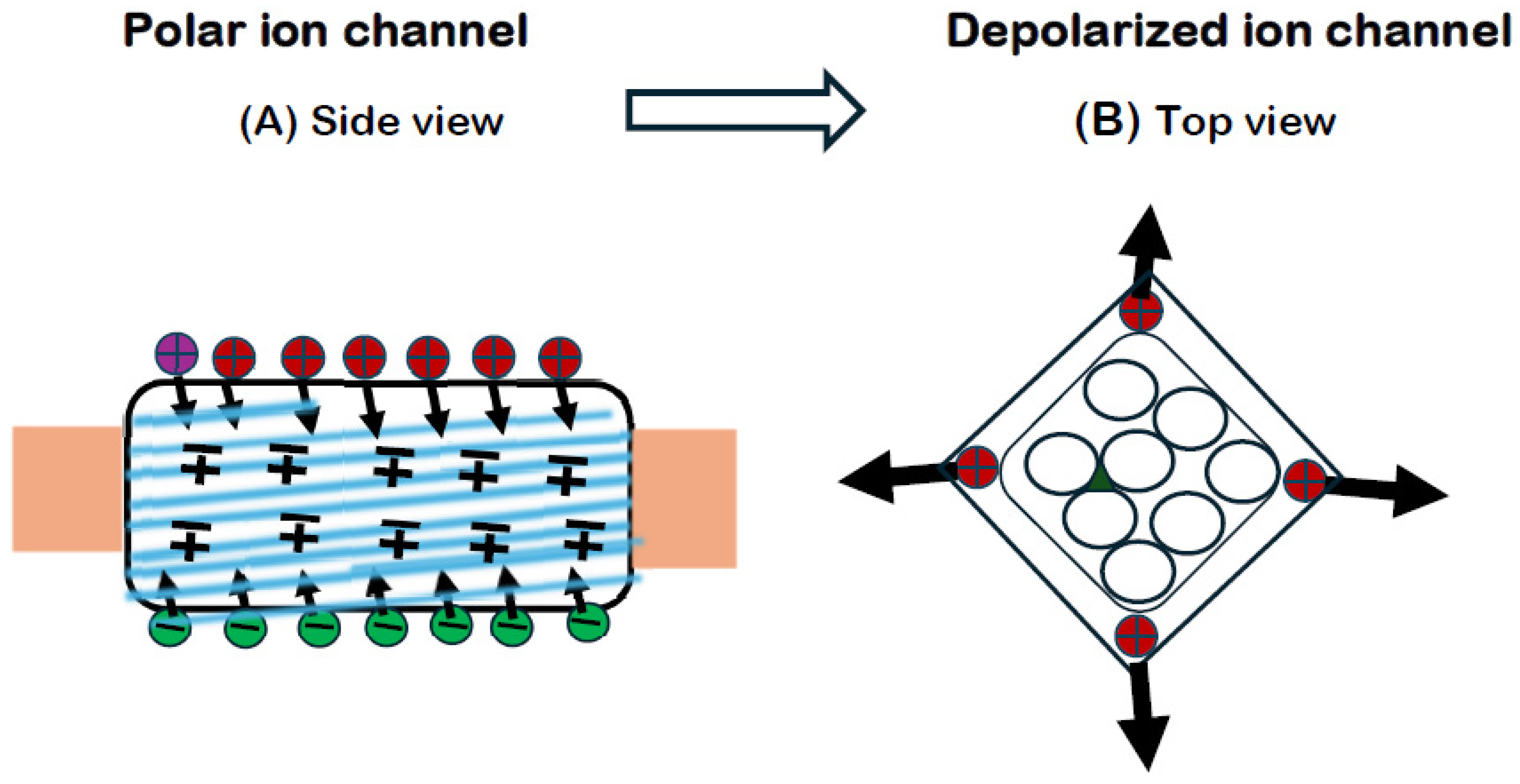
q1q2 is positive, such as the positive arginine and lysine residues of the S4 segments. The CAbER model claims that the dilation of the VSIC is due to an increase in the repulsion between positive S4 residues when a critical voltage reduction causes a reduction in the channel polarization and a concomitant reduction in the dielectric permittivity.
Experiments have demonstrated an outward motion of the positive S4 segments but have attributed this motion to a rigid motion of the entire S4 segment [
61,
62]. Outward movements of arginine residues were reported in skeletal muscle Na
+ channels [
62].
While we must accept the experimental observations, we must reject the assumption of rigidity of this hydrogen-bonded structure and the neglect of the established law of electrostatics. According to the CAbER hypothesis, the change in the size, shape and separation of the four S4 segments drives a discontinuous structural transition of the ion channel. Experiments support the outward movement of the S4 charges, which expands the segment; see
Figure 1B.
The ion channel is a structure with an outer jacket of four sets of voltage-sensing segments S1 to S4 and an inner pore domain of four sets of ion-conducting segments S5 and S6. Most of the positively charged residues on an S4 are on the outer part of the segment, while the inner part is anchored to the ectoplasm at the boundary with the axoplasm [
63]. The equilibrium configuration of the S4 segment is considered to be of the bent core or banana-shaped type due to the presence of a proline residue. When the electrostatic repulsions become large, the kink angle at the proline residue (due to its cyclic sidechain) goes toward zero, straightening the S4 segment; see
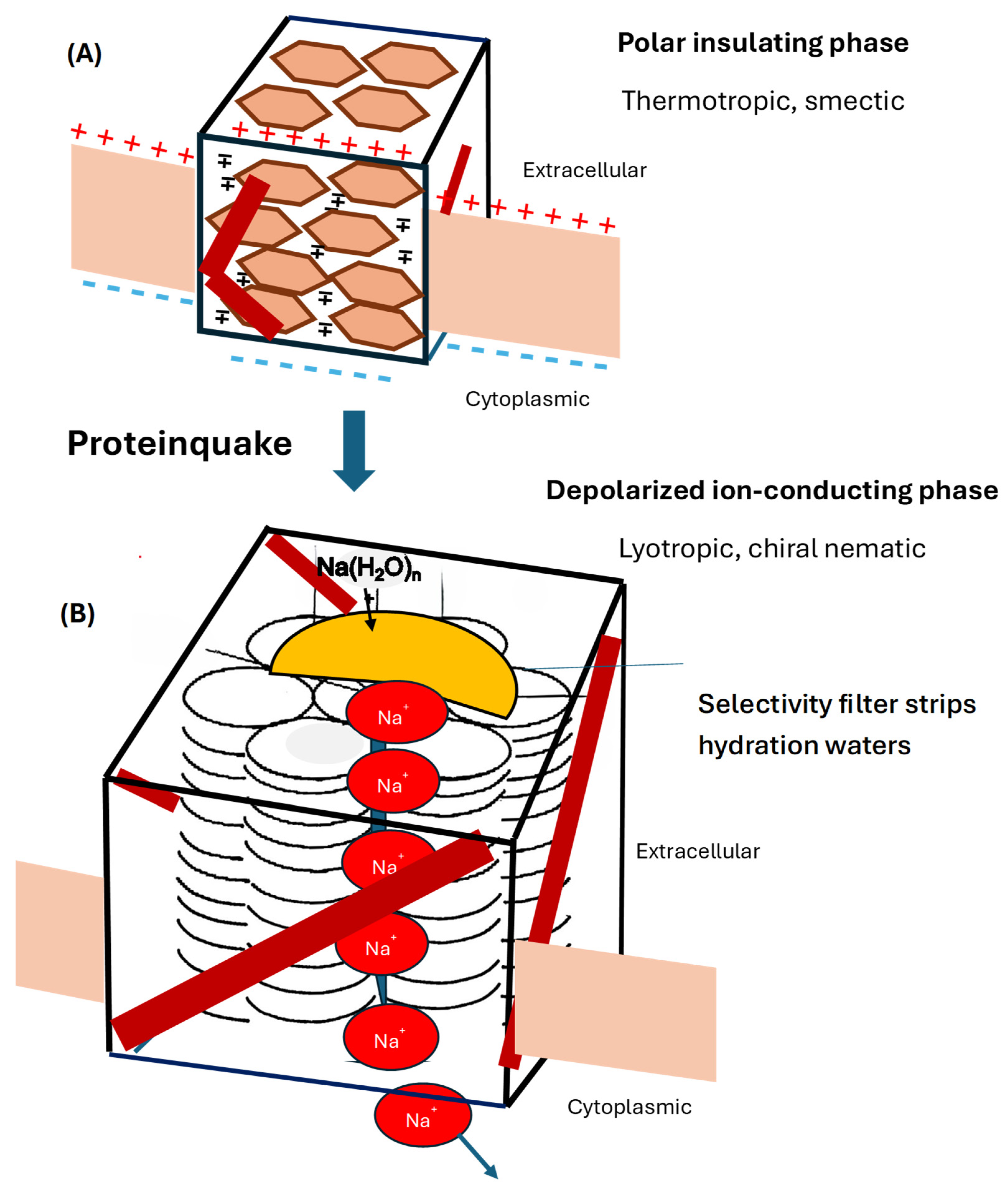
Figure 2A,B.
12. The Excitable VSIC Is Similar to a Blue Phase Under Electrostriction
Cryo-electron microscopy of a sodium channel [
64] shows a pseudocubical configuration topped by a dome. The new structure formed in the activation proteinquake [
65] leaves no room for the P loops, which are pushed out and everted to form a dome, which presumably is the selectivity filter. It acts as an enzyme that strips the selected ions as they enter the ion channel [
2].
Blue phases are liquid crystals that display cubical and tetragonal unit cells [
66]. Blue phases BPI and BPII are doubly twisted tubular structures stacked in three dimensions with lattices of cubic symmetry. Fluorescence resonance energy transfer experiments on
Shaker channels showed twisting of the S4 segments that produced S4 rotations on activation [
67].
Blue phases exhibit voltage-sensitive optical birefringence, like that observed to accompany action potentials [
68]. Blue phases can transition from a thermotropic, ferroelectric phase to a lyotropic, nonpolar phase. Subjecting the blue phase BPX to electric fields leads to electrostriction, a change in size and shape [
69]. The structure of a BPX is tetrahedral rather than cubic.
Line defects form at disordered intersections of three helices, where they can act as transient liquid pathways for the permeant ions. Thermal disorder must break the continuity of the liquid line defects, interrupting the ion current until a new pathway forms between the aqueous phases [
70].
13. Depolarization Increases the Repulsions, Causing a Proteinquake
The process of ion channel activation proposed by the CAbER hypothesis is shown in
Figure 2. On critical channel depolarization, the induced dipoles collapse, lowering
ε. The voltage-sensing S4 segments straighten, untilt, partially unwind and move apart. Translocations close to 1.0 nm were recorded [
71]. The VSIC relaxes into an expanded structure with a conformational transition to a chiral nematic, columnar phase, expanding the α helices of the four S5 and four S6 segments, which thread back and forth across their host lipid bilayer membrane forming a chiral nematic columnar system of parallel α helices. The expansion of the α helices widens the hydrogen bonds between their loops, allowing permeant sodium ions to replace the hydrogens. Line defects form at disordered intersections of three helices, where they act as transient liquid pathways for the permeant ions. Thermal disorder breaks the continuity of the liquid line defects, interrupting the ion current until a new pathway forms between the aqueous phases.
14. Widened Hydrogen Bonds Become Ion Sites
As the S5 and S6 segments (and probably transmembrane segments of the β subunit) relax and expand, hydrogen bonds between their core α helical loops widen. They are heteroconjugated bonds between a carbon and a nitrogen atom. This length change affects the chemical affinity properties of the bond, with the wider bond being occupied by a larger atom [
72]. Zundel and collaborators showed that the hydrogens of a widened H bond can be replaced by ions such as Na
+, Li
+ and K
+ [
73,
74]. The CAbER hypothesis proposes that the bare permeant ions enter the widened H bonds linking loops of the S5 and S6 α helices, replacing the protons and hopping from site to site [
4].
This model implies that the permeant ions compete for sites with the protons, so that the ion current varies with the hydrogen ion concentration. As a sodium ion moves from one hydrogen bond to an adjacent one, it leaves a site to be filled by a proton and displaces another proton further inward. The effect is equivalent to the effect of a proton jumping from ahead of the Na
+ to behind it. The net effect is that, as a Na
+ travels inward, a H
+ moves outward, reducing the [H
+] inside the axon and increasing it outside. As the pH increases inside and decreases outside, the Na
+ has to compete for sites with the H
+. Thus, lowering the pH outside will decrease the sodium current. This dependence, opposite to what was expected, has been observed: Hille [
1] points out that “As the pH
o is lowered below 6.0, [sodium conductance]
gNa begins to ‘titrate’ away … At pH 4 only 10% of the original
gNa remains.” This effect thus supports the CAbER hypothesis.
15. Traveling Phase Boundaries in the Action Potential
The CAbER hypothesis provides a new interpretation for the action potential mentioned at the beginning of this article. Patricia Cladis studies traveling phase boundaries in an experiment in which the sample is moved through a temperature gradient [
44]. In a squid axon, phase boundaries travel spontaneously in response to electrical stimuli. At the leading edge of the action potential, sodium channels sense a reduction in the electric field and, as described above, undergo a proteinquake from a smectic C* phase to a chiral nematic phase N*, filling the ion channel with liquid line defects. As some of these span the membrane, Na
+ ions pass chaotically inward across the membrane, further reducing the membrane voltage. Potassium channels behind the sodium channels sense this voltage change and undergo a similar proteinquake, creating liquid line defects that carry K
+ ions outward chaotically. First the sodium channel and then the potassium channel inactivate, undergoing phase transitions that return them to their insulating smectic C* phases to reestablish the membrane excitability. In Cladis’s terms, the excitable membrane exhibits one of the “broken symmetries of life.” The action potential briefly allows the membrane to relax into a more symmetrical “open” state, and inactivation restores it to the high electric field of the living, broken symmetry.
16. The CAbER Hypothesis Versus Conventional Models
Let us compare the CAbER hypothesis to earlier models, beginning at a time after the primary structures of several ion channels had been published. Since Hodgkin and Huxley had predicted that depolarization would move charges to open a gate, models were proposed on the basis of such an interaction. Data showed that the rise of the gating current was not instantaneous but a rapid rise and a slower decline [
75]. Currents of permeant ions followed this pattern after a slight delay [
76].
A model by Catacuzzeno et al. [
11] formalizes activation (from closed to open channel) as a system of reversible reactions between four states of the four voltage sensors (VS):
The model sees these reactions between the states as leading to a cooperative interaction between voltage sensors that move in several steps to open a gate in a water-filled pore through the center of the ion channel, S5, for a total of 13 reactions.
The CAbER hypothesis differs from conventional models in that it is based on the three dimensions of a varying tertiary model rather than the two dimensions of the secondary model. It does not assume that the four internally homologous repeats of the secondary structure form separate parts of the ion channel with a water-filled pore between them. Because it involves a structural transition of the entire molecule, every atom and every interatomic bond will be affected by the proteinquake, making the number of reactions extremely large. The ion channel is an open system and activation is not reversible. Activation is a dissipative process. Metabolic energy is required by pumps to work with inactivation to restore the system back to excitability.
CAbER considers activation in four steps, but not all of these steps are reversible within the activation process:
Collapse of branched-chain dipoles on critical depolarization, reducing the dielectric permittivity. The change from a ferroelectric phase to a nonpolar (paraelectric) phase would indicate that this step must exhibit hysteresis. This contribution to the gating current must depend on the concentrations of ions (permeant or not) in the aqueous media inside and outside, as well as the number of branched-chain amino acid residues in the channel.
Increase in the mutual electrostatic repulsions between the positive charges of the S4 segments (voltage sensors), expanding the ion channel. This major contribution to the gating current depends on the axial component of the motions of the S4 arginines and lysines. In a preparation with a reduced number, say half, of these residues, this part of the gating current can be expected to drop to one-half of the control. A mutation replacing one or more positive charges on the S4 segment of domain I of Na
+ channels with neutral or negative residues induced a decrease in the gating charge [
77].
A proteinquake that builds a selectivity filter and changes the smectic structure into a chiral nematic structure with liquid line defects. This process is so complex that it would be difficult to predict its influence on the gating current.
Defects sporadically form continuous pathways between the outer and inner aqueous phases, providing temporary pathways for the rapid transfer of permeant ions across the channel. As the gating process is now complete, the gating current can be expected to be zero.
In a 1986 review of electrostatic interactions in membranes and proteins, Honig et al. state that “channels in biological membranes are formed from single polypeptides or very stable subunits” and that “recent data give no indication of unusual charged sidechains in electrically active proteins” [
78]. We now know that these ion channels are highly unstable, as they switch from an insulating conformation to an ion-conducting one, and that the four S4 segments are very unusual in possessing arrays of amino acids with positively charged sidechains at every third position. The review does not refer to Coulomb’s law, the basic principle of electrostatic interactions. Hille [
1] refers to Coulomb’s law, but only in connection with the potential of a lone charge in pure water.
Tiwari-Woodruff et al. probed “structural interactions between transmembrane segments S2, S3, and S4 in Shaker channels. Charge reversal mutations of [glutamic acid]E283 in S2 and [lysine]K374 in S4 disrupt maturation of the protein. … indicating that electrostatic interactions exist between E283 in S2 and [arginine]R368 and R371 in S4, and between K374 in S4 and E293 in S2 and [aspartic acid]D316 in S3. Our data indicate that K374 interacts with E293 and D316 within the same subunit.” [
79] The study thus investigated attractions between negative residues E and D and positive residues K and R, while ignoring the existence of mutual repulsive interactions between positive residues such as the arginines R368 and R371.
The assumptions of the conventional gated pore have led to additional models filling in the details of these interactions. While the sliding helix model [
80,
81,
82] recognizes the role of chirality in activation, it does so at the expense of a rigidity that cannot exist in a molecule connected with hydrogen bonds. The CAbER hypothesis recognizes the role of chirality in amino acid residues, β sheets, α helices and the traveling action potential, whose image in a transverse mirror travels in the opposite direction.
The paddle model [
83] recognizes complex coordinated movements between parts of the ion channel, but again with a rigid device. The CAbER hypothesis recognizes complex coordinated movements, but these constitute phase transitions between liquid crystalline phases.
The CAbER model has no pore or gate. A liquid line defect is a disordered phase but not a pore. The CAbER ion channel undergoes a proteinquake from a thermotropic insulating phase to a lyotropic ion-conducting phase.
17. Summary: The Hypothetical Steps That Activate a Voltage-Sensitive Ion Channel
17.1. Thermotropic Excitability. The VSIC under a high electric field lies between opposite surface charges, which attract each other. They compress the ion channel by electrostriction. It is in a compact thermotropic phase, making the VSIC an insulator. Aromatic rings form a smectic configuration of layers parallel to the membrane plane. Branched sidechains are splayed out to form dipoles directed opposite to the imposed field, yielding a high dielectric permittivity, which minimizes the electrostatic forces. The four S4 segments are situated in a square array, most with a kink at a proline residue.
17.2. The action potential approaches. As the external electric field decreases to a critical threshold, the surface charges vanish, allowing the VSIC to expand outward. Dipoles disappear as the branched sidechains take on their equilibrium configurations. The dielectric permittivity plunges, greatly increasing the electrostatic forces. The S4 segments straighten, untilt and expand, losing their bent-core shape. They repel each other and separate, causing the VSIC to undergo a proteinquake.
The selectivity filter forms. The P segments evert from their membrane location and join to form a selectivity dome. This filter allows ions of the appropriate type to be stripped of their waters and to enter the VSIC.
The lyotropic structure forms. The eight S5 and S6 segments relax into the expanded volume. The α helices straighten and expand into a columnar configuration normal to the membrane plane in a hexagonal pattern. The hydrogen bonds linking their loops are stretched into a widened length. Permeant ions drift into the H bonds, replacing the protons.
Parallel columns of α helices form liquid defect lines. Certain intersections between three adjacent helices are disordered. These disclinations form defect lines [
84,
85], which become liquid pathways for the permeant ions. As the permeant ions enter them, they are driven by electrodiffusion and rapidly transported across the membrane. As thermal disorder breaks the continuity of one defect line, the current surge along it stops. After an unpredictable interval, another defect line connects the two aqueous surfaces and a new surge of ion current flows until it also breaks, and so on. Thus, the observed ion current flow is chaotic.
Inactivation restores the far-from-equilibrium polar state. A complex inactivation process returns the VSIC to its insulating thermotropic state, and the membrane potential difference is maintained by metabolically energized processes in ion pumps located in parallel with the ion channels.
18. Experimental Evidence Supporting the CAbER Model
As a hypothesis, the CAbER model is presented for experimental testing. However, there already exists a body of experimental data from diverse fields supporting it.
18.1. Electrical
Conserved arrays of positively charged arginine and lysine residues exist in all VSICs [
1]. Experiments have demonstrated an outward motion of the positive S4 segments but have attributed this motion to a rigid motion of the entire S4 segment [
61,
62]. These experiments support the outward movement of the S4 charges.
18.2. Mechanical
The CAbER model claims that the VSIC dilates, expanding outwardly on activation. This claim is supported by the elasticity of protein structures connected with hydrogen bonds, and optical and mechanical measurements on squid axon membrane demonstrating membrane swelling during passage of an action potential [
86,
87].
18.3. Ion-Conducting Medium
An early argument supposed that ions could only pass through a membrane either by a pore or a carrier; since we can rule out the carrier, it must be a pore. However, years of searching have revealed no water-filled pore—only a pore domain. The facts are more complex: Ions flow in distinct surges, not in steady flow. Because of the thermal motion, pathways form for brief intervals before breaking. Their content is not water but disordered regions of the cholesteric phase. Ions move through these liquid line segments by occupying hydrogen bonds in place of the protons, which they have ejected. Whereas the existence of a water phase has not been observed and would interfere with the insulating properties of the polar phase of the VSIC, the existence of transmembrane helices in ion channels is unquestioned, and the appearance of line defects in chiral liquid crystals is well established. Lavrentovich and Kleman [
84] point out that “the connection between symmetry and defects has been for decades at the very heart of physics …; nowadays, it becomes the subject of studies in biology.”
18.4. Optical
Experiments on light scattering and voltage-sensitive optical birefringence with polarized light have detected rapid structural changes accompanying the action potentials in two types of non-myelinated nerve fiber. “A large part of the birefringence change seems to be directly dependent on the potential difference across the axon membrane, and arises in radially oriented molecules associated with the membrane” [
88]. This experimental evidence of voltage-dependent rotation of the plane of polarization during an action potential supports the claim of the CAbER model that the VSIC is a chiral molecule, reflecting the chirality of many of its component amino acid residues, and that this chirality causes twisting motions in the ion channel during activation.
18.5. Structural
The CAbER model claims that the VSICs are a guest phase in the host phase of the excitable membrane, and that this biomembrane is a liquid crystal. Support for the classification of biomembranes and living matter as liquid crystals has been reviewed [
89]. Support for the assertion that VSIC activation is a phase transition is given by Tasaki [
50]. Support for the existence of structural changes in proteins is implicit in the concept of a proteinquake, in which a configuration change in a relaxing protein molecule releases energy in the form of waves [
90].
18.6. Ferroelectricity in Compounds Containing Branched-Chain Amino Acids
The CAbER model claims that the VSICs exist in a ferroelectric phase in their polar, crowded, insulating phase at high electric field and that they relax into a nonpolar, dilated, ion-conducting phase on lowering of the electric field. This claim is supported by an experimental study on compounds containing the sidechains of amino acids. This study found that branched-chain amino acids (and only they among amino acids) produce compounds that are ferroelectric with unusually high dielectric permittivities [
55]. These amino acid residues are present in the VSIC, particularly between the positive charges of the S4 segments [
91].
Other data supporting the CAbER model are cited above. The fact that the CAbER model explains the sequence of events underlying activation on the basis of established physical principles is support of the model in itself.
19. Conclusions: Electrostatic Repulsion Drives the Activation Proteinquake
As we change our paradigm from a gated pore to a repulsion-driven phase transition, a new level of understanding of ion channel activation emerges. Experimental data and physical law allow us to sketch a sequence of transitions of a voltage-sensitive ion channel that, on critical depolarization, convert a compact insulating phase at high electric field into a dilated ion-conducting phase. The similarities between a voltage-sensitive ion channel and both blue phases and Twist Grain Boundary Phases provide clues to a theoretical explanation of ion channel activation.
While many details remain to be filled in by further experimental and theoretical research, the Channel Activation by Electrostatic Repulsion hypothesis offers a new paradigm: an electrically driven phase transition of a chiral liquid crystalline structure from a compact thermotropic phase at high membrane voltage to a dilated lyotropic phase at a critically reduced voltage. A proteinquake driven by increasing electrostatic repulsions between positive charges on the four S4 segments converts an insulating phase with parallel planar sidechains of aromatic residues to a chiral nematic phase with liquid line defects at disordered intersections between three parallel α helices. Widened hydrogen bonds provide sites for permeant ions stripped of their waters in the selectivity filter to cross from one aqueous medium to the other through these liquid line defects. Thermal disorder breaks these transient ion pathways at indeterminate intervals, leading to the chaotic surges observed as single-channel currents.
Goals for future interdisciplinary research may be as follows:
Incorporate data from electrical, optical, frequency-dependent, and thermal experiments into a theoretical formulation.
Identify an order parameter, a quantity that incorporates the symmetry of the polar phase and goes to zero in the depolarized phase.
Write out a quantum-mechanical free energy (Hamiltonian) operator for the ion channel system.
Draw phase diagrams that display states of the system as functions of voltage, temperature, and ion concentrations.
Trace the effects of mutations on the function of a particular ion channel.
Apply the knowledge gained to medical problems posed by channelopathies.