


Abstract
The Senegal tree (Sengalia senegal) is the primary plant source of Gum Arabic (GA), a natural secretion rich in soluble fiber and bioactive polysaccharides. It has longstanding uses in traditional medicine, nutrition, and pharmaceuticals. The present study aimed to evaluate the phytochemical profile, antimicrobial, anti-inflammatory, and anticancer activities of GA methanolic extract (GAME), supported by molecular docking analysis of its key compounds. The gas chromatography–mass spectrometry (GCMS) analysis of the GAME identified many compounds, such as 9-octadecenoic acid (38.29%), methyl ester (15.52), 1,2-benzenedicarboxylic acid, 3-nitro (9.8%), hexadecadienoic acid, methyl ester (8.5), and á-d-mannofuranoside, methyl (7.38). The molecular docking analysis showed that 9-octadecenoic acid had strong binding affinity with target proteins, which included xanthine oxidase (XO), lipoxygenase (LOX), and cyclooxygenase-2 (COX-2), with the highest affinity to XO (−137.03 kcal/mol) and lipoxygenase (−135.09 kcal/mol). GAME possessed broad-spectrum antibacterial activity against Salmonella typhimurium (S. typhimurium), Escherichia coli (E. coli), Pseudomonas aeruginosa (P. aeruginosa), and Staphylococcus aureus (S. aureus), with a zone of inhibition from 16.28 to 16.93 mm. B. subtilis was resistant to the tested extract. The extract also showed good membrane stability and potent inhibition of albumin, XO, LOX, and COX-2, with IC50 values of 31.62, 13.02, 27.6, and 28.99 μg/mL, respectively. The cytotoxic assessment demonstrated moderate, dose-dependent effects on the Caco-2 (colorectal adenocarcinoma) and HeLa (cervical carcinoma) cell lines. These findings highlight the therapeutic potential of GA as a natural plant source of antibacterial, anti-inflammatory and anticancer agents. The combination of molecular docking with in vitro assays provides strong evidence supporting its application in the development of plant-based pharmaceuticals. This research suggests that GA could be a useful ingredient in the creation of anti-inflammatory and antibacterial drugs derived from plants.
1. Introduction
Modern research has demonstrated, based on the bioactive component contents of plants, that the linkage of plants with medicinal techniques is scientifically valid, paving the path for the discovery of more drugs derived from plants [1,2]. Natural products derived from plants have long served as a cornerstone of both traditional medicine and modern drug development. Their appeal lies in their natural origin, lower risk of side effects, and growing dissatisfaction with synthetic medications [3].
Senegalia senegal (S. senegal), previously referred to as Acacia senegal, is the currently accepted scientific name and is widely acknowledged as a significant species in agroforestry [4]. Acacia trees are among these plants, growing in large numbers in the central part of Sudan, in Africa, as well as in tropical and semitropical regions worldwide [5]. Following Sudan, Nigeria, Chad, Mali, and Senegal are all in the top-five acacia gum producers in the world [6]. In recent years, there has been an increasing interest in the phytochemical composition and pharmacological advantages of (GA), propelled by the increasing need for naturally derived, bioactive, and non-toxic alternatives to synthetic chemicals. GA was categorized as a solid exudate formed from the acacia tree’s stem and branches according to the Joint Expert Committee on Food Additives [7]. Additionally, it is referred to as Hashab, hard gum, or Acacia seyal gum (ASG) [8]. It comprises a complex polymer of polysaccharides, which is composed of arabinose, rhamnose, galactose, uronic acid and other polysaccharides associated with minerals [9].
In recent years, many studies have demonstrated that GA extracts contain numerous bioactive constituents that could efficiently interact with biological macromolecules, including enzymes and receptor proteins, which may contribute to their therapeutic potential [6].
The presence of numerous health-beneficial phytochemicals is responsible for the wide application of GA in medicinal and industrial sectors. Its importance has been extended by recent advances in the medicinal and industrial sectors, especially in drug delivery systems, cosmetic formulations, food preservation and hydrogel creation [10]. A diet rich in GA yields downstream health advantages for the gut microbiome, metabolic, and cognitive functions [11].
Researchers have utilized various methods and techniques to study these interactions, but computational approaches have become very important tools; among these tools, molecular docking has emerged as a powerful technique for examining the interaction patterns between small molecules and target proteins and for predicting their binding affinity [12]. This method significantly accelerates the search for natural compounds as potential therapeutic agents before applying it to the level of experimental validation [12].
GA has also been explored as a promising therapeutic drug for the treatment of a variety of disorders using various modes of administration. Moreover, GA is a rich source of non-viscous, soluble fiber with numerous health benefits and potent antioxidant properties [13]. Polymers generated from plants have attracted a lot of attention in recent years due to the wide variety of ways in which they can be used in medical treatments [14,15]. The pharmaceutical industry uses GA as a medication delivery vehicle since it is recognized as a material that does not have any detrimental effects on the human body. Moreover, GA exhibited strong antimicrobial, anti-inflammatory, antioxidant, and immunomodulatory effects in several studies in vivo and in vitro experiments [16,17,18]. It has some biological capabilities in the treatment of numerous disorders, including cardiovascular disease [19], dyslipidemia and renal failure [20]. Compared with previous studies that focused mainly on the nutritional, physicochemical, or general therapeutic properties of Gum Arabic, this work provides an in-depth characterization of the methanolic extract of Senegalia senegal, including GC–MS analysis and antimicrobial, anti-inflammatory, cytotoxic and molecular docking studies, thus providing a more comprehensive view of its phytochemical composition and pharmacological potential.
In this work, the biological activity of GAME was evaluated by combining in vitro and in silico approaches. Inhibition of albumin denaturation, membrane stabilization, and enzymatic tests that targeted lipoxygenase and xanthine oxidase were used to evaluate its anti-inflammatory potential. The interactions between certain therapeutic protein targets and bioactive compounds were further clarified by molecular docking analysis, which provided mechanistic insight into their molecular modes of action. Furthermore, the extract was evaluated for its anticancer activity against the Coca2 and HeLa human cell lines, as well as its antibacterial potential against selected human pathogenic bacteria.
2. Materials and Methods
2.1. Plant Materials
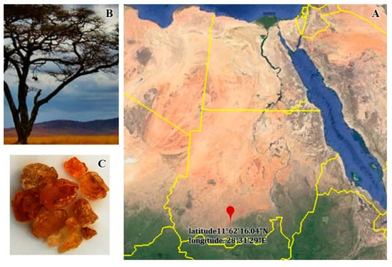
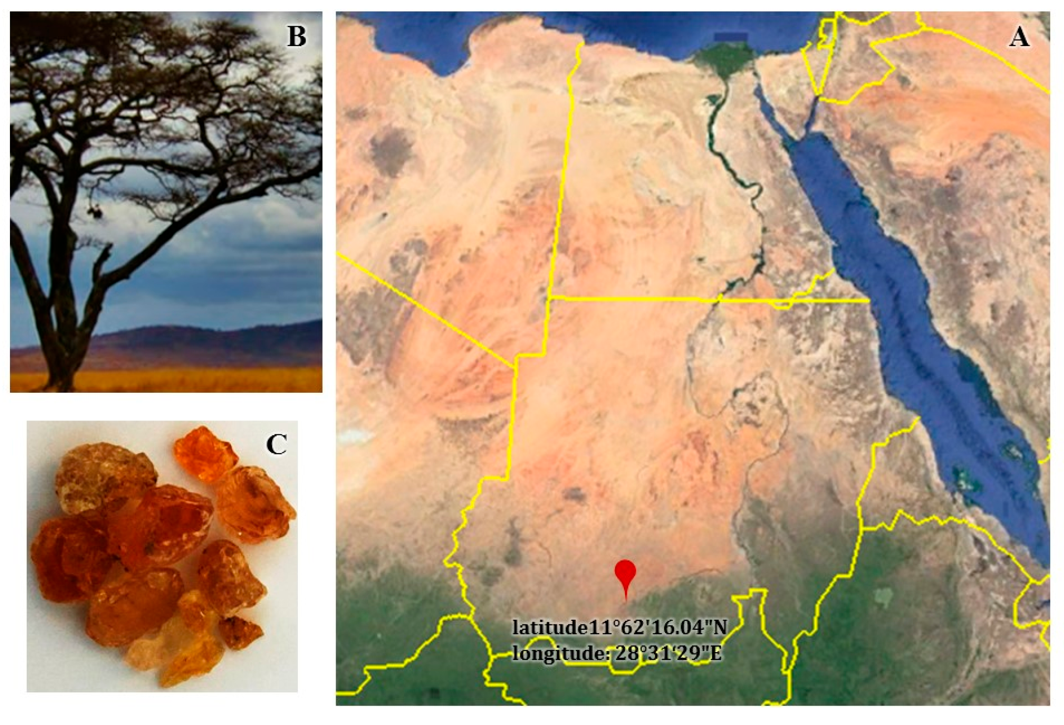
The Gum Arabic utilized in this study (Senegalia senegal) was bought from a certified local herbal store in AL Obeid City, North Kordofan State, Sudan. The identity and purity of the samples were verified by experts from the Ministry of Forestry and Agriculture, Khartoum Sudan. There was 2.13 percent moisture at the site, as well as 0.39 percent organic matter and 0.19 g of total nitrogen per kilogram of soil. The electric conductivity was 0.03 ds/m. Dried GA exudate was harvested from the stems and branches of S. Senegal trees. The collected exudate was solid, ranged in color from white to orange–brown, had a matte surface, and crumbled into spherical pieces of varying sizes upon close examination (Figure 1).
Figure 1.
Satellite map and field photographs of the study area and plant material: (A) satellite image showing the geographical location of the sampling sites; (B) Senegalia senegal tree; and (C) GA plant exudate collected from the selected site (latitude: 11°62′16.04″ N; longitude: 28°31′29″ E).
2.2. Preparation of Methanol Extract
Preparation and extraction were done according to Elnour et al. [21], with minor modifications. The crude GA was cleaned to remove extraneous materials, including bark and sand. The purified gum was then ground and passed through a 1.40 mm mesh sieve to obtain a fine powder. Subsequently, 2 g of the gum powder was transferred into a flask and extracted with 20 mL of absolute methanol. The mixture was placed on a magnetic stirrer and agitated for 24 h. The resulting supernatant was filtered through a 0.45 µm membrane filter.
2.3. Gas Chromatography–Mass Spectrometry Analysis
A gas chromatograph (GC-MS spectrometry) linked to an ISQ Single-Quadrupole Mass Spectrometer was used to analyze the GAME. In order to perform GC-MS analysis, a DB5-MS column (J & W Scientific, Folsom, CA, USA; 30 m, 0.25 mm inner diameter, and 0.25 m film thickness) was used under carefully optimized chromatographic conditions. Using helium as a carrier gas, a 1.5 mL/s flow and a 1:10 split ratio were employed. The applied temperature program was as follows: 40 °C for 3 min, 280 °C for 5 min, and 290 °C for 1 min at 7.5 °C/min. The injector and detector were kept at a temperature of 200 °C. An amount of 5 L of the diluted samples (1:10 diethyl ether, v/v) was introduced into the system. At 48 min, the program was over. Electron ionization at 70 eV, with a spectral range of m/z 40–450, was used to acquire the mass spectra. The retention index of each molecule was matched to the spectra in the Wiley 9 database to determine its identification [22].
2.4. Molecular Docking Study
Molecular docking simulations were performed using Molegro Virtual Docker (MVD, version 6.0, CLC Bio, Aarhus, Denmark) to explore the binding interactions of octadecenoic acid-9 with selected target enzymes, such as cyclooxygenase-2 (COX-2, PTGS2; PDB ID: 5IKT), albumin (PDB ID: 1AO6), xanthine oxidase (XO, PDB ID: 2E1Q) and lipoxygenase (LOX, PDB ID: 8VIY). All the target proteins were obtained from the Protein Data Bank (PDB) in the three-dimensional crystal structure format.
Protein preparation was performed by removing crystallographic water molecules and co-crystallized ligands and adding hydrogen atoms to ensure the correct protonation states under physiological settings (pH 7.0 ± 0.5). The preparation was carried out to preserve the critical structural integrity of the protein backbone. The octadecenoic acid-9 ligand was obtained by assigning correct bond ordering, formal charges and hydrogen atoms and then performing geometry optimization and energy minimization using a typical molecular mechanics force field to achieve a stable low-energy conformation.
The binding site was detected with the cavity identification technique used in MVD and validated based on the position of the co-crystallized ligand in each protein structure. Docking simulations were performed with the default MVD parameters and the MolDock scoring mechanism. For each protein–ligand complex, numerous poses were created and ranked by using the MolDock score, re-rank score, binding energy (BE), molecular docking score (MDS) and hydrogen bond interaction energy (HB).
To identify the most dependable and non-random binding poses, the most suitable docking conformations were selected on the basis of the lowest binding energy values, the most favorable Mol-Dock scores and the agreement among docking clusters. All findings were recorded and analyzed to evaluate the binding affinity and interaction profile of octadecenoic acid-9 against the specified target proteins.
2.5. Antimicrobial Activity
The well diffusion agar method was utilized in order to evaluate the antibacterial potential of the GAME against five pathogenic bacterial species according to Athanassiadis et al. [23]. Bacterial strains were obtained from the American Type Culture Collection (ATCC). The isolates included three Gram-negative species, S. typhimurium (ATCC 14028), E. coli (ATCC 25922), and P. aeruginosa (ATCC 27853), and two Gram-positive species, S. aureus (ATCC25923) and B. subtilis (ATCC6633). The bacteria (0.5 McFarland) were preserved in Mueller–Hinton Agar (MH). Using an ultrasonic sterile pipette, one hundred microliters of GAME was dissolved in 1% DMSO and injected into each well. The plates were kept in a 37 °C incubator for the next 48 h. Wells containing the same volume of DMSO (1%) served as the negative control, while standard antibiotics, i.e., chloramphenicol for S. typhimurium, ampicillin for E. coli, gentamycin for S. aureus and B. subtilis, and ciprofloxacin for P. aeruginosa, were used as the positive controls. All treatments were done in triplicate. Electronic calipers were used to measure the sizes of the growth inhibition zones in millimeters to the closest 0.1 mm. A total of five replicates of each experiment were used to compute the means and standard deviations for the bacterial strains.
2.6. Red Blood Cell Membrane Stabilization
An erythrocyte suspension was prepared according to Rashid et al. [24]; heparinized syringes were used to draw whole blood from rats by heart puncture. The red blood cells were washed three times with an isotonic buffered solution (154 mM NaCl; pH 7.4) in a 10 mM sodium phosphate buffer. In order to obtain red blood cells, the blood sample underwent centrifugation at a speed of 3000 rpm for 10 min. The method of inducing erythrocyte hemolysis using a hypotonic solution was employed to assess the membrane-stabilizing activity of the examined extract. The sample that was put through the test was made up of a stock erythrocyte (red blood cells, RBCs) suspension (0.50 mL) mixed with 5 mL of a hypotonic solution (50 mM NaCl) in 10 mM sodium phosphate-buffered saline (pH 7.4) and the extract at varying doses (1000–7.81 g/mL) or indomethacin (standard drug). The control sample contained 0.5 mL of red blood cells that were combined with a hypotonic buffered saline solution. After incubating the mixtures for ten minutes at room temperature, they were centrifuged for the same amount of time at three thousand rpm. It was determined that 540 nm was the optimal wavelength for measuring the absorbance of the supernatant in 96-well plates. The proportion of prevention of hemolysis or membrane stabilization was calculated utilizing a modified approach provided by Mounnissamy et al. [25]. The inhibition percentage (%) of hemolysis (membrane stabilization%) = 100 × (Ab1 − Ab2/Ab1). Ab1 represents the optical density of a 1-molar concentration hypotonic buffered saline solution. The optical density of the sample in the hypotonic solution was denoted by Ab2. To determine the IC50, the concentration of the sample needed to inhibit hemolysis in 50% of red blood cells was determined.
2.7. Albumin Denaturation Assay
According to Janani et al. [26], an albumin denaturation assay for detecting the anti-inflammatory activity of GAME was carried out. One percent bovine serum albumin (BSA) was mixed with 400 µL of different concentrations of GAME ranging from 1000 to 7.81 μg/mL, each generated by two-fold serial dilution. 1N HCl was used to bring the reaction mixture’s pH down to 6.8. The reaction mixture was kept at room temperature for 20 min before being heated to 55 °C in a water bath for another 20 min. A UV–VIS Spectrophotometer (Avi Scientific, Thane, India) was used to record the absorbance value at 660 nm after the mixture was cooled to room temperature. The negative control was a BSA combination with a 30% methanol solution. In this study, diclofenac sodium at various concentrations served as a positive control. Triplicate experiments were conducted. The proportion of protein denaturation inhibition was calculated using the following formula:
where
% Inhibition of protein denaturation = 1 − [ab 1/ab 2] × 100
ab 1 = absorbance of control;
ab 2 = absorbance of tested extract/standard drug with albumin solution.
2.8. Xanthine Oxidase (XO) Inhibitory Activity
The xanthine oxidase inhibitory (XO) action was analyzed using the methodology of Abdulhafiz et al. [27], with slight modifications. A UV–VIS microplate reader was used to measure the inhibitory activity of OX at 290 nm. The tested materials were dissolved in dimethyl sulfoxide (DMSO) before being diluted with phosphate buffer (pH = 7.5) to achieve a final concentration of less than 1% DMSO. The IC50 for each tested material was determined at concentrations ranging from 5 to 300 μg/mL for the XO enzyme activity. The reaction assay consisted of a mixture of XO solution (0.1 U/mL; pre-incubated for 15 min at 25 °C), 50 µL of the tested solution, phosphate buffer, and 35 µL of distilled water. The assay was initiated by adding 60 µL of xanthine substrate solution (0.15 mM). Then, the mixture was incubated for 30 min at 25 °C. Before measuring the absorbance, 25 µL of HCl solution (1 N) was added to cease the reaction. A positive control, allopurinol, was utilized. To produce the negative control (blank), HCl solution was added to the substrate. The XO inhibitory activity was expressed as a percentage of inhibition, which was computed as follows:
Percentage of inhibition: 1 − [tested inclination/blank inclination] × 100
The linear change in the absorbance of the test material was defined as test inclination, while the linear change in the absorbance of the blank material was defined as blank inclination. The ability of the samples or a reference substance (allopurinol) to inhibit the XO enzyme was assessed by determining the concentration that caused 50% enzyme inhibition in each sample or compound (IC50).
2.9. In Vitro Lipoxygenase (LOX) Inhibition Assay
To assess the anti-inflammatory efficacy resulting from the inhibition of the LOX enzyme in Glycine max, the samples and the reference chemical (ibuprofen) were subjected to analysis following the established Kumaraswamy and Satish [28] protocol, with slight modifications. All of the samples were mixed with a borate buffer solution (pH 9) at 25 °C for 15 min, and the final concentration ranged from 0.98 to 125 µg/mL. Pre-incubation of each concentration with 100 µL of linoleic acid (substrate) was maintained to initiate the reaction. To assess the inhibitory activity, a microplate reader (BIOTEK; Winooski, VT, USA) was used to monitor the rise in absorbance at 234 nm.
The formula for calculating inhibitory percentages was as follows:
Inhibitory activity (%) = (1 − absorbance of tested substance/absorbance of control) × 100
2.10. In Vitro Cyclooxygenase-2 (COX-2) Inhibition Assay
The inhibition of COX-2 enzyme activity was achieved by measuring the oxidation of N,N,N,N-tetramethyl-p-phenylenediamine (TMPD) in the presence of arachidonic acid. This assay provides a valuable tool for studying the effects of potential COX-2 inhibitors in a controlled in vitro environment [29,30]. A microplate reader (BIOTEK; USA) was used to track the rise in absorbance at 611 nm to measure the inhibitory activity using different concentrations ranging from 0.98 to 125 µg/mL.
The reaction mixture was prepared by adding the reaction buffer, enzyme solution, and substrate solution in the desired volumes to obtain the final concentrations required for the assay. Typically, the final concentrations of arachidonic acid and TMPD are in the micromolar range, while the enzyme concentration may vary depending on the experimental setup and specific assay requirements. Then, appropriate control samples were adjusted, including positive controls (known COX-2 inhibitors) and negative controls (reagent blanks with no enzyme or substrate), to validate the assay results. Then, the reaction mixture was incubated at 37 °C, which is physiologically relevant for most biological systems.
Inhibitory activity (percent) = (As/Ac)
As is the absorbance in the presence of the tested material, and Ac is the absorbance in the presence of the control. The tested material, as well as the standard drug (celecoxib), was effective at inhibiting the cox-2 isoenzyme at a concentration that caused 50% enzyme inhibition (IC50).
2.11. Cytotoxicity Evaluation Using Viability Assay
The Caco-2 (human colorectal adenocarcinoma) and Hela (cervical carcinoma) cell lines and Wi-38 (normal human lung fibroblasts) were obtained from the American Type Culture Collection (ATCC, Rockville, MD, USA). Wi-38 normal cells were used to evaluate the extract’s selective cytotoxicity toward cancer cells compared with non-cancerous cells. The MTT test was used to assess the quantity of viable cells following a 48 h incubation period, according to the methods of Mosmann [31]. The selectivity index (SI) (safety) (IC50) was calculated using the following equation.
Selectivity Index (SI) (Safety) = IC50 (Normal Cell)/IC50 Cancer Cell
2.12. Statistical Analysis
All records were organized in triplicate and were identified as the standard deviation (SD) of the mean. One-way ANOVA, SPSS 16, and Duncan’s multiple-range tests with p ≤ 0.05 were used in the statistical analysis.
3. Results
3.1. GC-MS Analysis of GAME
The phytochemical compounds of the methanol extract of GA were identified using GC–MS analysis and a database of the spectra of known components contained in the GC–MS library. The active ingredient included in the extract was identified using the retention duration, area percentage, molecular weights, and molecular formula, which were compared with the Wiley 9 GC-MS library, as shown in Table 1. The other compounds included 1, 9-octadecenoic acid (38.29%), 9-octadecenoic acid (Z)-, methyl ester (15.52%), 1,2-benzenedicarboxylic acid, 3-nitro (9.8%), hexadecadienoic acid, methyl ester (8.5), and á-d-mannofuranoside, methyl (7.38%).
Table 1.
Major phytochemical components identified in Gum Arabic collected from (Senegalia senegal) using GC–MS analysis.
Molecular Docking
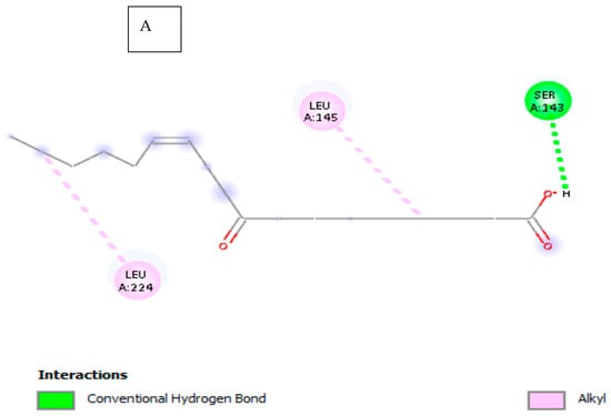
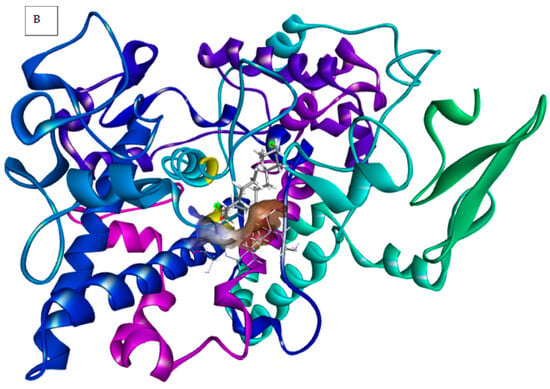
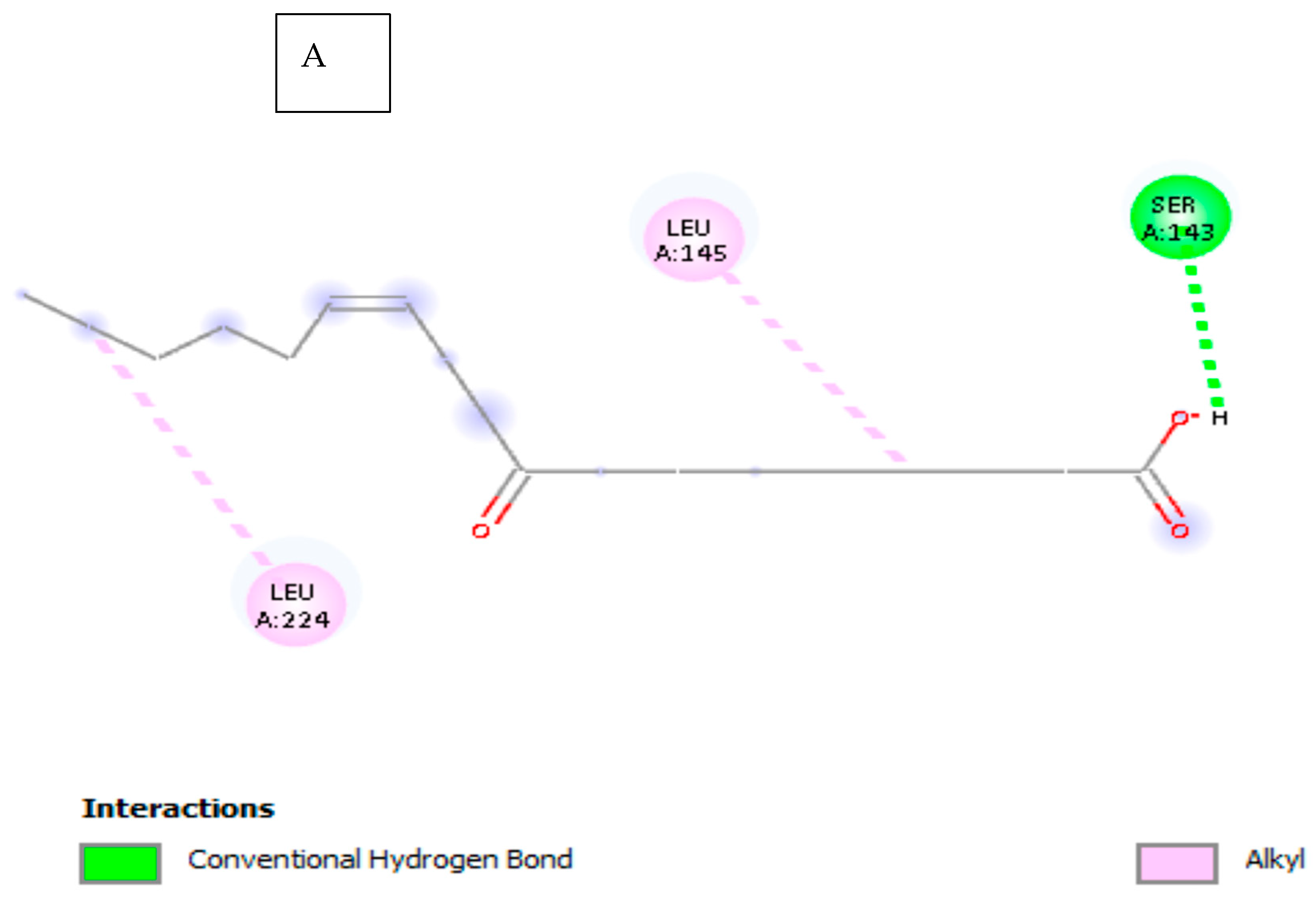
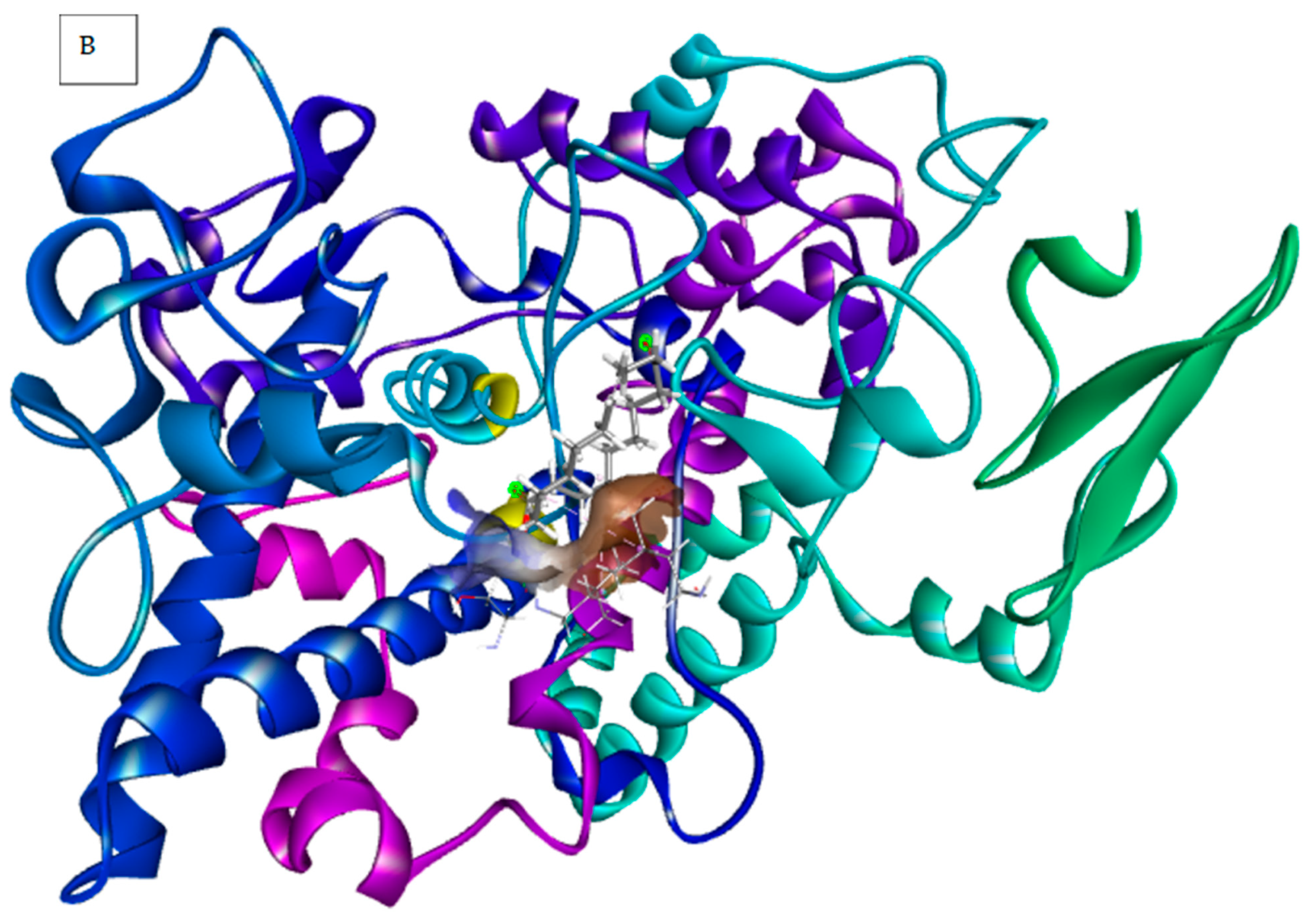
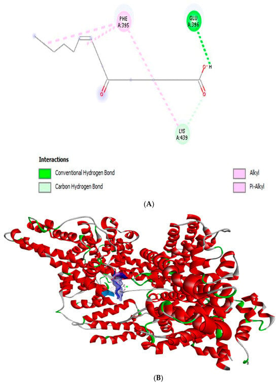
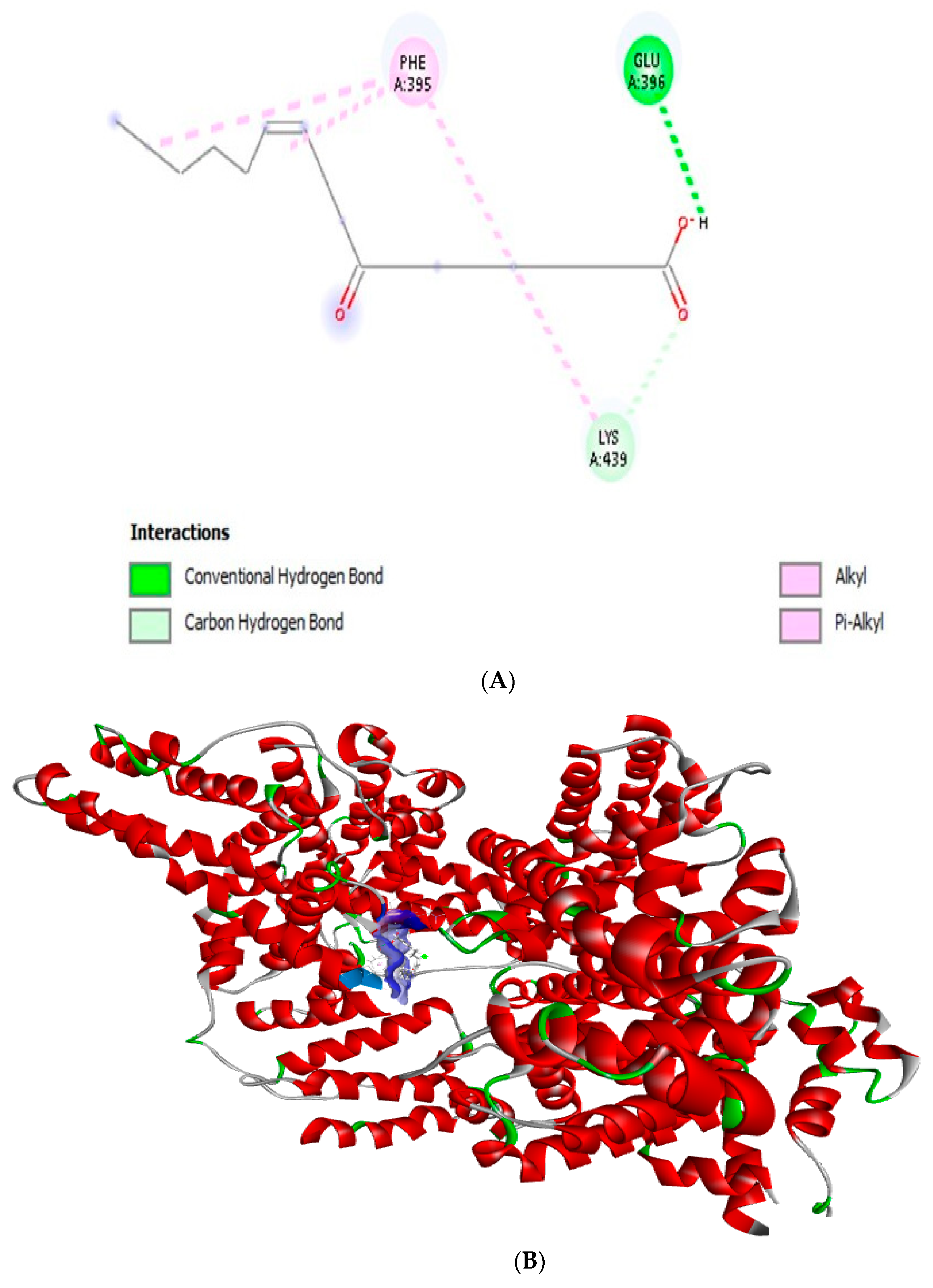
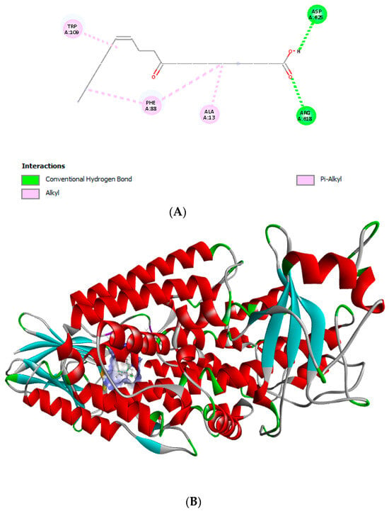
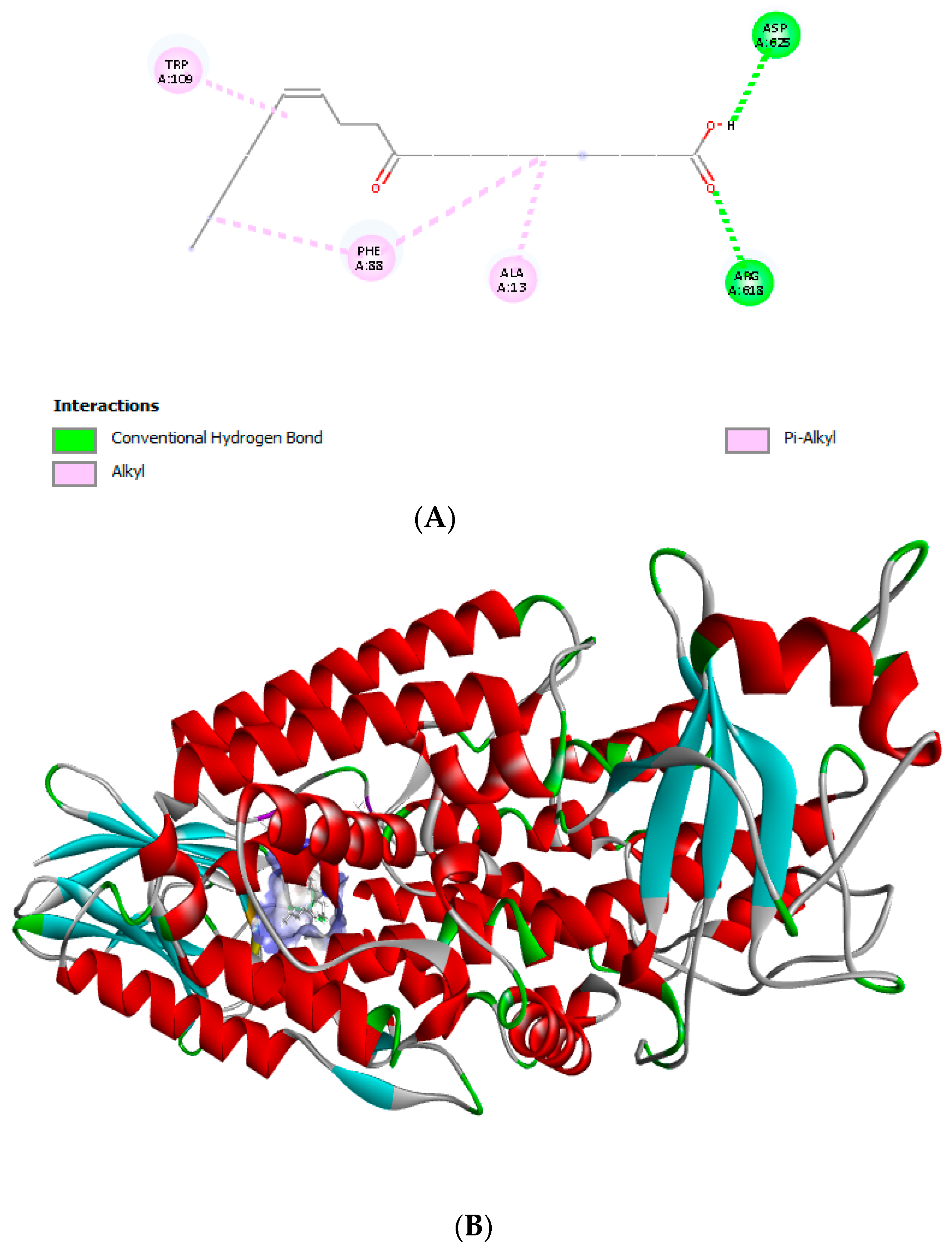
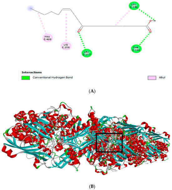
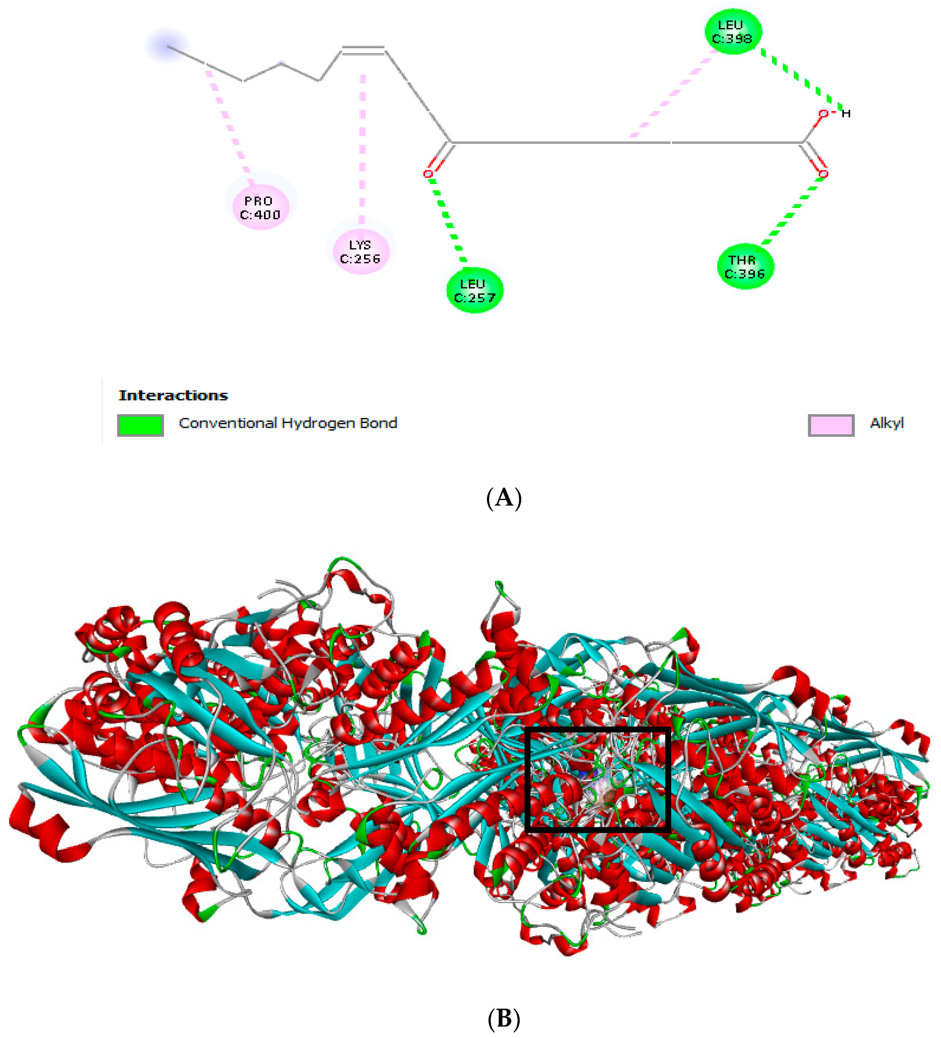
The molecular docking results revealed the binding affinity and interaction behavior of octadecenoic acid-9 with the selected target enzymes, including COX-2 (5IKT), albumin (1AO6), xanthine oxidase (XO, 2E1Q), and lipoxygenase (LOX, 8VIY). The calculated docking parameters comprised binding energy (BE), molecular docking score (MDS), re-rank score (RS), and hydrogen bond interaction energy (HB), as summarized in Table 2 and Figure 2, Figure 3, Figure 4 and Figure 5. The binding affinity values are expressed in kcal/mol. A lower binding energy (more negative value) signifies a stronger interaction between the ligand and the protein. Hydrogen bonds and hydrophobic interactions contribute to the stabilization of the complexes.
Table 2.
Molecular docking analysis of octadecenoic acid-9 with selected proteins.
Figure 2.
Molecular docking and binding interaction between octadecenoic acid-9 (grey stick), and COX-2 (5IKT): (A) 2D structure; (B) 3D structure (different colour of the COX-2 protein chain, denote different segments of protein (sequence-based coloring)), yellow residue- active site interacting with ligand.
Figure 3.
Molecular docking and binding interaction between octadecenoic acid-9 and albumin (1AO6): (A) 2D structure; (B) 3D structure (Albumin is represented as a cartoon with α-helices in red, loops and coils in grey, and chosen segments in green. Docked molecule of octadecenoic acid-9 in the binding cavity (blue). The semi-transparent surface shows the ligand-binding pocket, and the fatty acid is accommodated in the albumin structure).
Figure 4.
Molecular docking and binding interaction between octadecenoic acid-9 and xanthine oxidase (XO) (2E1Q6): (A) 2D structure; (B) 3D structure.The protein structure represented as a cartoon model, where α-helices are colored red, β-sheets cyan, loops gray, and turns green. Octadecenoic acid-9 is represented in blue and dominates the substrate-binding cavity. The transparent surface represents the active-site, highlighting the spatial complementarity between the ligand and the enzyme.
Figure 5.
Molecular docking and binding interaction between octadecenoic acid-9 and lipoxygenase (LOX) (8VIY): (A) 2D structure; (B) 3D structure ribbon representation of the modeled protein structure. α-helices are shown in red, β-sheets in cyan, loops/coils in gray, and terminal residues in green. The black box highlights the central catalytic/binding pocket region, where key amino acid residues are clustered and are predicted to participate in ligand interaction and enzymatic activity.
The docking analysis against COX-2 (5IKT) demonstrated a favorable binding interaction, with a binding energy of −83.7 kcal/mol, indicating a stable ligand–enzyme complex. The observed hydrogen bond interaction energy (−0.943 kcal/mol) indicates the contribution of hydrogen bonding to the overall binding stability (Table 2; Figure 2).
In the case of albumin (1AO6), octadecenoic acid-9 exhibited a stronger binding affinity, reflected by a more negative binding energy (−104.56 kcal/mol). The negative hydrogen bond interaction energy (−0.42 kcal/mol) indicates moderate hydrogen bonding interactions within the binding site (Table 2; Figure 3).
Notably, the strongest binding affinity was observed against xanthine oxidase (XO, 2E1Q), with a binding energy of −137.025 kcal/mol. This interaction was accompanied by a relatively high hydrogen bond interaction energy (−4.336 kcal/mol), suggesting enhanced stabilization of the ligand within the enzyme active site (Table 2; Figure 4).
Similarly, docking with lipoxygenase (LOX, 8VIY) showed a high binding affinity, with a binding energy of −135.09 kcal/mol and a hydrogen bond interaction energy of −3.7 kcal/mol, indicating strong ligand–enzyme interactions (Table 2; Figure 5).
Overall, octadecenoic acid-9 displayed pronounced binding affinities toward xanthine oxidase and lipoxygenase, followed by albumin and COX-2, highlighting its potential inhibitory activity against enzymes associated with inflammatory and oxidative pathways.
3.2. Antimicrobial Activity
The antimicrobial properties of GAME at a concentration of 100 μg/mL against S. typhimurium, E. coli, P. aeruginosa, and two Gram-positive species, S. aureus and B. subtilis, were assessed using the agar well diffusion technique (Table 3). Except for B. subtilis, GAME showed antibacterial action against all tested pathogens, with zone inhibition widths ranging from 16.28 to 16.93 mm. Four of the bacterial strains tested, namely, S. typhimurium (16.56 ± 1.35 mm), E. coli (16.28 ± 0.59 mm), P. aeruginosa (16.93 ± 0.83 mm), and S. aureus (16.76 ± 0.75 mm), were suppressed by the extract. However, B. subtilis was resistant to GAME. In contrast, the inhibition zone of solvent control was zero; however, the diameter of the zone of inhibition of the standard antibiotic for S. typhimurium (chloramphenicol) equaled 20.60 ± 1.06 mm; that for E. coli (ampicillin) equaled 9.83 ± 0.49 mm, which was less than the effect of the tested extract; those for S. aureus and B. subtilis (gentamycin) equaled 10.66 ± 2.07 mm, which was less than the effect of the tested extract, and 24.00 ± 0.75 mm, respectively; and that for ciprofloxacin, which was used for P. aeruginosa, equaled 21.13 ± 2.22 mm.
Table 3.
Agar well diffusion assay: zones of inhibition of methanol extract of Gum Arabic against tested pathogens measured by inhibition zone (mm).
3.3. Membrane Stabilization Activity
The results of hypotonicity-induced hemolysis show that GAME at concentrations between 1000 and 7.81 µg/mL considerably (p < 0.05) protected the erythrocyte membrane from lysis induced by a hypotonic solution (Table 4). The 1000 µg/mL concentration of indomethacin provided significant (p < 0.05) protection against the harmful effects of the hypotonic solution. Compared with the control, GAME demonstrated 92.25% protection at 1000 µg/mL, while indomethacin demonstrated 96.35% inhibition of RBCs hemolysis at the same concentration.
Table 4.
Effect of GAME on hypotonicity-induced hemolysis of erythrocytes.
3.4. Albumin Denaturation Assay for Anti-Inflammatory Activity
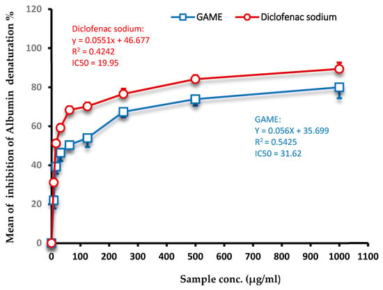
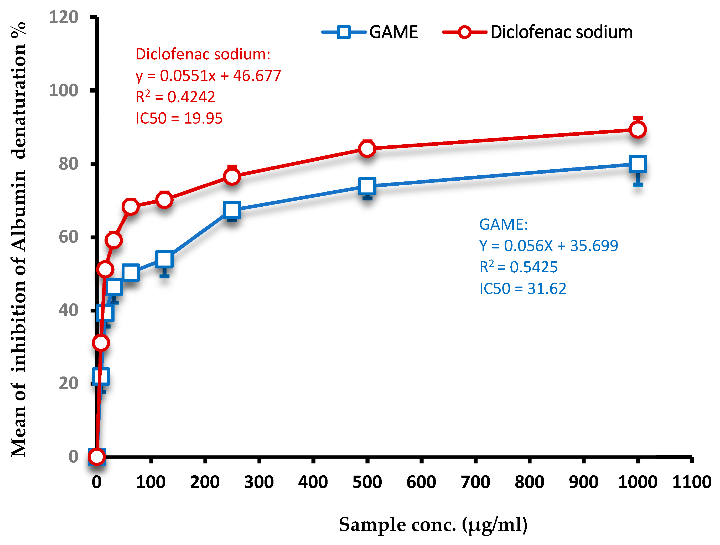
Various concentrations of GAME, ranging from 7.81 to 1000 µg/mL, were tested for anti-inflammatory action, and the results were compared with those obtained with a typical anti-inflammatory medication (diclofenac sodium). As the concentration of the GAME was increased, the percentage of inhibition steadily increased in proportion to the concentration. The results showed that the tested extract had a percentage of protein denaturation inhibition of up to 79.96%, which was comparable to that of the standard medicine employed (89%) (p < 0.05) (Figure 6). GAME had excellent anti-inflammatory properties when compared with the conventional medicine, demonstrating the efficacy of the extract.
Figure 6.
Inhibition of albumin denaturation by GAME. Values represent means of triplicate readings.
3.5. Xanthine Oxidase (XO) Inhibitory Percentage
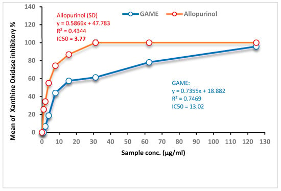
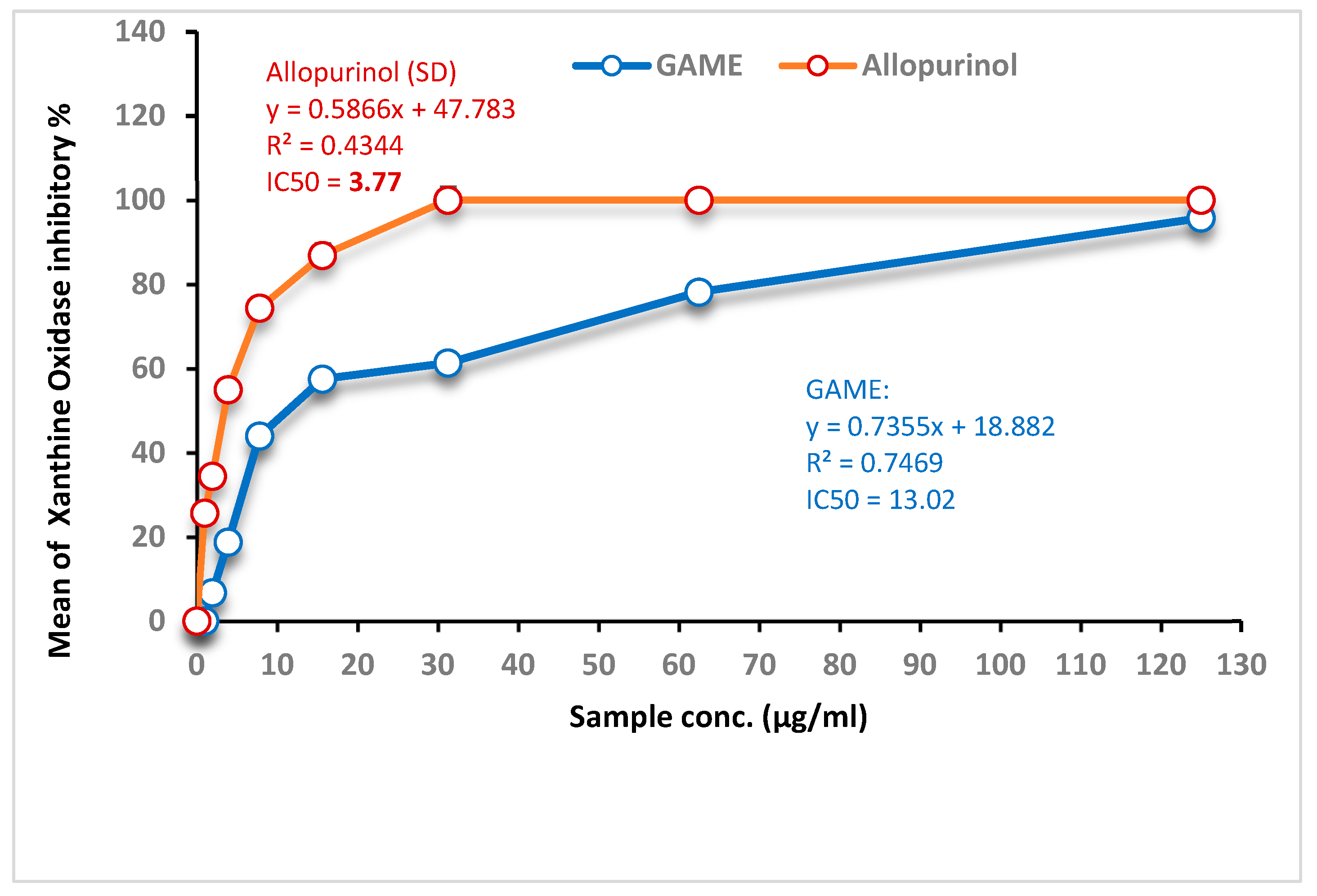
The GAME displayed XO inhibitory actions at various concentrations ranging from 1.95 to125 μg/mL in a concentration-dependent manner, with an IC50 value equal to 13.02 μg/mL, which can be recognized in Figure 7. Under the conditions of the experiment, the IC50 value of allopurinol, which is currently being utilized in clinical practice as an XO-inhibiting medication, was 3.77 μg/mL.
Figure 7.
Inhibition of xanthine oxidase by GAME. Values represent means of triplicate readings.
3.6. In Vitro Lipoxygenase (LOX) Inhibition
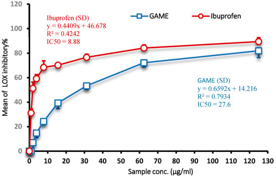
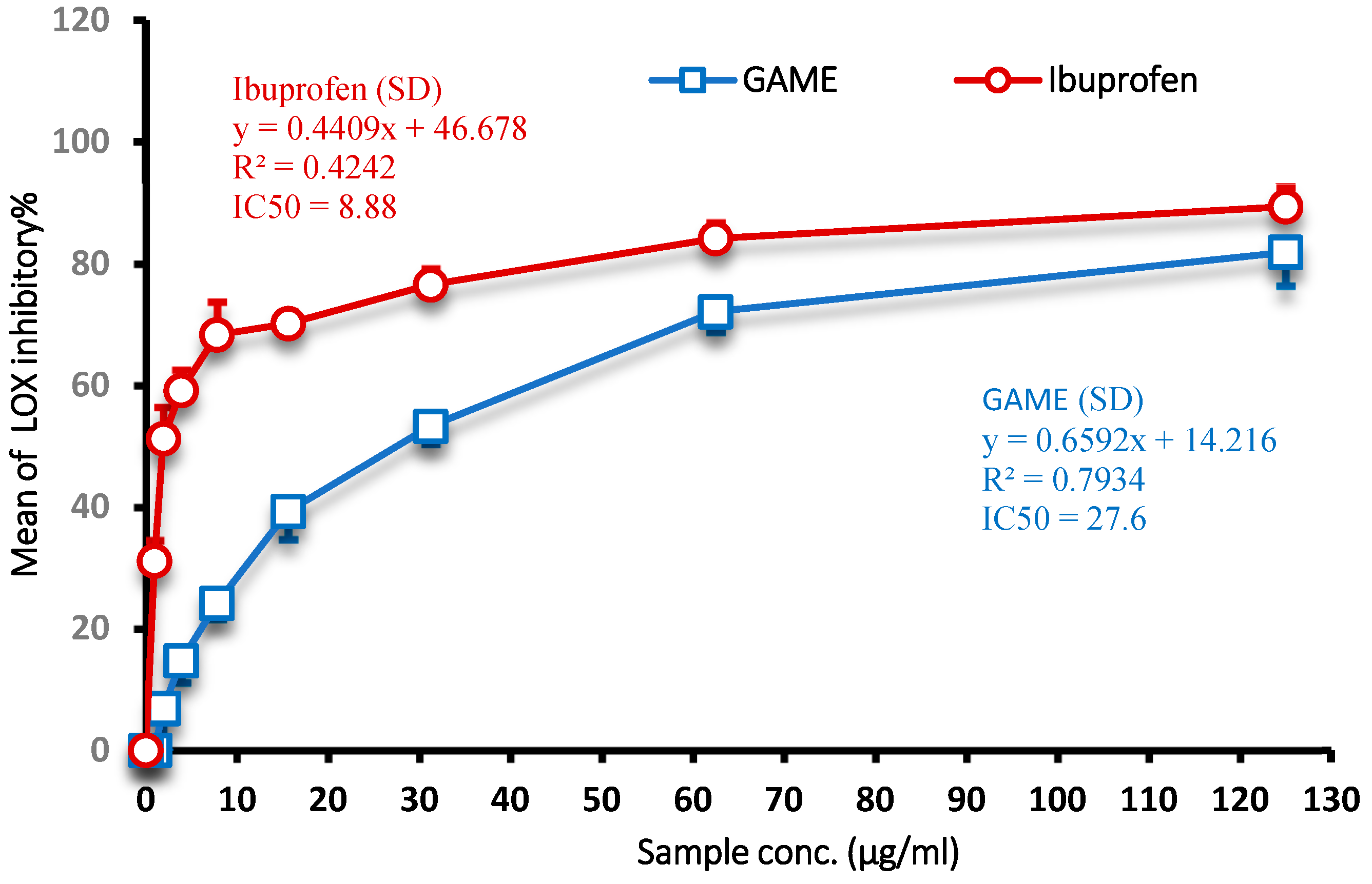
Figure 8 provides a graphical representation of the findings on the lipoxygenase inhibitory activity of GAME. When tested at doses ranging from 0.98 to 125 μg/mL, it demonstrated an enhanced capacity to inhibit lipoxygenase activity, with an inhibition rate that ranged from 10.2 to 74.8%.
Figure 8.
Inhibition of lipoxygenase (LOX) by GAME. Values represent means of triplicate readings.
3.7. In Vitro Cyclooxygenase 2 (COX-2) Inhibition
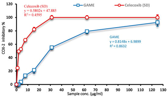
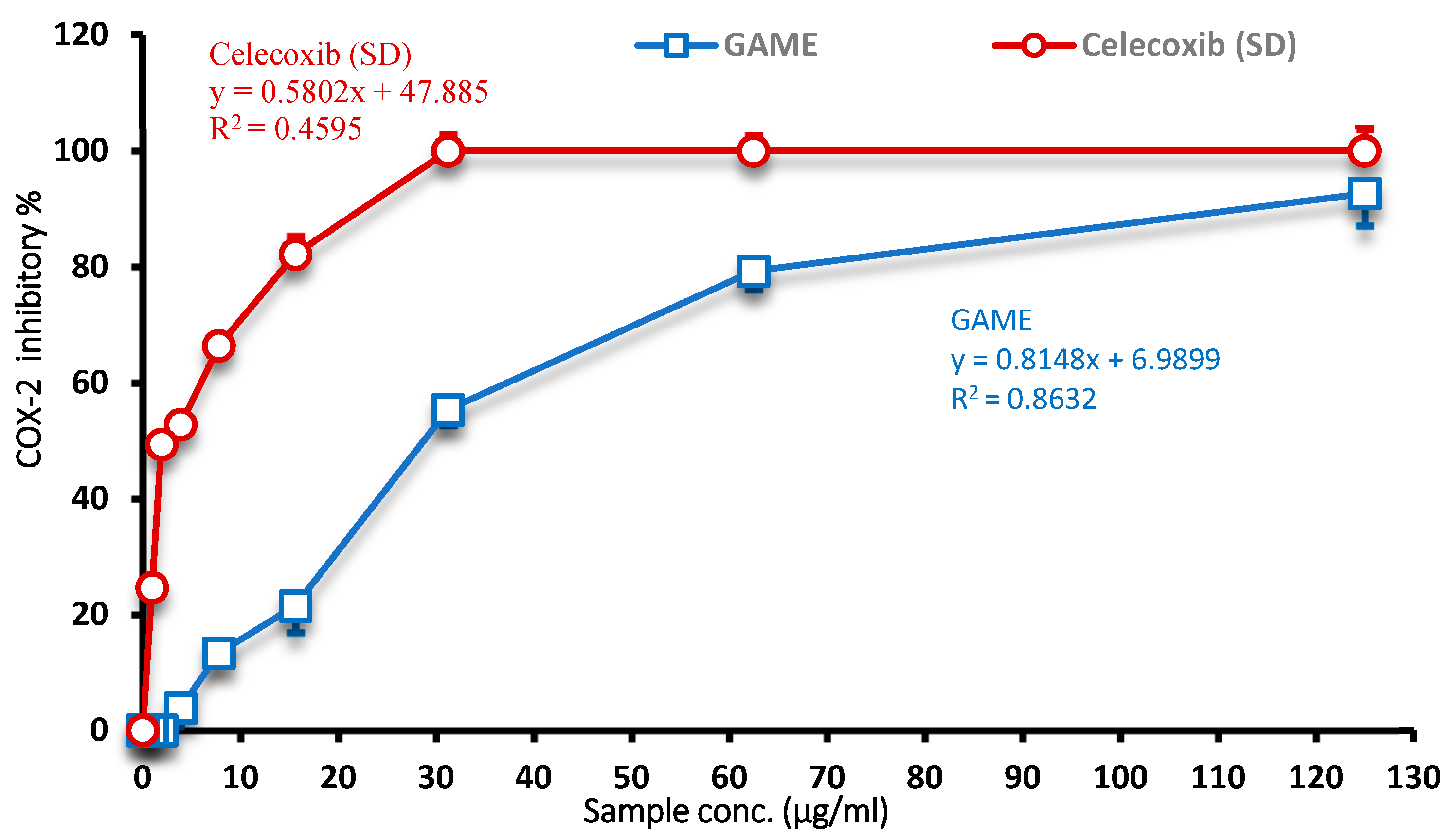
The IC50 value for GAME COX-2 was found to be 28.99 µg/mL, while the IC50 value for the luteolin standard medication celecoxib, which inhibits COX-2 activity, was 8.97 µg/mL (Figure 9). Based on these data, it appears that GAME, in particular, has the ability to suppress COX-2.
Figure 9.
Inhibition of cyclooxygenase 2 (COX-2) by GAME. Values represent means of triplicate readings.
3.8. Cytotoxicity Activities
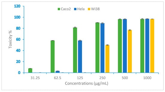
The cytotoxic effect of GAME was evaluated against the Caco-2 cell line (human colorectal adenocarcinoma) and HeLa cells (human cervical cancer) compared with the Wi-38 cell line at different concentrations, including 0, 31.25, 62.5, 125, 250, and 1000 µg/mL (Figure 10). The results validate that the cytotoxicity rises with higher concentrations of GAME. GAME is most effective against the Caco-2 cell line, followed by HeLa cells, and the IC50 values of GAME against Caco-2 and HeLa cells are 73.92 ± 0.67 and 114.57 ± 1.83, respectively. The selectivity index (SI) values, which were calculated using Wi-38 normal cells, demonstrate that GAME had a favorable selectivity for cancer cells. The SI values were 4.28 for Caco-2 and 2.85 for HeLa cell lines, suggesting that Caco-2 had higher therapeutic safety (Table 5).
Figure 10.
Toxicity percentage of GAME against Caco-2, HeLa and Wi38 cell lines.
- Selectivity Index (SI)
Table 5.
IC50 values and selectivity indices (SIs) (safety) of GAME against Caco-2 and Hela cell lines.
4. Discussion
This study aimed to evaluate the phytochemical composition and biological activities of GAME derived from the S. senegal plant by combining laboratory experiments and molecular docking analysis to elucidate its therapeutic potential. The results show that GAME possesses antibacterial and anti-inflammatory activity and has moderate cytotoxic activity, which can be attributed to its biologically important active compounds.
GC-MS analysis revealed that GAME is rich in fatty acids and their derivatives, particularly 9-octadecenoic acid and its methyl esters, as well as other compounds such as dicarboxylic acid derivatives and manfuranosides. Many researchers have demonstrated that these compounds possess antioxidant, antimicrobial, and anti-inflammatory properties [32]. Fatty acids, particularly unsaturated fatty acids, play a pivotal role in regulating inflammatory pathways and cellular signaling, thus explaining their important biological effects [33]. In addition, the presence of phenolic compounds and esters contributes to the elimination of free radicals and enhances the stability of cell membranes [34,35].
GA, owing its complex polysaccharide composition and dietary benefits, exhibits immunomodulatory and antioxidant properties [36]. This synergistic interplay between its polysaccharides and secondary metabolites positions GA as a highly promising multifunctional natural product [37].
The molecular docking analyses provided valuable mechanistic insights into the biological activities observed. Among the compounds, 9-octadecenoic acid demonstrated notable binding affinities for key enzymes associated with inflammation and oxidative stress, including XO, LOX, COX-2 and albumin. These findings support a multi-target mechanism, which is particularly advantageous in managing complex inflammatory conditions [38,39]. XO is a significant enzyme in uric acid production and the generation of reactive oxygen species, both of which contribute to oxidative stress and inflammation [40,41]. The robust binding affinity observed aligns with previous studies emphasizing the potential of natural compounds as XO inhibitors. Natural XO inhibitors have gained considerable attention for the management of gout and related diseases [42,43]. Molecular docking is a commonly used computer method to anticipate ligand–protein interactions and possible binding mechanisms of bioactive substances. Recent investigations on natural compounds have shown sustained and energetically favorable interactions with important biological targets, indicating their therapeutic potential. In line with these findings, the present investigation showed octadecenoic acid-9 has favorable binding energies and persistent interactions with the selected enzymes, indicating its potential multi-targeted inhibitory efficacy [44].
Molecular docking studies have revealed that natural compounds can effectively interact with the therapeutic protein targets, thus indicating their potential as lead bioactive agents. In silico studies have demonstrated strong binding affinities and stable interaction profiles between phytochemicals and critical enzymes in disease pathways. These findings are in agreement with the present study, which showed favorable binding interactions of octadecenoic acid-9 with the selected target proteins, suggesting its possible biological and inhibitory potential [45].
Similarly, the high binding affinity with LOX points to the ability of GAME to inhibit leukotriene biosynthesis, thereby mitigating inflammatory responses [46]. Meanwhile, the moderate interaction with COX-2 supports the in vitro results, indicating a comparable mechanism to that of nonsteroidal anti-inflammatory drugs, which inhibit prostaglandin synthesis [47]. Furthermore, the strong binding to albumin suggests favorable pharmacokinetic properties, such as enhanced bioavailability and systemic dispersion [30,48,49]. Collectively, these findings underscore the potential of molecular docking as a predictive tool for elucidating ligand–protein interactions and facilitate rational drug design [27,38,50]. The present study has some limitations, such as the absence of in vivo validation and enzyme inhibition experiments, and the use of computational docking, which only provides theoretical predictions of binding interactions.
GAME exhibits broad-spectrum antibacterial activity against both Gram-positive and Gram-negative bacteria. Pathogens such as S. typhimurium, E. coli, P. aeruginosa, and S. aureus showed susceptibility to GAME, whereas B. subtilis displayed resistance. The inhibition observed suggests that GAME may be as effective as, or even superior to, certain standard antibiotics in specific cases. This broad-spectrum activity can primarily be attributed to its bioactive components, including fatty acids, phenolics, and esters. These compounds are known for their ability to disrupt bacterial membranes, impair enzymatic activities, and instigate oxidative stress within microbial cells [51,52,53]. Additionally, GA has been reported to enhance immune system responses and modulate antimicrobial activity of host cells, which likely further boosts its antibacterial efficacy [54,55]. The resistance exhibited by B. subtilis could stem from inherent differences in its cell wall structure or intrinsic defense mechanisms, echoing prior findings about selective antibacterial effects seen with plant-based extracts [56].
The anti-inflammatory potential of GAME was validated through various in vitro assays, including tests for membrane stabilization, albumin denaturation inhibition, and enzymatic inhibition of XO, LOX, and COX-2. A significant percentage of erythrocyte membrane stabilization was observed, suggesting that GAME possesses the ability to stabilize lysosomal membranes, preventing the release of pro-inflammatory mediators [57]. Since protein denaturation often triggers inflammation, GAME’s ability to inhibit albumin denaturation underlies its capacity to mitigate inflammatory processes at the molecular level [58]. Furthermore, the dose-dependent inhibition observed correlates with earlier studies on plant-derived anti-inflammatory compounds [57]. The inhibitory effects of key inflammatory enzymes enhance GAME’s anti-inflammatory efficacy. By inhibiting the activity of the XO enzyme, GAME can lower uric acid levels and reduce the production of reactive oxygen species. Additionally, inhibiting LOX and COX-2 prevents the biosynthesis of pro-inflammatory molecules like leukotrienes and prostaglandins [58,59]. This multi-target approach positions GAME as a promising natural alternative to conventional anti-inflammatory drugs that are often associated with adverse side effects [60]. The assessment of cytotoxic activity revealed that GAME has moderate anticancer properties against the Caco-2 and HeLa cell lines, showing greater selectivity toward cancerous cells while sparing normal cells. This selective cytotoxicity is a highly desirable feature for potential therapeutic applications, minimizing harm to healthy tissues. The anticancer activities could be credited to bioactive compounds like fatty acids and phenolic derivatives known for their roles in inducing apoptosis, inhibiting cellular proliferation, and modulating key signaling pathways underlying cancer progression [61]. Previous research has similarly highlighted the anticancer properties of Gum Arabic, attributing its effects to the modulation of gene expression and the mitigation of oxidative stress [62].
The combined application of phytochemical analysis, in vitro assays, and molecular docking offers a holistic framework for evaluating the therapeutic potential of Gum Arabic. The multi-target activity identified in this investigation holds significant promise for addressing multifaceted conditions, such as inflammation and microbial infections, which involve numerous biological pathways.
5. Conclusions
The present study demonstrated that the methanolic extract of Senegalia senegal possesses a diverse phytochemical profile, as identified by GC–MS analysis. This study provides general evidence for the antibacterial and anti-inflammatory effects of Arabic gum methanol extract against clinically important pathogenic bacteria, such as S. typhimurium, E. coli, P. aeruginosa and S. aureus. The biological effects reported herein point to the therapeutic value of Arabic gum as a promising natural source of bioactive compounds with potential multi-target pharmacological activities. Importantly, this study shows the close association of the observed activities with the presence of essential phytoconstituents, such as octadecenoic acid, which have been reported to have antimicrobial and anti-inflammatory properties. This compound may have synergistic effects in modulating microbial growth and inflammatory responses, providing a mechanistic basis for the biological effects observed. Collectively, the results of the present study provide new knowledge regarding the pharmacological potential of Arabic gum and warrant further investigation of its use as a low-cost, natural candidate for the development of new antimicrobial and anti-inflammatory agents.
Author Contributions
N.M.D.: Conceptualization, funding acquisition, project administration, writing—review and editing, validation and resources. H.M.T.: Methodology, investigation, and writing—original draft. R.A.H.: Visualization, data curation, formal analysis, and supervision. All authors have read and agreed to the published version of the manuscript.
Funding
This research was funded by the Deanship of Scientific Research, the University of Jeddah, Jeddah, Saudi Arabia, grant number “UJ-20-112-DR”.
Institutional Review Board Statement
Not applicable.
Informed Consent Statement
Not applicable.
Data Availability Statement
The original contributions presented in this study are included in the article. Further inquiries can be directed to the corresponding author.
Conflicts of Interest
All authors in this article declare no conflicts of interest.
References
- Davis, C.C.; Choisy, P. Medicinal plants meet modern biodiversity science. Curr. Biol. 2024, 34, 158–173. [Google Scholar] [CrossRef] [Scilit]
- Ngo, L.T.; Okogun, J.I.; Folk, W.R. 21st Century natural product research and drug development and traditional medicines. Nat. Prod. Rep. 2013, 30, 584–606. [Google Scholar] [CrossRef] [Scilit]
- Yuan, H.; Ma, Q.; Ye, L.; Piao, G. The traditional medicine and modern medicine from natural products. Molecules 2016, 21, 559. [Google Scholar] [CrossRef] [Scilit] [PubMed]
- Kyalangalilwa, B.; Boatwright, J.S.; Daru, B.H.; Maurin, O.; van der Bank, M. Phylogenetic position and revised classification of Acacia s.l. (Fabaceae: Mimosoideae) in Africa, including new combinations in Vachellia and Senegalia. Bot. J. Linn. Soc. 2013, 172, 500–523. [Google Scholar] [CrossRef] [Scilit]
- Adam, H.E.; Eltahir, M.E.S.; Elhaja, M.E.; Ibrahim, A.H.I.; Abdelkareem, O.E.A.; Hammad, Z.M.; Khalifa, A.S.O.; Mahmoud, T.E.; Elsayed, M.E.O.; Elamin, H.M.A.; et al. Management of gum Arabic production potentialities in the gum belt in Kordofan, Sudan. Int. J. Environ. Plan. Manag. 2017, 3, 1–9. [Google Scholar]
- Elnour, A.A.M.; Al-Assaf, S.; Kabbashi, N.A.; Osman, M.E.; Abdelgadir, A.E. Analytical Study on Secondary Metabolites Extraction and Characterization from Acacia Gums (AGs). In Gum Arabic and Breast Cancer Biology; Elnour, A.A.M., Ed.; Springer: Singapore, 2025. [Google Scholar] [CrossRef] [Scilit]
- Williams, P.A.; Phillips, G.O. Gum arabic. In Handbook of Hydrocolloids, 3rd ed.; Phillips, G.O., Williams, P.A., Eds.; Woodhead Publishing: Cambridge, UK, 2021; pp. 627–652. [Google Scholar] [CrossRef] [Scilit]
- Eltarahony, M.M.; Elblbesy, M.A.; Hanafy, T.A.; Kandil, B.A. Synthesis, characterizations and disinfection potency of gelatin based Gum Arabic antagonistic films. Sci. Rep. 2025, 15, 8279. [Google Scholar] [CrossRef] [Scilit]
- Mudgil, D.; Mudgil, S. Acacia gum: Chemistry, properties and food applications. Food Humanit. 2024, 2, 100264. [Google Scholar] [CrossRef] [Scilit]
- Zahid, M.U.; Jatoi, N.R.; Gul, S.N.; Malik, S.; Kayani, W.K.; Rasheed, F. Molecular perspectives on acacia gum as a functional polysaccharide for biomedical and industrial applications: A review. Int. J. Biol. Macromol. 2025, 319, 145412. [Google Scholar] [CrossRef] [Scilit]
- Abi Assaf, J.; de Coriolis, J.C.; Godden, A.M.; Redhead, E.; Bartram, J.; Cohen-Krais, J.; Silova, K.; Crighton, Z.; Le Gall, G.; Sami, S.; et al. Gum Arabic modulates the microbiota-gut-brain axis and affects general fitness in zebrafish. Sci. Rep. 2025, 15, 34465. [Google Scholar] [CrossRef] [Scilit]
- González-Ruiz, D.; Gohlke, H. Targeting protein–protein interactions with small molecules: Challenges and perspectives for computational binding epitope detection and ligand finding. Curr. Med. Chem. 2006, 13, 2607–2625. [Google Scholar] [CrossRef] [Scilit]
- Ayaz, N.O.; Ramadan, K.S.; Farid, H.E.; Alnahdi, H.S. Protective role and antioxidant activity of arabic gum against trichloroacetate-induced toxicity in liver of male rats. Indian J. Anim. Res. 2017, 51, 303–309. [Google Scholar] [CrossRef] [Scilit]
- Satchanska, G.; Davidova, S.; Petrov, P.D. Natural and synthetic polymers for biomedical and environmental applications. Polymers 2024, 16, 1159. [Google Scholar] [CrossRef] [Scilit]
- Babiker, R.; Kaddam, L.; Mariod, A. The role of gum Arabic as an anti-inflammatory, antioxidant, and immune modulator in COVID-19: A review. Funct. Food Sci. 2022, 2, 242–257. [Google Scholar] [CrossRef] [Scilit]
- Idriss, M.M.; Jovial, S.A.; Sehgal, A.; Fatima, M.M. An in vitro antimicrobial activity of Acacia senegal as prebiotic against pathogens. J. Health Wellness Saf. Res. 2023, 2, 1–15. [Google Scholar] [CrossRef]
- Bhola, J.; Bhadekar, R. Enhancing antimicrobial efficacy: Gum acacia-enriched Lactobacillus consortium against multidrug-resistant pathogens. Med. Microecol. 2025, 25, 100132. [Google Scholar] [CrossRef] [Scilit]
- Mirghani, M.E.; Elnour, A.A.; Kabbashi, N.A.; Alam, M.Z.; Musa, K.H.; Abdullah, A. Determination of antioxidant activity of gum arabic: An exudation from two different locations. Sci. Asia 2018, 44, 179–186. [Google Scholar] [CrossRef] [Scilit]
- Jarrar, A.H.; Stojanovska, L.; Apostolopoulos, V.; Feehan, J.; Bataineh, M.F.; Ismail, L.C.; Al Dhaheri, A.S. The effect of gum Arabic (Acacia senegal) on cardiovascular risk factors and gastrointestinal symptoms in adults at risk of metabolic syndrome: A randomized clinical trial. Nutrients 2021, 13, 194. [Google Scholar] [CrossRef] [Scilit]
- Jaafar, N.S. Clinical effects of Arabic gum (Acacia): A mini review. Iraq J. Pharm. Sci. 2024, 28, 9–16. [Google Scholar] [CrossRef] [Scilit]
- Elnour, A.A.M.; Mirghani, M.E.S.; Kabbashi, N.A.; Alam, M.Z.; Musa, K.H. Gum Arabic: An optimization of ultrasonic-assisted extraction of antioxidant activity. Stud. UBB Chem. 2018, 63, 95–116. [Google Scholar] [CrossRef] [Scilit]
- Lisec, J.; Schauer, N.; Kopka, J.; Willmitzer, L.; Fernie, A.R. Gas chromatography mass spectrometry–based metabolite profiling in plants. Nat. Protoc. 2006, 1, 387–396, Erratum in: Nat. Protoc. 2015, 10, 1457. [Google Scholar] [CrossRef] [Scilit]
- Athanassiadis, B.; Abbott, P.V.; George, N.; Walsh, L.J. An in vitro study of the antimicrobial activity of some endodontic medicaments and their bases using an agar well diffusion assay. Aust. Dent. J. 2009, 54, 141–146. [Google Scholar] [CrossRef] [Scilit]
- Rashid, M.A.; Sikder, M.; Kaisar, M.; Miah, M.; Parvez, M.; Hossian, A. Membrane stabilizing activity: A possible mechanism of action for the anti-inflammatory activity of two Bangladeshi medicinal plants: Mesua nagassarium (BURM. F.) and Kigelia pinnata (JACK) DC. Int. J. Pharm. Res. Dev. 2011, 3, 1–5. [Google Scholar]
- Mounnissamy, V.M.; Kavimani, S.; Balu, V.; Quine, S.D. Evaluation of anti-inflammatory and membrane-stabilizing property of ethanol extract of Cansjera rheedii J. Gmelin (Opiliaceae). Iran. J. Pharmacol. Ther. 2007, 6, 230–235. [Google Scholar]
- Janani, K.; Geetha, R.V.; Rajeshkumar, S. In vitro evaluation of anti-inflammatory activity of Symplocos racemosa using protein denaturation assay. J. Pharm. Res. Int. 2021, 33, 715–720. [Google Scholar] [CrossRef] [Scilit]
- Abdulhafiz, F.; Mohammed, A.; Kayat, F.; Bhaskar, M.; Hamzah, Z.; Podapati, S.K.; Reddy, L.V. Xanthine oxidase inhibitory activity, chemical composition, antioxidant properties and GC–MS analysis of Keladi Candik (Alocasia longiloba Miq.). Molecules 2020, 25, 2658. [Google Scholar] [CrossRef] [Scilit]
- Kumaraswamy, M.; Satish, S. Antioxidant and anti-lipoxygenase activity of Thespesia lampas Dalz and Gibs. Adv. Biol. Res. 2008, 2, 56–59. [Google Scholar]
- Petrovic, N.; Murray, M. Using N,N,N′,N′-tetramethyl-p-phenylenediamine (TMPD) to assay cyclooxygenase activity in vitro. In Advanced Protocols in Oxidative Stress II; Springer: Berlin, Germany, 2010; pp. 129–140. [Google Scholar]
- Amessis-Ouchemoukh, N.; Madani, K.; Falé, P.L.; Serralheiro, M.L.; Araújo, M.E.M. Antioxidant capacity and phenolic contents of some Mediterranean medicinal plants and their potential role in the inhibition of cyclooxygenase-1 and acetylcholinesterase activities. Ind. Crops Prod. 2014, 53, 6–15. [Google Scholar] [CrossRef] [Scilit]
- Mosmann, T. Rapid colorimetric assay for cellular growth and survival: Application to proliferation and cytotoxicity assays. J. Immunol. Methods 1983, 65, 55–63. [Google Scholar] [CrossRef] [Scilit] [PubMed]
- Oluwole, O.; Fernando, W.B.; Lumanlan, J.; Ademuyiwa, O.; Jayasena, V. Role of phenolic acid, tannins, stilbenes, lignans and flavonoids in human health—A review. Int. J. Food Sci. Technol. 2022, 57, 6326–6335. [Google Scholar] [CrossRef] [Scilit]
- Calder, P.C. Omega-3 fatty acids and inflammatory processes. Nutrients 2010, 2, 355–374. [Google Scholar] [CrossRef] [Scilit] [PubMed]
- Yeshi, K.; Crayn, D.; Ritmejerytė, E.; Wangchuk, P. Plant secondary metabolites produced in response to abiotic stresses have potential application in pharmaceutical product development. Molecules 2022, 27, 313. [Google Scholar] [CrossRef] [Scilit] [PubMed]
- Vasile, F.E.; Romero, A.M.; Judis, M.A.; Mattalloni, M.; Virgolini, M.B.; Mazzobre, M.F. Phenolics composition, antioxidant properties and toxicological assessment of Prosopis alba exudate gum. Food Chem. 2019, 285, 369–379. [Google Scholar] [CrossRef] [Scilit] [PubMed]
- Barkeer, S.; Pothuraju, R.; Malakar, P.; Pimentel, T.C.; Siddiqui, J.A.; Nair, S.A. Gum acacia dietary fiber: Significance in immunomodulation, inflammatory diseases, and cancer. Phytother. Res. 2024, 38, 1509–1521. [Google Scholar] [CrossRef] [Scilit]
- Mohamed, S.A.; Elsherbini, A.M.; Alrefaey, H.R.; Adelrahman, K.; Moustafa, A.; Egodawaththa, N.M.; Crawford, K.E.; Nesnas, N.; Sabra, S.A. Gum Arabic: A Commodity with Versatile Formulations and Applications. Nanomaterials 2025, 15, 290. [Google Scholar] [CrossRef] [Scilit]
- Meng, X.-Y.; Zhang, H.-X.; Mezei, M.; Cui, M. Molecular docking: A powerful approach for structure-based drug discovery. Curr. Comput.-Aided Drug Des. 2011, 7, 146–157. [Google Scholar] [CrossRef] [Scilit]
- Pinzi, L.; Rastelli, G. Molecular docking: Shifting paradigms in drug discovery. Int. J. Mol. Sci. 2019, 20, 4331. [Google Scholar] [CrossRef] [Scilit]
- Harrison, R. Physiological roles of xanthine oxidoreductase. Drug Metab. Rev. 2004, 36, 363–375. [Google Scholar] [CrossRef] [Scilit]
- Glantzounis, G.K.; Tsimoyiannis, E.C.; Kappas, A.M.; Galaris, D.A. Uric acid and oxidative stress. Curr. Pharm. Des. 2005, 11, 4145–4151. [Google Scholar] [CrossRef] [Scilit]
- Serrano, J.L.; Figueiredo, J.; Almeida, P.; Silvestre, S. From xanthine oxidase inhibition to in vivo hypouricemic effect: An integrated overview of in vitro and in vivo studies with focus on natural molecules and analogues. Evid. Based Complement. Altern. Med. 2020, 2020, 9531725. [Google Scholar] [CrossRef] [Scilit]
- Alghamdi, A.A.; Althumali, J.S.; Almalki, M.M.M.; Almasoudi, A.S.; Almuntashiri, A.H.; Almuntashiri, A.H.; Mohammed, A.I.; Alkinani, A.A.; Almahdawi, M.S.; Mahzari, M.A.H. An overview on the role of xanthine oxidase inhibitors in gout management. Arch. Pharm. Pract. 2021, 12, 94–99. [Google Scholar] [CrossRef] [Scilit]
- Zazeri, G.; Povinelli, A.P.R.; Le Duff, C.S.; Tang, B.; Cornelio, M.L.; Jones, A.M. Synthesis and spectroscopic analysis of piperine-and piperlongumine-inspired natural product scaffolds and their molecular docking with IL-1β and NF-κB proteins. Molecules 2020, 25, 2841. [Google Scholar] [CrossRef] [Scilit]
- Zazeri, G.; Povinelli, A.P.R.; Pavan, N.M.; Jones, A.M.; Ximenes, V.F. Solvent-induced lag phase during the formation of lysozyme amyloid fibrils triggered by sodium dodecyl sulfate: Biophysical experimental and in silico study of solvent effects. Molecules 2023, 28, 6891. [Google Scholar] [CrossRef] [Scilit]
- Zafar, N.; Rafique, A.; Naz, S.; Nazir, M.M.; Ashraf, A. Unveiling the in vitro, in vivo anti-inflammatory potential of Ocimum basilicum and in silico analysis of its phytocompounds targeting COXs proteins. J. Pharm. Pharmacol. 2025, 77, 1133–1147. [Google Scholar] [CrossRef] [Scilit]
- Vane, J.R.; Botting, R.M. Mechanism of action of nonsteroidal anti-inflammatory drugs. Am. J. Med. 1998, 104, 2S–8S. [Google Scholar] [CrossRef] [Scilit]
- Kragh-Hansen, U.; Chuang, V.T.G.; Otagiri, M. Practical aspects of the ligand-binding and enzymatic properties of human serum albumin. Biol. Pharm. Bull. 2002, 25, 695–704. [Google Scholar] [CrossRef] [Scilit] [PubMed]
- Lionta, E.; Spyrou, G.; Vassilatis, D.K.; Cournia, Z. Structure-based virtual screening for drug discovery: Principles, applications, and recent advances. Curr. Top. Med. Chem. 2014, 14, 1923–1938. [Google Scholar] [CrossRef] [Scilit]
- Merkl, R.; Hrádková, I.; Filip, V.; Šmidrkal, J. Antimicrobial and antioxidant properties of phenolic acids alkyl esters. Czech J. Food Sci. 2010, 28, 275–279. [Google Scholar] [CrossRef] [Scilit]
- Abdllha, H.B.; Mohamed, A.I.; Almoniem, K.A.; Adam, N.I.; Alhaadi, W.; Elshikh, A.A.; Ali, A.J.; Makuar, I.G.; Elnazeer, A.M.; Elrofaei, N.A.; et al. Evolution of antimicrobial, antioxidant potentials and phytochemical studies of three solvent extracts of five species from Acacia used in Sudanese ethnomedicine. Adv. Microbiol. 2016, 6, 691–698. [Google Scholar] [CrossRef]
- Ding, Y.; Zong, Q.; Zhang, Q.; Wang, Y.; Wang, J.; Huang, W.; Sun, W.; Zhai, Y. Gum arabic based multifunctional antibacterial adhesion hydrogel dressings loaded with doxycycline hydrochloride for wound healing. Int. J. Biol. Macromol. 2025, 306, 141284. [Google Scholar] [CrossRef] [Scilit] [PubMed]
- Ali, B.H.; Ziada, A.; Blunden, G. Biological effects of gum Arabic: A review of some recent research. Food Chem. Toxicol. 2009, 47, 1–8. [Google Scholar] [CrossRef] [Scilit]
- Elmubarak, N.; Ibrahim, Y.; Gareeballah, A.; Sanhouri, N. In vitro investigation of antibacterial activity of gum Arabic prepared by two different processing methods against Enterococcus faecalis. Appl. Med. Res. 2023, 11, 1–10. [Google Scholar] [CrossRef] [Scilit]
- Esho, B.A.; Samuel, B.; Akinwunmi, K.F.; Oluyemi, W.M. Membrane stabilization and inhibition of protein denaturation as mechanisms of the anti-inflammatory activity of some plant species. Trends Pharmacol. Sci. 2021, 7, 269–278. [Google Scholar] [CrossRef]
- Opie, E.L. On the relation of necrosis and inflammation to denaturation of proteins. J. Exp. Med. 1962, 115, 597–608. [Google Scholar] [CrossRef] [Scilit]
- Umapathy, E.; Ndebia, E.; Meeme, A.; Adam, B.; Menziwa, P.; Nkeh-Chungag, B.; Iputo, J.E. An experimental evaluation of Albuca setosa aqueous extract on membrane stabilization, protein denaturation and white blood cell migration during acute inflammation. J. Med. Plants Res. 2010, 4, 789–795. [Google Scholar]
- Hudaib, M.M.; Tawaha, K.A.; Mohammad, M.K.; Assaf, A.M.; Issa, A.Y.; Alali, F.Q.; Aburjai, T.A.; Bustanji, Y.K. Xanthine oxidase inhibitory activity of the methanolic extracts of selected Jordanian medicinal plants. Pharmacogn. Mag. 2011, 7, 320–324. [Google Scholar] [CrossRef] [Scilit]
- Attiq, A.; Jalil, J.; Husain, K.; Ahmad, W. Raging the war against inflammation with natural products. Front. Pharmacol. 2018, 9, 976. [Google Scholar] [CrossRef] [Scilit]
- Panchal, N.K.; Sabina, E.P. Non-steroidal anti-inflammatory drugs (NSAIDs): A current insight into its molecular mechanism eliciting organ toxicities. Food Chem. Toxicol. 2023, 172, 113598. [Google Scholar] [CrossRef] [Scilit]
- Bagheri, F.; Rahi, A. Anti-diabetic, anti-obesity, antimicrobial, and anticancer applications of gum Arabic: Pharmaceutical advances. Micro Nano Bio Asp. 2024, 3, 1–6. [Google Scholar] [CrossRef]
- Alharthi, N.S.; Bel Hadj Salah, K.; Al-Zahrani, M.H.; Alyoubi, A.A.; Abuzinadah, G.M.S.A.; Alhazmi, W.A.; Momenah, M.A.; Alqahtani, M.A.; Mashraqi, A.; Sadan, M. Polyphenolic gum Arabic extract with antidiabetic, antioxidant, anticancer and antimicrobial activities enhances blood biochemistry, heat stress genes, and reproductive hormones in heat and cisplatin nephrotoxicity-stressed mice. Pak. Vet. J. 2025, 45, 162–173. [Google Scholar] [CrossRef] [Scilit]

Disclaimer/Publisher’s Note: The statements, opinions and data contained in all publications are solely those of the individual author(s) and contributor(s) and not of MDPI and/or the editor(s). MDPI and/or the editor(s) disclaim responsibility for any injury to people or property resulting from any ideas, methods, instructions or products referred to in the content. |
© 2026 by the authors. Licensee MDPI, Basel, Switzerland. This article is an open access article distributed under the terms and conditions of the Creative Commons Attribution (CC BY) license.