


Abstract
Background/Objectives: Chronic injuries of the radial collateral ligament (RCL) of the thumb metacarpophalangeal (TMCP) joint are becoming an increasingly common clinical challenge. In cases without degenerative changes, surgical treatment with ligament reconstruction may be effective. However, coexisting cartilage damage significantly worsens treatment outcomes and may necessitate arthrodesis, which limits joint mobility and carries a high risk of complications. In the present case, an alternative surgical approach was employed—combining RCL reconstruction using a palmaris longus (PL) tendon with interpositional arthroplasty. The aim of this report is to present an innovative method for treating chronic TMCP instability with focal cartilage damage through simultaneous ligament reconstruction and interpositional grafting using autologous tissue. Methods/Case presentation: A 60-year-old male manual laborer presented with chronic TMCP joint instability and pain impairing daily activities and preventing him from working. Intraoperatively, cartilage damage to the head of the first metacarpal was identified. Radial collateral ligament reconstruction was performed using the Glickel technique with the PL tendon, incorporating two modifications: (1) advancement of a capsuloligamentous flap as an interpositional graft, and (2) additional reinforcement of the dorsal capsule with a tendon strip. Results: At 24-month follow-up, complete pain relief was noted, handgrip strength increased by 10.4%, key pinch strength by 25%, palmar abduction improved by 27%, TMCP joint extension improved by 20% and TMCP joint total range of motion improved by 66.7%. Conclusions: The combination of ligament reconstruction with interposition of autologous tissue may be a promising alternative for treating TMCP instability with limited cartilage damage.
1. Introduction
Chronic injuries of the radial collateral ligament (RCL) of the thumb metacarpophalangeal joint (TMCPJ) are an increasingly recognizable problem of underestimated prevalence and consequences, as patients with RCL injuries typically present with a significant delay compared to those with injuries to its ulnar counterpart—the ulnar collateral ligament (UCL) [1]. While treatment for acute injuries is well established, chronic cases, particularly when accompanied by degenerative changes, pose a therapeutic challenge [2,3,4]. Conventionally, TMCP joint arthrodesis is a common solution in such instances. Nevertheless, arthrodesis is often associated with relatively high complication rates and leads to a diminished range of motion. In this case study, we present a novel surgical approach for chronic RCL injuries—a combination of RCL reconstruction using a palmaris longus (PL) tendon graft and interpositional arthroplasty. This surgical technique resulted in complete pain alleviation, preserved joint mobility, and improved function.
2. Clinical Case
A 60-year-old right-handed male heavy manual laborer presented with complaints of a “swollen” TMCP joint of the right hand. He was not able to determine the exact timing of the injury. Notably, the patient had a history of bilateral carpal tunnel syndrome surgery, with both procedures being refractory and requiring subsequent revisions on each hand. The patient complained of diminished handgrip strength due to joint pain and instability during grasping, which impaired his ability to work (meat cutting) along with daily life activities.
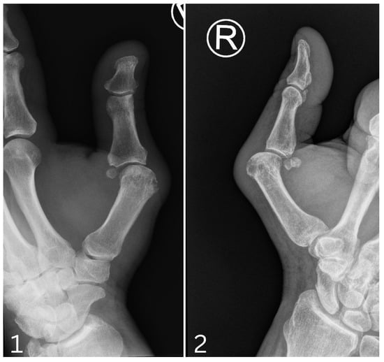
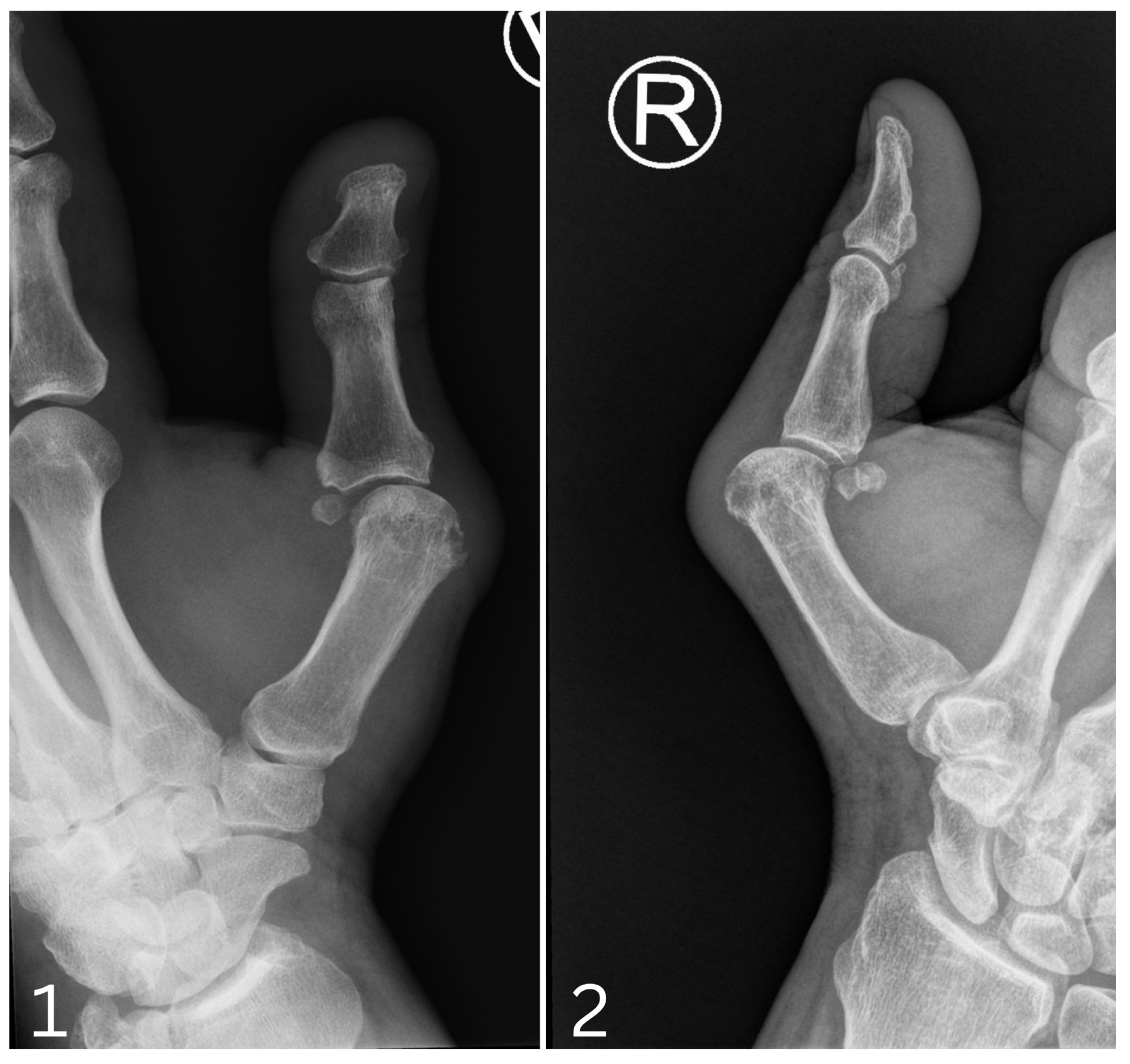
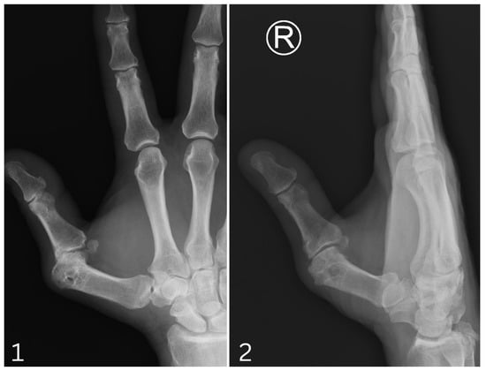
On physical examination, the following findings were noted: preserved integrity of the UCL under strain, complete insufficiency of the RCL, TMCP joint swelling with obvious ulnar and palmar translation (readily appreciated on standard radiographs—Figure 1), crepitus, and pain upon shear stress. A preoperative functional assessment, including handgrip strength, key pinch strength, and abduction, was performed, with results summarized in Table 1.
Figure 1.
Preoperative radiograph of TMCP joint: 1. Anteroposterior view. 2. Lateral view.
Table 1.
Pre- and postoperative assessments of various parameters.
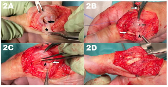
The patient was cautiously qualified for reconstruction of the RCL with palmaris longus tendon graft. Intraoperatively, chronic, irreparable proximal avulsion of the RCL was discovered, accompanied by substantial focal cartilage wear on the dorsal aspect of the metacarpal head (Figure 2A). As the patient was extremely reluctant to pursue salvage arthrodesis, we proceeded with ligament reconstruction using the standard technique described by Glickel et al. [5], but with two important modifications.
Figure 2.
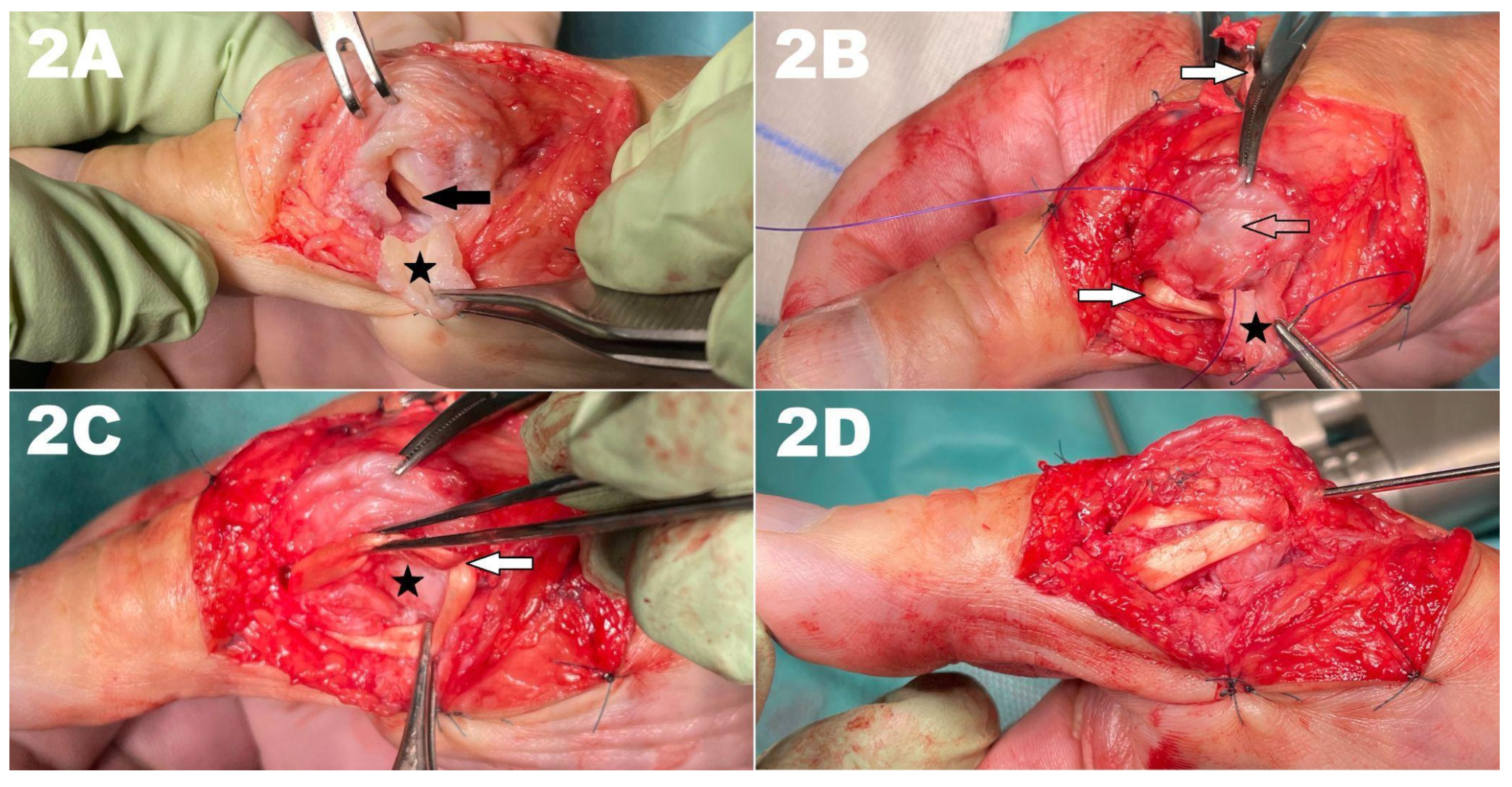
Consecutive steps of the surgical technique: (A)—focal cartilage wear on the dorsal aspect of the metacarpal head (black arrow), where the asterisk labels the RCL flap; (B)—mobilized broad capsular flap, where the white arrows marks the palmaris longus tendon graft and the translucent arrow indicates the dorsal joint capsule; (C)—the capsular flap covering the chondral defect in a “patch” manner; (D)—palmaris longus graft reinforcing the dorsal capsule in an oblique manner and a single K-wire transfixing the joint.
3. Surgical Technique
- Capsular flap mobilization and interposition:
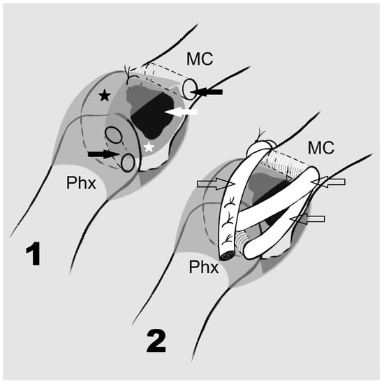
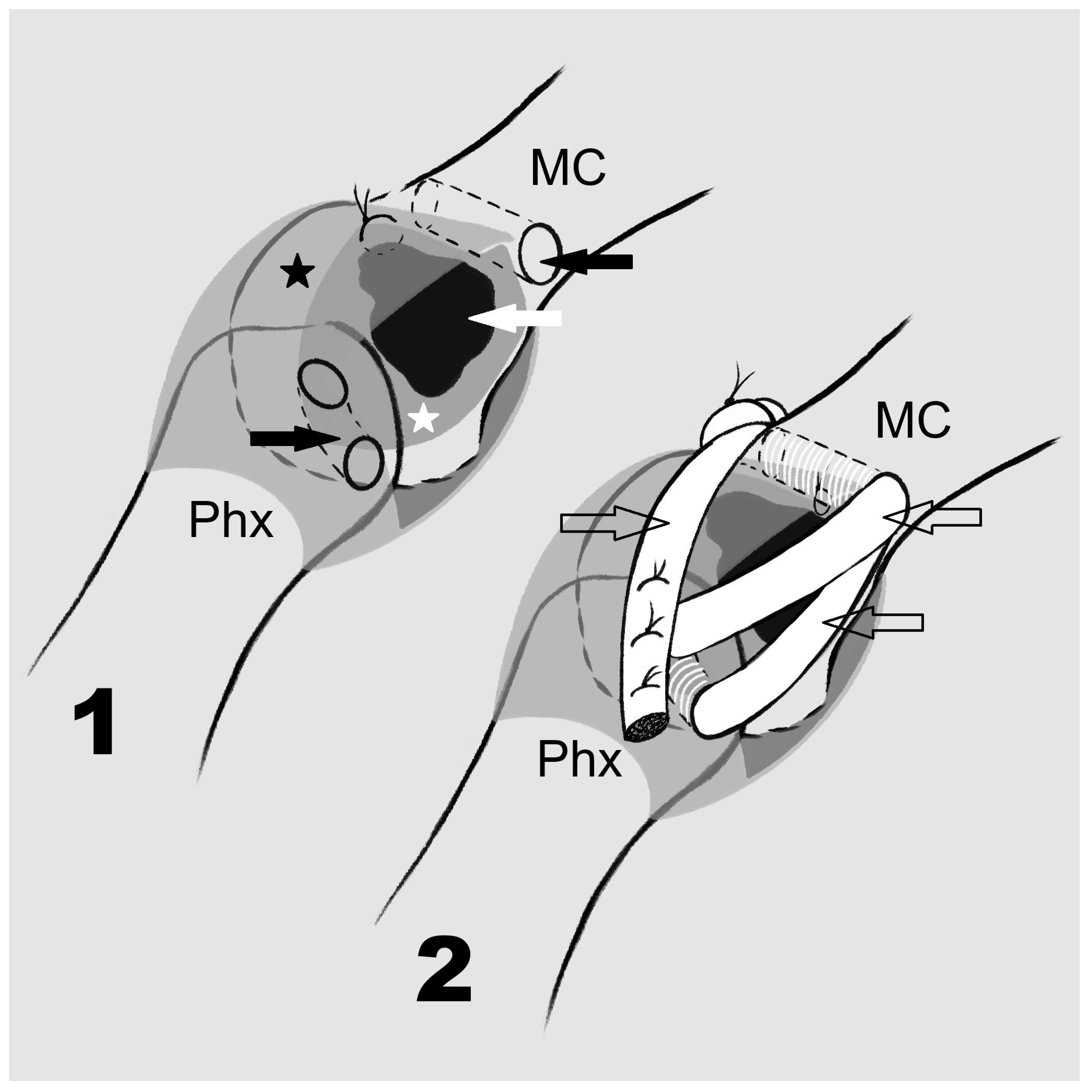
A broad radial capsular flap was mobilized while preserving its proximal attachment near the cartilage wear and maintaining the dorsal capsule for stability The flap incorporated remnants of the insufficient, irreparable RCL, as can be seen in Figure 2B. The flap was then used as an interpositional graft and laid down in a “patch” manner to cover the chondral defect. It was secured in place with transcapsular sutures on the dorsal side (Figure 2C).
- 2.
- Palmaris longus tendon grafting and dorsal reinforcement:
The palmaris longus tendon graft was properly tensioned and secured in place through bony tunnels in the first metacarpal and proximal phalanx. The redundant slip of the tendon graft was used to reinforce the dorsal capsule in an oblique manner (Figure 2D). This modification was intended to reinforce the dorsal capsule and potentially prevent collapse of the proximal phalanx base. Importantly, the aponeurosis and insertion of the abductor pollicis brevis (APB) were preserved and repaired en masse.
At the end of the procedure, the joint was temporarily transfixed with a single K-wire in a neutral position without any attempt to overcorrect in any plane, as we deemed this unnecessary in a long-standing case (Figure 2D). Figure 3 provides a diagram of the surgical technique.
Figure 3.
A graphic diagram to illustrate the ligament reconstruction with interposition arthroplasty: MC—Ist metacarpal; Phx—Ist Phalanx. 1. An illustration of the first modification: white arrow—chondral defect, black asterisk—capsule, white asterisk—capsular flap secured in place, black arrows—bony tunnels. 2. An illustration of the second modification: translucent arrows—PL tendon graft.
Postoperatively, the thumb was immobilized in a long-thumb spica cast. The K-wire was removed six weeks later. The patient did not undergo any formal rehabilitation and returned to his occupation after a two-month convalescence period. The same parameters as assessed preoperatively were evaluated during the 24-month follow-up by the designated trained personnel and are summarized in Table 1. All parameters demonstrated improvement during postoperative follow-up, and the patient expressed high satisfaction with the functional outcome.
4. Discussion
To date, none of the described methods for RCL reconstruction fully replicate the biomechanical properties of the native ligament, and progressive stretching of the reconstruction over time may lead to recurrent instability and inferior outcomes [6]. In our case involving a heavy manual laborer, improved stability with a firm endpoint during ulnar deviation of the joint was achieved at the two-year follow-up. Although ligament reconstruction is generally safe, it cannot be uniformly applied. We are still unable to fully prevent post-traumatic degeneration of the joint or reverse the natural course of osteoarthritis.
The presence of cartilage damage or osteoarthritic changes is widely regarded as a contraindication for “simple” ligament reconstruction [2,4]. Although a substantial array of operative solutions for osteoarthritis in hand joints exists—including denervation, resurfacing arthroplasty, and joint arthroplasty (both autoplasty and alloplasty)—the majority of these options are not suitable for TMCP joints. Most clinical strategies to address instability and arthritis in this region are extrapolated from treatment methods developed for the thumb CMC joint. TMCPJ symptomatic hyperextension is well recognized in this context.
Thus, the most obvious option for arthritic and severely unstable TMCP joints remains arthrodesis. Potential complications—such as breakage or migration of implants, infection, or nonunion—are highly technique-dependent and reported in up to 30% of cases [7,8]. A major drawback of arthrodesis would be the permanent loss of range of motion, which the patient was extremely reluctant to accept.
In contrast, interpositional arthroplasty techniques—though not uniformly appreciated—have a long and well-recognized role in treating several osteoarthritic conditions in hand joints. Among them, the most promising results have been noted in palmar plate arthroplasties for osteoarthrosis of proximal interphalangeal and metacarpophalangeal joints [9,10]. However, the evidence for comparable treatment of the thumb metacarpophalangeal joint remains sparse.
In the presented case, the modified technique salvaged joint mobility and resulted in complete pain resolution. Although postoperative metrics did not entirely match those of the unaffected hand, key pinch strength increased by 25%, reaching the level of the contralateral side, and overall handgrip strength improved by 10.4%, achieving 95% of the strength of the unaffected hand. The total range of motion of the TMCP joint improved by 66.7%, reaching 62.5% of the unaffected hand. Surprisingly, the total range of motion of the thumb reached 104% of the unaffected hand, mostly due to compensatory hyperextension of the interphalangeal joint. Postoperative thumb opposition was symmetrical to the contralateral, unaffected thumb. The clinical outcomes achieved in this case report were superior to the average improvements reported following TMCPJ arthrodesis, indicating that the modified technique may constitute a promising alternative to conventional arthrodesis [7].
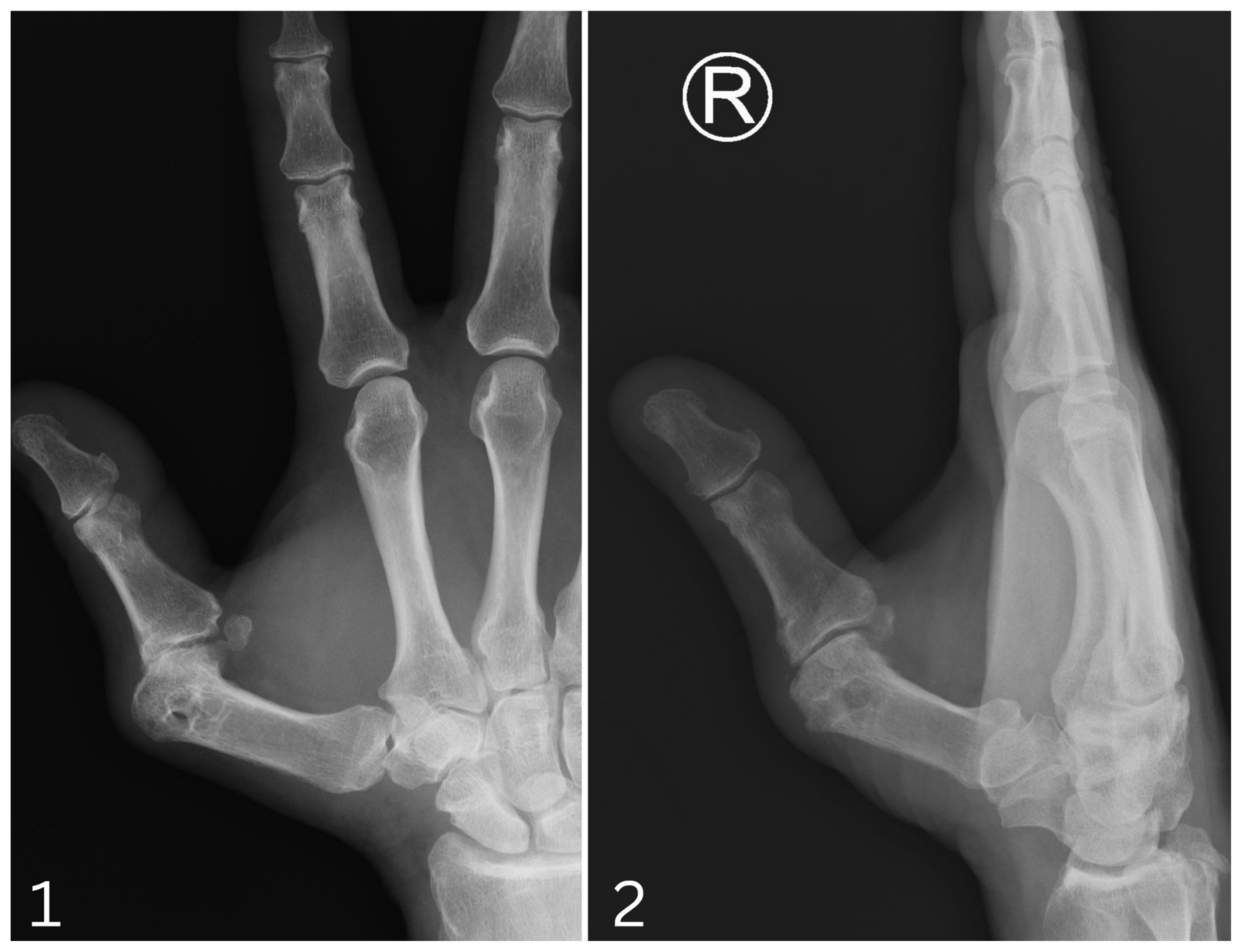
As expected, the follow-up radiograph (Figure 4) demonstrated some progression of intra-articular degeneration from preoperative early-stage 1 to end-stage 1 on the Kellgren–Lawrence scale, but without any clinical impact. The only evident drawback in this case was the persistence of joint deformity, which was not a concern for the patient.
Figure 4.
A 24-month follow-up radiograph of the TMCP joint: 1. Anteroposterior view. 2. Lateral view.
By applying two different approaches—ligament reconstruction and interpositional arthroplasty—we were able to retain effective function while avoiding the complications of total joint fusion. It should be noted that in this case only part of the joint surface exhibited a chondral defect. It is possible that in cases of complete joint degeneration or extensive chondral loss, such a procedure might be insufficient for relieving pain.
We also cannot confirm whether pain relief was solely due to the interpositional flap. Although not intended, a wide exposure of the joint during the procedure could have potentially resulted in partial denervation and contributed to pain relief. Nevertheless, this is only a speculative point and cannot be confirmed here. Nonetheless, the presented case highlights that not all possible treatment avenues have been fully explored, and interpositional arthroplasties utilizing autologous tissues may present a potential option in carefully selected cases.
5. Conclusions
In this case, RCL reconstruction combined with interpositional arthroplasty preserved joint motion, stability, and resulted in complete resolution of pain, meeting the expectations of the patient without the complications associated with hardware placement or bony nonunion. The results support the use of this procedure for this specific indication. Notably, the procedure does not preclude salvage arthrodesis or any other future treatment should they become necessary.
Further long-term follow-up data from analogous cases are needed to comprehensively assess this approach’s potential.
Author Contributions
Conceptualization, B.P.; validation, A.Ż.; formal analysis, B.P.; investigation, I.K.; resources, I.K.; writing—original draft preparation, B.P. and I.K.; writing—review and editing, A.Ż., visualization, B.P. and I.K.; supervision, A.Ż. All authors have read and agreed to the published version of the manuscript.
Funding
This research received no external funding.
Institutional Review Board Statement
According to Polish law (The Act on the Professions of Doctor and Dentist of 5 December 1996, Chapter 4, Articles 21, 22, 29), a retrospective case report based on standard, clinically indicated treatment does not constitute a medical experiment. Therefore, it does not require formal ethical review or approval by a local bioethics committee. Treatment was performed entirely according to standard clinical practice.
Informed Consent Statement
Informed consent was obtained from all subjects involved in this study. Written informed consent was obtained from the patient for the publication of this case.
Data Availability Statement
The original contributions presented in this study are included in the article. Further inquiries can be directed to the corresponding author.
Conflicts of Interest
The authors declare no conflicts of interest.
References
- Taylor, K.F.; Lanzi, J.T.; Cage, J.M.; Drake, M.L. Radial collateral ligament injuries of the thumb MCP joint: Epidemiology in a military population. J. Hand Surg. 2013, 38, 532–536. [Google Scholar] [CrossRef] [Scilit]
- Brock, J.; Jayaraju, U.; Trickett, R.W. Surgical interventions for chronic ulnar collateral ligament injuries of the thumb: A systematic review. J. Hand Surg. Asian Pac. 2023, 28, 548–554. [Google Scholar] [CrossRef] [Scilit]
- Glickel, S.Z.; Malerich, M.; Pearce, S.M.; Littler, J.W. Ligament replacement for chronic instability of the thumb MCP joint. J. Hand Surg. Am. 1993, 18, 930–941. [Google Scholar] [CrossRef] [Scilit]
- Oag, E.C.; Stirling, P.H.C.; McEachan, J.E. Long-term outcomes following surgical repair of acute thumb MCP joint UCL injuries. Hand Surg. Rehabil. 2022, 41, 210–213. [Google Scholar] [CrossRef] [Scilit]
- Glickel, S.Z. Thumb metacarpophalangeal joint ulnar collateral ligament reconstruction using a tendon graft. Tech. Hand Up. Extrem Surg. 2002, 6, 133–139. [Google Scholar] [CrossRef] [Scilit]
- Mitchell, T.W.; Mitchell, S.A.; Wu, C. Radial collateral ligament injuries of the thumb metacarpophalangeal joint. Cureus 2022, 14, e30182. [Google Scholar]
- Swaisi, M.; Igeta, Y.; Pavalache, R.; Vernet, P.; Facca, S.; Diaz, J.H.; Liverneaux, P. Arthrodesis of the thumb metacarpophalangeal joint: Conventional open technique with a locking plate or compression pins versus minimally invasive technique with compression pins or screws. Hand Surg. Rehabil. 2019, 38, 174–178. [Google Scholar] [CrossRef] [Scilit]
- Renfree, K.J.; Lara, D. Incidence of implant-related complications after arthrodesis of 30 thumb MCP joints with tension-band wires. J. Hand Surg. Eur. Vol. 2017, 42, 523–524. [Google Scholar] [CrossRef] [Scilit]
- Walker, K.L.; Johnson, A.N.; Marchessault, J.A. Dorsal capsule interpositional arthroplasty of the MCP joint. Hand 2022, 17, 68–73. [Google Scholar] [CrossRef] [Scilit]
- Piotuch, B.; Puchalski, P.; Żyluk, A. Modified proximal interphalangeal joint palmar plate arthroplasty after fracture dislocation: A case report. Handchir. Mikrochir. Plast. Chir. 2014, 46, 192–195. [Google Scholar] [CrossRef] [Scilit]

Disclaimer/Publisher’s Note: The statements, opinions and data contained in all publications are solely those of the individual author(s) and contributor(s) and not of MDPI and/or the editor(s). MDPI and/or the editor(s) disclaim responsibility for any injury to people or property resulting from any ideas, methods, instructions or products referred to in the content. |
© 2026 by the authors. Licensee MDPI, Basel, Switzerland. This article is an open access article distributed under the terms and conditions of the Creative Commons Attribution (CC BY) license.