


Abstract
Background/Objectives: Perilunate dislocations (PLDs) and perilunate fracture-dislocations (PLFDs) are uncommon but severe wrist injuries for which the prognostic influence of treatment timing and initial injury severity remains incompletely defined. This prospective study investigated the association between time to definitive surgery and patient-reported functional outcome and explored clinical and radiographic factors potentially influencing recovery. Methods: Thirty consecutive patients with perilunate injuries were screened at a specialized hand surgery referral center between June 2022 and June 2025. Twenty-five patients completing a minimum 12-month follow-up were included. Clinical outcomes included range of motion (ROM), grip strength, Mayo Wrist Score (MWS), and Patient-Rated Wrist Evaluation (PRWE). Radiographic assessment included scapholunate (SL) gap, carpal height ratio, and SL angle. Associations between treatment timing, radiographic variables, and final PRWE were explored using Spearman correlation and sensitivity analyses. Results: Wrist ROM improved significantly throughout the first postoperative year (all p < 0.0001). Median MWS improved from 70 to 80 (p = 0.0004), while PRWE decreased from 25.75 to 10.5 (p = 0.0002). At 1 year, grip strength reached a median 79% of the contralateral side. Longer time to definitive surgery was associated with worse final PRWE (ρ = 0.578, p = 0.012); this association persisted after exclusion of the most delayed case but disappeared when analysis was restricted to patients treated within 7 days (ρ = 0.111, p = 0.706). Greater preoperative SL gap was also associated with worse final PRWE (ρ = 0.517, p = 0.028), whereas final SL gap was not. Final outcomes did not significantly differ among the principal injury patterns. Conclusions: Perilunate injuries showed substantial functional recovery during the first postoperative year. Marked treatment delay and greater initial scapholunate disruption, rather than injury pattern alone, were associated with worse patient-reported outcome. These findings support prompt diagnosis and timely definitive reconstruction and suggest that quantitative measures of initial injury severity may provide prognostic information beyond conventional PLD/PLFD classification.
1. Introduction
Perilunate dislocations (PLDs) and perilunate fracture-dislocations (PLFDs) represent a spectrum of uncommon but severe carpal injuries, typically resulting from high-energy trauma to a hyperextended wrist. Although they account for only a small proportion of carpal trauma, their clinical relevance is disproportionate to their incidence because they predominantly affect young and working-age individuals and may result in persistent pain, loss of motion and grip strength, chronic carpal instability, median nerve dysfunction, and progressive post-traumatic degeneration [1,2,3,4]. Their pathoanatomic complexity, heterogeneous presentation, and relatively low incidence continue to make perilunate injuries among the most challenging traumatic conditions of the wrist.
The classical pathomechanical model proposed by Mayfield describes progressive perilunar instability as a sequential failure of the capsuloligamentous stabilizers around the lunate, generally beginning radially at the scapholunate complex and propagating through the midcarpal and lunotriquetral articulations [1,2]. According to the trajectory followed by the traumatic force, perilunate injuries can broadly be classified as lesser-arc or greater-arc injuries. In lesser-arc injuries, the disruptive force propagates predominantly through ligamentous structures, resulting in a pure PLD, whereas greater-arc injuries cross one or more osseous structures and produce a PLFD [2,3]. The scaphoid represents the most frequently involved bone, giving rise to the classic trans-scaphoid perilunate fracture-dislocation, although trans-radial styloid, trans-capitate, trans-triquetral, translunate, and combined patterns have also been described [1,3,5]. Trans-styloid perilunate fracture-dislocations are considerably less common and illustrate the substantial variability in the pathway through which traumatic energy may propagate around the lunate [5].
Despite the characteristic radiographic disruption of carpal alignment, perilunate injuries remain prone to delayed or missed diagnosis. In the landmark multicenter series by Herzberg et al., approximately one quarter of injuries were initially unrecognized, a figure that has remained remarkably consistent in subsequent literature [1,3]. This diagnostic challenge is particularly relevant in patients sustaining high-energy trauma, in whom associated injuries may divert attention from the wrist. Early restoration of carpal alignment is therefore considered a fundamental therapeutic goal, both to reduce abnormal loading across the carpus and to relieve soft-tissue and neural compression. Acute median neuropathy is a clinically relevant associated condition and may accompany displacement of the lunate or surrounding carpal structures, although the indications for concomitant carpal tunnel release remain debated [6,7,8].
Definitive surgical management is aimed at restoring the normal relationship among the lunate, capitate, scaphoid, and radius while addressing the specific osseous and ligamentous components of the injury. Open reduction and internal fixation, combined with stabilization or repair of disrupted intercarpal ligaments, remains a widely employed strategy, while arthroscopic-assisted techniques have increasingly been proposed for selected acute injuries [4,9,10]. Nevertheless, no single operative strategy has demonstrated unequivocal superiority across the full spectrum of perilunate injuries. Recent systematic reviews have emphasized substantial heterogeneity among published series in terms of injury configuration, surgical approach, fixation strategy, timing of treatment, follow-up, and outcome assessment [3,4,10]. Furthermore, post-traumatic arthritis, residual carpal instability, avascular necrosis, and persistent loss of motion remain relatively frequent even after surgical treatment, although radiographic deterioration does not invariably parallel patient-reported functional impairment [4,11].
The relationship between injury pattern and prognosis is similarly complex. Although greater-arc fracture-dislocations might intuitively be expected to carry a worse prognosis because of the additional osseous component, available evidence has not consistently demonstrated inferior functional outcomes compared with pure ligamentous PLDs [3,4,10,11,12]. Recent investigations suggest that the simple distinction between PLD and PLFD may therefore be insufficient to explain final outcome and that other factors—including the magnitude of initial carpal disruption, associated ligamentous injury, restoration of carpal alignment, median nerve involvement, and timing of definitive treatment—may contribute substantially to functional recovery [11,12]. This is particularly relevant when outcome is assessed not only through physician-based composite scores but also through patient-reported measures capable of capturing residual pain and disability.
Among these potential prognostic variables, time from injury to definitive treatment remains one of the most clinically relevant and yet incompletely defined factors. Current evidence supports early management, and systematic review data demonstrate substantially poorer results when treatment is postponed into the chronic stage, particularly beyond approximately six weeks [3]. However, the available literature has largely compared broad temporal categories—acute, delayed, and chronic injuries—rather than examining whether progressively increasing delay within the acute and subacute treatment window is itself associated with inferior functional recovery. Consequently, although prompt reduction and stabilization are widely advocated, the relationship between time to definitive surgery and subsequent patient-perceived wrist function remains insufficiently characterized.
The primary aim of the present prospective observational study was therefore to investigate the association between time from injury to definitive surgical treatment and final patient-reported functional outcome in patients with perilunate injuries. Secondary objectives were to characterize the longitudinal recovery of wrist function and range of motion, evaluate restoration and maintenance of carpal alignment, assess recovery of grip strength, and compare clinical, functional, and radiographic outcomes among different perilunate injury patterns. In addition, clinical and radiographic variables potentially associated with poorer final patient-reported outcome were explored. We hypothesized that a longer interval between injury and definitive surgical treatment would be associated with worse patient-reported functional recovery, whereas injury pattern alone would not fully account for variability in final outcome.
2. Materials and Methods
2.1. Study Design and Patient Population
This prospective, single-center observational cohort study included consecutive patients with perilunate dislocations (PLDs) and perilunate fracture-dislocations (PLFDs) referred to our specialized hand surgery referral center at Azienda Ospedaliera-Universitaria delle Marche, Ancona, Italy, between June 2022 and June 2025.
During the study period, 30 consecutive patients with a radiographically confirmed perilunate injury were screened for eligibility. Five patients were excluded from the final analysis because they did not complete the predefined follow-up protocol (2 relocation, 3 lost contact). The final study cohort therefore consisted of 25 patients who underwent definitive surgical treatment at our institution and completed a minimum follow-up of 12 months.
Patients were eligible for inclusion if they presented with a radiographically confirmed PLD or PLFD, underwent definitive surgical treatment at our institution, and completed the predefined clinical, functional, and radiographic follow-up protocol for a minimum of 12 months. Patients who did not complete the required follow-up assessments were excluded from the final analysis.
This cohort only includes PLFD through the radial styloid process, and the prognosis of PLFD through the ulnar styloid process remains to be studied in the future.
The study was conducted in accordance with the principles of the Declaration of Helsinki and was approved by the local Ethics Committee [protocol number: 126/2022—approval date: 12 April 2022]. Written informed consent was obtained from all participants.
2.2. Demographic and Injury-Related Assessment
Demographic and injury-related variables were prospectively collected, including sex, age, injured side, hand dominance, mechanism of injury, injury pattern, direction and degree of perilunate displacement, and time from injury to treatment.
Perilunate injuries were categorized according to their predominant osseous and ligamentous pattern as pure PLDs or PLFDs. Fracture-dislocations were further characterized according to the trajectory of the osseous injury, including trans-scaphoid perilunate and trans-styloid perilunate fracture-dislocations.
The direction and degree of perilunate displacement were characterized according to the Herzberg classification [1], whereas progressive perilunate instability was staged according to the Mayfield classification [2]. Scapholunate (SL) ligament injuries were classified according to the Andersson Garcia-Elias classification [13], while the severity of lunotriquetral (LT) ligament disruption and associated ulnar-sided perilunate instability was staged according to the Viegas classification [14].
Two treatment intervals were recorded when available: time from injury to attempted closed reduction and time from injury to definitive surgical treatment. Time to definitive surgery was expressed in hours and analyzed primarily as a continuous variable to investigate whether progressively increasing treatment delay was associated with subsequent functional outcome.
2.3. Surgical Treatment
Definitive surgical treatment was performed by the same two senior hand surgeons with extensive experience in hand and wrist surgery. Open reduction was performed through a standard dorsal approach. Arthroscopic treatment was used in selected cases according to the characteristics of the injury and intraoperative findings.
The surgical strategy was tailored to the individual injury pattern and aimed to restore anatomical carpal alignment, obtain stable fixation of associated fractures, and address the relevant ligamentous instability. In PLFDs, the type of osteosynthesis was selected according to fracture location, morphology, displacement, and degree of comminution.
Operative variables included surgical approach, fracture pattern and location, fixation devices, use of temporary radiolunate pinning, duration of postoperative immobilization, timing of Kirschner-wire removal, and concomitant median nerve decompression.
Median nerve release was recorded as a concomitant surgical procedure and was subsequently evaluated in exploratory analyses in relation to final functional outcome.
Complications, secondary procedures, and reoperations occurring during the follow-up period were prospectively recorded.
2.4. Radiographic Assessment
Radiographic evaluation was performed throughout follow-up to assess the restoration and maintenance of carpal alignment.
The scapholunate (SL) gap was measured preoperatively, immediately after reduction of the lunate, and at the 1-year follow-up.
The SL gap was measured on intraoperative anteroposterior radiographs obtained after lunate reduction, with the wrist in ulnar deviation. The same radiographic projection was obtained at the 1-year follow-up for comparison. Measurements were performed using Autodesk, 2024. AutoCAD, Version 2025 (Autodesk, Inc., San Francisco, CA, USA; https://www.autodesk.com; accessed on 30 April 2026), a computer-aided design (CAD) software application for 2D and 3D design and drafting. The measurement was defined as the narrowest distance between the proximal radial edge of the scaphoid and the proximal radial edge of the lunate, expressed in millimeters (mm).
Additional radiographic parameters included the carpal height ratio and scapholunate angle, both assessed at 6 months and 1 year postoperatively. Changes in these parameters over time were used to evaluate the maintenance of carpal alignment and the occurrence of residual or progressive carpal instability.
The magnitude of SL-gap correction was calculated as the difference between the preoperative and final postoperative measurements and was explored as a potential radiographic correlate of final patient-reported outcome.
2.5. Clinical and Functional Assessment
Clinical assessment included wrist range of motion (ROM), grip strength, and validated wrist-specific outcome measures.
Active wrist flexion, extension, pronation, and supination were measured at 3 months, 6 months, and 1 year postoperatively. Longitudinal changes in individual ROM components were evaluated to characterize the trajectory of functional recovery during the first postoperative year.
Grip strength was assessed at the 1-year follow-up using a Jamar dynamometer and compared with the contralateral, uninjured side. In addition to absolute grip-strength values, a grip-strength ratio was calculated by dividing the strength of the operated wrist by that of the contralateral wrist, thereby expressing recovery relative to the unaffected side.
Functional outcomes were assessed using the Mayo Wrist Score (MWS) and the Patient-Rated Wrist Evaluation (PRWE) at 6 months and 1 year postoperatively. The PRWE was used as the principal patient-reported outcome measure because it specifically assesses wrist-related pain and disability, whereas the MWS provided a complementary composite assessment of clinical wrist function.
2.6. Study Outcomes
The principal outcome of interest was the relationship between time from injury to definitive surgical treatment and final patient-reported wrist function, as assessed by the PRWE.
Secondary outcomes included longitudinal changes in PRWE and MWS between 6 months and 1 year, recovery of wrist ROM over time, grip-strength recovery relative to the contralateral side, and restoration and maintenance of radiographic carpal alignment.
Clinical, functional, and radiographic outcomes were also compared according to the principal injury patterns, including pure PLDs, trans-scaphoid perilunate fracture-dislocation, and trans-styloid perilunate fracture-dislocation.
Exploratory analyses investigated the relationships between final patient-reported outcome and selected clinical and radiographic variables, including preoperative SL gap, magnitude of SL-gap correction, and concomitant median nerve decompression. Given the limited sample size and subgroup distribution, these analyses were considered hypothesis-generating rather than confirmatory.
2.7. Statistical Analysis
Statistical analyses were performed using IBM SPSS Statistics, version 30.0 (IBM Corp., Armonk, NY, USA).
Categorical variables were summarized as absolute frequencies and percentages. Continuous variables were assessed for normality using the Shapiro–Wilk test and were additionally evaluated according to their distributional characteristics. Normally distributed variables were reported as mean ± standard deviation (SD), whereas non-normally distributed variables were reported as median and interquartile range (IQR).
Given the relatively small sample size (25 patients) and the non-normal distribution of several variables, non-parametric statistical methods were preferentially employed.
Paired comparisons between two time points were performed using the Wilcoxon signed-rank test. Longitudinal changes across three repeated measurements, including ROM assessments at 3, 6, and 12 months, were evaluated using the Friedman test. When the omnibus Friedman test was statistically significant, pairwise post hoc comparisons were performed using Wilcoxon signed-rank tests, with p-values adjusted for multiple comparisons according to the Holm procedure.
For comparisons between two independent groups, continuous variables were analyzed using the Mann–Whitney U test, whereas categorical variables were compared using Fisher’s exact test or the chi-square test, as appropriate. Effect size for two-group non-parametric comparisons was expressed as rank-biserial correlation where applicable.
Comparisons across the three principal injury patterns—pure perilunate dislocation, trans-scaphoid perilunate fracture-dislocation, and trans-styloid perilunate fracture-dislocation—were performed using the Kruskal–Wallis test for continuous variables. Because of the limited number of patients within individual injury-pattern subgroups, these comparisons were considered exploratory and were not intended to establish equivalence between injury types.
The relationship between time from injury to definitive surgery and final functional outcomes was evaluated using Spearman’s rank correlation coefficient (ρ). Final PRWE was considered the principal patient-reported outcome of interest. Spearman correlation was selected because of the limited sample size and the skewed distribution of treatment timing. Associations between selected radiographic variables, including preoperative SL gap and magnitude of SL-gap correction, and final PRWE were evaluated using the same approach. To assess the robustness of the association between treatment timing and final PRWE, sensitivity analyses were performed after exclusion of the patient with the longest treatment delay and after restricting the analysis to patients treated within 7 days of injury.
As an exploratory analysis, receiver operating characteristic (ROC) curves were constructed to assess the discriminatory ability of selected baseline variables for relatively unfavorable patient-reported outcome. Because no prespecified clinical threshold for final PRWE was available, final PRWE was dichotomized at the cohort median (>10.5) exclusively for exploratory purposes. Discrimination was quantified using the area under the ROC curve (AUC), with 95% confidence intervals estimated by bootstrap resampling. ROC-derived thresholds were explored using the Youden index but were not considered clinically validated cut-off values.
All statistical tests were two-sided, and a p-value < 0.05 was considered statistically significant. Given the observational design, limited sample size, and exploratory nature of subgroup and prognostic analyses, interpretation emphasized the magnitude and consistency of observed associations in addition to statistical significance.
3. Results
3.1. Study Population and Injury Characteristics
During the study period, 30 consecutive patients with radiographically confirmed perilunate injuries were screened for eligibility. Five patients were excluded because they did not complete the predefined follow-up protocol. Accordingly, 25 patients (83.3%) completed the minimum 12-month follow-up and were included in the final analysis.
The cohort consisted exclusively of male patients, predominantly young and middle-aged adults and showed a marked male predominance. Median age was 38.5 years ([IQR 22–59]); 25/25 (100%) patients were male. Body weight was 78.4 ± 4.7 kg/m2. The left wrist was involved in 14/25 (56%) patients and the right wrist in 11/25 (44%). The dominant hand was involved in 11/25 (44%) cases.
High-energy mechanisms accounted for the majority of injuries (21/25, 84%), including road-traffic and motorcycle accidents, falls from height, and other high-energy mechanisms. The remaining injuries resulted from lower-energy or miscellaneous traumatic mechanisms.
According to the principal injury pattern, patients were categorized as having a pure perilunate dislocation (PLD), trans-scaphoid perilunate fracture-dislocation, or trans-styloid perilunate fracture-dislocation (PLFD). The preliminary analysis showed trans-scaphoid PLFD to be the most frequent pattern, followed by trans-styloid PLFD and pure PLD; the definitive distribution for the 25-patient cohort is reported in Table 1. In the preliminary cohort, most injuries were classified as Mayfield stage III, while scapholunate and lunotriquetral ligament injuries showed heterogeneous patterns of severity.
Table 1.
Baseline demographic and injury characteristics of the study cohort.
Time from injury to definitive surgical treatment showed a markedly right-skewed distribution because of the presence of a limited number of delayed presentations. Accordingly, treatment timing was summarized using the median and interquartile range rather than the mean and was subsequently analyzed as a continuous variable in the assessment of its relationship with functional outcome.
3.2. Surgical Treatment and Perioperative Characteristics
All patients underwent definitive surgical treatment. Open reduction represented the predominant surgical strategy, whereas arthroscopic treatment was used in selected cases. The operative technique was tailored to the specific injury pattern, with fracture fixation and stabilization of the injured carpal structures performed according to the osseous and ligamentous components identified intraoperatively.
Fracture fixation was required in patients presenting with greater-arc injuries and was performed using Kirschner wires, plates, screws, or a combination of fixation devices according to fracture morphology and location. Temporary radiolunate pinning was additionally used when required to maintain carpal stability. In patients with pure ligamentous perilunate dislocations, treatment focused primarily on restoration of carpal alignment and stabilization of the disrupted intercarpal relationships.
Concomitant median nerve decompression was performed in selected patients according to the preoperative clinical presentation and intraoperative findings. Because carpal tunnel release was not systematically performed in all patients, its relationship with final functional outcome was subsequently evaluated as an exploratory analysis rather than as an independent treatment comparison.
Postoperative immobilization and timing of Kirschner-wire removal were adapted according to injury pattern, fixation construct, and progression of fracture. Surgical and perioperative characteristics are summarized in Table 2.
Table 2.
Surgical and perioperative characteristics.
3.3. Clinical and Functional Outcomes
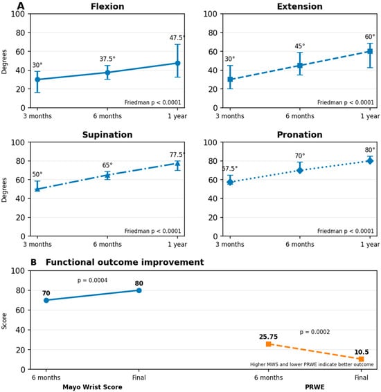
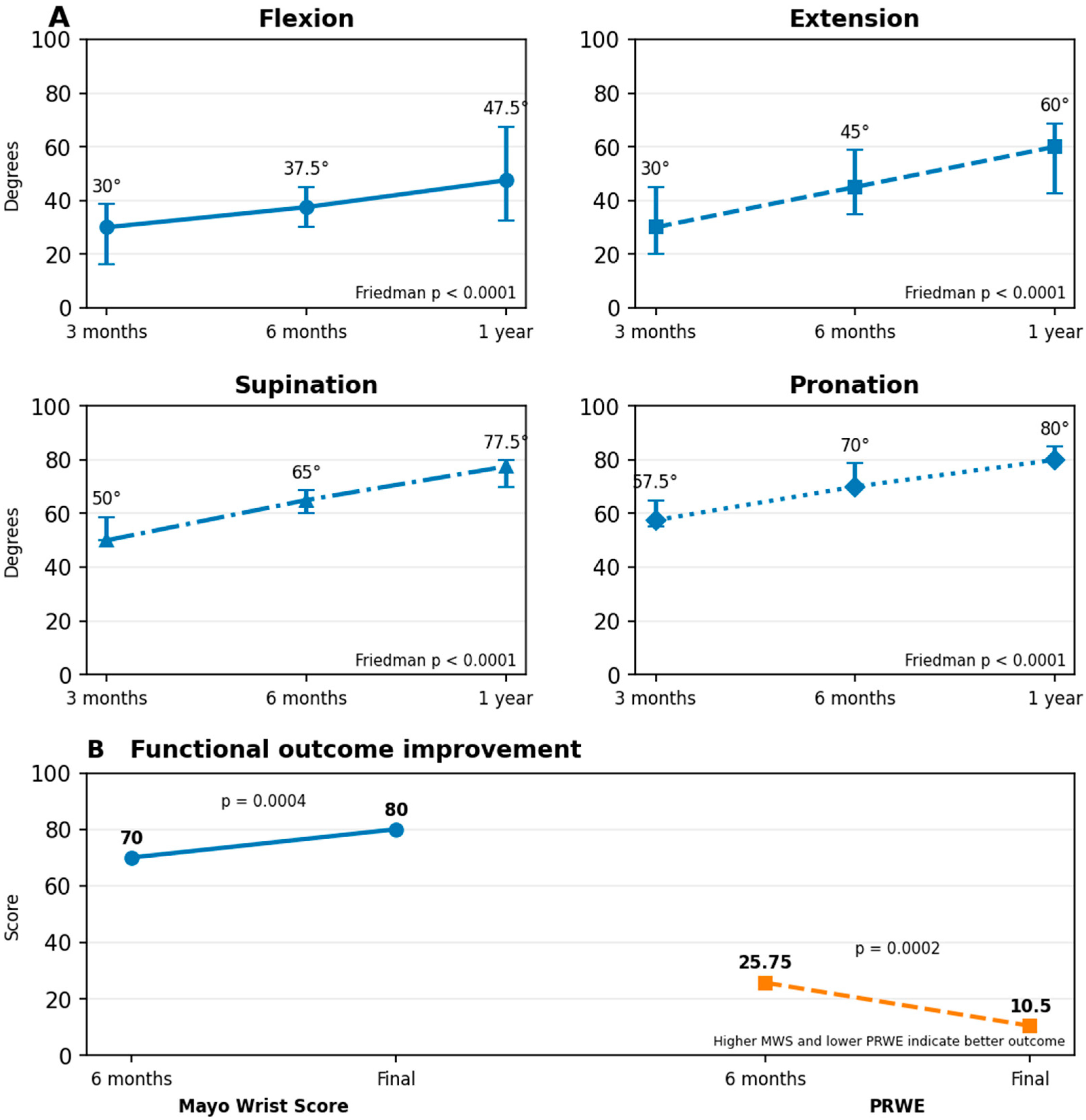
Clinical and functional outcomes showed a progressive improvement throughout the postoperative follow-up. Wrist range of motion showed a significant and progressive improvement throughout the first postoperative year (Table 3 and Figure 1).
Table 3.
Clinical and functional outcomes during follow-up.
Figure 1.
Longitudinal clinical and functional recovery following surgical treatment of perilunate injuries. (A) Median wrist flexion, extension, supination, and pronation progressively increased from 3 months to 6 months and 1 year after surgery. Significant changes over time were observed for all four range-of-motion components (Friedman test, all p < 0.0001); error bars represent the interquartile range. Holm-adjusted post hoc Wilcoxon signed-rank tests confirmed significant improvement across all pairwise follow-up comparisons (Supplementary Table S1). (B) Mayo Wrist Score significantly increased between 6 months and final follow-up (p = 0.0004), whereas Patient-Rated Wrist Evaluation (PRWE) significantly decreased (p = 0.0002), indicating reduced patient-reported pain and disability. Points represent medians, and error bars represent interquartile ranges.
Post hoc pairwise comparisons using Wilcoxon signed-rank tests with Holm adjustment confirmed significant improvements for all four ROM components between 3 and 6 months, between 6 months and 1 year, and between 3 months and 1 year (Supplementary Table S1). These findings indicate that improvement in wrist mobility continued significantly beyond the first 6 postoperative months.
At 1 year, the median flexion–extension arc was 107.5° [82.5–133.75], the median pronation–supination arc was 160.0° [150.0–160.0], and the median composite ROM was 265.0° [231.25–297.5].
Wrist-specific functional outcomes also improved significantly between 6 months and final follow-up. Median Mayo Wrist Score increased from 70.0 [65.0–73.75] to 80.0 [80.0–85.0], corresponding to a median within-patient improvement of 15 points (Wilcoxon signed-rank test, p = 0.0004). Median PRWE decreased from 25.75 [20.38–31.62] at 6 months to 10.5 [6.5–19.0] at final follow-up, with a median within-patient improvement of 13 points (p = 0.0002). Lower PRWE values indicate less wrist-related pain and disability.
Grip strength was assessed at the 1-year follow-up relative to the contralateral, uninjured wrist. Operated-side grip strength remained significantly lower than contralateral grip strength (Wilcoxon signed-rank test, p = 0.0004). The median operated-to-contralateral Jamar ratio was 0.79 [0.64–0.88], corresponding to approximately 79% of contralateral grip strength, with a median residual strength deficit of 21.1% [12.2–35.6].
3.4. Radiographic Outcomes
Radiographic assessment demonstrated significant restoration of scapholunate alignment following surgical treatment, with overall maintenance of carpal architecture during follow-up (Table 4). The median preoperative scapholunate (SL) gap was 3.15 mm [IQR 2.68–3.98] and decreased to 1.70 mm [1.32–1.90] at 1 year. The median absolute reduction in SL gap was 1.75 mm [1.20–1.98], corresponding to a median relative reduction of 49.36% [40.16–59.66]. The paired reduction in SL gap from the preoperative assessment to 1 year was statistically significant (Wilcoxon signed-rank test, p = 0.0002).
Table 4.
Radiographic outcomes during follow-up.
Carpal height was maintained during follow-up. Median carpal height ratio was 0.54 [0.50–0.57] at 6 months and 0.54 [0.49–0.57] at 1 year, with a median within-patient change of 0.00 [0.00–0.00]. No statistically significant difference was observed between the two time points (p = 0.1025), indicating overall preservation of carpal height during the observed follow-up period.
The scapholunate angle also remained largely stable at the group level, with a median value of 57.0° [48.38–62.22] at 6 months and 57.0° [49.62–62.38] at 1 year. Although the paired comparison reached statistical significance (p = 0.0273), the median within-patient change was 0.00° [0.00–0.27], indicating that the statistical difference reflected small individual changes rather than a clinically appreciable shift in the central tendency of scapholunate alignment.
Overall, these findings indicate substantial correction of the preoperative SL diastasis and maintenance of both carpal height and scapholunate alignment throughout the first postoperative year.
3.5. Association Between Treatment Timing and Functional Outcome
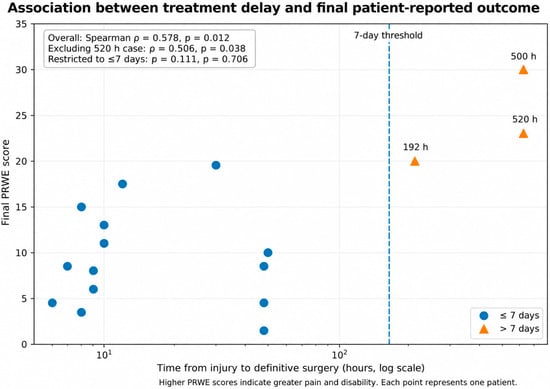
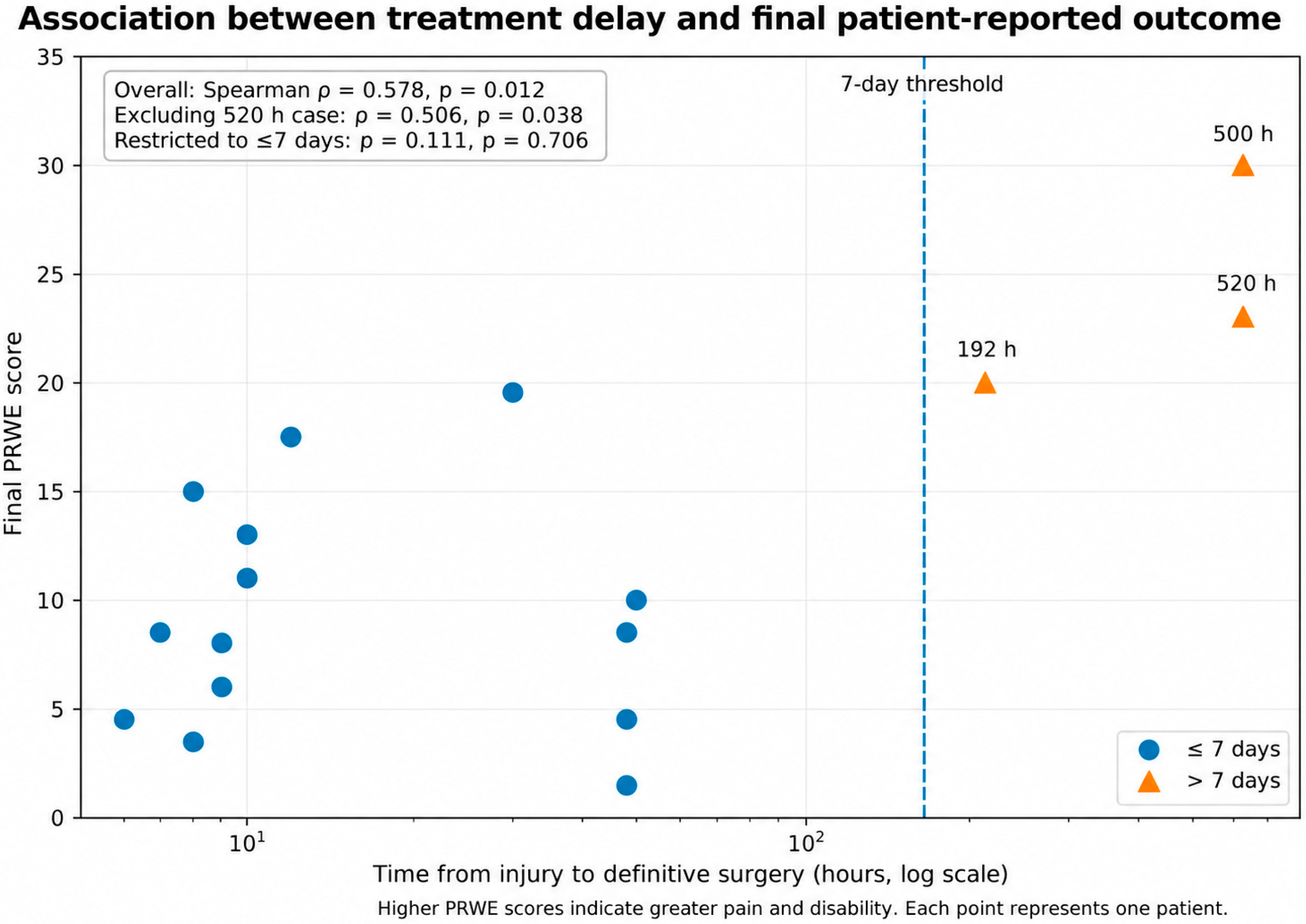
In the overall cohort, longer time from injury to definitive surgical treatment was significantly associated with worse final patient-reported wrist function. Time to surgery showed a moderate positive correlation with final PRWE score (Spearman ρ = 0.578, p = 0.012), indicating higher residual pain and disability with increasing treatment delay (Figure 2).
Figure 2.
Association between time from injury to definitive surgical treatment and final patient-reported outcome. Scatter plot showing the relationship between time to definitive surgery and final Patient-Rated Wrist Evaluation (PRWE) score. The x-axis is displayed on a logarithmic scale because of the markedly right-skewed distribution of treatment timing. In the overall cohort, longer treatment delay was significantly associated with higher final PRWE scores (Spearman ρ = 0.578, p = 0.012). The association remained significant after exclusion of the patient with the longest delay (520 h; ρ = 0.506, p = 0.038), whereas no significant association was observed when the analysis was restricted to patients treated within 7 days (≤168 h; ρ = 0.111, p = 0.706). Circles denote patients treated within 7 days, and triangles denote delayed presentations. Higher PRWE scores indicate greater residual pain and disability.
A sensitivity analysis excluding the patient with the longest treatment delay (520 h) demonstrated persistence of the association (Spearman ρ = 0.506, p = 0.038), indicating that the overall finding was not attributable to this single extreme observation. However, when the analysis was restricted to patients treated within 7 days of injury (≤168 h), no significant association between treatment timing and final PRWE was observed (ρ = 0.111, p = 0.706). These findings suggest that the association observed in the overall cohort was predominantly related to markedly delayed treatment rather than to incremental differences in timing within the early treatment period.
No significant association was observed between time to definitive surgery and final Mayo Wrist Score in the overall cohort (Spearman ρ = 0.085, p = 0.737).
3.6. Outcomes According to Injury Pattern
Clinical, functional, and radiographic outcomes were compared across the three principal injury patterns: pure perilunate dislocation (PLD), trans-scaphoid perilunate fracture-dislocation, and trans-styloid perilunate fracture-dislocation (Table 5). Given the limited number of patients within each subgroup, these comparisons were considered exploratory.
Table 5.
Exploratory comparison of treatment timing and final outcomes according to injury pattern.
Time from injury to definitive surgical treatment differed significantly among the three injury patterns (Kruskal–Wallis test, H = 7.812, p = 0.0201). Patients with pure PLD had the longest treatment interval, with a median time to surgery of 277.0 h [IQR 49.5–508.0], compared with 9.0 h [8.0–25.0] in the trans-scaphoid PLFD group and 30.0 h [11.5–84.0] in the trans-styloid PLFD group. The delay in treatment in this group was due to missed diagnosis at the referring peripheral centers. Once the patients were referred to our center, they were treated within a mean time of 24 to 48 h following clinical evaluation.
In contrast, no statistically significant differences were observed among the three injury patterns for the principal final clinical and functional outcomes. Median final Mayo Wrist Score was 80.0 [72.5–87.5] in the pure PLD group, 80.0 [80.0–85.0] in the trans-scaphoid PLFD group, and 82.5 [80.0–85.0] in the trans-styloid PLFD group (p = 0.9027). Final PRWE values were 16.5 [8.6–24.8], 8.25 [4.9–14.0], and 15.25 [11.9–18.1], respectively (p = 0.3401). Similarly, the operated-to-contralateral grip-strength ratio did not differ significantly among groups (p = 0.8419).
Range-of-motion outcomes at 1 year were also broadly similar across injury patterns. No significant between-group differences were observed for the flexion–extension arc (p = 0.8823) or total ROM (p = 0.7867). The pronation–supination arc showed a numerical difference among groups, but this did not reach statistical significance (p = 0.0773).
Radiographic outcomes were likewise not significantly different among injury patterns. Preoperative SL gap showed a borderline between-group difference (p = 0.0527), suggesting a possible difference in the magnitude of initial carpal disruption, whereas neither SL gap at 1 year nor the magnitude of SL-gap reduction differed significantly among groups (p = 0.1185 and p = 0.1275, respectively).
Overall, no specific injury pattern was associated with consistently worse final clinical, functional, or radiographic outcomes in this cohort. However, these findings should not be interpreted as evidence of equivalence between injury types, given the limited subgroup sample sizes.
3.7. Exploratory Associations with Final Outcomes
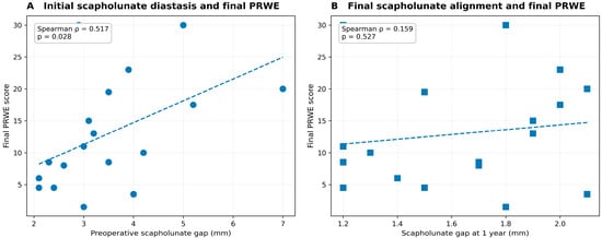
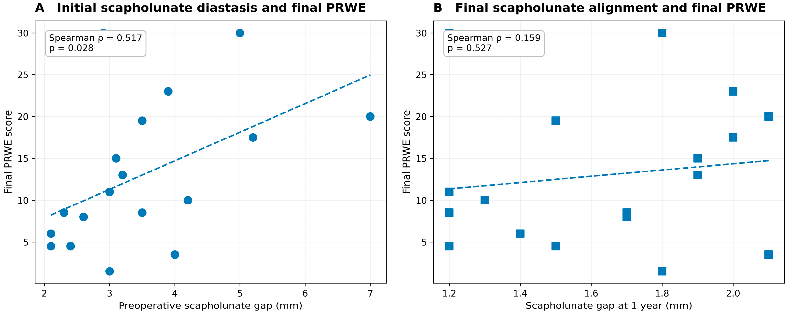
Exploratory analyses were performed to investigate potential associations between radiographic parameters and final clinical outcomes. Greater preoperative scapholunate (SL) diastasis was significantly associated with higher final PRWE scores (Spearman ρ = 0.517, p = 0.028), indicating that greater initial radiographic disruption was associated with greater residual patient-reported pain and disability at final follow-up (Figure 3A).
Figure 3.
Relationship between scapholunate alignment and final patient-reported outcome. (A) Greater preoperative scapholunate (SL) gap was significantly associated with higher final Patient-Rated Wrist Evaluation (PRWE) scores (Spearman ρ = 0.517, p = 0.028). (B) In contrast, SL gap at 1-year follow-up was not significantly associated with final PRWE (ρ = 0.159, p = 0.527). Each point represents an individual patient. Dashed lines are included for visualization of the overall trend. Higher PRWE scores indicate greater residual pain and disability.
In contrast, the SL gap measured at 1 year was not significantly associated with final PRWE (ρ = 0.159, p = 0.527; Figure 3B). The magnitude of SL-gap reduction was significantly correlated with final PRWE (ρ = 0.630, p = 0.005); however, SL-gap reduction was itself strongly correlated with the preoperative SL gap (ρ = 0.950, p < 0.0001). Accordingly, this association was interpreted as primarily reflecting the magnitude of the initial carpal disruption rather than an adverse effect of greater surgical correction.
No statistically significant association was observed between the 1-year scapholunate angle and final wrist motion, although borderline correlations were identified with total ROM (ρ = 0.466, p = 0.051) and the flexion–extension arc (ρ = 0.461, p = 0.054).
Taken together, these exploratory findings suggest that the severity of the initial scapholunate disruption may be more closely related to final patient-reported outcome than the residual radiographic SL gap after surgical reconstruction. Given the limited sample size and exploratory nature of these analyses, these findings should be considered hypothesis-generating.
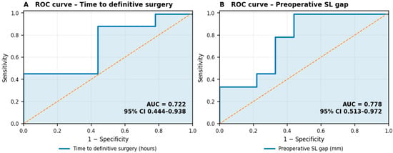
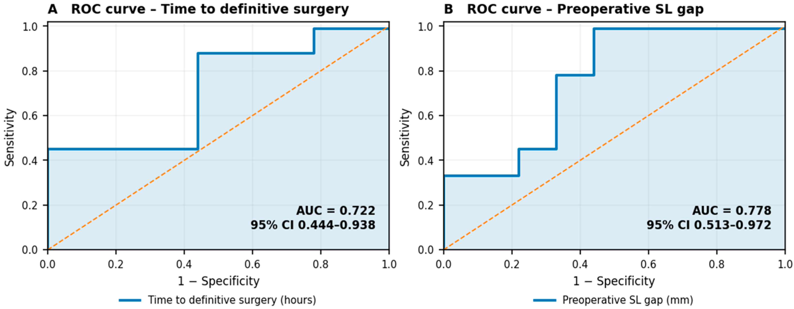
As an additional exploratory analysis, receiver operating characteristic (ROC) curves were constructed to assess the ability of treatment timing and initial radiographic severity to discriminate patients with relatively worse patient-reported outcomes. Given the absence of a prespecified clinically validated threshold for unfavorable final PRWE in this cohort, final PRWE was dichotomized at the cohort median (>10.5), yielding two equally sized groups (Figure 4). This threshold was used exclusively for exploratory discrimination analysis and should not be interpreted as a clinical definition of treatment failure.
Figure 4.
Exploratory receiver operating characteristic analysis for unfavorable final patient-reported outcome. Receiver operating characteristic (ROC) curves evaluating the discriminatory ability of time from injury to definitive surgery and preoperative scapholunate (SL) gap for a final Patient-Rated Wrist Evaluation (PRWE) score above the cohort median (>10.5). (A) Time to definitive surgery showed an area under the curve (AUC) of 0.722 (bootstrap 95% CI, 0.444–0.938). (B) Preoperative SL gap demonstrated an AUC of 0.778 (bootstrap 95% CI, 0.513–0.972). In both panels, the dashed diagonal represents discrimination no better than chance (AUC = 0.50). Because the PRWE threshold was defined by the cohort median rather than by a prespecified clinically validated cut-off, this analysis should be considered exploratory and hypothesis-generating.
Time from injury to definitive surgery demonstrated moderate discrimination for a final PRWE above the cohort median (AUC = 0.722; bootstrap 95% CI, 0.444–0.938), whereas preoperative SL gap showed somewhat greater discrimination (AUC = 0.778; bootstrap 95% CI, 0.513–0.972). SL-gap reduction yielded an AUC of 0.827 (bootstrap 95% CI, 0.597–1.000); however, because SL-gap reduction was strongly correlated with preoperative SL gap (ρ = 0.950, p < 0.0001), this finding was considered largely reflective of baseline radiographic severity rather than an independent prognostic effect of the magnitude of correction.
Because of the small sample size, wide confidence intervals, and exploratory data-driven dichotomization of PRWE, ROC-derived cut-off values were not interpreted as clinically actionable thresholds.
Median nerve decompression was performed in 11 patients. Final functional outcomes did not significantly differ according to median nerve release.
At 3 months, none of the patients reported symptoms of carpal tunnel syndrome. The median Semmes–Weinstein monofilament test value was 8 mm [IQR, 6–9] in the treated hand, compared with 7 mm in the contralateral hand. These values remained unchanged at the 1-year follow-up.
Median final PRWE was 8.25 [IQR 5.63–14.13] in patients undergoing median nerve decompression and 13.0 [8.88–19.88] in those without decompression (p = 0.306). Similarly, median final Mayo Wrist Score was 80.0 [77.5–81.25] and 85.0 [80.0–85.0], respectively (p = 0.310). Thus, no significant association between median nerve decompression and final patient-reported or clinician-rated functional outcome was identified in this cohort.
3.8. Complications, Reoperations, and Secondary Procedures
During follow-up, four patients required an unplanned secondary surgical procedure. One patient underwent removal of Kirschner wires, whereas two patients required secondary surgery because of scaphoid fixation-related complications, consisting of screw migration or mobilization, with hardware removal performed when indicated. One additional patient underwent secondary surgery for an extensor pollicis longus tendon lesion.
No clear association between the need for reoperation and final functional outcome was identified in the exploratory analyses; however, the small number of reoperations precluded meaningful inferential or prognostic assessment.
Median nerve decompression was performed concomitantly with definitive wrist surgery in selected patients according to clinical preoperative symptoms. As reported in the exploratory analysis, patients undergoing median nerve release did not demonstrate significantly different final PRWE or Mayo Wrist Scores compared with patients not requiring decompression.
Overall, no additional major treatment-related adverse events requiring further unplanned surgery were recorded during the available follow-up.
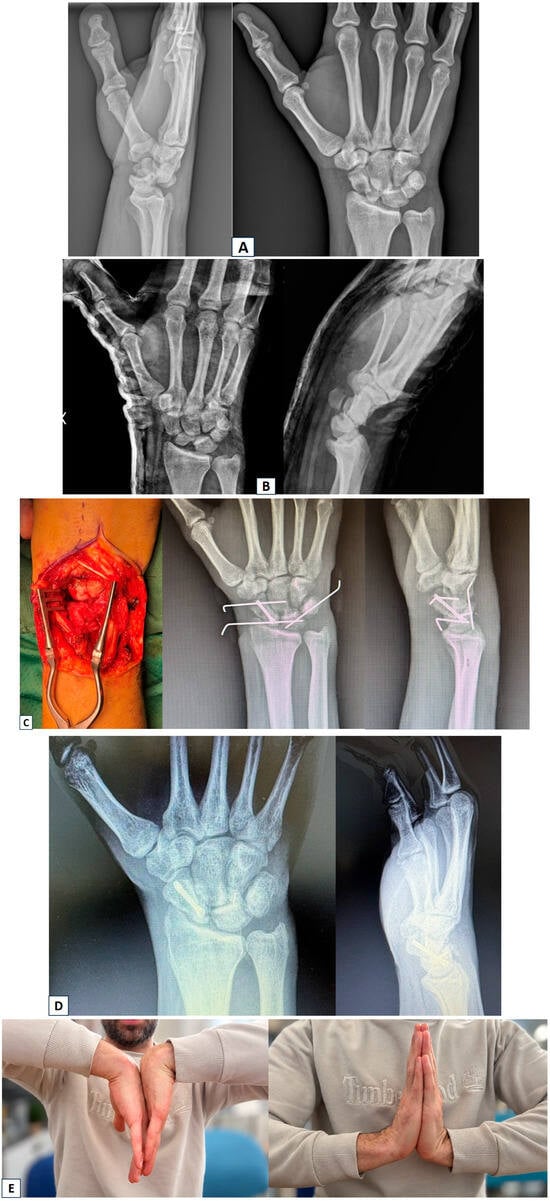
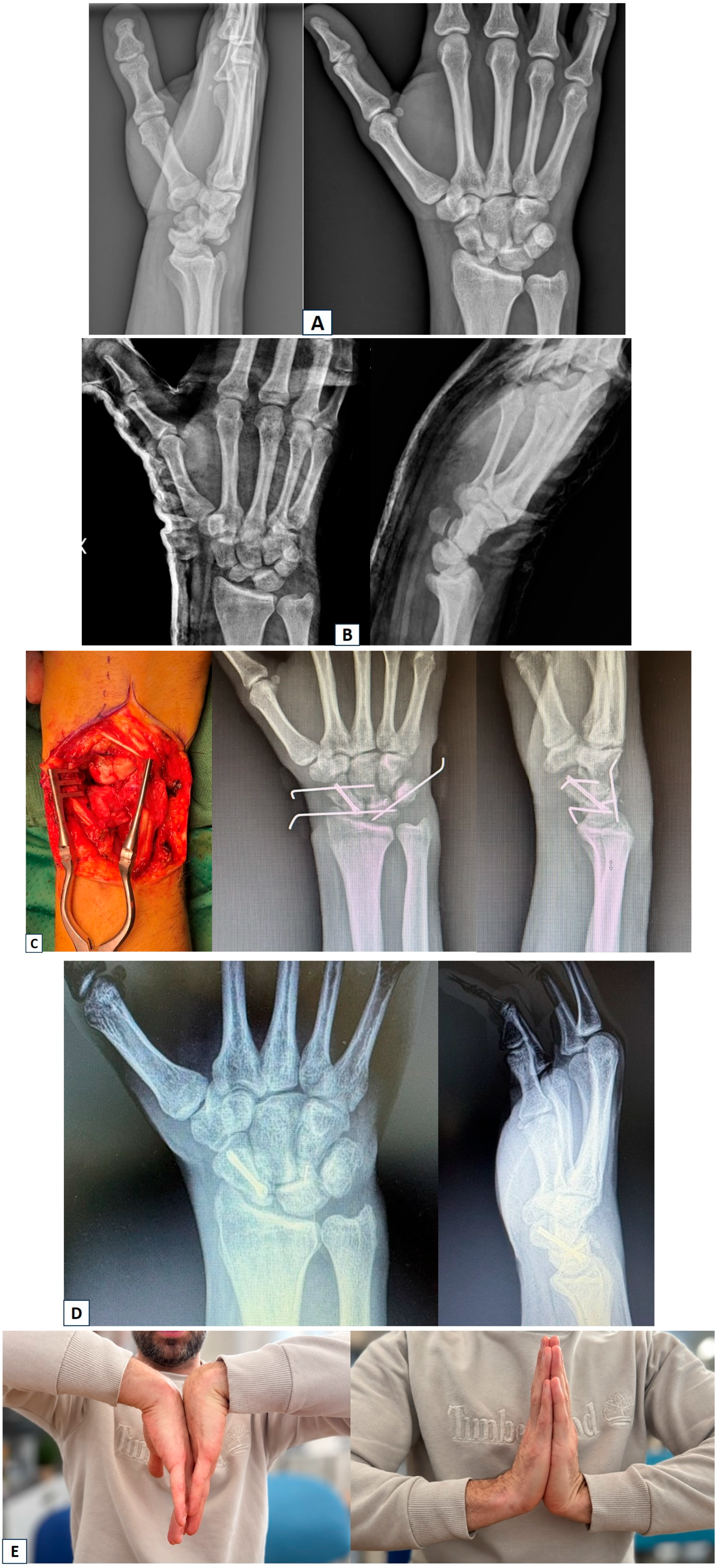
Figure 5.
(A) A 30-year-old man sustained a high-energy bicycle accident, resulting in an isolated injury to the right wrist with a trans-scaphoid perilunate fracture-dislocation. (B) Closed reduction of the dislocation and cast immobilization were performed at a peripheral hospital. Arrangements were subsequently made for transfer of the patient to our referral center. (C) Intraoperative photographs showing fracture reduction and screw fixation, scapholunate ligament repair with suture-anchor placement, and temporary stabilization with Kirschner wires. (D) Radiographic images obtained 1 year after the injury. (E) Clinical photographs obtained 1 year after the injury, showing excellent functional recovery of right wrist flexion and extension.
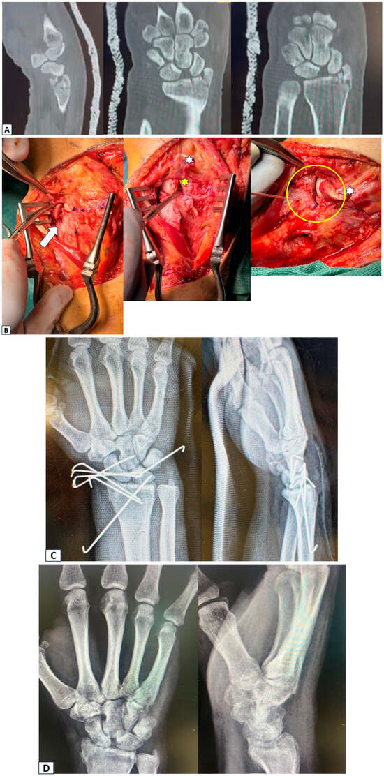
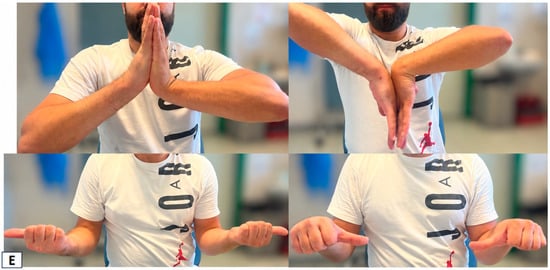
Figure 6.
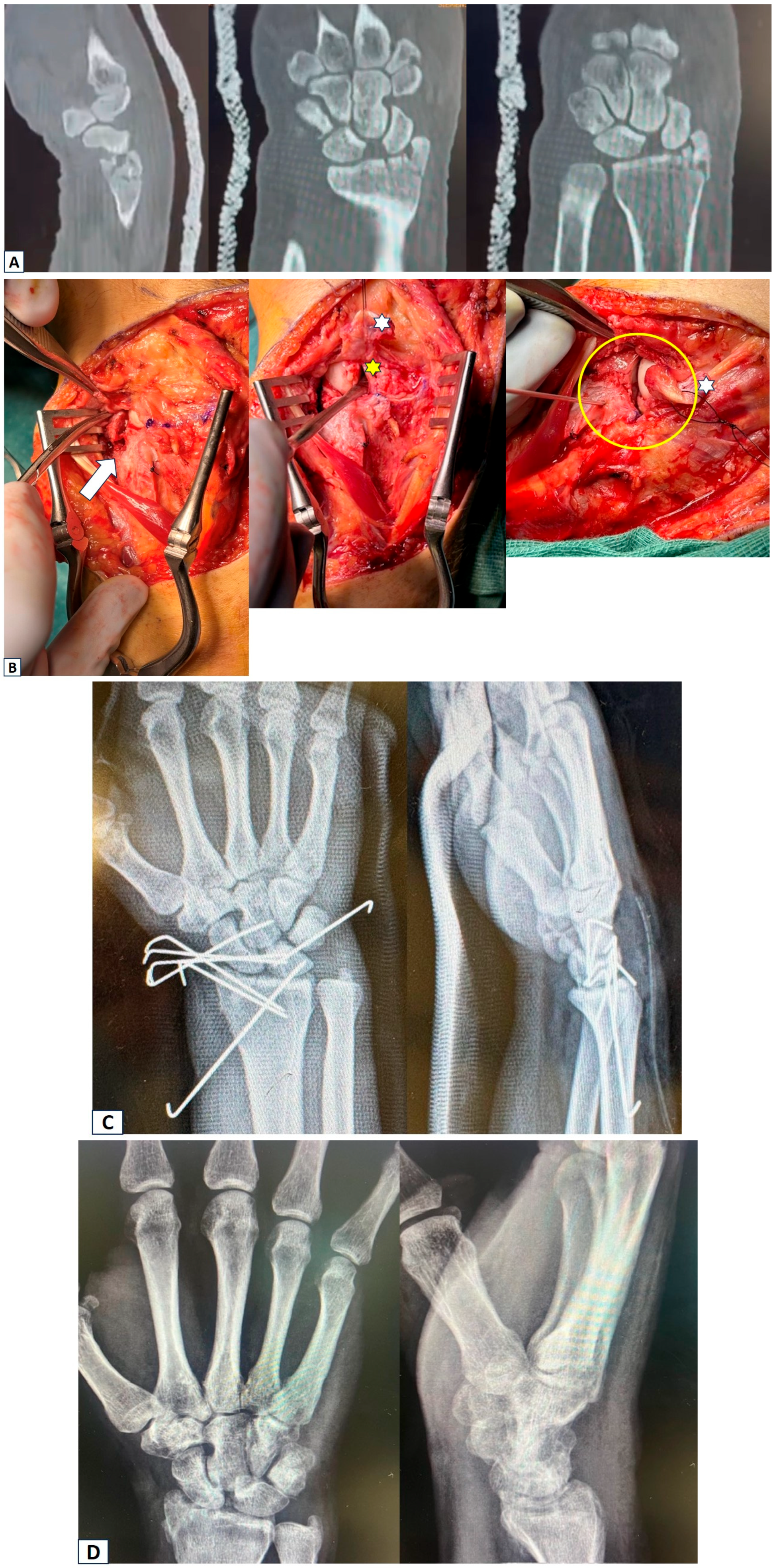
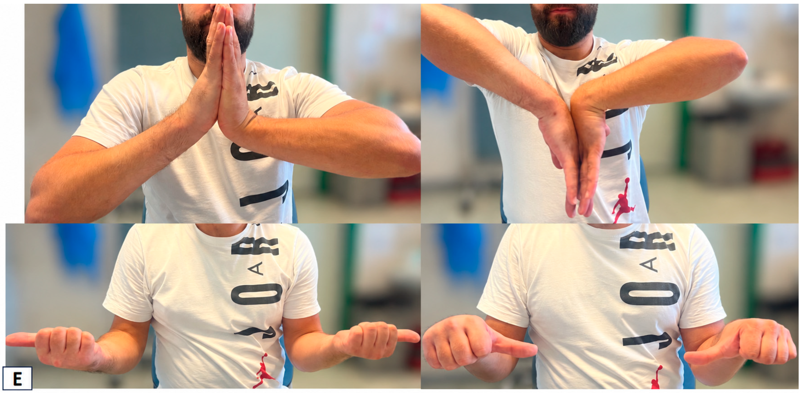
(A) A 32-year-old man sustained a fall from height (approximately 3 m, from a ladder) onto the right wrist, resulting in an isolated wrist injury with a trans-styloid perilunate fracture-dislocation. The images show a CT scan obtained after reduction and cast immobilization. Marked comminution, impaction, and bone loss of the radial styloid are evident, together with scapholunate diastasis. (B) Intraoperative images. The first image shows marked comminution and fragmentation of the radial styloid (white arrow). In the second image, the spatula indicates the scapholunate diastasis, with visualization of the scapholunate ligament (yellow star) and the dorsal intercarpal ligament (DIC) reflected dorsally (white star). The third image shows restoration of the comminuted articular surface after synthetic bone grafting and fixation with Kirschner wires (yellow circle), with the reflected DIC again visible (white star). (C) Immediate postoperative radiographs showing fracture fixation with Kirschner wires, scapholunate ligament repair with a suture anchor, and temporary stabilization of the carpal bones with Kirschner wires. (D) Radiographs obtained 1 year postoperatively, showing resorption of the radial styloid and the scaphoid fossa, with early radiographic evidence of post-traumatic osteoarthritis. (E) Clinical photographs obtained 1 year after injury, showing good recovery of right wrist flexion and extension, with the patient currently pain-free.
4. Discussion
The present prospective cohort study provides a longitudinal assessment of clinical, functional, and radiographic recovery after surgically treated perilunate injuries and, more importantly, explores factors potentially associated with residual patient-reported disability. The principal finding was that longer time from injury to definitive surgical treatment was associated with worse final PRWE, whereas the conventional distinction among pure perilunate dislocation, trans-scaphoid perilunate fracture-dislocation, and trans-styloid perilunate fracture-dislocation was not associated with significant differences in final functional outcome. However, sensitivity analyses provided an important qualification to the timing effect: the association persisted after exclusion of the patient with the longest treatment delay but disappeared when the analysis was restricted to patients treated within 7 days. Taken together, these observations suggest that the adverse influence of timing may be driven predominantly by substantial treatment delay rather than by relatively small differences in surgical timing within the early treatment window. A second relevant finding was that the magnitude of the initial scapholunate disruption was associated with final patient-reported outcome, whereas residual SL gap at 1 year was not. These results support the concept that the biological and mechanical severity of the initial injury, together with avoidance of prolonged delay before definitive reconstruction, may be more relevant to patient-perceived recovery than injury pattern alone.
The importance of treatment timing in perilunate injuries has long been recognized, although the precise temporal relationship between delay and functional outcome remains incompletely defined. Early anatomical reduction and maintenance of carpal alignment are widely regarded as fundamental principles in the management of these injuries [15,16]. The systematic review by van der Oest et al. categorized treatment as acute (<7 days), delayed (7–45 days), or chronic (>45 days) and reported generally favorable outcomes after acute treatment, whereas chronic injuries showed substantially poorer results [3]. The authors therefore emphasized early recognition, reduction, and referral to specialized centers, while also acknowledging the limited quality and heterogeneity of the available evidence. Our findings are consistent with this general principle but provide an additional perspective by analyzing treatment timing as a continuous variable rather than relying exclusively on predefined temporal categories. In the overall cohort, time to definitive surgery showed a moderate correlation with final PRWE (ρ = 0.578), and this relationship remained significant after removal of the most delayed case. Nevertheless, its disappearance among patients treated within 7 days suggests that the concept of “earlier is always better” should be interpreted cautiously within the acute window. The clinically relevant distinction may therefore not be between surgery performed several hours earlier or later during the acute phase, but rather between timely treatment and meaningfully delayed definitive reconstruction.
Nevertheless, the sample size of this subgroup is limited, and this result does not rule out the possibility of potential impact due to slight differences in timing within 7 days.
This interpretation is clinically relevant because perilunate injuries are particularly susceptible to diagnostic delay, consistent with our case series. Their rarity, complex radiographic appearance, and frequent occurrence in the setting of high-energy polytrauma contribute to missed or initially underestimated diagnoses. The classic multicenter study by Herzberg et al. highlighted this problem decades ago [1], and subsequent literature has continued to identify delayed recognition as an important challenge [3]. Prolonged carpal malalignment may theoretically expose cartilage, capsuloligamentous structures, and the median nerve to persistent abnormal mechanical conditions, while increasing the difficulty of obtaining anatomical reduction as fibrosis and tissue contracture develop. The current findings therefore reinforce the importance of early recognition and appropriate referral, but they do not establish a specific hourly threshold for surgery. Rather, they suggest that avoiding major delays is likely more important than attempting to define a narrow optimal interval within the first few days after injury.
Functional recovery in the present cohort was substantial and continued throughout the first postoperative year. All measured components of wrist motion improved significantly between 3, 6, and 12 months, while median MWS increased from 70 at 6 months to 80 at final follow-up and median PRWE decreased from 25.75 to 10.5. Importantly, significant improvement in ROM was still evident between 6 and 12 months, indicating that recovery after perilunate reconstruction should not be considered complete at the conventional 6-month assessment. At 1 year, the median flexion–extension arc reached 107.5°, while pronation–supination was relatively well preserved. Grip strength recovery was less complete, reaching a median of approximately 79% of the contralateral side. These findings are broadly consistent with contemporary systematic reviews demonstrating that surgically treated acute perilunate injuries generally permit useful functional recovery, although residual deficits in motion and strength remain common [4,9,10]. Liechti et al., in a systematic review including 553 operatively treated acute perilunate injuries, similarly emphasized that satisfactory patient-rated outcomes can coexist with persistent structural or functional abnormalities [4].
The surgical approach to acute perilunate injuries has progressively evolved from conventional open reduction and internal fixation toward arthroscopic-assisted and minimally invasive techniques in selected cases. Although open reduction remains a well-established treatment strategy, arthroscopy offers the potential advantages of direct assessment of chondral and ligamentous injury, accurate evaluation of carpal reduction, and reduced disruption of the already traumatized capsuloligamentous structures [17,18,19]. Liu et al. reported favorable early clinical and radiographic outcomes in 24 patients treated with arthroscopically assisted mini-invasive reduction and percutaneous fixation, with recovery of 86% of contralateral flexion–extension and 83% of grip strength, together with a mean PRWE of 10 [17]. Herzberg et al. similarly suggested that arthroscopy may represent a useful adjunct, either alone in selected injuries or combined with a limited open approach [18]. Importantly, Oh et al., in a comparative study of trans-scaphoid perilunate fracture-dislocations, found that both arthroscopic-assisted and open techniques achieved wrist stability, while the arthroscopic-assisted group demonstrated a greater flexion–extension arc and better functional scores, although clinically meaningful superiority was not demonstrated across all outcome measures [19]. These findings support the concept that the objective of treatment should not be defined by the surgical approach itself, but by restoration and maintenance of anatomical carpal relationships while minimizing additional soft-tissue injury. In the present cohort, treatment was individualized according to injury morphology and intraoperative findings. Given the limited sample size and non-randomized selection of surgical approach, our data do not permit a meaningful comparison between open and arthroscopic-assisted techniques. Nevertheless, the substantial functional recovery and maintenance of radiographic alignment observed during follow-up indicate that satisfactory outcomes can be achieved when stable anatomical reconstruction is obtained.
The distinction between patient-reported and clinician-based outcomes represents an additional clinically relevant finding of the present study. Treatment timing showed a moderate association with final PRWE (ρ = 0.578), whereas virtually no association was observed with the Mayo Wrist Score (ρ = 0.085). This divergence suggests that the PRWE may be more sensitive than the MWS to the residual consequences of treatment delay in patients with perilunate injuries. This interpretation is further supported by the significant association between greater preoperative SL diastasis and worse final PRWE, indicating that patient-perceived pain and disability may retain information related to the severity of the initial injury that is not fully captured by a composite clinician-based score. The two instruments assess partially different constructs: the MWS combines pain, motion, grip strength, and functional status into a composite score, whereas the PRWE directly quantifies wrist-specific pain and disability from the patient’s perspective. Accordingly, the present findings do not establish the PRWE as an independent prognostic instrument or demonstrate its statistical superiority over the MWS; rather, they suggest that PRWE may be a more sensitive outcome measure for detecting the functional consequences of prognostically relevant factors such as treatment delay and initial carpal disruption. This observation reinforces the importance of incorporating wrist-specific PROMs alongside objective and clinician-based measures when evaluating outcomes after complex carpal trauma.
Radiographic findings provide a second important component of the present study. Surgical treatment resulted in substantial correction of scapholunate diastasis, with median SL gap decreasing from 3.15 mm preoperatively to 1.70 mm at 1 year, while carpal height and scapholunate angle remained essentially stable during follow-up. This indicates that the reconstructive strategy was generally effective in restoring and maintaining carpal alignment during the first postoperative year. However, the relationship between radiographic parameters and patient-reported outcome was more complex. Greater preoperative SL gap was significantly associated with worse final PRWE, whereas SL gap measured at 1 year showed no such association. Furthermore, although greater SL-gap reduction correlated with worse PRWE, the magnitude of reduction was almost completely dependent on the preoperative SL gap. The apparently paradoxical association between greater correction and worse outcome should therefore not be interpreted as suggesting that anatomical correction is detrimental; rather, patients requiring greater correction had sustained greater initial carpal disruption.
This finding is conceptually consistent with previous evidence suggesting that the severity of the initial injury may exert a lasting influence on outcome even when satisfactory radiographic reconstruction is achieved. Garçon et al., in their long-term analysis of perilunate injuries, found that the magnitude of lunate displacement was associated with poorer functional outcome, supporting the prognostic relevance of initial carpal disruption [11]. Our findings extend this concept by identifying preoperative SL diastasis as a potential quantitative marker associated with subsequent patient-reported disability. Although the present sample is insufficient to establish SL gap as an independent prognostic factor, this observation raises the possibility that baseline radiographic measurements may contribute to risk stratification beyond categorical injury classification.
Conversely, the absence of a relationship between final SL gap and PRWE reinforces the increasingly recognized dissociation between structural and symptomatic outcomes after perilunate injury. Long-term studies frequently demonstrate radiographic deterioration despite acceptable patient-reported function. Garçon et al. reported radiographic osteoarthritis in 79% of patients after a mean follow-up approaching 10 years, despite considerably more variable functional impairment [11]. Similarly, systematic reviews by Liechti et al. and Abola et al. have highlighted that radiographic osteoarthritis becomes increasingly frequent with longer follow-up but does not consistently correlate with pain, disability, or patient dissatisfaction [4,9]. Accordingly, restoration of carpal anatomy remains an essential surgical objective, but postoperative radiographic appearance alone should not be regarded as a surrogate for patient-perceived success.
Another relevant observation was the absence of significant differences in final outcomes among pure PLD, trans-scaphoid PLFD, and trans-styloid PLFD. Neither PRWE, MWS, grip-strength recovery, ROM, nor final radiographic alignment differed significantly according to injury pattern. These findings should not be interpreted as demonstrating equivalence because the individual subgroups were small; nevertheless, they question the assumption that the presence of an associated fracture necessarily predicts a worse outcome. Contemporary evidence similarly provides no consistent demonstration that PLFDs uniformly result in poorer outcomes than pure ligamentous PLDs. Indeed, Liechti et al. reported a higher risk of secondary reduction loss in PLDs than in PLFDs, suggesting that purely ligamentous injuries may in some circumstances be mechanically less predictable than fracture-dislocations [4]. The recent study by Rachunek-Medved et al. likewise examined PLD and PLFD as potentially distinct prognostic entities and emphasized the complexity of predicting radiographic outcome from this binary classification alone [12].
The present results therefore support a more nuanced interpretation of perilunate injury severity. The conventional lesser-arc versus greater-arc distinction remains indispensable for understanding pathoanatomy and planning fixation, but it may not adequately capture the variables determining long-term patient experience. Within the current cohort, pure PLDs actually demonstrated the longest median time to surgery, while final functional outcomes remained statistically comparable among injury patterns. Consequently, the interaction among initial displacement, ligamentous disruption, treatment delay, quality of reduction, and subsequent biological healing may be more informative than the simple presence or absence of an associated fracture. Larger multicenter studies will be required to determine the relative contribution of these factors.
Median nerve involvement represents another important consideration in perilunate trauma. Acute median neuropathy is common; Wickramasinghe et al. reported median nerve symptoms in 47% of 71 patients with PLD or PLFD and were unable to identify reliable demographic or injury-related predictors [6]. In the present cohort, median nerve decompression was performed in eleven patients (44%) according to preoperative clinical and intraoperative findings, but neither final PRWE nor MWS differed significantly between patients undergoing and not undergoing release. These data should not be interpreted as evidence against carpal tunnel decompression when clinically indicated, particularly because treatment allocation was inherently confounded by indication. Rather, they suggest that appropriately selected patients requiring decompression can achieve functional outcomes comparable with those who do not require release. This remains an area of controversy, with published studies differing regarding routine versus selective carpal tunnel release [6,7,8,11].
Complications and reoperations must also be considered when interpreting the overall effectiveness of surgical treatment. Four patients in the present series required unplanned secondary procedures, including hardware-related procedures and treatment of an extensor pollicis longus tendon lesion. Contemporary systematic reviews demonstrate that complications remain relatively common following operative treatment of perilunate injuries. Lee et al., analyzing 43 studies and 880 patients, reported post-traumatic arthritis, carpal instability, lunate avascular necrosis, complex regional pain syndrome, and scaphoid nonunion or avascular necrosis among the principal complications [10]. Liechti et al. reported an overall complication rate of 15.0% and a reoperation rate of 10.4%, whereas the meta-analysis by Abola et al. found complication rates ranging from 0% to 22.5% across published series [4,9]. The limited number of events in our cohort prevents identification of predictors of reoperation, but the observed secondary procedures underscore the technical complexity of these injuries and the need for continued surveillance after reconstruction.
The exploratory ROC analyses further support the potential prognostic relevance of treatment delay and initial radiographic severity, but they require cautious interpretation. Time to surgery and preoperative SL gap demonstrated moderate discrimination for final PRWE above the cohort median, while SL-gap reduction produced a numerically higher AUC. However, the wide bootstrap confidence intervals, limited sample size, and use of a data-derived median PRWE threshold preclude translation of these findings into clinically applicable cut-off values. In particular, the strong correlation between baseline SL gap and its subsequent reduction demonstrates that these variables cannot be considered independent predictors. The ROC analysis should therefore be viewed as hypothesis-generating and as a framework for selecting variables to evaluate prospectively in larger cohorts rather than as a basis for immediate prognostic decision-making.
Several limitations should be acknowledged. First, despite prospective data collection, the study included only 25 patients, reflecting the rarity of perilunate injuries but limiting statistical power, particularly for subgroup comparisons, multivariable modeling, and complication analysis. Moreover, all patients were male, suggesting a prevalence of such injuries in males.
Second, the single-center design and treatment by two specialized hand surgeons improve procedural consistency but may preclude external generalizability. Third, injury patterns and surgical procedures were heterogeneous, as expected for this spectrum of trauma, and treatment was individualized rather than protocolized. In addition, formal interobserver and intraobserver reliability testing of SL-gap measurements was not performed, and the reproducibility of these radiographic measurements therefore warrants confirmation in future studies using prospectively standardized repeated assessments. Fourth, the observational design precludes causal inference regarding treatment timing. Patients undergoing delayed surgery may differ from acutely treated patients in ways not completely captured by the available variables, including referral pathways, initial diagnostic accuracy, soft-tissue injury, and trauma complexity. Fifth, although the 12-month evaluation adequately characterizes early functional recovery and maintenance of carpal alignment, it is insufficient to assess the full burden of post-traumatic osteoarthritis, avascular necrosis, and late carpal instability, which become increasingly relevant with longer follow-up [4,10,11]. Sixth, the exploratory ROC analyses were not externally validated and should not be interpreted as establishing prognostic thresholds.
Finally, the final follow-up (12 months) is too short to draw conclusions about post-traumatic arthritis and ischemic necrosis.
Nevertheless, the study also has several strengths. Consecutive enrollment, prospective follow-up, standardized clinical and radiographic assessments, use of both patient-reported and clinician-based functional measures, and longitudinal evaluation of ROM provide a detailed description of recovery during the first postoperative year. Furthermore, analyzing treatment timing as a continuous variable and performing sensitivity analyses allowed a more granular examination of timing than the conventional acute/delayed/chronic categorization used in much of the existing literature. The combined evaluation of timing, injury pattern, baseline radiographic disruption, postoperative alignment, and PROMs provides a clinically integrated perspective on factors potentially influencing recovery after these uncommon but severe wrist injuries. The differential association of treatment timing with PRWE but not MWS further highlights the value of patient-reported assessment in identifying residual disability that may not be fully reflected by conventional composite clinical scores.
Overall, our findings suggest that prognosis following perilunate injury cannot be adequately explained by injury pattern alone. Patients demonstrated substantial functional recovery during the first postoperative year, and anatomical carpal alignment was generally maintained; however, greater initial scapholunate disruption and markedly delayed definitive treatment were associated with greater residual patient-reported disability. Conversely, relatively small differences in timing among patients treated within the first week did not appear to influence final PRWE. These findings support prompt diagnosis, early reduction and referral, and timely definitive reconstruction, while suggesting that future prognostic models should incorporate quantitative measures of initial injury severity rather than relying exclusively on categorical PLD/PLFD classification.
5. Conclusions
PLD and PLFD remain complex injuries with heterogeneous anatomical patterns and substantial potential for persistent functional impairment. In the present prospective cohort, surgical treatment was associated with progressive improvement in wrist motion and functional scores during the first postoperative year, together with substantial restoration and maintenance of carpal alignment. However, grip strength remained incompletely recovered relative to the contralateral side.
The main finding of this study is that final patient-reported outcome appeared to be influenced more by marked treatment delay and the severity of the initial scapholunate disruption than by injury pattern alone. Longer time to definitive surgery was associated with worse final PRWE in the overall cohort, although this relationship was no longer evident when the analysis was restricted to patients treated within 7 days, suggesting that substantial delay rather than minor differences within the early treatment window may be the clinically relevant factor. Similarly, greater preoperative SL diastasis was associated with worse final PRWE, whereas residual SL gap at 1 year was not.
Conversely, no specific injury pattern was consistently associated with inferior final clinical, functional, or radiographic outcomes. These findings support early recognition, prompt reduction, timely referral to a specialized hand surgery unit, and definitive reconstruction before substantial delay occurs, while suggesting that quantitative measures of initial injury severity may provide additional prognostic information beyond the conventional PLD/PLFD classification.
Because of the limited sample size and the exploratory nature of the prognostic analyses, these results should be regarded as hypothesis-generating. Larger multicenter prospective studies with longer follow-up are required to validate the observed associations, define clinically meaningful thresholds, and clarify the long-term relationship between initial injury severity, treatment timing, radiographic evolution, and patient-reported function.
Supplementary Materials
The following supporting information can be downloaded at: https://www.mdpi.com/article/10.3390/surgeries7040115/s1, Table S1: Post hoc pairwise comparisons of wrist range of motion during postoperative follow-up.
Author Contributions
Conceptualization, L.S.; methodology, L.S. and F.D.F.; validation, F.D.F., A.P.G. and M.R.; formal analysis, F.D.F. and L.S.; investigation, L.S.; resources, M.R.; data curation, A.M., G.F., P.G. and O.M.; writing—original draft preparation, L.S. and F.D.F.; writing—review and editing, F.D.F.; supervision, M.R.; project administration, M.R. and F.D.F. All authors have read and agreed to the published version of the manuscript.
Funding
This research received no external funding.
Institutional Review Board Statement
The study was conducted in accordance with the Declaration of Helsinki and approved by the Institutional Review Board (or Ethics Committee) of AOU delle Marche (protocol code 126/2022 and date of approval: 12 April 2022).
Informed Consent Statement
Informed consent was obtained from all subjects involved in the study.
Data Availability Statement
The data presented in this study are available on request from the corresponding author due to ethical and privacy restrictions.
Acknowledgments
During the preparation of the Graphical Abstract associated with this manuscript, the authors used OpenAI ChatGPT (GPT-5.6 Sol) for the purposes of graphical design and visual composition. The authors have reviewed and edited the output and take full responsibility for the content of this publication.
Conflicts of Interest
The authors declare no conflicts of interest.
Abbreviations
The following abbreviations are used in this manuscript:
| AUC | Area under the curve |
| BMI | Body mass index |
| CI | Confidence interval |
| IQR | Interquartile range |
| LT | Lunotriquetral |
| MWS | Mayo Wrist Score |
| PLD | Perilunate dislocation |
| PLFD | Perilunate fracture-dislocation |
| PRWE | Patient-Rated Wrist Evaluation |
| PROM | Patient-reported outcome measure |
| ROC | Receiver operating characteristic |
| ROM | Range of motion |
| SD | Standard deviation |
| SL | Scapholunate |
References
- Herzberg, G.; Comtet, J.J.; Linscheid, R.L.; Amadio, P.C.; Cooney, W.P.; Stalder, J. Perilunate dislocations and fracture-dislocations: A multicenter study. J. Hand Surg. Am. 1993, 18, 768–779. [Google Scholar] [CrossRef] [Scilit] [PubMed]
- Mayfield, J.K.; Johnson, R.P.; Kilcoyne, R.K. Carpal dislocations: Pathomechanics and progressive perilunar instability. J. Hand Surg. Am. 1980, 5, 226–241. [Google Scholar] [CrossRef] [Scilit] [PubMed]
- van der Oest, M.J.W.; Duraku, L.S.; Artan, M.; Hundepool, C.A.; Power, D.M.; Rajaratnam, V.; Zuidam, J.M. Perilunate Injury Timing and Treatment Options: A Systematic Review. J. Wrist Surg. 2021, 11, 164–176. [Google Scholar] [CrossRef] [Scilit] [PubMed]
- Liechti, R.; Merky, D.N.; Grobbelaar, A.O.; van de Wall, B.J.M.; Vögelin, E.; Hirsiger, S. Outcomes of acute perilunate injuries-a systematic review. Eur. J. Trauma Emerg. Surg. 2023, 49, 2071–2084. [Google Scholar] [CrossRef] [Scilit] [PubMed]
- Chen, W.; Zhang, Y.; Pan, J. Transstyloid perilunate fracture-dislocations of the carpus, a review of two cases. J. Hand Microsurg. 2013, 5, 30–35. [Google Scholar] [CrossRef] [Scilit] [PubMed]
- Wickramasinghe, N.R.; Duckworth, A.D.; Clement, N.D.; Hageman, M.G.; McQueen, M.M.; Ring, D. Acute Median Neuropathy and Carpal Tunnel Release in Perilunate Injuries Can We Predict Who Gets a Median Neuropathy? J. Hand Microsurg. 2015, 7, 237–240. [Google Scholar] [CrossRef] [Scilit] [PubMed]
- Carroll, T.J.; Botros, M.; Lander, R.; Moody, S.; Reitenbach, M.L.; Wilbur, D. Predicting Acute Median Neuropathy in Perilunate Injuries. J. Hand Surg. Glob. Online 2023, 6, 58–61. [Google Scholar] [CrossRef] [Scilit] [PubMed]
- Dvorsky, J.L.; Green, A.; Fowler, J. A Review of Perilunate Dislocations and Concomitant Acute Carpal Tunnel Syndrome: When Should the Carpal Tunnel be Released? J. Hand Surg. Glob. Online 2025, 7, 100797. [Google Scholar] [CrossRef] [Scilit] [PubMed]
- Abola, M.V.; Gerber, B.A.; Rocks, M.C.; Chen, J.S.; Hacquebord, J.H.; Azad, A. A Comparison of Outcomes in Acute Perilunate Injuries: Systematic Review and Meta-Analysis of Treatment Approaches. Hand 2025, 20, 725–732. [Google Scholar] [CrossRef] [Scilit] [PubMed]
- Lee, C.H.; Lee, B.G.; Kim, J.H.; Yoon, H.S.; Han, K.J.; Choi, W.S. Complications and outcomes of operative treatment for acute perilunate injuries: A systematic review. J. Hand Surg. (Eur. Vol.) 2023, 48, 625–629. [Google Scholar] [CrossRef] [Scilit] [PubMed]
- Garçon, C.; Degeorge, B.; Coulet, B.; Lazerges, C.; Chammas, M. Perilunate dislocation and fracture dislocation of the wrist: Outcomes and long-term prognostic factors. Orthop. Traumatol. Surg. Res. 2022, 108, 103332. [Google Scholar] [CrossRef] [Scilit] [PubMed]
- Rachunek-Medved, K.; Medved, F.; Besz, W.; Illg, C.; Lauer, H.; Krauß, S.; Daigeler, A.; Thiel, J.T. Do perilunate dislocations and fracture-dislocations result in different radiological outcomes following wrist alignment reconstruction? A single-center retrospective study including 51 patients with perilunate injuries. Arch. Orthop. Trauma Surg. 2025, 145, 106. [Google Scholar] [CrossRef] [Scilit] [PubMed]
- Andersson, J.K.; Garcia-Elias, M. Dorsal scapholunate ligament injury: A classification of clinical forms. J. Hand Surg. (Eur. Vol.) 2013, 38, 165–169. [Google Scholar] [CrossRef] [Scilit] [PubMed]
- Viegas, S.F.; Patterson, R.M.; Peterson, P.D.; Pogue, D.J.; Jenkins, D.K.; Sweo, T.D.; Hokanson, J.A. Ulnar-sided perilunate instability: An anatomic and biomechanic study. J. Hand Surg. Am. 1990, 15, 268–278. [Google Scholar] [CrossRef] [Scilit] [PubMed]
- Herzberg, G. The treatment of perilunate injuries. J. Wrist Surg. 2015, 4, 75. [Google Scholar] [CrossRef] [Scilit] [PubMed] [PubMed Central]
- Liu, B.; Chen, S.L.; Zhu, J.; Tian, G.L. Arthroscopic Management of Perilunate Injuries. Hand Clin. 2017, 33, 709–715. [Google Scholar] [CrossRef] [Scilit] [PubMed]
- Liu, B.; Chen, S.L.; Zhu, J.; Wang, Z.X.; Shen, J. Arthroscopically assisted mini-invasive management of perilunate dislocations. J. Wrist Surg. 2015, 4, 93–100. [Google Scholar] [CrossRef] [Scilit] [PubMed]
- Herzberg, G.; Burnier, M.; Marc, A.; Merlini, L.; Izem, Y. The role of arthroscopy for treatment of perilunate injuries. J. Wrist Surg. 2015, 4, 101–109. [Google Scholar] [CrossRef] [Scilit] [PubMed]
- Oh, W.T.; Choi, Y.R.; Kang, H.J.; Koh, I.H.; Lim, K.H. Comparative Outcome Analysis of Arthroscopic-Assisted Versus Open Reduction and Fixation of Trans-scaphoid Perilunate Fracture Dislocations. Arthroscopy 2017, 33, 92–100. [Google Scholar] [CrossRef] [Scilit] [PubMed]

Disclaimer/Publisher’s Note: The statements, opinions and data contained in all publications are solely those of the individual author(s) and contributor(s) and not of MDPI and/or the editor(s). MDPI and/or the editor(s) disclaim responsibility for any injury to people or property resulting from any ideas, methods, instructions or products referred to in the content. |
© 2026 by the authors. Licensee MDPI, Basel, Switzerland. This article is an open access article distributed under the terms and conditions of the Creative Commons Attribution (CC BY) license.