


Abstract
Background/Objectives: The escalating incidence of cesarean deliveries has driven an increase in cesarean scar pregnancy (CSP). Given the risks of hemorrhage and uterine rupture, surgical intervention is recommended; however, the optimal modality remains unclear. This systematic review and network meta-analysis (NMA) evaluated the efficacy and safety of surgical and combined techniques for CSP management. Methods: Following PRISMA guidelines and PROSPERO registration, electronic databases were searched from January 2010 onwards. Eligible studies included randomized controlled trials and cohort studies (≥5 patients) evaluating primary uterus-preserving surgical interventions for CSP. A frequentist NMA calculated pooled odds ratios (OR) and mean differences (MD), with treatments ranked via surface under the cumulative ranking (SUCRA) curves. Results: Sixty-four studies (8970 women; 63 non-randomized, one randomized) were included. Combined hysteroscopic-laparoscopic surgery (COMB_HL) ranked highest for initial treatment success (SUCRA = 93.5%; OR vs. dilation and curettage [DC] = 6.05, 95% CI: 2.50–14.65), followed by high-intensity focused ultrasound plus curettage (HIFU_C) (SUCRA = 79.1%), laparoscopic resection (70.6%), and ultrasound-guided sclerotherapy plus curettage (64.5%). Methotrexate plus curettage was not statistically distinguishable from DC (OR = 0.71, 95% CI: 0.35–1.46). Blood loss (MD = −39.92 mL) and hospital stay (MD = −1.00 day) favoured advanced modalities, but heterogeneity was very high (I2 = 95.0% and 96.7%) and both prediction intervals crossed the null. HIFU_C reduced overall complications versus DC (OR = 0.23, 95% CI: 0.07–0.72). Reproductive outcomes were too sparsely reported for synthesis. Evidence certainty was low for treatment success and very low for secondary safety endpoints. Conclusions: The available observational evidence suggests that visually guided and combined modalities are associated with a higher probability of success than blind DC. Rankings are hypothesis-generating rather than proof of superiority; treatment should be individualized to CSP type, residual myometrial thickness, gestational age, fertility wishes and local expertise.
1. Introduction
Cesarean scar pregnancy (CSP) is a rare but recognized iatrogenic form of ectopic pregnancy, defined by the implantation of a gestational sac within the myometrial defect, or niche, of a previous cesarean section scar [1,2]. Driven by the escalating global rates of cesarean deliveries and advancements in transvaginal ultrasonography, the incidence of CSP has surged in recent decades, occurring in approximately 1 in 1800 to 1 in 2216 of all pregnancies, and representing over 6% of ectopic pregnancies in women with a prior hysterotomy [3,4]. The scarred myometrium exhibits myofiber disarray, poor vascularization, and defective decidualization, creating a hypoxic environment that facilitates abnormally deep trophoblastic invasion [5].
If left untreated or misdiagnosed, CSP can lead to severe maternal complications. The natural history of this condition often leads to massive hemorrhage, uterine rupture, and the need for emergency hysterectomy, which compromises reproductive capacity [6,7]. Furthermore, clinical evidence suggests a pathophysiological continuum between CSP and placenta accreta spectrum (PAS) disorders. Pregnancies that progress beyond the first trimester bear a high risk of developing severe PAS, requiring multidisciplinary surgical management at delivery [8,9,10]. Early diagnosis and active intervention during the first trimester are recommended over expectant management, which carries a disproportionately high burden of severe maternal morbidity [5,9].
Because medical management alone carries prolonged resolution times, unpredictable bleeding and high failure rates [7,11], primary surgical and combined interventional modalities have become the mainstay [12,13]. Uterus-preserving options include ultrasound-guided curettage, hysteroscopic resection, laparoscopic excision and transvaginal wedge resection, augmented by uterine artery embolization (UAE) or high-intensity focused ultrasound (HIFU) to limit hemorrhage [14,15,16,17,18,19].
Despite the proliferation of surgical options, a definitive consensus on the optimal management strategy remains elusive [11,15]. Treatment success and complication profiles vary across different modalities, confounded by the morphological severity of the CSP (e.g., Type I/endogenic versus Type II/III/exogenic implantations) and residual myometrial thickness [2,4]. Moreover, while immediate clinical efficacy and maternal safety are paramount, preserving long-term reproductive function and minimizing the risk of CSP recurrence are critical considerations in this demographic [10,20]. Previous meta-analyses have focused on isolated pairwise comparisons, such as HIFU versus UAE or simple curettage versus lesion resection [15,16,17], leaving a critical evidence gap regarding the evaluation of the full spectrum of surgical techniques.
Therefore, this systematic review and network meta-analysis aimed to evaluate the efficacy, safety, and reproductive outcomes of the different surgical techniques used for the management of cesarean scar pregnancy. By synthesizing contemporary evidence across heterogeneous treatment modalities and CSP classifications, this study aims to provide clinical evidence to optimize surgical decision-making, minimize severe maternal morbidity, and preserve future fertility in reproductive-aged women.
2. Materials and Methods
2.1. Protocol Registration and Reporting Guidelines
This systematic review and meta-analysis was conceived, conducted, and reported in accordance with the Preferred Reporting Items for Systematic Reviews and Meta-Analyses (PRISMA) 2020 updated guidelines (Supplementary Table S1) [21]. The study protocol was prospectively registered in the International Prospective Register of Systematic Reviews (PROSPERO) under registration number CRD420261382188.
2.2. Literature Search Strategy
A highly sensitive systematic literature search was performed across major electronic databases, including PubMed/MEDLINE, Embase, the Cochrane Central Register of Controlled Trials (CENTRAL), Scopus, and Web of Science. The search was restricted to articles published from 1 January 2010 to the present to capture the modern evolution of surgical approaches and contemporary CSP classification systems. The search syntax incorporated Medical Subject Headings (MeSH), Emtree terms, free-text keywords combined using Boolean operators (AND, OR, NOT). The search terms included variations of “Cesarean scar pregnancy”, “cesarean scar ectopic pregnancy”, “isthmocele pregnancy”, “surgical management”, “hysteroscopic resection”, “laparoscopic surgery”, and “minimally invasive surgery”. To mitigate publication bias, gray literature was explored using ClinicalTrials.gov and the WHO International Clinical Trials Registry Platform (WHO-ICTRP). Furthermore, a backward and forward (snowballing) citation search of the included studies and relevant review articles was conducted to identify any additional eligible literature.
2.3. Eligibility Criteria
Study inclusion was stringently defined based on the Population, Intervention, Comparator, Outcomes, and Study Design (PICOS) framework. The study population comprised adult women (≥18 years) with a confirmed diagnosis of CSP or cesarean scar ectopic pregnancy (CSEP) via transvaginal ultrasound (TVUS) or magnetic resonance imaging (MRI), regardless of viability or gestational trimester.
The intervention was primary uterus-preserving surgical intervention for CSP, including but not limited to ultrasound-guided suction curettage, hysteroscopic resection, laparoscopic excision/wedge resection, combined laparoscopic-hysteroscopic approaches, HIFU combined with suction curettage, and metroplasty. The comparator was alternative surgical techniques, adjunctive interventions (e.g., UAE + surgery vs. surgery alone), or medical management outcomes where comparative data were available.
The primary outcome was the initial treatment success rate, defined as the complete resolution of CSP without the need for additional interventions. The secondary outcomes included intraoperative hemorrhage (≥300 mL), need for emergency hysterectomy, time to serum β-hCG normalization, menstrual cycle resumption, recurrent CSP, hospital length of stay, overall complications, and subsequent live birth rates. Reproductive outcomes proved too sparsely reported for comparative synthesis.
Randomized controlled trials (RCTs), non-randomized comparative studies (prospective or retrospective cohorts), and case series comprising ≥5 participants were included in this review. Single case reports (<5 cases), studies evaluating exclusively expectant or medical management without surgical data, non-English publications, and qualitative opinion pieces were excluded.
2.4. Study Selection and Data Extraction
Titles and abstracts were independently screened by two investigators using the Covidence systematic review software (Veritas Health Innovation, Melbourne, Australia) [22]. Full-text articles of potentially eligible studies were evaluated. Inter-rater reliability (IRR) was quantified using Cohen’s kappa coefficient (κ). Disagreements were resolved by consensus or adjudication by a third reviewer. Data extraction was independently performed using a standardized, pre-piloted electronic form of data extraction.
2.5. Quality Assessment and Risk of Bias (RoB)
The methodological quality and risk of bias were independently appraised by two reviewers. For RCTs, the Cochrane Risk of Bias 2 (RoB 2) tool was utilized to evaluate domains such as the randomization process, deviations from intended interventions, missing outcome data, measurement of the outcome, and selection of the reported result [23]. For observational cohort and case–control studies, the Newcastle–Ottawa Scale (NOS) was applied, and studies scoring ≥7 stars were deemed high quality [24].
2.6. Statistical Analysis and Data Synthesis
All statistical analyses were performed using R software version 4.6.0 (R Foundation for Statistical Computing, Vienna, Austria) with the meta (v8.0-2), netmeta (v3.0-1), and dmetar (v0.1.0) packages.
2.6.1. Pairwise Meta-Analytic Model
For dichotomous outcomes (e.g., success rates and hysterectomy), effect sizes were pooled as odds ratios (OR) with 95% confidence intervals (CIs). Continuous outcomes (e.g., hospital stay, β-hCG normalization time) were pooled using mean differences (MD) or standardized mean differences (SMD) using Cohen’s d or Hedges’ g, depending on the uniformity of the measurement scales. Given the anticipated clinical and methodological heterogeneity in diverse surgical techniques, a random-effects (RE) model using the restricted maximum likelihood (REML) estimator was employed for all primary analyses [25].
2.6.2. Network Meta-Analysis Model
A frequentist network meta-analysis was conducted to compare the relative efficacy and safety of multiple surgical modalities [26]. Network plots were generated to visualize the geometry of the comparative evidence. Treatments were ranked using the Surface Under the Cumulative Ranking curve (SUCRA) and rankograms, where a SUCRA value approaching 100% indicates the highest probability of being the most effective or safest intervention [27]. Network consistency, the statistical agreement between direct and indirect evidence, was evaluated using both a global design-by-treatment interaction model and local node-splitting methods. A p-value < 0.05 indicated significant inconsistency [28]. Complete node-splitting output—direct, indirect and network estimates with 95% CIs for every closed loop—is given in Supplementary Table S2, since a non-significant global test does not exclude clinically relevant incoherence in an observational network.
Throughout this review, DC denotes dilation and curettage or suction curettage performed as the primary evacuation method, whether unguided or under ultrasonographic guidance, unless a specific guidance modality is stated. The abbreviations D&C and suction curettage are used only when reproducing the terminology of an individual primary study.
2.6.3. Node Definition, Transitivity and Interpretation of Rankings
Two reviewers independently mapped every intervention arm onto one of ten nodes using pre-specified rules based on the primary method of tissue removal and the adjuvant applied; the full mapping is given in Supplementary Table S3. Transitivity was assessed by tabulating CSP classification, gestational age at diagnosis, sac diameter, residual myometrial thickness, fetal cardiac activity, baseline β-hCG and publication year across nodes. Most of these were reported too inconsistently for node-level comparison, so transitivity could be examined only partially. Rankings (SUCRA/P-scores) were pre-specified to be interpreted alongside the precision of the contrasts, network geometry, heterogeneity and GRADE certainty, and not as stand-alone evidence of clinical superiority.
2.7. Heterogeneity Assessment and Exploration
Statistical heterogeneity was quantified using the I2 statistic, Cochran’s Q test, and τ2 (tau-squared). An I2 > 50% indicated substantial heterogeneity [29]. To explore the sources of heterogeneity, subgroup analyses (ANOVA F-test) were pre-specified based on the CSP classification systems (Type I, II, and III), gestational age at diagnosis, and fetal viability. Random-effects meta-regression (univariate and multivariate) was employed to evaluate continuous covariates, such as publication year and sample size. The robustness of the pooled estimates was tested using leave-one-out sensitivity analyses [30].
2.8. Publication Bias and Certainty of Evidence
Small-study effects and potential publication bias were visually assessed using contour-enhanced funnel plot. For outcomes comprising ≥10 studies, Egger’s continuous regression and Begg’s rank correlation tests were utilized [31]. If publication bias was detected, the non-parametric trim-and-fill method was applied to adjust the pooled estimates [32]. The overall certainty of the evidence for primary outcomes was graded using the Grading of Recommendations Assessment, Development, and Evaluation (GRADE) framework [33].
3. Results
3.1. Literature Search and Study Selection
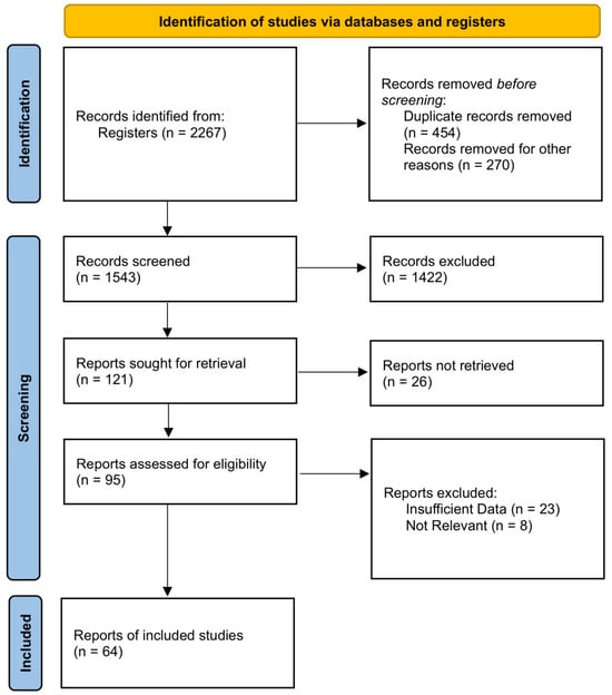
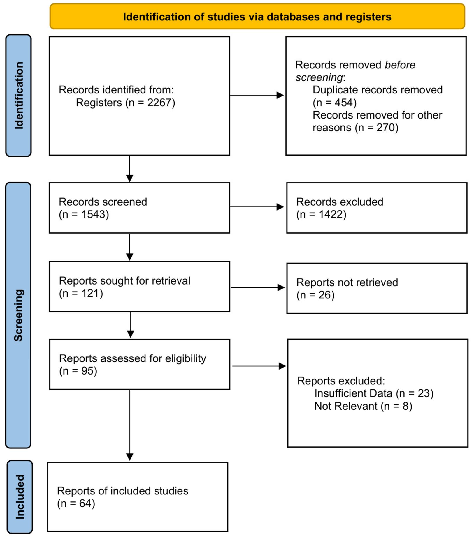
The initial systematic database search combined with clinical register screening yielded 2267 records. After the removal of 454 duplicate records and 270 records excluded before screening—non-English records (n = 46), abstracts, editorials and letters without extractable data (n = 168), and registry entries without a linked publication (n = 56)—1543 citations were evaluated based on their titles and abstracts. Of these, 1422 were excluded for failing to meet the predefined PICOS criteria. Full-text retrieval was sought for 121 reports, of which 26 could not be retrieved despite interlibrary requests and author correspondence; these were mainly non-indexed regional publications, and their exclusion is acknowledged as a potential source of selection bias. The remaining 95 full-text articles underwent an eligibility assessment. Thirty-one articles were excluded due to insufficient quantitative data (n = 23) or lack of relevance to the predefined surgical comparisons (n = 8). Sixty-four eligible studies [34,35,36,37,38,39,40,41,42,43,44,45,46,47,48,49,50,51,52,53,54,55,56,57,58,59,60,61,62,63,64,65,66,67,68,69,70,71,72,73,74,75,76,77,78,79,80,81,82,83,84,85,86,87,88,89,90,91,92,93,94,95,96,97] were included in the systematic review and network meta-analysis. The IRR for the screening process demonstrated excellent agreement, yielding a Cohen’s Kappa coefficient (κ) of 0.926 (Z = 39.72, p < 0.001, across the 1543 title/abstract records and 95 full-text reports screened in duplicate). The complete study selection process is delineated in Figure 1.
Figure 1.
PRISMA 2020 Flow Diagram.
3.2. Study Characteristics and Transitivity Assumption
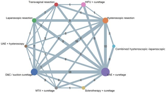
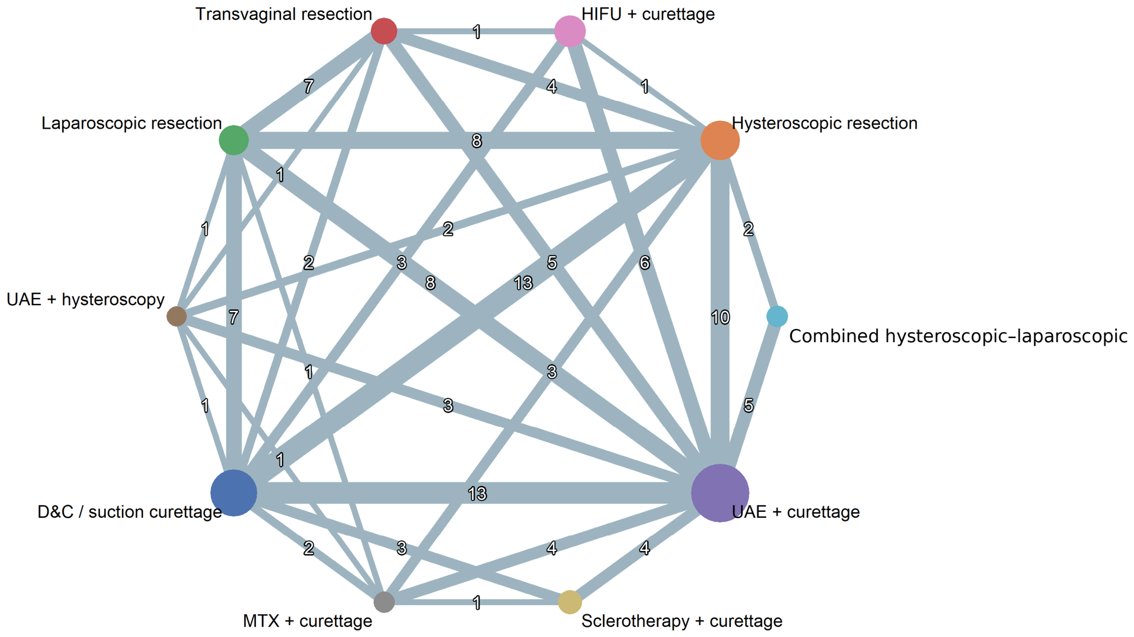
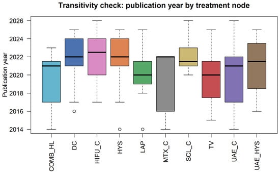
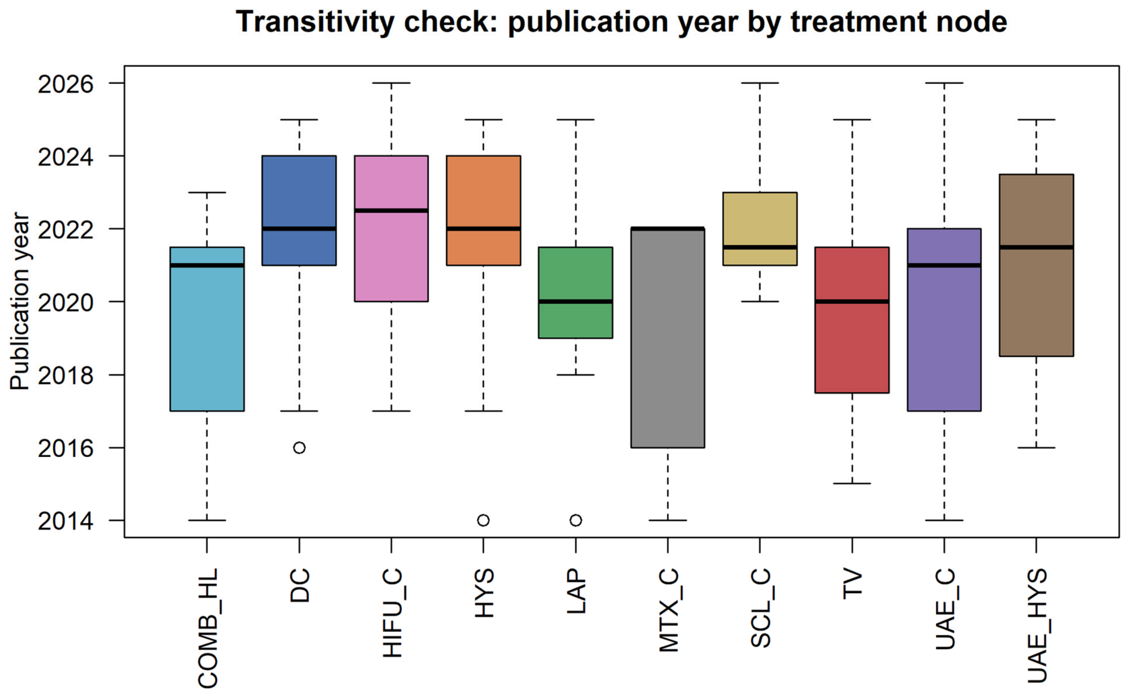
The 64 included studies, published between 2014 and 2026, comprised an aggregated cohort of 8970 adult women diagnosed with CSP (Table 1). The geographical distribution was predominantly from China, reflecting the high epidemiological prevalence of CSP in this region. The study designs consisted of one RCT [69] and 63 non-randomized comparative studies, comprising both prospective and retrospective cohorts. The studies investigated a diverse array of CSP classifications (Types I, II, III, and mixed cohorts) and compared ten distinct surgical and combined modalities: DC, hysteroscopic resection (HYS), laparoscopic resection (LAP), transvaginal resection (TV), combined hysteroscopic-laparoscopic surgery (COMB_HL), ultrasound-guided sclerotherapy plus curettage (SCL_C), high-intensity focused ultrasound plus curettage (HIFU_C), uterine artery embolization plus curettage (UAE_C), UAE plus hysteroscopy (UAE_HYS), and methotrexate plus curettage (MTX_C). Publication years were symmetrically distributed across nodes (Figure 2 and Figure 3), but publication year is a weak proxy for clinical comparability. Only 14 of 64 studies (22%) defined their cohort by CSP classification; 48 enrolled mixed cohorts, and gestational age, sac diameter, residual myometrial thickness and baseline β-hCG could not be tabulated by node at all. Where classification was available, allocation tracked lesion severity: 57% of COMB_HL studies were restricted to Type II/III disease and none to Type I, whereas no DC study was restricted to advanced disease and 13% to endogenic lesions. Transitivity could therefore be assessed only partially, and the estimates remain susceptible to confounding by indication.
Table 1.
Summary Characteristics of the 64 Included Studies.
Figure 2.
Network geometry of the ten treatment nodes for the primary outcome (initial treatment success). Each node represents one treatment modality; node size is proportional to the number of women allocated to that treatment and the thickness of each edge is proportional to the amount of direct evidence available for the corresponding comparison. The number placed on each edge is the number of studies contributing direct evidence to that comparison. Treatment abbreviations are defined in the Abbreviations list.
Figure 3.
Transitivity assessment. Boxplots illustrate the symmetric distribution of publication years across the different surgical intervention nodes. Each box spans the interquartile range with the horizontal line denoting the median, the whiskers extend to 1.5 × the interquartile range, and the open circles are individual studies falling outside that range (outliers). Treatment abbreviations are defined in the Abbreviations list.
3.3. Methodological Quality and Risk of Bias Assessment
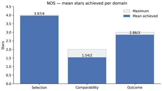
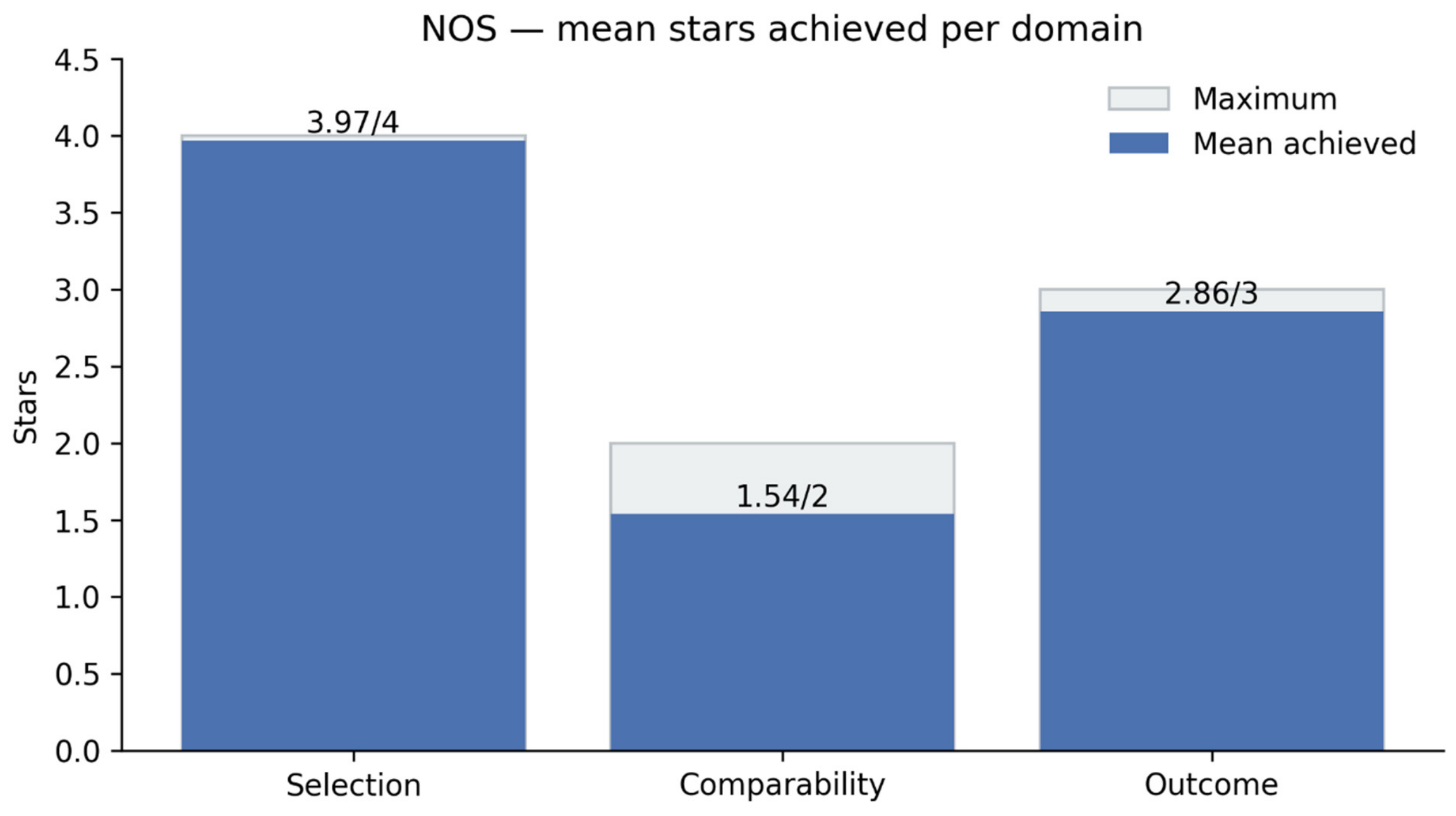
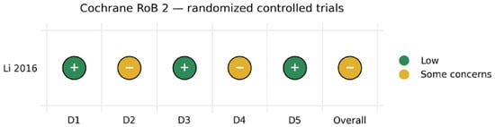
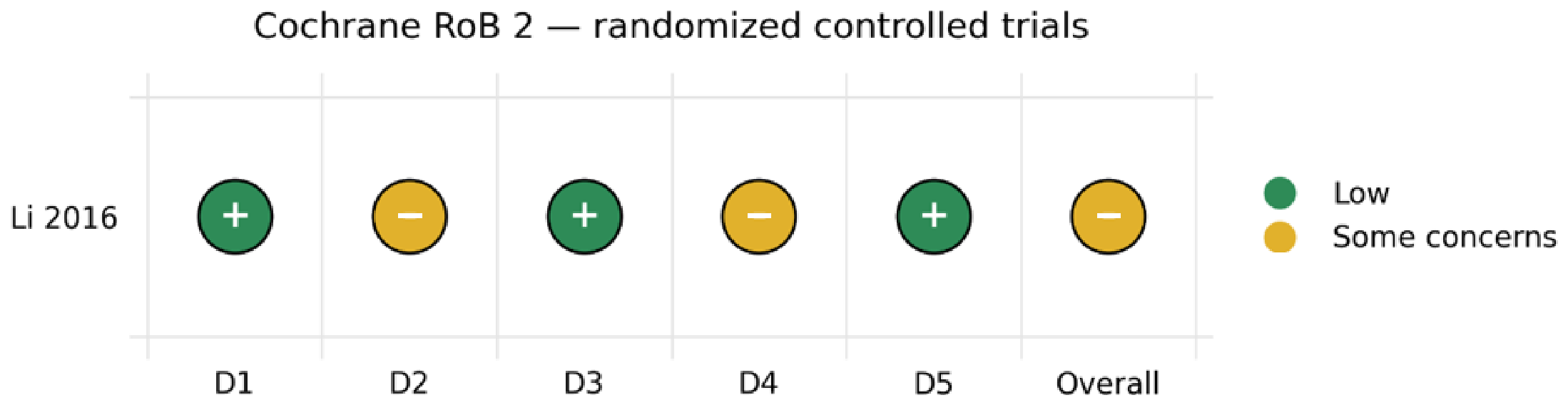
The methodological quality of the included observational cohorts was evaluated using NOS. The studies demonstrated robust quality, achieving mean scores of 3.97 out of 4 for the selection domain, 1.54 out of 2 for comparability, and 2.86 out of 3 for outcome ascertainment (Figure 4). Most studies exceeded the predefined high-quality threshold (≥7 stars) (Supplementary Figure S1). For the single RCT [69], the Cochrane RoB 2 tool indicated low risk in domains concerning the randomization process (D1), missing outcome data (D3), and measurement of the outcome (D5), with some concerns regarding deviations from intended interventions (D2) and selection of the reported result (D4), leading to an overall some concerns rating (Figure 5).
Figure 4.
Bar chart of mean Newcastle–Ottawa Scale (NOS) stars achieved per domain.
Figure 5.
Cochrane Risk of Bias 2 (RoB 2) traffic light plot and domain-level summary for the included randomized controlled trial (Li 2016 [69]). D1, randomization process; D2, deviations from intended interventions; D3, missing outcome data; D4, measurement of the outcome; D5, selection of the reported result. Green (+), low risk of bias; amber (−), some concerns.
3.4. Primary Outcome: Initial Treatment Success
3.4.1. Pairwise Meta-Analysis
Direct pairwise comparisons showed statistically significant differences among several treatments. HYS was associated with higher odds of treatment success than DC (OR = 2.33, 95% CI: 1.52 to 3.57, k = 12, p < 0.001), as were UAE_C (OR = 3.13, 95% CI: 1.96 to 5.00, k = 13), HIFU_C (OR = 3.85, 95% CI: 1.39 to 10.67, k = 3), SCL_C (OR = 3.23, 95% CI: 1.01 to 10.00, k = 2) and LAP (OR = 2.33, 95% CI: 1.28 to 4.17, k = 7). MTX_C was associated with lower odds of success than UAE_C (OR = 0.14, 95% CI: 0.04 to 0.47, k = 4). The point estimate favoured COMB_HL over UAE_C (OR = 2.17, 95% CI: 0.84 to 5.60, k = 5) and over HYS (OR = 3.33, 95% CI: 0.63 to 17.57, k = 1), but neither comparison reached statistical significance and both intervals were compatible with no difference.
3.4.2. Network Meta-Analysis (NMA)
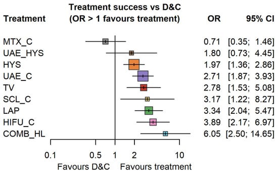
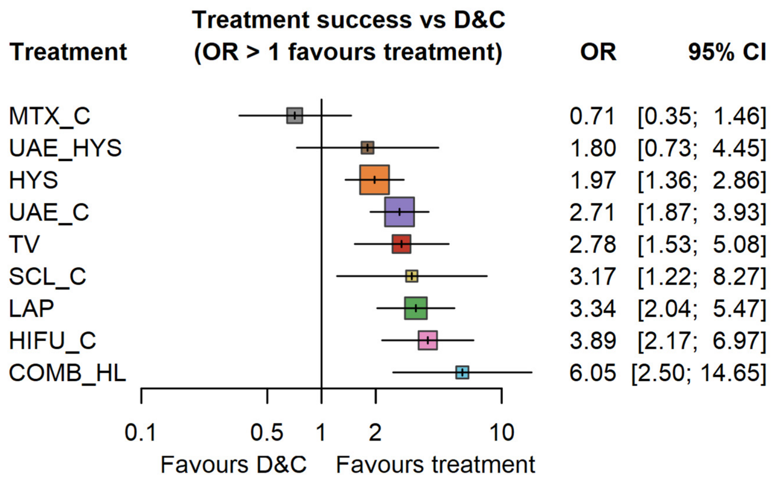
The NMA integrated direct and indirect evidence across the 10 treatment nodes. Using DC as the reference baseline, seven modalities demonstrated significantly higher odds of treatment success (Figure 6). The highest relative efficacy was observed for COMB_HL (OR = 6.05, 95% CI: 2.50 to 14.65), HIFU_C (3.89, 2.17 to 6.97), LAP (3.34, 2.04 to 5.47), SCL_C (3.17, 1.22 to 8.27), TV (2.78, 1.53 to 5.08), UAE_C (2.71, 1.87 to 3.93) and HYS (1.97, 1.36 to 2.86). Two estimates were compatible with no difference from DC: UAE_HYS (1.80, 0.73–4.45) and MTX_C (0.71, 0.35–1.46). MTX_C is therefore described throughout as showing no demonstrated advantage over DC rather than as inferior to it.
Figure 6.
Network Meta-Analysis Forest Plot. Comparative efficacy (Odds Ratios and 95% Confidence Intervals) for Initial Treatment Success across all surgical modalities relative to standalone Dilation and Curettage (DC).
3.4.3. Treatment Rankings and Network Consistency
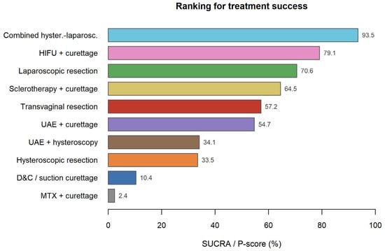
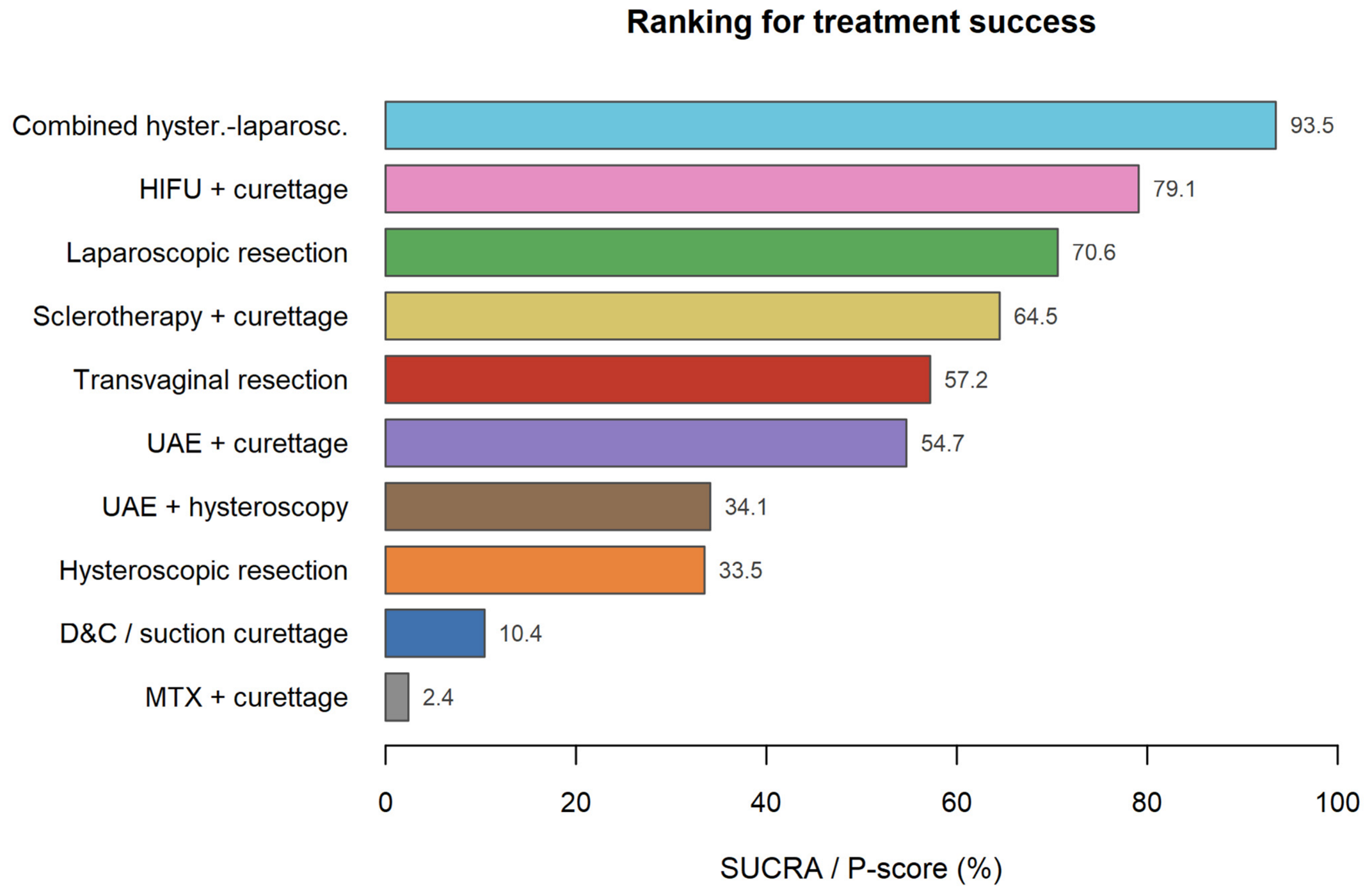
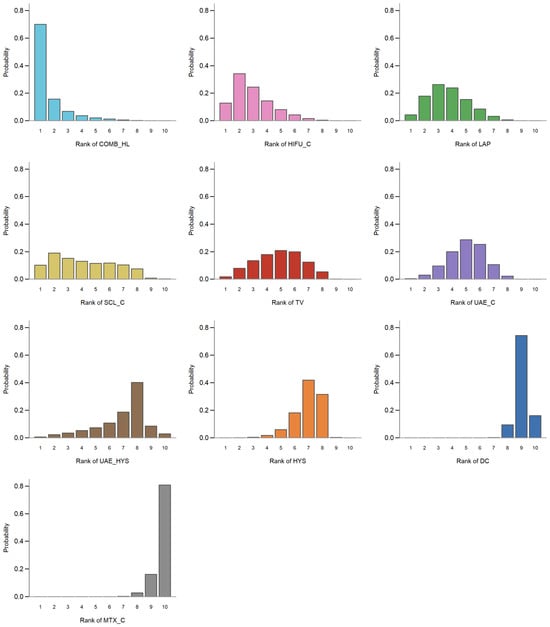
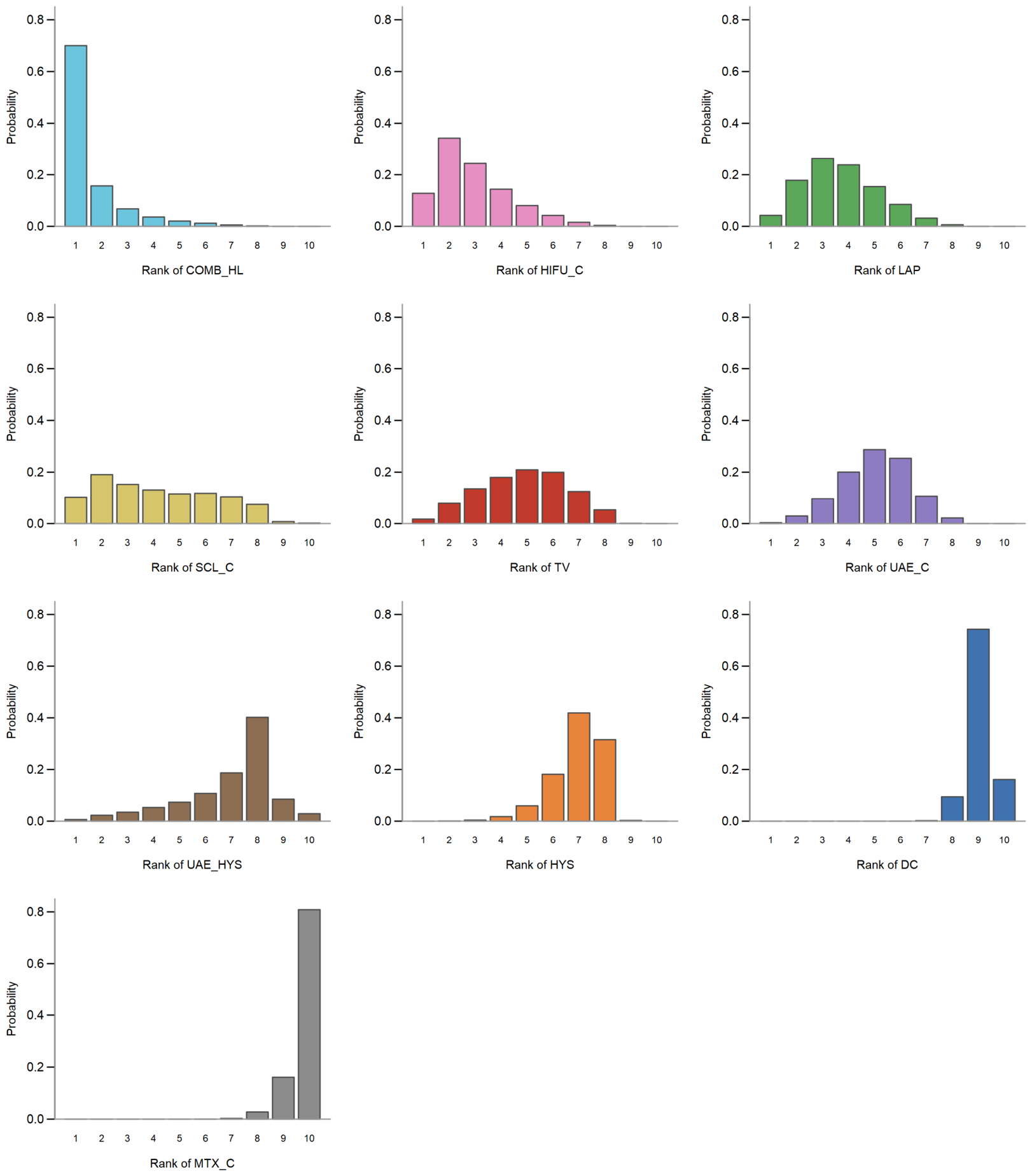
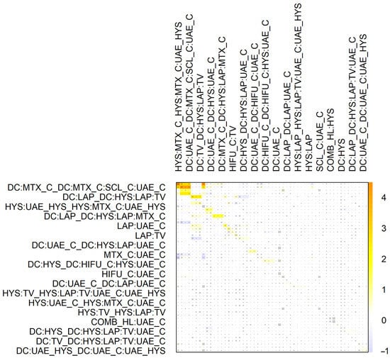
COMB_HL had the highest probability of being the most effective treatment for initial success (SUCRA = 93.5%). The hierarchical ranking was: HIFU_C (79.1%), LAP (70.6%), SCL_C (64.5%), TV (57.2%), UAE_C (54.7%), UAE_HYS (34.1%), HYS (33.5%), DC (10.4%) and MTX_C (2.4%) (Figure 7 and Figure 8). Global inconsistency testing indicated agreement between direct and indirect evidence (Q = 29.63, df = 39, p = 0.861); loop-level results are shown in the net heat plot (Figure 9). SUCRA ranks treatments without weighting the precision of the underlying contrasts; COMB_HL and SCL_C rest on a few small studies with wide intervals, so this hierarchy should be read together with the estimates in Figure 6 and the certainty ratings. Node-splitting for every closed loop is reported in Supplementary Table S2.
Figure 7.
Surface Under the Cumulative Ranking curve (SUCRA) bar plot indicating the probability hierarchy for initial treatment success.
Figure 8.
Rankograms depicting the probabilistic ranking distribution for each surgical intervention across the ten treatment nodes.
Figure 9.
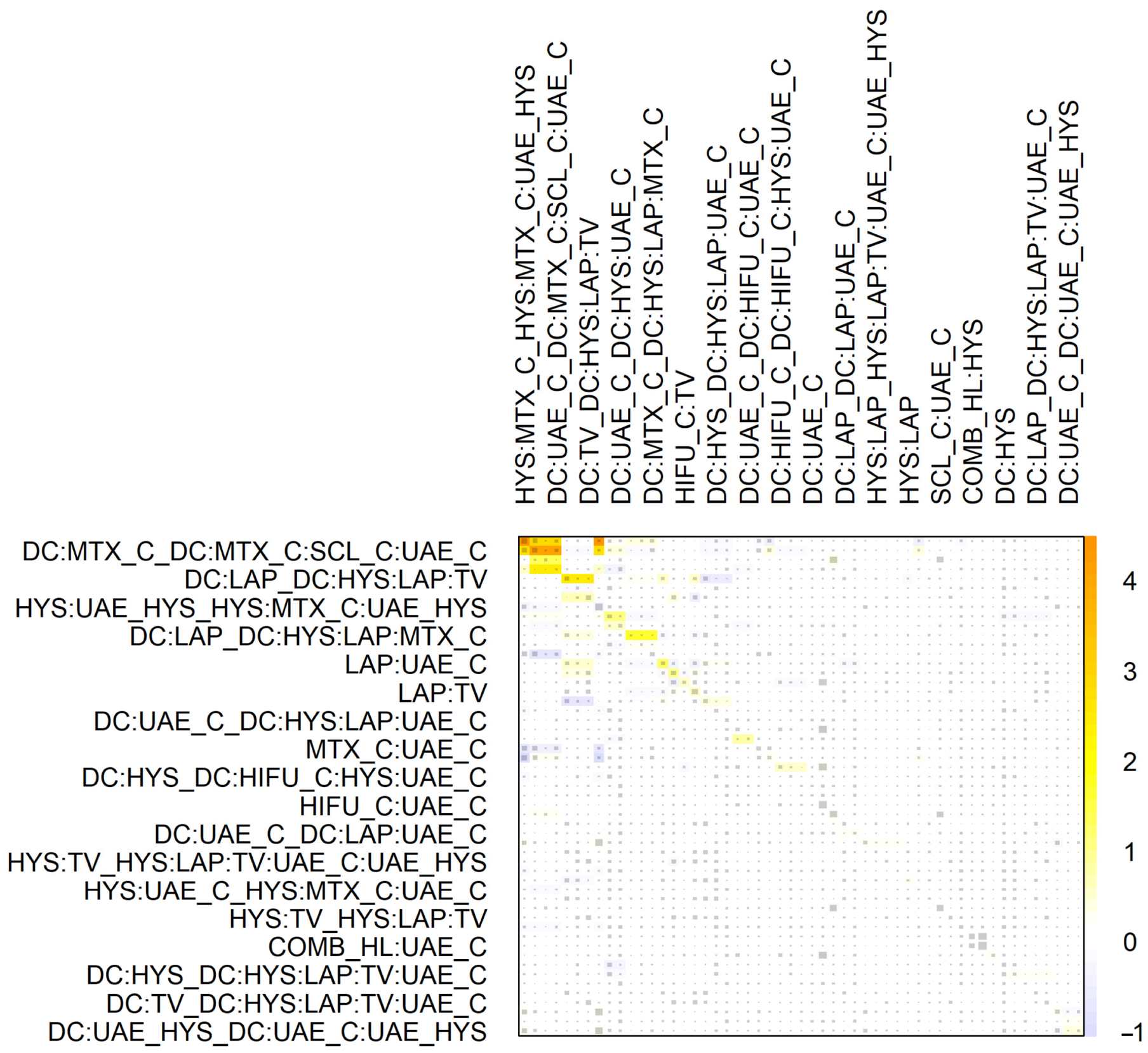
Net heat plot highlights localized inconsistencies within the network loops. Each row and each column corresponds to a treatment comparison (design). The area of every grey square is proportional to the contribution of the direct estimate of the design in the column to the network estimate of the design in the row, so that larger squares denote a greater contribution. The background colour shows the change in inconsistency when the design in the corresponding row is detached from the network, as indicated by the colour scale (−1 to 4); warmer colours denote greater inconsistency.
3.5. Secondary Outcomes: Operative Parameters and Safety
3.5.1. Continuous Metrics
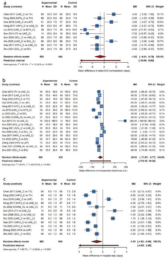
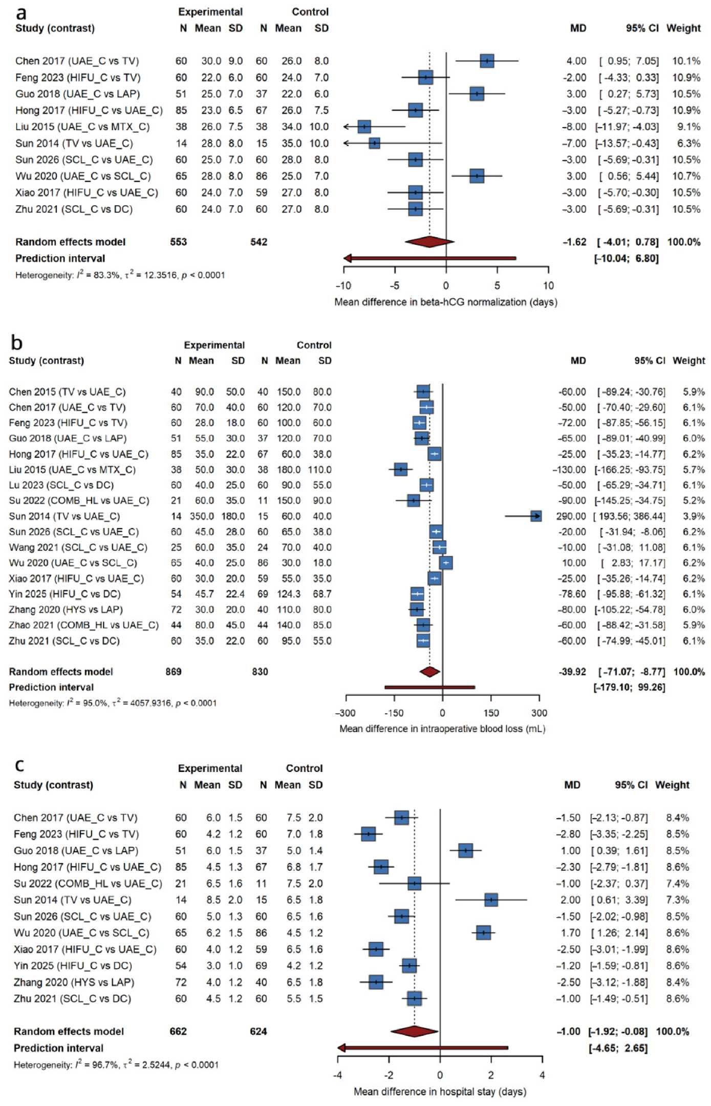
Random-effects pooling favoured combined and minimally invasive approaches, but with very high heterogeneity (Figure 10). Blood loss was lower (MD = −39.92 mL, 95% CI: −71.07 to −8.77; k = 17; I2 = 95.0%), hospital stay shorter (MD = −1.00 day, −1.92 to −0.08; k = 12; I2 = 96.7%), and β-hCG normalization non-significantly faster (MD = −1.62 days, −4.01 to 0.78; k = 10; I2 = 83.3%). All three 95% prediction intervals crossed the null (−179.10 to 99.26 mL; −4.65 to 2.65 days; −10.04 to 6.80 days), so these pooled means average highly disparate settings and do not predict the effect expected in an individual center.
Figure 10.
Pairwise meta-analyses for continuous operative parameters [34,36,42,47,49,59,64,66,68,71,72,73,75,76,78,81,88]. Forest plots detailing mean differences (MD) for (a) β-hCG normalization time in days, (b) intraoperative blood loss in mL, and (c) hospital length of stay in days; random-effects model (REML) with 95% prediction intervals. Squares are the individual study estimates, with the area of each square proportional to the study weight and the horizontal line showing the 95% confidence interval; arrows denote confidence intervals extending beyond the plotted axis. The diamond is the pooled random-effects estimate and the horizontal bar beneath it is the 95% prediction interval. The solid vertical line is the line of no effect (MD = 0) and the dotted vertical line marks the pooled estimate.
3.5.2. Complications and Hysterectomy Risk
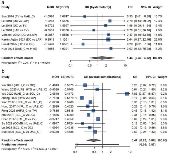
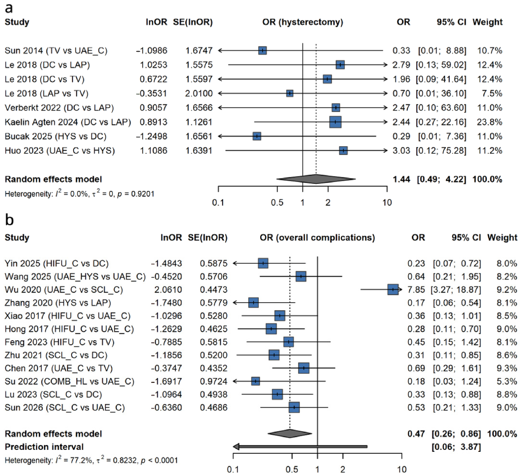
Overall complications favoured targeted, uterus-preserving modalities: HIFU_C versus DC, OR = 0.23 (0.07–0.72). The comparison of HIFU_C with UAE_C favoured HIFU_C but was not statistically significant (OR = 0.36, 0.13–1.01) and is reported as a trend. Pooled across contrasts, OR = 0.47 (0.26–0.86), k = 12, I2 = 77.2%, prediction interval 0.06–3.87. Emergency hysterectomy was rare and no contrast was significant (pooled OR = 1.44, 0.49–4.22; k = 8; I2 = 0%); the event rate was too low to support any inference about relative hysterectomy risk (Figure 11).
Figure 11.
Pairwise meta-analyses for safety endpoints. Forest plots depicting comparative odds ratios (OR) for (a) emergency hysterectomy [36,74,77,79,92,96] and (b) overall complications [34,47,48,49,59,68,72,73,75,76,78,88]. Squares are the individual study estimates, with the area of each square proportional to the study weight and the horizontal line showing the 95% confidence interval; arrows denote confidence intervals extending beyond the plotted axis. The diamond is the pooled random-effects estimate and, in (b), the horizontal bar beneath it is the 95% prediction interval. The solid vertical line is the line of no effect (OR = 1) and the dotted vertical line marks the pooled estimate.
3.5.3. Reproductive Outcomes
Menstrual resumption, recurrent CSP and subsequent live birth were pre-specified but reported by only six studies [37,58,77,79,87,95], none in a form permitting comparison between nodes: denominators were rarely restricted to women seeking conception, follow-up was unstated or highly variable, and definitions of subsequent pregnancy differed. No contrast reached the two studies needed for a stable estimate. Therefore, the comparative effect of modality on fertility, CSP recurrence and PAS risk remains unresolved.
3.6. Sensitivity Analysis and Publication Bias
A leave-one-out sensitivity analysis was performed by iteratively omitting individual studies to assess the stability of the pooled NMA estimates (specifically, COMB_HL vs. DC). Omission of individual studies did not alter the direction, the order of magnitude or the statistical significance of the pooled COMB_HL versus DC estimate, confirming that no single study disproportionately influenced the network trajectory (Figure 9; Supplementary Figure S2).
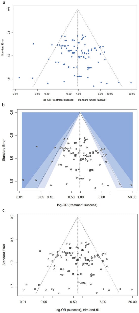
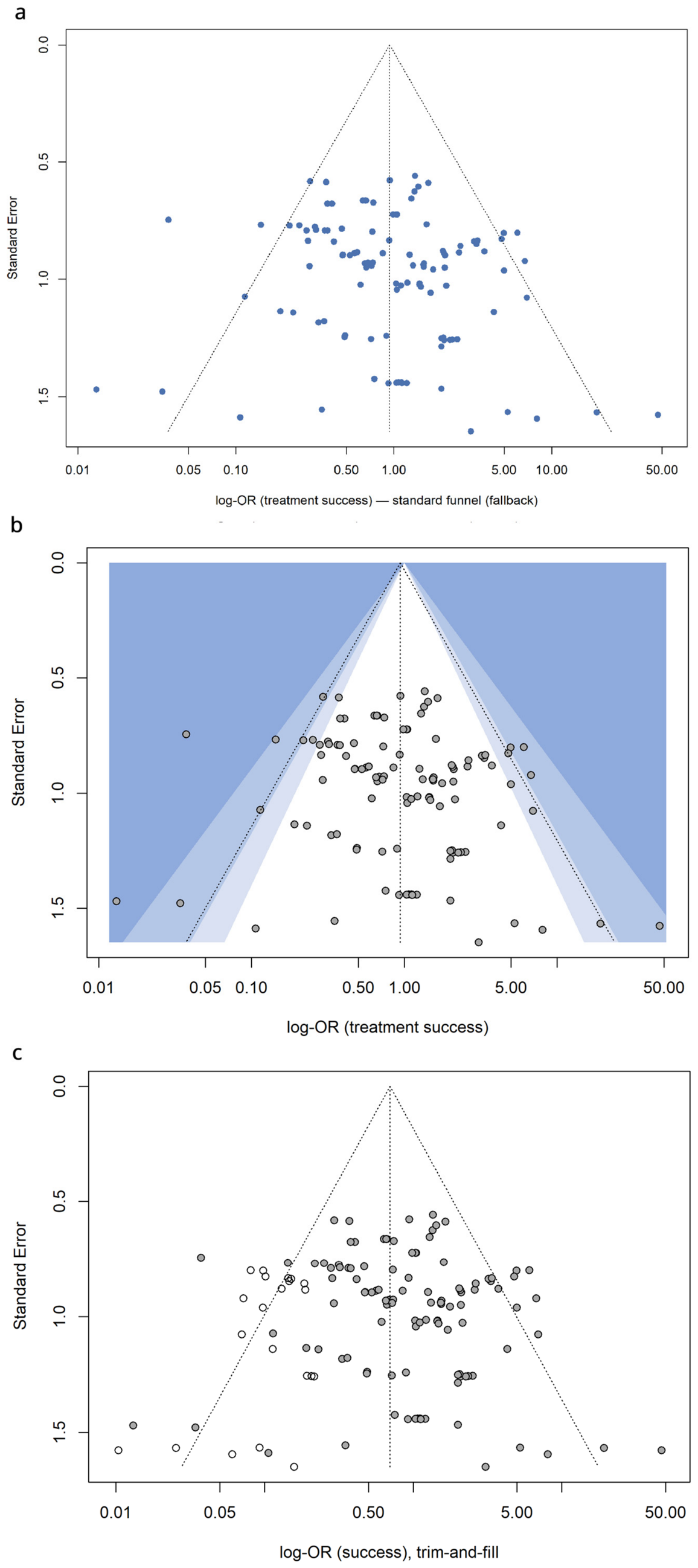
Potential publication bias and small study effects were evaluated using standard and contour-enhanced funnel plots. Visual asymmetry was observed in the standard log-OR plot. A subsequent non-parametric trim-and-fill analysis was performed, imputing theoretical missing studies to adjust for potential bias. Across the pairwise contrasts contributing to this outcome, the observed pooled OR was 0.94 (95% CI: 0.779–1.136); after trim-and-fill adjustment imputing 23 potentially missing contrasts (total contrasts analyzed = 145), the adjusted OR shifted to a more conservative 0.698 (95% CI: 0.570–0.854), suggesting that small-study effects slightly inflated the global comparative estimates (Figure 12).
Figure 12.
Publication bias and small-study effects assessment. (a) Standard funnel plot; (b) contour-enhanced funnel plot mapping regions of statistical significance; (c) trim-and-fill funnel plot showing observed versus imputed contrasts. In all three panels the dotted vertical line is the pooled log odds ratio and the dotted diagonal lines delimit the 95% pseudo-confidence region. In (b) the shaded contours delimit the regions in which a contrast would be statistically significant (p < 0.01, 0.01–0.05 and 0.05–0.10), the unshaded region corresponding to p > 0.10. In (c) the filled circles are the observed contrasts and the open circles the contrasts imputed by the trim-and-fill procedure.
3.7. Certainty of Evidence (GRADE Assessment)
The overall certainty of the evidence was systematically appraised using the GRADE framework (Table 2). The certainty for the primary outcome (Treatment Success) and Intraoperative Blood Loss was graded as low. These outcomes demonstrated substantial effect magnitudes and, for the COMB_HL versus DC contrast, the documented direction of confounding by indication is conservative, which together justified an upgrade; the scores were nonetheless penalized due to the observational nature of the foundational studies and the presence of significant statistical heterogeneity. The certainty for secondary safety endpoints, including hospital length of stay, β-hCG normalization time, overall complications, and hysterectomy, was graded as very low due to very serious imprecision (wide confidence intervals bridging clinical thresholds) and inconsistency (I2 > 90%).
Table 2.
GRADE Assessment: Certainty of Evidence for Surgical Interventions in Cesarean Scar Pregnancy.
4. Discussion
This systematic review and network meta-analysis integrated contemporary data from 64 studies and 8970 patients to formulate a comparative hierarchy of surgical interventions for CSP. Our findings suggest that advanced, visually guided, and combined modalities are associated with a higher probability of treatment success than blind DC. Specifically, COMB_HL achieved the highest probability of initial treatment success (SUCRA = 93.5%), followed by HIFU_C, standalone LAP and ultrasound-guided SCL_C. HIFU_C carried the lowest complication burden of any modality, although the pooled estimates for blood loss and hospital stay rest on prediction intervals that cross the null. MTX_C and standalone DC occupied the lowest ranks; for MTX_C the difference from DC did not reach statistical significance, so the appropriate conclusion is the absence of any demonstrated advantage over blind curettage rather than demonstrated inferiority.
This NMA reinforces the critical shift away from blind procedures. Blind DC alone is associated with an unacceptably high risk of massive hemorrhage and uterine perforation because it fails to address the hypervascular trophoblastic tissue deeply embedded in the myometrial niche [4,17]. Our data reflect this, placing DC near the bottom of the efficacy hierarchy.
The higher success probability observed for COMB_HL and LAP is biologically plausible given their anatomical and mechanistic rationale. Laparoscopy allows for the direct visualization and complete excision of exogenic (Type II and III) CSPs, excising the scarred and thinned myometrium, and facilitating multi-layer uterine reconstruction [12]. This anatomical restoration addresses the immediate ectopic pregnancy and may mitigate the risk of CSP recurrence and PAS in subsequent gestations, although the present review could not test that hypothesis. The addition of hysteroscopy (COMB_HL) refines this approach by precisely delineating the endogenic component of the gestational sac, minimizing damage to the healthy endometrium, and preventing residual trophoblastic tissue [14,47].
Less invasive adjunctive pre-treatments, such as HIFU and local sclerotherapy (SCL_C), showed competitive efficacy and a favourable safety profile. HIFU induces targeted coagulative necrosis of the gestational sac and surrounding microvasculature via thermal ablation, drastically reducing the risk of catastrophic bleeding during subsequent evacuation [16,18]. Our analysis places HIFU_C second in the efficacy hierarchy and found lower blood loss, shorter hospital stay and fewer overall complications, consistent with recent pairwise meta-analyses that advocate HIFU over UAE because of the latter’s potential detriment to the ovarian reserve and higher complication rates (e.g., post-embolization syndrome) [19,34].
Hysteroscopy was associated with higher odds of success than DC (OR = 1.97) yet ranked low (SUCRA 33.5%), reflecting case selection rather than inefficacy: hysteroscopic series are dominated by endogenic Type I lesions with preserved myometrium. A systematic review of hysteroscopic treatment reported 87.4% success, 4.1% complications and β-hCG normalization at a mean of 29.3 days, with a two-step diagnostic-then-resectoscopic technique performing best in selected patients [98]; where vascularity rather than depth dominates, UAE followed by hysteroscopic laser resection is an established alternative [99]. On the basis of that literature, hysteroscopy has been proposed as a first-line uterus-preserving option in Type I CSP, with the thresholds of residual myometrium > 3 mm and sac ≤ 30 mm used as selection criteria, and laparoscopic or combined excision, which repairs the niche, reserved for exogenic lesions and severely thinned myometrium [98]. These thresholds derive from case selection in the source series and could not be tested as effect modifiers within the present network, since the relevant covariates were unreported in most included studies.
The primary strength of this study is its unprecedented scale and robust methodological framework. To our knowledge, this is the largest NMA of CSP surgical management, encompassing nearly 9000 patients. Adherence to PRISMA guidelines, transitivity checks, and comprehensive sensitivity analyses, including trim-and-fill adjustments for publication bias, support the numerical stability of the pooled estimates.
However, several limitations must be acknowledged. The most important is confounding by indication: with 63 of 64 studies non-randomized, modality was chosen based on the features that predict outcome. Among studies specifying CSP classification, 57% of those contributing to COMB_HL were restricted to Type II/III disease and none to Type I, whereas no DC study was restricted to advanced disease. This channelling runs in opposite directions across nodes and cannot be removed by any adjustment of aggregate data; for COMB_HL its direction is conservative, but elsewhere it is unpredictable. Because the relevant covariates were largely unreported, the hierarchy may be biased to an unknown degree, and SUCRA values, which weight neither precision nor patient-level applicability, should not be read as evidence of clinical superiority. Substantial statistical heterogeneity (I2 > 80%) was observed in continuous secondary outcomes, driven by institutional variations in surgical protocols, discharge criteria, and timing of β-hCG assays, with prediction intervals crossing the null. Third, most studies originate from China, limiting generalizability. Fourth, reproductive outcomes could not be synthesized. Finally, the aggregate nature of the data precluded an individual patient data (IPD) meta-analysis, the design required to stratify treatment by CSP subtype and residual myometrial thickness and to address confounding by indication.
5. Conclusions
Within an almost entirely observational evidence base of low to very low certainty, visually guided and combined modalities were associated with a higher probability of treatment success than blind DC; COMB_HL ranked highest, followed by HIFU_C and LAP, and HIFU_C carried the lowest complication burden. These associations are hypothesis-generating and do not establish superiority for any individual patient. Therefore, the points that follow are offered as clinical considerations arising from this synthesis, not as treatment recommendations. Where curettage is chosen, the available evidence favours performing it under ultrasonographic or hysteroscopic vision rather than blind. Type I CSP with preserved residual myometrium and a smaller gestational sac appears amenable to hysteroscopic resection or ultrasound-guided suction curettage, with sclerosant or HIFU pre-treatment where vascularity is prominent; the numerical thresholds reported in the source literature reflect case selection in those series and were not testable within this network. Type II/III lesions, markedly thinned residual myometrium, or a large vascular mass are more often approached by excision with myometrial repair, laparoscopic, transvaginal or combined, which restores the scar defect. HIFU_C may be considered as an alternative to UAE where the technology and expertise are available. Methotrexate plus curettage showed no demonstrated advantage over DC in this analysis, and its role as a primary strategy warrants reconsideration pending adequately powered comparative data. Every decision must be individualized to CSP morphology, residual myometrial thickness, gestational age, β-hCG, hemodynamic status, fertility wishes and local expertise. Multicenter randomized trials stratified by standardized ultrasound criteria, and IPD meta-analysis with long-term reproductive follow-up, are the priorities for future research.
Supplementary Materials
The following supporting information can be downloaded at: https://www.mdpi.com/article/10.3390/surgeries7040117/s1, Supplementary Table S1: PRISMA 2020 checklist [21]; Supplementary Table S2: node-splitting output for every closed loop [34,35,36,37,38,39,40,41,42,43,44,45,46,47,48,49,50,51,52,53,54,55,56,57,58,59,60,61,62,63,64,65,66,67,68,69,70,71,72,73,74,75,76,77,78,79,80,81,82,83,84,85,86,87,88,89,90,91,92,93,94,95,96,97]; Supplementary Table S3: mapping of intervention arms onto the ten treatment nodes [34,35,36,37,38,39,40,41,42,43,44,45,46,47,48,49,50,51,52,53,54,55,56,57,58,59,60,61,62,63,64,65,66,67,68,69,70,71,72,73,74,75,76,77,78,79,80,81,82,83,84,85,86,87,88,89,90,91,92,93,94,95,96,97]; Supplementary Figure S1: Newcastle–Ottawa Scale item-level assessment [34,35,36,37,38,39,40,41,42,43,44,45,46,47,48,49,50,51,52,53,54,55,56,57,58,59,60,61,62,63,64,65,66,67,68,69,70,71,72,73,74,75,76,77,78,79,80,81,82,83,84,85,86,87,88,89,90,91,92,93,94,95,96,97]; Supplementary Figure S2: leave-one-out sensitivity analysis [34,35,36,37,38,39,40,41,42,43,44,45,46,47,48,49,50,51,52,53,54,55,56,57,58,59,60,61,62,63,64,65,66,67,68,69,70,71,72,73,74,75,76,77,78,79,80,81,82,83,84,85,86,87,88,89,90,91,92,93,94,95,96,97]. All references cited in the Supplementary Materials are also cited in the main text.
Author Contributions
Conceptualization, F.M.A. and A.S.M.; methodology, F.M.A., A.S.M. and A.M.H.; software, A.S.M.; validation, M.A., N.A. and K.M.A.-Q.; formal analysis, A.S.M. and F.M.A.; investigation, N.A.A. (Noor Ahmed Aljaber), J.S.A., H.S. and N.A.; resources, K.M.A.-Q. and A.G.A.; data curation, N.A.A. (Nesreen Abdullah Ahmed), A.E. and H.S.; writing—original draft preparation, F.M.A., A.S.M. and A.M.H.; writing—review and editing, M.A., N.A., K.M.A.-Q., N.A.A. (Noor Ahmed Aljaber), J.S.A., H.S., N.A.A. (Nesreen Abdullah Ahmed), A.E. and A.G.A.; visualization, A.S.M.; supervision, F.M.A.; project administration, F.M.A. All authors have read and agreed to the published version of the manuscript.
Funding
This research received no external funding.
Institutional Review Board Statement
Not applicable.
Informed Consent Statement
Not applicable.
Data Availability Statement
No new data were created or analyzed in this study. Data sharing is not applicable to this article.
Conflicts of Interest
The authors declare no conflicts of interest.
Abbreviations
The following abbreviations are used in this manuscript:
| CSP | Cesarean scar pregnancy |
| CSEP | Cesarean scar ectopic pregnancy |
| NMA | Network meta-analysis |
| PRISMA | Preferred Reporting Items for Systematic Reviews and Meta-Analyses |
| PROSPERO | International Prospective Register of Systematic Reviews |
| PICOS | Population, Intervention, Comparator, Outcomes, Study design |
| OR | Odds ratio |
| MD | Mean difference |
| SMD | Standardized mean difference |
| CI | Confidence interval |
| SUCRA | Surface Under the Cumulative Ranking curve |
| REML | Restricted maximum likelihood |
| RE | Random-effects |
| RoB 2 | Cochrane Risk of Bias 2 tool |
| NOS | Newcastle–Ottawa Scale |
| GRADE | Grading of Recommendations Assessment, Development, and Evaluation |
| IRR | Inter-rater reliability |
| RCT | Randomized controlled trial |
| DC | Dilation and Curettage/Suction Curettage (primary evacuation) |
| HYS | Hysteroscopic resection |
| LAP | Laparoscopic resection |
| TV | Transvaginal resection/repair |
| COMB_HL | Combined hysteroscopic–laparoscopic surgery |
| SCL_C | Ultrasound-guided sclerotherapy plus curettage |
| HIFU | High-intensity focused ultrasound |
| HIFU_C | HIFU plus curettage |
| UAE | Uterine artery embolization |
| UAE_C | UAE plus curettage |
| UAE_HYS | UAE plus hysteroscopy |
| MTX | Methotrexate |
| MTX_C | Methotrexate plus curettage |
| SC | Suction curettage |
| TIIAB | Temporary internal iliac artery blockage |
| PAS | Placenta accreta spectrum |
| TVUS | Transvaginal ultrasound |
| MRI | Magnetic resonance imaging |
| MeSH | Medical Subject Headings |
| WHO-ICTRP | WHO International Clinical Trials Registry Platform |
| β-hCG | Beta-human chorionic gonadotropin |
| IPD | Individual patient data |
References
- Gonzalez, N.; Tulandi, T. Cesarean Scar Pregnancy: A Systematic Review. J. Minim. Invasive Gynecol. 2017, 24, 731–738. [Google Scholar] [CrossRef] [Scilit] [PubMed]
- Hameed, M.S.; Wright, A.; Chern, B.S.M. Cesarean Scar Pregnancy: Current Understanding and Treatment Including Role of Minimally Invasive Surgical Techniques. Gynecol. Minim. Invasive Ther. 2023, 12, 64–71. [Google Scholar] [CrossRef] [Scilit] [PubMed]
- Birch Petersen, K.; Hoffmann, E.; Rifbjerg Larsen, C.; Svarre Nielsen, H. Cesarean Scar Pregnancy: A Systematic Review of Treatment Studies. Fertil. Steril. 2016, 105, 958–967. [Google Scholar] [CrossRef] [Scilit] [PubMed]
- Fu, P.; Sun, H.; Zhang, L.; Liu, R. Efficacy and Safety of Treatment Modalities for Cesarean Scar Pregnancy: A Systematic Review and Network Meta-Analysis. Am. J. Obstet. Gynecol. MFM 2024, 6, 101328. [Google Scholar] [CrossRef] [Scilit] [PubMed]
- Silva, B.; Viana Pinto, P.; Costa, M.A. Cesarean Scar Pregnancy: A Systematic Review on Expectant Management. Eur. J. Obstet. Gynecol. Reprod. Biol. 2023, 288, 36–43. [Google Scholar] [CrossRef] [Scilit] [PubMed]
- Maheux-Lacroix, S.; Li, F.; Bujold, E.; Nesbitt-Hawes, E.; Deans, R.; Abbott, J. Cesarean Scar Pregnancies: A Systematic Review of Treatment Options. J. Minim. Invasive Gynecol. 2017, 24, 915–925. [Google Scholar] [CrossRef] [Scilit] [PubMed]
- Kanat-Pektas, M.; Bodur, S.; Dundar, O.; Bakır, V.L. Systematic Review: What Is the Best First-Line Approach for Cesarean Section Ectopic Pregnancy? Taiwan. J. Obstet. Gynecol. 2016, 55, 263–269. [Google Scholar] [CrossRef] [Scilit] [PubMed]
- Timor-Tritsch, I.E.; Buca, D.; Di Mascio, D.; Cali, G.; D’aMico, A.; Monteagudo, A.; Tinari, S.; Morlando, M.; Nappi, L.; Greco, P.; et al. Outcome of Cesarean Scar Pregnancy According to Gestational Age at Diagnosis: A Systematic Review and Meta-Analysis. Eur. J. Obstet. Gynecol. Reprod. Biol. 2021, 258, 53–59. [Google Scholar] [CrossRef] [Scilit] [PubMed]
- Cali, G.; Timor-Tritsch, I.E.; Palacios-Jaraquemada, J.; Monteagudo, A.; Buca, D.; Forlani, F.; Familiari, A.; Scambia, G.; Acharya, G.; D’Antonio, F. Outcome of Cesarean Scar Pregnancy Managed Expectantly: Systematic Review and Meta-Analysis. Ultrasound Obstet. Gynecol. 2018, 51, 169–175. [Google Scholar] [CrossRef] [Scilit] [PubMed]
- Morlando, M.; Buca, D.; Timor-Tritsch, I.; Cali, G.; Palacios-Jaraquemada, J.; Monteagudo, A.; Khalil, A.; Zizzo, G.; Fichera, A.; Lanzone, A.; et al. Reproductive Outcome after Cesarean Scar Pregnancy: A Systematic Review and Meta-Analysis. Acta Obstet. Gynecol. Scand. 2020, 99, 1278–1289. [Google Scholar] [CrossRef] [Scilit] [PubMed]
- Timor-Tritsch, I.E.; Horwitz, G.; D’Antonio, F.; Monteagudo, A.; Bornstein, E.; Chervenak, J.; Messina, L.A.; Morlando, M.; Cali, G. Recurrent Cesarean Scar Pregnancy: Case Series and Literature Review. Ultrasound Obstet. Gynecol. 2021, 58, 121–126. [Google Scholar] [CrossRef] [Scilit]
- Alameddine, S.; Lucidi, A.; Jurkovic, D.; Timor-Tritsch, I.; Nazac, A.; Kaelin Agten, A.; Buca, D.; Di Mascio, D.; D’Antonio, F. Treatments for Cesarean Scar Pregnancy: A Systematic Review and Meta-Analysis. J. Matern. Fetal Neonatal Med. 2024, 37, 2327569. [Google Scholar] [CrossRef] [Scilit] [PubMed]
- Kłobuszewski, B.; Szmygin, M.; Nieoczym, K.; Kłobuszewska, O.; Woźniak, S.; Pyra, K.K. Advances in Treating Cesarean Scar Pregnancy: A Comprehensive Review of Techniques, Clinical Outcomes, and Fertility Preservation. Med. Sci. Monit. 2024, 30, e943550. [Google Scholar] [CrossRef] [Scilit] [PubMed]
- Du, Z.; Xu, W.; Lu, J.; Li, C. Comparison of Clinical Safety and Efficacy of Ultrasound-Guided Local Lauromacrogol Injection versus Uterine Artery Embolization in the Treatment of Caesarean Scar Pregnancy: A Systematic Review and Meta-Analysis. BMC Pregnancy Childbirth 2023, 23, 149. [Google Scholar] [CrossRef] [Scilit] [PubMed]
- Silva, M.S.; Pinto, P.V.; Vieira, R.J. The Role of Hysteroscopy in the Management of Caesarean Scar Pregnancy: A Systematic Review and Meta-Analysis. Eur. J. Obstet. Gynecol. Reprod. Biol. 2026, 323, 115192. [Google Scholar] [CrossRef] [Scilit] [PubMed]
- Li, J.; Li, H.; Jiang, J.; Zhang, X.; Shan, S.; Zhao, X.; Shi, B. Dilatation and Curettage versus Lesion Resection in the Treatment of Cesarean-Scar-Pregnancy: A Systematic Review and Meta-Analysis. Taiwan. J. Obstet. Gynecol. 2021, 60, 412–421. [Google Scholar] [CrossRef] [Scilit] [PubMed]
- Mi, W.; Pei, P.; Zheng, Y. Clinical Efficacy and Safety between High-Intensity Focused Ultrasound and Uterine Artery Embolization for Cesarean Scar Pregnancy: A Systematic Review and a Meta-Analysis. Ann. Palliat. Med. 2021, 10, 6379–6387. [Google Scholar] [CrossRef] [Scilit] [PubMed]
- Liu, Y.; Wang, L.; Zhu, X. Efficacy and Safety of High-Intensity Focused Ultrasound Compared with Uterine Artery Embolization in Cesarean Section Pregnancy: A Meta-Analysis. J. Minim. Invasive Gynecol. 2023, 30, 446–454. [Google Scholar] [CrossRef] [Scilit] [PubMed]
- Li, Y.; Hua, C. Is High-Intensity Focused Ultrasound Superior to Uterine Artery Embolization in Cesarean Scar Pregnancy and Subsequent Pregnancy Outcomes? A Meta-Analysis of the Chinese Population. J. Minim. Invasive Gynecol. 2023, 30, 180–191. [Google Scholar] [CrossRef] [Scilit] [PubMed]
- Xiao, X.; Feng, Z.; Li, T.; Yi, B.; Zhang, S.; Wang, W. Comparing the Efficacy and Safety of High-Intensity Focused Ultrasound and Uterine Artery Embolization in Caesarean Scar Pregnancy: A Meta-Analysis. Adv. Ther. 2019, 36, 1314–1325. [Google Scholar] [CrossRef] [Scilit] [PubMed]
- Page, M.J.; McKenzie, J.E.; Bossuyt, P.M.; Boutron, I.; Hoffmann, T.C.; Mulrow, C.D.; Shamseer, L.; Tetzlaff, J.M.; Akl, E.A.; Brennan, S.E.; et al. The PRISMA 2020 Statement: An Updated Guideline for Reporting Systematic Reviews. BMJ 2021, 372, n71. [Google Scholar] [CrossRef] [Scilit] [PubMed]
- Veritas Health Innovation. Covidence Systematic Review Software; Veritas Health Innovation: Melbourne, Australia, 2026; Available online: https://www.covidence.org (accessed on 1 July 2026).
- Sterne, J.A.C.; Savović, J.; Page, M.J.; Elbers, R.G.; Blencowe, N.S.; Boutron, I.; Cates, C.J.; Cheng, H.-Y.; Corbett, M.S.; Eldridge, S.M.; et al. RoB 2: A Revised Tool for Assessing Risk of Bias in Randomised Trials. BMJ 2019, 366, l4898. [Google Scholar] [CrossRef] [Scilit] [PubMed]
- Wells, G.A.; Shea, B.; O’Connell, D.; Peterson, J.; Welch, V.; Losos, M.; Tugwell, P. The Newcastle–Ottawa Scale (NOS) for Assessing the Quality of Nonrandomised Studies in Meta-Analyses; Ottawa Hospital Research Institute: Ottawa, ON, Canada, 2000. [Google Scholar]
- Veroniki, A.A.; Jackson, D.; Viechtbauer, W.; Bender, R.; Bowden, J.; Knapp, G.; Kuss, O.; Higgins, J.P.T.; Langan, D.; Salanti, G. Methods to Estimate the Between-Study Variance and Its Uncertainty in Meta-Analysis. Res. Synth. Methods 2016, 7, 55–79. [Google Scholar] [CrossRef] [Scilit] [PubMed]
- Rücker, G. Network Meta-Analysis, Electrical Networks and Graph Theory. Res. Synth. Methods 2012, 3, 312–324. [Google Scholar] [CrossRef] [Scilit] [PubMed]
- Salanti, G.; Ades, A.E.; Ioannidis, J.P.A. Graphical Methods and Numerical Summaries for Presenting Results from Multiple-Treatment Meta-Analysis: An overview and tutorial. J. Clin. Epidemiol. 2011, 64, 163–171. [Google Scholar] [CrossRef] [Scilit] [PubMed]
- Dias, S.; Welton, N.J.; Caldwell, D.M.; Ades, A.E. Checking Consistency in Mixed Treatment Comparison Meta-Analysis. Stat. Med. 2010, 29, 932–944. [Google Scholar] [CrossRef] [Scilit] [PubMed]
- Higgins, J.P.T.; Thompson, S.G.; Deeks, J.J.; Altman, D.G. Measuring Inconsistency in Meta-Analyses. BMJ 2003, 327, 557–560. [Google Scholar] [CrossRef] [Scilit] [PubMed]
- Patsopoulos, N.A.; Evangelou, E.; Ioannidis, J.P.A. Sensitivity of Between-Study Heterogeneity in Meta-Analysis: Proposed Metrics and Empirical Evaluation. Int. J. Epidemiol. 2008, 37, 1148–1157. [Google Scholar] [CrossRef] [Scilit] [PubMed]
- Egger, M.; Davey Smith, G.; Schneider, M.; Minder, C. Bias in Meta-Analysis Detected by a Simple, Graphical Test. BMJ 1997, 315, 629–634. [Google Scholar] [CrossRef] [Scilit] [PubMed]
- Duval, S.; Tweedie, R. Trim and Fill: A Simple Funnel-Plot-Based Method of Testing and Adjusting for Publication Bias in Meta-Analysis. Biometrics 2000, 56, 455–463. [Google Scholar] [CrossRef] [Scilit] [PubMed]
- Guyatt, G.H.; Oxman, A.D.; Vist, G.E.; Kunz, R.; Falck-Ytter, Y.; Alonso-Coello, P.; Schünemann, H.J. GRADE: An Emerging Consensus on Rating Quality of Evidence and Strength of Recommendations. BMJ 2008, 336, 924–926. [Google Scholar] [CrossRef] [Scilit] [PubMed]
- Yin, N.; Wei, F.; Gao, Z.; Yang, Z.; Zhou, X. Comparison of Curettage with and without High-Intensity Focused Ultrasound in the Management of Caesarean Scar Pregnancy. Curr. Probl. Surg. 2025, 72, 101884. [Google Scholar] [CrossRef] [Scilit] [PubMed]
- Wu, Y.; Sun, L.F.; Si, Y.N.; Luan, X.L.; Gao, Y.M. Clinical Efficacy Analysis of Different Therapeutic Methods in Patients with Cesarean Scar Pregnancy. Taiwan. J. Obstet. Gynecol. 2021, 60, 498–502. [Google Scholar] [CrossRef] [Scilit] [PubMed]
- Sun, Y.Y.; Xi, X.W.; Yan, Q.; Qiao, Q.Q.; Feng, Y.J.; Zhu, Y.P. Management of Type II Unruptured Cesarean Scar Pregnancy: Comparison of Gestational Mass Excision and Uterine Artery Embolization Combined with Methotrexate. Taiwan. J. Obstet. Gynecol. 2015, 54, 489–492. [Google Scholar] [CrossRef] [Scilit] [PubMed]
- Shao, M.; Tang, F.; Ji, L.; Hu, M.; Zhang, K.; Pan, J. The Management of Caesarian Scar Pregnancy with or without a Combination of Methods Prior to Hysteroscopy: Ovarian Reserve Trends and Patient Outcomes. J. Gynecol. Obstet. Hum. Reprod. 2022, 51, 102417. [Google Scholar] [CrossRef] [Scilit] [PubMed]
- He, Y.; Wu, X.; Zhu, Q.; Wu, X.; Feng, L.; Wu, X.; Zhao, A.; Di, W. Combined Laparoscopy and Hysteroscopy vs. Uterine Curettage in the Uterine Artery Embolization-Based Management of Cesarean Scar Pregnancy: A Retrospective Cohort Study. BMC Women’s Health 2014, 14, 116. [Google Scholar] [CrossRef] [Scilit] [PubMed]
- Niu, X.; Zhang, Y.; Ni, S.; Yu, J.; Li, S. Single-Center Insights into Cesarean Scar Pregnancy: Treatment Strategies and Determinants. Clin. Exp. Obstet. Gynecol. 2024, 51, 137. [Google Scholar] [CrossRef] [Scilit]
- Gan, Y.; Jiang, H.; Zhou, Y.; Chen, Z. Evaluation Effectiveness of Pre-Treatment Combined with Ultrasound-Guided Suction Curettage in Cesarean Scar Pregnancy: A Retrospective Cohort Study. Clin. Exp. Obstet. Gynecol. 2024, 51, 213. [Google Scholar] [CrossRef] [Scilit]
- Li, Y.Y.; Yin, Z.Y.; Li, S.; Xu, H.; Zhang, X.P.; Cheng, H.; Du, L.; Zhou, X.Y.; Zhang, B. Comparison of Transvaginal Surgery and Methotrexate/Mifepristone-Combined Transcervical Resection in the Treatment of Cesarean Scar Pregnancy. Eur. Rev. Med. Pharmacol. Sci. 2017, 21, 2957–2963. [Google Scholar] [PubMed]
- Guo, J.; Yu, J.; Zhang, Q.; Song, X. Clinical Efficacy and Safety of Uterine Artery Embolization (UAE) versus Laparoscopic Cesarean Scar Pregnancy Debridement Surgery (LCSPDS) in Treatment of Cesarean Scar Pregnancy. Med. Sci. Monit. 2018, 24, 4659–4666. [Google Scholar] [CrossRef] [Scilit] [PubMed]
- Huang, L.; Zhao, L.; Shi, H. Clinical Efficacy of Combined Hysteroscopic and Laparoscopic Surgery and Reversible Ligation of the Uterine Artery for Excision and Repair of Uterine Scar in Patients with Type II and III Cesarean Scar Pregnancy. Med. Sci. Monit. 2020, 26, e924076. [Google Scholar] [CrossRef] [Scilit] [PubMed]
- Xiong, J.; Fu, F.; Zhang, W.; Luo, J.; Xu, Y.Y.; Le, L.L.; He, X.J. Study on Influencing Factors and Related Clinical Issues in Cesarean Scar Pregnancy. Clin. Exp. Obstet. Gynecol. 2021, 48, 365–371. [Google Scholar] [CrossRef] [Scilit]
- Ling, L.; Fu, J.; Zhan, L.; Wang, W.; Su, Q.; Li, J.; Wei, B. Surgical Management for Type II Cesarean Scar Pregnancy. Clin. Exp. Obstet. Gynecol. 2021, 48, 555–560. [Google Scholar] [CrossRef] [Scilit]
- Wang, X.R.; Wang, Y.F.; Kang, S.W.; Zhang, Y. Treatment Outcomes of Uterine Lesion Resection versus Hysterectomy for Cesarean Scar Pregnancy. Clin. Exp. Obstet. Gynecol. 2020, 47, 882–886. [Google Scholar] [CrossRef] [Scilit]
- Zhang, X.; Pang, Y.; Ma, Y.; Liu, X.; Cheng, L.; Ban, Y.; Cui, B. A Comparison between Laparoscopy and Hysteroscopy Approach in Treatment of Cesarean Scar Pregnancy. Medicine 2020, 99, e22845. [Google Scholar] [CrossRef] [Scilit] [PubMed]
- Wang, L.; Cheng, W.; Zhao, Y.; Liu, H. Uterine Artery Chemoembolization Compared with Uterine Artery Embolization Combined with Prior Hysteroscopy and Curettage in the Treatment of Cesarean Scar Pregnancy. Afr. Health Sci. 2025, 25, 579–590. [Google Scholar] [CrossRef] [Scilit] [PubMed]
- Su, X.; Yang, M.; Na, Z.; Wen, C.; Liu, M.; Cai, C.; Zhong, Z.; Zhou, B.; Tang, X. Application of Laparoscopic Internal Iliac Artery Temporary Occlusion and Uterine Repair Combined with Hysteroscopic Aspiration in Type III Cesarean Scar Pregnancy. Am. J. Transl. Res. 2022, 14, 1737–1741. [Google Scholar] [PubMed]
- Liu, R.; Zhou, T.; Cui, P.; Wang, W.; Wang, S. Laparoscopy or Laparotomy for High-Risk Caesarean Scar Pregnancy? A Retrospective Study. Authorea 2020. preprint. [Google Scholar] [CrossRef] [Scilit] [PubMed]
- Cheng, Q.; Tian, Q.; Chang, K.-K.; Yi, X.-F. Comparison of the Efficacy and Safety of Different Surgical Strategies for Patients with Type II Cesarean Scar Pregnancy. Reprod. Dev. Med. 2020, 4, 89–96. [Google Scholar] [CrossRef] [Scilit]
- Yin, Y.; Huang, L.; Xu, N.; Ma, H.G.; Yuan, C. Comparative Analysis of the Efficacy of Four Different Surgical Modalities in the Treatment of Cesarean Scar Pregnancy: A Multicenter Retrospective Study. Adv. Ther. 2025, 42, 1448–1461. [Google Scholar] [CrossRef] [Scilit] [PubMed]
- Cao, S.; Qiu, G.; Zhang, P.; Wang, X.; Wu, Q. A Comparison of Transvaginal Removal and Repair of Uterine Defect for Type II Cesarean Scar Pregnancy and Uterine Artery Embolization Combined with Curettage. Front. Med. 2021, 8, 654956. [Google Scholar] [CrossRef] [Scilit] [PubMed]
- Shen, F.; Lv, H.; Wang, L.; Zhao, R.; Tong, M.; Lee, A.C.; Guo, F.; Chen, Q. A Comparison of Treatment Options for Type 1 and Type 2 Caesarean Scar Pregnancy: A Retrospective Case Series Study. Front. Med. 2021, 8, 671035. [Google Scholar] [CrossRef] [Scilit] [PubMed]
- Lin, Y.; Xiong, C.; Dong, C.; Yu, J. Approaches in the Treatment of Cesarean Scar Pregnancy and Risk Factors for Intraoperative Hemorrhage: A Retrospective Study. Front. Med. 2021, 8, 682368. [Google Scholar] [CrossRef] [Scilit] [PubMed]
- Tan, K.L.; Chen, Y.M.; Zeng, W.; Meng, Y.; Jiang, L. Local Methotrexate Injection Followed by Dilation and Curettage for Cesarean Scar Pregnancy. Front. Med. 2022, 8, 800610. [Google Scholar] [CrossRef] [Scilit] [PubMed]
- Teng, D.; Gao, H.; Li, Y.; Meng, T.; Shi, X.; Shi, J. Clinical Efficacy Analysis of Laparoscopic Uterine Artery Pre-Ligation Combined with Hysteroscopic Curettage for Cesarean Scar Pregnancy. Front. Med. 2023, 10, 1234499. [Google Scholar] [CrossRef] [Scilit] [PubMed]
- Huang, L.; Long, B.; Lin, F.; Ling, W.; Huang, L.; Xiong, X. Short- and Long-Term Efficacy Evaluation of High-Intensity Focused Ultrasound Ablation and Uterine Artery Embolization for Cesarean Scar Pregnancy. Front. Med. 2026, 13, 1805049. [Google Scholar] [CrossRef] [Scilit] [PubMed]
- Wu, Q.; Liu, X.; Zhu, L.; Zhu, Y.; Mei, T.; Cao, S.; Shen, Y.; Ding, J.; Lin, T. Clinical Assessment of Ultrasound-Guided Local Lauromacrogol Injection Combined with Curettage and Hysteroscopy for Cesarean Scar Pregnancy. Front. Pharmacol. 2020, 11, 601977. [Google Scholar] [CrossRef] [Scilit] [PubMed]
- Hong, T.; Chai, Z.; Liu, M.; Zheng, L.; Qi, F. The Efficacy and Health Economics of Different Treatments for Type 1 Cesarean Scar Pregnancy. Front. Pharmacol. 2022, 13, 822319. [Google Scholar] [CrossRef] [Scilit] [PubMed]
- Fang, S.; Zhang, P.; Zhu, Y.; Wang, F.; He, L. A Retrospective Analysis of the Treatment of Cesarean Scar Pregnancy by High-Intensity Focused Ultrasound. Front. Surg. 2020, 7, 23. [Google Scholar] [CrossRef] [Scilit] [PubMed]
- Yin, Y.; Pan, F.; He, M.; Zhang, C.; Liu, Y. High-Intensity Focused Ultrasound Combined with Ultrasound-Guided Suction Curettage Treatment for Cesarean Scar Pregnancy: A Comparison of Different HIFU Sonication Strategies. Int. J. Hyperth. 2022, 39, 390–396. [Google Scholar] [CrossRef] [Scilit] [PubMed]
- Wu, X.; Xue, X.; Wu, X.; Lin, R.; Yuan, Y.; Wang, Q.; Xu, C.; He, Y.; Hu, W. Combined Laparoscopy and Hysteroscopy vs. Uterine Curettage in the Uterine Artery Embolization-Based Management of Cesarean Scar Pregnancy: A Cohort Study. Int. J. Clin. Exp. Med. 2014, 7, 2793–2803. [Google Scholar] [PubMed]
- Chen, Y.Q.; Liu, H.S.; Li, W.X.; Deng, C.; Hu, X.W.; Kuang, P.J. Efficacy of Transvaginal Debridement and Repair Surgery for Cesarean Scar Pregnancy: A Cohort Study Compared with Uterine Artery Embolism. Int. J. Clin. Exp. Med. 2015, 8, 21187–21193. [Google Scholar] [PubMed]
- Yang, J.; Li, B.; Liu, J.; Zeng, W.; Hong, L. A New Modified Hysteroscopic-Laparoscopic Surgery for Cesarean Scar Pregnancy of Stable Type III. Int. J. Gen. Med. 2021, 14, 2289–2295. [Google Scholar] [CrossRef] [Scilit] [PubMed]
- Zhao, Q.; Sun, X.Y.; Ma, S.Q.; Miao, M.W.; Li, G.L.; Wang, J.L.; Guo, R.X.; Li, L.X. Temporary Internal Iliac Artery Blockage versus Uterine Artery Embolization in Patients after Laparoscopic Pregnancy Tissue Removal Due to Cesarean Scar Pregnancy. Int. J. Gen. Med. 2022, 15, 501–511. [Google Scholar] [CrossRef] [Scilit] [PubMed]
- Fu, P.; Zhou, T.; Cui, P.; Wang, W.; Wang, S.; Liu, R. Selection of Laparoscopy or Laparotomy for Treating Cesarean Scar Pregnancy. Int. J. Gen. Med. 2022, 15, 7229–7240. [Google Scholar] [CrossRef] [Scilit] [PubMed]
- Feng, M.; Shang, H.; Chen, X.; Chen, J.; Cui, Y.; Wang, H.; Ou, Y.; Huang, X. Comparative Evaluation of Focused Ultrasound Ablation Combined with Curettage and Transvaginal Repair in the Management of Cesarean Scar Pregnancy: A Retrospective Comparative Study. Int. J. Gen. Med. 2025, 17, 2685–2697. [Google Scholar] [CrossRef] [Scilit] [PubMed]
- Li, Y.L.; Gong, L.Y.; Wu, X.F.; Gao, H.; Zheng, H.B.; Lan, W.S. Randomized Controlled Trial of Hysteroscopy or Ultrasonography versus No Guidance during D&C after Uterine Artery Chemoembolization for Cesarean Scar Pregnancy. Int. J. Gynaecol. Obstet. 2016, 135, 158–162. [Google Scholar] [CrossRef] [Scilit] [PubMed]
- Li, Y.R.; Xiao, S.S.; Wan, Y.J.; Xue, M. Analysis of the Efficacy of Three Treatment Options for Cesarean Scar Pregnancy Management. J. Obstet. Gynaecol. Res. 2014, 40, 2146–2151. [Google Scholar] [CrossRef] [Scilit] [PubMed]
- Wang, J.; Zhao, R.; Qian, H.; Lv, H. Pituitrin Local Injection versus Uterine Artery Embolization in the Management of Cesarean Scar Pregnancy: A Retrospective Cohort Study. J. Obstet. Gynaecol. Res. 2021, 47, 1711–1718. [Google Scholar] [CrossRef] [Scilit] [PubMed]
- Zhu, L.; Gao, J.; Yang, X.; Qian, L.; Wang, Y.; Zhang, H.; Chen, B.; Li, D. The Novel Use of Lauromacrogol: A Retrospective Study of Ultrasound-Guided Sclerosant Injection for Cesarean Scar Pregnancy. J. Obstet. Gynaecol. Res. 2022, 48, 140–145. [Google Scholar] [CrossRef] [Scilit] [PubMed]
- Chen, H.; Zhou, J.; Wang, H.; Tan, W.; Yao, M.; Wang, X. The Treatment of Cesarean Scar Pregnancy with Uterine Artery Embolization and Curettage as Compared to Transvaginal Hysterotomy. Eur. J. Obstet. Gynecol. Reprod. Biol. 2017, 214, 44–49. [Google Scholar] [CrossRef] [Scilit] [PubMed]
- Le, A.; Li, M.; Xu, Y.; Wang, Z.; Dai, X.Y.; Xiao, T.H.; Zhuo, R.; Yuan, R.; Tulandi, T. Different Surgical Approaches to 313 Cesarean Scar Pregnancies. J. Minim. Invasive Gynecol. 2019, 26, 148–152. [Google Scholar] [CrossRef] [Scilit] [PubMed]
- Xiao, J.; Shi, Z.; Zhou, J.; Ye, J.; Zhu, J.; Zhou, X.; Wang, F.; Zhang, S. Cesarean Scar Pregnancy: Comparing the Efficacy and Tolerability of Treatment with High-Intensity Focused Ultrasound and Uterine Artery Embolization. Ultrasound Med. Biol. 2017, 43, 640–647. [Google Scholar] [CrossRef] [Scilit] [PubMed]
- Sun, Y.; Liu, L.; Sun, G.; Ying, L. Lauromacrogol Pericystic Injection Combined with Curettage vs. Uterine Artery Embolization with Curettage for Cesarean Scar Pregnancy. J. Matern. Fetal Neonatal Med. 2026, 39, 2630517. [Google Scholar] [CrossRef] [Scilit] [PubMed]
- Verberkt, C.; Lemmers, M.; de Leeuw, R.A.; van Mello, N.M.; Groenman, F.A.; Hehenkamp, W.J.K.; Huirne, J.A.F. Effectiveness, Complications, and Reproductive Outcomes after Cesarean Scar Pregnancy Management: A Retrospective Cohort Study. AJOG Glob. Rep. 2022, 3, 100143. [Google Scholar] [CrossRef] [Scilit] [PubMed]
- Hong, Y.; Guo, Q.; Pu, Y.; Lu, D.; Hu, M. Outcome of High-Intensity Focused Ultrasound and Uterine Artery Embolization in the Treatment and Management of Cesarean Scar Pregnancy: A Retrospective Study. Medicine 2017, 96, e7687. [Google Scholar] [CrossRef] [Scilit] [PubMed]
- Kaelin Agten, A.; Jurkovic, D.; Timor-Tritsch, I.; Jones, N.; Johnson, S.; Monteagudo, A.; Huirne, J.; Fleisher, J.; Maymon, R.; Herrera, T.; et al. First-Trimester Cesarean Scar Pregnancy: A Comparative Analysis of Treatment Options from the International Registry. Am. J. Obstet. Gynecol. 2024, 230, 669.e1–669.e19. [Google Scholar] [CrossRef] [Scilit] [PubMed]
- Wang, G.; Liu, X.; Bi, F.; Yin, L.; Sa, R.; Wang, D.; Yang, Q. Evaluation of the Efficacy of Laparoscopic Resection for the Management of Exogenous Cesarean Scar Pregnancy. Fertil. Steril. 2014, 101, 1501–1507. [Google Scholar] [CrossRef] [Scilit] [PubMed]
- Liu, W.; Shen, L.; Wang, Q.; Wang, W.; Sun, Z. Uterine Artery Embolization Combined with Curettage vs. Methotrexate plus Curettage for Cesarean Scar Pregnancy. Arch. Gynecol. Obstet. 2016, 294, 71–76. [Google Scholar] [CrossRef] [Scilit] [PubMed]
- Liu, G.; Wu, J.; Cao, J.; Xue, Y.; Dai, C.; Xu, J.; Jia, X. Comparison of Three Treatment Strategies for Cesarean Scar Pregnancy. Arch. Gynecol. Obstet. 2017, 296, 383–389. [Google Scholar] [CrossRef] [Scilit] [PubMed]
- Ou, J.; Peng, P.; Li, C.; Teng, L.; Liu, X. Assessment of the Necessity of Uterine Artery Embolization during Suction and Curettage for Caesarean Scar Pregnancy: A Prospective Cohort Study. BMC Pregnancy Childbirth 2020, 20, 378. [Google Scholar] [CrossRef] [Scilit] [PubMed]
- Yu, L.; Yang, B.; Xu, Q.; Teng, Y.; Xue, Z. A Study on the Timing of Uterine Artery Embolization Followed by Pregnancy Excision for Cesarean Scar Pregnancy: A Prospective Study in China. BMC Pregnancy Childbirth 2021, 21, 697. [Google Scholar] [CrossRef] [Scilit] [PubMed]
- Qu, W.; Li, H.; Zhang, T.; Zhang, Y.; Ban, Y.; Li, N.; Jiang, J.; Xie, J.; Shi, W.; Hao, Y.; et al. Comparison of Different Treatment Strategies in the Management of Endogenic Caesarean Scar Pregnancy: A Multicentre Retrospective Study. BMC Pregnancy Childbirth 2022, 22, 404. [Google Scholar] [CrossRef] [Scilit] [PubMed]
- Liu, Y.; Yin, Q.; Xu, F.; Luo, S. Clinical Efficacy and Safety of High-Intensity Focused Ultrasound (HIFU) Ablation in Treatment of Cesarean Scar Pregnancy (CSP) I and II. BMC Pregnancy Childbirth 2022, 22, 607. [Google Scholar] [CrossRef] [Scilit] [PubMed]
- Wang, X.; Yang, B.; Chen, W.; Chen, J. Clinical Efficacy and Re-Pregnancy Outcomes of Patients with Previous Cesarean Scar Pregnancy. BMC Pregnancy Childbirth 2023, 23, 84. [Google Scholar] [CrossRef] [Scilit] [PubMed]
- Lu, L.; Shao, Y.; Qu, Z.; Huang, G.; Lang, S.; Yang, C.; Lang, S.; Fang, S. Outcomes of Prophylactic Lauromacrogol Injection versus Non-Injection in Patients with Endogenic Cesarean Scar Pregnancy. BMC Pregnancy Childbirth 2023, 23, 771. [Google Scholar] [CrossRef] [Scilit] [PubMed]
- You, X.; Ruan, Y.; Weng, S.; Lin, C.; Gan, M.; Qi, F. The Effectiveness of Hysteroscopy for the Treatment of Cesarean Scar Pregnancy: A Retrospective Cohort Study. BMC Pregnancy Childbirth 2024, 24, 151. [Google Scholar] [CrossRef] [Scilit] [PubMed]
- Elmas, B.; Ozturk, N.; Kizil, E.; Laleli Koc, B.; Zorlu, U.; Tugrul Ersak, D.; Dikici Aktas, T.; Erten, A.; Erkaya, S. Comparison of Two Different Suction Curettage Methods in Cesarean Scar Pregnancy Treatment. BMC Pregnancy Childbirth 2024, 24, 717. [Google Scholar] [CrossRef] [Scilit] [PubMed]
- Sun, H.; Wang, J.; Fu, P.; Zhou, T.; Liu, R. Systematic Evaluation of the Efficacy of Treatments for Cesarean Scar Pregnancy. Reprod. Biol. Endocrinol. 2024, 22, 84. [Google Scholar] [CrossRef] [Scilit] [PubMed]
- Bucak, M.; Chawla, K.; Mark, K.S.; Turan, S.; Turan, O.M. Standardized Algorithm for Cesarean Scar Pregnancy Management: Single-Center Outcomes. J. Matern. Fetal Neonatal Med. 2025, 38, 2501693. [Google Scholar] [CrossRef] [Scilit] [PubMed]
- Qiu, J.; Fu, Y.; Xu, J.; Huang, X.; Yao, G.; Lu, W. Analysis on clinical effects of dilation and curettage guided by ultrasonography versus hysteroscopy after uterine artery embolization in the treatment of cesarean scar pregnancy. Ther. Clin. Risk Manag. 2019, 15, 83–89. [Google Scholar] [CrossRef] [Scilit] [PubMed]
- Fei, H.; Jiang, X.; Li, T.; Pan, Y.; Guo, H.; Xu, X.; Shu, S. Comparison of Three Different Treatment Methods for Cesarean Scar Pregnancy. Ther. Clin. Risk Manag. 2019, 15, 1377–1381. [Google Scholar] [CrossRef] [Scilit] [PubMed]
- Wu, Z.W.; Lin, C.B.; Lin, Y.Y.; Cai, Y.J.; Xie, L. Textbook Outcomes in Patients with a Cesarean Scar Pregnancy: A Comprehensive Analysis from a Single Eastern Center in China. Medicine 2025, 104, e45140. [Google Scholar] [CrossRef] [Scilit] [PubMed]
- Huo, S.; Shen, L.; Ju, Y.; Liu, K.; Liu, W. Treatments for Cesarean Scar Pregnancy: 11-Year Experience at a Medical Center. J. Matern. Fetal Neonatal Med. 2023, 36, 2162818. [Google Scholar] [CrossRef] [Scilit] [PubMed]
- Wu, D.F.; Zhang, H.X.; He, W.; Liu, X.; Song, H.M.; Yu, T.F. Experience in Management of Cesarean Scar Pregnancy and Outcomes in a Single Center. J. Int. Med. Res. 2022, 50, 3000605221123875. [Google Scholar] [CrossRef] [Scilit] [PubMed]
- Stabile, G.; Vona, L.; Sorrentino, F.; Etrusco, A.; Laganà, A.S.; Carlucci, S.; Restaino, S.; Vizzielli, G.; Nappi, L. Exclusive Hysteroscopic Approach for the Treatment of Cesarean Scar Pregnancy: Systematic Review of the Literature. Women’s Health 2025, 21, 17455057251387428. [Google Scholar] [CrossRef] [Scilit] [PubMed]
- Sorrentino, F.; De Feo, V.; Stabile, G.; Tinelli, R.; D’Alterio, M.N.; Ricci, G.; Angioni, S.; Nappi, L. Cesarean Scar Pregnancy Treated by Artery Embolization Combined with Diode Laser: A Novel Approach for a Rare Disease. Medicina 2021, 57, 411. [Google Scholar] [CrossRef] [Scilit] [PubMed]

Disclaimer/Publisher’s Note: The statements, opinions and data contained in all publications are solely those of the individual author(s) and contributor(s) and not of MDPI and/or the editor(s). MDPI and/or the editor(s) disclaim responsibility for any injury to people or property resulting from any ideas, methods, instructions or products referred to in the content. |
© 2026 by the authors. Licensee MDPI, Basel, Switzerland. This article is an open access article distributed under the terms and conditions of the Creative Commons Attribution (CC BY) license.