


Abstract
Context: Prosthetic rehabilitation of acquired maxillary defects with Maxillary Resection Prostheses (MRPs) remains biomechanically challenging, particularly in partially edentulous patients, where conventional clasp-retained designs often yield suboptimal retention, stability, and functional outcomes. Research Gap: The integration of telescopic crown systems with semi-precision attachments incorporating a rotational latching mechanism has not been previously described as a unified approach to optimise load distribution and prosthesis stability in maxillary defect rehabilitation. Objective: To describe and clinically evaluate a novel prosthetic design combining telescopic crowns and a semi-precision rotational latching attachment to enhance retention, stability, and functional performance of MRPs. Methodology: A 31-year-old patient with a unilateral maxillary defect following partial maxillectomy presented with an unstable interim prosthesis and impaired speech and mastication. A definitive MRP was designed using telescopic crowns on the remaining dentition to establish a controlled path of insertion and improved axial load transfer. A semi-precision attachment with a key–keyway rotational latching mechanism was incorporated into the secondary framework to engage specific undercuts while minimising lateral forces on abutment teeth. A provisional prosthesis was used for 3 months to evaluate base extension, phonetics, and functional parameters before fabrication of the definitive prosthesis. Results: Serial follow-up at 1, 3, and 6 months demonstrate consistent prosthesis stability, precise seating, and favourable retention. Marked improvements were observed in speech intelligibility, masticatory efficiency, and patient-reported comfort. Conclusions: This combined prosthetic strategy represents a novel and biomechanically optimised approach for the rehabilitation of partially edentulous maxillary defects, with promising clinical and functional outcomes.
1. Introduction
Maxillary resection performed for the treatment of malignant tumours and other destructive pathologies of the midfacial region often results in complex anatomical defects that significantly impair oral function and facial integrity. These defects may involve the palate, alveolar ridge, and surrounding structures, leading to disturbances in mastication, speech, swallowing, and facial aesthetics, with a substantial impact on patients’ quality of life [1,2,3]. Contemporary management of maxillary defects increasingly favours reconstructive approaches using microvascular pedicled skin–cutaneous flaps, which enable restoration of the palatal partition, midfacial contour, and soft-tissue bulk while providing a more stable anatomical foundation for functional rehabilitation [4,5,6,7]. Reconstruction, including various vascularized tissue transfers, has become an important strategy for achieving both structural restoration and improved long-term functional outcomes in patients undergoing partial or total maxillectomy [8,9].
Despite the advances in microvascular reconstructive techniques, prosthodontic rehabilitation often remains necessary to restore optimal occlusion, mastication, and aesthetics following maxillary resection and surgical reconstruction. The altered anatomy, scar tissue formation, and variable characteristics of reconstructed tissues may present challenges for prosthetic design and retention, particularly when residual dentition must serve as the primary source of prosthesis support and retention [10,11]. Furthermore, the biomechanical behaviour of maxillary prosthesis is influenced by functional forces and the distribution of abutment teeth, necessitating careful prosthodontic planning to achieve adequate retention and stability [12]. Therefore, advanced retention concepts such as telescopic crown systems and semi-precision attachments may offer improved biomechanical performance and prosthesis stability in patients rehabilitated after maxillary resection with reconstruction.
Furthermore, clasp assemblies are often aesthetically unfavourable, particularly in the anterior maxilla, and may contribute to patient dissatisfaction. Several studies have shown that clasp-retained removable partial dentures are associated with higher rates of abutment tooth loss than double-crown–retained prostheses [13,14]. These limitations have driven the search for alternative retention concepts that offer improved biomechanical behaviour while enhancing patient comfort and satisfaction.
Telescopic crown systems, also known as double crown systems, have gained increasing attention as an effective retention mechanism for removable prostheses. These systems consist of a primary crown cemented to the abutment tooth and a secondary crown incorporated into the removable prosthesis. Retention is achieved through frictional contact or conical geometry between the two components [15]. Telescopic systems offer several biomechanical advantages, including a controlled path of insertion, splinting of abutment teeth, axial force transmission, and the elimination of visible clasps [16].
Clinical and laboratory investigations have consistently shown superior retention, stability, and patient-reported outcomes for telescopic crown–retained prosthesis compared with clasp-retained designs. Studies have reported improved oral health–related quality of life, reduced abutment tooth loss, and favourable long-term survival rates for double crown–retained removable partial dentures [15,17,18]. Advances in materials science, including the use of zirconia, PEEK, PEKK, and CAD/CAM-fabricated components, have further enhanced the clinical performance and versatility of telescopic systems [19,20,21].
In addition to rigid retention mechanisms, controlled prosthesis movement is often desirable in MRP design. Rotational or hinged attachments allow limited prosthesis movement during insertion and function, enabling engagement of favourable undercuts while minimising stress on abutment teeth [22].
Semi-precision attachments are particularly advantageous in patients with complex defect anatomy, restricted mouth opening, or pronounced tissue undercuts [23,24]. These attachments typically consist of a patrix component connected to the abutment crown and a matrix component incorporated into the removable prosthesis framework. When the denture is inserted, the patrix engages the matrix in a key–keyway relationship, creating a latching effect that secures the prosthesis while still allowing intentional removal by the patient. Unlike precision attachments, semi-precision attachments are usually fabricated in the dental laboratory, making them more adaptable and cost-effective.
This case is of clinical significance as it presents a previously unreported retention approach integrating telescopic crowns with a rotational latching semi-precision attachment for maxillary resection prosthesis rehabilitation. This novel biomechanical configuration may constitute an advancement in the prosthodontic management of partially edentulous patients with acquired maxillary defects. The present clinical report documents the application of this integrated retention strategy in the rehabilitation of a partially edentulous patient following maxillary resection.
2. Case Description
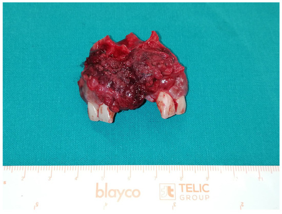
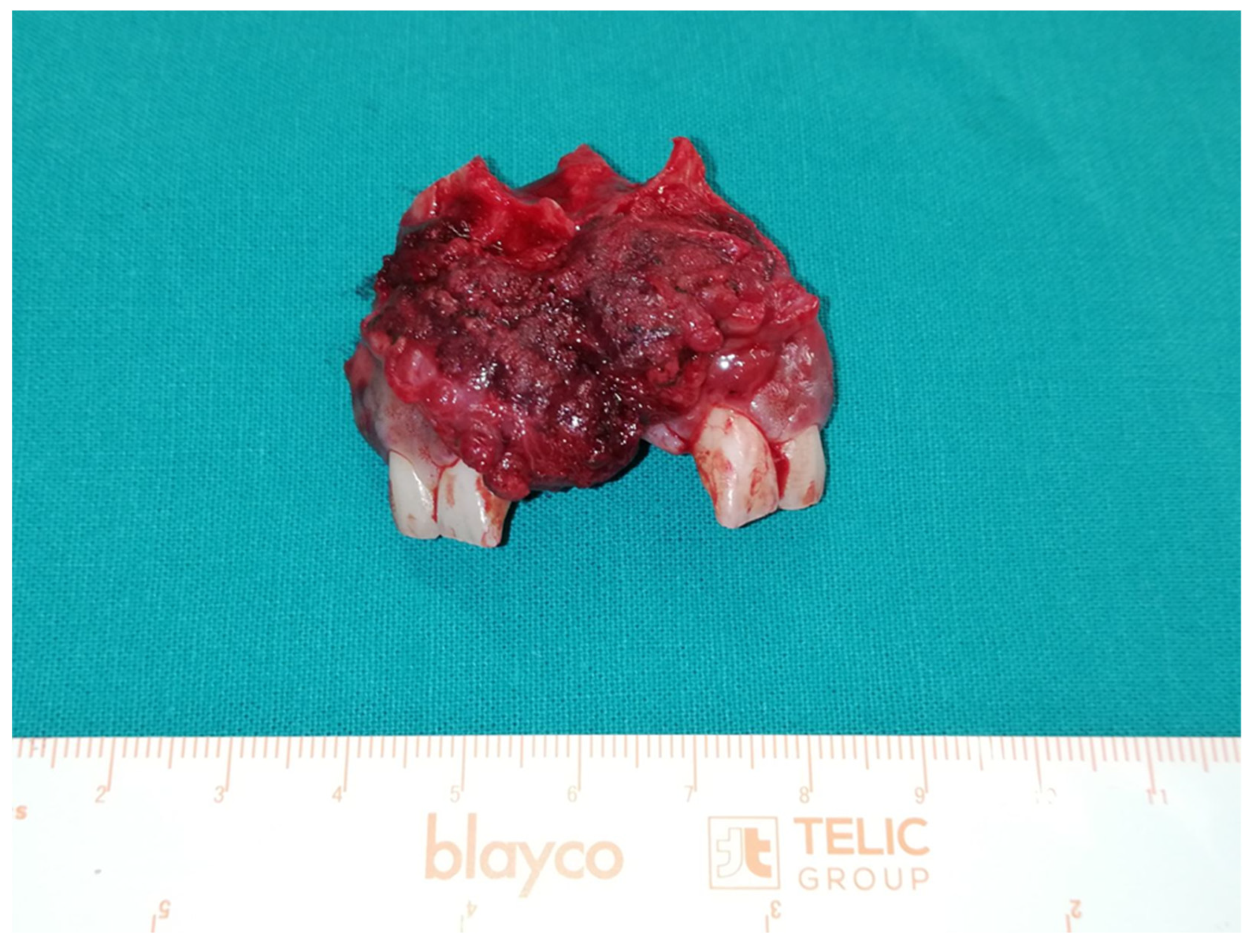
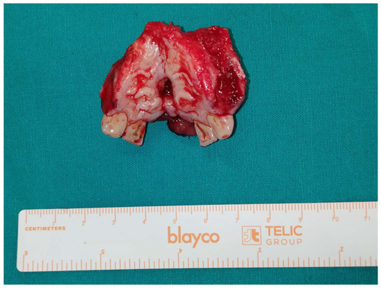
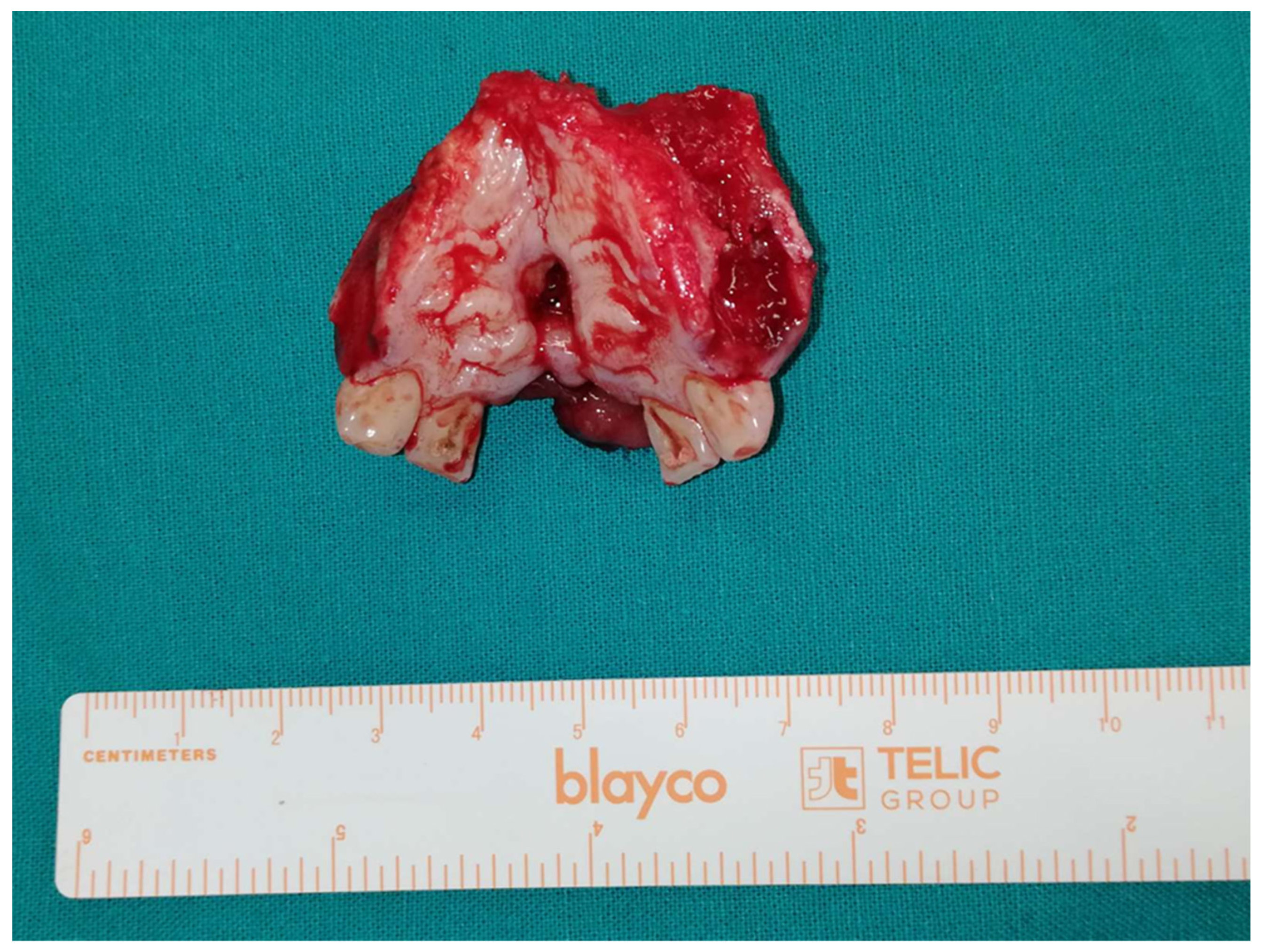
A 31-year-old male patient was referred for definitive prosthetic rehabilitation after partial maxillectomy performed one year earlier for tumour resection. Histopathological evaluation established a diagnosis of spindle cell carcinoma (sarcomatoid carcinoma). The patient underwent an en bloc radical anterior partial maxillectomy, with specimen dimensions measuring 4.5 × 3.3 × 2.1 cm (Figure 1 and Figure 2). The superior margin corresponded to the anterior nasal floor, while the lateral margins extended to the interdental alveolar bone between the maxillary first and second premolars bilaterally. Given the lesion’s anatomical location and a prior incisional biopsy confirming malignancy, surgical management was complemented by bilateral modified radical neck dissection to address potential regional lymphatic dissemination.
Figure 1.
En bloc radical anterior partial maxillectomy sagittal view.
Figure 2.
En bloc radical anterior partial maxillectomy palatal view.
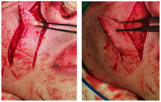
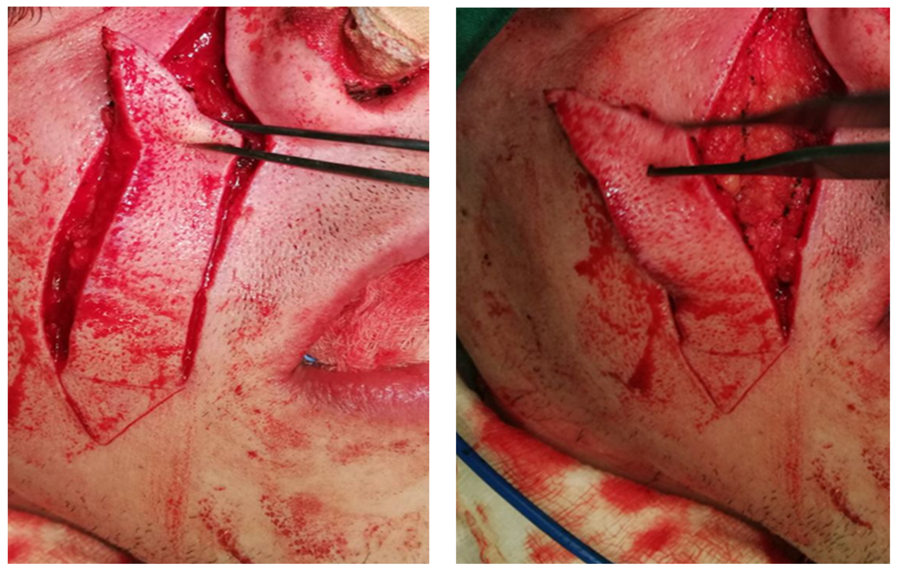
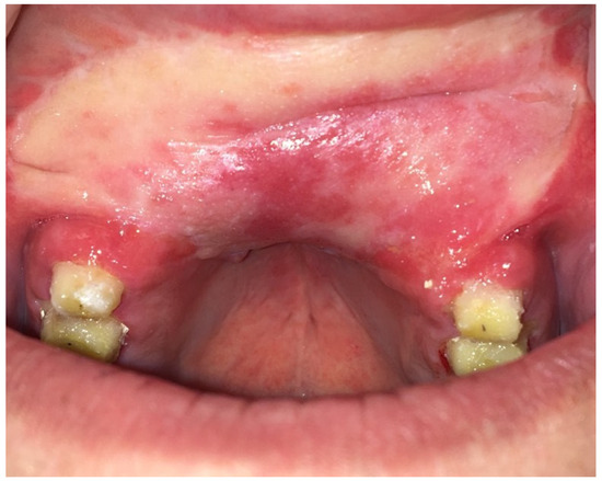
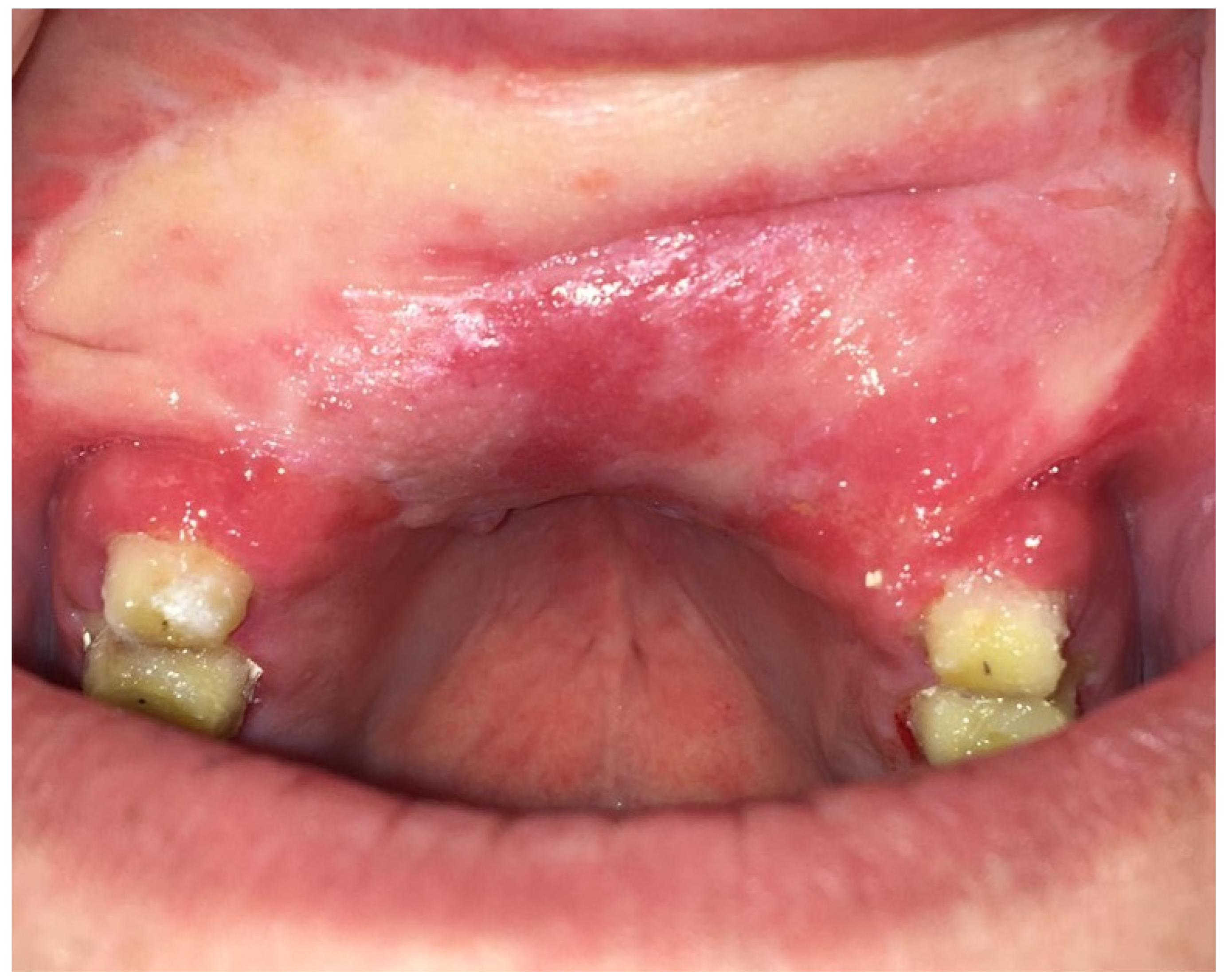
Reconstruction of the maxillary defect was achieved using bilateral nasolabial pedicled skin–cutaneous flaps (Figure 3), ensuring adequate soft tissue coverage and restoration of oronasal separation (Figure 4). In the context of comprehensive oncologic management, the patient subsequently received adjuvant radiotherapy in combination with systemic chemotherapy to reduce the risk of locoregional recurrence and improve overall disease control.
Figure 3.
Nasolabial pedicled skin–cutaneous flaps.
Figure 4.
Soft tissue closure and restoration of oronasal separation.
During the healing phase, the patient had been rehabilitated with a clasp-retained interim MRP. His chief complaints included prosthesis instability, impaired mastication, compromised speech, and reduced confidence in social interactions.
2.1. Clinical Examination
Extraoral examination revealed mild facial asymmetry without signs of tumour recurrence or soft tissue pathology. Intraoral examination demonstrated an acquired anterior maxillary defect involving the hard palate and alveolar ridge (Class I Brown’s classification) [1]. The defect was lined with well-healed mucosa, with areas of scar tissue and pronounced anatomical undercuts. Teeth #15–17 and #25–27 were present, with adequate periodontal support.
The existing interim MRP exhibited poor retention and limited stability, particularly during speech and mastication. Further evaluation revealed an acceptable vertical dimension of occlusion but uneven occlusal force distribution during function with fractured clasp assemblies, indicating possible mechanical overload.
2.2. Treatment Planning
One of the primary objectives of the selected prosthetic design was to improve prosthesis retention and stability while protecting the remaining abutment teeth. Telescopic crown systems provide a reliable frictional or conical interface between the primary crowns cemented on the abutment teeth and the secondary crowns incorporated into the removable prosthesis. This mechanism enables secure seating of the prosthesis and resistance to dislodging forces during mastication, speech, and swallowing. In the present case, the telescopic crowns also provided a controlled and predictable path of insertion, which was particularly advantageous given the complex anatomy of the maxillary defect and the presence of undercuts within the surgical site.
Another critical consideration in treatment planning was achieving favourable stress distribution along the long axes of the abutment teeth. Telescopic crowns are well documented to transmit functional loads primarily in an axial direction, thereby reducing harmful bending moments and shear stresses. This biomechanical advantage is especially important in MRPs, where uneven load distribution can compromise abutment teeth and support periodontal structures. By splinting the remaining teeth through the primary crowns, the design could further enhance load sharing among the abutments and reduce the risk of overloading individual teeth.
Minimising lateral forces on the abutment teeth was also a key objective. Excessive lateral stresses are commonly associated with clasp-retained prosthesis and may lead to tooth mobility, periodontal breakdown, and eventual abutment loss. The use of semi-precision attachments with a rotational latching mechanism enables engagement of favourable undercuts in the interproximal areas of the primary crowns, thereby enhancing retention while reducing the risk of mechanical or biological complications associated with rigid retention systems [25].
Ease of insertion and removal was also a key concern, especially considering the patient’s preference for a prosthesis that could be handled comfortably and independently. The combined use of telescopic crowns and semi-precision attachments with a rotational latching mechanism could facilitate smooth and guided insertion of the MRP, reducing the need for excessive manual force or complex insertion paths. This feature could also improve patient confidence and compliance with daily prosthesis use.
Finally, aesthetic considerations were important in selecting the treatment design. The elimination of visible clasp assemblies in the anterior maxilla produces a more natural and aesthetically acceptable appearance, which patients highly value. The proposed treatment plan, including alternative options such as conventional clasp-retained MRPs and implant-supported prosthesis, was thoroughly discussed with the patient. Given the anatomical limitations, financial considerations, and the patient’s informed preference, the telescopic crown–retained MRP combined with a semi-precision attachment incorporating a rotational latching mechanism was accepted as the definitive treatment. The timeline of the Episode of Care for Maxillary Obturator Rehabilitation is presented in Table A1.
2.3. Prosthetic Procedure and Follow-Up
Following a comprehensive clinical examination, the patient underwent periodontal and endodontic evaluation to confirm the suitability of the remaining maxillary teeth as abutments for definitive prosthetic rehabilitation. All selected abutment teeth demonstrated satisfactory periodontal support and endodontic status, allowing their use in a telescopic crown–retained MRP design. Tooth preparation emphasised parallel axial walls and a common path of insertion, both essential for the predictable function of splinted telescopic crown systems. A controlled reduction protocol was implemented to achieve uniform coping thickness and optimise rigidity of the primary crowns. Equigingival finishing margins were intentionally selected to facilitate periodontal accessibility and minimise plaque-induced biomechanical compromise at the tooth–prosthesis interface.
Impression procedures employed a dual retraction cord technique with polyether material (Impregum, 3M ESPE) to ensure high-fidelity capture of both hard and soft tissue structures, including defect morphology critical for load distribution analysis. The interim prosthesis served as a functional biomechanical simulator, allowing iterative assessment of occlusal load transfer, cantilever behaviour, and prosthesis stability under functional excursion.
The patient was instructed to wear the provisional MRP for a 3-month period, during which function, phonetics, comfort, and prosthesis stability were carefully evaluated. This phase allowed identification and correction of issues affecting speech intelligibility, masticatory efficiency, insertion and removal mechanics, and patient tolerance during daily activities. The MRP borders within the defect were carefully assessed to ensure adequate coverage without irritation of the surrounding soft tissues. Chairside adjustments were performed to adjust occlusal contacts, optimise phonetics, and improve base extensions, based on clinical findings and patient feedback.
Once the provisional restoration proved satisfactory in function, stability, and patient acceptance, it served as a diagnostic reference for the fabrication of the definitive prosthesis. The dental technician duplicated the provisional MRP using conventional techniques, employing silicone putty (Express XT putty quick 3M ESPE) to accurately transfer its contours, extensions, and functional morphology. This process ensured that the definitive MRP preserved the clinically validated contours and functional characteristics established during the provisional phase.
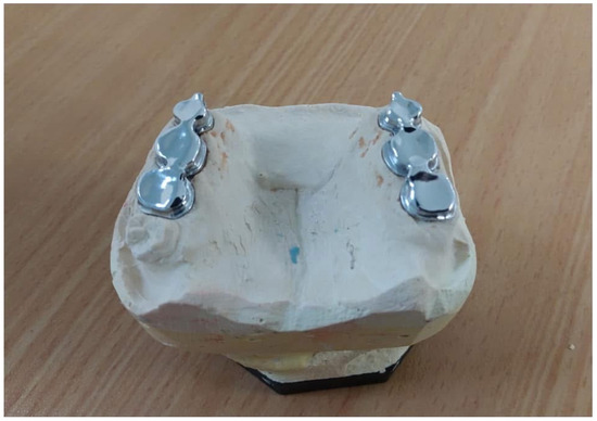
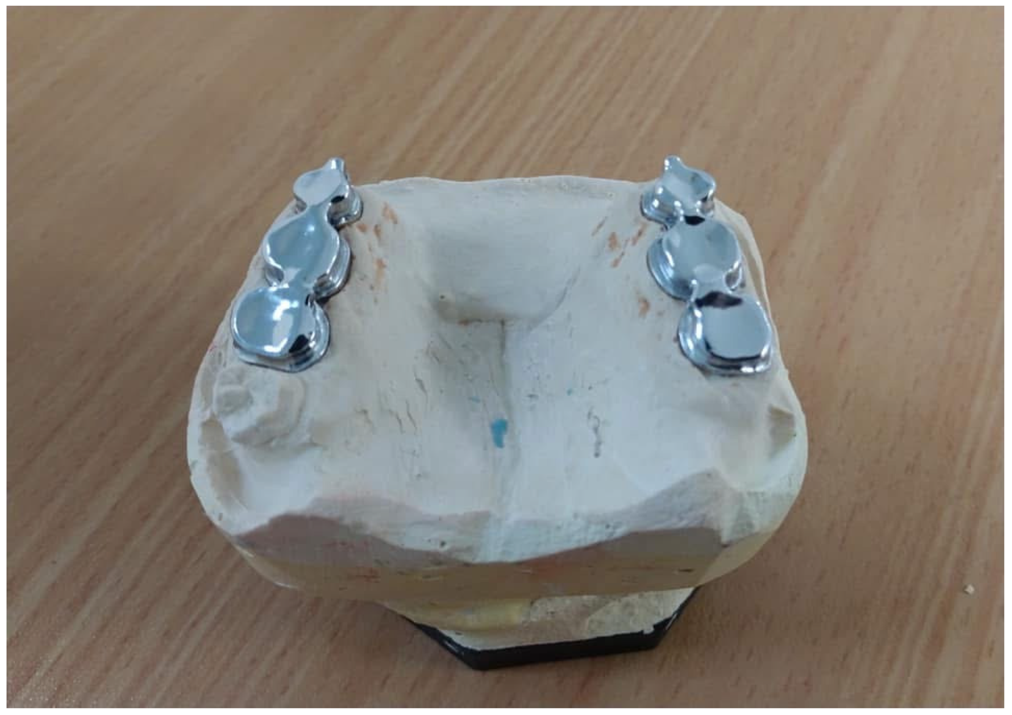
Definitive impressions were subsequently obtained using a double retraction cord and polyether impression material to accurately capture the prepared abutment teeth, residual ridge, and defect morphology. Attention was given to recording defect undercuts and soft-tissue contours to facilitate optimal prosthesis design. Master casts were fabricated, and the primary telescopic crowns were designed and cast in a cobalt–chromium alloy (Figure 5), selected for its favourable mechanical properties, corrosion resistance, and long-term clinical performance. The primary crowns were evaluated intraorally for marginal integrity, passive fit, and parallelism. Retention was verified intraorally through standardised seating force assessment, ensuring homogenous frictional engagement across all abutments and minimising asymmetric load distribution.
Figure 5.
Primary crowns on definitive cast model.
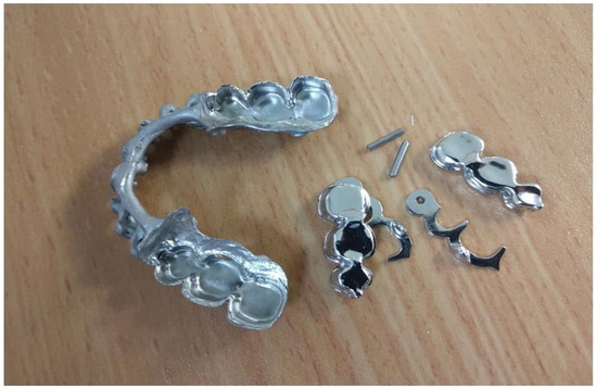
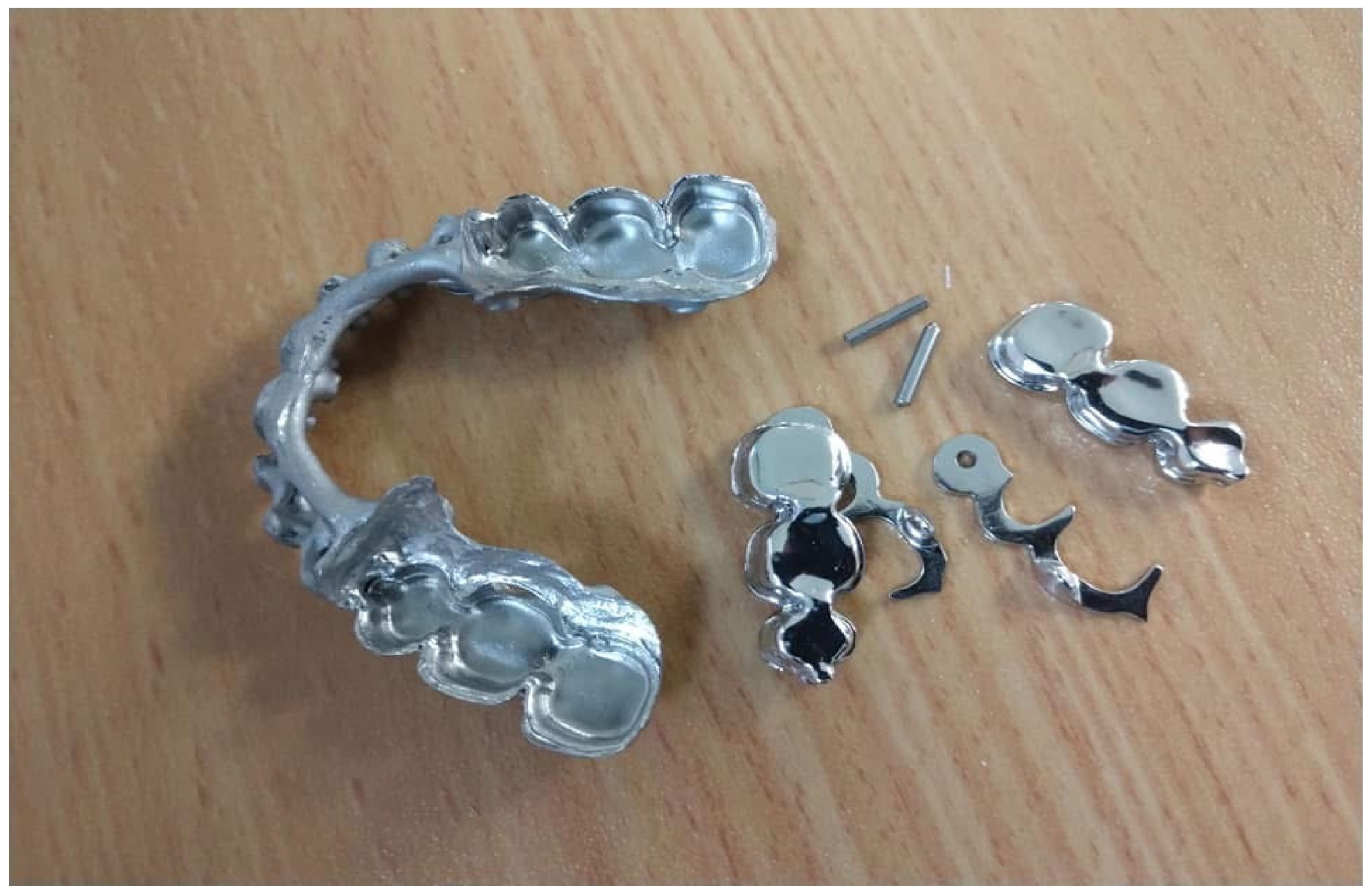
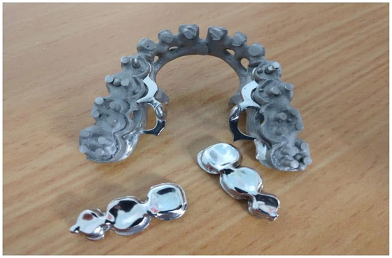
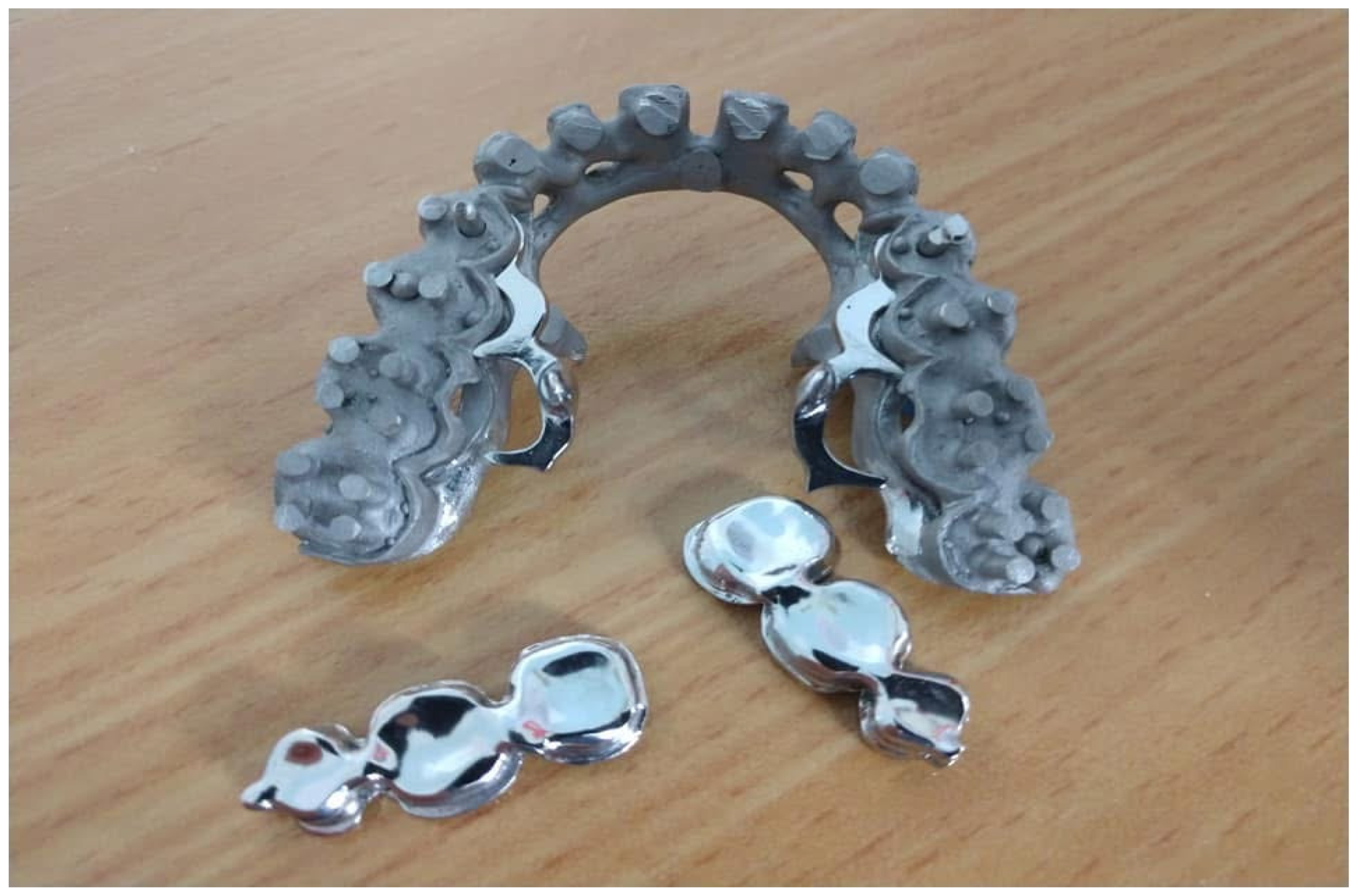
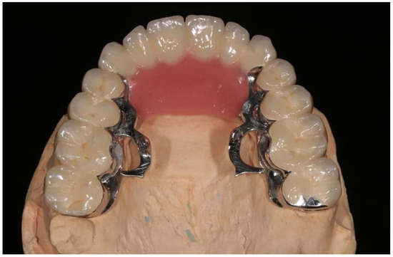
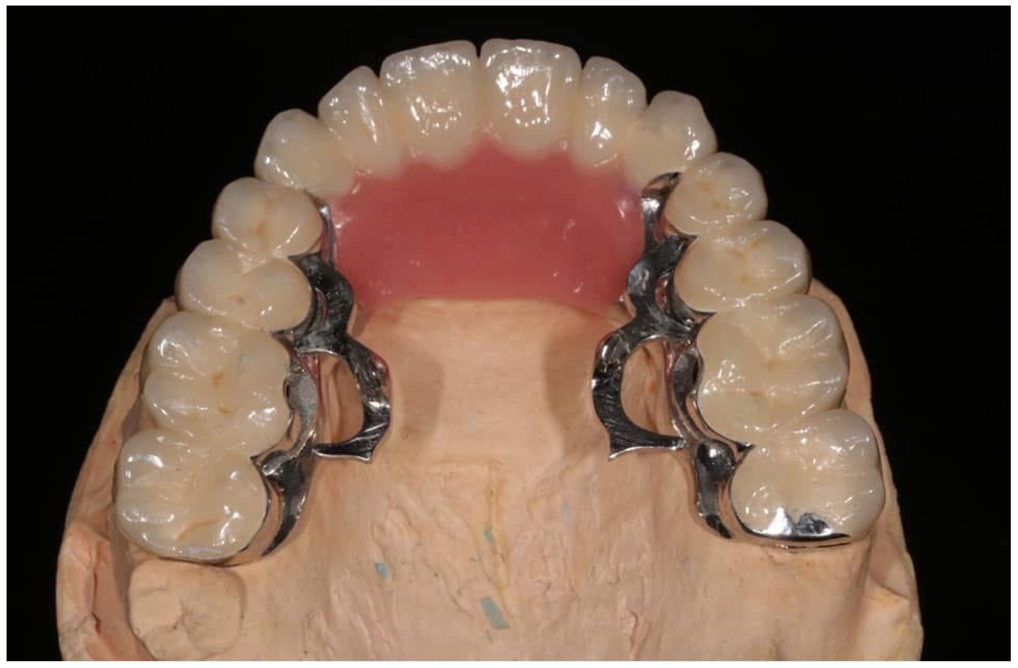
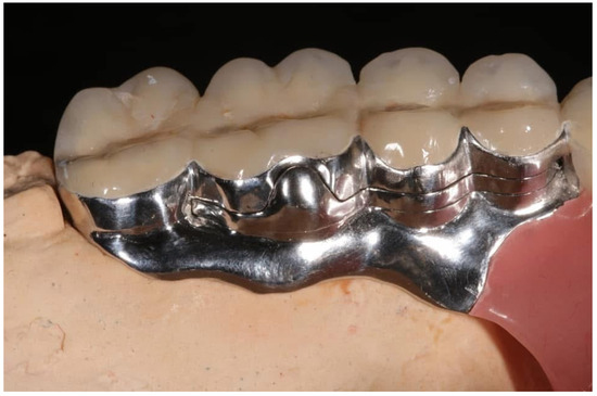
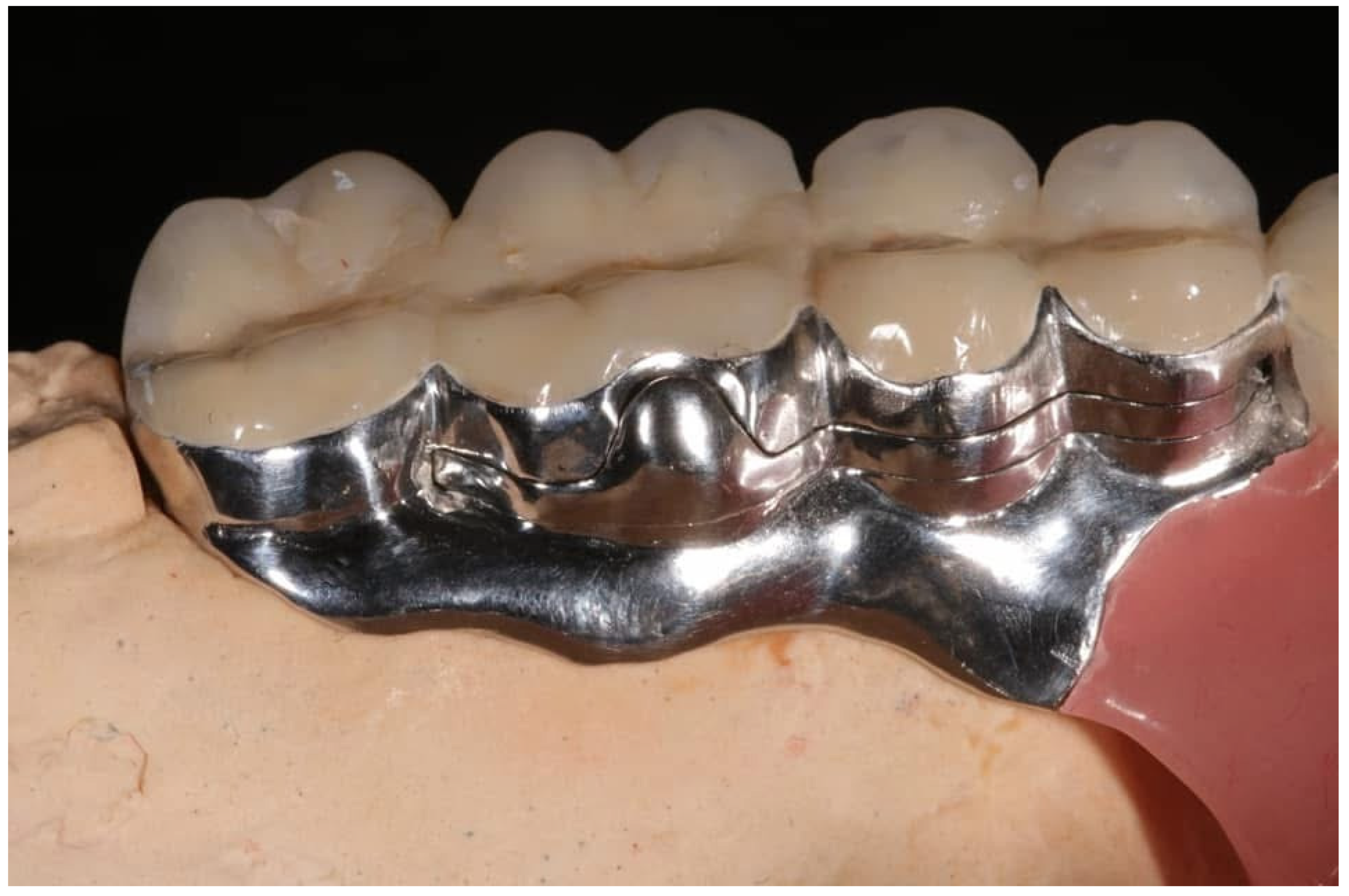
Secondary telescopic crowns were subsequently fabricated and incorporated into the metal framework of the MRP. This integration ensured precise alignment between primary and secondary components, providing reliable frictional retention and a controlled insertion path. Semi-precision attachments with a rotational latching mechanism were strategically incorporated into the metal framework of the major connector, extending from the distal interproximal area of the second molars to the mesial interproximal space of the second premolars bilaterally (Figure 6 and Figure 7). This configuration effectively redistributed functional stresses away from the anterior defect extension, reduced lever-arm magnitude, and introduced controlled rotational degrees of freedom during insertion, thereby decreasing peak stress concentrations at the abutment–prosthesis interface.
Figure 6.
Secondary metal framework, primary metal framework and rotational elements of the latching mechanism.
Figure 7.
Occlusal view of secondary metal framework, primary metal framework and rotational elements of the latching mechanism incorporated into the secondary metal framework.
A diagnostic wax-up was performed on the secondary metal framework and evaluated during the clinical try-in appointment. Occlusal relationships were assessed and refined to ensure even and harmonious contacts, and phonetic evaluation confirmed adequate speech intelligibility. Minor adjustments to the framework and occlusion were made to optimise comfort and function while maintaining prosthesis stability and retention. The MRP acrylic resin base was designed to maximise support and stability while minimising prosthesis weight, particularly in the region of the maxillary defect.
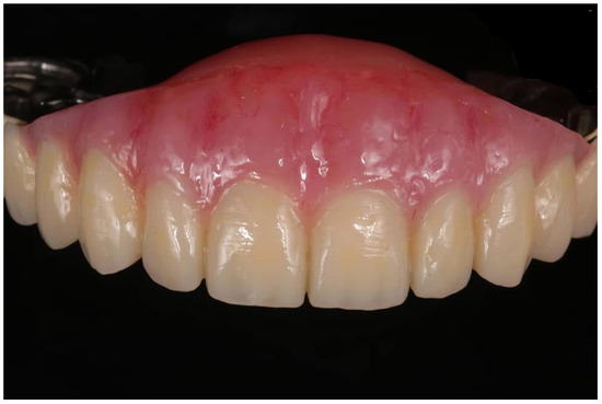
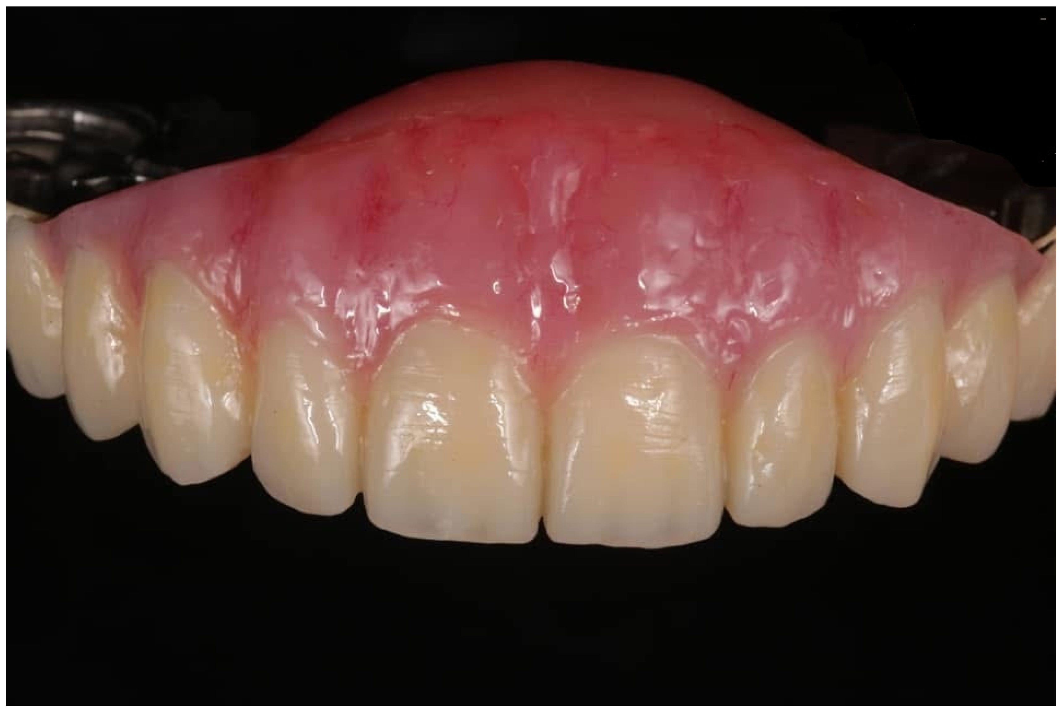
The prosthesis was fabricated using the Anaxdent flasking system, and the metal secondary framework was veneered with ANAXBLEND composite facing material according to the manufacturer’s instructions. The primary crowns were then definitively cemented using polycarboxylate luting cement (ANAXUSA). After verification of all clinical parameters, the definitive MRP prosthesis—consisting of an ANAXBLEND veneered cobalt–chromium secondary framework—was delivered (Figure 8, Figure 9 and Figure 10). A group function occlusal scheme was implemented.
Figure 8.
Occlusal view with latching mechanism in opened position.
Figure 9.
Latching mechanism in closed position.
Figure 10.
Frontal view of maxillary resection prosthesis.
From a biomechanical perspective, the combined system functioned as a rigid–resilient hybrid construct: telescopic crowns provided primary vertical support and load-sharing, whereas the rotational latching mechanism facilitated adaptive engagement of defect undercuts without generating deleterious shear forces. This dual-mechanism design improved stress homogenization across abutments and reduced localised strain accumulation, particularly in cantilevered anterior segments.
The patient received detailed instructions regarding prosthesis insertion and removal, daily cleaning procedures, and maintenance of oral and periodontal hygiene. Regular follow-up visits were scheduled to monitor abutment health and prosthesis performance. Maintenance protocols were structured to preserve the biological integrity of the telescopic interface. Mechanical cleaning protocols were specified to prevent abrasive wear of frictional surfaces, while chemical cleansing agents were selected based on non-corrosive compatibility with cobalt–chromium alloys. Periodic recall was emphasised to monitor frictional coefficient stability, as progressive wear at the primary–secondary interface is a known determinant of long-term retention loss in double-crown systems.
Follow-up evaluations were conducted at 1, 3, and 6 months after prosthesis delivery. Clinical examination demonstrated stable prosthesis retention, satisfactory tissue adaptation, and absence of mucosal irritation. The abutment teeth remained stable, with no evidence of increased mobility or periodontal deterioration. The patient reported marked improvement in speech clarity, masticatory efficiency, and overall comfort compared with the previously used prosthesis. These findings indicated a favourable short-term outcome and supported the clinical effectiveness of combining telescopic crowns with semi-precision attachments incorporating a rotational latching mechanism in maxillary MRP rehabilitation.
3. Discussion
Based on clinical findings and the patient’s functional and aesthetic expectations, a definitive MRP retained by telescopic crowns, in combination with a semi-precision attachment incorporating a rotational latching mechanism, was selected as the treatment of choice. This decision was guided by the need to achieve predictable retention, stability, and controlled prosthesis insertion in the presence of a large anterior maxillary defect and a limited number of remaining posterior teeth. Conventional retention methods were considered insufficient to meet the biomechanical and functional demands of this case, particularly given the irregular defect anatomy and the patient’s prior experience with an unstable interim MRP.
By basing the definitive MRP on a well-adjusted provisional prosthesis, the need for extensive post-delivery adjustments was minimised and patient adaptation to the final restoration was facilitated. Consequently, the definitive MRP provided predictable fit, improved function, and enhanced patient comfort, highlighting the clinical advantages of incorporating a provisional diagnostic phase in maxillary MRP rehabilitation.
Successful rehabilitation of acquired maxillary defects with MRPs depends largely on achieving adequate retention, stability, and support while preserving the health of the remaining oral structures. In partially edentulous patients, these objectives are particularly challenging due to limited abutment availability, unfavourable tooth distribution, compromised ridge anatomy, and the presence of scar tissue or undercuts within the defect [26,27,28].
Numerous clinical and in vitro studies have demonstrated the biomechanical superiority of telescopic systems. Arnold et al. (2020) [29] reported significantly higher and more stable retention forces for telescopic crowns than for conventional clasp assemblies. Finite element analyses have further confirmed that telescopic systems distribute stress more evenly across abutment teeth and supporting structures [30]. These findings are clinically relevant in maxillary MRP rehabilitation, where uneven force distribution may compromise prosthesis stability and tissue health.
In the present case, telescopic crowns provided a controlled path of insertion and predictable seating of the MRP despite the irregular defect anatomy. The splinting effect of the primary crowns enhanced the load-bearing capacity of the remaining abutment teeth, reducing the risk of individual tooth overload. Additionally, eliminating visible clasps improved aesthetics, an important factor for patient acceptance and satisfaction.
While telescopic crowns provide rigid support, exclusive reliance on rigid retention may not always be optimal in MRPs. Extensive maxillary defects often present favourable undercuts that can enhance retention if engaged appropriately. However, rigid engagement of these undercuts may lead to excessive stress on abutment teeth and difficulty during insertion and removal. Semi-precision attachments incorporating a rotational latching mechanism address this challenge by allowing controlled prosthesis movement and rotational seating paths.
Saverio et al. (2020) emphasised that attachment systems in MRPs should provide a degree of resilience to accommodate functional movements and tissue displacement [31]. Semi-precision attachments with a rotational latching mechanism allow the prosthesis to rotate into position during insertion, facilitating engagement of defect undercuts without imposing excessive lateral forces on abutment teeth. This concept is particularly beneficial in patients with limited mouth opening or complex anatomical constraints [24].
Indirect retention represents the resistance that prevents rotational displacement of a removable prosthesis around its fulcrum line. In removable prosthodontics, indirect retainers are typically positioned away from the rotational axis to counteract dislodging forces. In the present case, the rotational axis passed approximately through the second premolar region, while the semi-precision latching attachments were positioned distally on the molars. This configuration helped reduce rotational forces generated by the large anterior cantilever MRP while allowing controlled resilience to accommodate tissue displacement. Such a design enhances prosthesis stability while protecting abutment teeth from excessive mechanical stress.
The synergistic use of telescopic crowns and semi-precision attachments with a rotational latching mechanism in the present case reflects a rigid-flexible retention philosophy. Telescopic crowns provided primary support and axial load transfer, while the semi-precision attachment enhanced retention by utilising defect undercuts in a controlled manner. Finite element studies have demonstrated that resilient attachment systems can reduce stress concentrations around abutments compared with rigid connectors, supporting the biomechanical rationale for this combined approach [30].
Patient-reported outcomes are increasingly recognised as critical measures of prosthodontic success. Studies have shown that double-crown–retained prosthesis are associated with significant improvements in oral health–related quality of life, speech, comfort, and masticatory efficiency compared with clasp-retained designs [15,16]. The favourable patient feedback observed in the present case aligns with these findings.
Overall, this design was considered superior to alternative prosthetic modalities due to anatomical and patient-specific limitations. A swing-lock obturator was contraindicated because of the patient’s young age, heightened aesthetic demands, and inadequate anterior sulcus depth precluding labial bar placement. Magnetic attachments were unsuitable owing to limited prosthetic space and their reduced capacity to resist horizontal and rotational forces, rendering them inadequate for controlling movement associated with the large anterior cantilever. Implant-supported rehabilitation was precluded by the absence of osseous reconstruction, as the defect had been restored exclusively with bilateral nasolabial pedicled skin–cutaneous flaps. Conventional obturators were likewise unsuitable because the soft tissue-lined defect lacked adequate dentoalveolar and osseous support for predictable retention and stability.
Despite its advantages, the combined use of telescopic crowns and semi-precision attachments with a rotational latching mechanism is not without limitations. The technique requires meticulous clinical execution, precise laboratory fabrication, and higher treatment costs compared with conventional MRP designs. Material selection also plays a crucial role in long-term performance. Recent studies have investigated a wide range of materials for telescopic systems, including cobalt-chromium alloys, zirconia, PEEK, and PEKK, each demonstrating distinct mechanical and retentive properties [21,32,33].
Long-term clinical studies have reported favourable survival rates for telescopic crown–retained prosthesis, with follow-up periods exceeding 10 years [18,34]. However, complications such as loss of retention, wear of crown interfaces, and maintenance requirements must be considered during treatment planning [35]. Regular follow-up and patient compliance with hygiene protocols are essential for long-term success.
Another limitation of this case report is its inherent lack of generalisability. While the presented approach proved effective in this patient, treatment decisions must be individualised based on defect size, remaining dentition, patient expectations, and financial considerations. Future research should focus on prospective clinical studies and comparative trials evaluating different retention strategies for maxillary MRP prosthesis.
Additionally, comparative evidence suggests that prosthetic rehabilitation, while effective, may still result in reduced quality of life compared with surgical reconstruction in certain cases, highlighting persistent functional limitations of conventional designs [36]. Within this evolving landscape, telescopic crown–retained removable partial dentures have demonstrated favourable long-term outcomes, with [37]. reporting stable performance and predictable function over extended follow-up periods. Collectively, these findings underscore a gradual transition toward more biomechanically refined and attachment-based prosthetic solutions for maxillary defect rehabilitation.
Recent research highlights that retention in maxillofacial prostheses can be achieved through a variety of systems, including precision attachments, telescopic mechanisms, and magnets [38,39]. Moreover, a two-piece, magnet-retained obturator may offer improved patient satisfaction and more effective restoration of orofacial function compared to conventional designs in the management of completely edentulous patients [40]. While magnetic retainers offer ease of insertion and removal, they are associated with reduced retentive stability over time and require strict maintenance due to corrosion risk and plaque accumulation [41]. In contrast, mechanically based systems such as telescopic crowns provide superior long-term stability and controlled load distribution [42]. Implant-supported obturators are strongly recommended for the rehabilitation of permanent oral and maxillofacial defects. Moreover, telescopic crown attachments appear to offer better longevity. In contrast, clasp-retained obturators and surgical splints tend to have shorter survival times and are therefore more suitable for temporary use [43]. Furthermore, patient compliance [44,45] and hygiene maintenance remain critical determinants of prosthesis longevity across all retention modalities [46].
In parallel, attachment-retained prostheses, particularly those employing swing-lock or precision attachment concepts, have been explored to improve retention and distribution of functional loads in complex maxillofacial cases. Swing-lock systems have been reported as effective adjuncts in removable prosthodontics, offering enhanced retention and adaptability in cases with compromised dentition [47,48]. Their application has also been extended to patients with congenital syndromes and extensive tooth loss, demonstrating improved prosthesis stability and retention [49,50]. More recently, swing-lock–based prosthetic reconstruction in maxillectomy patients was introduced [51], highlighting its ability to improve prosthesis engagement and functional performance through controlled locking mechanisms. These systems collectively demonstrate the biomechanical advantage of controlled engagement of undercuts while minimising deleterious lateral forces on abutments. However, despite these advancements, the authors did not identify any previous report that has described the combined application of telescopic crown systems with a semi-precision attachment incorporating a rotational latching mechanism for maxillary resection prostheses. The present case, therefore, introduces a novel rigid–flexible hybrid design, where telescopic crowns provide axial support and controlled insertion, while the rotational latching mechanism enables adaptive engagement of defect undercuts without overloading abutment teeth. This combination represents a novel treatment protocol in maxillofacial prosthodontics, integrating principles of precision retention and controlled resilience within a single prosthetic framework, thereby addressing a persistent gap in current rehabilitative strategies.
Although this study is limited by its design as a single case report with short-term follow-up, absence of objective biomechanical measurements, and lack of validated patient-reported outcome instruments, these constraints do not diminish its clinical relevance. As an initial observation, it offers meaningful insights and serves to generate hypotheses, providing a basis for future research with larger cohorts, longer follow-up, and standardised outcome measures.
4. Conclusions
The use of telescopic crowns in combination with a semi-precision attachment incorporating a rotational latching mechanism provides a predictable and effective strategy for rehabilitating maxillary defects in partially edentulous patients. Telescopic crowns offer rigid support, splint the remaining abutments, and allow controlled axial load transfer, thereby minimising lateral forces and preserving abutment health. Semi-precision attachments with a rotational latching mechanism complement this system by permitting controlled engagement of defect undercuts, enhancing retention without compromising prosthesis insertion or patient comfort.
The integration of these two retention mechanisms addresses common challenges associated with MRP instability, including uneven force distribution, difficult seating, and functional limitations. Clinically, this hybrid approach may improve masticatory efficiency, speech intelligibility, and overall patient satisfaction.
While meticulous planning and laboratory precision are required, the technique may offer advantages over conventional clasp-retained MRPs, combining biomechanical efficiency with improved aesthetics. Within the limitations of a single clinical case, this report supports the use of telescopic crown–retained MRPs with semi-precision attachments incorporating a rotational latching mechanism as a valuable treatment option in complex maxillofacial prosthetic rehabilitation.
Author Contributions
Conceptualization, P.C.; methodology, P.C.; software, J.F.; validation, P.C., S.K. and O.N.; formal analysis, P.C., S.K. and O.N.; investigation, P.C., S.K. and O.N.; resources, P.C., S.K. and O.N.; data curation, P.C., S.K. and O.N.; writing—original draft preparation, P.C., S.K. and O.N.; writing—review and editing, P.C., S.K., J.F., M.T. and O.N.; visualisation, P.C., S.K. and O.N.; supervision, S.K. and O.N.; project administration, P.C. All authors have read and agreed to the published version of the manuscript.
Funding
This publication is financed by the Project “Strengthening and optimising the operation of MODY services and academic and research units of the Hellenic Mediterranean University”, funded by the Public Investment Program of the Greek Ministry of Education and Religious Affairs.
Institutional Review Board Statement
All procedures were performed as part of routine clinical care. Ethical approval was not required for this case report in accordance with institutional guidelines of the School of Dentistry, Aristotle University of Thessaloniki.
Informed Consent Statement
Informed consent, including consent for treatment and the use of clinical photographs, was obtained from the patient before treatment.
Data Availability Statement
The original contributions presented in this study are included in the article. Further inquiries can be directed to the corresponding author.
Acknowledgments
This publication is financed by the Project “Strengthening and optimizing the operation of MODY services and academic and research units of the Hellenic Mediterranean University”, funded by the Public Investment Program of the Greek Ministry of Education and Religious Affairs. The authors would like to acknowledge Konstantinos Vactshevanos, maxillofacial surgeon Antonis Tsekos and his surgical team for their clinical support. The authors also thank J. Eimecke, dental technician, for exceptional dental technology skills and technical assistance.
Conflicts of Interest
The authors declare no conflicts of interest.
Abbreviations
The following abbreviations are used in this manuscript:
| MRP | Maxillary Resection Prosthesis |
| RPD | Removable Partial Denture |
| CAD/CAM | Computer-Aided Design/Computer-Aided Manufacturing |
| PEEK | Polyether Ether Ketone |
| PEKK | Polyether Ketone Ketone |
| Co-Cr | Cobalt-Chromium |
| VDO | Vertical Dimension of Occlusion |
Appendix A
Table A1.
Timeline of the Episode of Care for Maxillary Obturator Rehabilitation.
References
- Brown, J.S.; Rogers, S.N.; McNally, D.N.; Boyle, M. A modified classification for the maxillectomy defect. Head Neck 2000, 22, 17–26. [Google Scholar] [CrossRef] [Scilit]
- Kornblith, A.B.; Zlotolow, I.M.; Gooen, J.; Huryn, J.M.; Lerner, T.; Strong, E.W.; Shah, J.P.; Spiro, R.H.; Holland, J.C. Quality of life of maxillectomy patients using an obturator prosthesis. Head Neck 1996, 18, 323–334. [Google Scholar] [CrossRef]
- Mittal, M.; Sharma, R.; Kalra, A.; Sharma, P. Form, Function, and Esthetics in Prosthetically Rehabilitated Maxillary Defects. J. Craniofacial Surg. 2018, 29, E8–E12. [Google Scholar] [CrossRef] [Scilit] [PubMed]
- Andrades, P.; Militsakh, O.; Hanasono, M.M.; Rieger, J.; Rosenthal, E.L. Current Strategies in Reconstruction of Maxillectomy Defects. Arch. Otolaryngol. Head Neck Surg. 2011, 137, 806–812. [Google Scholar] [CrossRef] [Scilit] [PubMed]
- Moreno, M.A.; Skoracki, R.J.; Hanna, E.Y.; Hanasono, M.M. Microvascular free flap reconstruction versus palatal obturation for maxillectomy defects. Head Neck 2010, 32, 860–868. [Google Scholar] [CrossRef] [Scilit]
- Hanasono, M.M.; Silva, A.K.; Yu, P.; Skoracki, R.J. A Comprehensive Algorithm for Oncologic Maxillary Reconstruction. Plast. Reconstr. Surg. 2013, 131, 47–60. [Google Scholar] [CrossRef] [Scilit]
- Costa, H.; Zenha, H.; Sequeira, H.; Coelho, G.; Gomes, N.; Pinto, C.; Martins, J.; Santos, D.; Andresen, C. Microsurgical reconstruction of the maxilla: Algorithm and concepts. J. Plast. Reconstr. Aesthetic Surg. 2015, 68, e89–e104. [Google Scholar] [CrossRef] [Scilit]
- Alwadeai, M.S.; Al-Aroomy, L.A.; Shindy, M.I.; Amin, A.A.-W.; Zedan, M.H. Aesthetic reconstruction of onco-surgical maxillary defects using free scapular flap with and without CAD/CAM customized osteotomy guide. BMC Surg. 2022, 22, 362. [Google Scholar] [CrossRef] [Scilit] [PubMed]
- Cuéllar, C.N.; Martínez, E.B.; Cuéllar, I.N.; López, A.M.L.; Rial, M.T.; Pérez, A.S.; Escobar, J.I.S. Primary Maxillary Reconstruction with Fibula Flap and Dental Implants: A Comparative Study Between Virtual Surgical Planning and Standard Surgery in Class IIC Defects. J. Oral Maxillofac. Surg. Off. J. Am. Assoc. Oral Maxillofac. Surg. 2021, 79, 237–248. [Google Scholar] [CrossRef] [Scilit]
- Haug, S.P. Maxillofacial Prosthetic Management of the Maxillary Resection Patient. Atlas Oral Maxillofac. Surg. Clin. N. Am. 2007, 15, 51–68. [Google Scholar] [CrossRef] [Scilit]
- Okay, D.J.; Genden, E.; Buchbinder, D.; Urken, M. Prosthodontic guidelines for surgical reconstruction of the maxilla: A classification system of defects. J. Prosthet. Dent. 2001, 86, 352–363. [Google Scholar] [CrossRef] [Scilit] [PubMed]
- Parr, G.R.; Tharp, G.E.; O Rahn, A. Prosthodontic principles in the framework design of maxillary obturator prostheses. J. Prosthet. Dent. 1989, 62, 205–212. [Google Scholar] [CrossRef] [Scilit]
- Ishida, K.; Nogawa, T.; Takayama, Y.; Saito, M.; Yokoyama, A. Prognosis of double crown-retained removable dental prostheses compared with clasp-retained removable dental prostheses: A retrospective study. J. Prosthodont. Res. 2017, 61, 268–275. [Google Scholar] [CrossRef] [Scilit]
- Oh, W.-s.; Oh, J.-y.; May, K. Loss of abutment teeth for double crown-retained removable partial dentures compared to clasp-retained removable partial dentures: A systematic review and meta-analysis. Quintessence Int. 1985, 52, 412–424. [Google Scholar] [CrossRef] [Scilit]
- Seo, J.-G.; Cho, J.-H. Clinical outcomes of rigid and non-rigid telescopic double-crown-retained removable dental prostheses: An analytical review. J. Adv. Prosthodont. 2020, 12, 38–48. [Google Scholar] [CrossRef] [Scilit] [PubMed] [PubMed Central]
- Arnold, C.; Hey, J.; Setz, J.M.; Boeckler, A.F.; Schweyen, R. Retention force of removable partial dentures with different double crowns. Clin. Oral Investig. 2018, 22, 1641–1649. [Google Scholar] [CrossRef] [Scilit]
- Stober, T.; Danner, D.; Bömicke, W.; Hassel, A.J. Improvement of oral health-related quality-of-life by use of different kinds of double-crown-retained removable partial dentures. Acta Odontol. Scand. 2016, 74, 1–6. [Google Scholar] [CrossRef] [Scilit]
- Kern, J.-S.; Hanisch, O.; Hammächer, C.; Yildirim, M.; Wolfart, S. Telescopic Crowns on Implants and Teeth: Evaluation of a Clinical Study After 8 to 12 Years. Int. J. Oral Maxillofac. Implant. 2019, 34, 977–986. [Google Scholar] [CrossRef] [Scilit]
- Stock, V.; Schmidlin, P.R.; Merk, S.; Wagner, C.; Roos, M.; Eichberger, M.; Stawarczyk, B. PEEK Primary Crowns with Cobalt-Chromium, Zirconia and Galvanic Secondary Crowns with Different Tapers—A Comparison of Retention Forces. Materials 2016, 9, 187. [Google Scholar] [CrossRef] [Scilit]
- Priester, M.; Müller, W.-D.; Beuer, F.; Schmidt, F.; Schwitalla, A.D. Performance of PEEK based telescopic crowns, a comparative study. Dent. Mater. 2021, 37, 1667–1675. [Google Scholar] [CrossRef] [Scilit]
- Tasaka, A.; Schwindling, F.S.; Rues, S.; Rammelsberg, P.; Yamashita, S. Retentive force of telescopic crowns combining fiber-reinforced composite and zirconia. J. Prosthodont. Res. 2022, 66, 265–271. [Google Scholar] [CrossRef] [Scilit]
- Ghiaz, K.; Shaik, S.S.; John, P.; Kumar, A.; Vijayakumar, N. Precision Attachment Boon to Prosthodontists. J. Pharm. Bioallied Sci. 2022, 14, S1030–S1032. [Google Scholar] [CrossRef] [Scilit] [PubMed]
- dos Santos Nunes Reis, J.M.; da Cruz Perez, L.E.; Alfenas, B.F.; de Oliveira Abi-Rached, F.; Filho, J.N. Maxillary Rehabilitation Using Fixed and Removable Partial Dentures with Attachments: A Clinical Report. J. Prosthodont. Off. J. Am. Coll. Prosthodont. 2014, 23, 58–63. [Google Scholar] [CrossRef] [Scilit]
- Palmeiro, M.R.L.; Piffer, C.S.; Brunetto, V.M.; Maccari, P.C.; Shinkai, R.S.A. Maxillary Rehabilitation Using a Removable Partial Denture with Attachments in a Cleft Lip and Palate Patient: A Clinical Report. J. Prosthodont. Off. J. Am. Coll. Prosthodont. 2015, 24, 250–253. [Google Scholar] [CrossRef] [Scilit]
- Chan, M.F.; Adams, D.; Brudvik, J.S. The swing-lock removable partial denture in clinical practice. Dent. Update 1998, 25, 80–84. [Google Scholar]
- Aramany, M.A. Basic principles of obturator design for partially edentulous patients. Part I: Classification. J. Prosthet. Dent. 1978, 40, 554–557. [Google Scholar] [CrossRef] [Scilit] [PubMed]
- Cao, Y.; Yu, C.; Liu, W.; Miao, C.; Han, B.; Yang, J.; Li, L.; Li, C. Obturators versus flaps after maxillary oncological ablation: A systematic review and best evidence synthesis. Oral Oncol. 2018, 82, 152–161. [Google Scholar] [CrossRef] [Scilit]
- Ahmed, Z.U.; Flynn, J.; Riedel, E.R.; Huryn, J.M.; Rosen, E.B. Definitive maxillary obturator prosthesis: Timelines for fabrication and follow-up. Spec. Care Dent. Off. Publ. Am. Assoc. Hosp. Dent. Acad. Dent. Handicap. Am. Soc. Geriatr. Dent. 2020, 40, 315–319. [Google Scholar] [CrossRef] [Scilit]
- Arnold, C.; Schweyen, R.; Boeckler, A.; Hey, J. Retention Force of Removable Partial Dentures with CAD-CAM-Fabricated Telescopic Crowns. Materials 2020, 13, 3228. [Google Scholar] [CrossRef] [Scilit] [PubMed]
- Al-Kordy, N.M.A.; Al-Saadi, M.H. Finite Element Study of Stress Distribution with Tooth-Supported Mandibular Overdenture Retained by Ball Attachments or Resilient Telescopic Crowns. Eur. J. Dent. 2023, 17, 539–547. [Google Scholar] [CrossRef] [Scilit]
- Saverio, C.; Antonio, B.; Hao, H.Z.; Silvia, P.; Gianluigi, C.; Dorina, L.; Carinci, F. Definitive Palatal Obturator Applications: A Systematic Literature Review. Prosthesis 2025, 7, 112. [Google Scholar] [CrossRef] [Scilit]
- Merk, S.; Wagner, C.; Stock, V.; Eichberger, M.; Schmidlin, P.R.; Roos, M.; Stawarczyk, B. Suitability of Secondary PEEK Telescopic Crowns on Zirconia Primary Crowns: The Influence of Fabrication Method and Taper. Materials 2016, 9, 908. [Google Scholar] [CrossRef] [Scilit]
- Hussein, M. Biomechanical Performance of PEEK and Graphene-Modified PMMA as Telescopic Removable Partial Denture Materials: A Nonlinear 3D Finite Element Analysis. Int. J. Prosthodont. 2022, 35, 793–800. [Google Scholar] [CrossRef] [Scilit]
- Mautsch, C.; Klenke, J.; Kern, T.; Wolfart, S.; Kern, J.-S. Electroplated double-crowns on implants and teeth after up to 12 years—A retrospective clinical study. Int. J. Implant. Dent. 2025, 11, 9. [Google Scholar] [CrossRef] [Scilit]
- Kamel, A.; Badr, A.; Fekry, G.; Tsoi, J. Parameters Affecting the Retention Force of CAD/CAM Telescopic Crowns: A Focused Review of In Vitro Studies. J. Clin. Med. 2021, 10, 4429. [Google Scholar] [CrossRef] [Scilit] [PubMed]
- Breeze, J.; Rennie, A.; Morrison, A.; Dawson, D.; Tipper, J.; Rehman, K.; Grew, N.; Snee, D.; Pigadas, N. Health-related quality of life after maxillectomy: Obturator rehabilitation compared with flap reconstruction. Br. J. Oral Maxillofac. Surg. 2016, 54, 857–862. [Google Scholar] [CrossRef] [Scilit] [PubMed]
- Frisch, E.; Ratka-Krüger, P.; Wenz, H. Unsplinted implants and teeth supporting maxillary removable partial dentures retained by telescopic crowns: A retrospective study with >6 years of follow-up. Clin. Oral Implant. Res. 2015, 26, 1091–1097. [Google Scholar] [CrossRef] [Scilit]
- Bulut, A.; Türksayar, A.D.; Saglam, S. Retention systems used in maxillofacial prostheses: A review. Niger. J. Clin. Pract. 2019, 22, 1629–1634. [Google Scholar] [CrossRef] [Scilit] [PubMed]
- Mathew, R.S.; Raghavan, R.; Pa, S. Maxillofacial prosthetic rehabilitation using different retention systems: A review. IP Ann. Prosthodont. Restor. Dent. 2022, 8, 143–149. [Google Scholar] [CrossRef] [Scilit]
- Sharaf, M.Y.; Eskander, A.E. Patient Satisfaction regarding Conventional One-Piece Versus Two-Piece Magnet-Retained Obturator for Patients with Complete Edentulism: A Preliminary Crossover Clinical Trial. Open Dent. J. 2022, 16, e187421062203140. [Google Scholar] [CrossRef] [Scilit]
- Nichlani, G.; Mittal, D.A.; Joshi, M.; Ghadage, M.; Kadam, N.; Kamble, S.S. Retention in maxillofacial prostheses: A literature review. Int. J. Health Sci. 2022, 6, 2151–2163. [Google Scholar] [CrossRef] [Scilit]
- A Nazar, S.; Nair, V.V.; K, H.K.; Ravichandran, R. Retention in maxillofacial prosthetics: A review. Int. J. Appl. Dent. Sci. 2021, 7, 568–573. [Google Scholar] [CrossRef] [Scilit]
- Zierden, K.; Koch, C.J.; Wöstmann, B.; Rehmann, P. Clinical Longevity of Obturators in Patients with Jaw Defects: A Retrospective Cohort Study. Clin. Oral Investig. 2024, 28, 284. [Google Scholar] [CrossRef] [Scilit] [PubMed] [PubMed Central]
- Kamalakidis, S.; Papaemmanouil, G.; Anastassiadou, V. Exploring factors influencing complete denture management of patient with Myasthenia Gravis. Balk. J. Dent. Med. 2020, 24, 113–117. [Google Scholar] [CrossRef] [Scilit]
- Kamalakidis, S.; Anastassiadou, V.; Pissiotis, A. Exploring adaptation and satisfaction in copied complete dentures regarding two different occlusal schemes. Int. J. Prosthodont. 2023, 36, 13–19. [Google Scholar] [CrossRef] [Scilit] [PubMed]
- Costa, L.d.O.; Soares, S.G.M.; Barreto, B.N.; Almeida, F.C.; Dias, A.M.; Miranda, J.S. Recommendations for post-rehabilitation care of maxillofacial prostheses. Braz. J. Oral Sci. 2024, 23, e249184. [Google Scholar] [CrossRef] [Scilit]
- Lynch, C.D.; Allen, P.F. The Swing-Lock Denture: Its Use in Conventional Removable Partial Denture Prosthodontics. Dent. Updat. 2004, 31, 506–508. [Google Scholar] [CrossRef] [Scilit]
- McKenna, G.; Ziada, H.; Allen, P.F. Prosthodontic rehabilitation of a patient using a swing-lock lower denture after segmental mandibulectomy. Eur. J. Prosthodont. Restor. Dent. 2013, 21, 141–144. [Google Scholar] [PubMed]
- Razaq, I.; Durey, K.; Nattress, B. Provision of a swing lock denture for a patient with Gorlin Goltz syndrome. Eur. J. Prosthodont. Restor. Dent. 2012, 20, 141–144. [Google Scholar]
- Aggarwal, H.; Cho, S.-H. Complete removable dental prosthesis with the swing lock system: A clinical report. J. Prosthet. Dent. 2014, 112, 1035–1037. [Google Scholar] [CrossRef] [Scilit]
- Seong, D.-J.; Hong, S.-J.; Ha, S.-R.; Hong, Y.-G.; Kim, H.-W. Prosthetic reconstruction with an obturator using swing-lock attachment for a patient underwent maxillectomy: A clinical report. J. Adv. Prosthodont. 2016, 8, 411–416. [Google Scholar] [CrossRef] [Scilit] [PubMed]
Disclaimer/Publisher’s Note: The statements, opinions and data contained in all publications are solely those of the individual author(s) and contributor(s) and not of MDPI and/or the editor(s). MDPI and/or the editor(s) disclaim responsibility for any injury to people or property resulting from any ideas, methods, instructions or products referred to in the content. |
© 2026 by the authors. Licensee MDPI, Basel, Switzerland. This article is an open access article distributed under the terms and conditions of the Creative Commons Attribution (CC BY) license.