


Abstract
Background/Objectives: Minimally invasive aortic valve replacement via right anterior thoracotomy (Mini-AVR) has been proven safe and effective. However, the restricted surgical field through this approach makes this surgery challenging and therefore limits its application. A simple modification of an exposure technique involving third-rib detachment from the sternum in a wedge shape allows for expansion of the surgical field to the left, facilitating surgical exposure and performance, which may shorten the learning curve for surgeons, and make this surgery applicable to patients with less favorable anatomy. Methods: This is a retrospective study. From 2019 to 2024, 176 patients aged 62.9 ± 17.5 years old underwent Mini-AVR via right anterior thoracotomy with third-rib detachment at our hospital in Vietnam. Results: A mechanical prosthesis was used in 98 patients (55.7%) and bioprosthesis in 78 patients (44.3%). Leftward and deep aorta position were seen in 57 (32.4%) and 18 (10.2%) patients, respectively. The aortic cross-clamp and bypass time were 78.69 ± 24.1 and 128.1 ± 26.3 min, respectively. Root enlargement was performed in 2 patients (1.1%). Conclusions: Wedge-shape detachment of the third rib from the sternum in Mini-AVR allows for expansion of the surgical field to the left, facilitating surgical exposure and performance, especially in patients with less favorable anatomy.
1. Introduction
Minimally invasive aortic valve replacement surgery (Mini-AVR) has undergone a long development and refinement process, with the first cases described in the 1990s. The advantages of Mini-AVR are shorter ventilation time and hospital stay, fewer complications, and better cosmesis and quality of life [1]. Currently, Mini-AVR is mainly performed via upper hemi-sternotomy or right anterior thoracotomy, and the latter is more suitable for patients with isolated aortic valve disease [2,3,4,5]. However, the right anterior thoracotomy has a restricted surgical field, which limits its application to only patients with favorable anatomical conditions, i.e., the aortic valve should be positioned more to the right than the left of the right sternal border, with a short distance between the aorta and the chest wall (less than 10 cm), and a specific alpha angle (angle between the midline and the inclination of the ascending aorta) of at least 45 degrees [6,7].
In order to have better visualization and manipulation of the aortic valve, some authors proposed third-rib dislocation, which allowed the surgical field to be expanded vertically (by separating the third rib) and laterally (by extending the skin incision rightwards) [6,8]. However, expansion of the surgical field towards the left is still limited by the sternum body, hence access to the more leftward and distant aorta is very challenging. Wedge-shaped resection of the sternum to detach the third rib may help to provide further access to the left and less challenging surgery in these patients.
2. Patients and Methods
2.1. Study Design and Patient Selection
This is a retrospective study. All patients with aortic valve diseases who underwent Mini-AVR via a right anterior thoracotomy approach at Hanoi Heart Hospital, Vietnam from 2019 to 2024 were enrolled in this study.
All patients underwent a preoperative computed tomography (CT) scan to evaluate the aortic valve pathology, anatomy of the aortic valve, ascending aorta, chest wall, and the width of the sternum bone at the level of the 3rd rib. The aorta is considered leftward when at least half of the aorta is to the left of the right sternal border at the level of pulmonary bifurcation.
2.2. Surgical Technique
General anesthesia was administered with single-lumen endotracheal intubation. The patient was positioned supine with a pillow under the shoulders. External defibrillator pads were placed.
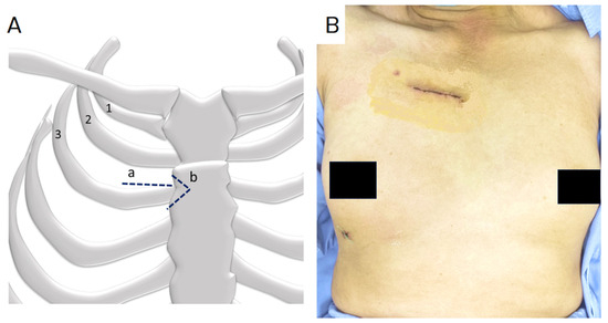
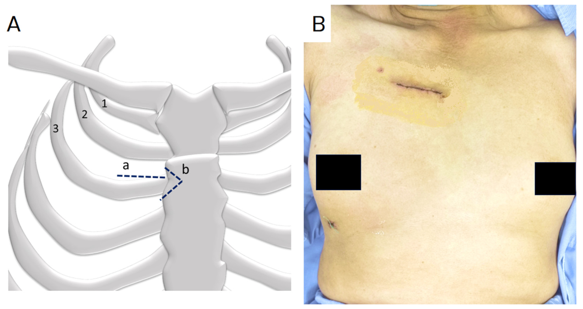
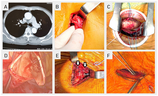
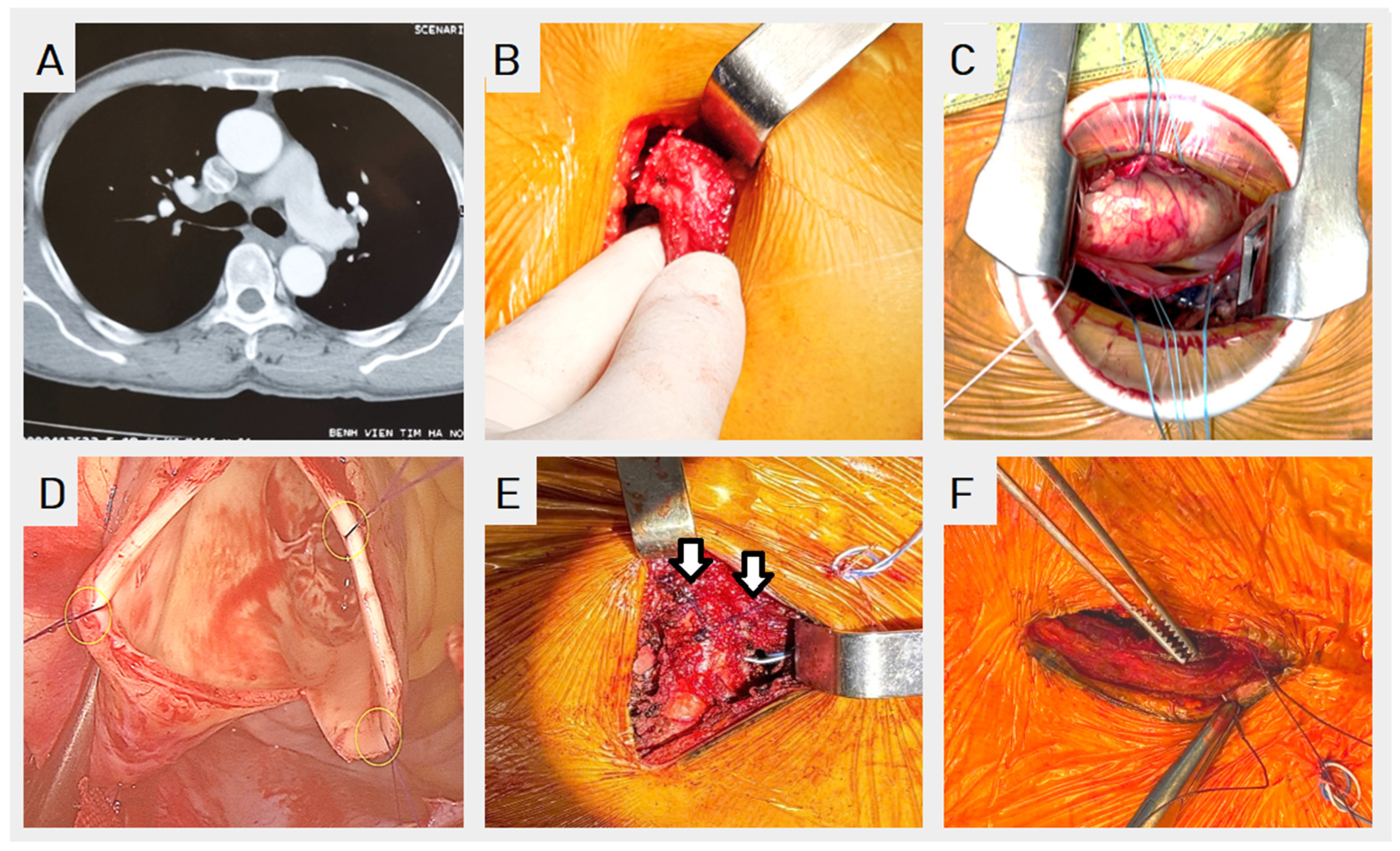
A 4–5 cm skin incision was made parasternally at the level of the right second inter-costal space (ICS) (Figure 1), superior to the 3rd rib, and extended to the right edge of the sternum. The chest wall muscles were dissected to expose the 2nd ICS and the 3rd rib. The right internal mammary artery and vein were dissected and ligated. The width of the sternum at the level of the 3rd rib was examined. The V-line of the wedge-shape cut was marked using a cautery. The third rib was detached from the sternum using an oscillating saw in a wedge shape (Figure 2). The depth of the wedge should be less than 1.5 cm or a half of the sternal width to avoid the risk of sternal bone fracture, especially in the narrow ones. The pleura was then opened and a medium-sized RingProtect® (Grena, London, UK) soft tissue retractor was placed, then a Haight’s rib spreader (Aesculap, Tuttlingen, Germany) was used to expand the surgical field by retracting the 3rd rib inferiorly. At the end of surgery, the detached rib was returned to the original position and re-attached to the sternum using two Vicril® size 2 sutures (Ethicon, Raritan, NJ, USA) (Figure 2E). The muscle layers were closed, followed by the subcutaneous and skin layers (Figure 2F). The pericardium was opened and stay sutures were placed to retract the aorta closer to the incision. The ascending aorta was dissected away from the pulmonary arteries for placement of an umbilical tape to support mobilization of the aorta.
Figure 1.
Approach to the aortic valve via right anterior minithoracotomy. (A) A schematic drawing shows (1), (2), (3) the first 3 ribs on the right chest wall, (a) a skin incision over the right second intercostal space and (b) wedge-shape resection to the sternum at the tip of the third rib. (B) Skin closure.
Figure 2.
Exposure of the aortic valve. (A) Preoperative CT scan shows leftward ascending aorta. (B) A wedge-shape resection for 3rd rib detachment allows for (C) extended surgical view, with (D) subsequent adequate exposure of the aortic valve with 3 staying sutures. (E) The 3rd rib is later reattached to the sternum using two Vicril® size 2 sutures (arrows), and (F) the wound is closed in layers.
2.3. Cardiopulmonary Bypass
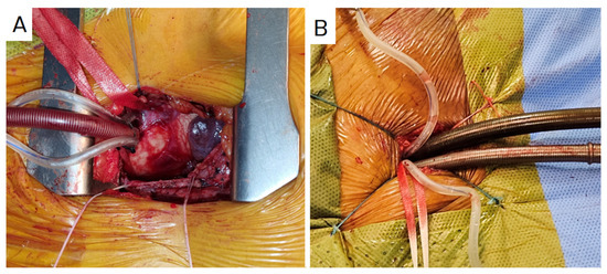
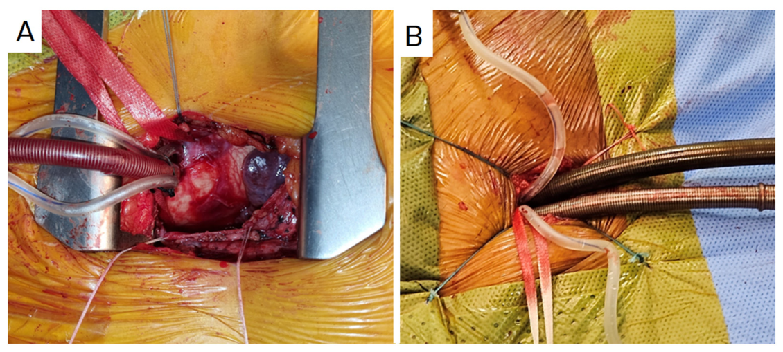
Cardiopulmonary bypass was established between the femoral artery and vein or ascending aorta and femoral vein. The femoral vessels were exposed through a 2–3 cm incision in the groin crease. The arterial cannula was placed either in the femoral artery or directly into the ascending aorta. A femoral cannula was inserted in most patients using the Seldinger’s technique with a 15–19 Fr femoral arterial cannula (Biomedicus, Medtronic, Minneapolis, MN, USA) (Figure 3). The alternative approach was used for patients with peripheral arterial disease (e.g., femoral or iliac arteries atherosclerosis) or older patients at high risk for stroke from retrograde perfusion, using a flexible cannula of 18–20 Fr (EOPA, Medtronic, Minneapolis, MN, USA). A 23–25 Fr RAV femoral venous cannula (LivaNova, Mirandola, Italy) was inserted via the femoral vein using Seldinger’s technique, with the tip extending into the superior vena cava for effective drainage. Negative pressure of 30–50 mmHg was applied to the venous drainage system for better flow. All patients were cooled to 32 °C.
Figure 3.
Cardiopulmonary bypass setup, with arterial cannulation via ascending aorta (A) or femoral artery (B).
2.4. Aortic Valve Replacement
A left heart venting cannula was placed through the right superior pulmonary vein. The aorta was occluded with a standard DeBakey’s aortic clamp. An aortotomy was made transversely just below the fat strip of the ascending aorta. Myocardial protection was achieved with antegrade cardioplegia through the aortic root or directly through the coronary ostia using the Custodiol® solution (Dr Franz Köhler Chemie GmbH; Alsbach-Hähnlein, Germany). The aortic valve was exposed using three retracting 5-0 Prolene® sutures (Ethicon, Raritan, NJ, USA); one suture was placed in the middle of the upper edge of the aortotomy line, and the other 2 sutures were placed in the lower edge (Figure 2D). The diseased aortic valve was replaced using a prosthetic valve under direct vision. Standard surgical instruments for open AVR were used, in combination with long-shaft MICS instruments whenever necessary.
The aorta was closed with two layers running suture using 5-0 Prolene® (Ethicon, Raritan, NJ, USA) (Figure 2). The suture line was reinforced using a Teflon or pericardial felt, and biological glue (Bioglue®, Cryolife, Kennesaw, GA, USA) if necessary. A temporary epicardial pacing wire was anchored to the right ventricular wall before remove the aortic clamp. After the weaning off bypass, the pericardium was closed. A 25 Fr chest tube was placed in the right pleural cavity, and a 14–16 Fr tube was placed in the transverse sinus, just behind the ascending aorta. The third rib was reattached to the sternum and the wound was closed in layers.
3. Results
In the five years from 2019 to 2024, a total of 176 patients with aortic valve diseases underwent Mini-AVR via a right anterior thoracotomy approach at our hospital. Baseline demographic characteristics, preoperative conditions and comorbidities are summarized in Table 1.
Table 1.
Baseline characteristics.
Anatomy characteristics of the ascending aorta and aortic annulus using preoperative CT angiography are shown in Table 2. The ascending aorta was considered distal from the sternum when the distance between the aorta and the chest wall was greater than 10 cm.
Table 2.
CT angiography findings.
Pathological characteristics of aortic valves are shown in Table 3. The majority of patients had aortic stenosis (53.4%) or combined aortic stenosis and regurgitation (31.8%). Most valve diseases were degenerative (88.1%).
Table 3.
Aortic valve characteristics.
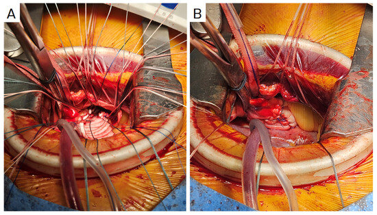
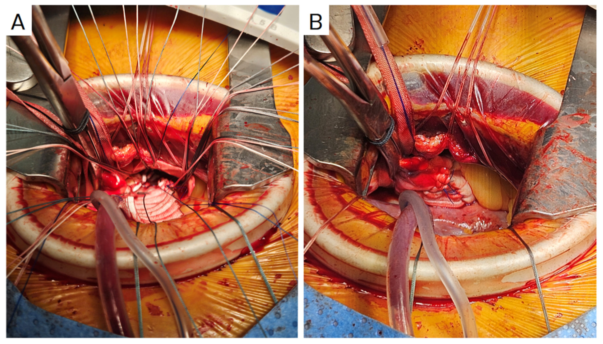
Mini-AVR with wedge-shaped detachment of the third rib could be completed within an aortic cross-clamp time of 78.69 ± 24.1 min. This approach allowed for root enlargement using Yang’s technique (Figure 4) in three patients (1.7%), two cases with bioprosthetic valves, and one with a mechanical valve. The average aortic cross-clamp time for these cases was 106 min. Two patients required conversion to a full sternotomy for bleeding control due to a tear in the aortic annulus (1.1%). Intraoperative and postoperative characteristics are shown in Table 4 and Table 5.
Figure 4.
Right anterior minithoracotomy for aortic valve replacement and root enlargement using Yang’s technique (A) and after closure of the aorta (B).
Table 4.
Intraoperative characteristics.
Table 5.
Postoperative outcomes.
4. Discussion
Minimally invasive aortic valve surgery is associated with less bleeding and fewer sternum-related complications, shorter recovery and hospital stay, and better cosmesis. The right anterior thoracotomy approach through the second intercostal space is less invasive, better in hemostasis and simpler in chest closure, and does not affect the stability of the chest wall, as compared to the upper hemi-sternotomy approach [9].
Narrow intercostal spaces (especially combined with a rigid chest wall) can limit exposure in Mini-AVR, for which some surgeons proposed that the solution is to transect the costochondral cartilage with diathermia to greatly increase the exposure. The divided costochondral cartilage can be sutured with a Vicryl® suture at the end of the procedure. An alternative is to perform a partial rib resection, but this is less preferable because of increased invasiveness, potentially increased postoperative pain and reduced aesthetic appearance.
In their first report about Mini-AVR, Glauber et al. used a 5–7 cm incision, without rib resection [1]. Similarly, Ribeiro et al. (2015) stopped their incision at the border of the sternum [4]. In order to enhance visualization of the aortic valve and attain better manipulation, expansion of the surgical field with rib dislocation or detachment had been proposed. Ribeiro et al. (2015) used the “box principle” to mobilize the upper and lower ribs for better exposure [4]. Lamenas et al. (2015) reported the “Miami method”, in which the authors transected the cartilage of the inferior rib, and reattached it later [9]. Alexiou et al. described their technique in Mini-AVR using an oscillating saw, in which the third rib was meticulously detached from the sternum in a wedge shape, but the benefit of 3rd rib wedge resection was not discussed in this study [10].
The expansion of surgical view can be achieved with rib resection or costochondral cartilage resection of either the upper or lower ribs. Normally, the third-rib mobilization is preferred with rib resection or wedge-shape resection of the sternum at the tip of the third rib. Lamenas et al. transected the cartilage [9], and Bethencourt et al. caused fracture of the third rib [11]. In our study, wedge resection preserves the continuity of costochondral structure, with better stabilization upon surgical closure with re-attachment of the third rib. This modification allows for better exposure of the aortic valve, while preserving the continuity of the costochondral structure for better stabilization upon closure.
Surgical time may be reduced with better exposure. The aortic cross-clamp time in our study was shorter than those in the report by Glauber et al. (78 vs. 87 min) [1]; and similar to those reported by Ribeiro et al. (78.6 ± 24.1 min vs. 73.2 ± 22.8 min), who also achieved better exposure with the “box principle” by mobilizing the third rib [4].
The scope of surgical intervention may be expanded to more complex surgery and less favorable anatomy. Initially, Mini-AVR was preferred in patients with rightward ascending aorta, where the ascending aorta is on the right of the midline, and less than 10 cm from the sternum. With more experience, Mini-AVR could be performed in patients with leftward and deep ascending aorta. As shown in our study, 32% of patients had leftward aorta and 10% had deeper aorta. Especially with the wedge resection, the surgical view became sufficient to perform aortic root enlargement with Yang’s technique [12] (Figure 4). Mobilizing the third rib allows better surgical exposure, and thereby preoperative CT scan for surgical planning and exclusion of unfavorable anatomy may not be necessary. Although we find that a depth of 1.0–1.5 cm wedge-cut to the sternum bone at the level of the third rib is less than 50% of the width of the sternum in most patients, preoperative CT scan examination of the sternum might be useful in small-sized patients in whom their narrow sternum may require careful pre- and intraoperative examination to avoid the risk of sternal bone fracture associated with a deep wedge-cut.
Expanding the surgical field to the left, even in anatomically favorable cases like those with a leftward aorta with an alpha angle near 45 degrees, made the procedure easier to perform, thus shortening the learning curve for surgeons. Once surgeons were familiar and comfortable with the procedure, they could apply this technique to more challenging cases involving leftward aorta, deep aorta positions from the chest wall, unfavorable alpha angles, dilated ascending aorta, heavily calcified valves, or small aortic roots.
We performed Mini-AVR and aortic root enlargement via right anterior thoracotomy for three patients using a Y-shaped incision technique described by Yang [12]. Of note, these patients had favorable aorta positions relative to the chest wall. We recommended caution in patient selection for right anterior thoracotomy if they present with multiple adverse factors. High-risk patients can also undergo minimally invasive AVR instead of TAVR. Mini-AVR retains the advantages of SAVR over TAVR, such as lower cost, reduced stroke risk, and reduced paravalvular leak risk, while also minimizing surgical trauma [13]. This study has several limitations. Its retrospective design without a control group limits its statistical power and ability to reach a meaningful conclusion about the benefit of wedge-shape rib detachment in Mini-AVR. Although we did not encounter any patients with sternal bone instability, our data were not sufficient to exclude the risk of sternal fracture. Subgroup analysis of patients with extreme leftward and deep aorta was not possible due to insufficient follow-up data. Although we found it easier for young surgeons to perform Mini-AVR with wedge detachment of the third rib, the study design is not appropriate to correlate this modification with the surgeon’s learning curve.
5. Conclusions
Simple modification of the exposure technique for Mini-AVR involving a wedge-shape detachment of the third rib from the sternum allows for expansion of the surgical field to the left. Although this approach helps perform complex AVR in patients with a deep and more leftward ascending aorta without any sternal bone fracture, further study in a larger patient population might be required to validate the safety and efficacy of this technique.
Author Contributions
Conceptualization, H.S.N., T.D.V. and N.M.N.; methodology, H.S.N., N.M.N., K.T.N. and T.D.V.; formal analysis, H.S.N., K.T.N. and N.M.N.; investigation, H.S.N. and N.M.N.; writing—original draft preparation, H.S.N. and T.D.V.; writing—review and editing, T.D.V., K.T.N. and N.M.N.; visualization, H.S.N., N.M.N. and K.T.N.; and supervision, H.S.N. and T.D.V. All authors have read and agreed to the published version of the manuscript.
Funding
This research received no external funding.
Institutional Review Board Statement
The retrospective study was conducted in accordance with the Declaration of Helsinki and was approved by the Ethics Committee of Hanoi Heart Hospital (No. 1232/BVT-BBHĐĐĐ) on 24 April 2024.
Informed Consent Statement
Patient consent was waived due to the retrospective nature of the study. All data were anonymized, and no identifiable personal information was included.
Data Availability Statement
The data presented in this study are available on request from the corresponding author. The data are not publicly available due to institutional and ethical restrictions regarding patient confidentiality.
Conflicts of Interest
The authors declare no conflicts of interest.
Abbreviations
| TAVR | Transcatheter aortic valve replacement |
| SAVR | Surgical aortic valve replacement |
| Mini-AVR | Minimally invasive aortic valve replacement |
References
- Glauber, M.; Gilmanov, D.; Farneti, P.A.; Kallushi, E.; Miceli, A.; Chiaramonti, F.; Murzi, M.; Solinas, M. Right anterior minithoracotomy for aortic valve replacement: 10-year experience of a single center. J. Thorac. Cardiovasc. Surg. 2015, 150, 548–556 e542. [Google Scholar] [CrossRef] [Scilit] [PubMed]
- Miceli, A.; Murzi, M.; Gilmanov, D.; Fugà, R.; Ferrarini, M.; Solinas, M.; Glauber, M. Minimally invasive aortic valve replacement using right minithoracotomy is associated with better outcomes than ministernotomy. J. Thorac. Cardiovasc. Surg. 2014, 148, 133–137. [Google Scholar] [CrossRef] [Scilit] [PubMed]
- Mikus, E.; Turci, S.; Calvi, S.; Ricci, M.; Dozza, L.; Del Giglio, M. Aortic valve replacement through right minithoracotomy: Is it really biologically minimally invasive? Ann. Thorac. Surg. 2015, 99, 826–830. [Google Scholar] [CrossRef] [Scilit] [PubMed]
- Ribeiro, I.B.; Ruel, M. Right Anterior Minithoracotomy for Aortic Valve Replacement: A Widely Applicable, Simple, and Stepwise Approach. Innovations 2019, 14, 321–329. [Google Scholar] [CrossRef] [Scilit] [PubMed]
- Yousuf Salmasi, M.; Hamilton, H.; Rahman, I.; Chien, L.; Rival, P.; Benedetto, U.; Young, C.; Caputo, M.; Angelini, G.D.; Vohra, H.A. Mini-sternotomy vs right anterior thoracotomy for aortic valve replacement. J. Card. Surg. 2020, 35, 1570–1582. [Google Scholar] [CrossRef] [Scilit] [PubMed]
- Glauber, M.; Miceli, A.; Bevilacqua, S.; Farneti, P.A. Minimally invasive aortic valve replacement via right anterior minithoracotomy: Early outcomes and midterm follow-up. J. Thorac. Cardiovasc. Surg. 2011, 142, 1577–1579. [Google Scholar] [CrossRef] [Scilit] [PubMed]
- Klein, P.; Klop, I.D.G.; Kloppenburg, G.L.T.; van Putte, B.P. Planning for minimally invasive aortic valve replacement: Key steps for patient assessment. Eur. J. Cardio-Thorac. Surg. Off. J. Eur. Assoc. Cardio-Thorac. Surg. 2018, 53, ii3–ii8. [Google Scholar] [CrossRef] [Scilit] [PubMed]
- Lamelas, J.; Alnajar, A. Recent advances in devices for minimally invasive aortic valve replacement. Expert Rev. Med. Devices 2020, 17, 201–208. [Google Scholar] [CrossRef] [Scilit] [PubMed]
- Lamelas, J. Minimally invasive aortic valve replacement: The “Miami Method”. Ann. Cardiothorac. Surg. 2015, 4, 71–77. [Google Scholar] [PubMed]
- Taghizadeh-Waghefi, A.; Arzt, S.; Wenzel, L.; Petrov, A.; Wilbring, M.; Matschke, K.; Kappert, U.; Alexiou, K. Right Anterior versus Right Transaxillary Access for Minimally Invasive Aortic Valve Replacement: A Propensity Matched Competitive Analysis. J. Clin. Med. 2024, 13, 985. [Google Scholar] [CrossRef] [Scilit] [PubMed]
- Bethencourt, D.M.; Le, J.; Rodriguez, G.; Kalayjian, R.W.; Thomas, G.S. Minimally Invasive Aortic Valve Replacement via Right Anterior Minithoracotomy and Central Aortic Cannulation: A 13-Year Experience. Innovations 2017, 12, 87–94. [Google Scholar] [CrossRef] [Scilit] [PubMed]
- Yang, B. A novel simple technique to enlarge the aortic annulus by two valve sizes. JTCVS Tech. 2021, 5, 13–16. [Google Scholar] [CrossRef] [Scilit] [PubMed]
- Sayed, A.; Almotawally, S.; Wilson, K.; Munir, M.; Bendary, A.; Ramzy, A.; Hirji, S.; Abushouk, A.I. Minimally invasive surgery versus transcatheter aortic valve replacement: A systematic review and meta-analysis. Open Heart 2021, 8, e001535. [Google Scholar] [CrossRef] [Scilit] [PubMed]

Disclaimer/Publisher’s Note: The statements, opinions and data contained in all publications are solely those of the individual author(s) and contributor(s) and not of MDPI and/or the editor(s). MDPI and/or the editor(s) disclaim responsibility for any injury to people or property resulting from any ideas, methods, instructions or products referred to in the content. |
© 2026 by the authors. Licensee MDPI, Basel, Switzerland. This article is an open access article distributed under the terms and conditions of the Creative Commons Attribution (CC BY) license.