


Abstract
Background and Clinical Significance: With increasing demand for total knee arthroplasty (TKA), the incidence of post-operative instability has also risen. Although fracture of the polyethylene tibial post in posterior-stabilized (PS) prostheses is relatively uncommon, it should not be overlooked because delayed recognition may lead to severe late sequelae. Case Presentation: Between April 2008 and January 2020, 132 revision TKAs were performed at our institution due to suspected instability with or without component loosening. Among these, three patients were identified as having instability associated with fracture of the polyethylene tibial post in PS implants. All three patients presented late, with a mean interval of 49.66 months (range 34–74) after the index TKA. At presentation, all demonstrated multiplanar global instability, and two showed recurvatum deformity. Pre-operatively, revision surgery was indicated due to progressive global instability. Although tibial post fracture was suspected as a possible underlying cause, the diagnosis could not be definitively established until intraoperative inspection, which confirmed polyethylene tibial post fracture in all cases. Each patient underwent revision TKA using a semi-constrained prosthesis with an increased polyethylene insert height. At final follow-up (12–18 months), all patients achieved complete resolution of clinical instability and demonstrated meaningful improvement in the Hospital for Special Surgery (HSS) and Western Ontario and McMaster Universities Osteoarthritis Index (WOMAC) scores. Conclusions: Although rare, polyethylene tibial post fracture should be considered in the differential diagnosis of post-operative knee instability following PS TKA. Early recognition may allow management with isolated polyethylene exchange, whereas delayed diagnosis can result in progressive ligamentous insufficiency and global multiplanar instability, ultimately necessitating revision using a semi-constrained implant.
1. Introduction and Clinical Significance
With increasing demand for total knee replacement surgeries, there has been an increase in the incidence of post-operative knee instability. Post-operative instability following total knee arthroplasty (TKA) may present in various forms. Clinical presentations of TKA instability may also be different in each patient, including knee swelling, foreign body sensation, a locking sensation during range of motion, and dislocation. During the evaluation of this instability, thorough physical examination and evaluation of radiographs, as well as laboratory studies, are essential. Merchan-Rodriguez describes the most common causes of instability following TKA as ligament imbalance, component malalignment, and implant loosening [1]. Tibial post fracture of polyethylene bearing is a relatively uncommon complication in the posterior stabilized design with a cam–post mechanism. In a systematic review by Lachiewicz, the author reported the highest incidence of tibial post fracture as 1.2% [2], with others reporting an incidence of less than 1% [3,4]. Its potential causes are hyperextension due to extension gap laxity, flexion position of a femur implant causing an early post–cam mechanism, anatomical variation in the femur with anterior bowing, increased tibial slope, and a more anteriorly located polyethylene post. Tibial post fractures are easily missed on simple radiographs, and evaluations using MRI and CT scans are often limited due to artifacts from metallic implants obscuring vision. In the setting of post-operative instability, when swelling and dislocation occur for uncertain reasons, the possibility of tibial post fracture should be considered. From 2008 to 2020, 132 revision TKAs were performed at our clinic due to suspected TKA instability with or without implant loosening. Among the 132 cases included, we experienced 3 cases of unstable total knee arthroplasty with polyethylene tibial post fractures, and herein, report the treatment options and their post-operative results. This case report suggests that tibial post fracture should be included in the differential diagnosis of unexplained instability to ensure appropriate surgical management. Furthermore, unlike earlier reports, this series emphasizes the diagnostic challenges and management considerations in late-detected tibial post fractures, providing new insights into this rare complication
2. Case Presentation
2.1. Case 1
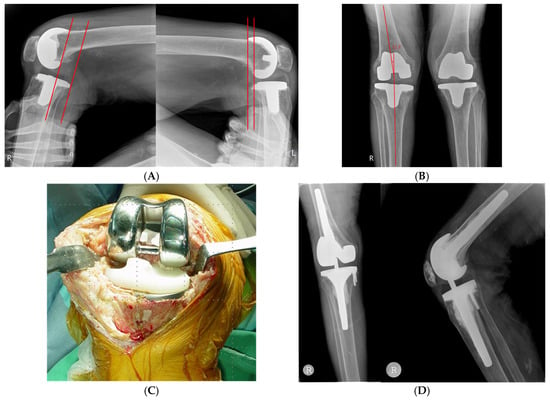
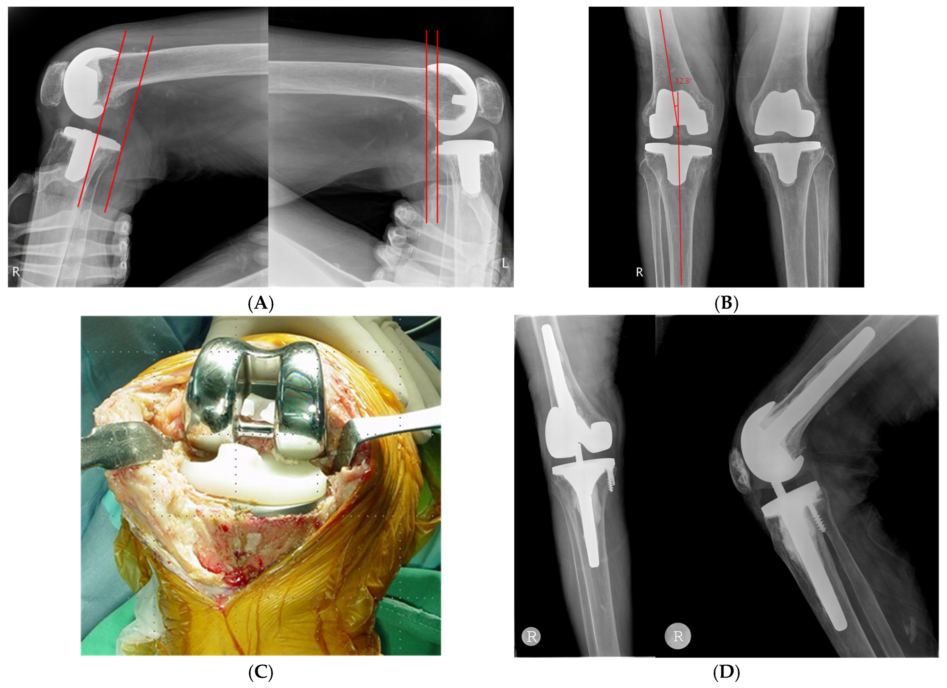
An 80-year-old female patient with a history of right knee TKA 41 months prior at another hospital presented to our clinic with complaints of right knee pain, swelling, and a clicking sound during active knee flexion. The patient reported no history of trauma. The initial surgery was performed using a posterior stabilized prosthesis (TC-Plus, Plus Orthopedics, Rotkreuz, Switzerland) with a tibial bearing of 10 mm. The patient also described occasional episodes of the knee feeling “locked” or giving way backward, which are findings suggestive of posterior tibial subluxation. On simple radiographs, the patient had a neutral mechanical alignment of 0.0°. Tibio-femoral component alignments were evaluated by measuring alpha, beta, gamma, and delta angles, which were 91.0°, 89.0°, 2.0°, and 89.1°, respectively. On stress examination, the patient showed flexion (anterior–posterior) instability, showing posterior translation of the tibia on the posterior drawer test with a side-to-side difference of 10.2 mm (Figure 1A), as well as a valgus instability of 12.3° on the right knee (Figure 1B). On valgus-stressed anteroposterior radiographs, the right knee showed greater valgus laxity (12.3°) than the contralateral knee (3.6°), consistent with medial-sided laxity. No frank medial collateral ligament tear was identified on pre-operative stress examination, indicating that the MCL was attenuated but remained intact despite the valgus laxity. The preserved ligament continuity and absence of abrupt opening suggested chronic attenuation rather than an acute or primary MCL rupture. On initial presentation to our clinic, the patient also exhibited a recurvatum gait. Intraoperative inspection confirmed a polyethylene tibial bearing post fracture with associated wear at the waist of the post (Figure 1C). Based on the physical examination and intraoperative findings, revision surgery was performed for global instability using a semi-constrained implant (Triathlon TS knee system, Stryker) with a bearing height of 16 mm (Figure 1D). Given the presence of multiplanar instability and ligamentous insufficiency, isolated polyethylene insert exchange was considered insufficient to adequately restore stability. At the 12-month follow-up, the patient reported no clinical signs or symptoms of instability. The last follow-up Hospital for Special Surgery (HSS) score also improved to 82 from a pre-operative score of 43. Pre-operative and post-operative Western Ontario and McMaster (WOMAC) scores were 91 and 19, respectively.
Figure 1.
(A) Posterior stressed lateral radiographs demonstrate significant difference with 15.1mm posterior laxity on right side and 4.9mm posterior laxity on left side. (B) Valgus stressed anterior–posterior radiographs showing significant difference with 12.3° valgus laxity on right side and 3.6° valgus laxity on left side. (C) Intraoperative photograph showing fractured polyethylene post in middle third portion with polyethylene debris. No femoral or tibial component loosenings were seen. (D) Patient underwent revision total knee arthroplasty under impression of global knee instability, which could not be recovered with simple up-sized polyethylene bearing change.
2.2. Case 2
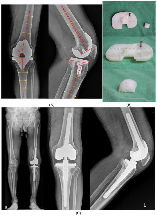
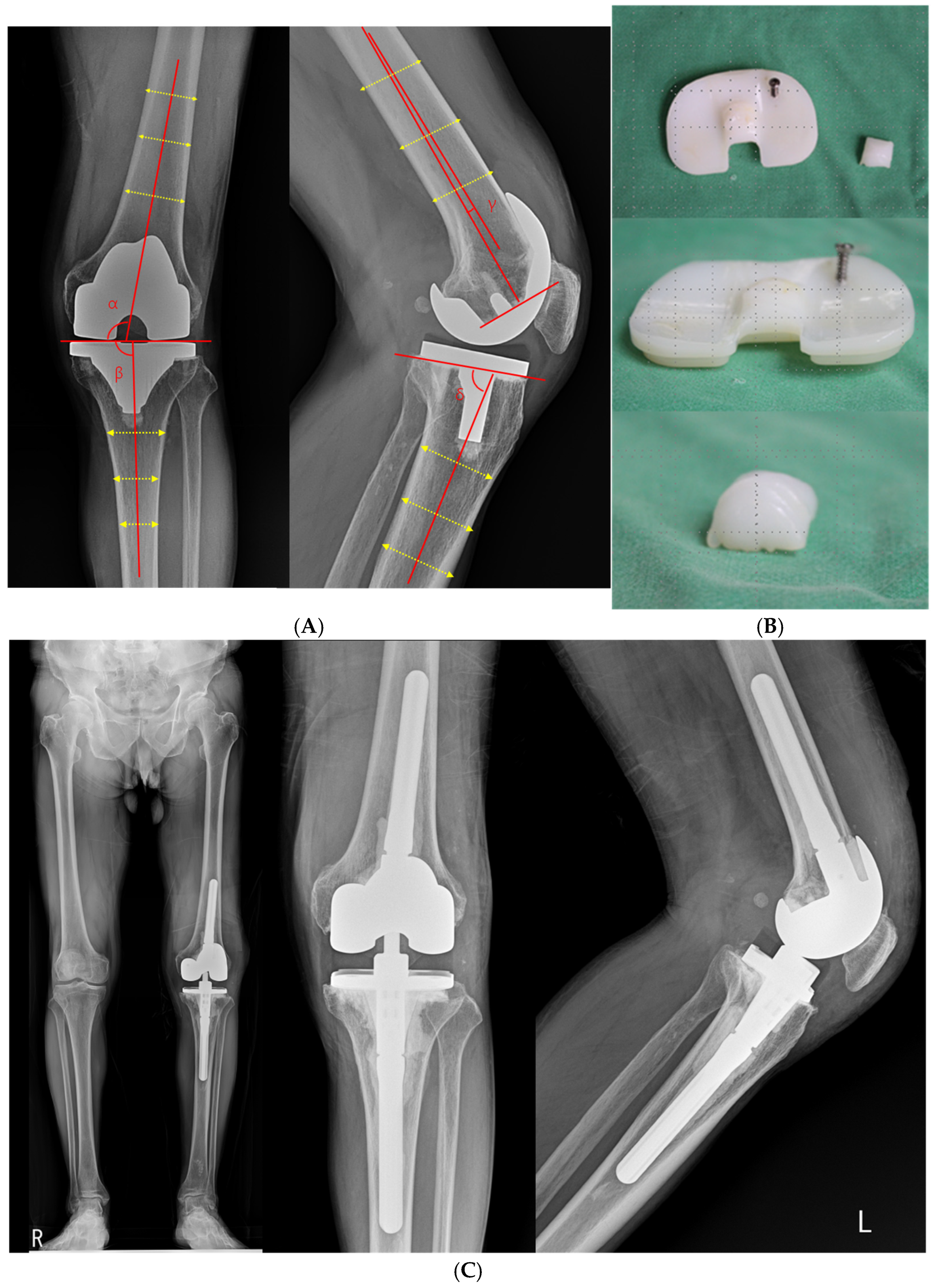
A 75-year-old male patient who underwent left knee TKA 34 months prior at another hospital presented to our clinic with complaints of left knee swelling, effusion, and pain. The patient reported no history of trauma. The implant used for the initial surgery was a posterior stabilized type (Scorpio II NRG, Stryker, NJ, USA) with a tibial bearing of 12 mm. On long bone radiographs, the patient demonstrated a valgus mechanical alignment of 7.5°, with alpha, beta, gamma, and delta angles of 96.5°, 91.0°, 1.0°, and 83.3°, respectively (Figure 2A).
Figure 2.
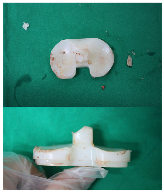
(A) Antero-posterior and lateral radiographs showing 7.5° valgus mechanical alignment with alpha, beta, gamma, and delta angles of 96.5°, 91.0°, 1.0°, and 83.3°, respectively.(The yellow dotted arrows indicate the midpoints of the femoral and tibial shafts used to determine the anatomical axes). (B) Removed polyethylene bearing post with fracture in middle third portion without evidence of polyethylene bearing wear. (C) Post-operative radiograph of revision total knee arthroplasty using semi-constrained condylar type implant (LCCK, Zimmer) with 17 mm polyethylene bearing.
On stress examination, the patient showed mild flexion instability with posterior tibial subluxation, varus instability of 6.0°, and valgus instability of 8.6°. On physical examination, the patient also exhibited 15° of knee recurvatum.
Intraoperatively, a fracture of the polyethylene tibial bearing post near the base was identified (Figure 2B). Based on physical examination and intraoperative findings, revision surgery was performed due to global instability, using a semi-constrained implant (LCCK, Zimmer Biomet, Warsaw, IN, USA) with a bearing height of 17 mm (Figure 2C).
At the 12-month follow-up after revision surgery, no clinical or radiographic evidence of instability was observed. The HSS score improved from 58 pre-operatively to 91 at the final follow-up. Pre-operative and post-operative WOMAC scores were 83 and 17, respectively.
2.3. Case 3
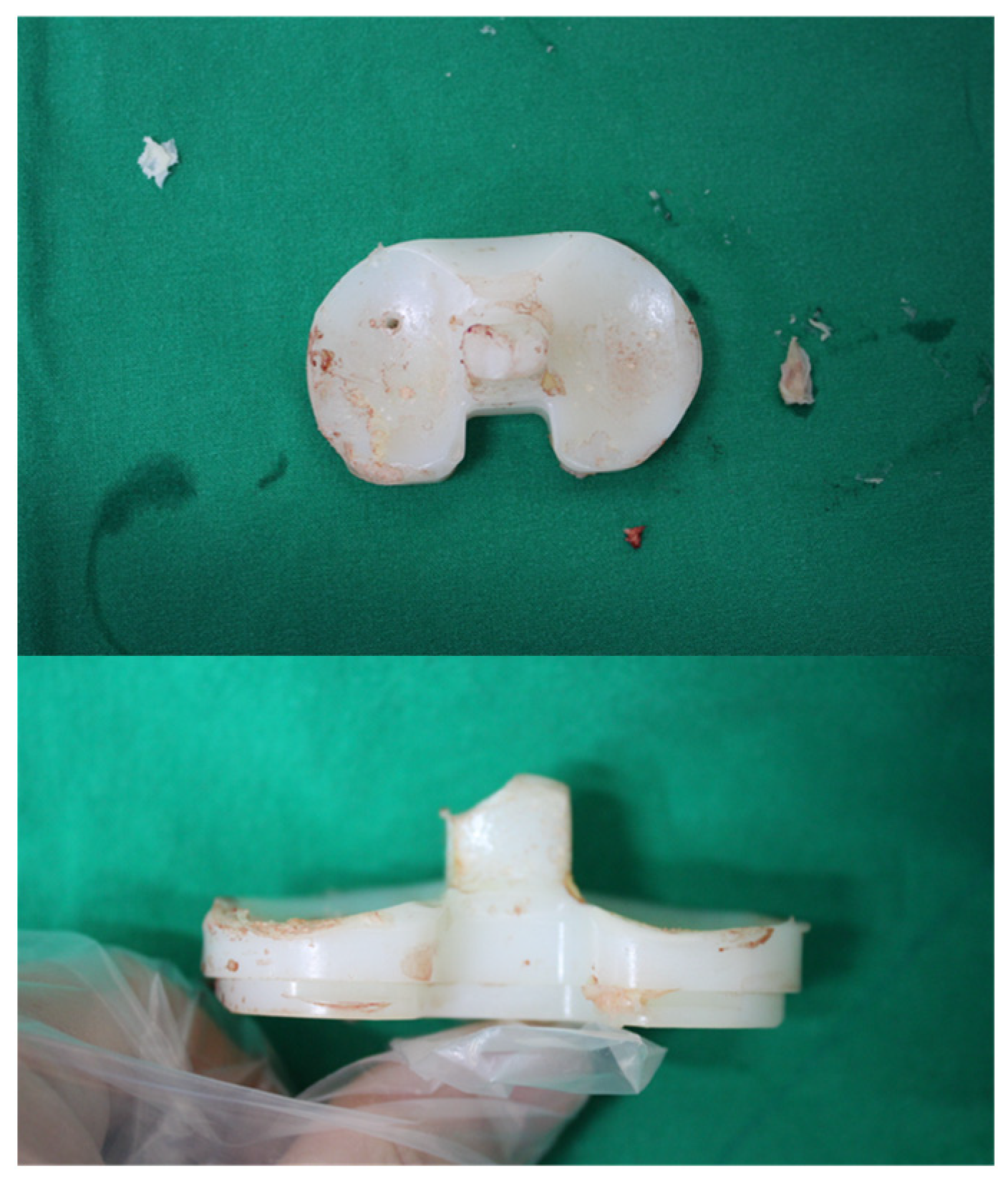
A 73-year-old female patient who underwent right knee TKA at our clinic 74 months prior presented with right knee pain, swelling, and a feeling of ambulatory instability. There was no apparent history of trauma. The implant used for the primary TKA was a posterior stabilized type (Scorpio II NRG, Stryker, NJ, USA) with a 10 mm polyethylene bearing. On simple radiographs, the patient had 4.9° valgus mechanical alignment with alpha, beta, gamma, and delta angles of 94.1°, 90.8°, 0.9°, and 83.4°, respectively. No obvious tibial post fracture was identified on these radiographs, consistent with the known difficulty in visualizing such failures on standard imaging. Under stress examination, the patient had varus instability of 15.4° and recurvatum of 20.0°. Notably, no flexion instability was observed. Intraoperatively, the upper-medial corner of the tibial post was found to be fractured, and severe synovial proliferation was observed (Figure 3). Intraoperative microbiology and pathology reports confirmed no signs of infection. Revision TKA was performed using a semi-constrained implant (LCCK, Zimmer Biomet, Warsaw, IN, USA) with a bearing height of 14 mm. At the 18-month follow-up, the patient did not show any clinical discomfort or instability. The HSS score had also improved to 89 compared to a pre-operative score of 61. Pre-operative and post-operative WOMAC scores were 77 and 21, respectively.
Figure 3.
Removed polyethylene bearing with fracture on the upper-medial corner of the tibial post.
3. Discussion
Post-operative instability is one of the most common causes for undergoing revision TKA, which, if unaddressed, may lead to poor patient satisfaction and functional limitation. Al-Jabri et al. stated that 10–26% of revision surgeries following total knee arthroplasty (TKA) were due to instability [5]. Parratte and Pagnano reported that 10–22% of revision surgeries were due to post-operative knee instability [6]. Many authors, including Kelly Vince et al., have broadly classified instability as varus–valgus (extension), anterior–posterior (flexion), recurvatum, and global types [7]. There are multiple factors that may contribute to instability following TKA. In a systematic review by Al-Jabri et al., the authors classified the contributing factors according to patient factors, implant factors, and those related to suboptimal surgical technique [5]. Among the causes of post-operative instability following TKA, Al-Jabri et al. described polyethylene wear and delayed ligamentous dysfunction as common causes of late post-operative instability [5].
In a case series of tibial bearing post fractures by Diamond et al., the authors reported an incidence of 5 cases out of 955 knees (0.52%) [3]. In another case report by Bonner et al., the incidence of tibial bearing post fracture was described as less than 1% [4]. Despite its low incidence, in evaluation of post-operative knee instability following TKA, the possibility of post fracture of the tibial polyethylene bearing should be considered.
While there are numerous speculations about contributing factors to tibial post fracture following TKA, for better clarification, many authors have broadly classified the factors as patient-related, specific to implant design, and related to suboptimal surgical technique [1,2,5]. Patient-related factors may be further classified as traumatic, anatomic, and habitual factors. In a systematic review by Lachiewicz, out of the 23 patients studied, only 6 patients reported a definite history of a traumatic event [2]. None of our three patients reported any clear history of trauma prior to initial presentation. According to Pesciallo et al., neuromuscular disorders lead to weak quadriceps strength and connective tissue diseases, which result in ligamentous laxity and may produce recurvatum of the knee alongside repetitive stress on the anterior aspect of the tibial post [8]. In addition to anatomic factors, habitual behavioral patterns of patients may induce rotational stress on the tibial post, such as squatting and cross-leg sitting, and may also contribute to tibial post fracture [9]. In our three cases, two patients showed recurvatum on presentation. Incidentally, the two patients with recurvatum had a complete fracture of the tibial post identified intraoperatively. Although the exact causal relation between recurvatum of the knee and tibial post fracture remains to be investigated, in our cases, the incidence of tibial post fracture seemed to have a strong correlation with recurvatum of the knee. None of the three patients in this case series had a known history of neuromuscular disorders or connective tissue disease. However, all three patients admitted having frequently sat cross-legged and squatted post-operatively.
In addition to patient-related factors, there have been speculations on possibility of specific implant design leading to tibial post fracture. In a systematic review by Lachiewicz, the author mentioned a higher incidence of tibial bearing post fracture in prosthesis designs where a hole in the bearing post is present for fixation of polyethylene bearing onto a tibial tray [2]. Furman et al. speculated that implant designs with a bearing post located more anteriorly were more susceptible to tibial bearing post fracture [10]. Bonner et al. described highly cross-linked polyethylene bearing as more susceptible to post fractures due to decreased resistance to mechanical strain [4]. However, contradicting reports by Minoda et al. found no significant difference in longevity between highly cross-linked and conventional polyethylene bearing [11]. Out of the three cases included in this study, one patient with a TC-Plus implant (Plus Orthopedics, Rotkreuz, Switzerland) had notably more anterior positioning of the tibial bearing post compared to the other two patients with a Scorpio II NRG implant (Stryker, NJ, USA). In our three cases, all patients had an index operation performed using a conventional polyethylene bearing with no hole in the bearing post for fixation.
Suboptimal surgical techniques that increase stress may also contribute to tibial post fracture. Previous studies by Bonner et al. and Lachiewicz have suggested that tibio-femoral component malalignment can increase abnormal stress on the polyethylene post and predispose it to fracture [2,4]. In particular, anterior placement of the tibial component, posterior positioning, or excessive flexion of the femoral component may alter cam–post interaction by shifting the contact point anteriorly, thereby leading to abnormal or excessive cam–post contact and anterior post impingement during knee motion. Such altered contact mechanics can result in repetitive anterior impingement and localized stress concentration on the anterior or anteromedial aspect of the polyethylene post. In the presented cases, the fracture configurations predominantly involved the anterior or anteromedial portion of the post, supporting the possibility that abnormal anterior impingement related to component positioning contributed to post failure [4].
Notably, despite the advanced age of our patients (73–80 years) and presumably lower activity levels, these mechanical factors still led to relatively early tibial post failure, with fractures occurring at a mean of approximately 4 years after primary TKA. This finding suggests that even low-demand patients remain at risk for tibial post fracture when unfavorable mechanical conditions, such as component malposition or ligamentous laxity, are present.
Of the three cases in this study, one patient had neutral alignment, and two patients had valgus alignment of 7.5° and 4.9°. All three cases had a posterior tibial slope of less than 7° and the femoral component was inserted with less than 2° flexion.
Bal et al. reported a favorable short-term outcome following polyethylene bearing exchange to increased thickness [12]. Hendel et al. suggested that revision surgery using more constrained design implants causes sustained instability [13].
All three cases in this study presented to our clinic approximately 3–6 years after the index operation (mean 49.66 months; range 34–74). However, because none of the patients had interval evaluations specifically assessing tibial post integrity, we were unable to determine the exact timing of tibial post fracture. At the time of presentation, all patients demonstrated multiplanar global instability, and two exhibited knee recurvatum.
Accordingly, it remains uncertain whether the observed recurvatum preceded the tibial post fracture or developed as a consequence of post failure. We speculate that pre-existing extension gap laxity leading to hyperextension may have increased stress on the tibial post and predisposed it to fracture; conversely, once the post failed, loss of posterior stability likely exacerbated recurvatum and overall instability, creating a vicious cycle.
From a treatment standpoint, isolated polyethylene insert exchange has been reported as a viable option when tibial post fracture is recognized early and when collateral ligament integrity and overall knee stability are preserved. In contrast, delayed or neglected post fractures, particularly those associated with global instability or ligamentous insufficiency, often require revision surgery with a higher level of constraint.
In the present series, all patients demonstrated advanced global instability at presentation, making isolated insert exchange insufficient. Therefore, revision TKA using a semi-constrained prosthesis was required in all cases to restore stability.
4. Conclusions
In evaluation of post-operative instability following TKA, a tibial bearing post fracture may easily be overlooked. Tibial bearing post fracture is easily missed on simple radiographs and is often difficult to identify on CT and MRI scans due to metal artifacts obscuring the view. Although of low incidence, possibility of tibial post fractures should never be overlooked, for it may lead to catastrophic late sequelae, such as global instability. Therefore, clinicians should maintain a high index of suspicion for tibial post fracture in patients with posterior-stabilized TKA who develop unexplained knee pain, recurrent effusions, audible clicking, or new-onset instability, particularly hyperextension or excessive laxity. If this complication is suspected, varus–valgus and anterior–posterior stress radiographs may demonstrate abnormal laxity, and early surgical exploration should be considered, given the limitations of advanced imaging modalities.
The choice between isolated polyethylene insert exchange and revision total knee arthroplasty should be guided primarily by the degree of instability and ligamentous competence at presentation. While early-recognized tibial post fractures with preserved collateral ligament integrity may be amenable to isolated insert exchange, delayed or neglected cases are more likely to be associated with progressive ligamentous insufficiency and global instability, necessitating revision surgery with a higher level of constraint.
Furthermore, to reduce the incidence of tibial bearing post fractures, careful attention should be paid to implant positioning and surgical techniques, with efforts made to minimize stress factors from arising on the polyethylene post, including recurvatum, excessive femoral component flexion, and increased posterior tibial slope. In addition, post-operative patient education and lifestyle modification are important to avoid habitual activities that increase repetitive stress on the tibial post following posterior-stabilized TKA.
Author Contributions
Conceptualization, I.-S.S.; methodology, I.-S.S. and J.-B.B.; formal analysis, J.K.; investigation, J.K. and J.-B.B.; data curation, J.K.; writing—original draft preparation, J.-B.B.; writing—review and editing, I.-S.S. and J.K.; visualization, J.-B.B.; supervision, I.-S.S. All authors have read and agreed to the published version of the manuscript.
Funding
This research received no external funding.
Institutional Review Board Statement
The study was conducted in accordance with the Declaration of Helsinki and was approved by the Institutional Review Board of Daejeon Sun Hospital (protocol code DSH-In-25-01; 31 January 2025).
Informed Consent Statement
Written informed consent was obtained from the patients for publication of this case report and for the use of anonymized clinical data and images.
Data Availability Statement
The data used and analyzed during the study are available on request from the corresponding author.
Conflicts of Interest
The authors declare no conflicts of interest.
References
- Rodriguez-Merchan, E.C. Instability following total knee arthroplasty. HSS J. 2011, 7, 273–278. [Google Scholar] [CrossRef] [Scilit] [PubMed]
- Lachiewicz, P.F. How to treat a tibial post fracture in total knee arthroplasty? A systematic review. Clin. Orthop. Relat. Res. 2011, 469, 1709–1715. [Google Scholar] [CrossRef] [Scilit] [PubMed]
- Diamond, O.J.; Howard, L.; Masri, B. Five cases of tibial post fracture in posterior stabilized total knee arthroplasty using Prolong highly cross-linked polyethylene. Knee 2018, 25, 657–662. [Google Scholar] [CrossRef] [Scilit] [PubMed]
- Bonner, B.; Patel, S.S.; Lange, J. Tibial Post Fracture in Revision Posterior Stabilized Total Knee Arthroplasty. Ortho. J. Harvard Med. Sch. 2019, 20, 23–28. [Google Scholar]
- Al-Jabri, T.; Brivio, A.; Maffulli, N.; Barrett, D. Management of instability after primary total knee arthroplasty: An evidence-based review. J. Orthop. Surg. Res. 2021, 16, 729. [Google Scholar] [CrossRef] [Scilit] [PubMed]
- Parratte, S.; Pagnano, M.W. Instability after total knee arthroplasty. J. Bone Joint Surg. Am. 2008, 90, 184–194. [Google Scholar] [PubMed]
- Vince, K.G.; Abdeen, A.; Sugimori, T. The unstable total knee arthroplasty. J. Arthroplast. 2006, 21, 44–49. [Google Scholar] [CrossRef] [Scilit] [PubMed]
- Pesciallo, C.; Lopreite, F.H.; Garabano, G.; Tillet, F.; Rodríguez, J.; Del Sel, H. Fracture of a polyethylene tibial post in posterior stabilized total knee replacement: Two case reports. Rev. Asoc. Argent. Ortop. Traumatol. 2021, 86, 99–106. [Google Scholar] [CrossRef] [Scilit]
- Haas, B.D.; Komistek, R.D.; Stiehl, J.B.; Anderson, D.T.; Northcut, E.J. Kinematic comparison of posterior cruciate sacrifice versus substitution in a mobile bearing total knee arthroplasty. J. Arthroplast. 2002, 17, 685–692. [Google Scholar] [CrossRef] [Scilit] [PubMed]
- Furman, B.D.; Lipman, J.; Kligman, M.; Wright, T.M.; Haas, S.B. Tibial post wear in posterior-stabilized knee replacements is design-dependent. Clin. Orthop. Relat. Res. 2008, 466, 2650–2655. [Google Scholar] [CrossRef] [Scilit] [PubMed]
- Minoda, Y.; Aihara, M.; Sakawa, A.; Fukuoka, S.; Hayakawa, K.; Tomita, M.; Umeda, N.; Ohzono, K. Comparison between highly cross-linked and conventional polyethylene in total knee arthroplasty. Knee 2009, 16, 348–351. [Google Scholar] [CrossRef] [Scilit] [PubMed]
- Bal, B.S.; Greenberg, D. Failure of a metal-reinforced tibial post in total knee arthroplasty. J. Arthroplast. 2007, 22, 464–467. [Google Scholar] [CrossRef] [Scilit] [PubMed]
- Hendel, D.; Garti, A.; Weisbort, M. Fracture of the central polyethylene tibial spine in posterior-stabilized total knee arthroplasty. J. Arthroplast. 2003, 18, 672–674. [Google Scholar] [CrossRef] [Scilit] [PubMed]
Disclaimer/Publisher’s Note: The statements, opinions and data contained in all publications are solely those of the individual author(s) and contributor(s) and not of MDPI and/or the editor(s). MDPI and/or the editor(s) disclaim responsibility for any injury to people or property resulting from any ideas, methods, instructions or products referred to in the content. |
© 2026 by the authors. Licensee MDPI, Basel, Switzerland. This article is an open access article distributed under the terms and conditions of the Creative Commons Attribution (CC BY) license.