


Abstract
The avocado oil industry discards residues from the peeling and destoning steps primarily as mixtures with high biofunctional potential. Extracts from a residual avocado oil industry (RAOI) mixture were evaluated for the effects of green technologies Naviglio® (rapid solid–liquid dynamic extraction), ultrasound, and maceration on their functional compounds and biological activity. The Naviglio® extract excelled for total flavonoid content (7.29 ± 1.09 mg QE/g), minimum inhibitory concentration (MIC) against Escherichia coli (25 mg/mL) and Staphylococcus aureus (25 mg/mL), and minimum bactericidal concentration (MBC) against Staphylococcus aureus (50 mg/mL), with similar anti-inflammatory activity and total phenolic content (17.32 ± 0.59 mg GAE/g) than the maceration extract. Maceration was superior in seven polyphenol contents, β-sitosterol (9135.87 ± 468.83 mg/kg), and antioxidant activities (116.71 ± 16.09, 63.85 ± 3.97 and 49.63 ± 1.83 µmol TE/g for ABTS, FRAP and DPPH, respectively). At the evaluated MIC and MBC, the Naviglio® extract was non-toxic, while maceration and ultrasound extracts were moderately toxic; at the anti-inflammatory concentrations tested, the Naviglio® and ultrasound extracts were non-toxic. Naviglio® and ultrasound extracts have pharmaceutical potential as antioxidants and anti-inflammatory agents, while the macerated extract is a potential source of β-sitosterol. For the first time, Naviglio® technology was applied to RAOI mixtures, and the biological properties of the extracts were evaluated.
1. Introduction
The avocado (Persea americana Mill.), a subtropical fruit of the Lauraceae family, is native to Mexico and Central America, but is grown and consumed worldwide [1]. In addition to being eaten fresh, the flesh of the avocado is used to make a variety of products. The demand for processed avocado-based products, including extra virgin avocado oil (AO), has increased in recent years due to the health benefits associated with their consumption. This increased demand has concurrently led to a rise in the volume of industrial byproducts and residues, including not only pure seeds and peels, but also a higher proportion of wastewater, pomace, and seed-peel mixtures [2]. It has been demonstrated that avocado seeds and peels contain carotenoids, tocopherols, polyphenols, acetogenins, and other functional compounds. These compounds have been shown to have potential health applications associated with their antihypertensive, antimicrobial, antioxidant, larvicidal, and hypolipidemic properties [3,4].
The recovery of bioactive compounds from industrial food waste presents a substantial challenge. Maceration is the primary conventional solid–liquid extraction technique employed for the recovery of thermolabile active principles. However, this method involves long processing times, high amounts of organic or inorganic solvents, and low selectivity and yield. Among green non-conventional alternatives, ultrasound-assisted extraction (UAE) entitles the transmission of high-intensity ultrasonic waves from titanium probes. The physical and mechanical action of ultrasound has been demonstrated to disrupt the cellular walls of plants, thereby facilitating the diffusion of active substances from the vegetal material into the solvent [5]. Extraction based on Naviglio’s principle constitutes an emerging rapid solid–liquid dynamic technology. This technology involves a negative pressure gradient between the internal (high-pressure) and external (low-pressure) regions of a solid matrix. Upon removal of the gradient, the liquid rapidly flows outward, carrying the substances non-chemically bonded to the matrix. In this active method, extraction is independent of the compounds’ affinity for the solvent. Consequently, water is a suitable extraction solvent [6]. The Naviglio® extraction offers several advantages. It operates at room temperature without inducing temperature increase associated with high energy input, thereby preserving thermolabile bioactive compounds, in contrast to ultrasound-assisted extraction. Additionally, the extraction time is considerably shorter than the maceration time required [5,6,7]. Green non-conventional and emerging alternatives contribute to the development of sustainable extraction processes. These processes have been demonstrated to allow shorter time consumption and are generally recognized as safe solvents [6], enhancing the selective recovery of target molecules and yielding extracts with higher purity for more reliable detection and separation. These green extraction technologies promote a more sustainable approach to the utilization of vegetable matrices and their byproducts for waste valorization, thereby contributing to the principles of the circular economy [8]. As residues from the avocado industry are frequently a mixture of different waste materials rather than individual materials, further studies are necessary to evaluate their sustainable use for the production of extracts with potential applications in the pharmaceutical and food industries.
This study aimed to compare the functional compound content, in vitro biological activity, and toxicity of extracts derived from peel and seed residual mixtures from the avocado oil industry, utilizing three green extraction technologies.
2. Materials and Methods
2.1. Standards and Reagents
Standards of the gallic, 3,4-dihydroxybenzoic, 3,4-dihydroxyphenylacetic, 4-hydroxybenzoic, chlorogenic, vanillic, caffeic, syringic, p-coumaric, 4-hydroxy-3-methoxycinnamic, sinapic, and 2-hydroxycinnamic acids, as well as hydroxytyrosol, tyrosol, epigallocatechin, catechin hydrate, kaempferol, apigenin, Trolox (6-hydroxy-2,5,7,8-tetramethylchroman-2-carboxylic acid), and β-sitosterol, were acquired from Sigma-Aldrich (St. Louis, MO, USA). Also, Sigma-Aldrich supplied the Folin–Ciocalteu phenol reagent, ABTS (2,2-azinobis-(3-ethylbenzothiazoline)-6-sulfonic acid), DPPH (2,2-diphenyl-1-picrylhydrazyl), FRAP (2,4,6-tripyridyl-S-triazine), phosphoric acid, hydrochloric acid, anhydrous sodium carbonate, silver sulfate, sodium chloride, dimethyl sulfoxide (DMSO), resazurin, sterile saline solution, and hexane. Potassium hydroxide, sodium hydroxide, potassium chloride, potassium persulfate, sodium acetate trihydrate, glacial acetic acid, sodium nitrite, aluminum chloride, and ferric chloride hexahydrate were from Golden Bell (Zapopan, Jalisco, Mexico). J.T. Baker (Xalostoc, Ecatepec de Morelos, Edo. de Mex., Mexico) supplied ethanol, absolute ethanol, and acetonitrile, while Thermo Fisher Scientific (Waltham, MA, USA) provided the phosphate buffer (0.15 M), pH 7.4. The culture media used were Mueller–Hinton broth (MHB) (Sigma-Aldrich, Darmstadt, Germany), trypticase soy broth (Neogen, Lansing, MI, USA), and Mueller–Hinton agar (BD Bioxon, Nuevo Leon, Mexico). Furthermore, the pharmaceutical agents utilized as standards were ampicillin (100 μg/mL) and dexamethasone (Amsa Laboratorios, Cd. Mexico, Mexico). Synthetic seawater (24 g NaCl/L) was from Grupo Acuario (Cd. de Mexico, Mexico). All reagents used were of analytical grade, while the solvents were of HPLC grade. The sterile Alsever solution was prepared with reagent-grade substances. Ultrapure water was obtained from a Milli-Q purification system (Millipore, Bedford, MA, USA).
2.2. Residue Samples
The MEVI Oil Company, located in Jalisco, Mexico, provided three residue samples. Each sample comprises five kilograms of a fresh solid mixture of crushed peels and seeds. These samples were obtained from the peeling and destoning stages of the industrial production of extra virgin AO. The samples of the residues of the AO industry (RAOI) underwent a drying process in a laboratory oven (50 °C) to a constant weight, followed by grinding (1 mm particle size) in a knife mill (Thomas-Wiley, 3375-E15, New York, NY, USA). Finally, the samples were light- and air-protected in plastic bags and stored (−20 °C) until extraction experiments.
2.3. Extraction Methodologies
The extraction conditions were established in accordance with the extant literature on the subject, which has previously reported higher recovery of bioactive compounds [9,10]. These conditions were further constrained by equipment-related factors, including system dimensions and the homogeneity of the solid–liquid mixture within the system. Briefly, for maceration extraction (ME), RAOI was mixed with 96% ethanol at a 1:5 (w/v) ratio and incubated for 48 h under agitation at 50 rpm (Benchmark, Edison, NJ, USA) at ambient temperature and protected from light. Naviglio’s extraction (NE) was performed in an Armfield extractor (FT110, Hampshire, UK), first filling the extraction bag (Ø 5 µm) with a mixture of RAOI and solvent in a 1:10 w/v proportion; then, under 7 bar of maximum pressure were applied 15 times with a program of 10 min cycles divided in 5 min of static phase and 5 min of dynamic phase according to Vega-Hernández et al. [11] using a mixture of RAOI and solvent in a 1:10 w/v ratio. During the ultrasound-assisted extraction (UAE) process, the mixture of RAOI and solvent (1:20 w/v) underwent sonication using a Branson SFX-550 instrument (Emerson Electric Co., St. Louis, MO, USA) at a cavitation energy of 140 Joules and a 40% wave amplitude. After the extraction process, the mixtures were filtered (2200-070, 70 mm, Whatman, EE, USA), and the solvent was vacuum-evaporated to dryness at 45 °C in a rotary evaporator (RV 10 basic, IKA, Boutersem, Belgium). The recovered extracts were stored at −80 °C until analysis.
2.4. Fourier Transform Infrared (FT-IR) Spectroscopy
The extracts from RAOI (ERAOIs) were evaluated using a Cary 630 spectrophotometer (Agilent Technologies, Santa Clara, CA, USA), recording the spectra acquired at 32 scans per sample with a nominal resolution of 4 cm−1 over a 4000 to 500 cm−1 wavelength range [11].
2.5. Contents of Functional Compounds and Antioxidant Capacity
2.5.1. Total Phenolic Content (TPC)
Appropriate methanolic dilutions of ERAOIs were assayed according to a microscale adaptation of the Folin–Ciocalteu method [12]. Absorbance was measured at 750 nm (Multiskan Go, Thermo Scientific, Waltham, MA, USA). TPC was interpolated from a gallic acid calibration curve (0–0.8 mg/mL) with concentrations expressed as milligrams of gallic acid equivalents (GAE) per gram of extract (mg GAE/g).
2.5.2. Identification and Quantification of Individual Phenolic Compounds by HPLC
Filtration of the methanolic dilutions of ERAOIs through 0.45 μm PTFE membranes was performed prior to HPLC phenol profiling using an Agilent 1260 Infinity II system (Agilent, Waldbronn, Germany) equipped with a UV diode-array detector set at 280 nm and the OpenLAB CDS ChemStation software V 2.3. Phenols were separated on a Poroshell 120, EC-C18, 3.9 × 150 mm, 2.7 µm column using a modification of the method described by Escarpa and González [13]. A sample of 5 μL was injected and eluted at a flow rate of 1.0 mL/min. The elution was performed using a binary gradient of (A) 0.01 M phosphoric acid aqueous solution and (B) 100% methanol. The program began with 5 min at 5% (B), followed by 25 min at 40%, 15 min at 50%, 5 min at 70%, 10 min at 80%, and 5 min at 100% (B) for a total time of 65 min. The external standard method was used to quantify phenolic compounds. These compounds were identified by their retention times and the UV spectra of the standards. Results were expressed as mg of the phenolic compound per gram of extract (mg/g).
2.5.3. Total Flavonoid Content (TFC)
The TFC quantification was performed on methanolic ERAOIs dilutions using the aluminum chloride colorimetric method [14]. Absorbance was measured at 490 nm in a plate reader. The results were expressed as mg of quercetin equivalents (QE) per gram of extract (mg QE/g) after interpolation from a quercetin curve (0–100 mg/mL).
2.5.4. Total Anthocyanin Content (TAC)
The TAC of ERAOIs dilutions was measured using the pH-differential method [15]. Absorbance readings were obtained at 520 nm and 700 nm in a Jenway 6320D spectrophotometer (Dunmow, Essex, England, UK). The TAC calculations were based on a molar weight of 449.2 g/mol for the cyanidin 3–O glucoside, a molar extinction coefficient of 26,900 L/mol cm, and 1 cm of path length cell-wide to express results as milligrams of cyanidin 3–O glucoside equivalents (CGE) per gram of extract (mg CGE/g).
2.5.5. β-Sitosterol Content
The recovery of β-sitosterol was conducted according to a modification of the procedure described by Nzekoue et al. [16]. Briefly, 0.1 g extract, 100 μL HCl 1N, and 300 μL water were sonicated (10 min). Sonication was followed by the addition of 1000 μL KOH (50%) and 2000 μL absolute ethanol and incubation (80 °C, 40 min). Then, 2000 μL of hexane and 2000 μL of saturated NaCl solution were incorporated. The extraction process was repeated in the organic phase with 1000 μL of hexane. The supernatant was dried under a nitrogen current. Next, it was resuspended in 1000 μL of acetonitrile and methanol (55:45% v/v), and filtered (PTFE, 0.45 µm, Ø 13 mm, J.G. Finneran, Vineland, NJ, USA). The extract was analyzed according to the method proposed by Oliveira et al. [17] using the HPLC-DAD system described in Section 2.5.2. The β-sitosterol was eluted using a Zorbax Eclipse XDB-C18 column (4.6 × 250 mm, 5 µm), preceded by an XDB-C18 precolumn (4.6 × 12.5 mm, 5 µm) at 30 °C. The quantification and identification were performed as described for the phenolic profile, using, in this case, a β-sitosterol standard. Results were presented as milligrams per kilogram of extract (mg/kg).
2.5.6. Antioxidant Activity (AOX)
The AOX of the ERAOIs was assessed using the synthetic radicals DPPH [18], ABTS [19], and FRAP [20]. Post-addition tests were performed in the absence of the sample to monitor radical formation until a stable signal was obtained. Measuring the decoloration of the DPPH (purple) and ABTS (green) solutions or the blue color formation in the FRAP assay as the reducing ability of the sample. Samples previously diluted to give a linear response were deposited in 96-well plates, followed by the addition of the respective radical solution. After mixing, the absorbance was measured at 515, 734, and 450 nm for the DPPH, ABTS, and FRAP methods, respectively. The absorbance was converted to AOX using different concentration ranges in the calibration curves according to the method used to calculate the μmol of Trolox equivalents (TE) per gram of extract (μmol TE/g). All determinations were performed in triplicate.
2.6. Biological Activity
2.6.1. Antimicrobial Activity
The antimicrobial activity of the ERAOIs was evaluated by determining the minimum inhibitory concentration (MIC) and the minimum bactericidal concentration (MBC) [11] against two bacterial strains: Escherichia coli ATCC 8739 and Staphylococcus aureus ATCC 25923. The MICs were determined by the broth microdilution method, as stipulated by the Clinical and Laboratory Standards Institute (CLSI) [21]. The bacterial strain was activated in trypticase soy broth (37 °C for 18 h). This was followed by dilution with sterile saline solution to adjust the culture inoculum concentration to 5 × 10⁵ CFU/mL (0.5 McFarland turbidity scale). Serial dilutions of ERAOIs were prepared at an initial concentration of 200 mg/mL. These dilutions were prepared in MHB containing 25% (v/v) DMSO and subsequently filtered using a 20 μm SFCA+PF filter (Corning, Kaiserslautern, Germany). A total of 100 μL of MHB was utilized to fill a 96-well plate under aseptic conditions. In the initial well of each column, 100 μL of extract was added, followed by a 1:2 serial dilution to obtain concentrations ranging from 1.6 to 100 mg/mL. Ampicillin (100 μg/mL) was used as the positive control, while MHB was used as the negative control. After incubation (37 °C, 24 h), 30 μL of the resazurin solution (0.015%) was added to all the wells. Color changes were observed after incubation (1 h). The presence of microbial growth was confirmed by a change from blue to pink, while inhibition led to no change or a blue color. MIC was defined as the lowest extract concentration (mg/mL) at which no color change occurred [21]. All assays were performed in triplicate. Conversely, for the MBC determination, 10 µL of cultures exhibiting no evident growth during the MIC assay were inoculated onto Mueller–Hinton agar and subsequently incubated at 37 °C for 24 h. This procedure was carried out in accordance with the established protocol [18], incorporating positive and negative controls. The lower concentration without visible bacterial growth was taken as the MBC (mg/mL). Studies were carried out in triplicate.
2.6.2. Acute Toxicity Assay
The acute toxicity of the ERAOIs was determined using a lethality test with Artemia salina L. [22]. Disks with a diameter of 7 mm (Whatman No. 4) were used to deposit dilutions of each ERAOI (0.01, 0.1, 1, 10, and 100 mg/mL). These disks were transferred to 6-well plates. Immediately, 10 nauplii previously incubated in a solution of artificial seawater (48 h, 28 °C) under light and aeration were transferred to each well, and 5 mL volume of synthetic seawater was added. After 24 h of incubation, nauplii counts were obtained and used to calculate the LD50 with the PROBIT method. Based on LD50 (µg/mL), results were categorized as very high toxicity (LD50 < 10 µg/mL), very toxic (10 < LD50 < 100 µg/mL), moderate toxicity (100 < LD50 < 1000 µg/mL), and non-toxic (LD50 < 1000 µg/mL) [23]. A 100% acetone-positive control and a seawater negative control were also included. Tests were carried out in triplicate.
2.6.3. In Vitro Anti-Inflammatory Activity
The anti-inflammatory activity of ERAOI was determined by using the stabilization of the human red blood cell membrane method [24]. Blood samples were collected and placed into heparinized tubes. Then, they were mixed with a similar volume of sterile Alsever’s solution (NaCl 0.42%, 0.05% sodium citrate, 0.05% citric acid, and 2% dextrose) and centrifuged (5 min, 3000 rpm). A 10% (v/v) suspension of human red blood cells was prepared by first washing the cells four times with a NaCl isotonic solution (0.85%; pH 7.2). Subsequently, ERAOI solutions at concentrations ranging from 50 µg/mL to 1000 µg/mL were prepared in phosphate-buffered solution (pH of 7.4). The hemolysis-positive control was distilled water, the negative control was an isotonic NaCl solution, and dexamethasone was used as a reference anti-inflammatory drug. After mixtures were incubated (37 °C for 30 min) and centrifuged (3000 rpm for 5 min), the supernatant was recovered to measure the absorbance of hemoglobin at 560 nm (UV/VIS spectrophotometer, Metash, Shanghai, China). The hemolysis protection (HP) (%) was estimated (Equation (1)) assuming hemolysis in the presence of water to be 100%.
HP (%) = (100 − (Absorbance of the test sample/Absorbance of the control) × 100
2.7. Statistical Analysis
The data were expressed as the mean ± standard deviation. Comparisons according to the extraction method were performed using ANOVA and Fisher’s least significant difference post hoc tests (p < 0.05) in STATGRAPHICS Centurion®.
3. Results and Discussion
3.1. FT-IR Spectroscopy Characterization
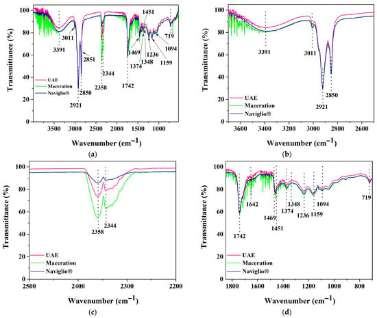
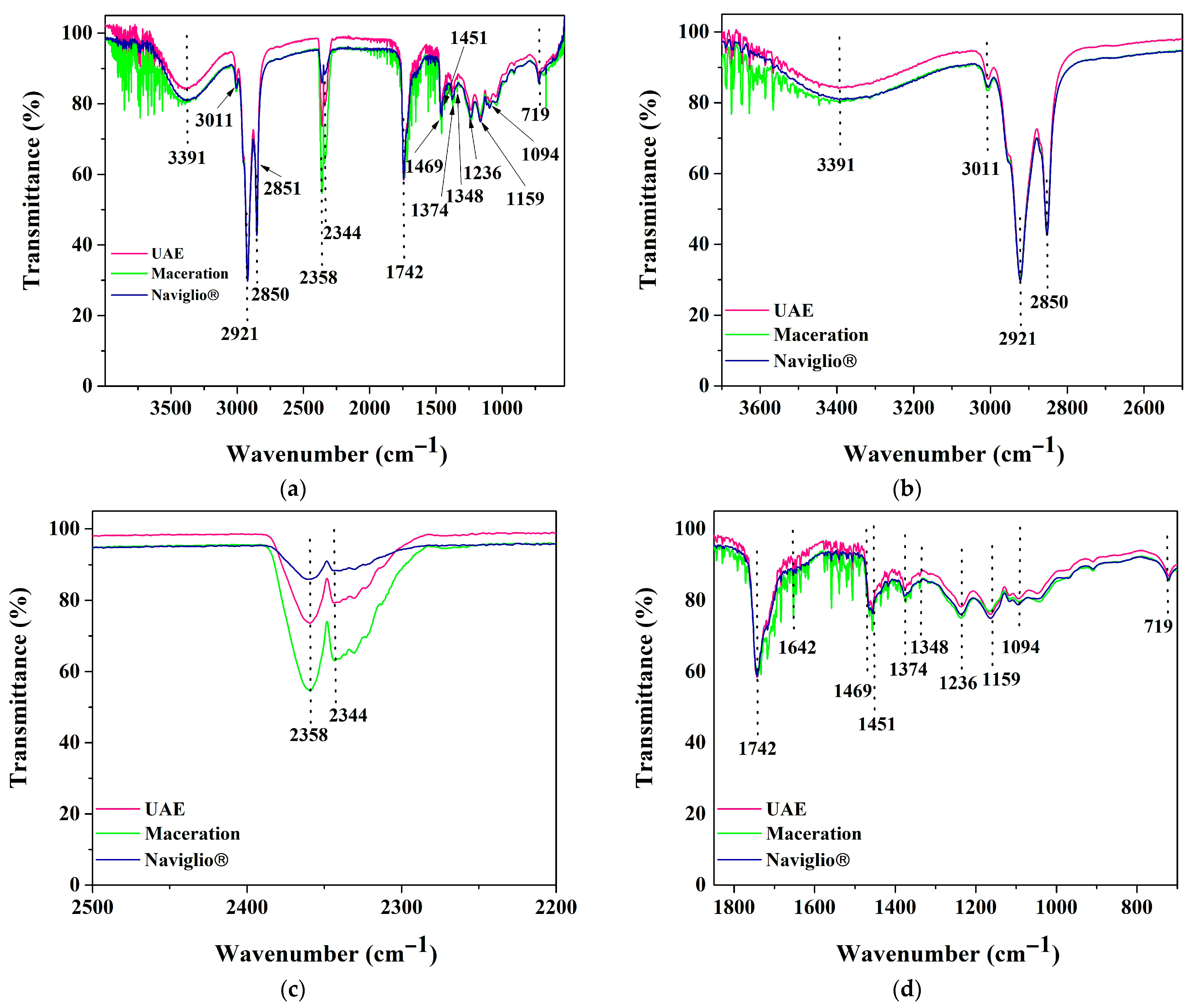
The FTIR spectra of ERAOIs obtained by NE, UAE, and ME exhibited absorption peaks at 3391, 3011, 2920, 2850, 2358, 2344, 1744, 1469, 1451, 1374, 1348, 1236, 1159, 1094, and 707 cm−1 (Figure 1a). These peaks were similar to those reported for avocado, avocado seed, and avocado peel oils [25,26]. The broad band around 3500 to 3000 cm−1 and centered at 3391 cm−1 was ascribed to hydroxyl stretching associated with the OH groups of unsaturated fatty acids (=C–H(trans-)), aromatic alkenes (C=C, C=O), and some phytosterols (β-sitosterol and campesterol) in the ERAOIs [27,28].
Figure 1.
FTIR spectra of avocado oil industrial residue extracts obtained from Naviglio®, ultrasound-assisted, and maceration technologies: changes at (a) 3600–800 cm−1, (b) 3600–2600 cm−1, (c) 2500–2200 cm−1, and (d) 1800–800 cm−1. UAE: Ultrasound-assisted extraction.
The peak around 3011 cm−1 was ascribed to the stretching mode of cis C=C, cis C–H, and cis C=H [25,27]. Furthermore, peaks around 2920 and 2851 cm−1 were ascribed to the asymmetric and symmetric stretching of the –c–CO–c aromatics and stretching of the methylene group (–CH2– stretching of alkene) [26]. Additionally, peaks at 2358 and 2344 cm−1 were assigned to the CH2 stretching mode. These signals have been reported in avocado seed oil, indicating the presence of longer fatty acid chains [25]. The peak around 1734 cm−1 indicated the presence of a carbonyl group of aldehydes and ketones (ester linkage of triacylglycerol; C=O stretching) [29,30]. The peak centered at 1642 cm−1 corresponded to –C=C stretching of cis-olefins, an unsaturated alkene in avocado seed oils [26,30]. The peaks located at 1469, 1451, 1374, and 1348 cm−1 were ascribed to –CH2, cis C=H, and –CH3 scissoring vibrations and CH bending rocking of alkene in avocado fruit or avocado byproduct-based oils, respectively [26,27,30]. The functional groups centered at 1236 cm−1 (–C–O stretch), 1159 cm−1 (carbon–oxygen bonds and carbonyl group), and 1094 cm−1 (C–O stretching vibration, –C–H bending, –C–stretch) were detected in UAEE, NEE, and MEE. These findings are consistent with those previously reported for avocado seed oils [29]. These peaks are associated with a cyclic ester of saturated oil [27,31]. Additionally, a peak around 719 cm−1 was ascribed to the cis–CH=CH–bending of olefins and to the CH2 rocking mode [28,30].
3.2. Contents of Functional Compounds and Antioxidant Capacity
3.2.1. Total Phenolic Content
The ERAOIs obtained by the three extraction methods averaged 16.21 ± 4.47 mg GAE/g. As shown in Table 1, the lowest value was measured in the UAEE, while contents in the NEE and MEE were not significant (p < 0.05). Grisales-Mejía et al. [32] reported TPC values ranging from 283 ± 26 to 321 ± 26 mg GAE/g for ethanolic extracts of seed and peel mixtures obtained by pressurized liquid extraction (PLE). These TPC values from mixed residue extracts were higher than those from avocado seed extracts (ASE).
Table 1.
Bioactive compounds and antioxidant activity of avocado oil industrial residue extracts obtained from Naviglio®, ultrasound-assisted, and maceration technologies.
Most studies have focused on extracts from avocado peels or seeds. The present study found values within the range of those previously reported for aqueous ASEs (6.31 mg GAE/g) obtained through UAE (3 h, 40 kHz, 20 °C) [33], or those reported for hydroethanolic (70%) UAEEs obtained from peel (23.06 ± 0.03 to 42.84 ± 1.76 mg GAE/g) and seed (33.03 ± 0.08 to 55.57 ± 1.23 mg GAE/g) [3]. García-Ramón et al. [34] reported values ranging from 30.35 ± 0.05 mg GAE/g to 44.24 ± 0.06 mg GAE/g in peel hydroethanolic MEEs.
3.2.2. Profile of Phenolic Compounds (HPLC)
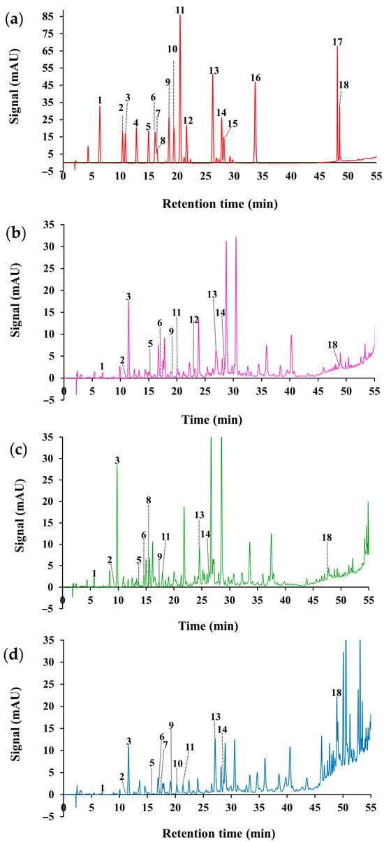
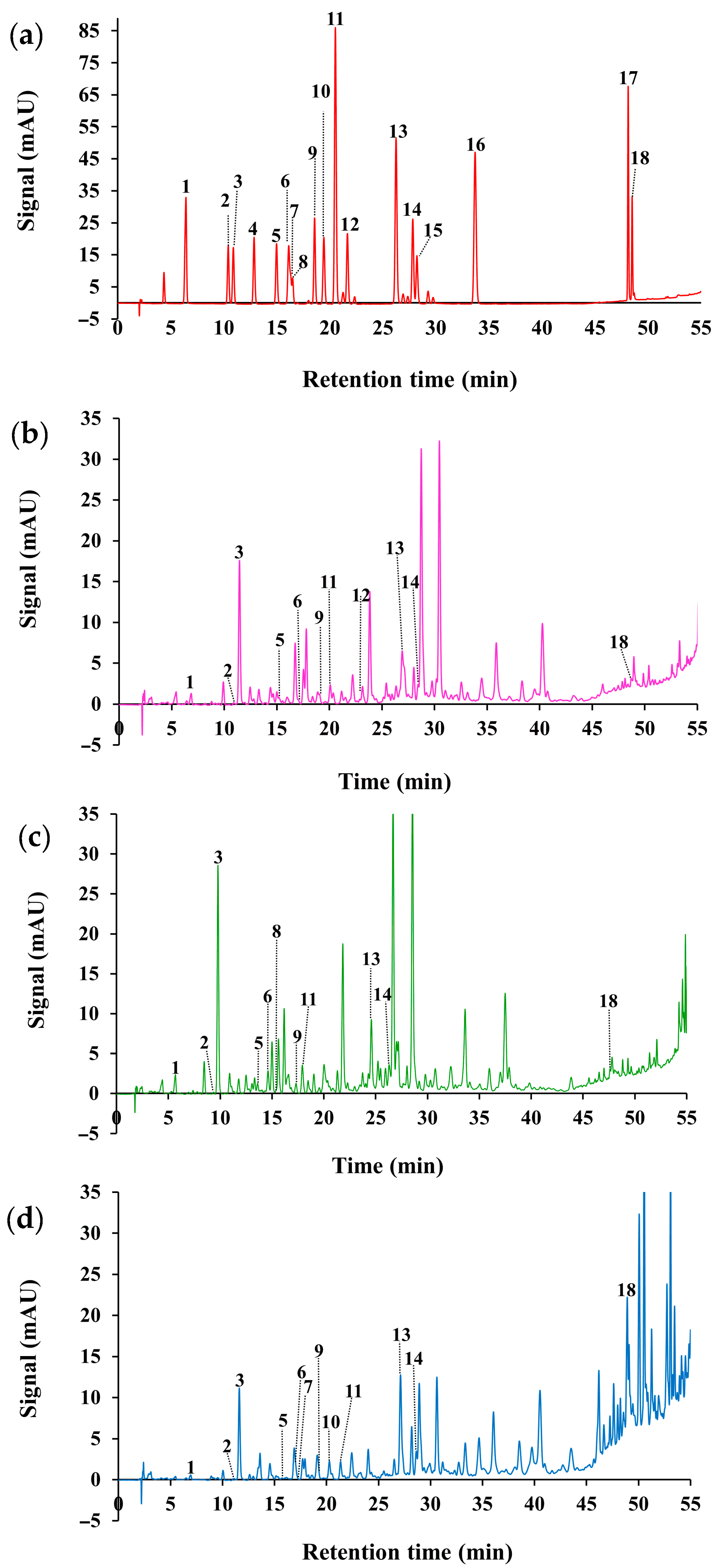
During the course of phenol profiling, eight of the twelve phenolic acids, the two phenolic alcohols, and two of the four flavonoids under investigation were found to be present in quantifiable amounts (Table 2). Peak numbers (Table 2) were assigned according to the retention time of the reference compounds (Figure 2a).
Table 2.
Profile of phenolic compounds of avocado oil industrial residue extracts obtained from Naviglio®, ultrasound-assisted, and maceration technologies.
Figure 2.
HPLC chromatograms of avocado oil industrial residue extracts. (a) Reference compounds, (b) ultrasound-assisted extraction, (c) maceration extraction and (d) Naviglio® extraction. Peak numbers: (1) Gallic acid, (2) hydroxytyrosol, (3) 3,4-dihydroxybenzoic acid, (4) 3,4-dihydroxyphenylacetic acid, (5) tyrosol, (6) 4-hydroxybenzoic acid, (7) epigallocatechin, (8) catechin, (9) chlorogenic acid, (10) vanillic acid, (11) caffeic acid, (12) syringic acid, (13) p-coumaric acid, (14) 4-hydroxy-3-methoxycinnamic acid, (15) sinapic acid, (16) 2-hydroxycinnamic acid, (17) kaempferol and (18) apigenin.
The most predominant phenolic compounds identified were 3,4-dihydroxybenzoic, p-coumaric, and vanillic acids, as well as apigenin, irrespective of the extraction method employed (Figure 2). Both NEE and MEE exhibited the same number of detected compounds (12). The MEE stood out for its gallic and caffeic acid content (p < 0.05). The presence of hydroxytyrosol was detected in trace amounts in all of the examined extracts. Catechin was also found in trace amounts in NEE and MEE, but not in UAEE. Tyrosol content was not different between UAEE and MEE (p > 0.05). However, it was present in trace amounts in the NEE. The contents of 3,4-dihydroxybenzoic, 4-hydroxybenzoic, vanillic and p-coumaric acids were not different between NEE and UAEE, being lower than those in MEE. Only the 4-hydroxy-3-methoxycinnamic acid level was lower in the MEE (p < 0.05). The chlorogenic acid content did not differ among the ERAOIs studied (p > 0.05). Finally, the apigenin NEE content was significantly higher than that of the ME and UAE extracts (p < 0.05). Maceration was the extraction method that improved the recovery of phenolic acids and tyrosol, while the NE excelled at the apigenin recovery. The UAE performed similarly to NE, surpassing it in the recovery of gallic and caffeic acids.
The amounts of each phenolic compound quantified were consistent with previous reports as follows: gallic acid has been quantified at lower concentrations in seed aqueous UAEE (0.031 mg/g) and in peel hydroethanolic MEEs (0.0004 mg/g to 0.002 mg/g) [33,34]. Tyrosol was detected only in avocado seed extract by Lyu et al. [35]. The 3,4-dihydroxybenzoic acid MEE content was higher than that reported in previous studies for peel MEEs, which ranged from 0.013 to 0.015 mg/g [34]. In contrast to the absence of epigallocatechin in the ERAOIs, Sánchez-Quezada et al. [36] detected its presence (0.011 mg/mL) in UAEE-derived seed oil extraction residues. The value obtained for 4-hydroxybenzoic acid in UAEE was lower than that reported in seed aqueous UAEEs (0.15 mg/g) [33]. Higher catechin content was measured in avocado peel (7.12 mg/g) and seed (14.31 mg/g) hydroethanolic UAEEs [3]. Similar chlorogenic acid content was reported in aqueous UAEEs from seeds (0.094 to 0.007 mg/g) and in hydroethanolic peel MEEs (0.084 mg/g) [33,34]. In the preceding study [34], vanillic acid was the most abundant phenol (2.624–3.258 mg/g). The values obtained for vanillic acid in this study were substantially lower than those measured in peel (0.48 ± 0.00 to 0.65 ± 0.00 mg/g) and seed (0.58 ± 0.00 to 1.37 ± 0.00 mg/g) hydroalcoholic UAEEs [3]. Concerning caffeic acid, other authors [36] reported a comparatively lower concentration (0.002 mg/mL) in hydroethanolic UAEE from seed oil extraction residues. The p-coumaric acid content in UAEE surpasses that previously measured in seed UAEE (0.032 mg/g) [33]; nevertheless, it was similar to the values reported for peel UAEEs (0.09 ± 0.00 to 0.18 ± 0.00 mg/g) [3]. The content of 4-hydroxy-3-methoxycinnamic acid in the present study UAEE was higher than that quantified in seed UAEE (0.049 mg/g) [33]. Otherwise, while this phenolic acid was present at trace levels in MEE, previously reported amounts ranged from 0.064 to 0.048 mg/g in hydroethanolic peel MEEs [34]. Although the 2-hydroxycinnamic acid was not detected, it was already measured in hydroethanolic peel MEEs in contents of 0.161 mg/g to 0.083 mg/g [34]. All these authors stated that factors such as maturity stage, anatomical structure, and avocado variety influence the type and concentration of phenolic compounds in avocado fruits and byproducts [3,33,34,35,36].
It is relevant that the absence of the flavonol kaempferol and the flavan-3-ol epigallocatechin, as well as the low levels of catechin, were observed in the studied ERAOIs. Previous studies on avocado peel and seed extracts, both crude and in their fractions after an enrichment step [37]. These authors identified these compounds, along with many others from the same and other classifications, such as flavonoid glycosides, anthocyanins, and numerous glycosylated derivatives. These findings indicate that an enrichment step during the extraction process should improve the flavonoid detection and quantification, and that a more extensive study of specific flavonoids is necessary, given their high proportions within the TPC in the NEE (42.09%), UAEE (33.24%), and MEE (24.97%).
This study is pioneering in its approach, as it is the first to report the identification and quantification of individual phenolic compounds in extracts from mixtures of solid waste provided directly by the AO industry. Furthermore, to our knowledge, this is the first report on extracts obtained from avocado processing waste using Naviglio® technology.
3.2.3. Total Flavonoid Content
On average, the TFC of the three experimental ERAOIs was 5.35 ± 1.67 mg CE/g extract. The utilization of NE resulted in an extract exhibiting the highest TFC (Table 1), while UAEE and MEE did not demonstrate a significant difference (p > 0.05).
The values of TFC in the seed and peel MEEs have been previously documented [34]. In the hydroethanolic peel MEEs, the TFC amounts reported (786.08 mg QE/g and 642.85 mg QE/g) surpassed those observed in this study. Conversely, lower TFCs were quantified in two mixtures of hydrophilic and lipophilic ASE (0.176 mg/g) and APE (0.122 mg/g). These mixtures were obtained through successive maceration with methanol and petroleum ether [38]. With respect to UAEE, the lower TFCs ranging from 1.46 to 3.38 mg QE/g and from 1.31 to 6.90 mg QE/g have been measured in hydroethanolic APE and ASE, respectively [35].
3.2.4. Total Anthocyanin Content
As indicated in Table 1, trace amounts of TAC were detected in the NEE, while no presence of the substance was noted in the other extracts. This finding was unexpected, since anthocyanins have previously been identified and quantified in peel and its aqueous PLE extracts [32]. The polarity of solvents is a critical factor in the recovery of anthocyanins during extraction processes, as these compounds exhibit high water solubility [39]. Additionally, ripening influences TAC, as it increases as the avocado fruit ripens, resulting in the characteristic violet external color of mature avocado Hass fruits [39].
3.2.5. β-Sitosterol
The average β-sitosterol content of the studied ERAOIs attained 7614.35 ± 1526.07 mg/kg. The highest content was observed in the MEE (p < 0.05). However, there were no statistically significant differences (p > 0.05) between the UAEE and NEE (Table 1). To our knowledge, β-sitosterol content has not been reported in avocado peels, seeds, or laboratory extracts. The absence of phytosterols was reported in APE and ASE obtained from byproducts of the AO industry [38]. In the present study, β-sitosterol was detected during the course of FT-IR characterization. The presence of this compound is attributed to pulp residues adhering to the peel and seed, as the pulp and AO are sources of this compound [40]. The findings of this study suggest that the studied extracts represent a potential source of β-sitosterol that can be incorporated into developing products focused on correcting dyslipidemia [41] or as a control agent against dengue due to its larvicidal effect [42].
3.2.6. Antioxidant Activity
A higher ABTS AOX was measured in the MEE (p < 0.05), while the NEE and UAEE values were not different (p > 0.05) (Table 1). The same occurred with the AOX measured by the FRAP method. AOX results obtained by the DDPH method showed differences among the three studied ERAOIs. The MEE standing was the highest, followed by NEE and UAEE.
Other authors have reported a wide range of AOX values for avocado seed and peel extracts. However, it is noteworthy that no AOX values have been reported for residual peel and seed mixtures from AO extraction to date. A similar magnitude of values (4.07 to 14.90 µmol TE/g) was measured in UAEEs from the seed and peel using the ABTS method. However, higher values (322.11 to 859.27 µmol TE/g) were measured using the FRAP method [3]. Furthermore, other authors reported AOX values much higher than those obtained in our study. For instance, the AOX of UAEEs from peel and seed measured using DPPH (1138.70 and 1348.0 µmol TE/g) and ABTS (339.9 and 343.3 µmol TE/g) [32]. The AOX value of 760.5 µmol TE/mL (DPPH) in seed oil was obtained by UAE and emulsified with aqueous extracts of its residual flour [36]. While AOX of hydroethanolic MEEs of avocado residues was determined by the ABTS, FRAP and DPPH methods in immature (1006.21, 804.40, and 564.82 µmol TE/g, respectively) and mature (763.73, 456.40, and 382.07 µmol TE/g, respectively) APE [34]. Moreover, the AOX values of 496 and 1510 µmol TE/mL were obtained when measuring (ABTS) ASE and APE via a 3-cycle ME using hydroethanolic mixtures at different temperatures [43]. Differences in AOX are associated with the use of multiple extraction techniques, various solvent types and proportions, and differences in the origin, supply sources, and conditioning procedures of the avocado subproducts [34,43].
3.3. Biological Activities
3.3.1. Antimicrobial Activity
All ERAOIs showed antimicrobial activity against both Gram-positive and Gram-negative bacteria (Table 3). There are previous reports on the antimicrobial activity of APEs against S. aureus and E. coli, obtained by maceration, Soxhlet extraction (ethanol), supercritical fluid extraction (CO2), and UAE with water [33].
Table 3.
Minimum inhibitory concentration and minimum bactericidal concentration of avocado oil industrial residue extracts obtained from Naviglio®, ultrasound-assisted, and maceration technologies.
The MIC and MBC values (Table 3) ranged from 25 to 100 mg/mL. The comparison of MICs across different ERAIs reveals that the NEE and UAEE did not differ and had the lowest MICs for both bacteria. Regarding the MBC for E. coli, the three studied extracts showed significant differences (p < 0.05). Among these extracts, MEE exhibited the highest value (100 mg/mL), followed by NEE (50 mg/mL) and UAEE (25 mg/mL). Meanwhile, the MBC values for S. aureus did not differ between the UAEE and MEE (100 mg/mL), while the NEE MBC reached 50 mg/mL. A previous study of an aqueous seed UAEE [33] reported MIC values of 0.21 mg/mL for E. coli and 2.78 mg/mL for S. aureus. In the case of ASE, Romaní et al. [44] reported lower E. coli MIC (0.625 mg/mL) and MBC (1.250 mg/mL) values when testing an extract obtained by maceration (ethanol) and ethyl acetate fractionation. The solvent and extraction method differentially enrich extracts with antimicrobial phytoconstituents, as those actually studied, and others, such as acetogenins, which have been shown to influence the amounts of antimicrobial activity [4,33,44].
3.3.2. Brine Shrimp Bioassay
The average lethal doses (LD50) resulting from the PROBIT method analysis were 2880 µg/mL, 960 µg/mL, and 250 µg/mL for the NE, UAE, and ME extracts, respectively. According to the degree of toxicity of vegetal extracts (Section 2.6.3) [23], NEE was classified as non-toxic, while MEE and UAEE were moderately toxic. However, MEE and UAEE extracts were toxic at the same MIC and MBC values, making them unsuitable as antimicrobial food ingredients. However, they could be used for other antimicrobial purposes. There were no reports on APEs’ toxicity in the shrimp model. However, those about ASEs referred to LD50 values similar to the NEE values, reaching 2370 µg/mL for Soxhlet hexane extracts [42]. Other authors measured comparatively higher LD50 values for UAE oil (4556.27 ± 363 µg/mL) and for the aqueous extracts from the residual oil extraction flour (3320.83 ± 385 µg/mL) [36].
3.3.3. In Vitro Anti-Inflammatory Activity
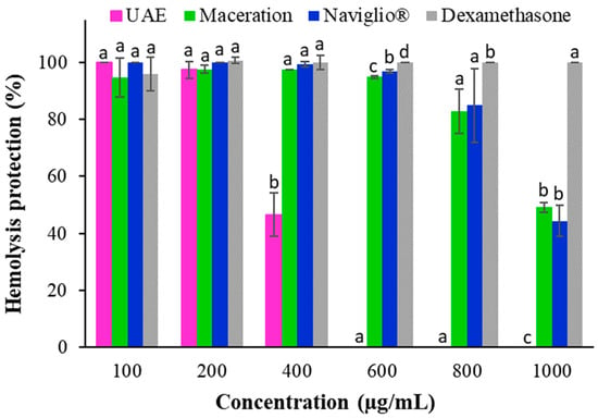
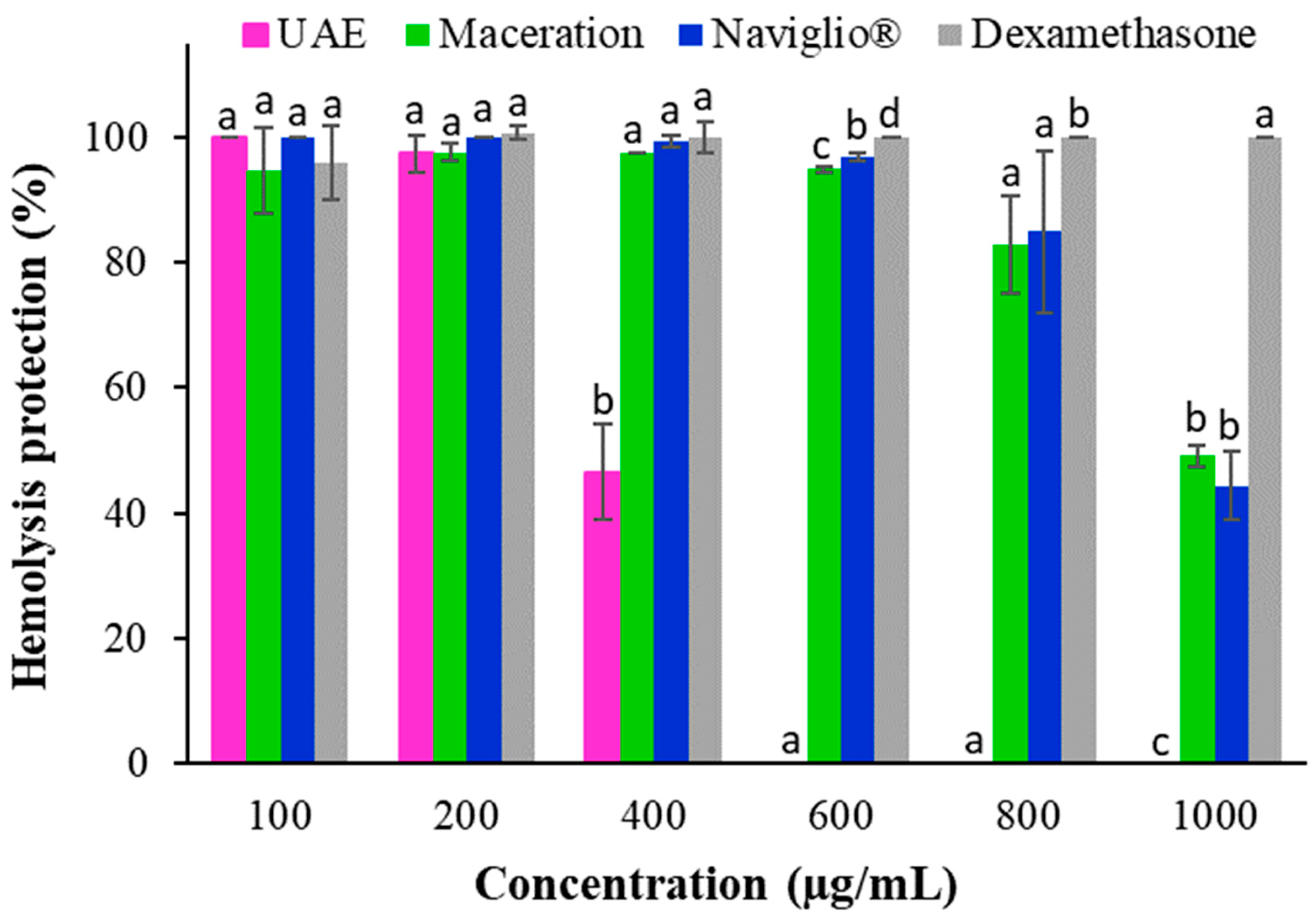
All ERAOIs showed anti-inflammatory activity (Figure 3). The NE and ME extracts exhibited comparable anti-inflammatory effects (p ≥ 0.05), though they differed significantly from the measurements obtained in UAEE at 400 µg/mL. The maximum effect of UAEE was observed at 100 to 200 µg/mL, whereas NEE and MEE demonstrated superior anti-inflammatory effects across 100 to 800 µg/mL. However, all extracts displayed a lower effect on HP than that of dexamethasone. UAEE from 400 µg/mL (46.57%) to a null HP at 600 µg/mL. Furthermore, the NEE and MEE decreased from 600 µg/mL to a minimum HP at 1000 µg/mL (44.39% and 49.21% for the NEE and MEE, respectively).
Figure 3.
Human red blood cell membrane stabilization using different concentrations of dexamethasone and avocado oil industrial residue extracts obtained from Naviglio®, ultrasound-assisted (UAE), and maceration technologies. a–d Means in the same concentration without a common letter are significantly different (p < 0.05).
Ovalle-Marín et al. [45] observed that hydroalcoholic avocado peel MEE had an anti-inflammatory effect on the cellular line of the mouse macrophage RAW264.7 when measuring two targets for anti-inflammatory drugs (NO generation and cytokine TNF-α) as the inflammatory markers. The observed activity was ascribed to procyanidin dimers and trimers present in the peel and chlorogenic acid found in the leaves. For its part, Tremocoldi et al. [46] did not find that Hass APE and ASE could inhibit both markers in LPS-stimulated RAW264.7 macrophage cultures. However, these compounds demonstrated an inhibitory effect when assayed with Fuerte APEs. These findings suggest that Fuerte APEs can be considered a natural anti-inflammatory product rich in bioactive phenolic compounds.
4. Conclusions
The extracts obtained by three green technologies from the peel and seed residual mixture of the avocado oil industry (Persea americana, Mill.) showed FT-IR spectra suggesting the presence of polysaccharides (pectins, starch, and lignin), fatty acids, phytosterols, and phenolic compounds. Moreover, extraction techniques influence the concentration and type of the extracted compounds. Although NEE proved to be non-toxic, ME and UAE extracts were found to be moderately toxic at the evaluated MIC and MBC. However, NEE and UAEE were not toxic at the anti-inflammatory tested concentrations. The NEE and UAEE have pharmaceutical potential as antioxidants and anti-inflammatories, while MEE is a potential source of β-sitosterol.
Key limitations identified are related to the enrichment of the crude extract and its subsequent separation into hydrophilic and lipophilic fractions. A more extensive chemical characterization of both functional and potentially toxic compounds is necessary. This characterization should employ in vitro (cell cultures) or in vivo biological models to test biological activity. Finally, it is essential to test the antimicrobial activity against a broader spectrum of microorganisms of food, medical, and even agricultural relevance.
Further studies on other RAOI mixtures, such as pomace, are needed to evaluate their sustainable real application in the obtention of extracts with potential in the pharmaceutical and food industries.
Author Contributions
Conceptualization, K.D.V.-B., J.d.P.R.-A. and A.C.-G.; methodology, K.D.V.-B., J.d.P.R.-A., A.C.-G., J.C.S.-N. and L.M.A.-E.; software, J.C.S.-N. and L.M.A.-E.; validation, M.C.C.-S. and A.C.-G.; formal analysis, K.D.V.-B., J.C.S.-N. and L.M.A.-E.; investigation, K.D.V.-B. and J.d.P.R.-A.; resources, J.d.P.R.-A.; data curation, J.d.P.R.-A. and M.C.C.-S.; writing—original draft preparation, K.D.V.-B., J.d.P.R.-A., A.C.-G. and L.M.A.-E.; writing—review and editing, J.d.P.R.-A., L.M.A.-E. and M.C.C.-S.; visualization, K.D.V.-B.; supervision, J.d.P.R.-A.; project administration, J.d.P.R.-A.; funding acquisition, J.d.P.R.-A. All authors have read and agreed to the published version of the manuscript.
Funding
This research was funded by the Centro Universitario del Sur, Universidad de Guadalajara, under grant number SAC/CIP/097/2023.
Institutional Review Board Statement
Not applicable.
Informed Consent Statement
Not applicable.
Data Availability Statement
The original contributions presented in this study are included in the article. Further inquiries can be directed to the corresponding authors.
Acknowledgments
The authors acknowledge Mevi Mexico oil industry for providing the solid waste samples, and to S.T. Martín-del-Campo, for the review and correction of the revised manuscript.
Conflicts of Interest
The authors declare no conflicts of interest. The funders had no role in the design of the study; in the collection, analyses, or interpretation of data; in the writing of the manuscript; or in the decision to publish the results.
Abbreviations
The following abbreviations are used in this manuscript:
| AO | Avocado oil |
| AOX | Antioxidant activity |
| APE | Avocado peel extract |
| ASE | Avocado seed extract |
| CGE | Cyaniding 3-O glucoside equivalents |
| DMSO | Dimethyl sulfoxide |
| ERAOIs | Extracts from residues of the avocado oil industry |
| GAE | Gallic acid equivalents |
| HP | Hemolysis protection |
| ME | Maceration extraction |
| MEE | Maceration extraction extract |
| MBC | Minimum bactericidal concentration |
| MIC | Minimum inhibitory concentration |
| MHB | Mueller–Hinton broth |
| NE | Naviglio’s extraction |
| NEE | Naviglio’s extraction extract |
| n | Number of samples |
| nd | Not detected |
| PLE | Pressurized liquid extraction |
| QE | Quercetin equivalents |
| RAOI | Residues of the avocado oil industry |
| TAC | Total anthocyanin content |
| TE | Trolox equivalents |
| TFC | Total flavonoid content |
| TPC | Total phenolic content |
| tz | Traces |
| UAE | Ultrasound-assisted extraction |
| UAEE | Ultrasound-assisted extraction extract |
References
- FAOSTAT. Avocado World Production. 2024. Available online: https://www.fao.org/faostat/en/#data/QCL (accessed on 12 April 2025).
- Permal, R.; Leong Chang, W.; Seale, B.; Hamid, N.; Kam, R. Converting industrial organic waste from the cold-pressed avocado oil production line into a potential food preservative. Food Chem. 2020, 306, 125635. [Google Scholar] [CrossRef] [Scilit] [PubMed]
- Oliveira, C.S.d.; Andrade, J.K.S.; Rajan, M.; Narain, N. Influence of the phytochemical profile on the peel, seed and pulp of Margarida, Breda and Geada varieties of avocado (Persea americana Mill) associated with their antioxidant potential. Food Sci. Technol. 2022, 42, 25822. [Google Scholar] [CrossRef] [Scilit]
- Rodríguez-López, C.E.; Hernández-Brenes, C.; Treviño, V.; Díaz de la Garza, R.I. Avocado fruit maturation and ripening: Dynamics of aliphatic acetogenins and lipidomic profiles from mesocarp, idioblasts and seed. BMC Plant Biol. 2017, 17, 159. [Google Scholar] [CrossRef] [Scilit]
- Gallo, M.; Ferrara, L.; Naviglio, D. Application of ultrasound in Food Science and Technology: A perspective. Foods 2018, 7, 164. [Google Scholar] [CrossRef] [Scilit] [PubMed]
- Naviglio, D.; Scarano, P.; Ciaravolo, M.; Gallo, M. Rapid solid-liquid dynamic extraction (RSLDE): A powerful and greener alternative to the latest solid-liquid extraction techniques. Foods 2019, 8, 245. [Google Scholar] [CrossRef] [Scilit] [PubMed]
- Reyes-Becerril, M.; Angulo, C.; Silva-Jara, J. Antibacterial and immunomodulatory activity of moringa (Moringa oleifera) seed extract in Longfin yellowtail (Seriola rivoliana) peripheral blood leukocytes. Aquac. Res. 2021, 52, 4076–4085. [Google Scholar] [CrossRef] [Scilit]
- Won, S.; Kwon, K.H. Green technology for extraction of bioactive compounds from edible plants. J. Oleo Sci. 2024, 73, 1249–1265. [Google Scholar] [CrossRef] [Scilit]
- Reyes-Becerril, M.; Guerra-Contreras, K.; Gijón-Díaz, D. Plantas aromáticas: Métodos para medir su actividad. CIBNOR 2020, 2, 47. [Google Scholar]
- Bimakr, M.; Rahman, R.A.; Taip, F.S.; Ganjloo, A.; Salleh, L.M.; Selamat, J.; Hamid, A.; Zaidul, I.S.M. Comparison of different extraction methods for the extraction of major bioactive flavonoid compounds from spearmint (Mentha spicata L.) leaves. Food Bioprod. Process. 2011, 89, 67–72. [Google Scholar] [CrossRef] [Scilit]
- Vega-Hernández, L.C.; Serrano-Niño, J.C.; Velázquez-Carriles, C.A.; Martínez-Preciado, A.H.; Cavazos-Garduño, A.; Silva-Jara, J.M. Improving foodborne pathogen control using green nanosized emulsions of Plectranthus hadiensis phytochemicals. Colloids Interfaces 2024, 8, 3. [Google Scholar] [CrossRef] [Scilit]
- Arnous, A.; Makris, D.P.; Kefalas, P. Correlation of pigment and flavanol content with antioxidant properties in selected aged regional wines from Greece. J. Food Compost. Anal. 2002, 15, 655–665. [Google Scholar] [CrossRef] [Scilit]
- Escarpa, A.; González, M.C. Optimization strategy and validation of one chromatographic method as approach to determine the phenolic compounds from different sources. J. Chromatogr. A 2000, 897, 161–170. [Google Scholar] [CrossRef] [Scilit]
- Panche, A.N.; Diwan, A.D.; Chandra, S.R. Flavonoids: An overview. J. Nutr. Sci. 2016, 5, e47. [Google Scholar] [CrossRef] [Scilit]
- Lee, J.; Durst, R.; Wrolstad, R. Determination of total monomeric anthocyanin pigment content of fruit juices, beverages, natural colorants, and wines by the pH differential method: Collaborative Study. J. AOAC Int. 2005, 88, 1269–1278. [Google Scholar] [CrossRef] [Scilit]
- Nzekoue, F.k.; Khamitova, G.; Angeloni, S.; Sempere, A.N.; Tao, J.; Maggi, F.; Xiao, J.; Sagratini, G.; Vittori, S.; Caprioli, G. Spent coffee grounds: A potential commercial source of phytosterols. Food Chem. 2020, 325, 126836. [Google Scholar] [CrossRef] [Scilit]
- Oliveira, A.P.; Baptista, P.; Andrade, P.B.; Martins, F.; Pereira, J.A.; Silva, B.M.; Valentão, P. Characterization of Ficus carica L. cultivars by DNA and secondary metabolite analysis: Is genetic diversity reflected in the chemical composition? Food Res. Int. 2012, 49, 710–719. [Google Scholar] [CrossRef] [Scilit]
- Brand-Williams, W.; Cuvelier, M.E.; Berset, C. Use of a free radical method to evaluate antioxidant activity. LWT—Food Sci. Technol. 1995, 28, 25–30. [Google Scholar] [CrossRef] [Scilit]
- Pellegrini, N.; Ke, R.; Yang, M.; Rice-Evans, C. Screening of dietary carotenoids and carotenoid-rich fruit extracts for antioxidant activities applying 2,2′-azinobis(3-ethylenebenzothiazoline-6-sulfonic acid radical cation decolorization assay. Methods Enzymol. 1999, 299, 379–389. [Google Scholar] [CrossRef] [Scilit]
- Benzie, I.F.F.; Strain, J.J. The ferric reducing ability of plasma (FRAP) as a measure of “Antioxidant power”: The FRAP assay. Anal. Biochem. 1996, 239, 70–76. [Google Scholar] [CrossRef] [Scilit] [PubMed]
- Clinical and Laboratory Standards Institute. Methods for Determining Bactericidal Activity of Antimicrobial Agents; CLSI: Wayne, PA, USA, 1999. [Google Scholar]
- Krishnaraju, A.V.; Rao, T.V.N.; Sundararaju, D.; Vanisree, M.; Tsay, H.-S.; Subbaraju, G.V. Assessment of bioactivity of Indian medicinal plants using brine shrimp (Artemia salina) lethality assay. Int. J. Appl. Sci. Eng. 2005, 3, 125–134. [Google Scholar] [CrossRef]
- Ntungwe, N.E.; Domínguez-Martín, E.M.; Roberto, A.; Tavares, J.; Isca, V.; Pereira, P.; Cebola, M.-J.; Rijo, P. Artemia species: An important tool to screen general toxicity samples. Curr. Pharm. Des. 2020, 26, 2892–2908. [Google Scholar] [CrossRef] [Scilit] [PubMed]
- Rajakumar, P.; Anandhan, R. Synthesis and In-vitro anti-inflammatory activity of novel glycodendrimers with benzene 1,3,5 carboxamide core and triazole as branching unit. Eur. J. Med. Chem. 2011, 46, 4687–4695. [Google Scholar] [CrossRef] [Scilit] [PubMed]
- Reyes-Cueva, E.; Nicolalde, J.F.; Martínez-Gómez, J. Characterization of unripe and mature avocado seed oil in different proportions as phase change materials and simulation of their cooling storage. Molecules 2021, 26, 107. [Google Scholar] [CrossRef] [Scilit]
- Sathish, S.; Narendrakumar, G.; Vaithyasubramanian, S.; Sinduri, E. Mechanistic model for the batch extraction of oil from avocado seeds available for biofuel production. Int. J. Green Energy 2021, 18, 1645–1657. [Google Scholar] [CrossRef] [Scilit]
- Del Castillo-Llamosas, A.; Rodríguez-Martínez, B.; del Río, P.G.; Eibes, G.; Garrote, G.; Gullón, B. Hydrothermal treatment of avocado peel waste for the simultaneous recovery of oligosaccharides and antioxidant phenolics. Bioresour. Technol. 2021, 342, 125981. [Google Scholar] [CrossRef] [Scilit]
- Rohman, A.; Windarsih, A.; Riyanto, S.; Sudjadi; Shuhel Ahmad, S.A.; Rosman, A.S.; Yusoff, F.M. Fourier transform infrared spectroscopy combined with multivariate calibrations for the authentication of avocado oil. Int. J. Food Prop. 2016, 19, 680–687. [Google Scholar] [CrossRef] [Scilit]
- Tsegay, H. Determination of oil and biodiesel content, physicochemical properties of the oil extracted from avocado seed (Persea americana) grown in Wonago and Dilla (Gedeo zone), Southern Ethiopia. Chem. Int. 2017, 3, 311–319. [Google Scholar]
- Rodríguez-Martínez, B.; Ferreira-Santos, P.; Gullón, B.; Teixeira, J.A.; Botelho, C.M.; Yáñez, R. Exploiting the potential of bioactive molecules extracted by ultrasounds from avocado peels—Food and nutraceutical applications. Antioxidants 2021, 10, 1475. [Google Scholar] [CrossRef] [Scilit]
- Uzoh, C.F.; Onukwuli, O.D. Self-cured alkyd resin using non-drying avocado seed oil as a material of regenerative resource. Bull. Korean Chem. Soc. 2018, 39, 643–650. [Google Scholar] [CrossRef] [Scilit]
- Grisales-Mejía, J.F.; Álvarez-Rivera, G.; Torres-Castañeda, H.G.; Andrade-Mahecha, M.M.; Martínez-Correa, H.A.; Mendiola, J.A.; Cifuentes, A.; Ibañez, E. Hass avocado (Persea americana Mill.) residues as a new potential source of neuroprotective compounds using pressurized liquid extraction. J. Supercrit. Fluids 2024, 204, 106117. [Google Scholar] [CrossRef] [Scilit]
- Kupnik, K.; Primožič, M.; Kokol, V.; Knez, Ž.; Leitgeb, M. Enzymatic, antioxidant, and antimicrobial activities of bioactive compounds from avocado (Persea americana L.) seeds. Plants 2023, 12, 1201. [Google Scholar] [CrossRef] [Scilit] [PubMed]
- García-Ramón, F.; Malnati-Ramos, M.; Rios-Mendoza, J.; Vivar-Méndez, J.; Nieva-Villegas, L.M.; Cornelio-Santiago, H.P.; Sotelo-Méndez, A. Avocado Hass peel from industrial by-product: Effect of extraction process variables on yield, phenolic compounds and antioxidant capacity. Front. Sustain. Food Syst. 2023, 7, 1255941. [Google Scholar] [CrossRef] [Scilit]
- Lyu, X.; Agar, O.T.; Barrow, C.J.; Dunshea, F.R.; Suleria, H.A.R. Phenolic compounds profiling and their antioxidant capacity in the peel, pulp, and seed of Australian grown avocado. Antioxidants 2023, 12, 185. [Google Scholar] [CrossRef] [Scilit]
- Sánchez-Quezada, V.; Gaytán-Martínez, M.; Recio, I.; Loarca-Piña, G. Avocado seed by-product uses in emulsion-type ingredients with nutraceutical value: Stability, cytotoxicity, nutraceutical properties, and assessment of in vitro oral-gastric digestion. Food Chem. 2023, 421, 136118. [Google Scholar] [CrossRef] [Scilit] [PubMed]
- Rosero, J.C.; Cruz, S.; Osorio, C.; Hurtado, N. Analysis of phenolic composition of byproducts (seeds and peels) of avocado (Persea americana Mill.) cultivated in Colombia. Molecules 2019, 24, 3209. [Google Scholar] [CrossRef] [Scilit]
- Rashama, C.; Ijoma, G.N.; Matambo, T.S. Applying phytochemical analysis in the pre-selection process of plant-based substrates for biomethanation: The case of avocado oil processing byproducts. J. Mater. Cycles Waste Manag. 2022, 24, 1623–1632. [Google Scholar] [CrossRef] [Scilit]
- Khoo, H.E.; Azlan, A.; Tang, S.T.; Lim, S.M. Anthocyanidins and anthocyanins: Colored pigments as food, pharmaceutical ingredients, and the potential health benefits. Food Nutr. Res. 2017, 61, 1361779. [Google Scholar] [CrossRef] [Scilit]
- King-Loeza, Y.; Ciprián-Macías, D.A.; Cardador-Martínez, A.; Martín-del-Campo, S.T.; Castañeda-Saucedo, M.C.; Ramírez-Anaya, J.d.P. Functional composition of avocado (Persea americana Mill. Var. Hass) pulp, extra virgin oil, and residues is affected by fruit commercial classification. J. Agric. Food Res. 2023, 12, 100573. [Google Scholar] [CrossRef] [Scilit]
- Park, E.; Edirisinghe, I.; Burton-Freeman, B. Avocado fruit on postprandial markers of cardio-metabolic risk: A randomized controlled dose response trial in overweight and obese men and women. Nutrients 2018, 10, 1287. [Google Scholar] [CrossRef] [Scilit] [PubMed]
- Leite, J.J.G.; Brito, É.H.S.; Cordeiro, R.A.; Brilhante, R.S.N.; Sidrim, J.J.C.; Bertini, L.M.; Morais, S.M.d.; Rocha, M.F.G. Chemical composition, toxicity and larvicidal and antifungal activities of Persea americana (avocado) seed extracts. Rev. Soc. Bras. Med. Trop. 2009, 42, 110–113. [Google Scholar] [CrossRef] [Scilit]
- Rojas-García, A.; Fuentes, E.; Cádiz-Gurrea, M.D.; Rodriguez, L.; Villegas-Aguilar, M.D.; Palomo, I.; Arráez-Román, D.; Segura-Carretero, A. Biological evaluation of avocado residues as a potential source of bioactive compounds. Antioxidants 2022, 11, 11061049. [Google Scholar] [CrossRef] [Scilit] [PubMed]
- Romaní, L.; Enciso, E.; Cárdenas, V.; Condorhuamán, Y.M. Actividad antibacteriana de compuestos fenólicos de semillas de Persea americana Mill. “palta hass” frente a Escherichia coli. Cienc. Investig. 2017, 20, 19–22. [Google Scholar] [CrossRef] [Scilit]
- Ovalle-Marín, A.; Parra-Ruiz, C.; Rivas-Zambrano, F.; Orellana, J.; García-Díaz, D.; Jiménez-Patiño, P. Characterization of Persea americana Mill. peels and leaves extracts and analysis of its potential in vitro anti-inflammatory properties. BLACPMA 2020, 19, 395–407. [Google Scholar] [CrossRef] [Scilit]
- Tremocoldi, M.A.; Rosalen, P.L.; Franchin, M.; Massarioli, A.P.; Denny, C.; Daiuto, É.R.; Paschoal, J.A.R.; Melo, P.S.; Alencar, S.M.d. Exploration of avocado by-products as natural sources of bioactive compounds. PLoS ONE 2018, 13, e0192577. [Google Scholar] [CrossRef] [Scilit] [PubMed]

Disclaimer/Publisher’s Note: The statements, opinions and data contained in all publications are solely those of the individual author(s) and contributor(s) and not of MDPI and/or the editor(s). MDPI and/or the editor(s) disclaim responsibility for any injury to people or property resulting from any ideas, methods, instructions or products referred to in the content. |
© 2026 by the authors. Licensee MDPI, Basel, Switzerland. This article is an open access article distributed under the terms and conditions of the Creative Commons Attribution (CC BY) license.