


Abstract
Objectives: The aim of the study was to compare CBCT (Cone Beam Computed Tomography)-derived radiographic hard tissue gain after hydraulic transcrestal sinus floor elevation performed with versus without cross-linked hyaluronic acid (HA) and to assess associated clinical, radiographic, and patient-reported outcomes. Materials and Methods: This single-center, prospective clinical study enrolled 58 patients with 2–6 mm residual bone height in the posterior maxilla, assigned in a non-randomized parallel-group design to HA (n = 29) or No Graft (n = 29). The primary endpoint was CBCT-based 3D CBCT-derived radiographic hard-tissue volume gain at 12 months; secondary endpoints included vertical bone gain, marginal bone loss (MBL) at 12 and 24 months, implant stability quotient (ISQ), implant survival/success, complications, and patient-reported outcomes (VAS pain, OHIP-14). Analyses used ANCOVA and appropriate comparative statistics. Results: At 12 months, mean CBCT-derived radiographic hard-tissue volume gain was higher with HA (1210 +/− 610 mm3) than with No Graft (858 +/− 605 mm3; adjusted difference +352 mm3, 95% CI: 140–564; p = 0.002). ISQ at 12 months was greater with HA (p = 0.028), while MBL at 12/24 months did not differ significantly. Twenty-four-month implant survival was 98.1% (HA) vs. 94.3% (No Graft; p = 0.041). Complication rates were low and comparable; VAS pain at 72 h was modestly lower with HA. Conclusions: In this non-randomized prospective clinical study, cross-linked hyaluronic acid was associated with greater CBCT-derived radiographic hard-tissue volume gain and vertical radiographic gain after hydraulic transcrestal sinus floor elevation compared with clot stabilization alone. Clinical outcomes were favorable in both groups during the 24-month observation period. These findings support the interpretation of cross-linked hyaluronic acid as a potential space-maintaining adjunct in posterior maxillary sites with limited residual bone height; however, because no histological or histomorphometric assessment was performed, they should not be interpreted as proof of vital mature bone regeneration or superior bone quality.
1. Introduction
Sinus lift procedures are widely regarded as predictable techniques for achieving the tridimensional bone volume required to place implants in the posterior atrophic maxilla [1]. The earliest descriptions of sinus floor elevation and implant placement employed a lateral window approach [2], which, despite allowing significant elevation of the antral floor and providing excellent intraoperative visibility, was associated with notable drawbacks, including surgical invasiveness and a higher risk of Schneiderian membrane perforation. Shortly thereafter, a transalveolar approach was proposed for ridges with at least 4–5 mm of residual bone height, aiming to reduce morbidity and postoperative discomfort [3]. The Osteotome Sinus Floor Elevation technique [4] introduced the use of progressively larger diameter osteotomes, activated by a mallet, to fracture and simultaneously compact the apical bone, thereby elevating the maxillary sinus floor without initially placing grafting material [5,6]. This technique was later adapted to allow the sequential insertion of grafts into the subantral space to achieve the desired bone gain.
Among transalveolar sinus elevation techniques, attention has progressively shifted towards more conservative approaches designed to preserve the Schneiderian membrane during detachment and elevation. The hydraulic pressure method described by Sotirakis and Gonshor was developed to elevate the membrane in a less traumatic manner, exploiting its elasticity through the injection of sterile saline with a disposable plastic syringe; the solution is then drained from the subantral compartment to accommodate grafting materials [7].
A long-standing debate persists regarding whether graft material is necessary to maintain space for bone formation following membrane elevation via the osteotome approach. Several parameters have been examined, including vertical bone gain at the mesial and distal aspects of implants, short- and long-term radiographic changes, intrasinus bone gain and peri-implant bone stability over extended follow-up periods [8]. While some authors report no statistical or clinical advantage in using autografts, allografts, xenografts, or synthetic materials, others suggest that grafting increases the likelihood of new bone formation [9]. The former view attributes bone regeneration to the mere stabilization of the blood clot, whereas the latter links enhanced outcomes to the osteoconductive and osteoinductive properties of grafting materials.
Previous studies assessing bone regeneration after sinus lift have largely relied on bidimensional radiographic techniques, such as orthopantomography, periapical radiographs, or 2D CBCT slices, using linear measurements [10,11]. Although informative, these methods do not capture the true tridimensional changes in crestal bone volume. Advances in imaging now allow three-dimensional CBCT analysis to provide more accurate volumetric assessments of regenerated bone [12].
The aim of this prospective study was to quantify CBCT-derived radiographic hard-tissue volume gain obtained after hydraulic transcrestal sinus floor elevation with or without cross-linked hyaluronic acid. Secondary objectives included evaluating vertical radiographic gain, marginal bone loss, implant survival and success, implant stability, complications, and patient-reported outcomes. This approach sought to clarify whether the addition of HA was associated with greater radiographically detectable hard-tissue fill while maintaining the minimally invasive advantages of the hydraulic transcrestal technique.
2. Materials and Methods
2.1. Study Design
A single-center, prospective non-randomized, parallel-group clinical study was conducted to evaluate CBCT-derived radiographic hard-tissue volume gain in the posterior maxilla following transcrestal sinus floor elevation using a hydraulic pressure technique, with or without the use of cross-linked hyaluronic acid (HA) as a grafting material. The patients enrolled were treated during the activity at University Albanian of Tirana between July 2023 and February 2024. All surgical procedures and assessments were standardized and performed by the same operator (E.Q.), reducing inter-operator variability and enhancing the reproducibility of the protocol. The clinical investigation was conducted at the Department of Medical Sciences of the University of Tirana, Albania, registered with Nr 276 prot. dated 4 July 2023.
2.2. Patient Selection
The following inclusion criteria were considered [13]:
- Age ≥ 18 years.
- Partially edentulous patients in the posterior maxilla (premolar/molar) requiring implant-supported rehabilitation with sinus floor elevation.
- Residual alveolar bone height between 2 mm and 6 mm (measured on preoperative CBCT).
- Crestal bone width ≥ 6 mm at the planned implant site.
- Healthy maxillary sinus on CBCT (patent osteomeatal complex, Schneiderian membrane ≤ 2 mm, absence of sinus pathology).
- Adequate oral hygiene and compliance (Full Mouth Plaque Score < 20%, Full Mouth Bleeding Score < 10%).
- Exclusion criteria [13]:
- Active sinus pathology (e.g., acute sinusitis, mucoceles, cysts).
- Systemic contraindications to oral surgery (e.g., immunosuppression, uncontrolled hypertension, recent myocardial infarction).
- History of head and neck radiotherapy within the last 5 years.
- Uncontrolled diabetes mellitus (HbA1c > 7.5%).
- Current or past use of antiresorptive drugs.
- Pregnancy or lactation.
- Substance abuse or psychiatric disorders that could impair compliance.
2.3. Sample
According to the prospectively defined study protocol approved by the Institutional Review Board/Ethics Committee of the University of Tirana, Albania, protocol No. 276, dated 4 July 2023, a total planned sample of 58 patients was established on the basis of an a priori sample-size calculation, as detailed in the Section 2.7. A total of 58 patients (31 females, 27 males; mean age 65.8 ± 7.4 years) were consecutively treated between July 2023 and February 2024, in accordance with the calculated sample size from the a priori power analysis. Patients were assigned to HA or No Graft in a non-randomized manner according to a predefined allocation rule. All patients were treated by the same experienced surgeon (E.Q.), ensuring procedural consistency. Twenty-nine patients underwent sinus lift with cross-linked hyaluronic acid (HA group) and 29 without graft material (No Graft group). All participants completed the 12-month follow-up; three patients (one in HA, two in No Graft) were lost to the 24-month implant survival assessment due to relocation or withdrawal from follow-up. Therefore, all 58 patients were included in the primary 12-month volumetric analysis. The three patients lost after the 12-month evaluation were included in the 24-month survival analysis as censored observations.
2.4. Surgical Protocol
All interventions were performed under local anesthesia (3% mepivacaine). A full thickness mucoperiosteal flap was raised via a palatal intrasulcular incision extending to adjacent teeth to optimize surgical access [14].
Osteotomy preparation was performed using the M.I.S.E. kit, starting with a 2.0 mm pilot drill at 800 rpm, followed by 2.5 mm and 3.0 mm diameter drills, all with depth stops 1 mm short of the sinus floor [15]. A chamfered 3.0 mm drill (C3.0) was used to deform the cortical sinus floor without perforating the Schneiderian membrane. Controlled advancement in 1 mm increments allowed gradual thinning of the cortex and partial membrane elevation by bone compression. Cortical breach was refined with a rounded drill (R3.0) at low speed (150 rpm).
The Schneiderian membrane was elevated using the Flusilift® hydraulic system. Saline injection verified membrane integrity.
- HA group: 2 mL of cross-linked, high molecular weight (1300 kDa) hyaluronic acid gel was injected beneath the membrane to maintain the subantral space.
- No graft group: No biomaterial was placed; space stabilization relied on the blood clot.
In both groups, dental implants (3.5–4.5 mm diameter; 11.5–13 mm length) were placed immediately if insertion torque ≥ 35 Ncm; otherwise, placement was delayed by 6 months [16].
Flaps were sutured with interrupted monofilament sutures. Postoperative care included amoxicillin 1 g t.i.d. × 6 days, ibuprofen 600 mg as needed, chlorhexidine 0.2% rinse b.i.d. × 7 days, and sinus precautions [17]. Sutures were removed at 10 days.
2.5. Imaging and Volumetric Analysis
CBCT scans were acquired preoperatively (T0) and at 12 months postoperatively (T12) using identical parameters [18]: Voxel size: 0.2 mm; field of view: 8 × 8 cm; tube voltage: 90 kVp; tube current: 10 mA; exposure time: 10.8 s; bite-block stabilization for reproducible positioning. DICOM data were imported into Archiplan 3D, version 5.4.2 (Sweden & Martina SpA, Due Carrare, Padova, Italy) for segmentation of bone and teeth generated STL models for T0 and T12. Registration was based on stable anatomical landmarks (palatal vault, residual dentition). Digital subtraction (T12–T0) isolated the CBCT-derived regenerated volume (mm3). To reduce methodological variability, all CBCT scans were acquired using the same device and identical acquisition parameters. Segmentation was performed using standardized thresholding followed by manual refinement when required. Registration was based on stable anatomical structures, including the palatal vault and residual dentition, rather than on implant surfaces, in order to minimize the influence of implant-related artifacts. No dedicated metal artifact reduction algorithm beyond the standard reconstruction and segmentation workflow available in the software was applied. Therefore, potential beam-hardening artifacts at the bone–implant interface were considered a possible source of measurement bias. The term “regenerated volume” was used to indicate the CBCT-derived radiographic volume obtained after digital subtraction between T12 and T0 datasets. Since CBCT imaging cannot distinguish mature vital bone from immature mineralized tissue, connective tissue, or residual biomaterial, the volumetric measurements were interpreted as radiographic estimates of regenerated hard tissue rather than direct histological confirmation of vital bone regeneration. For this reason, the primary endpoint was deliberately defined as CBCT-derived radiographic hard-tissue volume gain rather than histologically confirmed bone regeneration. No histological or histomorphometric sampling was performed, and therefore no conclusions regarding the proportion of mature vital bone, immature mineralized tissue, connective tissue, or residual non-bone components can be drawn from the present imaging protocol. In addition, radiographic density was not used as a validated surrogate marker of bone maturity or implant osseointegration.
Two independent, blinded examiners (G.R. and R.S.) performed the volumetric measurements. Agreement was assessed in a 25% random subsample using Bland–Altman analysis.
2.6. Aims
The primary aim of the study was to compare CBCT-derived radiographic hard-tissue volume gain, expressed in cubic millimeters (mm3), between the HA and No Graft groups at 12 months post-surgery. Several secondary aims were also evaluated. Vertical bone gain, in millimeters, was assessed at both 6 and 12 months using standardized CBCT cross-sections. Implant performance was monitored through survival and success rates at 12 and 24 months, with success defined according to the Albrektsson criteria [19]. Marginal bone loss was measured in millimeters on standardized periapical radiographs at 12 and 24 months [20]. Implant stability was quantified by the implant stability quotient (ISQ), recorded at the time of implant placement and again at 12 months [21].
Demographic, clinical, surgical, and postoperative variables reported in the tables were extracted from the patients’ clinical records and perioperative charts. No standardized questionnaire was used for these parameters, except for patient-reported outcomes specifically assessed using VAS pain scores and OHIP-14.
Intraoperative and postoperative safety was evaluated by recording complications, including membrane perforation, postoperative sinusitis, and implant failure. Finally, patient-reported outcomes were collected to capture subjective recovery experiences. Pain intensity was recorded using a 0–10 visual analogue scale (VAS) at 24 and 72 h after surgery [22], while oral health–related quality of life was assessed with the OHIP-14 questionnaire at 1 and 6 months [23,24].
2.7. Statistical Analysis
All statistical analyses were performed using IBM SPSS Statistics version 29.0 (IBM Corp., Armonk, NY, USA) and R version 4.3.1 (R Foundation for Statistical Computing, Vienna, Austria). Descriptive statistics were first generated for all study variables. Continuous data are expressed as mean ± standard deviation (SD) when normally distributed, and as median with interquartile range (IQR) when normality was not satisfied. Categorical variables are reported as absolute frequencies and percentages.
Normality of continuous variables was assessed using the Shapiro–Wilk test, supplemented by visual inspection of histograms and Q–Q plots [25]. Homogeneity of variance was evaluated with Levene’s test [26]. For the primary ANCOVA model, normality of model residuals was also examined. Parametric tests were used only when distributional assumptions were acceptable; otherwise, non-parametric tests were applied.
Between-group comparisons (HA vs. No Graft) were conducted using independent-samples t-tests [27] for normally distributed variables with equal variances, or Welch’s t-test when variances were unequal [28]. For variables not meeting normality assumptions, the Mann–Whitney U test was applied [29]. Categorical data were compared using Pearson’s χ2 test [30], or Fisher’s exact test [31] in cases where expected frequencies were <5.
Within-group changes over time (e.g., baseline vs. 12 months) were assessed with paired-samples t-tests [27] for normally distributed variables, and with Wilcoxon [32] signed-rank tests for non-parametric data.
The primary outcome, CBCT-derived regenerated volume at 12 months, was analyzed using an analysis of covariance (ANCOVA) [33] model, adjusting for baseline residual bone height, implant site (premolar vs. molar), and implant timing (immediate vs. delayed). Results are reported as adjusted mean differences with corresponding 95% confidence intervals (CI). The primary 12-month volumetric analysis was performed as a complete-case analysis, since all enrolled patients completed the 12-month CBCT follow-up. For the 24-month implant survival analysis, patients lost after the 12-month visit were treated as censored observations in the Kaplan–Meier analysis.
The primary endpoint, CBCT-derived regenerated volume at 12 months, was a continuous outcome. Therefore, ANCOVA was selected as the primary analytical model because it allowed comparison between groups while adjusting for clinically relevant covariates without dichotomizing the volumetric data. Logistic regression was not used for the primary endpoint because it would have required conversion of the continuous regenerated-volume outcome into a binary variable, resulting in loss of information, reduced statistical power, and dependence on an arbitrary threshold. Binary clinical outcomes, such as implant survival, were analyzed using Kaplan–Meier survival analysis and the log-rank test; multivariable logistic regression was not performed for these outcomes because of the low number of events.
Secondary outcomes were analyzed using methods tailored to their nature: vertical bone gain was assessed with ANCOVA [33] using the same covariates as the primary outcome; marginal bone loss was compared with either independent-samples t-tests [21] or Mann–Whitney U tests [29]; implant stability quotient (ISQ) values were examined with repeated-measures ANOVA [34], with time (placement, 12 months) as the within-subject factor and treatment group as the between-subject factor; implant survival and success rates were evaluated with Kaplan–Meier [35] survival curves and compared using the log-rank test [36]; patient-reported outcomes (VAS pain scores and OHIP-14 scores) were compared using the Mann–Whitney U test.
p-values were calculated using independent-samples t-tests for normally distributed continuous variables, Mann–Whitney U tests for non-normally distributed continuous variables, χ2 or Fisher’s exact tests for categorical variables, ANCOVA for adjusted comparisons of regenerated bone volume and vertical bone gain, repeated-measures ANOVA for ISQ changes over time, and Kaplan–Meier analysis with log-rank test for implant survival.
To assess measurement reliability, 25% of cases were randomly selected for repeat volumetric analysis. Inter-examiner agreement was evaluated using Bland–Altman plots (bias and limits of agreement) [37] and intraclass correlation coefficients (ICC) calculated with a two-way random-effects model and absolute agreement definition. ICC values were interpreted as poor (<0.5), moderate (0.5–0.75), good (0.75–0.9), or excellent (>0.9).
All statistical tests were two-tailed, and a p-value of <0.05 was considered statistically significant. No formal correction for multiple comparisons was applied to secondary outcomes, which were interpreted as exploratory.
The sample size was determined a priori for the primary endpoint, namely CBCT-derived regenerated bone volume at 12 months, using G*Power software, version 3.1.9.7, according to the approach described by Faul et al. [25]. The calculation was based on a two-sided α level of 0.05, a statistical power of 80%, and a 1:1 allocation ratio. A clinically relevant between-group difference of approximately 350 mm3 in regenerated bone volume was assumed, together with the expected residual variability after adjustment for baseline residual bone height and other covariates included in the ANCOVA model. This calculation indicated that approximately 25 patients per group were required. To account for an anticipated dropout rate of approximately 15%, the target sample was increased from 25 to 29 patients per group, resulting in a total planned sample of 58 participants.
3. Results
3.1. Study Population
The study included the planned sample of 58 patients, with 29 patients allocated to each group, as defined by the a priori sample-size calculation reported in the Section 2.7. All patients completed the 12-month CBCT follow-up and were included in the primary volumetric analysis. Baseline demographic and clinical characteristics are presented in Table 1. Groups were balanced with respect to age, gender distribution, smoking status, residual bone height, implant site (premolar vs. molar), and timing of implant placement (immediate vs. delayed).
Table 1.
Baseline demographic and clinical characteristics.
3.2. Primary Outcome
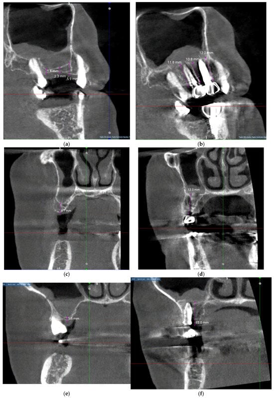
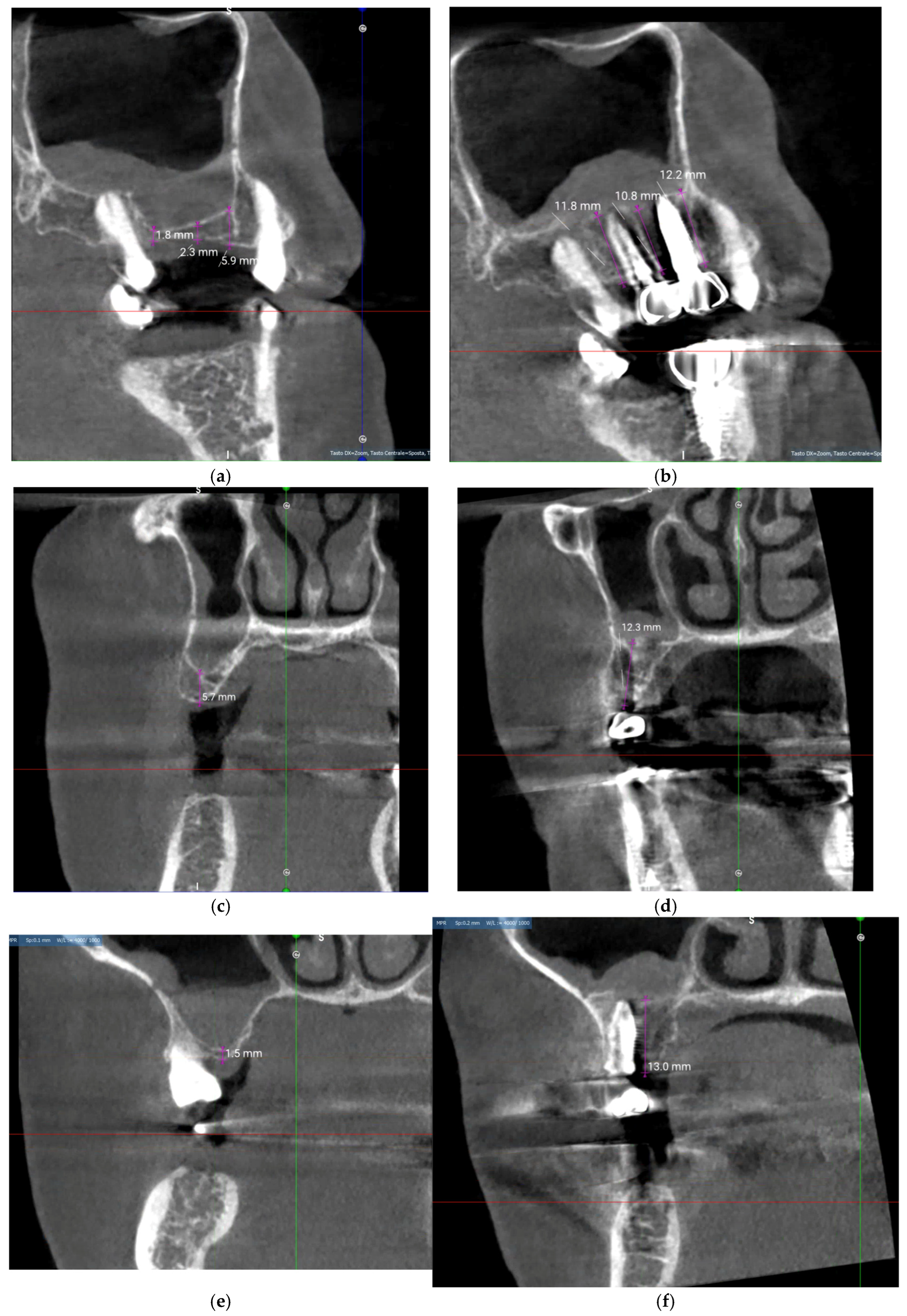
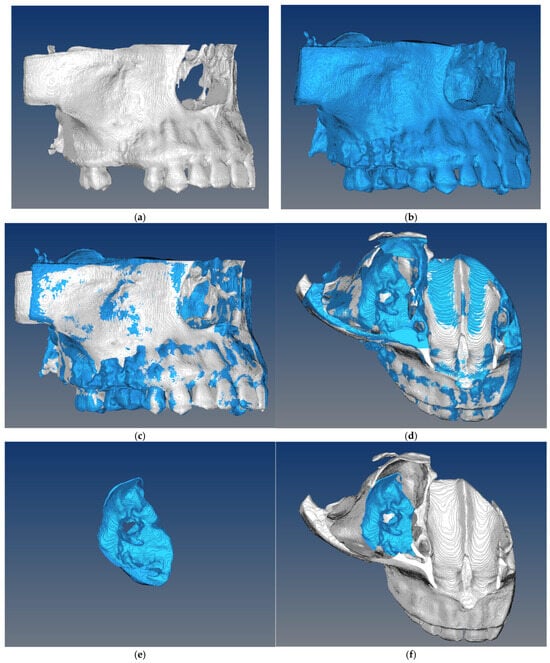
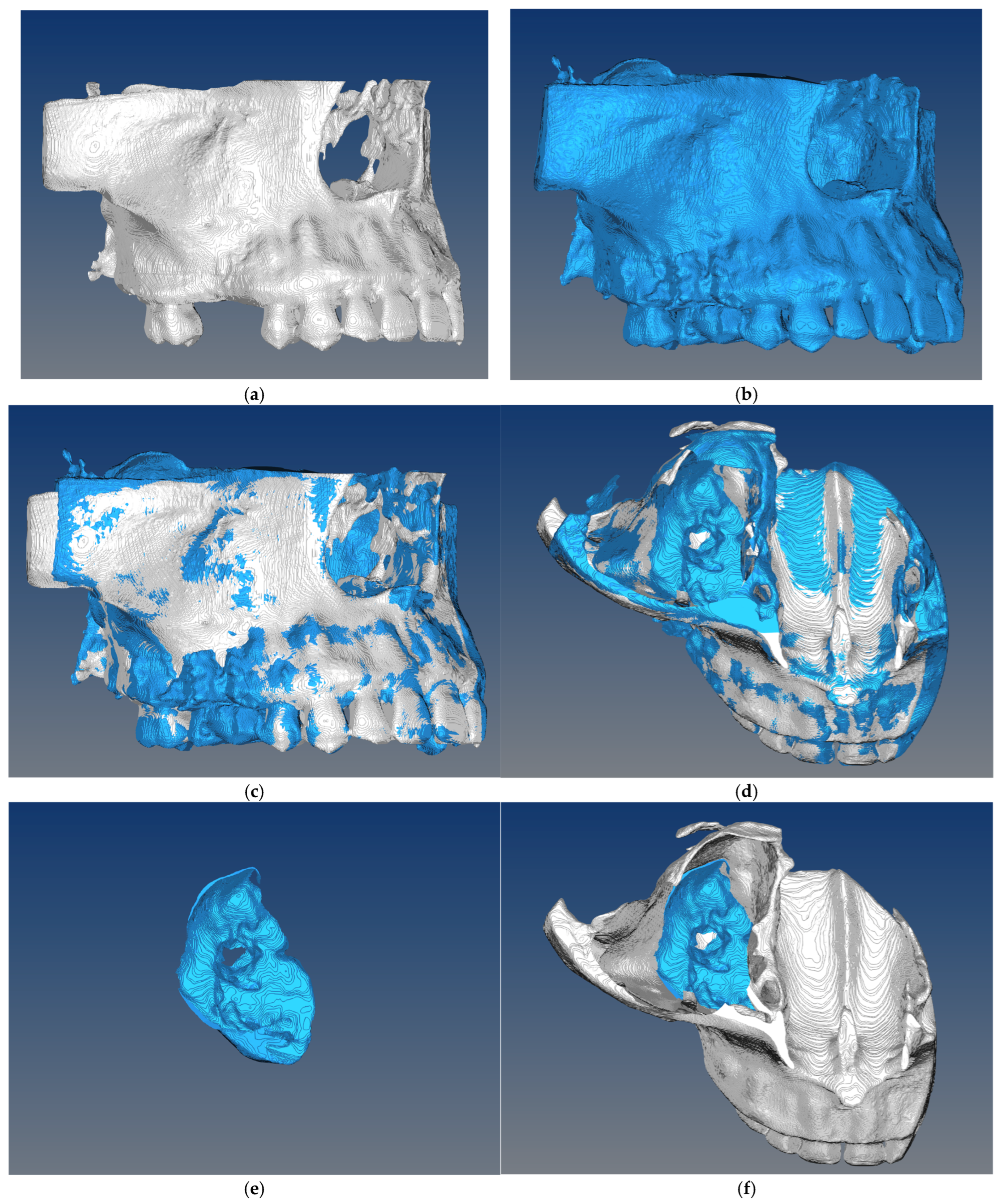
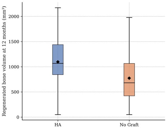
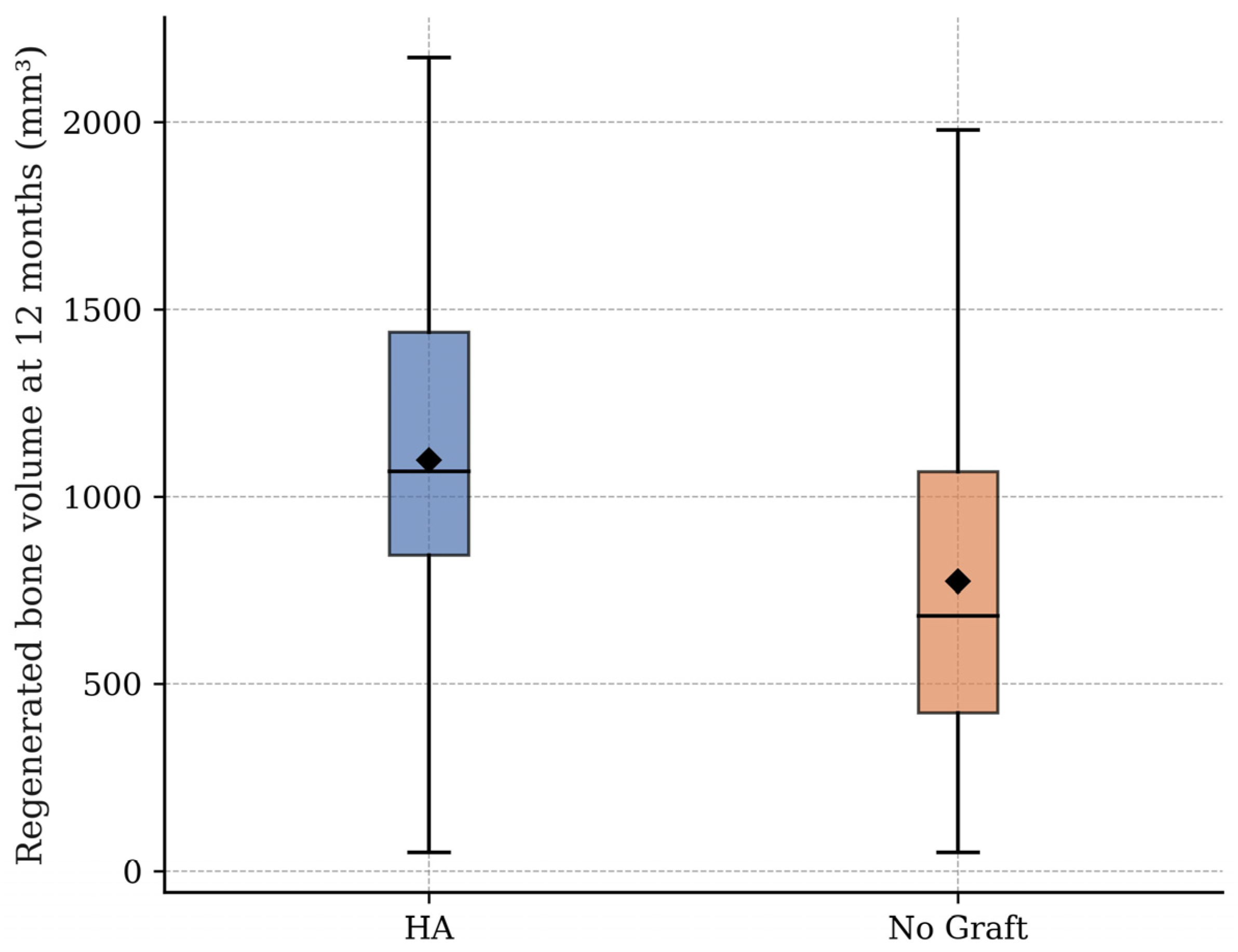
Representative CBCT cross-sectional images are shown in Figure 1 to illustrate the radiographic context of the vertical measurements performed at baseline and at 12 months. These images were not used as stand-alone evidence of vital bone regeneration or implant osseointegration. Representative cases are also shown in Figure 2a–f, depicting the STL models at baseline and 12 months, their matching, axial views, and subtraction mapping of the regenerated volume. At 12 months, mean CBCT-derived radiographic hard-tissue volume gain was significantly greater in the HA group (1210 ± 610 mm3) compared to the No Graft group (858 ± 605 mm3) (p = 0.002; Figure 3). The adjusted ANCOVA model, controlling for baseline residual bone height, implant site, and implant timing, yielded an adjusted mean difference of +352 mm3 (95% CI: 140 to 564 mm3) in favor of the HA group Table 2, Figure 3. Because the study was not powered for subgroup comparisons according to residual bone height strata, no confirmatory stratified analysis was performed. Instead, baseline residual bone height was included as a continuous covariate in the primary ANCOVA model. After adjustment for baseline residual bone height, implant site, and implant timing, the HA group still showed a significantly greater CBCT-derived radiographic hard-tissue volume gain than the No Graft group, with an adjusted mean difference of +352 mm3 (95% CI: 140 to 564 mm3; p = 0.002). Taken together, these findings indicate that the HA group showed a significantly greater imaging-based hard-tissue volume within the elevated subantral compartment. Because the primary endpoint was derived from CBCT subtraction, this result supports a radiographic volumetric gain associated with HA, but does not allow determination of the histological composition, maturity, or vital bone fraction of the newly detected hard tissue.
Figure 1.
Representative CBCT images of a single clinical case illustrating the radiographic assessment of vertical hard-tissue gain after hydraulic transcrestal sinus floor elevation. (a) Sagittal cross-sectional view at baseline (T0); (b) corresponding sagittal view at 12 months (T12); (c) coronal view at baseline (T0); (d) corresponding coronal view at 12 months (T12); (e) additional cross-sectional view at baseline (T0); (f) corresponding cross-sectional view at 12 months (T12). Purple lines indicate the linear measurements used to assess vertical radiographic hard-tissue gain (values expressed in millimeters). The green, blue and red lines represent reference axes of the CBCT software and were not used as outcome measurements. The images illustrate the radiographic context of the vertical measurements and should not be interpreted as stand-alone evidence of histologically confirmed vital bone regeneration or implant osseointegration.
Figure 2.
Workflow of three-dimensional CBCT-derived volumetric analysis. (a) STL model generated from baseline CBCT segmentation; (b) STL model generated from the 12-month CBCT scan; (c) surface registration of T0 and T12 models using stable anatomical landmarks; (d) axial verification of model alignment; (e) subtraction model (T12–T0) used to isolate the CBCT-derived radiographic hard-tissue volume gain; (f) axial view of the segmented radiographic volume in the elevated sinus compartment.
Figure 3.
Box-and-whisker plot of CBCT-derived radiographic hard-tissue volume gain at 12 months in the HA and No Graft groups.
Table 2.
Primary and secondary outcomes at 12 and 24 months.
3.3. Secondary Outcomes
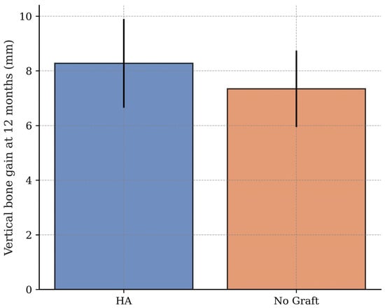
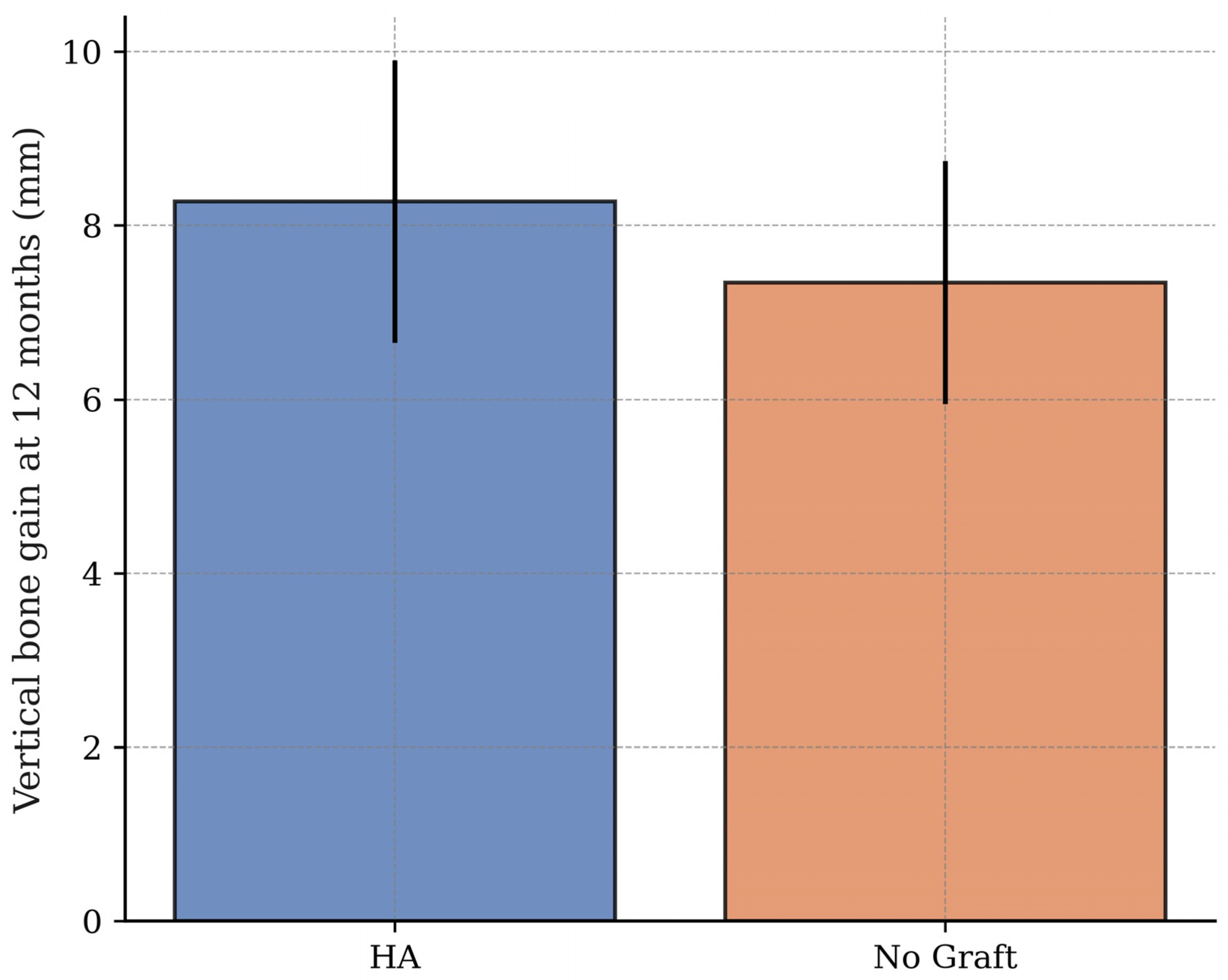
At the 12-month evaluation, the HA group demonstrated a mean vertical bone gain of 8.2 ± 1.6 mm, which was significantly greater than the 7.4 ± 1.6 mm observed in the No Graft group (p = 0.010; Figure 4). This secondary vertical-gain finding was directionally consistent with the primary volumetric outcome and supports the interpretation that HA contributed to greater radiographically detectable maintenance/fill of the elevated sinus compartment. However, the clinical secondary outcomes should be interpreted as exploratory and should not be used to infer long-term biological superiority.
Figure 4.
Mean vertical radiographic hard-tissue gain at 12 months in the HA and No Graft groups.
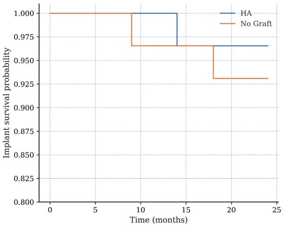
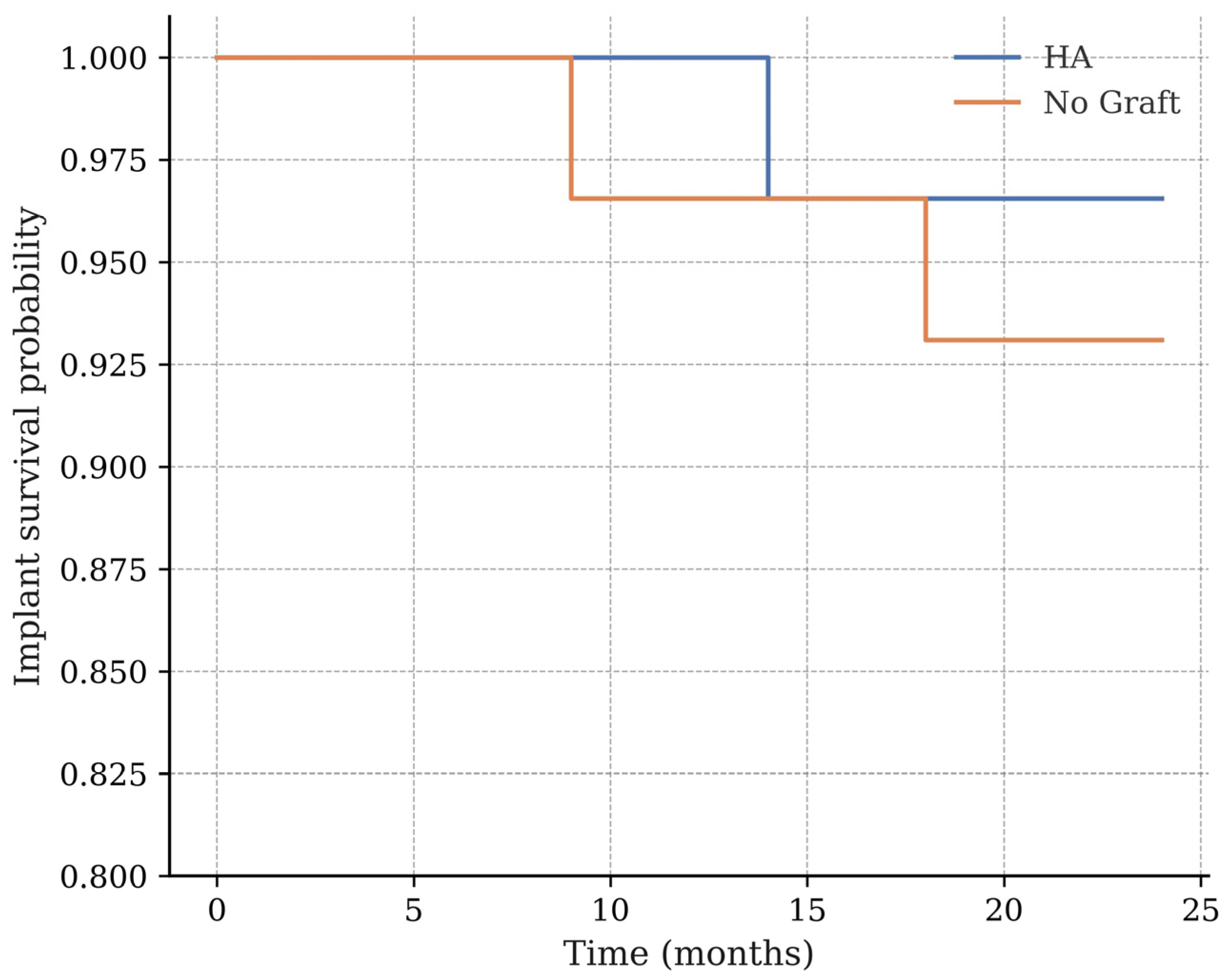
The adjusted ANCOVA model suggested that the observed effect was independent of baseline residual bone height, indicating that the benefit of HA was independent of the initial vertical dimension. Marginal bone loss (MBL) was modest in both groups. After 12 months, the mean MBL measured 0.65 ± 0.35 mm for the HA group and 0.75 ± 0.35 mm for the No Graft group, with no statistically significant difference (p = 0.080). By 24 months, MBL values had increased slightly in both groups, to 0.83 ± 0.42 mm and 0.96 ± 0.45 mm, respectively, yet the between-group difference remained non-significant (p = 0.067). Implant stability, assessed by the implant stability quotient (ISQ), was similar at placement in both cohorts (HA: 67.8 ± 5.2; No Graft: 66.9 ± 5.4; p = 0.420). Over the course of the first year, ISQ values increased in both groups, reflecting favorable implant stability over time. However, at 12 months the HA group exhibited significantly higher stability scores (74.2 ± 4.5) than the No Graft group (72.1 ± 4.9; p = 0.028). With regard to implant survival, Kaplan–Meier analysis at 24 months revealed a cumulative survival rate of 98.1% (28/29) for the HA group compared with 94.3% (27/29) for the No Graft group (log-rank p = 0.041; Table 3; Figure 5).
Table 3.
Implant survival, success, and complications at 24 months.
Figure 5.
Kaplan–Meier curves showing implant survival up to 24 months in the HA and No Graft groups.
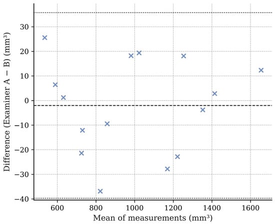
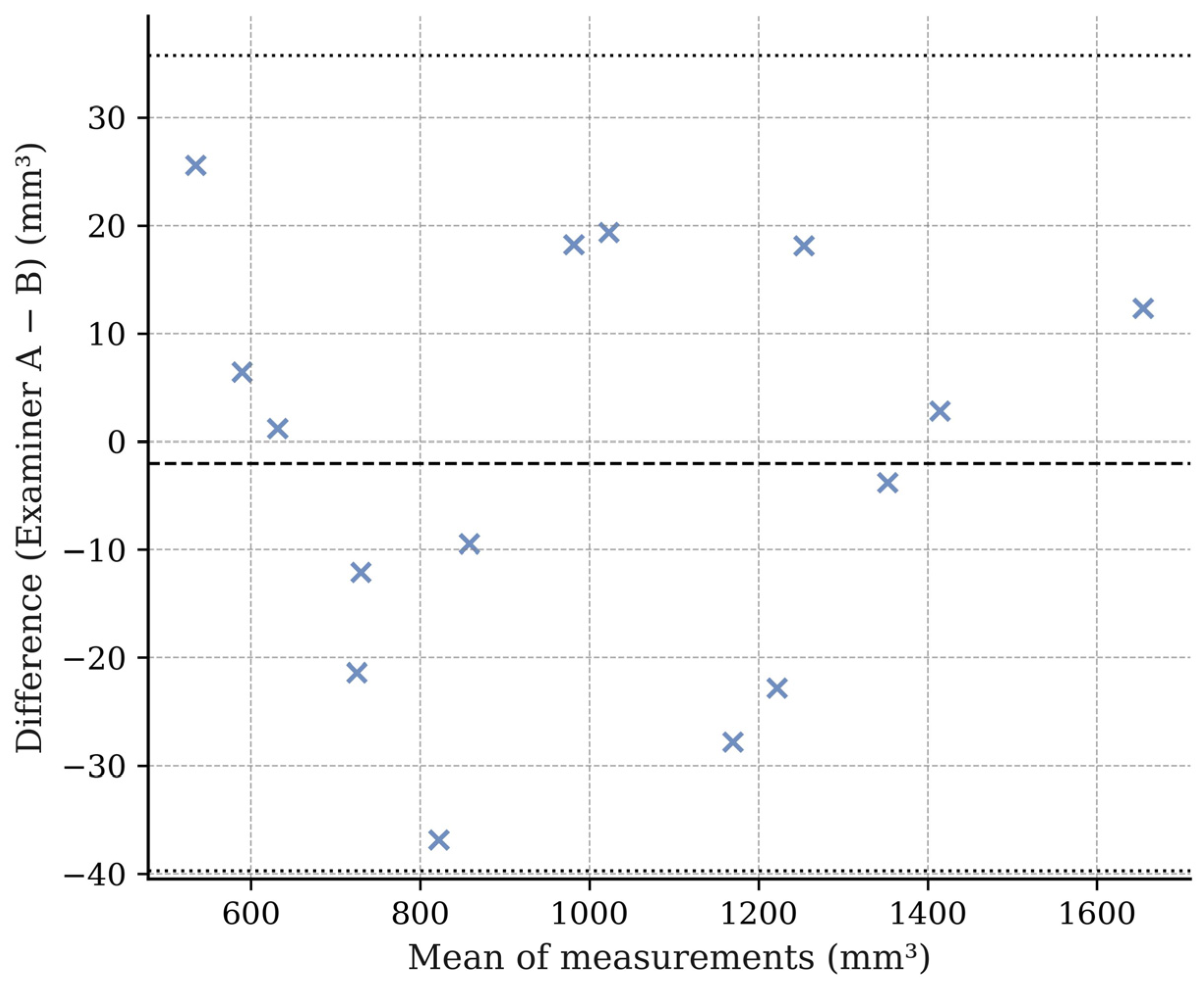
Implant success rates, based on Albrektsson’s criteria, were high in both groups, 96.2% and 92.5%, respectively, without a statistically significant difference (p = 0.312). Intraoperative membrane perforation occurred infrequently, affecting 3.4% of cases in the HA group (n = 1) and 6.9% in the No Graft group (n = 2; p = 0.648). Postoperative sinusitis was observed in one patient in each group, both resolving uneventfully with medical therapy. No late sinus-related complications were recorded during follow-up. Patient-reported outcomes showed that postoperative discomfort was slightly lower in the HA group. Median VAS pain scores at 72 h were 3.0 [IQR: 2–4] compared to 4.0 [IQR: 3–5] in the No Graft group, a statistically significant difference (p = 0.021). Oral Health Impact Profile (OHIP-14) scores improved significantly from baseline to 6 months in both groups, with no difference between groups at the 6-month mark (p = 0.312). Finally, the reliability analysis confirmed the robustness of the volumetric measurements. In the 25% subset of cases re-evaluated, inter-examiner agreement was excellent, with an intraclass correlation coefficient (ICC) of 0.982 (95% CI: 0.969–0.990). Bland–Altman analysis revealed a negligible mean bias of +3 mm3, with 95% limits of agreement ranging from −42 to +48 mm3 (Figure 6). Because CBCT-derived regenerated volume is a continuous variable, inter-examiner reliability was assessed using the intraclass correlation coefficient and Bland–Altman analysis. Cohen’s kappa/K-index was not used because it is primarily intended for agreement assessment of categorical or ordinal ratings rather than continuous volumetric measurements.
Figure 6.
Bland–Altman plot showing inter-examiner agreement for CBCT-derived volumetric measurements. The symbol (×) represents the mean difference (bias) between measurements, while the dashed lines represent the upper and lower 95% limits of agreement.
4. Discussion
This prospective clinical investigation evaluated volumetric bone regeneration following transcrestal sinus floor elevation with a hydraulic pressure technique [7], comparing cases in which cross-linked hyaluronic acid (HA) was used as a grafting material to those in which no biomaterial was added and the clot alone supported the subantral space [3,8,12]. The primary endpoint, CBCT-derived regenerated volume at 12 months, was significantly higher in the HA group, and the data therefore allow us to reject the null hypothesis that no difference exists between the two treatment modalities in terms of radiographically detected volumetric hard-tissue gain. In addition to the volumetric advantage, a modest but significant gain in vertical bone height was also observed in the HA group, suggesting that the presence of HA within the compartment may have contributed to greater maintenance or radiographic fill of the elevated subantral compartment. These findings directly address our primary research objective, confirming that the addition of HA was associated with a statistically significant CBCT-derived radiographic volumetric gain. From a comparative perspective, it is important to contextualize these results with respect to other commonly used approaches in transcrestal sinus floor elevation. Clinical evidence indicates that PRF-assisted transcrestal sinus floor elevation may result in mean vertical bone gains of approximately 2–3 mm when used alone, with values approaching 3–4 mm when combined with adjunctive techniques. These outcomes are typically assessed using linear radiographic measurements and may vary depending on baseline anatomical conditions [38,39]. Similarly, several studies have reported that the use of deproteinized bovine bone mineral (DBBM) in transcrestal sinus floor elevation is associated with mean vertical bone gains of approximately 3–4 mm. In the present study, the volumetric bone gains observed with cross-linked hyaluronic acid appear to fall within the range reported for both PRF- and DBBM-assisted procedures. However, direct comparisons should be interpreted with caution due to differences in outcome assessment methods (linear versus volumetric) and study design. These results are in line with a growing body of literature highlighting the biological potential of HA in bone regeneration [40]. Laboratory studies have demonstrated that HA, particularly in cross-linked, high molecular weight formulations, can serve as a temporary extracellular matrix analogue, providing a hydrated, viscoelastic scaffold that facilitates the migration and differentiation of mesenchymal cells and supports angiogenesis through interactions with CD44 receptors [41]. High molecular weight, cross-linked HA is characterized by increased resistance to enzymatic degradation compared to its linear counterpart, resulting in prolonged residence time within the surgical site. This extended persistence may lead to a more sustained interaction with cell surface receptors such as CD44 and RHAMM, which regulate key processes including mesenchymal cell migration, proliferation, and angiogenesis. In contrast, linear HA is rapidly degraded into lower molecular weight fragments, resulting in a more transient activation of these signaling pathways. The prolonged availability of cross-linked HA may therefore extend the temporal window of receptor engagement, potentially enhancing early tissue organization and stabilization of the regenerative microenvironment. This effect may be particularly relevant in sinus augmentation, where maintenance of the subantral space during the initial healing phase is critical for successful bone formation. In the context of sinus augmentation, these properties translate into an improved ability to maintain the elevated space in the early healing phase, a period in which the clot is still fragile and prone to collapse. Our findings resonate with experimental data showing enhanced early bone formation in HA-treated defects, and they echo clinical reports, such as the work by Goetz and colleagues, where HA enrichment improved volumetric outcomes even when combined with xenografts in lateral window procedures [42]. In addition, cross-linked HA may offer an advantage for patients seeking alternatives to xenogeneic grafts. As an animal-free biomaterial, HA may be more acceptable for individuals who decline porcine- or bovine-derived materials for ethical, cultural, religious, or personal reasons. This aspect does not replace the need for biological and clinical validation, but it may contribute to patient acceptance and shared decision-making in elective regenerative procedures. It is worth noting that the magnitude of the volumetric difference we observed is clinically relevant, particularly in sites with a residual bone height of only 2–4 mm. Sites with lower residual bone height are biologically and mechanically more demanding because the elevated Schneiderian membrane receives less support from the native crestal bone, increasing the importance of early space maintenance. In the present study, the effect of baseline residual bone height was addressed statistically by including RBH as a continuous covariate in the primary ANCOVA model. This approach avoided the loss of statistical power associated with post hoc dichotomization of a relatively small sample into 2–4 mm and 4–6 mm subgroups. Nevertheless, the study was not specifically powered to test whether the effect of HA differed between extremely atrophic and moderately atrophic sites. Therefore, the possibility that HA may provide a proportionally greater benefit in sites with 2–4 mm residual bone height remains biologically plausible but should be confirmed in future randomized studies specifically designed for subgroup or interaction analysis. In such challenging scenarios, the mechanical support provided by the material may reduce the risk of insufficient graft maturation before implant placement [43]. This is further supported by the slightly higher implant survival rate at 24 months in the HA group. While both groups achieved survival rates exceeding 94%, the difference in favour of the HA-treated sites suggests that greater regenerated volume may contribute to improved load distribution [44] and bone-to-implant contact [45], particularly in the early functional phase. Interestingly, marginal bone loss [20] around the implant necks did not differ significantly between the groups. This finding aligns with earlier systematic reviews indicating that while space-maintaining materials can influence the sinus floor region, they do not necessarily affect crestal bone remodelling [46]. It also underlines that the benefits of HA in this setting may be confined to the subantral compartment rather than extending to peri-implant tissues coronally. From a patient-centred perspective, the HA group reported slightly lower postoperative pain scores, a result that could be attributed to the biomaterial’s anti-inflammatory properties described in preclinical models [47]. Although the absolute difference was small, such improvements in patient comfort can play a role in treatment acceptance, especially for elective procedures like sinus augmentation. Furthermore, in line with current patient-centred clinical research principles, the present study incorporated Patient-Reported Outcome Measures (PROMs), including both the Visual Analogue Scale (VAS) and the OHIP-14 questionnaire. While previous sinus floor elevation studies have primarily focused on clinician-reported outcomes, the inclusion of these validated instruments allows a more comprehensive assessment of patient experience and aligns the present study with current patient-centred research standards [48]. The robustness of our radiographic findings is supported by the excellent inter-examiner reliability in volumetric measurements, with an ICC well above 0.9. This is particularly important given that CBCT-based volume assessment, while highly informative, is sensitive to factors such as voxel size, patient positioning, and segmentation thresholds [49]. By maintaining strict imaging protocols and using stable anatomical landmarks for registration [50], we minimised these potential sources of error. Nevertheless, implant-related beam-hardening and metal artifacts may have influenced the segmentation of peri-implant structures at T12. To reduce this risk, the registration process was based on stable anatomical landmarks rather than implant surfaces, and volumetric assessment focused on the subantral compartment. However, no dedicated metal artifact reduction algorithm was applied beyond the standard reconstruction and segmentation workflow available in the software. This should be considered when interpreting the absolute volumetric values. There are, however, limitations that must be acknowledged. The non-randomized design represents an inherent limitation of the present study, as it introduces a potential risk of selection bias. Patient allocation was based on a predefined alternating sequence applied to consecutively enrolled subjects, rather than true randomization. Although this approach was chosen to ensure feasibility and maintain a controlled clinical workflow in a single-center setting, it cannot fully replicate the methodological robustness of randomized allocation.
To mitigate this limitation, several precautions were implemented. First, strict inclusion and exclusion criteria were applied to reduce clinical heterogeneity. Second, consecutive enrollment minimized investigator-driven selection. Third, baseline characteristics were carefully assessed and demonstrated no statistically significant differences between groups. Finally, the primary outcome was analyzed using an ANCOVA model adjusting for key clinical covariates, thereby reducing the impact of potential confounding factors.
Nevertheless, residual confounding cannot be completely excluded, and the results should therefore be interpreted with caution. Future multicenter randomized controlled trials, potentially incorporating stratified randomization or propensity score-based approaches, are warranted to confirm the present findings and further strengthen the level of evidence. The follow-up period of 24 months is sufficient to assess early- to mid-term implant performance, but it cannot establish long-term predictability under functional loading. In implant therapy and sinus floor elevation procedures, 5-year follow-up data are generally considered more appropriate to support claims of long-term clinical stability. Therefore, the present survival data should be interpreted as preliminary functional outcomes rather than definitive evidence of long-term superiority. Furthermore, the study evaluated only one specific HA formulation in conjunction with a single hydraulic sinus lift system, which limits the extrapolation of results to other products or techniques. A further limitation is that the study was powered for the overall comparison between HA and No Graft groups and not for subgroup analyses according to baseline residual bone height. Although RBH was included as a continuous covariate in the primary adjusted model, the sample size did not allow a reliable post hoc comparison between extremely atrophic sites and moderately atrophic sites. Future trials should stratify recruitment according to RBH categories and should be powered to test treatment-by-RBH interactions.
Finally, although CBCT volumetry is a valuable research tool, it cannot distinguish mature vital bone from immature mineralized tissue, connective tissue, or residual biomaterial. Therefore, the present findings should be interpreted as radiographic evidence of volumetric hard-tissue gain rather than direct histological confirmation of vital bone regeneration. Histological or histomorphometric analysis of bone cores would provide additional insight into the biological events underpinning these radiological measurements. Future investigations should therefore explore the histomorphometric [51] profile of HA-regenerated sites, compare HA directly with other bioactive space maintainers such as platelet-rich fibrin [52] or collagen-based materials [53], and extend follow-up to assess whether the volumetric advantage observed at 12 months persists and translates into durable implant success. Multicenter randomized controlled trials with larger patient cohorts, multiple operators, standardized imaging protocols, longer follow-up periods, and histological or histomorphometric validation are needed to confirm the generalizability of these findings beyond a single-center and single-operator setting. In summary, the present results contribute to a nuanced understanding of how bioactive agents like HA can augment the biological potential of minimally invasive sinus lift techniques. While both approaches, HA and no graft, were clinically successful, the use of HA provided measurable advantages in regenerated volume and vertical gain, benefits that may be particularly valuable in cases with minimal residual bone height. Another important aspect to consider is the control of potential confounding variables. In the present study, several strategies were adopted to minimize their impact. Baseline comparability between groups was verified, and no significant differences were observed in key clinical parameters. In addition, residual bone height (RBH), a known determinant of sinus augmentation outcomes, was included as a continuous covariate in the ANCOVA model. Strict inclusion and exclusion criteria and consecutive patient enrollment further contributed to reducing selection bias and clinical heterogeneity. However, as this was a non-randomized study, residual confounding from unmeasured variables (e.g., bone quality, patient-related factors, or site-specific characteristics) cannot be completely excluded. A key limitation of the present study is that CBCT volumetric analysis provides radiographic evidence of three-dimensional hard-tissue gain but does not allow histological confirmation of vital bone formation. The lower radiographic density of the tissue within the elevated sinus compartment compared with adjacent native bone, as observed during image interpretation, may reflect differences in tissue mineralization, maturation stage, or overall tissue composition. Therefore, the present findings should be interpreted as evidence of CBCT-derived radiographic hard-tissue volume gain rather than definitive proof of mature vital bone regeneration or superior bone quality. Histological and histomorphometric evaluation of bone-core biopsies would be required to determine the proportion of vital bone and to characterize the biological quality of the regenerated tissue more accurately.
5. Conclusions
Within the limitations of this non-randomized prospective clinical study, the use of cross-linked hyaluronic acid during hydraulic transcrestal sinus floor elevation was associated with significantly greater CBCT-derived radiographic hard-tissue volume gain and greater vertical radiographic gain at 12 months compared with clot stabilization alone. These results support the interpretation that cross-linked hyaluronic acid may act as a space-maintaining adjunct within the elevated subantral compartment in posterior maxillary sites with limited residual bone height.
Peri-implant marginal bone levels, implant stability, implant survival, complication rates, and patient-reported outcomes were favorable in both groups during the 24-month observation period; however, these secondary clinical outcomes should be interpreted as exploratory and do not establish long-term superiority. Because CBCT volumetry cannot determine tissue composition or confirm vital mature bone formation, the present findings should be interpreted as radiographic evidence of hard-tissue volume gain rather than histological proof of bone regeneration. Further randomized controlled trials with longer follow-up and histological or histomorphometric correlation are warranted to determine whether the observed CBCT-derived volumetric differences persist over time and correspond to differences in vital bone formation or functional outcomes.
Author Contributions
Conceptualization, G.M.; methodology, E.Q.; validation, F.B.; investigation, A.S.; writing—original draft preparation, A.M. and G.R.; writing—review and editing, A.M. and G.R.; supervision, R.S.; All authors have read and agreed to the published version of the manuscript.
Funding
This research received no external funding.
Institutional Review Board Statement
The study was conducted in accordance with the Declaration of Helsinki and approved by the Institutional Review Board of University of Tirana (protocol code Nr 276 prot. and date of approval 4 July 2023).
Informed Consent Statement
Informed consent was obtained from all subjects involved in the study. Written informed consent has been obtained from the patients to publish this paper.
Data Availability Statement
The original contributions presented in this study are included in the article. Further inquiries can be directed to the corresponding authors.
Conflicts of Interest
The authors declare no conflicts of interest.
References
- Stern, A.; Green, J. Sinus lift procedures: An overview of current techniques. Dent. Clin. N. Am. 2012, 56, 219–233. [Google Scholar] [CrossRef] [Scilit] [PubMed]
- Boyne, P.J.; James, R.A. Grafting of the maxillary sinus floor with autogenous marrow and bone. J. Oral Surg. 1980, 38, 613–616. [Google Scholar] [PubMed]
- Albash, Z.; Khalil, A.; Sleman, N. Influence of implant protrusion length and initial bone height on intrasinus bone gain in transalveolar sinus floor elevation without bone graft: A 2-year retrospective study. Ann. Med. Surg. 2023, 85, 3291–3297. [Google Scholar] [CrossRef] [Scilit]
- Summers, R.B. The osteotome technique: Part 3—Less invasive methods of elevating the sinus floor. Compendium 1994, 15, 698–708. [Google Scholar] [PubMed]
- He, L.; Chang, X.; Liu, Y. Sinus floor elevation using osteotome technique without grafting materials: A 2-year retrospective study. Clin. Oral Implant. Res. 2013, 24, 63–67. [Google Scholar]
- Sammartino, G.; Mariniello, M.; Scaravilli, M.S. Benign paroxysmal positional vertigo following closed sinus floor elevation procedure: Mallet osteotomes vs. screwable osteotomes. A triple blind randomized controlled trial. Clin. Oral Implant. Res. 2011, 22, 669–672. [Google Scholar]
- Sotirakis, E.G.; Gonshor, A. Elevation of the maxillary sinus floor with hydraulic pressure. J. Oral Implantol. 2005, 31, 197–204. [Google Scholar] [CrossRef] [Scilit] [PubMed]
- Silva, L.F.; de Lima, V.N.; Faverani, L.P.; de Mendonça, M.R.; Okamoto, R.; Pellizzer, E.P. Maxillary sinus lift surgery-with or without graft material? A systematic review. Int. J. Oral Maxillofac. Surg. 2016, 45, 1570–1576. [Google Scholar] [CrossRef] [Scilit] [PubMed]
- Andrés-García, R.; Ríos-Santos, J.V.; Herrero-Climent, M.; Bullón, P.; Fernández-Farhall, J.; Gómez-Menchero, A.; Fernández-Palacín, A.; Ríos-Carrasco, B. Sinus Floor Elevation via an Osteotome Technique without Biomaterials. Int. J. Environ. Res. Public Health 2021, 18, 1103. [Google Scholar] [CrossRef] [Scilit] [PubMed]
- Y Baena, R.R.; Lupi, S.M.; Pastorino, R.; Maiorana, C.; Lucchese, A.; Rizzo, S. Radiographic evaluation of regenerated bone following poly(lactic-co-glycolic) acid/hydroxyapatite and deproteinized bovine bone graft in sinus lifting. J. Craniofac. Surg. 2013, 24, 845–848. [Google Scholar] [CrossRef] [Scilit] [PubMed]
- Ozyuvaci, H.; Aktas, I.; Yerit, K.C.; Aydin, K.; Firatli, E. Radiological evaluation of sinus lift operation: What the general radiologist needs to know. Dentomaxillofac. Radiol. 2005, 34, 199–204. [Google Scholar] [CrossRef] [Scilit] [PubMed]
- Cho, Y.S.; Hwang, K.G.; Jun, S.H.; Tallarico, M.; Kwon, A.M.; Park, C.J. Radiologic comparative analysis between saline and platelet-rich fibrin filling after hydraulic transcrestal sinus lifting without adjunctive bone graft: A randomized controlled trial. Clin. Oral Implant. Res. 2020, 31, 1087–1093. [Google Scholar] [CrossRef] [Scilit]
- Ceruso, F.M.; Lumbau, A.I.; Pernice, F.; Mastroianni, A.; Miranda, M.; Meloni, S.M.; Gargari, M.; Tallarico, M.; Pisano, M. Crestal Hydraulic Sinus Lift with Simultaneous Implant Insertion: A Retrospective Case Series. Dent. J. 2025, 13, 193. [Google Scholar] [CrossRef] [Scilit]
- Kaufman, E. Maxillary sinus elevation surgery: An overview. J. Esthet. Restor. Dent. 2003, 15, 272–283. [Google Scholar] [CrossRef] [Scilit] [PubMed]
- Esposito, M.; Felice, P.; Worthington, H.V. Interventions for replacing missing teeth: Augmentation procedures of the maxillary sinus. Cochrane Database Syst. Rev. 2014, 2014, CD008397. [Google Scholar] [CrossRef] [Scilit] [PubMed]
- Meredith, N. Assessment of implant stability as a prognostic determinant. Int. J. Prosthodont. 1998, 11, 491–501. [Google Scholar] [PubMed]
- Esposito, M.; Worthington, H.V.; Grusovin, M.G. Interventions for replacing missing teeth: Antibiotics at dental implant placement to prevent complications. Cochrane Database Syst. Rev. 2013, 2013, CD004152. [Google Scholar] [CrossRef] [Scilit] [PubMed]
- Scarano, A.; Luongo, R.; De Filippis, I.; Scarano, A.; Qorri, E.; Sforza, F.; Rampino, M.; Bugea, C. Fluid-Dynamic Crestal Sinus Floor Elevation in Atrophic Posterior Maxilla Implant Rehabilitation with Hyaluronic Acid: A Prospective Study. Materials 2025, 18, 2230. [Google Scholar] [CrossRef] [Scilit] [PubMed]
- Albrektsson, T.; Zarb, G.; Worthington, P.; Eriksson, A.R. The long-term efficacy of currently used dental implants: A review and proposed criteria of success. Int. J. Oral Maxillofac. Implant. 1986, 1, 11–25. [Google Scholar]
- Nandal, S.; Ghalaut, P.; Shekhawat, H. A radiological evaluation of marginal bone around dental implants: An in-vivo study. Natl. J. Maxillofac. Surg. 2014, 5, 126–137. [Google Scholar] [CrossRef] [Scilit] [PubMed]
- Sennerby, L.; Meredith, N. Resonance frequency analysis: Measuring implant stability and osseointegration. Compend. Contin. Educ. Dent. 1998, 19, 493–498. [Google Scholar] [PubMed]
- McCormack, H.M.; Horne, D.J.L.; Sheather, S. Clinical applications of visual analogue scales: A critical review. Psychol. Med. 1988, 18, 1007–1019. [Google Scholar] [CrossRef] [Scilit] [PubMed]
- Slade, G.D. Derivation and validation of a short-form Oral Health Impact Profile (OHIP-14). Community Dent. Oral Epidemiol. 1997, 25, 284–290. [Google Scholar] [CrossRef] [Scilit] [PubMed]
- Corridore Denise, C.D.; Campus Gugliemo, C.G.; Guerra Fabrizio, G.F.; Ripari Francesca, R.F.; Sale Silvana, S.S.; Ottolenghi Livia, O.L. Validation of the Italian version of the Oral Health Impact Profile-14 (IOHIP-14). Ann. Stomatol. 2013, 4, 239–243. [Google Scholar]
- Shapiro, S.S.; Wilk, M.B. An analysis of variance test for normality (complete samples). Biometrika 1965, 52, 591–611. [Google Scholar] [CrossRef] [Scilit]
- Levene, H. Robust tests for equality of variances. In Contributions to Probability and Statistics; Olkin, I., Ed.; Stanford University Press: Stanford, CA, USA, 1960; pp. 278–292. [Google Scholar]
- Hogben, A. A practical and simple equivalent for student’s t test of statistical significance. J. Lab. Clin. Med. 1964, 64, 815–819. [Google Scholar] [PubMed]
- West, R.M. Best practice in statistics: Use the Welch t-test when testing the difference between two groups. Ann. Clin. Biochem. 2021, 58, 267–269. [Google Scholar] [CrossRef] [Scilit] [PubMed]
- Mann, H.B.; Whitney, D.R. On a test of whether one of two random variables is stochastically larger than the other. Ann. Math. Stat. 1947, 18, 50–60. [Google Scholar] [CrossRef] [Scilit]
- Pearson, K.X. On the criterion that a given system of deviations from the probable in the case of a correlated system of variables is such that it can be reasonably supposed to have arisen from random sampling. Philos. Mag. Ser. 1900, 50, 157–175. [Google Scholar] [CrossRef] [Scilit]
- Jung, S.H. Stratified Fisher’s exact test and its sample size calculation. Biom. J. 2014, 56, 129–140. [Google Scholar] [PubMed]
- Wilcoxon, F. Individual Comparisons by Ranking Methods. Biom. Bull. 1945, 1, 80–83. [Google Scholar] [CrossRef] [Scilit]
- Im, S. Understanding and Application of Analysis of Covariance (Research Methods 11); Kyoyookbook: Seoul, Republic of Korea, 2002. [Google Scholar]
- Thompson, H.W.; Mera, R.; Prasad, C. The Analysis of Variance (ANOVA). Nutr. Neurosci. 1999, 2, 43–55. [Google Scholar] [CrossRef] [Scilit] [PubMed]
- Kaplan, E.L.; Meier, P. Nonparametric estimation from incomplete observations. J. Am. Stat. Assoc. 1958, 53, 457–481. [Google Scholar] [CrossRef]
- Bland, J.M.; Altman, D.G. The logrank test. BMJ 2004, 328, 1073. [Google Scholar] [CrossRef] [Scilit] [PubMed]
- Altman, D.G.; Bland, J.M. Measurement in Medicine: The Analysis of Method Comparison Studies. J. R. Stat. Soc. Ser. D 1983, 32, 307–317. [Google Scholar] [CrossRef] [Scilit]
- Mancini, A.; Inchingolo, F.; Inchingolo, A.M.; Carpentiere, V.; Vecchio, G.D.; Palermo, A.; Inchingolo, A.D.; Dipalma, G. Concentrated Growth Factors and Bone Grafting in Maxillary Sinus Lift: A Systematic Review. Int. J. Dent. 2025, 2025, 6681492. [Google Scholar] [CrossRef] [Scilit] [PubMed]
- Orth, C.C.; da Silva, R.C.; de Barros Carrilho, G.P.; de Carvalho, P.F.M.; Joly, J.C.; Haas, A.N. Vertical Bone Gain Post-Sinus Lifting and Simultaneous Implant Placement with Osseodensification: A Retrospective Study. Clin. Implant Dent. Relat. Res. 2025, 27, e13430. [Google Scholar] [CrossRef] [Scilit] [PubMed]
- D’Albis Giuseppe, G.D.A.; D’Albis Vincenzo, V.D.A.; Palma Micol, M.P.; Plantamura Marianna, M.P.; Nizar Al Krenawi, A.K.N. Use of hyaluronic acid for regeneration of maxillofacial bones. Genesis 2022, 60, e23497. [Google Scholar] [CrossRef] [Scilit] [PubMed]
- Karam, J.; Singer, B.J.; Miwa, H.; Chen, L.H.; Maran, K.; Hasani, M.; Garza, S.; Onyekwere, B.; Yeh, H.C.; Li, S.; et al. Molecular weight of hyaluronic acid crosslinked into biomaterial scaffolds affects angiogenic potential. Acta Biomater. 2023, 169, 228–242. [Google Scholar] [CrossRef] [Scilit] [PubMed]
- Friedmann, A.; Goetz, W. Combined Sinus Grafting and Lateral Augmentation by a Hyaluronic Acid-Facilitated Guided Bone Regeneration Protocol—Case Series Supported by Human Histologic Analysis. J. Biomed. Res. Environ. Sci. 2022, 3, 065–073. [Google Scholar] [CrossRef] [Scilit]
- Yuksel, I.B.; Altiparmak, F.; Gurses, G.; Akti, A.; Alic, M.; Tuna, S. Radiographic Evidence of Immature Bone Architecture After Sinus Grafting: A Multidimensional Image Analysis Approach. Diagnostics 2025, 15, 1742. [Google Scholar] [CrossRef] [Scilit] [PubMed]
- Faegh, S.; Müftü, S. Load transfer along the bone-dental implant interface. J. Biomech. 2010, 43, 1761–1770. [Google Scholar] [CrossRef] [Scilit] [PubMed]
- Albrektsson, T.; Johansson, C. Osteoinduction, osteoconduction and osseointegration. J. Eur. Spine 2001, 10, S96–S101. [Google Scholar] [CrossRef] [Scilit]
- Kang, M.S.; Kim, S.H.; Lee, J.H.; Yeo, G.H.; Im, K.M.; Kim, C.H.; Kim, B.J.; Kim, J.H. Comparative study on lateral maxillary sinus augmentation of without grafting, particle bone grafting and recombinant human bone morphogenetic protein-2+absorbable collagen sponge grafting with simultaneous dental implant placement: A retrospective study. J. Korean Assoc. Oral Maxillofac. Surg. 2025, 51, 217–227. [Google Scholar] [CrossRef] [Scilit] [PubMed]
- Abatangelo, G.; Vindigni, V.; Avruscio, G.; Pandis, L.; Brun, P. Hyaluronic Acid: Redefining Its Role. Cells 2020, 9, 1743. [Google Scholar] [CrossRef] [Scilit] [PubMed]
- Lang, N.P.; Zitzmann, N.U. Working Group 3 of the VIII European Workshop on Periodontology. Clinical research in implant dentistry: Evaluation of implant-supported restorations, aesthetic and patient-reported outcomes. J. Clin. Periodontol. 2012, 39, 133–138. [Google Scholar] [CrossRef] [Scilit] [PubMed]
- Ozdede, M.; Akay, G.; Karadag Atas, O. Influence of CBCT device, voxel size, and segmentation software on the accuracy of tooth volume measurements. BMC Oral Health 2025, 25, 1063. [Google Scholar] [CrossRef] [Scilit] [PubMed]
- Park, J.; Baumrind, S.; Curry, S.; Carlson, S.K.; Boyd, R.L.; Oh, H. Reliability of 3D dental and skeletal landmarks on CBCT images. Angle Orthod. 2019, 89, 758–767. [Google Scholar] [CrossRef] [Scilit] [PubMed]
- Chavassieux, P.; Chapurlat, R. Interest of Bone Histomorphometry in Bone Pathophysiology Investigation: Foundation, Present, and Future. Front. Endocrinol. 2022, 13, 907914. [Google Scholar] [CrossRef] [Scilit]
- Song, P.; He, D.; Ren, S.; Fan, L.; Sun, J. Platelet-rich fibrin in dentistry. J. Appl. Biomater. Funct. Mater. 2024, 22, 22808000241299588. [Google Scholar] [CrossRef] [Scilit] [PubMed]
- Canullo, L.; Del Fabbro, M.; Colantonio, F.; Iacono, R.; Raffone, C.; Pedetta, A.; Khijmatgar, S.; Shapira, L. Sinus floor augmentation using crestal approach in conjunction with hydroxyapatite/cross-linked collagen sponge: A pilot study. Clin. Implant Dent. Relat. Res. 2023, 25, 974–983. [Google Scholar] [CrossRef] [Scilit] [PubMed]

Disclaimer/Publisher’s Note: The statements, opinions and data contained in all publications are solely those of the individual author(s) and contributor(s) and not of MDPI and/or the editor(s). MDPI and/or the editor(s) disclaim responsibility for any injury to people or property resulting from any ideas, methods, instructions or products referred to in the content. |
© 2026 by the authors. Licensee MDPI, Basel, Switzerland. This article is an open access article distributed under the terms and conditions of the Creative Commons Attribution (CC BY) license.