1. Introduction
Intelligent lower-limb exoskeletons have emerged as an important research area in wearable robotics and rehabilitation engineering, offering potential benefits for locomotor assistance, functional recovery, and mobility enhancement in individuals with movement impairments [
1,
2,
3,
4]. Advances in biomedical sensors, embedded systems, and artificial intelligence have accelerated the development of adaptive biomechatronic devices capable of interacting more effectively with human users [
1,
2,
3]. In addition to rehabilitation applications, lower-limb exoskeletons are increasingly investigated for industrial and assistive scenarios where reducing physical effort and improving locomotor stability are essential requirements [
3,
4,
5].
Several sensing modalities have been investigated for movement intention recognition in lower-limb exoskeletons, including electroencephalography (EEG), inertial measurement units (IMUs), force sensors, vision-based systems, and electromyographic (EMG) signals [
2,
3,
4,
5,
6,
7,
8]. EEG-based approaches provide information related to neural activity but usually require complex signal processing and are highly sensitive to noise and artifacts. IMU and force sensors provide useful kinematic and kinetic information; however, they mainly describe movement after its initiation rather than the user’s muscular intention. Vision-based systems can support motion tracking, but their performance depends strongly on environmental conditions and computational resources.
Compared with these approaches, EMG signals provide a direct and physiologically relevant representation of muscle activation preceding movement execution [
9,
10,
11,
12,
13]. Therefore, EMG is particularly suitable for movement intention recognition in assistive and rehabilitation systems, where early detection of the user’s intended movement is essential for adaptive exoskeleton control.
Recent studies have explored both conventional machine learning and deep learning techniques for the classification of locomotor activities using EMG signals. Traditional approaches typically rely on handcrafted features extracted from time, frequency, or time–frequency domains and subsequently classified using algorithms such as Support Vector Machines (SVMs), Random Forests, or Artificial Neural Networks [
9,
10,
11]. More recently, deep learning architectures, including Convolutional Neural Networks (CNNs), Recurrent Neural Networks (RNNs), and Long Short-Term Memory (LSTM) networks, have demonstrated promising results by enabling end-to-end learning directly from EMG data while reducing the need for manual feature engineering [
10,
11,
12,
13].
Several intelligent exoskeleton systems have also adopted multimodal sensing strategies that combine EMG signals with inertial measurement units (IMUs), force sensors, or biomechanical measurements to improve robustness and movement recognition accuracy. Although these systems often achieve high classification performance under controlled laboratory conditions, significant challenges remain regarding inter-subject variability, electrode placement sensitivity, signal quality, and real-time implementation in practical rehabilitation environments [
12,
13,
14,
15,
16,
17].
Compared with studies focused exclusively on EMG classification, the development of integrated experimental platforms remains an important research challenge. Such platforms must support synchronized data acquisition, embedded processing, dataset generation, movement classification, and future integration within adaptive biomechatronic control architectures, as demonstrated by recent studies on deep-learning-based activity recognition and lower-limb exoskeleton design [
18,
19]. Furthermore, many published studies evaluate performance only through conventional cross-validation approaches, providing limited insight into subject-independent generalization capabilities.
In this context, this work presents an integrated experimental platform for an intelligent lower-limb exoskeleton based on multichannel EMG acquisition, inertial monitoring, embedded processing, and artificial intelligence. The proposed system integrates MyoWare 2.0 EMG sensors, MPU9250/6500 inertial sensors, Arduino Mega and Banana Pi embedded platforms, and ESCON motor controllers within a unified biomechatronic architecture. A dataset containing locomotor activities, including stepping, kneeling, and dash movements, was acquired and used for movement intention recognition through a lightweight one-dimensional convolutional neural network (1D-CNN) [
18].
The main contributions of this work are:
Development of a modular biomechatronic platform integrating EMG acquisition, inertial sensing, embedded processing, and exoskeleton control components;
Implementation of an automated framework for EMG acquisition, validation, buffering, and dataset generation;
Development and evaluation of a 1D-CNN model for lower-limb movement classification based on multichannel EMG signals;
Assessment of model performance using both Stratified 5-Fold Cross-Validation and Leave-One-Subject-Out (LOSO) protocols to evaluate classification accuracy and inter-subject generalization;
Demonstration of the feasibility of integrating EMG-based movement intention recognition mechanisms into future adaptive control systems for intelligent lower-limb exoskeletons.
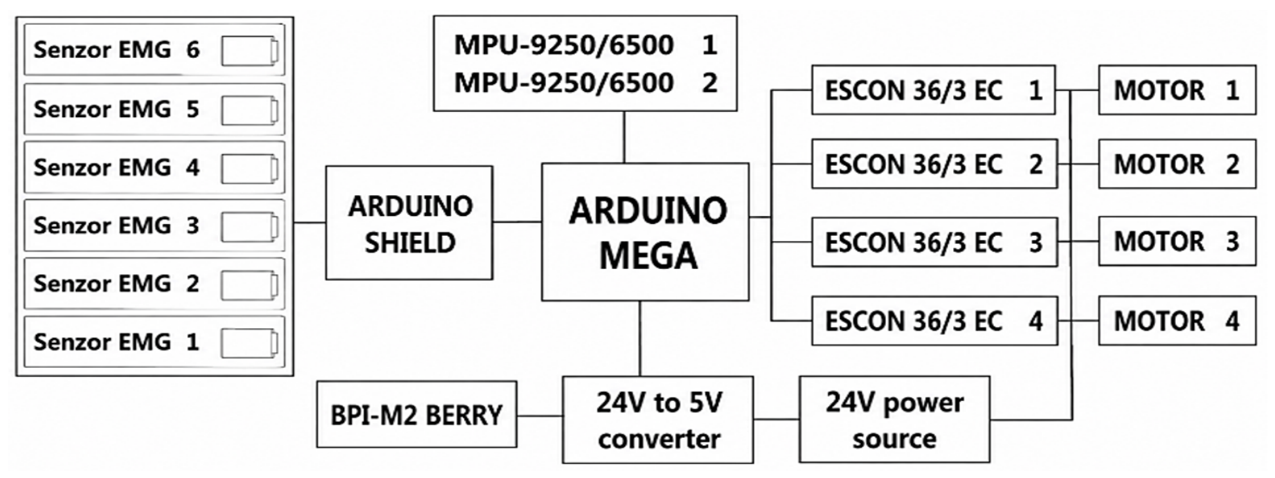
The overall platform architecture integrates multichannel EMG acquisition, inertial sensing, embedded processing, and actuator control modules, as illustrated in
Figure 1.
This work aims to validate an integrated biomechatronic platform for EMG-based movement intention recognition and to assess its potential for future adaptive exoskeleton control in locomotor assistance and rehabilitation applications.
2. Materials and Methods
The hardware–software architecture of the experimental platform was designed for electromyographic signal acquisition and lower-limb movement classification using artificial intelligence algorithms. The proposed methodology integrates EMG sensors, embedded platforms, and biomechatronic mechanisms into a modular system intended for the acquisition, processing, and analysis of muscular activity.
Within the study, various experimental scenarios associated with lower-limb locomotor movements were analyzed and used for EMG dataset generation and validation of the classification process. The system architecture enables synchronized acquisition and processing of data from electromyographic and inertial sensors, as well as the integration of experimental inference and control mechanisms based on artificial intelligence.
2.1. General System Architecture
The proposed hardware–software platform was developed as a modular biomechatronic system for lower-limb movement monitoring and EMG-based movement classification. The architecture integrates four main subsystems: electromyographic signal acquisition, inertial sensing, embedded data processing, and biomechatronic actuator control. This modular design enables synchronized acquisition and processing of physiological and motion-related data for movement intention recognition.
The platform supports a complete data pipeline, from signal acquisition and preprocessing to AI-based classification and control integration. Its modular structure allows further expansion through additional sensing modules, advanced classification algorithms, and adaptive control mechanisms, providing a flexible framework for future intelligent exoskeleton applications.
2.2. Mechanical Structure and Evolution of the Biomechatronic Platform
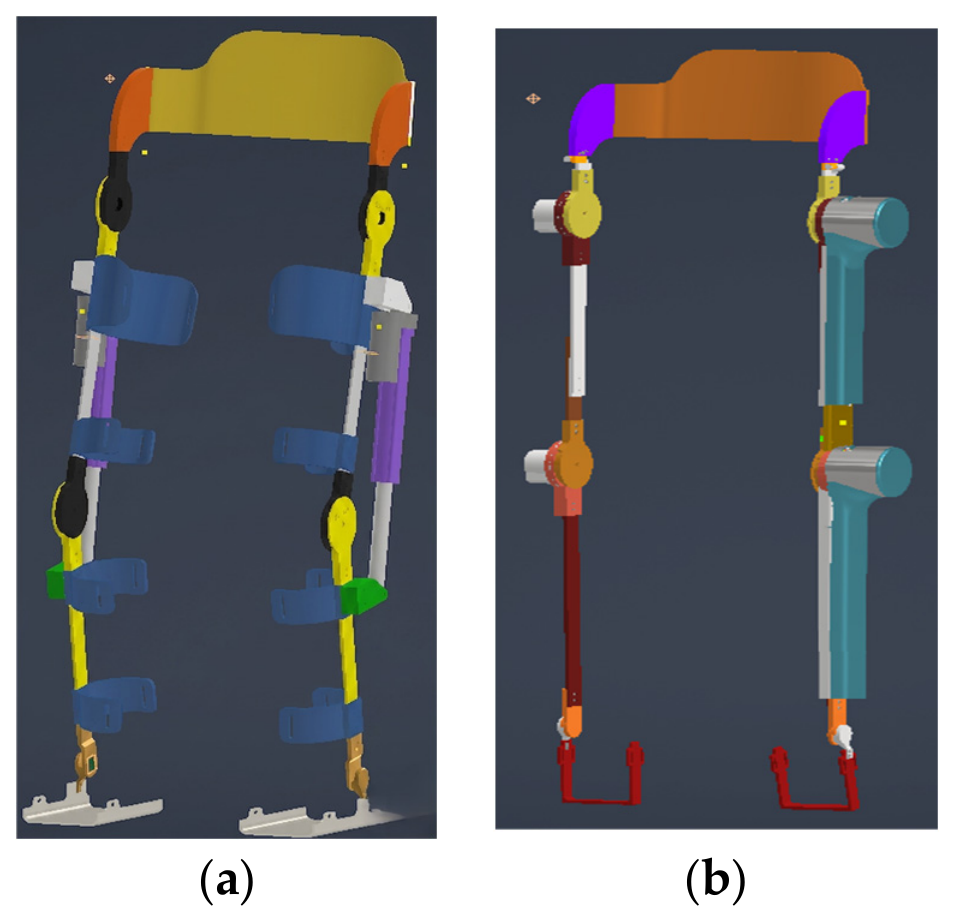
The development of the proposed exoskeleton platform involved two successive design stages, as illustrated in
Figure 2. Prototype I represents the initial mechanical platform, while Prototype II extends the system by integrating EMG acquisition, inertial sensing, embedded processing, and AI-based movement classification.
The mechanical design was developed using parametric CAD modeling, enabling modular joint configuration and actuator integration. The exoskeleton includes hinge-based lower-limb joint modules designed to reproduce human biomechanical movements while ensuring proper alignment with the user’s joint axes to improve comfort and reduce undesired mechanical loads [
1,
2,
3,
4,
20].
Figure 2 illustrates the evolution of the system from the initial mechanical prototype to the enhanced biomechatronic platform.
The initial exoskeleton prototype was developed to validate the mechanical design and analyze lower-limb biomechanics [
21]. Based on the results obtained, a second-generation biomechatronic platform was developed, integrating EMG and inertial sensors, embedded processing, and artificial intelligence algorithms for movement intention recognition. The mechanical structure employs modular hinge elements and bearing-supported revolute joints to reproduce physiological lower-limb movements while ensuring stability and low-friction operation. The functional and technological evolution of the two prototype generations developed within the research is presented in
Table 1.
2.3. Electronic Architecture and Embedded Integration of the Biomechatronic Platform
The proposed biomechatronic platform employs a modular electronic architecture integrating EMG acquisition, embedded processing, inertial sensing, and actuator control [
12,
13,
20,
22,
23,
24]. The system enables synchronized acquisition of multichannel electromyographic signals and motion-related data required for movement classification and intention recognition.
Communication between subsystems is implemented through serial interfaces and a compact binary protocol to ensure efficient and reliable data transmission between acquisition and processing modules [
14,
23]. This architecture supports real-time signal processing and AI-based inference, enabling the implementation of movement classification algorithms within the embedded subsystem [
7,
9,
10,
11,
12,
13,
15,
22].
To improve acquisition quality, separate power and signal acquisition circuits were implemented to reduce electromagnetic interference during EMG measurements. The modular architecture also enables future integration of additional sensing modules and adaptive control mechanisms for intelligent exoskeleton applications [
5,
6,
7,
8,
25].
The main electronic components integrated into the platform and their functional roles are summarized in
Table 2.
2.4. EMG System
The MyoWare 2.0 system was used for electromyographic signal acquisition, selected due to its small size, easy integration with embedded platforms, and ability to operate in multiple EMG signal processing modes [
23,
24]. Electromyographic signals represent electrical variations generated during muscle activation and provide relevant information regarding the user’s movement intention. For this reason, they are widely used in biomechatronic systems and intelligent exoskeletons [
5,
6,
7,
8,
12].
In biomechatronic applications dedicated to locomotor assistance, the analysis of muscle activity enables the identification of patterns associated with different phases of movement and contributes to the development of experimental mechanisms for predictive exoskeleton control [
6,
12].
The EMG system used within the platform enables the monitoring of muscle activity associated with lower-limb movements and provides the data required for the intelligent movement classification process [
9,
10,
11,
12,
13,
15].
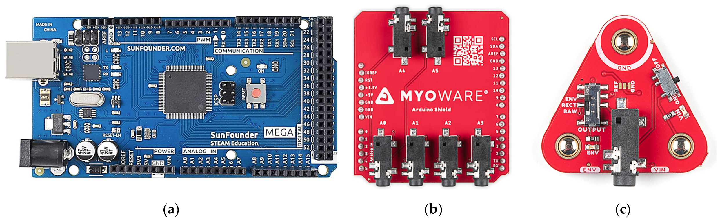
Figure 3 presents the hardware configuration of the EMG system used for multichannel electromyographic signal acquisition and processing. The use of six EMG channels allows simultaneous monitoring of the main muscle groups involved in the analyzed movements, offering a trade-off between system hardware complexity and the amount of neuromuscular information required for the classification process.
The MyoWare 2.0 sensors allow operation in three main modes:
RAW, for direct acquisition of the raw electromyographic signal;
RECT, for the rectified signal;
ENV, for extracting the EMG signal envelope [
24].
Within the proposed platform, the ENV mode was used because it provides the envelope of the electromyographic signal and enables a more robust representation of muscle activity for subsequent processing and AI-based classification. The use of the EMG envelope is frequently reported in the literature to reduce the influence of local fluctuations and improve the robustness of the inference process [
12,
13].
The EMG data are acquired through a dedicated shield connected to the Arduino Mega platform, which is responsible for reading, buffering, and transmitting the electromyographic data to the embedded system for further processing [
23].
2.5. Experimental Protocol and Data Acquisition
The experimental study involved ten healthy male volunteers recruited from the Military Institute of the Armed Forces of the Republic of Moldova. The participants were military students aged between 19 and 24 years, with heights ranging from 175 to 180 cm and body weights ranging from 60 to 70 kg. All participants were physically active and regularly engaged in training activities as part of their military education. The demographic characteristics of the study participants are summarized in
Table 3.
The participant cohort consisted of 10 volunteers, which was considered sufficient for the preliminary validation of the proposed biomechatronic platform and AI-based movement classification framework. The primary objective of this study was proof-of-concept validation under controlled experimental conditions rather than large-scale statistical generalization.
Inclusion criteria required participants to be healthy, physically active, and free from any known neurological, musculoskeletal, or cardiovascular disorders that could affect locomotor performance or electromyographic signal acquisition. Exclusion criteria included a history of lower-limb injury, neurological disease, chronic musculoskeletal conditions, or any medical condition that could interfere with the execution of the experimental tasks.
The study was conducted in accordance with the ethical principles of the Declaration of Helsinki and was approved by the Ethics Committee of the Technical University of Moldova (Approval No. 25.80013.5007.09RE). All participants were informed about the objectives, procedures, and potential risks associated with the study before participation. Written informed consent was obtained from all participants prior to data collection.
Electromyographic recordings were acquired during the execution of predefined locomotor activities, including stepping, kneeling, and dash movements, following a standardized experimental protocol and sensor placement procedure.
The experimental dataset used for training and validation of the artificial intelligence model consisted of 608 electromyographic recordings collected from the 10 participants during the execution of the analyzed locomotor activities.
2.6. EMG Sensor Placement
The placement of electromyographic sensors was determined based on the analysis of the muscle groups primarily involved in the lower-limb movements considered in the classification experiments [
5,
6,
7,
8]. The EMG acquisition sites were selected to capture muscle activity relevant to locomotor movement execution and movement intention recognition [
9,
10,
11,
12,
13].
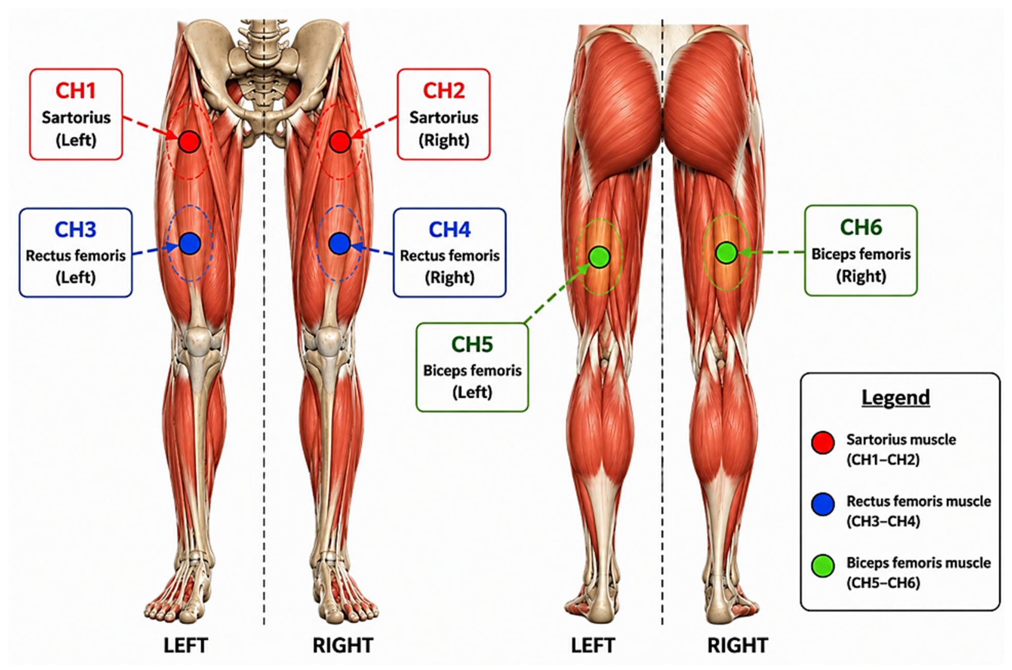
As illustrated in
Figure 4, six EMG channels were used to monitor three major muscle groups bilaterally: sartorius, rectus femoris, and biceps femoris. These muscles were selected due to their significant involvement in lower-limb flexion, extension, and stabilization during stepping, kneeling, and dash movements [
3,
6].
The rectus femoris primarily contributes to knee extension and hip flexion, the biceps femoris is involved in knee flexion and hip extension, while the sartorius contributes to lower-limb flexion and stabilization. Bilateral monitoring of these muscle groups provides a representative characterization of neuromuscular activity during locomotor tasks.
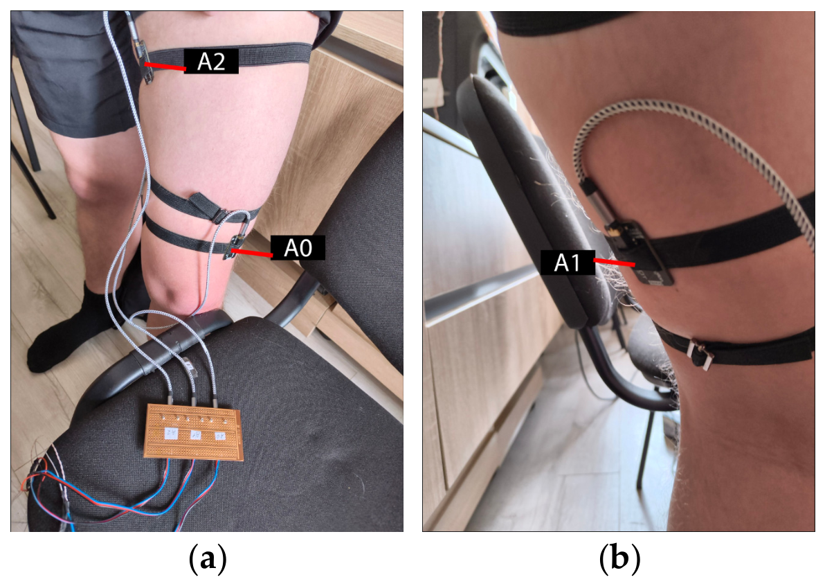
The practical placement of the six EMG sensors on the lower limbs is presented in
Figure 5.
A consistent electrode configuration was maintained for all participants to minimize acquisition variability and improve the quality and reproducibility of the experimental dataset used for AI model training and validation [
5,
6,
7,
8,
12,
13,
15].
2.7. EMG Data Acquisition and Processing
Electromyographic signal acquisition is performed using the Arduino Mega platform, which is responsible for the simultaneous reading of EMG channels and the transmission of data to the embedded subsystem dedicated to movement processing and classification [
24,
25]. The MyoWare 2.0 sensors used in this study operate in ENV mode and incorporate an analog band-pass filter with cutoff frequencies of 20.8 Hz and 498.4 Hz. Additionally, the sensors implement an envelope extraction stage based on a first-order low-pass filter with a cutoff frequency of 3.6 Hz, providing a smoothed representation of muscle activation suitable for movement classification tasks. No additional digital notch filtering was applied.
In the initial development stage of the platform, the acquisition process was based on segmenting the signals into fixed windows of 64 samples for each EMG channel between the frequency 60 and 65 Hz. This sampling rate was determined by the combined influence of the embedded acquisition architecture, communication overhead, and inertial sensor processing routines implemented on the microcontroller platform.
It should be noted that the reported sampling frequency corresponds to the envelope output generated by the MyoWare 2.0 sensors operating in ENV mode rather than to raw electromyographic signals. The envelope signal represents a low-frequency estimate of muscle activation and is commonly employed in embedded myoelectric control applications where the objective is movement intention recognition rather than spectral analysis of EMG envelope data.
To ensure dimensional consistency for the Convolutional Neural Network (CNN), a fixed-length segmentation strategy was adopted instead of a sliding-window approach with overlap. Each movement recording was treated as a single experimental sample. Because the duration of the recordings varied among trials, all time-series signals were resampled to a fixed length using linear interpolation implemented through the scipy.interpolate.interp1d function. This procedure standardized all recordings to 64 samples while preserving the temporal evolution of the signal.
Following resampling, each EMG channel was normalized independently using Z-score normalization. The normalization procedure was performed separately for each recording according to:
where
and
represent the mean value and standard deviation of the corresponding channel within the analyzed recording. This normalization reduces inter-recording amplitude variability while preserving temporal activation patterns.
Experimental tests revealed limitations of threshold-based segmentation, as the generated segments did not always correspond to complete movement cycles, leading to labeling inconsistencies and reduced dataset quality for AI training [
9,
10,
11,
12,
13]. Therefore, the current configuration employs continuous EMG acquisition without activation thresholds, preserving the full electromyographic information and reducing the risk of losing relevant movement intention patterns [
12,
13].
Data are transmitted to the processing system via a compact binary protocol implemented on the Arduino Mega platform. Dedicated buffers manage the continuous data stream, while a Python 3.10.7-based processing module automatically decodes and stores the EMG signals in CSV format for dataset organization, AI model training, and classification tasks [
9,
10,
11,
12,
13].
Figure 6 illustrates the continuous electromyographic data acquisition and transmission flow from the EMG sensors to the embedded subsystem for movement processing and classification.
In the proposed configuration, EMG data are acquired continuously without applying muscle activation thresholds, preserving complete signal information and reducing the risk of losing relevant movement intention patterns [
9,
10,
11,
12,
13]. Data buffering organizes the signal stream into temporal segments for subsequent analysis.
EMG data are transmitted via a serial interface using a binary protocol implemented on the Arduino Mega platform. Within the processing subsystem, the data stream is decoded and automatically stored in CSV format for dataset generation and AI model training.
Figure 7 presents the experimental dataset processing workflow, including validation, conversion, and organization stages required for AI model training and inference.
The stages of the electromyographic signal acquisition and processing workflow used for generating the experimental dataset are presented in
Table 4. The proposed structure enables the organization of the data flow from the EMG channels and the integration of validation and processing mechanisms required for lower-limb movement classification [
9,
10,
11,
12,
13].
For experimental validation, an additional EMG signal quality verification mechanism was implemented to identify segments affected by noise, motion artifacts, or electromagnetic interference [
12,
13]. The signals deemed valid were used for generating the experimental dataset and for training the AI model.
The staged organization of the processing workflow enables integrated management of EMG data acquisition, validation, and dataset preparation for locomotor movement classification. Continuous data collection and the use of a binary protocol help reduce the risk of losing relevant information and increase the integrity of the dataset used for training and validating AI models [
9,
10,
11,
12,
13]. The implemented validation process allows the identification of noise-affected segments and ensures the generation of a dataset suitable for developing EMG-based movement intention recognition mechanisms.
Table 5 represents the EMG recordings among movements. The dataset consisted of six movement classes corresponding to left and right variants of kneeling, stepping, and dash movements. Although the acquisition protocol was designed to collect a comparable number of recordings for each class, minor differences in class distribution were observed due to variations in recording quality and data validation procedures.
To reduce the influence of class imbalance during training, class weights were computed automatically using the compute_class_weight function from the Scikit-learn library and incorporated into the training process. This approach assigns a higher penalty to misclassified samples belonging to underrepresented classes, promoting a more balanced learning process and reducing classification bias.
2.8. AI Model and Training Process
A one-dimensional convolutional neural network (1D-CNN) model was developed for lower-limb movement classification using datasets generated through the electromyographic signal acquisition and processing pipeline. The model aimed to identify neuromuscular patterns associated with different locomotor movements and to recognize movement intention based on the monitored muscle activity [
9,
10,
11,
12,
13,
27].
A 1D-CNN architecture was selected for movement classification due to its suitability for processing temporal multichannel electromyographic signals and its favorable balance between classification performance and computational efficiency [
16,
17,
21]. Unlike conventional machine-learning approaches, which typically require manual extraction of handcrafted features from time, frequency, or time–frequency domains, 1D-CNN models enable automatic feature learning directly from EMG signal sequences, thereby reducing preprocessing complexity and minimizing feature engineering requirements.
The choice of the 1D-CNN architecture was also motivated by the computational constraints associated with embedded implementation in biomechatronic and exoskeleton control systems. Compared with more complex deep learning architectures such as LSTM, CNN-LSTM, or Transformer-based models, 1D-CNN architectures require fewer parameters, lower computational resources, and reduced inference latency, making them particularly suitable for real-time movement intention recognition.
Alternative architectures such as LSTM, CNN-LSTM, and Transformer-based models were not evaluated in the present study, as the primary objective was to validate the feasibility of EMG-based movement classification using a computationally efficient architecture suitable for embedded deployment in real-world biomechatronic systems. Comparative evaluation with alternative deep learning architectures represents an important direction for future research.
The experimental data were generated and organized in CSV format based on the decoded EMG stream and were subsequently preprocessed prior to the training phase, including signal validation and the removal of segments affected by noise, motion artifacts, or electromagnetic interference [
12,
13].
The main parameters used for the training and validation process of the AI model are presented in
Table 6.
The architecture of the proposed 1D-CNN model was configured to process multichannel electromyographic sequences using convolutional layers for temporal feature extraction, followed by pooling and classification layers for locomotor movement recognition. The network structure was optimized to balance classification performance and computational efficiency for future embedded biomechatronic applications. The detailed architecture of the proposed 1D-CNN model is presented in
Table 7.
The experimental dataset included 608 EMG files from 10 participants and was organized into six experimental classes associated with different types of locomotor movements. The EMG signals were normalized using the Z-score method performed per recording and processed through a 1D-CNN architecture based on end-to-end learning, without manual extraction of features such as RMS, MAV, or FFT.
To prevent information leakage, data augmentation was applied exclusively to the training subset after the generation of the Stratified 5-Fold and LOSO partitions. Validation and testing subsets contained only original recordings and were never augmented. For performance evaluation, two complementary validation strategies were used—Stratified 5-Fold Cross-Validation and Leave-One-Subject-Out (LOSO)—which enabled the assessment of both classification performance and the model’s inter-subject generalization capability.
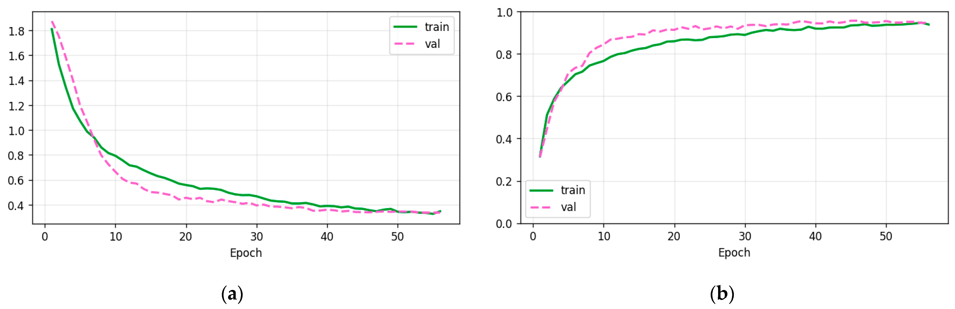
The evolution of the AI model’s performance metrics during the training and validation phases is shown in
Figure 8.
The training results show a progressive decrease in loss and a consistent increase in accuracy for both the training and validation datasets. Performance stabilization after approximately 40–50 epochs indicates convergence of the optimization process and stable model learning under the analyzed experimental conditions.
The Leave-One-Subject-Out (LOSO) strategy was used to evaluate the model’s inter-subject generalization capability and simulate scenarios involving previously unseen users. The performance metrics obtained using both Stratified 5-Fold Cross-Validation and LOSO evaluation are presented in
Table 8.
The results presented in
Table 8 highlight clear differences between the two validation strategies. Stratified 5-Fold Cross-Validation achieved higher performance, whereas LOSO evaluation showed a substantial decrease in accuracy due to inter-subject variability. These findings confirm that LOSO provides a more realistic assessment of model generalization for real-world EMG-based movement recognition applications.
These findings indicate that while cross-validation demonstrates strong classification performance under controlled conditions, it may overestimate generalization to unseen users [
11,
12,
13,
20,
21,
22,
23]. Consequently, the distinct configurations detailed in
Table 9 reflect these validation trade-offs; the Stratified 5-Fold architecture maximizes baseline classification, whereas the sequential, lightweight LOSO setup explicitly addresses and evaluates robustness against inter-subject variability in real-world scenarios.
The comparative configurations presented in
Table 8 highlight the distinct objectives of the two experimental approaches. The Stratified 5-Fold Cross-Validation configuration was optimized to maximize classification performance under controlled experimental conditions, whereas the LOSO configuration focused on evaluating inter-subject generalization and real-world applicability.
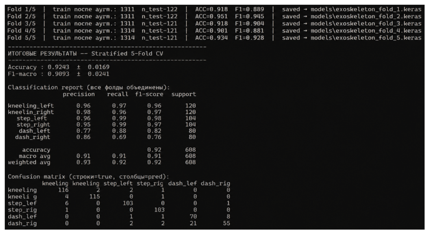
From a biomechatronic perspective, these results confirm the potential of EMG-based movement classification for intelligent exoskeleton control while emphasizing the need for improved adaptation mechanisms, personalization strategies, and multimodal sensing integration to enhance robustness across users (
Figure 9).
The confusion matrix shows a dominant distribution along the main diagonal, indicating strong classification performance across all movement classes. Minor classification errors occur mainly between locomotor movements with similar biomechanical characteristics, suggesting partial overlap in the associated EMG activation patterns.
2.9. Ethical Considerations
The experimental study was conducted in compliance with ethical principles regarding research involving human participants. The experimental protocol used for electromyographic signal acquisition and validation of the biomechatronic platform was reviewed and approved by the Ethics and Professional Deontology Commission for Research Activities of the Technical University of Moldova, within the research project “Development of passive and active exoskeletons for military use” (project code 25.80013.5007.09RE), through the approval issued on 9 April 2026.
The experimental process involved 10 healthy male volunteers aged between 19 and 24 years. Participants were fully informed in advance about the purpose of the research, the experimental procedures, the methods of data collection and usage, as well as their right to withdraw from the study at any time without any consequences. All participants provided written informed consent prior to the commencement of the experiments.
Electromyographic data were collected solely for scientific research purposes, anonymized, and processed without using any personally identifiable information. The experiments focused on lower limb locomotor movements, including stepping, kneeling, and dash movements. The experimental procedures were non-invasive and did not involve any significant additional risks for the participants.
3. Results
This section presents the experimental results obtained from EMG signal acquisition, AI model training, and movement classification using the proposed biomechatronic platform [
5,
6,
7,
8,
9,
10,
11,
12,
13]. The evaluation was performed using a dataset of 608 multichannel EMG recordings collected from 10 participants and organized into six locomotor movement classes: kneeling_left, kneeling_right, step_left, step_right, dash_left, and dash_right.
The experimental results are analyzed in terms of EMG signal quality, AI model performance, and inference capability. Model performance was evaluated using Stratified 5-Fold Cross-Validation and Leave-One-Subject-Out (LOSO) protocols to assess both classification accuracy and inter-subject generalization [
9,
10,
11,
12,
13,
14,
15,
16,
17].
3.1. Results of EMG Signal Acquisition
Experimental validation of the proposed platform was performed through multichannel EMG acquisition during the execution of lower-limb locomotor movements [
16,
17,
18].
The implemented system enabled simultaneous acquisition from all six EMG channels and continuous data transmission to the embedded subsystem. No observable interruptions occurred during acquisition, buffering, or transmission, allowing the collection of complete EMG recordings for preprocessing, validation, and AI-based classification.
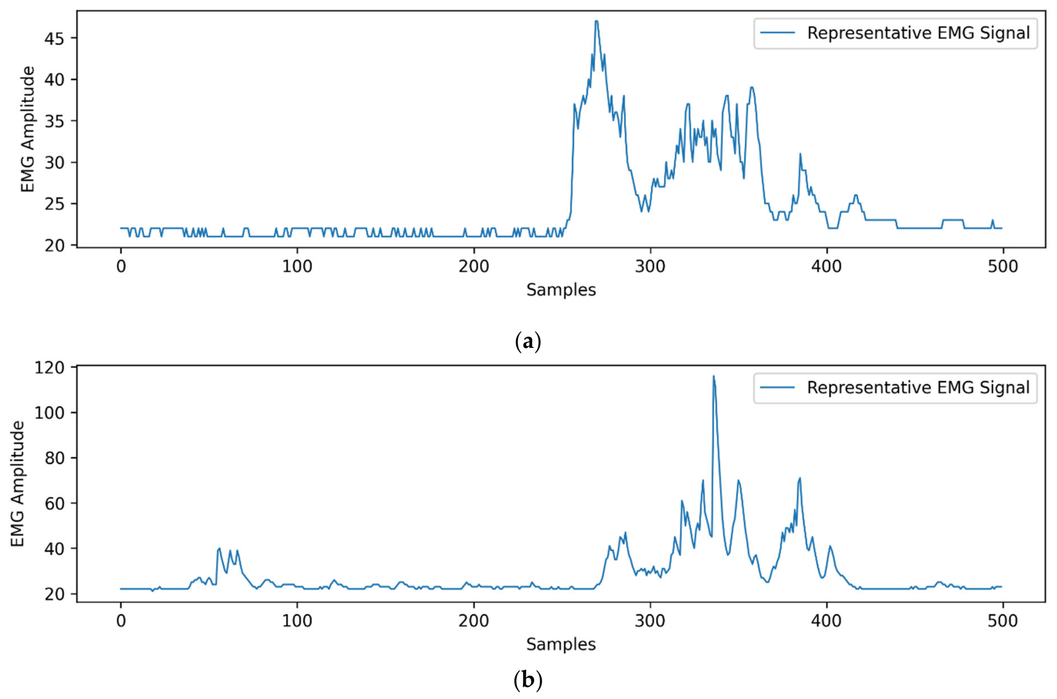
For the experimental analysis, the evolution of the electromyographic signal amplitude and the distribution of muscle activity throughout the execution of locomotor movements were evaluated. Representative examples of the EMG signals acquired during the experiments are shown in
Figure 10.
The representative EMG signals shown in
Figure 10 indicate clear differences between stepping and kneeling movements. Stepping generated moderate and repetitive amplitude variations, whereas kneeling produced higher activation peaks, reflecting the distinct biomechanical demands of the analyzed movements. These differences support the separability of the movement classes used for AI-based classification.
The signal validation process enabled the removal of segments affected by noise and electromagnetic interference, improving the quality of the dataset used for AI model training.
3.2. Results of AI Model Training
The AI model was trained using the validated EMG dataset consisting of 608 recordings from 10 participants, organized into six lower-limb movement classes.
The training process included dataset preprocessing, validation of the electromyographic signals, and data organization for the classification task. To reduce the impact of noise on the AI model’s performance, sequences considered irrelevant or affected by electromagnetic interference were removed [
12,
13].
The results of the training process are presented in
Figure 8, which highlights the evolution of the accuracy parameter and the variation in the loss function during the AI model’s training and validation stages.
As shown in
Figure 8, training accuracy increased progressively while the loss function decreased during training. The stabilization of these parameters indicates model convergence and effective feature learning from multichannel EMG signals [
9,
10,
11,
12,
13].
The results of the training process indicate good model stability and the absence of pronounced overfitting, as evidenced by the convergent evolution of the accuracy and loss parameters for both the training and validation sets.
LOSO evaluation yielded an average accuracy of 62.11 ± 23.26% and a macro F1-score of 58.28 ± 24.76%, substantially lower than the Stratified 5-Fold Cross-Validation results. This indicates reduced generalization performance when the model is applied to previously unseen participants.
3.3. Results of AI Inference
The inference process was evaluated using EMG recordings selected from the experimental dataset to assess the model’s ability to classify lower-limb movements.
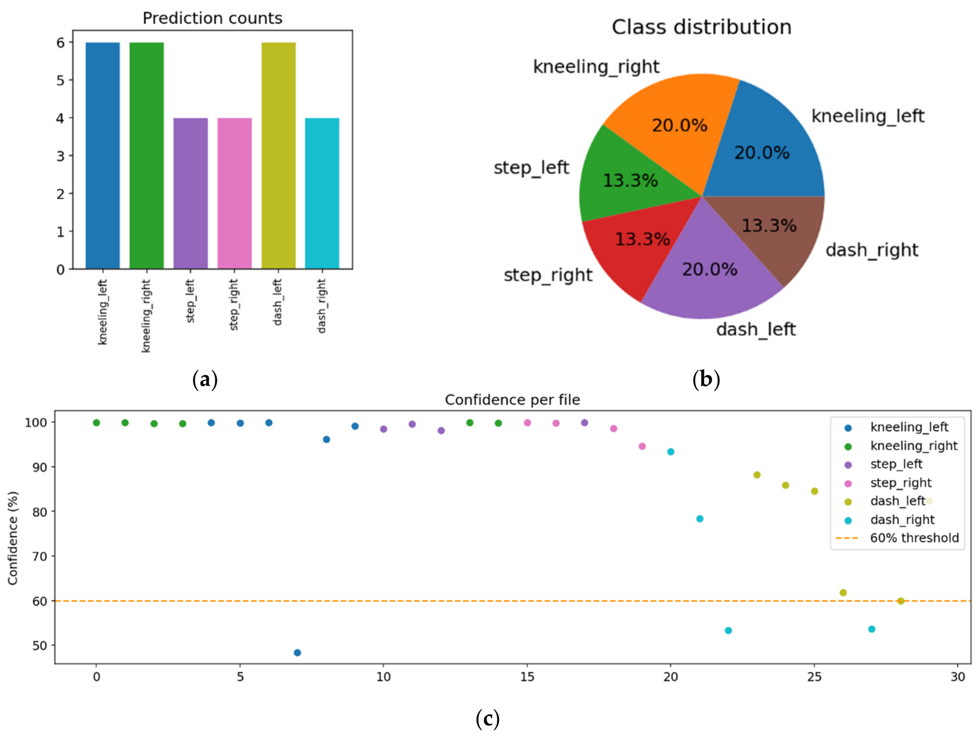
The inference and classification results obtained using the AI model are presented in
Figure 11.
The inference results presented in
Figure 11 show that the model successfully classified all six movement classes included in the study. The evaluation set consisted of 30 EMG files distributed across the six classes (6 kneeling_left, 6 kneeling_right, 4 step_left, 4 step_right, 5 dash_left, and 5 dash_right), enabling assessment of prediction behavior and confidence scores.
In the inference test conducted using the model obtained through Stratified 5-Fold Cross Validation, all 30 analyzed files were correctly classified, corresponding to an accuracy of 100% for the evaluated experimental sample. The average confidence level of the predictions was 96.12%, while the median confidence score reached 99.51%, indicating stable and consistent predictions for most of the analyzed electromyographic sequences.
The 100% inference result should be interpreted in the context of the limited evaluation sample and does not represent the overall system performance. A more robust estimate is provided by Stratified 5-Fold Cross-Validation, which yielded an average accuracy of 92.43 ± 1.69%.
These inference results confirm the model’s ability to correctly classify the evaluated EMG sequences, while overall system performance is more accurately represented by the cross-validation and LOSO evaluation results.
4. Discussion
4.1. Comparison with Literature
To assess the relevance of the obtained results and position them within the context of recent research on EMG-based movement intention recognition, the performance of the proposed model was compared with results reported in the specialized literature. Recent studies have demonstrated the effectiveness of using CNN, CNN-LSTM, and other deep learning architectures for classifying lower-limb movements and developing intelligent systems for exoskeletons and rehabilitation applications [
14,
15,
16,
17,
26,
28,
29,
30].
Table 10 presents a comparison between the results obtained in this study and those reported in recent research using electromyographic signals and artificial intelligence algorithms for movement classification and movement intention recognition in locomotor assistance and exoskeleton control applications.
In addition to the performance obtained through Stratified 5-Fold Cross-Validation, LOSO evaluation yielded an accuracy of 62.11 ± 23.26%. Although lower than the cross-validation result, this provides a more realistic estimate of the model’s generalization capability to previously unseen users.
A direct comparison with conventional machine-learning methods such as SVM, Random Forest, and MLP classifiers were not included in the present study. The primary objective of this work was the development and validation of an integrated EMG-based biomechatronic platform and the assessment of a lightweight 1D-CNN architecture suitable for future embedded implementation. Nevertheless, comparative evaluations with classical machine-learning and recurrent neural network approaches represent an important direction for future work and will be considered in subsequent studies.
A substantial performance decrease was observed when transitioning from Stratified 5-Fold Cross-Validation (92.43%) to Leave-One-Subject-Out validation (62.11%). This result represents one of the most important findings of the present study, as it highlights the significant impact of inter-subject variability on EMG-based movement classification. While cross-validation demonstrates strong classification capability under controlled experimental conditions, LOSO evaluation provides a more realistic estimate of model performance when applied to previously unseen users. These findings are consistent with recent studies indicating that subject-independent EMG classification remains a major challenge in intelligent exoskeleton control systems.
However, direct comparison between studies should be interpreted with caution because performance is strongly influenced by factors such as the number of subjects, movement classes, sensor configuration, and validation methodology. Many published studies rely exclusively on conventional cross-validation procedures, which may overestimate the generalization capability of the classifier.
Although the proposed approach achieved lower subject-independent accuracy than subject-dependent approaches reported in the literature, the inclusion of LOSO evaluation represents a more rigorous assessment of real-world applicability and identifies important directions for future work involving transfer learning, personalization strategies, and multimodal EMG–IMU fusion.
4.2. Study Limitations
Although the obtained results demonstrate the feasibility of using electromyographic signals for locomotor movement classification and movement intention recognition, several limitations should be acknowledged.
First, the participant cohort was relatively small, consisting of only 10 volunteers. Although the dataset included 608 movement recordings and was sufficient for preliminary validation, a larger and more diverse population would improve statistical robustness and better represent real-world variability [
11,
14,
17].
Second, all participants were healthy individuals, and the experimental validation was conducted under controlled conditions using a relatively homogeneous cohort. Therefore, the results cannot be directly generalized to broader demographic groups, clinical populations, or rehabilitation scenarios involving neurological or musculoskeletal impairments.
A significant performance decrease was observed under Leave-One-Subject-Out validation, where accuracy dropped to 62.11 ± 23.26%. This result confirms the strong influence of inter-subject variability and highlights the need for improved subject-independent generalization strategies [
12,
13,
16].
Finally, the present study focused exclusively on EMG-based movement intention recognition. Although inertial sensors were integrated into the platform architecture, multimodal EMG–IMU fusion and real-time closed-loop exoskeleton control were not evaluated. Future research will focus on expanding the experimental dataset, improving inter-subject generalization through advanced adaptation and transfer learning strategies, integrating multimodal sensing approaches, and validating the proposed platform in real-time adaptive exoskeleton control scenarios.
5. Conclusions
This study demonstrated the feasibility of developing an integrated biomechatronic platform for lower-limb movement monitoring and classification using electromyographic signals and artificial intelligence techniques. The proposed platform combines multichannel EMG acquisition, embedded processing, inertial sensing, and AI-based classification to support the development of intelligent exoskeletons for locomotion assistance and rehabilitation.
The experimental results confirmed that electromyographic signals contain relevant neuromuscular information for distinguishing locomotor movement patterns and recognizing movement intention. The proposed 1D-CNN model demonstrated strong classification performance under controlled experimental conditions, achieving high accuracy and macro F1-score values using the Stratified 5-Fold Cross-Validation strategy. These results validate the effectiveness of combining EMG-based sensing with deep learning approaches for lower-limb movement classification.
The additional evaluation using Leave-One-Subject-Out validation revealed a substantial reduction in performance, highlighting the strong influence of inter-subject variability on model generalization. This result confirms that subject-independent movement intention recognition remains one of the major challenges in the development of real-world AI-based exoskeleton control systems. Physiological variability and signal acquisition differences significantly affect classification robustness when the model is evaluated on previously unseen users.
Despite these limitations, the obtained results demonstrate the practical potential of EMG-based movement intention recognition for intelligent assistive systems and provide valuable insight into the challenges associated with deploying adaptive biomechatronic control systems in real-world environments. Future research will focus on expanding the experimental dataset, improving inter-subject generalization through transfer learning and personalization strategies, integrating multimodal EMG–IMU sensing approaches, and validating the proposed platform in real-time adaptive exoskeleton control scenarios.



 ,
,