


Abstract
Tail inflammation and necrosis in dairy cows are increasingly reported, yet the early stages of their development and dynamics remain unclear. We conducted a longitudinal study to characterize the macroscopic pathogenesis of tail lesions throughout the entire lactation period to identify the initial stages and vulnerable phases of lactation. Fifty-one cows were examined weekly. We scored the distal spineless tail for bloody lesions. Change point analysis and first-order Markov chains were used to describe the timing and transitions probabilities. Overall, 60.8% of the cows developed at least one bloody lesion, with 49.0% experiencing lacerations and 25.5% undergoing autoamputation. On average, autoamputation shortened tails by 4.0 cm. Three progression types were identified: laceration with healing and preservation of the tip, autoamputation after laceration and spontaneous autoamputation. Regarding the frequency of occurrence, 65.5% began as bloody lacerations and healed in 93.2% of cases. Of these, 4.0% progressed to autoamputation, 12.7% underwent spontaneous autoamputation from an intact tip. Significant changes in lesion severity occurred in lactation weeks 8 and 40. These patterns are consistent with an endogenous microcirculatory mechanism and support the hypothesis that tail inflammation and necrosis may represent a manifestation of Bovine Inflammation and Necrosis Syndrome.
1. Introduction
The tails of dairy cows have gained significant attention as an indicator of animal health [1]. Tail lesions have been associated with pain and impaired behavior. Therefore, tail integrity has been included into numerous animal welfare assessment protocols [2,3,4,5,6]. Recent studies have focused on the distal tail end, where a wide variety of lesions with high prevalences have been documented across housing and management systems [1,7,8,9,10].
Macroscopically, the lesions in dairy cattle are highly consistent across cases and closely resemble those observed in tail necrosis in fattening cattle. For several decades, scientific literature has documented inflammation and necrosis of the tail tip in beef cattle [11,12,13,14,15]. In addition to the frequently cited housing conditions that were suspected as potential causes of inflammation and necrosis of the tail tip [16,17], endogenous circulatory disorders have repeatedly been described as a possible trigger for the inflammation within the tail since the beginning [18]. Such disturbances have been linked to ruminal acidosis, a result of intensive feeding practices in fattening cattle [19,20].
Similar mechanisms are now being discussed in dairy cows, where tail tip inflammation and necrosis may also arise from impaired blood circulation in the distal tail region [8,9,18]. In addition to the pain associated with these lesions, they seem to be linked to broader health impairments [1]. Although distal tail lesions in dairy cows have been increasingly reported, their etiology and early development are insufficiently understood. Previous studies primarily describe the presence of lesions, but not how they emerge or progress over time.
Against this background, the present study aims to trace the pathogenesis of tail inflammation and necrosis through repeated, weekly macroscopic examinations. Particular emphasis is placed on identifying early visible changes, describing the initial stages of the onset of the lesions, documenting how these lesions evolve over time as well as determining the stage of lactation at which cows are most vulnerable.
2. Materials and Methods
2.1. Animals, Housing and Management
Data collection was carried out on a conventional dairy farm in Germany, Bavaria between February 2023 and February 2025. The cows were housed in a free-stall barn with solid floors, deep cubicles and an automatic scraper for removing manure. The farm had a total of 132 cows, most of which were Simmental, followed by a small number of Holstein-Friesian, Yellow Cattle, and crossbreeds. The cows were kept in two separate barns (herd 1, herd 2) with different housing and milking systems. Table 1 shows the differences in between the herds. The cows were fed with a partial mixed ration based on grass and maize silage, straw, grain, maize and barley, as well as soybean and rapeseed extraction meals. Herd 1 received milk performance feed via concentrate feed stations, while herd 2 received concentrate via the automatic milking system.
Table 1.
Overview of the two herds and different characteristics.
Furthermore, samples of the total mixed ration were collected every four months (n = 7) during the data collection and analyzed for the presence of mycotoxins.
Ventilators and curtains were installed to regulate the climatic conditions in the barn. Both herds also had access to either paved outdoor walking area (herd 1) or pasture (herd 2). The regular herd management included frequent tail hair clipping (every four weeks) to maintain hygiene and to improve the milking process.
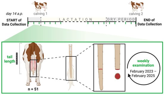
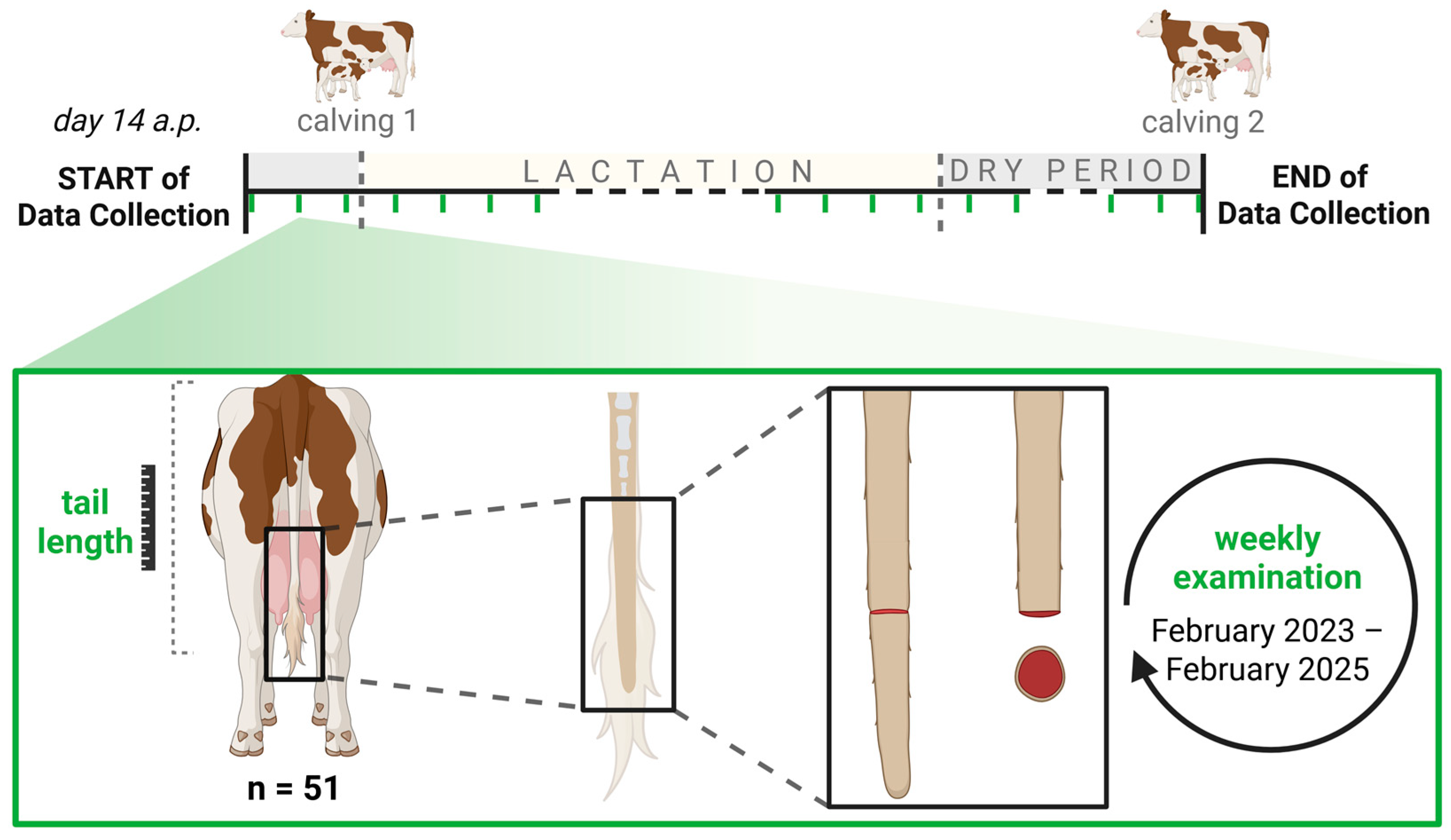
The cows were selected for the weekly examination based on their stage of pregnancy. All cows from both herds (primiparous and multiparous) due to calve between March and December 2023 were included. Animals that had already undergone tail amputation were excluded from the study. Weekly data collection started around 14 days before calving (antepartum, a.p.) and continued throughout the lactation and dry period until the next calving (Figure 1). Initially, 82 cows were included in the study (Table 1).
Figure 1.
Overview of the data collection period with focus on bloody lesions at the tail tip in form of a laceration and/or a bloody tail tip as well as the tail length; examined weekly from February 2023 until February 2025 (a.p. = antepartum; created with BioRender.com).
Only animals that completed the full data collection period were selected for statistical analysis. Cows that had been culled (n = 28) or had their tails amputated for medical reasons during the time of data collection (n = 3) were excluded. A total of 51 cows and 2786 examinations were included in the analysis. Due to their individual calving intervals, these cows were in the study for between 50 and 67 weeks (mean: 54.6 weeks).
The dairy cows selected for the statistical analysis were primarily Simmental (n = 42), as well as Holstein–Friesian (n = 4), crossbreeds (n = 4) and Yellow Cattle (n = 1). The cows were in their first to sixth lactation (Table 2).
Table 2.
Overview of the lactation stages of the cows involved in the statistical analysis.
2.2. Data Collection
Data collection was consistently performed by the same, well-trained animal scientist and an assistant on the same day each week. As the data collection process was painless and the cows were accustomed to human contact, most examinations were carried out with the animals standing or lying in the cubicles. The cows were only secured if necessary during data collection. No separation from the herd was required. The weekly examination typically took about five minutes per animal.
Because all treatments performed on the animals were routine agricultural activities related to regular herd management, the competent authority did not require an ethical evaluation of the study.
Tail Lesions and Tail Length
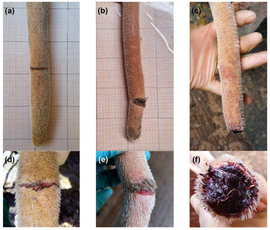
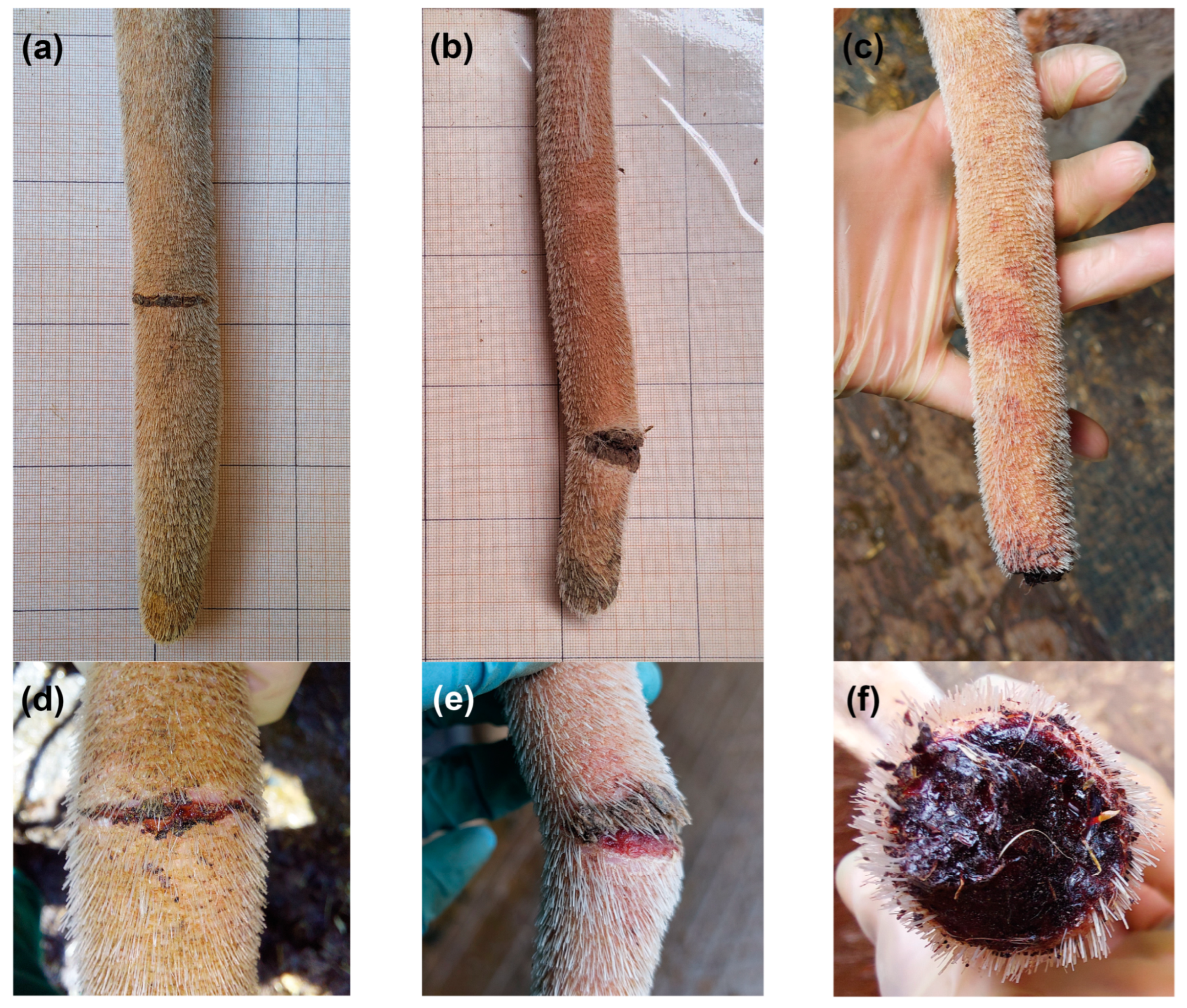
The lower third of the tail was trimmed, after which the distal spineless part was examined on a weekly basis for the lesions described in detail by Kremer-Rücker et al. [7]. All lesions were documented with a special focus on bloody lesions in form of lacerations or bloody tail tips (Figure 2).
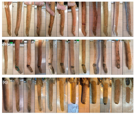
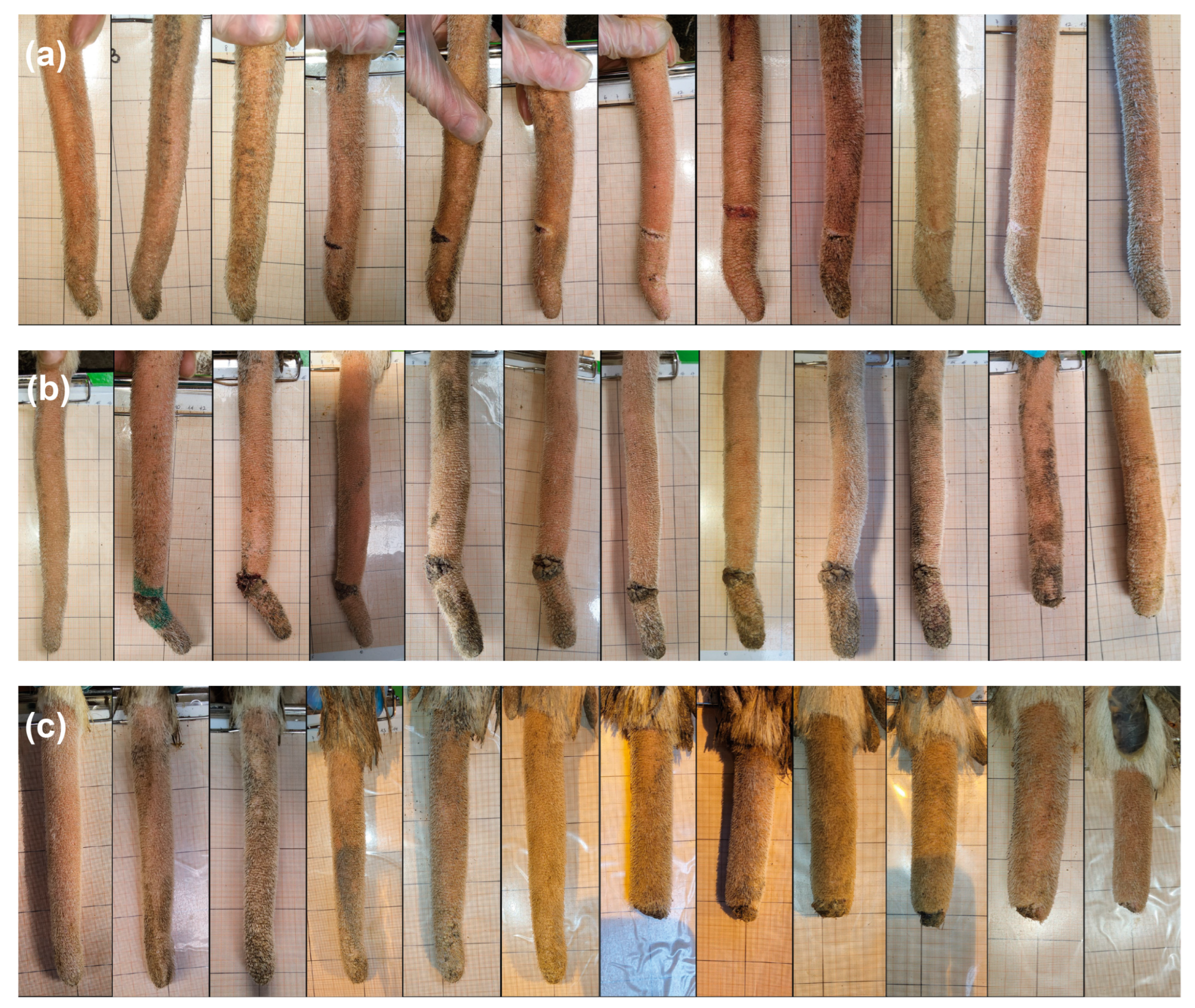
Figure 2.
Bloody lesions at the tail tips of dairy cows: lacerations of varying severity (a,b,d,e) or bloody tail tip (c,f).
The length of the tail was measured using a standard tape measure. To improve repeatability, a narrow strip of fur was shaved at the approximate level of the fourth tail vertebra (at the base of the tail) and the distance from there to the tail tip was measured weekly using a tape measure (Figure 1).
After the examination of the tail, a photograph of the spineless tail tip was taken on graph paper using a Samsung Galaxy Tab S8 Tablet (Samsung Electronics Co., Ltd., Suwon, Republic of Korea). In each case, the dorsal view of the tail tip was taken. If any changes were observed, further photographs were taken from the lateral or ventral side, as well as a frontal image of the tail’s tip.
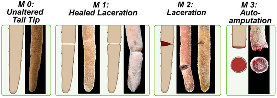
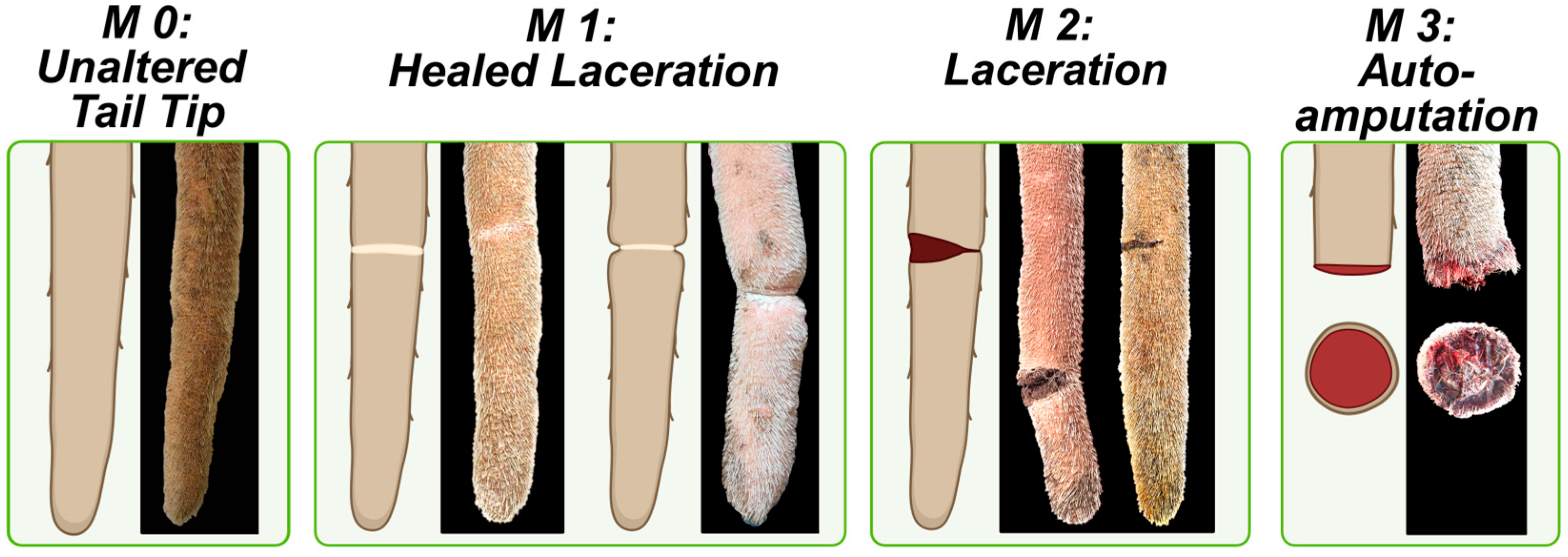
To standardize the terminology, all circumferent lesions are now termed lacerations, in accordance with the current state of knowledge [18]. Four different manifestations (M) are differentiated as follows: unaltered tail tip (M0), healed laceration (M1), laceration (M2) and autoamputation (M3; Figure 3).
Figure 3.
Different manifestations (M) of lacerations (modified after Kremer-Rücker and Weber [18]; created with BioRender.com).
After the data collection period, all photographs of each cow were organized chronologically and analyzed. Then a more detailed description of the lacerations was formulated. This description was based on the collected images and utilized the following parameters: In the case of multiple lacerations a distinction was made between lacerations of varying numbers, ranging from one laceration (A) up to five lacerations (E). The location was classified according to the dorsal view of the tail (dorsal, lateral or ventral). The size of the laceration was categorized as a quarter, semi-, three-quarter or full circle.
2.3. Statistical Analysis
2.3.1. Descriptive Statistics
The prevalence of bloody lesions among all cows (n = 51) was calculated by dividing the number of affected cows by the total number of cows.
Changes in tail length over the data collection period were compared between cows with and without autoamputation. For each animal, the difference was calculated by subtracting the average of the final three measurements from the average of the initial three measurements.
For the incident case analysis, we included only cows that showed no laceration at baseline and developed a laceration for the first time during the study period. Therefore, animals that already presented any manifestation of laceration at baseline (M1, M2, M3; Figure 3) as well as animals that never developed a laceration throughout the study (M0; Figure 3) were excluded. Since individual cows could present with more than one manifestation of laceration (ranging from A to E), position-specific panels were combined into a single dataset representing only newly formed lacerations. The final incident dataset contained 48 lacerations across 28 animals. Weekly examinations were harmonized to biologically relevant discrete time points (2 weeks ante partum, calving, 2 weeks postpartum, 8 weeks postpartum, mid-lactation, end of lactation, drying off, next calving). Data were reshaped into long format, and point prevalence of lacerations was calculated for each time point.
2.3.2. Empirical Transitions and Markov Modeling
Within each laceration panel, records were ordered by cow and observation date. The subsequent state was defined as the one-step lead of the current state. First-order, discrete-time empirical transition probabilities were estimated as
based on observed transitions. The transitions were also considered according to herd system (herd 1 vs. herd 2).
Laceration dynamics were modelled as a time-homogeneous, first-order Markov chain with four states (intact, healed laceration, laceration, autoamputation; Figure 3), treating ‘autoamputation’ as absorbing in accordance with standard Markov-chain theory [21]. Empirical transition matrices (overall and by herd system) were used to parameterize discrete-time Markov chains using the markovchain package [22]. The model assumes the Markov property and stationarity of transition mechanisms across observation intervals [21].
2.3.3. Longitudinal Trend and Changepoint Detection
To identify common shifts in laceration severity across lactation, scores were z-standardized within each animal to remove between-animal effects and subsequently averaged by lactation week:
Changepoint detection for mean shifts was performed on the weekly aggregated z-score series using the Pruned Exact Linear Time (PELT) algorithm with an Akaike Information Criterion (AIC) penalty [23]. Trend plots summarize mean trajectories with standard errors.
2.3.4. Software
All analyses were conducted in R Version 4.3.0 [24]. Data wrangling was performed using dplyr and tidyr [25], visualization using ggplot2 and ggpubr [26,27], data reshaping using reshape2 [28], Markov modeling using markovchain [22], changepoint analysis using changepoint [23], and state graph visualization using diagram [22,23,26,29].
3. Results
3.1. Prevalence of Bloody Lesions and Loss of Tail Length
Throughout the duration of the study, it was observed that 60.8% of the cows (n = 31; herd 1: 60.0%, n = 18; herd 2: 61.9%, n = 13) exhibited at least one instance of a bloody lesion, manifesting either as a laceration or a bloody tail tip (Figure 2). Since the cows could have had both conditions during the study period, the prevalence of lacerations was 49.0% (n = 25), and the prevalence of bloody tail tips was 25.5% (n = 13). Additionally, it was revealed that 25.5% of the cows (n = 13; herd 1: 30.0%, n = 9; herd 2: 19.0%, n = 4) underwent an autoamputation.
Table 3 shows how the onset of lesions is distributed depending on the animals’ lactation number. As shown, all age groups were affected by severe tail lesions in this study.
Table 3.
Overview of the number of cows and their lactation number when they suffered from a laceration (M2) and/or autoamputation (M3) in this study (n = 51 cows).
The autoamputations were confirmed through weekly measurements of the tail length and the subsequent photographic documentation of each tail tip. On average, the cows lost 4.0 cm ± 3.2 cm (standard deviation, SD) of tail length compared to their average tail length before part loss (Table 4).
Table 4.
Comparison of the difference in tail length between cows with and without autoamputation (n = 51 cows).
3.2. Pathogenesis of Bloody Lesions
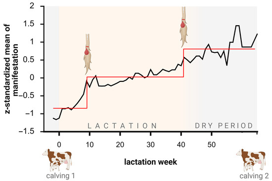
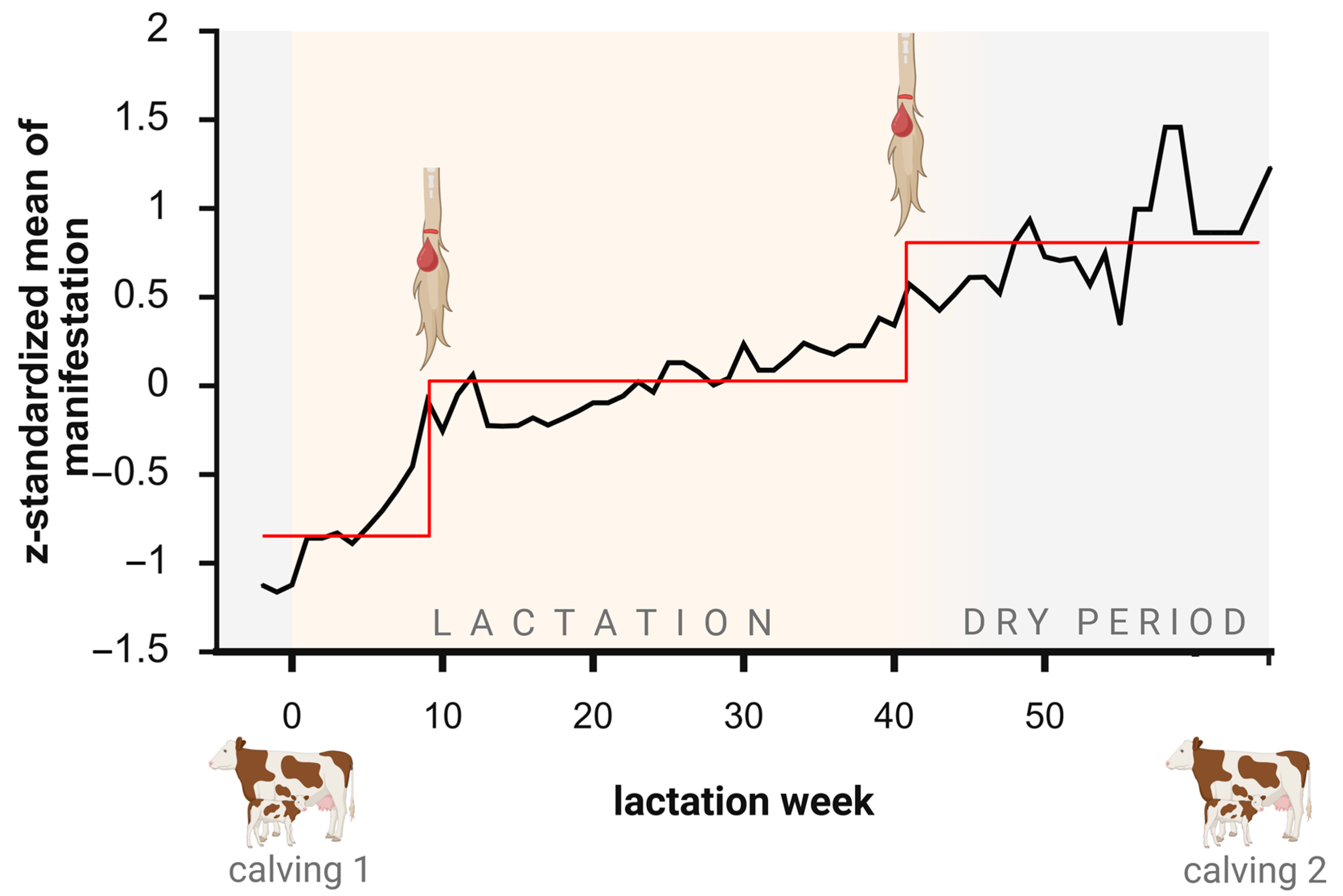
In order to describe the pathogenesis of the lacerations, the focus was on the onset of new lesions, i.e., when an unaltered tail tip first showed signs of a laceration (M1, M2, or M3; Figure 3). Change-point analysis revealed significant changes in the occurrence of the lesions at weeks 8 and 40 of lactation (Figure 4). Due to the varying calving intervals, the cows entered the dry period at different points in their lactation. The mean lactation period was 42.0 weeks (SD ± 3.9 weeks). Thus, week 35 postpartum (p.p.) marked the end of the lactation period, with all cows still producing milk. The start of the dry period varied for each cow, occurring between lactation weeks (lw) 36 and 52. The mean dry period was 10.0 weeks (SD ± 2.9 weeks).
Figure 4.
The results of the change-point analysis revealed significant changes in lesion severity in weeks 8 and 40 of lactation (n = 28 cows; mean lactation period: 42.0 ± 3.9 weeks; mean dry period: 10.0 ± 2.5 weeks; black line: z-standardized mean of manifestation; red line: estimated change points over time; created with BioRender.com).
Table 5 illustrates the prevalence of new-onset lacerations at specific time points during the study period. The lactation time points were chosen based on the results of the change-point analysis.
Table 5.
The prevalence of new-onset developed lacerations at specific points in time during the study period (n = 28 cows; lw = lactation week; a.p. = antepartum; p.p. = postpartum; 0: unaltered tail tip; 1: healed laceration; 2: laceration; 3: autoamputation).
We were able to identify three different courses of progression for bloody lesions (Figure 5). Figure 5 shows excerpts from the weekly documentation of the tail tips of three cows. The first progression (Figure 5a) represents the onset of laceration followed by complete healing and preservation of the tip. The tail tip showed no visible alterations at the beginning of the observation. Then a laceration occurred and remained bloody for another four weeks before healing. The next course of progression (Figure 5b) was onset of a laceration followed by autoamputation. The laceration remained bloody for eight weeks before autoamputation, resulting in the loss of the tail tip. The remaining bloody tail stump healed over time. The third course of progression (Figure 5c) was the spontaneous loss of the tail tip. Within a week, a piece of the tail got lost through autoamputation, despite there being no obvious signs at the examination the previous week.
Figure 5.
The progression types of bloody lesions as an excerpt from the weekly documentations: (a) laceration with healing and preservation of the tip, (b) autoamputation after laceration, (c) spontaneous autoamputation.
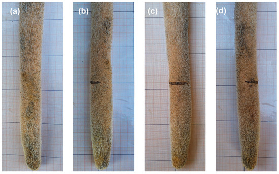
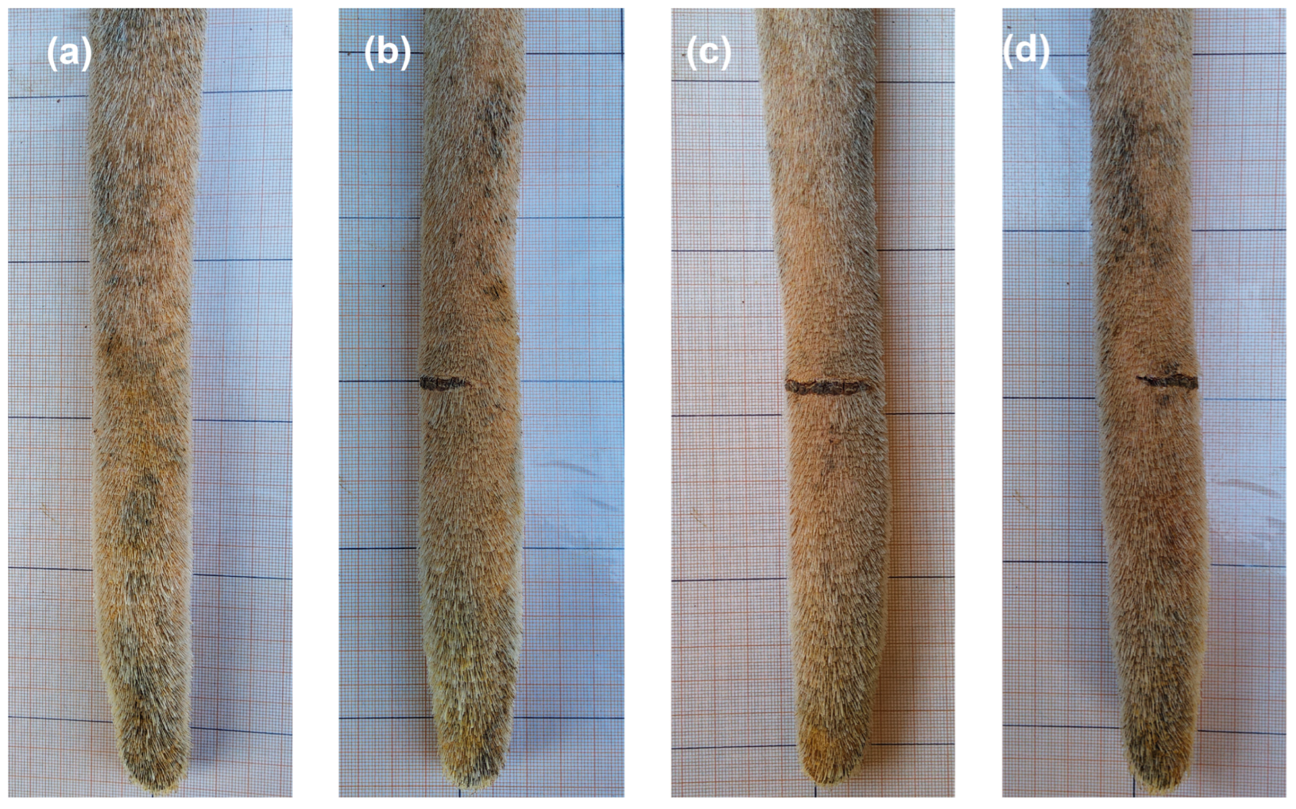
Regarding the location of the lacerations, 50.0% were lateral at the tail tip (Figure 6). The next most common locations were lateral and dorsal (19.1%), dorsal (14.3%), lateral and ventral (9.5%), and ventral (7.1%). Most lacerations were the size of a quarter circle (54.6%), followed by a semicircle (38.6%), a three-quarter circle (4.6%), and a full circle (2.3%). It was possible to have multiple lesions per tail tip. In this study, up to five lesions in different manifestations were observed at the same time point.
Figure 6.
Sample images from the tail tip of one cow with a laceration that is semicircular in shape and visible from the left lateral (b), ventral (c), and right lateral (d) views. The laceration is not visible from the dorsal view of the tail (a).
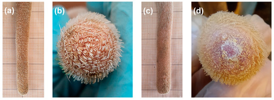
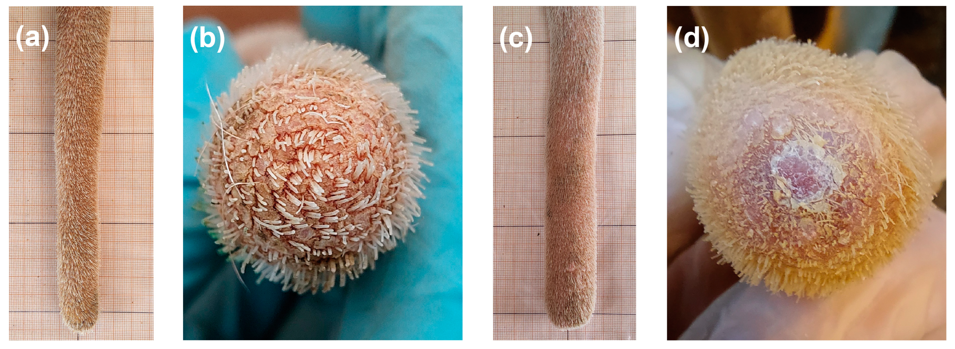
After autoamputation the remaining tail stumps healed over time without any kind of medical treatment. The amputation stumps were hairless and possibly covered in crusts (Figure 7d). The stumps were not tapered anymore and remained evenly wide (Figure 7c). Figure 7 shows a comparison between an unaltered tail tip, which is fully covered by hair (Figure 7b), free from lesions, tapering towards the tip (Figure 7a) compared to a remaining tail stump after autoamputation.
Figure 7.
Comparison of an unaltered tail tip and a healed tail stump after autoamputation: unaltered tail tip (a,b); remaining and healed tail stump after autoamputation (c,d).
In order to assess the pathogenesis of the new-onset lacerations transition probabilities were evaluated (Table 6). These probabilities represented the probability of a change in the manifestation of lesions within a week. The values are summed up for all cows and the entire study period. We checked the transition probabilities between the two herds and found no differences. Therefore, Table 6 shows the transition probabilities for the whole sample. The vertical column (0–3) shows the initial manifestation of the tail tip for each week. The horizontal row (0–3) shows the manifestation the following week. The values in the matrix indicate the probability that a cow will transition from a specific initial manifestation to the subsequent manifestation. For example, 96.04% of cows with an unaltered tail tip (0) remain in manifestation 0 in the following week, while 2.59% transition to the ‘laceration’ manifestation (2). Of the cows with an existing laceration (2), 54.64% remain in the same state, but 42.27% show healing (1). Autoamputations (3) are stable and 100% remain in the same state.
Table 6.
Transition matrix with transition probabilities for the different manifestations of lacerations (0: unaltered tail tip; 1: healed laceration; 2: laceration; 3: autoamputation; n = 28 cows). The manifestations (0–3) listed in the vertical column reflect the previous manifestation. The manifestation at the top of each row indicates the following manifestation (0–3) after a week.
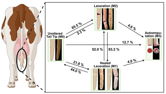
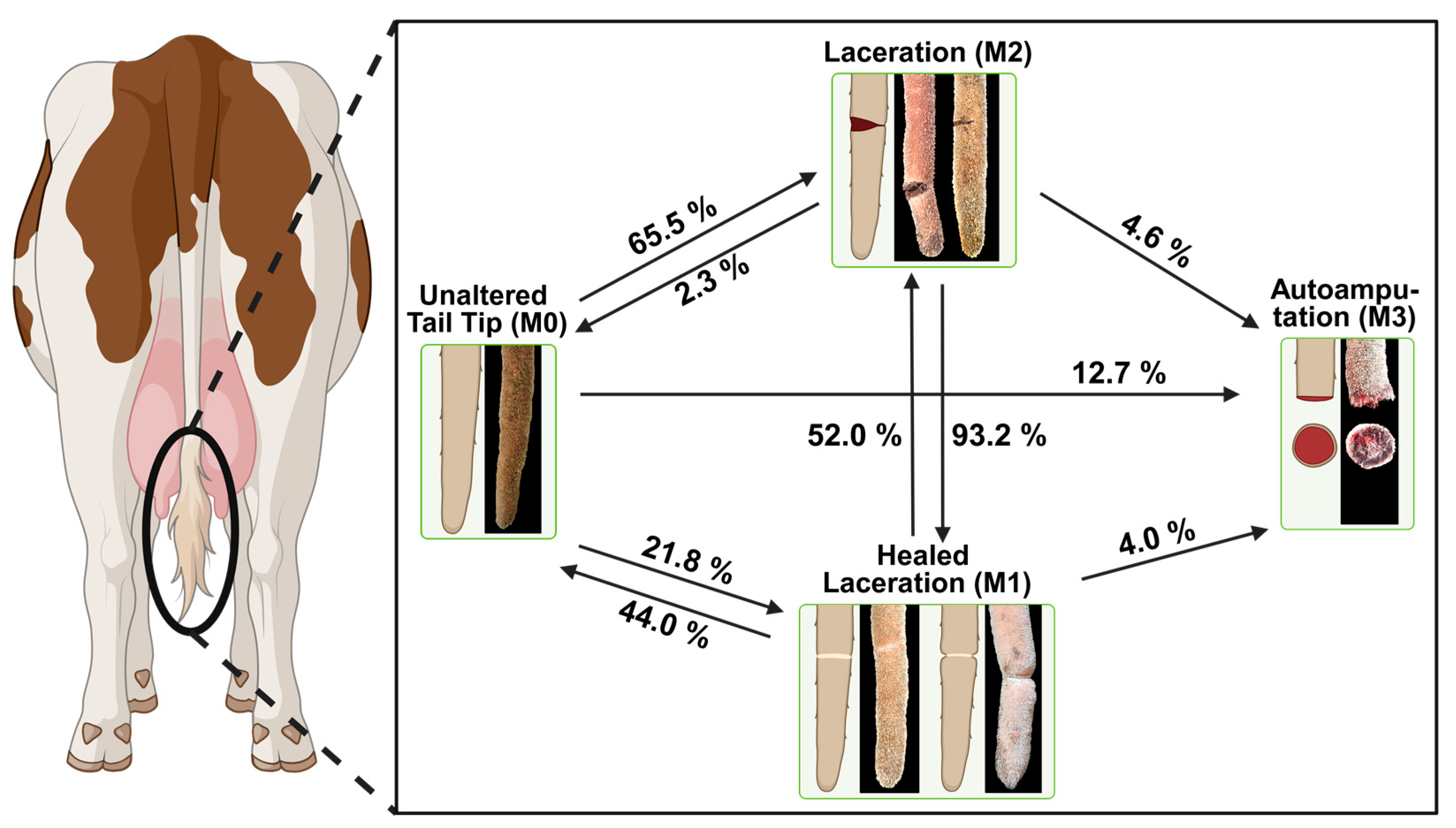
To improve practical application, we only considered cases where a change in manifestation occurred between two subsequent data collection times. This is known as the frequency of occurrence (Figure 8). The frequency of occurrence revealed that in 65.5% of cases the laceration occurred first. On average, bloody lacerations remained bloody for 2.2 weeks (SD ± 1.4). In 93.2% of cases this stage was followed by healing. In 52.0% of cases, a transition from a scar formation after healing to a bloody laceration was possible. In 4% of cases, the presence of a laceration or healed laceration was followed by autoamputation and loss of part of the tail. In 12.7% of cases, autoamputation of a part of the tail occurred directly from the unaltered tail tip.
Figure 8.
Frequency of occurrence of newly developed lacerations (n = 28 cows), 124 transitions (created with BioRender.com).
4. Discussion
The aim of this study was to identify early changes at the tail tip of dairy cows, to describe initial stages of lesions, and to document how these lesions evolve over time. Consequently, a weekly examination of the tail tips of 51 cows was carried out over the course of a lactation period.
4.1. Lacerations and Their Origin
This study focused on initial stages of lesions, such as lacerations or bloody tail tips, which were found to be prevalent in 60.8% of cases (Figure 2), posing a significant issue for cow welfare. In general, lacerations, or lesions with a similar clinical presentation, have been described several times in the literature [4,7,9,18,30].
During this study 49.0% of the cows sustained at least one manifestation of laceration during their lactation period. As shown by Volhøj et al. in their study, 22% of observed live cows and 67% of cows at the abattoir had band-shaped wounds [9]. Thus, these lesions seem to be quite prevalent in dairy herds. It is reasonable to assume that such changes at the tail tip negatively impact the animals’ health and well-being. As Deiss demonstrated through histological and histochemical examinations of tail tips from newborn calves to adult animals after slaughter that the skin at the tip of the tail is sensitive to touch and pain [31]. Köhler et al. proved that cows generally became more sensitive to pressure at the tail tip when changes occurred [1].
Several hypotheses have been proposed regarding the causes of these lacerations. These included the automatic manure scraper [4,32], tail tapes for marking and tracking the cows [4,32], wires [33], cubicle design, and other housing conditions [32,34], as well as interactions with infrastructure [35]. Poor handling was also mentioned [35], as a fracture or a dislocation of the vertebra of the tail requires considerable force and does not occur accidentally without outside input. Rather, it occurs through significant tail manipulation, such as lifting or twisting, when animals are forced to run or stand still [2,36]. Therefore, Volhøj et al. hypothesized that tail fractures would lead to a disturbed blood supply, resulting in local thrombosis and ischemic necrosis. However, there appears to be no relationship between band-shaped lesions or lacerations and deviations in the proximal part of the tail, as lacerations occurred more often on intact tails [9]. Thus, contrary to the listed causes, the lacerations seem to originate from a different source. Nevertheless, Volhøj et al. suspected a connection with blood circulation in the tip of the tail, as the described lesions followed the anatomical course of the tail’s blood vessels. Kremer-Rücker and Weber followed up on this and formulated the hypothesis that the changes, as frequently described for fattening bulls, represent the clinical picture of tail inflammation and necrosis caused by microcirculatory disorders [18].
Recent studies also suggested circulatory disorders as a possible cause of the alterations at the tip of the tail [1,8,9]. However, since these studies were all based on one-time surveys, the exact pathogenesis of the alterations at the tail tip can only be hypothesized. To our knowledge, our study is the first longitudinal follow-up study on this topic that observed cows over a complete lactation period.
In terms of pathogenesis, it is interesting to note the lesions that originated from an unaltered tail tip. As Figure 8 shows, in 65.5% of cases a bloody laceration was the first sign of onset. Regarding the vascular anatomy of the tail end, as described by Brown and Carrow [37] and Schlerka et al. [38], and the etiology summarized by Kremer-Rücker and Weber [18], systemic inflammation could trigger an immune response that affects also the terminal blood flow area, leading to circulatory disorders within the capillaries of the tail tip. This may result in insufficient blood flow to the capillaries of the tail tip and an insufficient oxygen supply to the tissue, resulting in the onset of lacerations. The histopathological finding of coagulation necrosis in the tails with “annular lesions”, clinically equal to lacerations, points as well towards ischemia as an underlying cause [8,39]. A similar process was considered central to the development of laminitis [40,41,42,43]. Since the claws and the tail tip have comparable terminal blood flow areas, this process could be responsible for the changes at the tail tip as well.
Köhler et al. demonstrated the importance of blood circulation in the tail tip in relation to observed tail lesions and different health parameters [1]. They used infrared thermography to measure the temperature of dairy cows’ tail tips. They attributed variations in tail tip temperature to differences in blood circulation in this area. These findings supported the hypothesis that the observed lesions originated internally and resulted from reduced blood flow, as the tail tips of the healthiest and best-performing cows had the warmest tail tips and therefore, the best blood circulation. Additionally, these cows exhibited fewer changes at the tail tips than the average of all observed cows during the study. As tail tip temperature decreased, changes in the tail tip and other health problems increased, confirming the link between animal health and undisturbed blood circulation in the acral regions [1].
Therefore, the location of the lacerations at the tail tip supports the hypothesis of tail inflammation and necrosis in the area of terminal blood flow. Most findings were located laterally (50.0%; Figure 6). The middle coccygeal artery runs ventrally along the tail and branches laterally [37,38]. Reduced blood flow in the fine capillaries could lead to lacerations, especially laterally. As observed in this study and previously described by Volhøj et al., several lacerations could occur simultaneously in parallel at the tail tip. They hypothesized that the segmented appearance of the lesions might be related to the segmented blood supply of the tail [9].
In our study, healing of the lacerations was observed in 93.2% of the cases (Figure 8). Volhøj et al. also observed these healed lacerations, described as band-shaped connective tissue formation, slightly elevated, faded, with increased texture and varying degrees of hair loss (from thinning to alopecia) [9]. Histological findings of such lesions showed granulation tissue extending into the central structures and complete atrophy of the hair follicles, despite a superficially intact epidermis. Thus, from a histological point of view, there was no evidence that these lesions were of traumatic origin [8,9]. However, Volhøj et al. did not conclude that these formations are consistent with healed band-shaped wounds [9]. Their histological description and lesion distribution suggest this, though.
Furthermore, the transition from a healed laceration back to an open wound was also observed. This indicates a dynamic nature of these lesions. Metzner et al. already described the signs of tail inflammation and necrosis in fattening bulls as recurrent [13]. Similarly, during our study, a transition from a laceration or a healed laceration back to an unaltered tail tip was observed. These findings suggest that the lesions are reversible, similar to those observed and described in fattening bulls [13,44,45].
In the present study, lacerations remained bloody for an average of 2.2 weeks (SD ± 1.4). The direct transition from an unaltered tail tip to a healed laceration suggests that lacerations could develop and heal within a week. However, secondary infections of tail tip necrosis could influence wound healing and the progression of the lesion, as often described in fattening bulls [12,46,47,48].
Regarding the size of the lacerations, Volhøj et al. reported an average circumference of 46% for band-shaped lesions [9], while Lorenz et al. stated circumferences ranging from 20% to 95% [8]. In this study, most of the lacerations were quarter-circles (54.6%) or semicircles (38.6%).
4.2. Autoamputations and Tail Length
Notably, 25.5% of the cows in this study lost their tail tips through autoamputation. This occurred as a direct result of a laceration, a healed laceration, or spontaneously from an unaltered tail tip (Figure 5 and Figure 7). This longitudinal study confirmed the suspicion of Volhøj et al. that band-shaped wounds could lead to autoamputation [9]. The Danish researchers described band-shaped wounds that ranged from superficial to deep-seated, with only a few soft tissue structures present, and they hypothesized that ‘deep and circumferential ulcerations may lead to the autoamputation of the distal part of the tail’ [9].
Numerous studies have described shortened tails in dairy cows [4,5,33,49]. In their study on tail damage in dairy cattle, Cuttance et al. published that most tails were shortened in the lower third of the tail [33]. Volhøj et al. also observed cows with shortened tails and hairless tail tips [9]. Additionally, cows with missing tail tips showed wound or scar tissue formation at the tip [9]. This study did not explicitly include the differences regarding the macroscopic appearance of an intact tail tip and a tail after autoamputation. However, it was found that trained people could easily and consistently classify the shape of the tail tips as conical or blunt and thus determine whether the amputation was intact or autoamputation (see Figure 7). Distinguishing between the two in future studies could therefore provide valuable information about the frequency of autoamputation in dairy cows. Autoamputation, however, contributes to the healing process of the tail. In addition to Volhøj et al. [9], numerous authors have previously described cases of healing through autoamputation following annular demarcation and tissue loss in fattening bulls [11,13,45,46]. Similarly, animals with autoamputations were less likely to develop new lesions [9,13,46], a finding that aligns with the results of our study.
This study found that the loss of length due to autoamputation was most prevalent in the lowest 4.0 cm (SD ± 3.2 cm) of the tail (Table 4). Similar areas were reported in other studies in relation to the position of the examined lesions. The median location of band-shaped lesions was 7 cm from the tail tip [9]. Ural et al. reported two cases in dairy cows with hairless areas and circumscribed crusts 4–6 cm above the tails’ tips [50]. In fattening bulls, rings of demarcation were observed 5 cm proximally to the tail’s tip [46]. Therefore, due to the anatomy of the blood vessels in this area and their fine capillary structure, it appears to be particularly susceptible to circulatory disorders.
Conversely, the tail length of cows that did not undergo autoamputation increased during the study period (Table 4). The increase in tail length was highest in first-lactation cows. This seems plausible, given that cows have not yet finished growing by the time they first calve. The cows also generally had different tail lengths. In this study, the tail length of the cows was not comparable due to the choice of measurement method. While the measurement point at the base of the tail was repeatable for individual animals, it was not comparable across all animals. Only the delta of the tail length could be compared. For tail length measurements to be used in further studies, comparable measurement points must first be clearly defined for all animals.
4.3. The Distribution of the Onset of Tail Inflammation and Necrosis in Relation to the Lactation Number and Stage of the Cows
This study found that cows with different numbers of lactations were equally affected by severe tail lesions (Table 3). Volhøj et al. reported a higher prevalence in cows with higher parity, which the researchers assumed was due to a longer period of risk [9]. Regarding the previously discussed and suspected etiology and pathogenesis, systemic inflammation could play a role, particularly during the transition period [51]. Cows that have completed multiple lactations have experienced negative energy balances several times and have been exposed to systemic inflammation more often. Since cows at different stages of lactation were affected equally in this study, it suggests that some cows may be more resilient and reach their physiological limits later than others [52,53,54,55].
With regard to critical times during lactation for the development of bloody lesions at the tail tip, lactation weeks 8 and 40 proved to be significant. These time periods also exhibited the highest prevalence of bloody lacerations (Figure 4). On one hand, there is an increased prevalence of bloody lesions during peak lactation in week 8. At this time, cows have their highest milk yield. However, maximum feed intake has not yet been reached, resulting in a negative energy balance. In addition, this period is particularly vulnerable to a wide variety of (metabolic) diseases [43,56,57,58]. For example, the incidence of mastitis is greatest between 40 and 70 days in milk [59]. The highest risk of subacute rumen acidosis occurs within the first 60 days after calving. In summary, dairy cows exhibited impaired immune function and increased inflammation during the peripartum period due to alterations in metabolic, immune, and endocrine pathways [43]. On the other hand, the severity of the lesions changed significantly during lactation week 40. At this point, the cows were either at the end of their lactation period or had already entered their dry period. This drying-off process is also marked by a change in feed from a lactating ration to a dry ration, as well as a regrouping of the herd. These changes may trigger adjustments in the digestive system, including alterations to the environment and microbiota in the gut, and cause general stress. These are associated with inflammatory processes in animals [51]. Furthermore, dairy cows experience increased systemic and mammary inflammation during the early dry period because of the transition from milking to not being milked [60]. The cessation of milk production as well as remodeling and involution of the mammary tissue are associated with the activation of inflammation [61,62]. The onset of the dry-off period is also characterized by reduced liver function, systemic inflammation, and a depleted antioxidant system [60,62,63,64,65]. Thus, bloody lesions at the tail tip occurred significantly more often when dairy cows were in a period characterized by systemic inflammation, particularly during the early dry period and peak lactation.
Taken together, cows undergo several inflammatory processes during the dry period, transition period and lactation. Additionally, an increased release of endotoxins is described to result from dietary changes, alterations in gastrointestinal tract permeability (leaky gut), mobilization of adipose tissue, remodeling of mammary gland tissue, or uterine tissue trauma after calving [61,66,67]. Thus, inflammatory processes are not limited to a single area but also affect several areas simultaneously [51,61,67]. Furthermore, metabolic and infectious diseases tend to occur together as a complex interplay [68], which for example has also already been observed in pigs and is described as ‘Swine Inflammation and Necrosis Syndrome’ (SINS) [69].
Altogether, the clinical patterns observed in this study, namely, the high frequency of initially bloody lacerations, their typical localization in the terminal vascular region, the known histological indications of ischemic processes [8,9,39], and their pronounced occurrence during the inflammatory phases of lactation, suggest an intrinsic origin of these lesions [18]. These findings consistently indicate an endogenous process involving impaired microcirculation in the tail tip. This pathophysiological pattern could be indicative of potential ‘Bovine Inflammation and Necrosis Syndrome’ (BINS), wherein systemic inflammatory processes in various body parts lead to circulatory disturbances in the body’s distal regions. Therefore, tail inflammation and necrosis in dairy cattle with lacerations and autoamputations as one of the cardinal symptoms may represent another clinical manifestation of BINS, as Meier et al. [70] and Kremer-Rücker and Weber [18] have previously suggested.
5. Conclusions
The findings of this longitudinal study show that bloody lesions on the tail tip of dairy cows are dynamic, widespread, and often progressive. Lacerations appear to be an early visible manifestation of the underlying pathology. The temporal onset patterns of the lesions, their previously proven histopathological characteristics, and their consistent anatomical localization support the hypothesis that these lesions originate from inflammatory and circulatory disturbances in the terminal blood flow area. The recurrence and partial reversibility of the lesions, together with their association with physiologically demanding phases of lactation, further underline the multifactorial and systemic nature of the condition and may indicate the presence of ‘Bovine Inflammation and Necrosis Syndrome’ (BINS). Overall, these results highlight the need for further research to elucidate the underlying mechanisms and to develop early detection and prevention strategies to ensure animal health and welfare.
Author Contributions
Conceptualization, P.V.K.-R., C.S. and K.M.W.; methodology, K.M.W. and P.V.K.-R.; software, P.V.K.-R.; validation, K.M.W., P.V.K.-R., C.S. and R.T.A.; formal analysis, C.S., K.M.W., P.V.K.-R. and R.T.A.; investigation, K.M.W.; resources, P.V.K.-R.; data curation, K.M.W. and C.S.; writing—original draft preparation, K.M.W., P.V.K.-R. and C.S.; writing—review and editing, K.M.W., P.V.K.-R. and C.S.; visualization, K.M.W. and R.T.A.; supervision, P.V.K.-R.; project administration, P.V.K.-R.; funding acquisition, P.V.K.-R. and K.M.W. All authors have read and agreed to the published version of the manuscript.
Funding
The authors would like to thank the Tönnies Forschung, Gemeinnützige Gesellschaft zur Förderung der Forschung über die Zukunft des Tierschutzes in der Nutztierhaltung mbH, Rheda-Wiedenbrück, Germany, for funding the project TINCa Dairy II on Tail Inflammation and Necrosis in Dairy Cattle, from which this study originates. The authors declare that this study received funding from Tönnies Forschung gGmbH, Rheda-Wiedenbrück, Germany. The funder was not involved in the study design; data collection, analysis, or interpretation; the writing of this article; or the decision to submit it for publication.
Institutional Review Board Statement
Because all treatments performed on the animals were routine agricultural activities related to regular herd management, the competent authority did not require an ethical evaluation of the study.
Informed Consent Statement
Not applicable.
Data Availability Statement
This article includes the original contributions presented in this study. Further inquiries can be directed to the corresponding authors.
Acknowledgments
We thank the dairy farm for their support in collecting the data and Philipp Kreß for his very helpful assistance. Furthermore, we thank Annalena Keil for her technical support in creating the graphs. During the preparation of this manuscript, the authors used DeepL-write for language editing. The authors have reviewed and edited the output and take full responsibility for the content of this publication.
Conflicts of Interest
The authors declare no conflicts of interest.
Abbreviations
The following abbreviations are used in this manuscript:
| AIC | Akaike Information Criterion |
| a.p. | Antepartum |
| BINS | Bovine Inflammation and Necrosis Syndrome |
| lw | Lactation week |
| M0 | Unaltered tail tip |
| M1 | Healed laceration |
| M2 | Laceration |
| M3 | Autoamputation |
| p | Prevalence |
| PELT | Pruned Exact Linear Time |
| p.p. | Postpartum |
| SD | Standard deviation |
References
- Köhler, A.A.; Scholz, A.M.; Kremer-Rücker, P.V. Tail Tip Temperature Measured by Thermography as an Indicator of Animal Health in Holstein Cows. Animal 2025, 19, 2–20. [Google Scholar] [CrossRef] [Scilit] [PubMed]
- Laven, R.A.; Jermy, M.C. Measuring the Torque Required to Cause Vertebral Dislocation in Cattle Tails. N. Z. Vet. J. 2020, 68, 107–111. [Google Scholar] [CrossRef] [Scilit] [PubMed]
- PraeRi. Animal Health, Hygiene and Biosecurity in German Dairy Cow Operations—A Prevalence Study (PraeRi) Final Report. Available online: https://ibei.tiho-hannover.de/praeri/pages/69 (accessed on 22 December 2022).
- Crossley, R.E.; Bokkers, E.A.M.; Browne, N.; Sugrue, K.; Kennedy, E.; De Boer, I.J.M.; Conneely, M. Assessing Dairy Cow Welfare during the Grazing and Housing Periods on Spring-Calving, Pasture-Based Dairy Farms. J. Anim. Sci. 2021, 99, 1–15. [Google Scholar] [CrossRef] [Scilit] [PubMed]
- Moono, P.; Fruean, S.N.; Hampson, D.J.; Bryan, M.A. The Frequency of Tail Damage amongst Cows from a Sample of New Zealand Dairy Farms Participating in an Animal Welfare Programme. N. Z. Vet. J. 2022, 70, 248–255. [Google Scholar] [CrossRef] [Scilit] [PubMed]
- Jafari-Gh, A.; Laven, R.; Khaloubagheri, F.; Mirrahimi, M.H.; Jafari-Gh, S.; Banadaky, M.D.; Mueller, K.R.; Vallee, E. An Investigation of Dairy Cattle Welfare in Commercial Iranian Farms: Results from Animal- and Stockperson-Based Measures. Animals 2025, 15, 359. [Google Scholar] [CrossRef] [Scilit] [PubMed]
- Kremer-Rücker, P.V.; Abel, K.M.; Lorenz, L.M.; Schmidt, C.; Lechner, M.; Schubert, K.F.; Köhler, A.A.; Meier, S.; Scholz, A.M. A Pilot Study: Tail Tip Lesions in Dairy Cows—An Unnoticed Animal Welfare Issue? Arch. Anim. Breed. 2024, 67, 271–284. [Google Scholar] [CrossRef] [Scilit] [PubMed]
- Lorenz, L.M.; Volkwein, M.E.; Schmidt, C.; Lechner, M.; Kremer-Rücker, P.V. A Look Inside—Histopathological Examinations of Different Tail Tip Lesions in Dairy Cows. Animals 2024, 14, 2094. [Google Scholar] [CrossRef] [Scilit] [PubMed]
- Volhøj, T.; Nielsen, C.K.; Schjermer, D.M.; Jensen, N.S.; Jørgensen, B.M.; Nielsen, S.S.; Jensen, H.E. Prevalence and Characterisation of Band-Shaped Tail Lesions in Holstein Cows. Acta Vet. Scand. 2024, 66, 19. [Google Scholar] [CrossRef] [Scilit] [PubMed]
- Abel, K.M.; Lorenz, L.M.; Kremer-Rücker, P. Prävalenzen von Schwanzspitzenveränderungen Bei Milchkühen—Schwelt Hier Ein? In Proceedings of the 29 Internationale DVG-Fachtagung zum Thema Tierschutz, Schwerpunkt: “Tierschutz ohne Grenzen” Gleiches Recht für alle? München, Deutschland, 29 February–2 March 2024; Deutsche Veterinärmedizinische Gesellschaft: Gießen, Germany, 2024; pp. 331–334. [Google Scholar]
- Bertocchi, D.; Franciosi, C.; Mondini, S.; Zoboli, C. Necrosi Della Coda in Torelli All’ingrasso. Atti Soc. Ital. Buiatrica Riv. Zootec. Vet. 1973, 6, 585–592. [Google Scholar]
- Madsen, E.B.; Nielsen, K. A Study of Tail Tip Necrosis in Young Fattening Bulls on Slatted Floors. Nord. Vet. Med. 1985, 37, 349–357. [Google Scholar] [PubMed]
- Metzner, M.; Hofmann, W.; Heckert, H.P.; Koberg, J.; Raue, F. Zur Epidemiologie der Schwanzspitzenentzündung bei Mastbullen. Tierarztl. Umsch. 1994, 49, 348–355. [Google Scholar]
- Schrader, L.; Roth, H.R.; Winterling, C.; Brodmann, N.; Langhans, W.; Geyer, B.; Graf, B. The Occurrence of Tail Tip Alterations in Fattening Bulls Kept under Different Husbandry Conditions. Anim. Welf. 2001, 10, 119–130. [Google Scholar] [CrossRef] [Scilit]
- Rouha-Muelleder, C.; Absmanner, E.; Kahrer, E.; Zeiner, H.; Scharl, T.; Leisch, F.; Stanek, C.; Troxler, J. Alternative Housing Systems for Fattening Bulls under Austrian Conditions with Special Respect to Rubberised Slatted Floors. Anim. Welf. 2012, 21, 113–126. [Google Scholar] [CrossRef] [Scilit]
- Keane, M.P.; Cuevas-Gómez, I.; Earley, B. A Review: Effect of Different Floor Surfaces and Space Allocation on the Welfare and Performance of Indoor Finishing Beef Cattle. J. Appl. Anim. Res. 2025, 53, 2566390. [Google Scholar] [CrossRef] [Scilit]
- Nielsen, S.S.; Alvarez, J.; Boklund, A.; Dippel, S.; Dorea, F.; Figuerola, J.; Herskin, M.S.; Michel, V.; Miranda Chueca, M.A.; Nannoni, E.; et al. Welfare of Beef Cattle. EFSA J. 2025, 23, e9518. [Google Scholar] [CrossRef] [Scilit] [PubMed]
- Kremer-Rücker, P.V.; Weber, K.M. Review: Tail Lesions in Dairy Cows—A Summary of What’s New about the Old. Animal 2026, 101785. [Google Scholar] [CrossRef] [Scilit] [PubMed]
- Kordowitzki, P. Untersuchungen zum Auftreten der Schwanzspitzennekrose bei Mastbullen. Ph.D. Dissertation, Freie Universität Berlin, Berlin, Germany, 2015. [Google Scholar]
- Heers, P.; Beune, H.; Freitag, M. Schwanzspitzennekrosen bei Mastbullen–Ursachenforschung in Mastbetrieben. In Forschungsnotizen des Fachbereichs Agrarwirtschaft Soest; Fachhochschule Südwestfalen: Iserlohn, Germany, 2017; p. 43. [Google Scholar] [CrossRef]
- Norris, J.R. Markov Chains; Cambridge University Press: Cambridge, UK, 1997. [Google Scholar]
- Spedicato, G.A. Discrete Time Markov Chains with R. R. J. 2017, 9, 84. [Google Scholar] [CrossRef] [Scilit]
- Killick, R.; Eckley, I.A. Changepoint: An R Package for Changepoint Analysis. J. Stat. Softw. 2014, 58, 1–19. [Google Scholar] [CrossRef] [Scilit]
- R Core Team. R: A Language and Environment for Statistical Computing; R Core Team: Vienna, Austria, 2024. [Google Scholar]
- Wickham, H.; François, R.; Henry, L.; Müller, K.; Vaughan, D. Dplyr: A Grammar of Data Manipulation. 2023. Available online: https://dplyr.tidyverse.org/ (accessed on 12 February 2026).
- Wickham, H. Ggplot2: Elegant Graphics for Data Analysis, 2nd ed.; Springer: New York, NY, USA, 2016. [Google Scholar]
- Kassambara, A. Ggpubr: ‘Ggplot2’ Based Publication Ready Plots. 2023. Available online: https://rpkgs.datanovia.com/ggpubr/ (accessed on 12 February 2026).
- Wickham, H. Journal of Statistical Software Reshaping Data with the Reshape Package. J. Stat. Softw. 2007, 21, 1–20. [Google Scholar] [CrossRef] [Scilit]
- Soetaert, K. Diagram: Functions for Visualising Simple Graphs, Plotting Flow Diagrams, Etc.; Comprehensive R Archive Network: Vienna, Austria, 2014. [Google Scholar]
- Cuttance, E.L.; Mason, W.A.; Bryan, M.A.; Laven, R.A. The Prevalence of Damaged Tails in Beef Cows, Pregnant Dairy Heifers and Weaned Dairy Calves. N. Z. Vet. J. 2025, 73, 407–413. [Google Scholar] [CrossRef] [Scilit] [PubMed]
- Deiss, E. Histologische Und Histochemische Untersuchung Der Haut Des Rinderschwanzes—Ein Beitrag Zur Problematik Der Schwanzspitzenveränderungen Des Mastrindes. Ph.D. Dissertation, Veterinär-Anatomisches Institut, Zürich, Switzerland, 1994. [Google Scholar]
- Crossley, R.E.; Bokkers, E.A.M.; Browne, N.; Sugrue, K.; Kennedy, E.; Conneely, M. Risk Factors Associated with Indicators of Dairy Cow Welfare during the Housing Period in Irish, Spring-Calving, Hybrid Pasture-Based Systems. Prev. Vet. Med. 2022, 208, 105760. [Google Scholar] [CrossRef] [Scilit] [PubMed]
- Cuttance, E.L.; Mason, W.A.; Hea, S.Y.; Bryan, M.A.; Laven, R.A. The Prevalence of Damaged Tails in New Zealand Dairy Cattle. N. Z. Vet. J. 2024, 72, 123–132. [Google Scholar] [CrossRef] [PubMed]
- Crossley, R.E.; Bokkers, E.A.M.; Browne, N.; Sugrue, K.; Kennedy, E.; Engel, B.; Conneely, M. Risk Factors Associated with the Welfare of Grazing Dairy Cows in Spring-Calving, Hybrid Pasture-Based Systems. Prev. Vet. Med. 2022, 204, 105640. [Google Scholar] [CrossRef] [Scilit] [PubMed]
- Cuttance, E.; Mason, W.; Bryan, M.; Laven, R. Risk Factors Associated with Tail Damage in New Zealand Dairy Cattle. Vet. J. 2025, 314, 106439. [Google Scholar] [CrossRef] [Scilit] [PubMed]
- Laven, R.A. Broken Tails on New Zealand Dairy Farms. In Proceedings of the Proceedings for Australian Cattle Veterinarians Conference, Adelaide, South Australia, 11–13 March 2020; Adelaide, Australia, 11–13 March 2020, Australian Veterinary Association: St Leonards, NSW, Australia, 2022; pp. 47–49. [Google Scholar]
- Brown, R.; Carrow, R. Vascular Anatomy of the Bovine Tail. J. Am. Vet. Med. Assoc. 1963, 143, 1214–1215. [Google Scholar] [PubMed]
- Schlerka, G.; Baumgartner, W.; Galle, U. Risks of Intravenous Injection into the Tail of Cattle. Berl. Munch. Tierarztl. Wochenschr. 1989, 102, 77–84. [Google Scholar] [PubMed]
- Abdel-Wahed, R.E.; El-Sayad, G.A. Clinical and Histopathological Evaluation of Tail Necrosis in Some Farm Animals. Kafr EL-Sheikh Vet. Med. J. 2004, 2, 93–105. [Google Scholar] [CrossRef] [Scilit]
- Vermunt, J.J.; Greenough, P.R. Predisposing Factors of Laminitis in Cattle. Br. Vet. J. 1994, 150, 151–160. [Google Scholar] [CrossRef] [Scilit] [PubMed]
- Noronha Filho, A.D.F.; Freitas, S.L.R.; Rodrigues, D.F.; Mendes, F.F.; Miguel, M.P.; Cunha, P.H.J.; Fioravanti, M.C.S.; Silva, L.A.F. Characterization of Ruminal Acidosis and Initial Phase of Laminitis Inducted by Oligofructose in Crossbred Calves. Pesqui. Vet. Bras. 2019, 39, 99–106. [Google Scholar] [CrossRef] [Scilit]
- Passos, L.T.; Bettencourt, A.F.; Ritt, L.A.; Canozzi, M.E.A.; Fischer, V. Systematic Review of the Relationship between Rumen Acidosis and Laminitis in Cattle. Res. Vet. Sci. 2023, 161, 110–117. [Google Scholar] [CrossRef] [Scilit] [PubMed]
- Tufarelli, V.; Puvača, N.; Glamočić, D.; Pugliese, G.; Colonna, M.A. The Most Important Metabolic Diseases in Dairy Cattle during the Transition Period. Animals 2024, 14, 816. [Google Scholar] [CrossRef] [Scilit] [PubMed]
- Heckert, H.P.; Appel, G.; Hofmann, W.; Metzner, M. Untersuchungen zur Schwanzspitzenentzündung bei Mastbullen—Pathomorphologische und histopathologische Veränderungen. Tierarztl. Umsch. 1995, 50, 155–159. [Google Scholar]
- Hünermund, G.; Romer, H.; Wagenseil, F.; Albrecht, E. Schwanzspitzennekrose—Erfahrungsbericht des Rindergesundheitsdienstes Südwürrtemberg. Tierarztl. Umsch. 1980, 35, 238–245. [Google Scholar]
- Kunz, W.; Vogel, O. Schwanzspitzenentzündung—Ein neues Problem in der Rindermast. Tierarztl. Umsch. 1978, 6, 344–353. [Google Scholar]
- Drolia, H.; Luescher, U.A.; Meek, A.H.; Wilcock, B.P. Tail Tip Necrosis in Ontario Beef Feedlot Cattle. Can. Vet. J. 1991, 32, 23–29. [Google Scholar] [PubMed]
- Dirksen, G. Schwanzspitzenentzündung Der Mastrinder. In Innere Medizin und Chirurgie des Rindes; Dirksen, G., Gründer, H.-D., Stöber, M., Eds.; Parey in MVS Medizinverlag GmbH & Co. KG: Stuttgart, Germany, 2006; pp. 787–792. [Google Scholar]
- Claussen, R.; Merle, R.; Volland, M.; Magnus, S.; Müller, K.-E. The Prevalence of Tail Alterations on German Dairy Farms. Animals 2025, 15, 2644. [Google Scholar] [CrossRef] [Scilit] [PubMed]
- Ural, K.; Alıç, D.; Karakurum, M.; Aktaş, M.; Haydardedeoğlu, A.; Cıngı, C. Tail-Tip Necrosis in Beef and Dairy Cattle: A Report of Seven Cases in Ankara. Vet. Fak. Derg. 2007, 13, 203–207. [Google Scholar]
- Bradford, B.J.; Yuan, K.; Farney, J.K.; Mamedova, L.K.; Carpenter, A.J. Invited Review: Inflammation during the Transition to Lactation: New Adventures with an Old Flame. J. Dairy Sci. 2015, 98, 6631–6650. [Google Scholar] [CrossRef] [Scilit] [PubMed]
- Gross, J.J. Dairy Cow Physiology and Production Limits. Anim. Front. 2023, 13, 44–50. [Google Scholar] [CrossRef] [Scilit] [PubMed]
- Huber, K. Invited Review: Resource Allocation Mismatch as Pathway to Disproportionate Growth in Farm Animals—Prerequisite for a Disturbed Health. Animal 2018, 12, 528–536. [Google Scholar] [CrossRef] [Scilit] [PubMed]
- Huber, K. Review: Welfare in Farm Animals from an Animal-Centred Point of View. Animal 2024, 18, 101311. [Google Scholar] [CrossRef] [Scilit] [PubMed]
- Luy, J. Leistungsabhängige Gesundheitsstörungen bei Nutztieren—Die ethische Dimension. Züchtungskunde 2012, 1, 39–51. [Google Scholar]
- Strucken, E.M.; Laurenson, Y.C.S.M.; Brockmann, G.A. Go with the Flow—Biology and Genetics of the Lactation Cycle. Front. Genet. 2015, 6, 118. [Google Scholar] [CrossRef] [Scilit] [PubMed]
- Mulligan, F.J.; Doherty, M.L. Production Diseases of the Transition Cow. Vet. J. 2008, 176, 3–9. [Google Scholar] [CrossRef] [Scilit] [PubMed]
- Martens, H. Invited Review: Increasing Milk Yield and Negative Energy Balance: A Gordian Knot for Dairy Cows? Animals 2023, 13, 2097. [Google Scholar] [CrossRef] [Scilit] [PubMed]
- Trevisi, E.; Cattaneo, L.; Piccioli-Cappelli, F.; Mezzetti, M.; Minuti, A. International Symposium on Ruminant Physiology: The Immunometabolism of Transition Dairy Cows from Dry-off to Early Lactation—Lights and Shadows. J. Dairy Sci. 2025, 108, 7662–7674. [Google Scholar] [CrossRef] [Scilit] [PubMed]
- Gao, J.; Marins, T.N.; Calix, J.O.S.; Qi, Z.; Savegnago, C.G.; Roper, A.M.; Woldemeskel, M.W.; Bernard, J.K.; Tao, S. Systemic and Mammary Inflammation and Mammary Gland Development of Holstein Dairy Cows around Dry-off and Calving. J. Dairy Sci. 2025, 108, 2090–2110. [Google Scholar] [CrossRef] [Scilit] [PubMed]
- Bruinjé, T.C.; LeBlanc, S.J. Invited Review: Inflammation and Health in the Transition Period Influence Reproductive Function in Dairy Cows. Animals 2025, 15, 633. [Google Scholar] [CrossRef] [Scilit] [PubMed]
- Zobel, G.; Weary, D.M.; Leslie, K.E.; von Keyserlingk, M.A.G. Invited Review: Cessation of Lactation: Effects on Animal Welfare. J. Dairy Sci. 2015, 98, 8263–8277. [Google Scholar] [CrossRef] [Scilit] [PubMed]
- Mezzetti, M.; Minuti, A.; Piccioli-Cappelli, F.; Trevisi, E. Inflammatory Status and Metabolic Changes at Dry-off in High-Yield Dairy Cows. Ital. J. Anim. Sci. 2020, 19, 51–65. [Google Scholar] [CrossRef] [Scilit]
- Cattaneo, L.; Minuti, A.; Dahl, G.E.; Trevisi, E. Graduate Student Literature Review: The Challenge of Drying-off High-Yielding Dairy Cows. J. Dairy Sci. 2023, 106, 6416–6426. [Google Scholar] [CrossRef] [Scilit] [PubMed]
- Putman, A.K.; Brown, J.L.; Gandy, J.C.; Wisnieski, L.; Sordillo, L.M. Changes in Biomarkers of Nutrient Metabolism, Inflammation, and Oxidative Stress in Dairy Cows during the Transition into the Early Dry Period. J. Dairy Sci. 2018, 101, 9350–9359. [Google Scholar] [CrossRef] [Scilit] [PubMed]
- Brown, W.E.; Bradford, B.J. Invited Review: Mechanisms of Hypophagia during Disease. J. Dairy Sci. 2021, 104, 9418–9436. [Google Scholar] [CrossRef] [Scilit] [PubMed]
- Horst, E.A.; Kvidera, S.K.; Baumgard, L.H. Invited Review: The Influence of Immune Activation on Transition Cow Health and Performance—A Critical Evaluation of Traditional Dogmas. J. Dairy Sci. 2021, 104, 8380–8410. [Google Scholar] [CrossRef] [Scilit] [PubMed]
- Sordillo, L.M. Nutritional Strategies to Optimize Dairy Cattle Immunity. J. Dairy Sci. 2016, 99, 4967–4982. [Google Scholar] [CrossRef] [Scilit] [PubMed]
- Reiner, G.; Kuehling, J.; Loewenstein, F.; Lechner, M.; Becker, S. Swine Inflammation and Necrosis Syndrome (SINS). Animals 2021, 11, 1670. [Google Scholar] [CrossRef] [Scilit] [PubMed]
- Meier, S.; Abel, K.; Kremer-Rücker, P.V. Development of a Tail Scoring as Health Indicator for Dairy Cows. In Proceedings of the ICAR Meeting (Virtual meeting), Animal Welfare Workshop, Leeuwarden, The Netherlands, 26 April 2021. [Google Scholar]

Disclaimer/Publisher’s Note: The statements, opinions and data contained in all publications are solely those of the individual author(s) and contributor(s) and not of MDPI and/or the editor(s). MDPI and/or the editor(s) disclaim responsibility for any injury to people or property resulting from any ideas, methods, instructions or products referred to in the content. |
© 2026 by the authors. Licensee MDPI, Basel, Switzerland. This article is an open access article distributed under the terms and conditions of the Creative Commons Attribution (CC BY) license.