


Abstract
Milk sample collection before attaching the milking cluster for bacteriological and cytological examination is a standard clinical diagnostic procedure in dairy production. Collecting samples premilking can be difficult under certain field conditions. Therefore, we aimed to determine the effect of the timing of milk sampling on the results of bacteriologic analysis and somatic cell count. The study was conducted on a dairy herd of 1520 German Holstein cows. From 110 cows selected because of previously high somatic cell count, paired milk samples (premilking and postmilking) were taken and submitted for bacteriological and cell count analysis. The somatic cell score (SCS) was significantly higher in postmilking samples than in premilking samples (p = 0.001). A total of 113 of 433 premilking and 69 of 433 postmilking samples had positive bacteriologic culture. Quarter samples with a positive bacteriologic culture had numerically higher SCS estimates than negative samples. Mammary quarters with positive premilking and negative postmilking cultures had a significantly lower SCS than quarters with positive premilking and postmilking cultures (p < 0.05). Our findings suggest that premilking and postmilking samples differ in bacteriological and cytological composition. Therefore, the sampling strategy has to be taken into account when making treatment and prevention strategy decisions based on milk samples.
1. Introduction
Mastitis is a common disease in dairy cattle, impacting animal welfare [1,2] and farm economics [3]. Clinical udder examination and milk sample collection for somatic cell count (SCC) and bacteriologic milk culture (BC) are commonly used to characterize the type of mastitis, which is critical for treatment decisions and the design of control strategies [4]. Milk sampling is therefore a standard clinical diagnostic procedure in dairy production and is part of the daily farm routine. Furthermore, the European Medicines Agency recommends, in the scientific advice of EU regulation 2019/16, target pathogen identification and antimicrobial susceptibility testing for the use of drugs outside the term of marketing authorization [5]. The choice of milk sampling time during the milking process affects the sensitivity and specificity of mastitis detection, as milk composition, SCC, and bacterial load vary among milk fractions [6,7].
A key step in a standard operating procedure for BC is the collection of duplicate (within one milking) or consecutive (in different milkings) premilking samples of the cleaned teat after udder preparation, before the attachment of the milking cluster [8,9]. Establishing standard operating procedures for clinical diagnostic methods facilitates consistent, comparable test results. Under field conditions, usually only one premilking sample is obtained for mastitis diagnosis [10]. Foremilk is possibly recommended as the bacterial count is higher than in milk collected later in the milking process. This has been reported by several authors for nearly 100 years [11]. However, postmilking samples obtained after detachment of the milking cluster were reported to be a valid alternative to premilking samples for detecting mastitis pathogens by one study in cattle [12]. Strip milk collected at the end of milking had, according to Sears [12], a higher specificity than foremilk for Staphylococcus (Staph.) aureus (96 vs. 92%), non-aureus staphylococci (NAS, 98 vs. 86%), and Streptococcus (Strep.) spp. other than agalactiae (99 vs. 95%). Similar results have been reported for goats [13]. In contrast, Godden et al. [14] found the lowest sensitivity for detection of subclinical mastitis caused by Staph. aureus in fresh postmilking samples compared to premilking samples.
Collecting milk samples before attaching the milking cluster (premilking) can be difficult under field conditions. Delayed automatic animal identification, or technical restraints (poor udder access in automatic milking systems (AMSs)), make sample collection before milking impractical. This problem has become particularly relevant with the increasing implementation of AMSs. Safe aseptic milk sample collection without disturbing the milking process is difficult in an AMS; therefore, in our experience, producers with AMSs often separate sampling and milking and collect samples after milking. Scheduling sampling before milking is labor-intensive because the cows must be separated from the herd. In addition, the risk of udder infection could increase due to milk leakage after udder stimulation [15].
As the standard aseptic milk sampling protocol in dairy cows is carried out at the beginning of the milking process [9], and reports of milk sampling at the end of the process are limited [12,13,14], we aimed to determine the effect of the timing of the milk sampling process (premilking vs. postmilking) on the results of bacteriologic analysis and somatic cell count on a farm with udder health problems. In addition, we determined the relationship of the palpation score to the somatic cell score (SCS) and BC.
2. Materials and Methods
2.1. Farm and Cows
The study was conducted in a dairy herd of 1520 German Holstein cows. The mean 305-day milk production was 9925 ± 2221 kg. The cows were housed in different production groups in a free-stall barn with deep bedded cubicles (lime, straw). Dry cows were transferred to a deep straw-bedded pen in groups of two to four cows a few days before calving. Cows were moved to a deep straw-bedded, fresh cow pen for 1 to 2 days after calving, and then to a transition cow group with cubicles.
The cows were fed a total mixed ration and milked twice a day in a double parallel milking parlor (Boumatic), with three milkers and one assistant per milking. The reported mastitis incidence was 2.9% per month, and the annual replacement rate was 37.8%. The main reason for culling was udder health (51% of culls). The average SCC from the monthly milk test was 372,000 cells/mL in the summer and 260,000 cells/mL in the winter.
Milk samples were collected as part of a problem-oriented herd health visit due to suboptimal udder health. The results were used for treatment decisions and management adjustments. Inclusion criteria for animals selected for this analysis were cows (n = 110) with a composite milk SCC exceeding 300,000 cells/mL in the last monthly milk test, and that had not been treated with intramammary antibiotics on the day of sampling. The mean SCC in the previous monthly milk test for all selected cows was 635,910 (range 14,000 to 8,770,000) cells/mL. The mean lactation number was 2.76; 26 cows were in lactation 1, and 84 cows were in lactation 2 or higher. Statistical analysis used 2 lactation classes (class 1, lactation 1; class 2, lactation > 1) to ensure a sufficient number of observations. Mean days in milk (DIMs) at the day of sampling was 192 days.
2.2. Examination, Sampling, and Analysis
Milk sampling was carried out during a herd health farm visit. Paired samples were collected before and after milking (premilking and postmilking sample) during one milking in the milking parlor. Trained veterinarians premilked the cows and collected the samples. The farmer consented to further analysis and processing of the samples. In the milking parlor, the foremilk of each teat was evaluated macroscopically using a 4-well plastic paddle. The teats were then cleaned thoroughly and disinfected using alcohol swabs before collecting premilking samples according to the protocol described by Dirksen et al. [8]. After detachment of the milking cluster, the teats were cleaned and disinfected again, and postmilking samples were obtained using the same protocol. Theoretically, a total of 880 milk samples (440 pre- and 440 postmilking) were to be collected from 110 cows. However, only 3 samples were collected (total minus 5 pre- and 5 postmilking samples) in 5 cows that had only 3 functional quarters. Two cows had clinical mastitis in one quarter (total minus 2 pre- and 2 postmilking samples). A total of 866 milk samples (433 premilking and 433 postmilking) were submitted to the laboratory for bacteriologic culture. The SCC could not be determined in 77 samples for technical reasons; insufficient sample volume was the primary reason.
One of the veterinarians (bovine specialist) who collected the milk samples palpated the mammary gland after milking using an adapted method by Dirksen et al. [8]. The palpation findings were scored as follows: 1 = udder parenchyma fine or coarse-grained but soft, 2 = udder parenchyma coarse-grained with single lumps, 3 = udder parenchyma diffusely lumpy, 4 = udder parenchyma lumpy with patches of diffuse hardening, 5 = udder parenchyma diffusely hardened throughout, 6 = udder parenchyma acutely swollen, 7 = udder parenchyma difficult to feel because of edema of the udder skin (calving related until 10 days postpartum), 8/9 = special findings (e.g., rubbery or fluctuant). To assess the relationship among the palpation score, the somatic cell score (SCS) (see Statistical Analysis), and the BC, the palpation scores were grouped into 3 classes according to clinical relevance: class 1 (scores 1, 2), class 2 (scores 3, 4, 5), and class 3 (scores 6, 7, 8, 9). This approach facilitated statistical evaluation by eliminating scores with low prevalence. The palpation score was available for 840 quarters.
The milk samples were cooled to 4 °C within 1 h after collection and transported on the same day to the local state laboratory (Landesuntersuchungsanstalt Sachsen). Bacteriologic analysis was done using standard bacterial culture plate procedures according to the German Veterinary Medical Society [16]. The culture results were grouped in 12 categories for statistical analysis: 0 = negative, 1 = Bacillus spp., 2 = Enterobacter spp., 3 = Enterococcus spp., 4 = Yeast, 5 = Proteus spp., 6 = Staph. aureus, 7 = non-aureus Staph. (NAS), 8 = Strep. spp., 9 = Strep. dysgalactiae spp. dysgalactiae (Strep. dysgalactiae), 10 = Strep. uberis, 11 = mixed flora. The SCC was assessed using fluoro-opto-electronic cell counting (Fossomatic Minor, FOSS GmbH, Hamburg, Germany) in the same laboratory as the bacteriological analysis.
2.3. Statistical Analysis
The traits under investigation included negative and positive cultures, different pathogens, somatic cell score and palpation findings; therefore, various statistical methods for trait analysis were used. All analyses were carried out using SAS 9.4 (SAS Institute 2016).
2.4. Analysis of the Somatic Cell Count
According to the international standard [17,18], the SCC was logarithmically transformed into somatic cell score (SCS) using the formula:
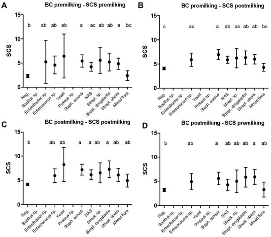
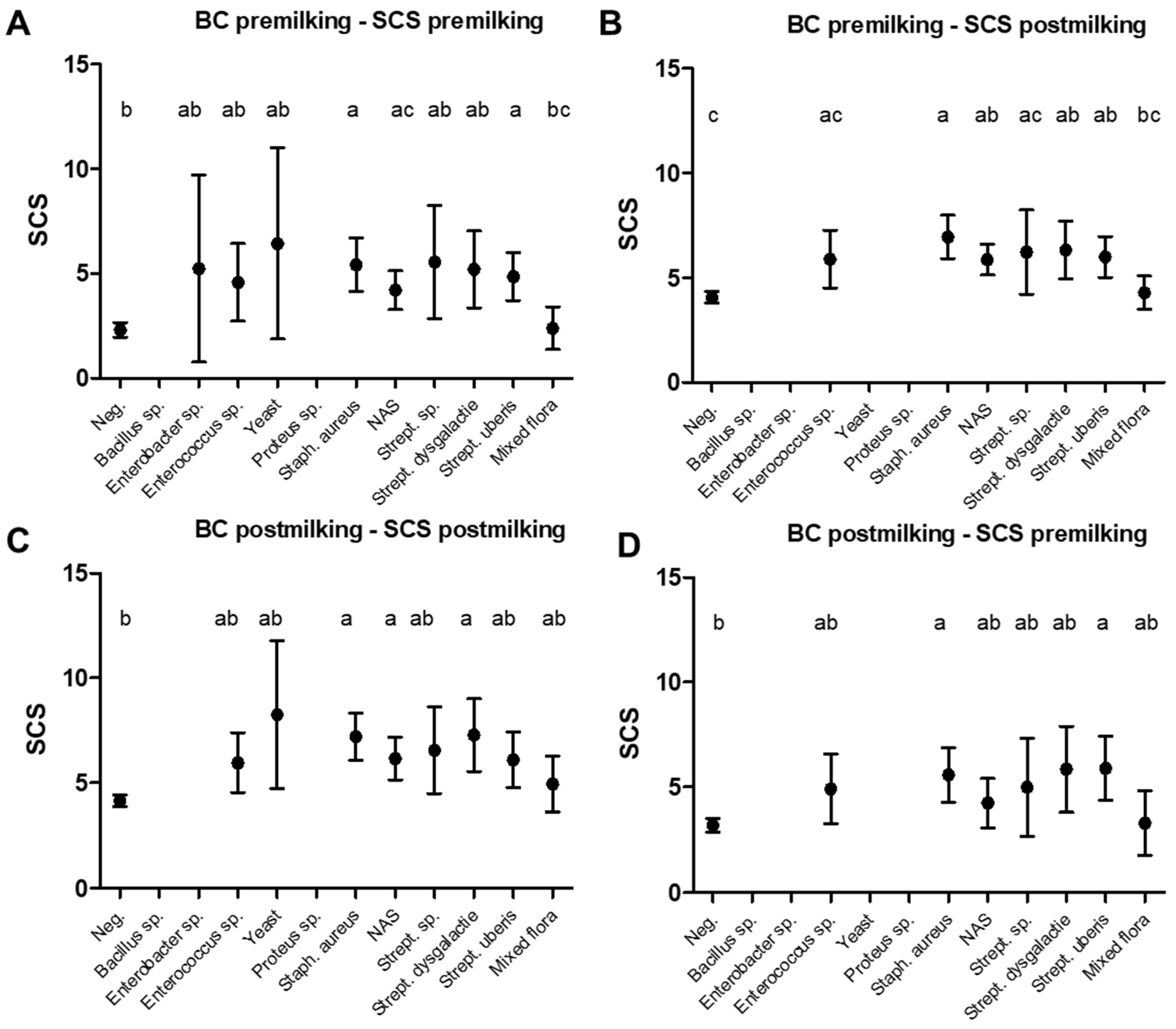
The SCS was considered a quantitative trait with a normal distribution of random effects. The investigation model included the fixed effects of premilking and postmilking samples, lactation class, and DIMs as fixed covariates. The effect of repeated measures in the same cow in premilking and postmilking samples was modeled by random effects for animal and udder quarter. A two-trait model was formulated to estimate the correlation between premilking and postmilking sample results. Thus, the analysis was based on a linear mixed model. This linear mixed model approach, as a single-trait model, was also used to analyze the relationship between BC results of premilking and postmilking samples with the SCSs of the same samples. To keep the model complexity low, the possible combinations of the relationship between bacteriologic culture and SCS in premilking and postmilking samples (BC bin pre:SCS pre; BC bin pre:SCS post; BC bin post:SCS post; BC bin post:SCS pre; Figure 1) were analyzed separately.
Figure 1.
The association between somatic cell scores and bacteriologic culture results (least square mean (LSM) with 95% CI) (n = 789). (A): Association between bacteriologic culture and somatic cell score in premilking samples; (B): association between bacteriologic culture in premilking samples and somatic cell scores in postmilking samples; (C): association between bacteriologic culture and somatic cell scores in postmilking samples; (D): association between bacteriologic culture in postmilking samples and somatic cell scores in premilking samples; BC, bacteriologic culture; within a–c different superscript letters indicate significant differences (p ≤ 0.05).
2.5. Analysis of Bacteriologic Milk Culture Bacteriologic Analysis
The BC results were available in 1 of 12 categories per udder quarter for pre- and postmilking samples. When selecting the statistical method, it is important to note that BC is a nominal characteristic (findings vs. no findings) on an unordered scale of the 12 categories. For comparison of premilking and postmilking samples for the estimated probabilities of a finding within the categories a generalized linear mixed model was used, assuming a Bernoulli distribution. The model considered repeated premilking and postmilking samples within cow and udder quarter.
Because the number of observations per level was sometimes small, only simple models were applicable. For more complex analyses, the observations were converted to 0 (no findings = negative culture) and 1 (findings) regardless of the level. The resulting (0, 1) trait (BC bin) could then be evaluated using a generalized linear mixed model, assuming a Bernoulli distribution. The model for the latent variable contains the same fixed and random effects as described for SCS.
2.6. Relationship of the Traits
The correlation between SCS and bacteriologic culture was estimated by converting SCS into a discrete scale, forming classes (SCS dis) and using BC bin. The correlation between the two discrete variables was then estimated using the logit model for each udder quarter. The results reported were the averages of these estimates across the udder quarters.
3. Results
3.1. Somatic Cell Score
The timing of milk sampling and DIMs affected the SCS (p < 0.05), but lactation number did not (p = 0.39). Quarters with higher DIMs had larger SCSs; this effect was more pronounced in postmilking than premilking samples. The somatic cell score was significantly higher in postmilking samples than in premilking samples (SCS difference −1.68; p = 0.001). In premilking samples, the SCC was 92,986 ± 1087 cells/mL (median ± SE) cells/mL (n = 415), corresponding to an SCS of 2.89 ± 0.169 cells/mL (LSMean ± SE). In postmilking samples, the SCC was 299,464 ± 2613 (median ± SE) cells/mL (n = 374), corresponding to an SCS of 4.58 ± 0.14 cells/mL (LSMean ± SE). The correlation coefficient of the cow effect between the SCS of premilking and postmilking samples was 0.763 (confidence interval 0.601 to 0.895).
3.2. Bacteriologic Analysis
Bacteriologic culture was positive in 182 out of 866 samples. A total of 113 of 433 premilking samples and 69 of 433 postmilking samples were positive; this difference was significant (p < 0.05). Consequently 70 cows had a positive BC result out of the premilking samples and 47 cows did out of the postmilking samples. The correlation coefficient of the cow effect between the BC of premilking and postmilking samples was 0.870 (confidence interval 0.845 to 0.881). Most cows with a positive BC were positive in one quarter (61% of positive cows in premilking samples and 74% of positive cows in postmilking samples). Three or more quarters were affected in 14% of positive cows in the premilking sample and 14.8% of positive cows in the postmilking sample. The bacteria culture differed between premilking and postmilking samples in six quarters of six cows.
In quarters with a high cell count (>100,000 cells/mL) BC was positive in 39% of the samples and in low-cell-count samples (≤100,000 cells/mL) 12.1% had a positive BC. The higher bacterial detection rate in premilking samples than in postmilking samples was evident independent of cell count class (≤/>100,000 cells/mL) (Table 1). NAS, Strep. uberis and mixed flora were more often present in premilking samples than in postmilking samples. The same bacteria were usually recovered from premilking and postmilking samples in mammary quarters that were positive (n = 44 quarters). In 21 quarters bacteria were detected in premilking samples but not in postmilking samples. This occurred mainly when NAS or Strep. uberis was cultured (Table 2).
Table 1.
Clinical infection status of the quarters according to BE and SCC in premilking and postmilking samples and test of differences (F-test, p-value).
Table 2.
Comparison of the numbers of positive cultures and the SCS in quarters with positive premilking or postmilking samples only and those with paired positive premilking and postmilking samples (BC pos, NAS, Strep. uberis). Data presented are quarters where complete datasets were available (BC and SCS in premilking and postmilking sample).
In addition to the timing of milk sampling, the lactation number and DIMs also significantly affected the results of BC bin (p < 0.05). Samples from cows in lactation 1 had a higher probability (0.33) of a positive bacteriologic culture than samples from cows in lactation 2 or higher (0.16). The higher the DIMs, the larger the probability of a positive bacteriologic culture.
3.3. Palpation Score
Averaged over both lactation classes, palpation score 1 was present in a relative frequency of 72.5% of the quarters (n = 609). The second highest palpation score was 2 in 10.5% of the quarters (n = 88). All other palpation scores were less common or not present (score 3—5.5%; score 4—1.2%; score 5—1.5%; score 6—1.4%; score 7—0.9%; score 9—6.4%). Palpation scores were summarized in the classes, according to clinical relevance. DIMs and lactation class had an effect on palpation. The probability of palpation class 1 (score 1, 2) from DIMs 5–300 was higher in cows with lactation number 1 than in older cows (p = 0.0035), and the probability increased with DIMs. In palpation classes 2 and 3, probabilities decreased with DIMs. There was a nonsignificant difference between lactation classes 1 and 2 in the probability of palpation classes 2 and 3 (p = 0.1). In this finding, however, particular attention should be paid to the low frequency of palpation classes 2 and 3 for lactation number 1.
3.4. Relationship Between Traits
Samples with a positive bacteriologic culture had higher SCS estimates than negative samples, regardless of sampling time, but the difference was not consistently significant (Figure 1). Moderate positive correlations ranging from 0.44 to 0.58 were found in all possible combinations of the SCS and BC (BC bin) in premilking and postmilking samples.
Mammary quarters with positive premilking and negative postmilking cultures had a significantly lower SCS than quarters with positive premilking and postmilking cultures (Table 2). The number of positive Strep. uberis and NAS cultures allowed us to test the relationship with the SCS. Premilking samples positive for Strep. uberis accompanied by postmilking samples negative for Strep. uberis had a significantly lower SCS than quarters with positive premilking and postmilking samples. This relationship did not apply to NAS; the SCS was not lower in an NAS-positive premilking culture accompanied by a negative postmilking culture compared with cows that were NAS-positive before and after milking.
Quarters of palpation class 3 had a significantly higher SCS (3.70 ± 0.48) in premilking samples than quarters of palpation class 1 (2.49 ± 0.17), but were not significantly different from palpation class 2 quarters (3.17 ± 0.48). There was a similar effect of palpation on the SCS in postmilking samples. With an increasing palpation class, the probability of a positive BC increased. This was most evident in lactation class 2 in postmilking samples, where the probability of a positive BC in the premilking sample was 0.11 in palpation class 1, 0.15 in class 2 and 0.24 in class 3.
4. Discussion
We analyzed the effect of the timing of milk sampling (premilking vs. postmilking) on the results of bacteriologic analysis and SCC on a farm with udder health problems. The relationship of the palpation score to the SCS and BC was also determined.
In our study, SCC was lower in premilking samples than in corresponding postmilking samples, and it increased with increasing DIMs. It is well known that the SCC rises gradually during lactation [19], but the effect of sample collection relative to the milking process is less well defined. It has been reported that SCC increases during milking [6,20]. Possible reasons include the higher cell count in alveolar milk because of its proximity to blood vessels [21] and epithelial cell exfoliation during milking [22]. Interestingly, the bacterial numbers of the milk fraction during the same process tend to point in the opposite direction.
Premilking samples yielded more positive bacterial cultures than postmilking samples, particularly Strep. uberis, NAS, and mixed flora. The latter cultures were more often restricted to premilking than to postmilking samples. Older experimental studies, as well as current milk hygiene doctrines, suggest that the first milk fractions can have a higher bacterial load than the main fraction or postmilking samples [11]. A more recent study showed that more premilking samples yielded Staph. aureus, NAS, and Staph. spp. than postmilking samples [12]. Possible explanations include commensal microorganisms in the teat cistern and teat canal, or on the teat skin [23], and contaminants from the sampler’s hand [24].
The teat canal and teat skin are colonized by bacteria [23,24,25]. Paduch et al. found that the prevalence of bacterial colonization of the teat canal was 84.5% in healthy dairy cows. In 25% of the cases, more than two species were cultured [23]. It is generally assumed that the bacterial load in the teat cistern is higher than in the teat canal, considering that the pathogenesis of bacterial infection of the mammary gland is ascending from the teat canal to the teat cistern into the glandular cistern and parenchyma [26]. However, this has not been substantiated scientifically. The hypothesis that bacteria cultured only from foremilk samples originate from the teat canal or teat skin is supported by studies that showed a reduced number and a smaller bacteriologic variety in samples collected via teat cannula or centesis, i.e., not via the teat canal, compared with conventionally stripped milk samples [27,28,29]. Furthermore, another study showed that the microbiota of the teat canal were more similar to that of conventional premilking samples than cisternal milk collected via centesis [30].
It is conceivable that a flushing effect during milking might reduce the bacterial load of the teat canal and on the teat skin in postmilking samples. This has not been proven experimentally, but desquamation of bacteria-coated keratin by the shearing forces associated with milk flow has been reported [31]. As a result, fewer bacteria remain in the teat canal or on the teat skin to be shed into a postmilking sample.
In line with these considerations, the first milk fractions may contain bacteria originating from the teat canal, teat skin, and teat cistern that are not associated with intramammary inflammation. In our study, the SCS of bacteriologically positive premilking samples accompanied by bacteriologically negative postmilking samples was significantly lower than the SCS in cases with positive premilking and postmilking samples. This suggests that microorganisms cultured from foremilk samples are not necessarily related to intramammary inflammation. However, it should be noted that these cows had SCCs higher than the 100,000 cell/mL threshold because cows with a history of high SCC were selected. An in-depth analysis was only possible for NAS and Strep. uberis because of the small sample size. The SCS did not differ between NAS-positive premilking samples accompanied by negative NAS postmilking samples and NAS-positive samples collected before and after milking. This suggests that both samples were reasonably associated with udder infection status. This is of particular interest because NAS are often characterized as commensals on the skin with little or no mammary pathogenicity [32,33]. When Strep. uberis was limited to premilking samples, the SCS was lower than when the pathogen was cultured from premilking and postmilking samples. Based on these questions, it can be hypothesized that some Strep. uberis detections are the consequence of a teat canal colonization or are a contamination of the neighboring skin. The prevalence of colonization of the teat canal with Strep. uberis was 68.7% in healthy cows [23].
However, in the present study, some quarters with positive bacteriologic cultures limited to premilking samples had an SSC > 100,000 cells/mL and were thus considered infected.
The handler is a possible source of bacterial contamination of a milk sample. De Visscher et al. detected NAS on 60% of the milkers’ skin or gloves [24]. We suspect this was a minor issue in the present study because the cleanliness of the samplers’ hands was comparable between the pre- and postmilking sampling, and the mixed flora population was lower in postmilking samples.
Veterinary practitioners and farmers routinely use palpation of the udder parenchyma and skin, in addition to macroscopic and laboratory examination, to diagnose diseases of the udder [8]. The increased SCS in class 3 palpation in our study reflects active inflammation because class 3 included score 6 (udder parenchyma acutely swollen). Udder palpation has high interobserver variability [34]; focusing on variables such as udder firmness or elasticity with dynamometry or ultrasonography may expand and improve clinical examination of the udder [35,36,37].
In summary, more positive bacterial cultures were obtained from premilking samples than from postmilking samples in dairy cows with subclinical mastitis. Cultures of mixed flora were more common in premilking than in postmilking samples. Positive premilking bacterial cultures were less strongly associated with a high SCC than positive postmilking bacterial cultures.
Limitation
The focus of this study was the timing of milk sampling in cows with subclinical mastitis. Further studies are needed to determine whether the findings also apply to cows with clinical mastitis. All cows originated from the same farm, and the number of samples was limited. Udder pathogens are often farm-specific, and the range of isolated microorganisms was relatively small. The premilking sampling protocol should be repeated with a larger number of cows from different farms. Furthermore, there was a lack of a true diagnostic gold standard in our study protocol, and therefore the analysis of protocol sensitivity and specificity and the use of agreement statistics could not be done.
5. Conclusions
Our findings suggest that premilking and postmilking samples differ in bacteriologic and cytologic composition. Therefore, the sampling protocol must be taken into account when decisions about mastitis treatment and prevention strategies are based on bacteriologic and cytologic results. In future studies, the meaning of the increased bacterial count in premilking samples should be further evaluated.
Author Contributions
Conceptualization, A.S.; methodology, A.S.; formal analysis, L.B.-S. and J.S.; investigation, R.D.O., A.K., A.S. and A.G.; resources, A.S.; data curation, T.K.; statistics, J.S.; writing—original draft preparation, L.B.-S.; writing—review and editing, L.B.-S., A.S. and J.S.; visualization, L.B.-S.; supervision, A.S. and J.S.; project administration, A.S.; validation, A.W. and S.D. All authors have read and agreed to the published version of the manuscript.
Funding
This research received no external funding.
Institutional Review Board Statement
These samples were taken in preparation for an animal experiment in accordance with the German Animal Welfare Act and the Saxony Veterinary and Food Safety Office Landesdirektion Sachsen (permit number TVV 48/14) on the farm. Prior to commencing an experiment, we routinely do a systematic herd health analysis. During this process milk samples were collected.
Informed Consent Statement
The animals were privately owned and verbal informed consent was obtained from animal owner. The rationale for utilizing verbal consent is that data collection was performed during routine diagnostic visit and no extra procedures were undertaken.
Data Availability Statement
Datasets are available on request from the authors.
Acknowledgments
We thank the farm for providing samples. This research was supported by the Open Access Publishing Fund of Leipzig University.
Conflicts of Interest
Author A.K. is currently employed by the company “VM Striegistal”. The research was conducted under employment of the university. The remaining authors declare that the research was conducted in the absence of any commercial or financial relationships that could be construed as a potential conflict of interest.
Abbreviations
The following abbreviations are used in this manuscript:
| SCC | somatic cell count |
| BC | bacteriologic milk culture |
| Staph. | Staphylococcus |
| Strep. | Streptococcus |
| AMS | automatic milking systems |
| DIM | days in milk |
| NAS | non-aureus Staph. |
| SCS | somatic cell score |
References
- Fogsgaard, K.K.; Bennedsgaard, T.W.; Herskin, M.S. Behavioral changes in freestall-housed dairy cows with naturally occurring clinical mastitis. J. Dairy Sci. 2015, 98, 1730–1738. [Google Scholar] [CrossRef] [Scilit] [PubMed]
- Petersson-Wolfe, C.S.; Leslie, K.E.; Swartz, T.H. An Update on the Effect of Clinical Mastitis on the Welfare of Dairy Cows and Potential Therapies. Vet. Clin. Food Anim. Pract. 2018, 34, 525–535. [Google Scholar] [CrossRef] [Scilit] [PubMed]
- Seegers, H.; Fourichon, C.; Beaudeau, F. Production effects related to mastitis and mastitis economics in dairy cattle herds. Vet. Res. 2003, 34, 475–491. [Google Scholar] [CrossRef] [Scilit] [PubMed]
- Ruegg, P.L. A 100-Year Review: Mastitis detection, management, and prevention. J. Dairy Sci. 2017, 100, 10381–10397. [Google Scholar] [CrossRef] [Scilit] [PubMed]
- European Medicine Agency. Scientific Advice Under Article 107(6) of Regulation (EU) 2019/6 for the Establishment of a List of Antimicrobials Which Shall not be Used in Accordance with Articles 112, 113 and 114 of the Same Regulation or Which Shall only be Used in Accordance with These Articles Subject to Certain Conditions. Available online: https://www.ema.europa.eu/system/files/documents/regulatory-procedural-guideline/vet_reg_cascade_list_report_en.pdf?utm_source=chatgpt.com (accessed on 21 May 2026).
- Vangroenweghe, F.; Dosogne, H.; Burvenich, C. Composition and milk cell characteristics in quarter milk fractions of dairy cows with low cell count. Vet. J. 2002, 164, 254–260. [Google Scholar] [CrossRef] [Scilit] [PubMed]
- Woolford, M.W.; Williamson, J.H.; Henderson, H.V. Changes in electrical conductivity and somatic cell count between milk fractions from quarters subclinically infected with particular mastitis pathogens. J. Dairy Res. 1998, 65, 187–198. [Google Scholar] [CrossRef] [Scilit] [PubMed]
- Dirksen, G.; Rosenberger, G.; Gründer, H.-D.; Stöber, M. (Eds.) Die Klinische Untersuchung des Rindes, 4th ed.; Unveränderter Nachdr. der 3. Aufl.; Enke: Stuttgart, Germany, 2012. [Google Scholar]
- National Mastitis Council. Microbiological Procedures for the Diagnosis of Bovine Udder Infection and Determination of Milk Quality; NMC: Singapore, 2004. [Google Scholar]
- Torres, A.H.; Rajala-Schultz, P.J.; DeGraves, F.J. Diagnosis of intramammary infections at dry-off based on sampling strategy, epidemiology of pathogens, and agreement beyond chance. J. Vet. Diagn. Investig. 2009, 21, 427–436. [Google Scholar] [CrossRef] [Scilit] [PubMed]
- Faber, J. A Study of the Bacterial Content of the Fore Milk of Cows. J. Dairy Sci. 1930, 13, 449–452. [Google Scholar] [CrossRef] [Scilit]
- Sears, P.M.; Wilson, D.J.; Gonzalez, R.N.; Hancock, D.D. Microbiological results from milk samples obtained premilking and postmilking for the diagnosis of bovine intramammary infections. J. Dairy Sci. 1991, 74, 4183–4188. [Google Scholar] [CrossRef] [Scilit] [PubMed]
- Sánchez, A.; Contreras, A.; Corrales, J.C.; Muñoz, P. Influence of sampling time on bacteriological diagnosis of goat intramammary infection. Vet. Microbiol. 2004, 98, 329–332. [Google Scholar] [CrossRef] [Scilit] [PubMed]
- Godden, S.M.; Jansen, J.T.; Leslie, K.E.; Smart, N.L.; Kelton, D.F. The effect of sampling time and sample handling on the detection of Staphylococcus aureus in milk from quarters with subclinical mastitis. Can. Vet. J. 2002, 43, 38–42. [Google Scholar] [PubMed]
- Elbers, A.R.; Miltenburg, J.D.; Lange, D.d.; Crauwels, A.P.; Barkema, H.W.; Schukken, Y.H. Risk factors for clinical mastitis in a random sample of dairy herds from the southern part of The Netherlands. J. Dairy Sci. 1998, 81, 420–426. [Google Scholar] [CrossRef] [Scilit] [PubMed]
- Fehlings, K.; Zschöck, M.; Baumgärtner, B.; Geringer, M.; Hamann, J.; Knappstein, K. Leitlinien zur Entnahme von Milchproben unter Antiseptischen Bedingungen und Leitlinien zur Isolierung und Identifizierung von Mastitiserregern; Dt. Veterinärmed. Ges. Sachverständigenausschuss Subklinische Mastitis: Gießen, Germany, 2009. [Google Scholar]
- Reents, R.; Dekkers, J.C.; Schaeffer, L.R. Genetic evaluation for somatic cell score with a test day model for multiple lactations. J. Dairy Sci. 1995, 78, 2858–2870. [Google Scholar] [CrossRef] [Scilit] [PubMed]
- Norman, H.D.; Miller, R.H.; Wright, J.R.; Wiggans, G.R. Herd and state means for somatic cell count from dairy herd improvement. J. Dairy Sci. 2000, 83, 2782–2788. [Google Scholar] [CrossRef] [Scilit] [PubMed]
- Laevens, H.; Deluyker, H.; Schukken, Y.H.; Meulemeester, L.d.; Vandermeersch, R.; de Muêlenaere, E.; de Kruif, A. Influence of parity and stage of lactation on the somatic cell count in bacteriologically negative dairy cows. J. Dairy Sci. 1997, 80, 3219–3226. [Google Scholar] [CrossRef] [Scilit] [PubMed]
- Paape, M.J.; Tucker, H.A. Somatic cell content variation in fraction-collected milk. J. Dairy Sci. 1966, 49, 265–267. [Google Scholar] [CrossRef] [Scilit] [PubMed]
- Khatun, M.; Bruckmaier, R.M.; Thomson, P.C.; House, J.; García, S.C. Suitability of somatic cell count, electrical conductivity, and lactate dehydrogenase activity in foremilk before versus after alveolar milk ejection for mastitis detection. J. Dairy Sci. 2019, 102, 9200–9212. [Google Scholar] [CrossRef] [Scilit] [PubMed]
- Herve, L.; Quesnel, H.; Lollivier, V.; Portanguen, J.; Bruckmaier, R.M.; Boutinaud, M. Mammary epithelium disruption and mammary epithelial cell exfoliation during milking in dairy cows. J. Dairy Sci. 2017, 100, 9824–9834. [Google Scholar] [CrossRef] [Scilit] [PubMed]
- Paduch, J.H.; Krömker, V. Colonization of the teat skin and the teat canal of lactating dairy cattle by mastitis pathogens. Tierarztl. Prax. Ausg. G Grosstiere/Nutztiere 2011, 39, 71–76. [Google Scholar] [PubMed]
- Visscher, A.d.; Supré, K.; Haesebrouck, F.; Zadoks, R.N.; Piessens, V.; van Coillie, E.; Piepers, S.; Vliegher, S.d. Further evidence for the existence of environmental and host-associated species of coagulase-negative staphylococci in dairy cattle. Vet. Microbiol. 2014, 172, 466–474. [Google Scholar] [CrossRef] [Scilit] [PubMed]
- Braem, G.; Vliegher, S.d.; Verbist, B.; Heyndrickx, M.; Leroy, F.; Vuyst, L.d. Culture-independent exploration of the teat apex microbiota of dairy cows reveals a wide bacterial species diversity. Vet. Microbiol. 2012, 157, 383–390. [Google Scholar] [CrossRef] [Scilit] [PubMed]
- Germon, P.; Foucras, G.; Smith, D.G.E.; Rainard, P. Invited review: Mastitis Escherichia coli strains—Mastitis-associated or mammo-pathogenic? J. Dairy Sci. 2025, 108, 4485–4507. [Google Scholar] [CrossRef] [Scilit] [PubMed]
- Friman, M.; Hiitiö, H.; Niemi, M.; Holopainen, J.; Pyörälä, S.; Simojoki, H. The effect of a cannula milk sampling technique on the microbiological diagnosis of bovine mastitis. Vet. J. 2017, 226, 57–61. [Google Scholar] [CrossRef] [Scilit] [PubMed]
- Bexiga, R.; Pereira, H.; Pereira, O.; Leitão, A.; Carneiro, C.; Ellis, K.A.; Vilela, C.L. Observed reduction in recovery of Corynebacterium spp. from bovine milk samples by use of a teat cannula. J. Dairy Res. 2011, 78, 9–14. [Google Scholar] [CrossRef] [Scilit] [PubMed]
- Hiitiö, H.; Simojoki, H.; Kalmus, P.; Holopainen, J.; Pyörälä, S.; Taponen, S. The effect of sampling technique on PCR-based bacteriological results of bovine milk samples. J. Dairy Sci. 2016, 99, 6532–6541. [Google Scholar] [CrossRef] [Scilit] [PubMed]
- Dahlberg, J.; Williams, J.E.; McGuire, M.A.; Peterson, H.K.; Östensson, K.; Agenäs, S.; Dicksved, J.; Waller, K.P. Microbiota of bovine milk, teat skin, and teat canal: Similarity and variation due to sampling technique and milk fraction. J. Dairy Sci. 2020, 103, 7322–7330. [Google Scholar] [CrossRef] [Scilit] [PubMed]
- Besier, J.; Lind, O.; Bruckmaier, R.M. Dynamics of teat-end vacuum during machine milking: Types, causes and impacts on teat condition and udder health–a literature review. J. Appl. Anim. Res. 2016, 44, 263–272. [Google Scholar] [CrossRef] [Scilit]
- Thorberg, B.-M.; Kühn, I.; Aarestrup, F.M.; Brändström, B.; Jonsson, P.; Danielsson-Tham, M.-L. Pheno- and genotyping of Staphylococcus epidermidis isolated from bovine milk and human skin. Vet. Microbiol. 2006, 115, 163–172. [Google Scholar] [CrossRef] [Scilit] [PubMed]
- Vanderhaeghen, W.; Piepers, S.; Leroy, F.; van Coillie, E.; Haesebrouck, F.; de Vliegher, S. Identification, typing, ecology and epidemiology of coagulase negative staphylococci associated with ruminants. Vet. J. 2015, 203, 44–51. [Google Scholar] [CrossRef] [Scilit] [PubMed]
- Fossing, C.; Vaarst, M.; Houe, H.; Enevoldsen, C. Contributions to variability of clinical measures for use as indicators of udder health status in a clinical protocol. Acta. Vet. Scand. 2006, 48, 15. [Google Scholar] [CrossRef] [Scilit] [PubMed]
- Rees, A.; Fischer-Tenhagen, C.; Heuwieser, W. Udder firmness as a possible indicator for clinical mastitis. J. Dairy Sci. 2017, 100, 2170–2183. [Google Scholar] [CrossRef] [Scilit] [PubMed]
- Tommasoni, C.; Fiore, E.; Lisuzzo, A.; Gianesella, M. Mastitis in dairy cattle: On-farm diagnostics and future perspectives. Animals 2023, 13, 2538. [Google Scholar] [CrossRef] [Scilit] [PubMed]
- Flöck, M.; Winter, P. Diagnostic ultrasonography in cattle with diseases of the mammary gland. Vet. J. 2006, 171, 314–321. [Google Scholar] [CrossRef] [Scilit] [PubMed]

Disclaimer/Publisher’s Note: The statements, opinions and data contained in all publications are solely those of the individual author(s) and contributor(s) and not of MDPI and/or the editor(s). MDPI and/or the editor(s) disclaim responsibility for any injury to people or property resulting from any ideas, methods, instructions or products referred to in the content. |
© 2026 by the authors. Licensee MDPI, Basel, Switzerland. This article is an open access article distributed under the terms and conditions of the Creative Commons Attribution (CC BY) license.