Review

The Hoof as the Sentinel of Systemic Failure: The Lameness Triad and the Susceptibility–Resilience Gate in Dairy Cows


Department of Agricultural, Food and Nutritional Science, University of Alberta, Edmonton, AB T6G 2P5, Canada
Dairy 2026, 7(4), 61; https://doi.org/10.3390/dairy7040061
Submission received: 18 May 2026 / Revised: 23 July 2026 / Accepted: 3 August 2026 / Published: 5 August 2026
(This article belongs to the Section Dairy Animal Health)

Abstract

Lameness is a major welfare and economic disorder in dairy production and a clinical sign with multiple causes. This review focuses on claw horn disruption lesions (CHDL), a major cause of periparturient lameness. Although hoof-centered prevention and treatment remain essential, preclinical systemic alterations, recurrence, and clustering with other transition-period disorders suggest that CHDL may sometimes represent the local expression of broader pathophysiology. We propose the Lameness Triad, in which three mechanisms converge: sustained endotoxemic pressure and TLR4-mediated innate immune activation, endothelial glycocalyx degradation and vascular dysfunction, and metabolic triage that reallocates resources from production toward defense. Their combined pathogenic pressure is proposed to produce clinical disease when it exceeds a cow-specific Susceptibility–Resilience Gate comprising mucosal barrier integrity, hepatic endotoxin clearance, immune calibration, redox reserve, hoof structural reserve, and prior damage. Ruminal dysbiosis, mammary involution, and postpartum uterine contamination may contribute to inflammatory and endotoxemic pressure, whereas the confined, continuously loaded digital corium may become a principal site of clinical expression. Longitudinal multi-omics studies identified inflammatory, urinary, and milk-metabolite alterations before diagnosis, with within-cohort serum and urine models discriminating cows that later became lame as early as eight weeks prepartum. These findings require prospective validation and do not establish the complete causal sequence. The framework extends, rather than replaces, established hoof-health programs and provides testable hypotheses for earlier detection, prevention, and selection for resilience.

1. Introduction: From the Hoof Outward to the System Inward

Lameness is among the most prevalent and costly disorders of dairy cattle [1,2,3,4,5]. It may result from infectious foot disease, arthritis, nerve injury, or other musculoskeletal conditions, but this review focuses on CHDL, principally sole ulcers, sole hemorrhage, and white line disease, which account for a substantial proportion of periparturient cases. Lameness reduces milk yield and reproductive performance, increases culling, and compromises welfare [6,7,8,9,10,11,12,13]. Reported prevalence in Canadian Holstein-Friesian herds ranges from approximately 15% to 21%, with sole hemorrhage documented in 23.8% of cows examined during hoof trimming in Alberta [1,14,15]. Prompt treatment improves outcomes [16], yet lameness remains prevalent despite advances in housing, trimming, and therapy [5].
The dominant hoof-centered paradigm attributes lameness to local convergence of mechanical, environmental, nutritional, and infectious factors [17,18,19,20,21,22,23,24,25,26]. It is strongly supported by the relationships among visible lesions, pain, gait impairment, concrete flooring, prolonged standing, inadequate lying time, rapidly fermentable diets, and infectious agents. Accordingly, improved housing, preventive and therapeutic trimming, biosecurity, and lesion-specific treatment remain indispensable.
Several observations nevertheless suggest that local factors do not fully explain individual risk. Susceptibility varies among cows under broadly comparable management [14]; lameness clusters with mastitis, metritis, retained placenta, and ketosis [27,28,29]; and longitudinal studies have detected systemic metabolic and immunological alterations before diagnosis [30,31,32,33,34,35]. Within-cohort serum and urine models distinguished cows that later became lame as early as eight weeks prepartum [31], although their high accuracies require independent validation. Genetic evidence likewise supports host heterogeneity: heritability estimates range from 0.01 to 0.39 [36], candidate loci involve immune regulation, keratinization, and tissue remodeling [37,38,39], and susceptibility and recovery appear to have partly distinct genetic architectures [40].
This review therefore considers CHDL as potential local manifestations of systemic immune, vascular, and metabolic dysregulation interacting with biomechanical stress. The digital corium may be especially vulnerable because it is confined within a rigid keratin capsule and continuously loaded. The hoof is thus conceptualized as a sentinel site at which systemic compromise may become clinically evident, analogous, but not mechanistically identical, to the kidney in hypertension or the retina in diabetes.
The proposed architecture has two coupled components: the Lameness Triad—Element I (sustained endotoxemic pressure), Element II (endothelial glycocalyx degradation and vascular dysfunction), and Element III (metabolic triage)—which generates pathogenic pressure, and the Susceptibility–Resilience Gate, which influences its clinical expression. The Gate comprises mucosal barrier integrity, hepatic endotoxin clearance, immune-response calibration, redox homeostasis, hoof structural reserve, and prior tissue damage. Their conceptual relationship is:
(Element I × Element II × Element III) × Susceptibility > Resilience
This is a testable conceptual formulation, not a validated quantitative equation. It links systemic pathogenic pressure with host capacity and may explain heterogeneous susceptibility and recurrence.
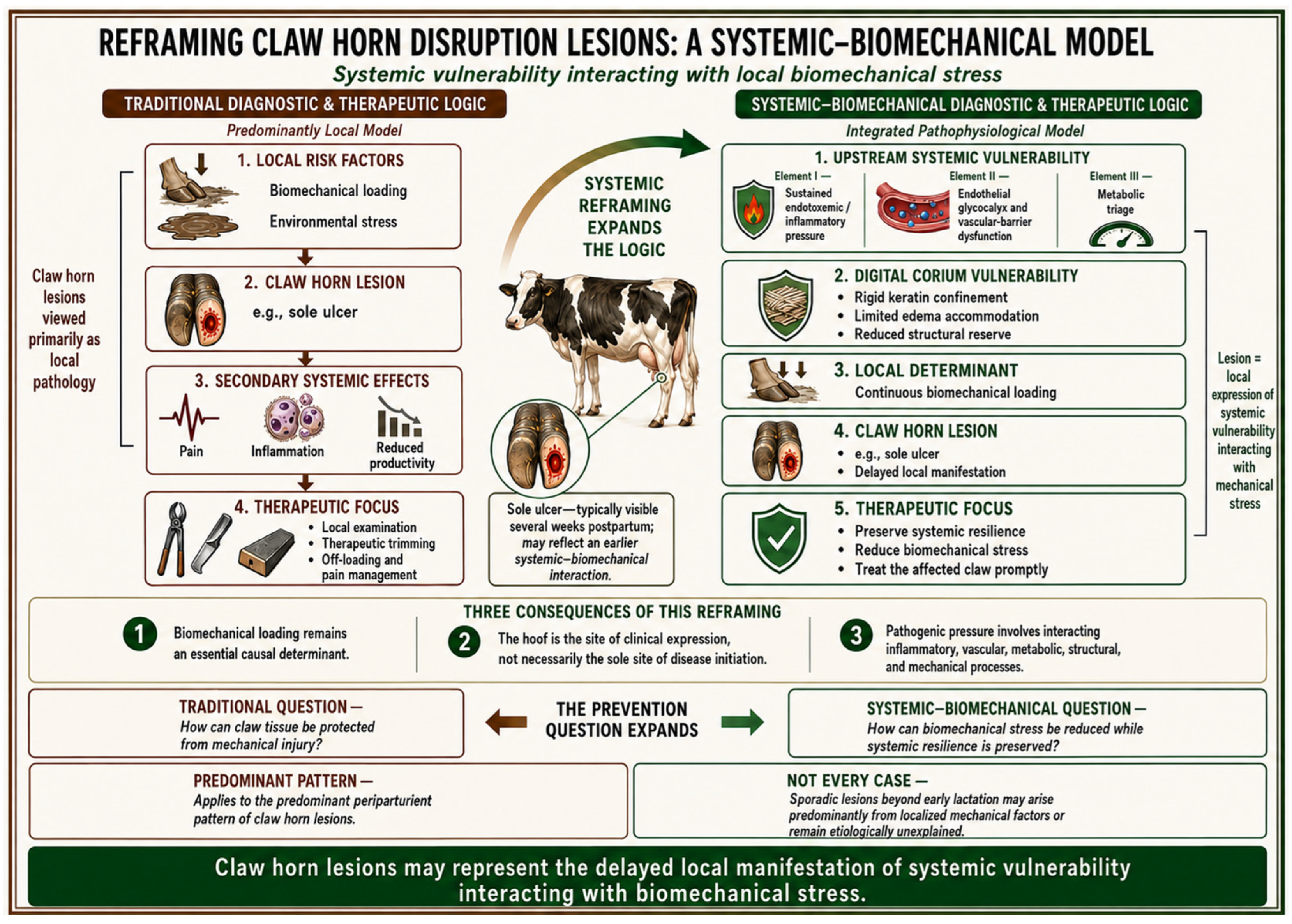
Upstream ruminal–intestinal, mammary, and uterine inputs may contribute to overlapping inflammatory pressure during the periparturient period. The Triad integrates this pressure with endothelial–vascular dysfunction and metabolic triage; clinical expression then depends on the corium’s constrained anatomy, continuous loading, and the cow’s Gate state. The following sections evaluate the longitudinal and multi-omics evidence for this architecture and its implications for diagnosis and prevention. Figure 1 summarizes the proposed extension of the traditional local model.

2. The Hoof as Sentinel Organ of Failure, Not Origin of Disease

The hoof-centered model is supported by visible lesions, plausible mechanical injury, and established environmental risks [21,22,23,24]. Its sequence, local insult, tissue injury, inflammation, pain, and altered gait, provides effective targets for prevention and treatment.
However, marked between-cow heterogeneity persists under broadly comparable conditions. Jewell et al. [14] found behavioral evidence of lameness in approximately 25% of cows in Canadian tiestall herds, while approximately 75% remained unaffected. Exposure is never identical within a facility, but the distribution indicates that structural integrity, inflammatory responsiveness, metabolic capacity, previous damage, and other host characteristics modify clinical expression.
Systemic changes may also precede diagnosis. In longitudinal studies, altered lactate, IL-6, haptoglobin, and serum amyloid A occurred before clinical lameness, while multivariate models separated pre-lame and unaffected cows from −8 weeks prepartum onward [30,31]. These high internal accuracies require external validation, but the early separation is compatible with a preclinical state that increases vulnerability when transition-period stress converges with postpartum loading.
Lameness is also associated with other transition-period disorders [27,28,29]. In cows later diagnosed with lameness, urinary SDMA was elevated at −8 weeks prepartum and at diagnosis, and five acylcarnitines remained elevated across sampled time points [33,41,42]. These findings do not prove vascular causation, but their timing is difficult to attribute solely to an already visible claw lesion.
The digital corium provides an anatomical setting in which systemic vulnerability and local forces may interact. Its rigid capsule limits expansion during inflammation; the solar corium can be compressed between the distal phalanx and horn capsule; and weight bearing continues during systemic stress [43,44]. These features may impair perfusion and horn production without making the claw a passive recipient of systemic injury.
Around calving, weakening of the suspensory apparatus may further increase vulnerability [45]. Concurrent endothelial, inflammatory, and mitochondrial disturbances, reflected provisionally by SDMA, acylcarnitines, and glycerophospholipid changes [33], could reduce the corium’s capacity to tolerate loading, thereby impairing perfusion, repair, and keratinization.
This interpretation extends rather than invalidates local diagnosis and treatment (Figure 1). CHDL still require prompt examination, therapeutic trimming, off-loading, and analgesia. The proposal concerns biological origin: in many periparturient cases, systemic vulnerability may magnify mechanical injury. Infectious foot diseases such as digital dermatitis and foot rot remain etiologically distinct.
The systemic reframing applies to the predominant periparturient CHDL pattern, not every case; some sporadic lesions may arise primarily from local mechanical factors or individual susceptibility.

3. Preclinical Inflammatory Activation

If the hoof represents a site at which systemic vulnerability becomes clinically expressed, when does that vulnerability begin? Among the earliest detectable changes preceding lameness are activation of innate immune pathways, systemic inflammation, acute-phase protein release, and metabolic and vascular disturbances occurring before claw lesions become clinically visible. These findings suggest that immunological priming may precede and interact with mechanical loading rather than arise solely as a consequence of an established hoof lesion.
Our longitudinal study of transition dairy cows followed from −8 weeks prepartum to +8 weeks postpartum identified systemic inflammatory alterations several weeks before clinical lameness was diagnosed [30]. All lame cows were diagnosed between +1 and +3 weeks postpartum by locomotion scoring [24] and received a score of 5, indicating severe lameness characterized by pronounced back arching, reluctance to move, and marked unloading of the affected limb. At −8 weeks prepartum, approximately two months before diagnosis, serum lactate concentrations were significantly higher in cows that subsequently developed lameness than in unaffected controls (5428 vs. 2455 µmol/L, p = 0.04) [30]. Because this difference occurred during the dry period, before the intensive concentrate feeding characteristic of early lactation, it cannot readily be attributed to postpartum dietary carbohydrate fermentation alone. Nevertheless, lactate is a nonspecific metabolite, and its elevation should be interpreted as evidence of altered systemic metabolism potentially associated with inflammatory or endotoxemic activity rather than as direct proof of inflammation.
Ametaj et al. [46] demonstrated strong correlations between plasma lactate and ruminal endotoxin concentrations in cows fed high-grain diets, supporting an association between lactate accumulation and endotoxemic states. Elevated lactate before intensive postpartum grain feeding therefore raises the possibility of an endotoxin source outside the rumen or of altered lactate production and clearance during prepartum inflammatory activation. One potential source is subclinical Gram-negative intramammary infection during the dry period, which could expose the host to bacterial products, including lipopolysaccharide. Consistent with this possibility, milk somatic cell counts were strongly correlated with serum lactate across the measured time points (r = 0.82–0.92, p < 0.01) [30]. This association does not establish directionality, but it supports further examination of a potential relationship between mammary immune activity and systemic metabolic disturbance.
By −4 weeks prepartum, the inflammatory signature had intensified. Serum IL-6 was approximately fivefold higher in cows that subsequently developed lameness than in controls (251 vs. 48 pg/mL, p = 0.03), while haptoglobin was moderately increased (0.20 vs. 0.15 mg/mL, p = 0.05) and serum amyloid A was nearly threefold higher (9732 vs. 3461 µg/mL, p = 0.03) [30]. Ishikawa et al. [47] showed that elevated prepartum serum IL-6 was associated with postpartum reproductive disease, supporting its potential value as a marker of periparturient inflammatory risk. Hagiwara et al. [48] reported pronounced increases in IL-6 in serum and whey during mastitis, consistent with cytokine production by immune cells responding to mammary infection. Pyörälä [49] further established that subclinical mastitis can elicit measurable inflammatory responses even in the absence of overt clinical signs. Collectively, these findings support the biological plausibility of clinically inapparent mammary inflammation contributing to systemic immune activation, although they do not demonstrate that the mammary gland was the source of IL-6 in the pre-lame cows.
Tumor necrosis factor-α showed an overall tendency to be elevated in cows that subsequently developed lameness (p = 0.09), with significant differences at −4 weeks prepartum and at diagnosis [30]. Kushibiki [50] described the central role of TNF-α in coordinating systemic inflammatory responses and hepatic acute-phase protein synthesis in cattle. Petersen et al. [51] similarly identified haptoglobin and serum amyloid A as among the most responsive indicators of inflammation in this species. At lameness diagnosis, haptoglobin was threefold higher in affected cows than in controls (0.36 vs. 0.12 mg/mL, p = 0.05), and serum amyloid A was also nearly threefold higher (29,300 vs. 10,401 µg/mL, p = 0.05) [30]. The magnitude and temporal development of these responses are consistent with a substantial systemic inflammatory challenge. Accordingly, what appears clinically as an acute episode of lameness may represent the delayed manifestation of biological processes that began during the prepartum period.
The IL-1 findings reveal a potentially important distinction between circulating and tissue-local inflammatory responses. Whereas circulating IL-6, TNF-α, haptoglobin, and serum amyloid A were elevated in cows that subsequently developed lameness, circulating IL-1 was lower at −4 weeks prepartum (p = 0.05) and showed a tendency to be lower at −8 weeks (p = 0.07) [30]. Fontaine et al. [52] reported increased IL-1β mRNA expression in perivascular cells of laminar tissue in horses with experimentally induced laminitis, demonstrating that local tissue expression of IL-1β can increase even when circulating concentrations do not show a parallel response. Although findings from equine laminitis cannot be directly extrapolated to bovine CHDL, they illustrate that systemic and tissue-local cytokine profiles may differ. The lower circulating IL-1 observed in pre-lame cows therefore does not exclude local IL-1 activity within the digital corium. Rather, it supports the need to distinguish circulating inflammatory patterns from inflammatory signaling within the affected tissue.
Serum phosphoric acid (PA) was elevated throughout the observation period: 3.9-fold at −8 weeks, 3.1-fold at −4 weeks, 6.4-fold during the week of lameness diagnosis, and 11.4-fold at +6 weeks after diagnosis [32]. Evidence from human and experimental models links phosphate excess with inflammatory signaling and TNF-α-associated responses [53,54,55]; however, these findings provide biological context rather than direct evidence of causation in dairy cows. Elevated PA may therefore reflect, or potentially amplify, a persistent inflammatory–metabolic disturbance rather than represent an established initiating cause of lameness. Its progressive elevation through +6 weeks indicates that the associated systemic disturbance persisted beyond clinical diagnosis, although the mechanistic significance of PA remains to be determined. Accordingly, PA should be interpreted as contextual evidence of persistent systemic disturbance rather than as a validated mechanistic biomarker.
Milk somatic cell counts were strongly and positively correlated with serum lactate (r = 0.82–0.92), IL-6 (r = 0.73–0.79), TNF-α (r = 0.80–0.84), and serum amyloid A (r = 0.70–0.86) across the measured time points (all p < 0.01) [30]. These associations connect mammary inflammatory activity with systemic inflammatory and metabolic alterations, but they cannot alone establish that mammary inflammation caused the systemic response. Todhunter et al. [56] showed that new Gram-negative intramammary infections increase during the dry period, with bacterial growth rates reported to be three- to fourfold higher than during lactation. Bradley and Green [57] likewise identified the nonlactating period as a major risk window for new intramammary infections and a critical stage in mastitis epidemiology. Refaai et al. [58] reported an epidemiological association between subclinical intramammary infection and claw lesions in dairy cows, providing further support for a possible mammary–claw connection.
On the basis of these converging observations, it is proposed that subclinical mammary inflammation during the dry period may function as one source of inflammatory priming in cows that subsequently develop CHDL. In susceptible animals, this prepartum immune activation could reduce systemic and tissue resilience, thereby increasing vulnerability when additional metabolic, inflammatory, and biomechanical pressures emerge during early lactation. This mammary–lameness axis remains a mechanistic hypothesis requiring direct testing through longitudinal mammary microbiology, circulating endotoxin measurements, tissue-specific inflammatory markers, and prospective assessment of claw-lesion development.

4. Multi-Matrix Metabolomic Profiling: Evidence Relevant to the Proposed Triadic Architecture

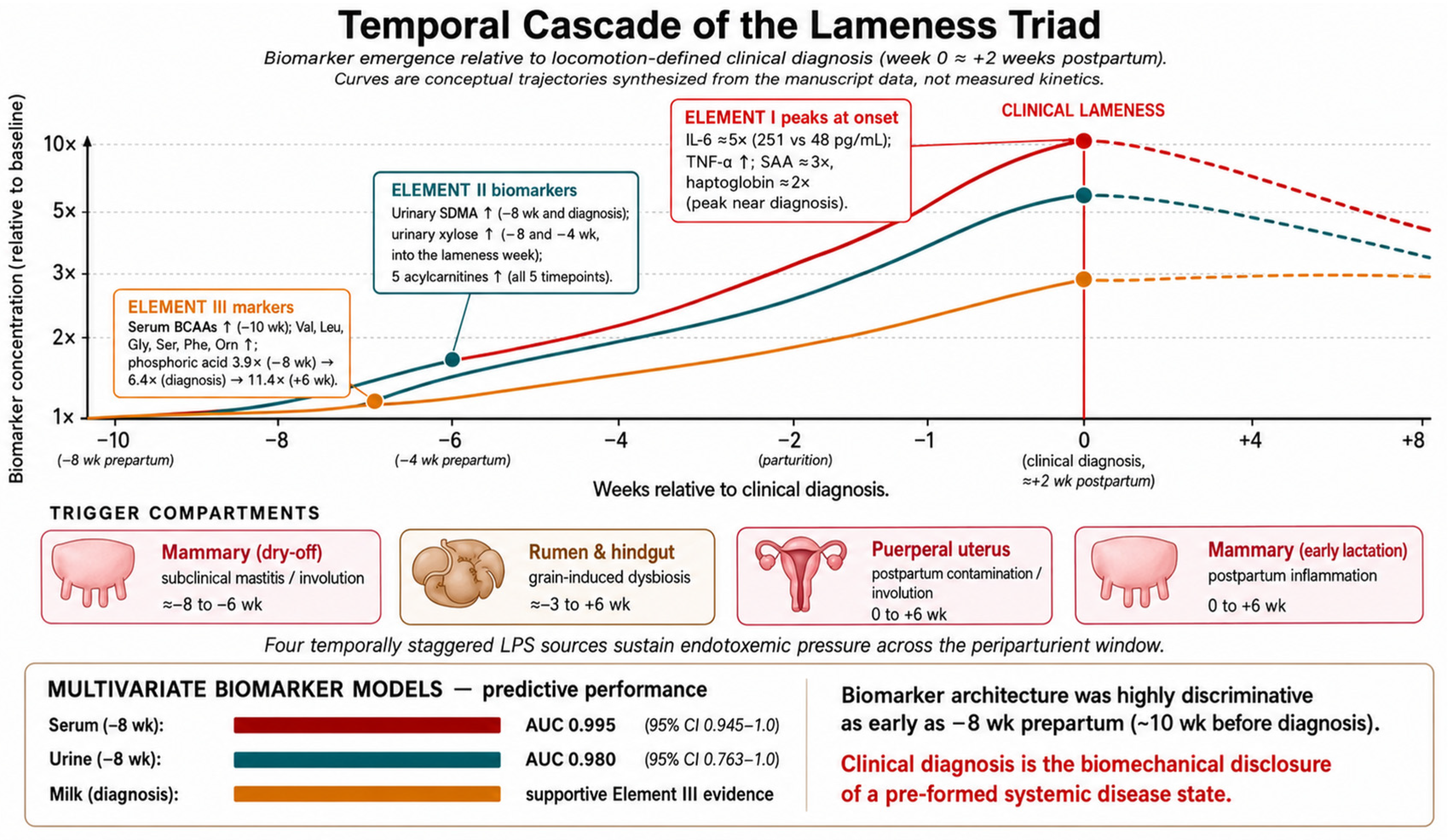
Longitudinal metabolomic profiling of serum, urine, and milk identified systemic changes before clinical lameness [34,59,60]. To organize these findings, they are considered provisionally in relation to the three components introduced in Section 1 and developed mechanistically in Section 6: serum findings primarily with Elements I and III; urinary SDMA, xylose, and acylcarnitines with Element II; and coordinated milk-metabolite suppression with Element III. Figure 2 integrates their temporal patterns. These assignments are mechanistic hypotheses rather than validated biomarker classifications.

4.1. Serum Metabolomics: Mobilization and Immune Reprogramming

Serum metabolomic analysis using targeted approaches (Biocrates AbsoluteIDQ p180 platform—BIOCRATES Life Sciences AG, Innsbruck, Austria) identified 5-metabolite combinations that differentiated healthy from pre-lame cows with high within-cohort accuracy [31,32]. At −8 weeks prepartum, a panel comprising lysine, lysophosphatidylcholine a C28:0, isoleucine, lysophosphatidylcholine a C17:0, and glutamate achieved area under the curve (AUC) of 0.995 (95% CI: 0.945–1.0). At −4 weeks, a different 5-metabolite panel (lysine, arginine, isoleucine, leucine, glutamate) achieved AUC 0.992 (95% CI: 0.938–1.0) [31]. These within-cohort classification accuracies were higher than those reported by other investigators. Randall et al. [59] achieved 82% accuracy in current lameness detection and 71–75% accuracy when models trained on lactating cow data were applied to pre-calving samples. Barden et al. [34] used nuclear magnetic resonance metabolomics to discriminate sole lesion development but did not report prediction at −8 weeks prepartum.
Quantitative enrichment analysis identified four metabolic pathways consistently altered throughout lameness development: lysine degradation, biotin metabolism, tryptophan metabolism, and valine-leucine-isoleucine degradation [31]. The involvement of branched-chain amino acid catabolism is particularly significant. Elevated circulating BCAAs in pre-lame cows are consistent with altered protein mobilization and amino-acid use providing substrates for immune cell activation and proliferation, a biochemical pattern consistent with Element III, the metabolic triage that diverts amino acids from anabolic to immunological use.
An independent serum GC-MS analysis [32] reinforces this interpretation and provides complementary mechanistic clues. At −10 weeks before lameness diagnosis, pre-lame cows exhibited elevated valine (3.9-fold), glycine (3.0-fold), serine (3.4-fold), leucine (4.7-fold), phenylalanine (3.5-fold), and ornithine (4.3-fold), with continued elevation at −6 weeks and at the week of clinical lameness. Critically, the same pre-lame cows showed elevated lactate and a tendency toward elevated β-hydroxybutyrate without compensatory increase in gluconeogenesis-related metabolites, a pattern consistent with routing of catabolized amino acids toward lactate and ketone-body production rather than glucose synthesis [32]. Leu (a ketogenic AA) generates acetyl-CoA which feeds ketone synthesis; Phe (both glucogenic and ketogenic) similarly contributes to ketone bodies. Elevated systemic concentrations of these AAs without proportionate gluconeogenic output are consistent with skeletal muscle protein mobilization and altered carbon and nitrogen use, providing a candidate biochemical signature of Element III operating during the dry period, before grain feeding could plausibly explain ketone elevation.
The identification of biotin metabolism as a consistently altered pathway provides mechanistic rationale for empirical observations. Lean et al. [25] reviewed nutritional impacts on lameness and concluded that biotin supplementation reduces heel erosion and sole lesions in some studies, though mechanisms remained unclear. Biotin functions as a cofactor for four carboxylases essential for glucose metabolism (pyruvate carboxylase), fatty acid synthesis (acetyl-CoA carboxylase), and amino acid catabolism (propionyl-CoA carboxylase, β-methylcrotonyl-CoA carboxylase). Perturbations in biotin metabolism therefore affect fundamental cellular energetics, with disproportionate consequences for tissues of high metabolic demand such as the hoof corium, placing this finding squarely at the interface of Element III (metabolic) and the manifestation layer (corium vulnerability).
Temporal analysis demonstrated progressive metabolite alterations from −8 weeks through disease onset. During the clinical disease week, 13 metabolites were consistently elevated throughout a 16-week observation period [31]. Additionally, metabolic alterations persisted at +4 and +8 weeks postpartum after clinical diagnosis, indicating that improvement in locomotion may not coincide with complete metabolic normalization. This persistence may have implications for reproductive performance, recurrence risk, and treatment evaluation, and is consistent with the genetic architecture finding that recovery and susceptibility are governed by distinct biological systems [40].

4.2. Urinary Metabolomics: Direct Signatures of Endothelial Glycocalyx Degradation

Targeted urinary metabolomics detected 153 metabolites and numerous alterations from −8 weeks prepartum to +8 weeks postpartum [33]. Complementary 1H NMR profiling in the same population identified prepartum changes that included xylose [61]. Four findings are particularly relevant to the proposed endothelial–vascular Element II.
First, urinary SDMA was elevated at −8 weeks and again at diagnosis [33]. SDMA is a renal-filtration marker associated with cardiovascular risk and endothelial dysfunction and may inhibit cellular arginine transport [41,42]. Its timing places altered renal–vascular physiology before clinical lameness, without establishing causation.
Second, five acylcarnitines remained elevated at all sampled time points [33], consistent with altered mitochondrial fatty-acid flux. Acylcarnitines have also been associated with laminitis in an equine gut-derived sepsis model [62]. Their accumulation may indicate systemic metabolic stress relevant to endothelial homeostasis, but it is not endothelial-specific.
Third, urinary phosphatidylcholines and lysophosphatidylcholines were altered across the observation period [33]. Because lysophosphatidylcholines participate in monocyte chemotaxis and macrophage activation [63], this pattern is compatible with leukocyte–endothelial signaling before diagnosis.
Fourth, urinary xylose was elevated at −8 and −4 weeks prepartum [61]. Xylose forms the proximal sugar of the Ser–Xyl–Gal–Gal–GlcA linker that anchors heparan- and chondroitin-sulfate chains to proteoglycans. Its excretion is therefore consistent with proteoglycan turnover predicted during glycocalyx degradation [64,65], although dietary absorption and tissue origin preclude treating it as direct proof.
Additional amino-acid and sphingomyelin changes support broader metabolic and immune disturbance. Multivariable urinary models achieved within-cohort AUCs from 0.980 at −8 weeks to 1.0 at +4 and +8 weeks [33]; these exceptionally high estimates require independent validation.
Urinary alterations, including numerous acylcarnitines, persisted after diagnosis [33], further indicating that clinical and biochemical recovery may not be synchronous [40].
Direct validation of Element II should quantify glycocalyx-specific products, including heparan- and chondroitin-sulfate disaccharides, hyaluronan fragments, and shed syndecans. Current evidence supports temporally early endothelial–vascular dysfunction as a plausible component while remaining insufficient to prove glycocalyx degradation [61,64,65].

4.3. Milk Metabolomics: The Quantitative Signature of Metabolic Triage

Milk metabolomic analysis using both DI/LC-MS/MS and 1H NMR platforms revealed perhaps the most striking quantitative finding in the dataset: 37 metabolites were altered in lame cows at the week of clinical signs, with 35 of these decreased in concentration [66]. The breakdown is mechanistically informative: 11 glycerophospholipids (4 lysophosphatidylcholines and 7 phosphatidylcholines), 7 sphingomyelins, 10 amino acids, 4 acylcarnitines, and 3 biogenic amines were all reduced; only sn-glycero-3-phosphocholine and phosphatidylethanolamine ae C42:1 were elevated. Milk is conventionally regarded as a product of mammary gland function, but its composition simultaneously reflects whole-body metabolic prioritization. The coordinated suppression of structural lipids, immune-related signaling lipids, and proteinogenic amino acids in the milk of lame cows is quantitatively consistent with Element III of the Lameness Triad, the coordinated organism-level resource reallocation proposed for Element III from productive to defensive functions.
Two findings within this signature are particularly diagnostic of the metabolic-triage logic. First, the most extreme depletions are tryptophan (4.18-fold reduction) and methionine (3.43-fold reduction) [66]. These specific amino acids are mechanistically central to immune regulation: tryptophan is the substrate for the indoleamine 2,3-dioxygenase (IDO) pathway and the kynurenine axis that governs immune tolerance and inflammatory regulation, while methionine is the precursor for S-adenosylmethionine, the universal methyl donor required for epigenetic regulation of immune gene expression and for glutathione synthesis (the principal cellular antioxidant). Their reduced concentrations in milk are consistent with altered availability, utilization, or mammary secretion for immune and antioxidant defense rather than non-specific protein deprivation.
Second, the lysophosphatidylcholine-to-phosphatidylcholine ratio falls dramatically: 6.17 in healthy cows versus 1.25 in lame cows [66]. This ratio is compatible with lower apparent secretory phospholipase A2 (sPLA2)-related conversion of PCs to LPCs; sPLA2 also releases arachidonic acid for prostaglandin and leukotriene synthesis [67,68]. However, the ratio does not establish an adaptive host effort or demonstrate that inflammatory resolution was enhanced. It is therefore interpreted here as evidence of altered mammary lipid metabolism that is consistent with, but does not prove, the metabolic-triage framework.
The specificity of metabolic reallocation is further suggested by the lipid pattern. The milk study showed decreases in phosphatidylcholine and sphingomyelin species in lame cows [66], while related serum and urinary studies in lameness cohorts document systemic phosphatidylcholine, lysophosphatidylcholine, and sphingolipid perturbations [31,32,33]. These data support altered lipid partitioning or utilization, but they should not be read as direct proof of active mammary-to-systemic transport without flux studies. Mechanistically, phosphatidylcholine can attenuate LPS-induced NF-κB activation and modulate macrophage activity [69,70], and ceramide can inhibit macrophage responses to LPS [71], providing plausible pathways through which these lipid classes may participate in systemic immunoregulation.
Lame cows produced 5.37 kg/day less milk during the first 56 days postpartum compared with healthy controls [66]. Warnick et al. [6] and Green et al. [7] previously interpreted decreased milk yield in lame cows as a consequence of disease-associated stress or reduced feed intake. An alternative interpretation is that decreased production may partly reflect biological reprioritization, although reduced intake and other consequences of disease remain plausible contributors. The observed metabolite pattern is consistent with redistribution of nutrients away from mammary synthesis during systemic inflammatory and metabolic stress, rather than proof of intentional resource allocation. The 35-of-37 suppression pattern, inverse milk–serum partitioning, and LPC/PC ratio inversion therefore provide complementary observations consistent with the same triadic logic.

5. Heterogeneous Sources of a Common Inflammatory Driver

Before developing the Lameness Triad itself, the upstream trigger layer must be characterized. The current literature has frequently treated mammary, ruminal, and uterine LPS sources as parallel etiologies competing for primacy. The framework proposed here addresses this question by proposing that the three compartments may contribute, to differing degrees, to a single biological variable, the systemic LPS load, and that what matters mechanistically is the integrated endotoxin pressure on the immune system, as well as the timing, magnitude, and anatomical origin of that pressure. This model predicts that intervention at a single source may have only a partial effect when other inflammatory sources remain active.

5.1. Mammary Compartment: Subclinical Colonization During Involution

Subclinical mastitis during the dry period may function as a quantitatively important source of systemic inflammatory and endotoxemic pressure. Cows harboring subclinical intramammary infections exhibit elevated serum levels of pro-inflammatory cytokines (IL-6, TNF-α) and acute-phase proteins (SAA, haptoglobin, lipopolysaccharide-binding protein) [47,48,49]. These mediators do not remain localized within the mammary gland; they enter systemic circulation, promoting endothelial activation, increased vascular permeability, and upregulation of adhesion molecules that facilitate neutrophil margination [72,73]. Todhunter et al. [56] demonstrated that the ionic composition and reduced antimicrobial factor concentrations of dry secretions create conditions favorable for bacterial multiplication, with Gram-negative bacteria proliferating 3–4-fold more rapidly than during lactation.
A plausible mechanistic sequence is as follows. Pathogenic bacteria may colonize mammary tissue during the dry period, establishing chronic low-grade infection. Gram-negative species may release LPS from their outer membranes. This LPS may reach the systemic circulation, bind LPS-binding protein, and activate TLR4 on hepatic Kupffer cells and circulating monocytes [74,75,76]. Activated immune cells release IL-1β, IL-6, and TNF-α, which stimulate hepatic acute-phase protein synthesis. Accumulating inflammatory mediators may activate endothelial cells throughout the vasculature, upregulate adhesion molecules, and contribute to the vascular dysfunction associated with the SDMA alterations discussed in Section 4.2 [72,73]. The mammary gland may therefore contribute to systemic inflammatory signaling during the dry period, although the magnitude and duration of this contribution require direct measurement.
Empirical support for the mammary–lameness axis comes from multiple independent observations. Refaai et al. [58] reported an epidemiological association between subclinical intramammary infection and claw lesions in dairy cows. The strong correlations between milk somatic cell count and serum lactate, IL-6, TNF-α, and SAA in pre-lame cows (r = 0.70 to 0.92) [30] indicate that, within the studied cohort, mammary inflammatory status and systemic inflammatory state are quantitatively coupled in the same animals. Eckel et al. [33] explicitly proposed, on the strength of their urinary metabolomic data, that chronic low-grade inflammation during the dry period might increase susceptibility to lameness once high-grain feeding is introduced, a sequential dry-period-then-grain-feeding model that is compatible with the temporal staggering described in Section 5.4.

5.2. Ruminal Compartment: High-Grain Dysbiosis and Barrier Failure

Postpartum grain feeding may act not only as a direct metabolic and local risk factor but also as an amplifier of the inflammatory state already established during the dry period. The abrupt dietary shift toward high-starch rations in early lactation, intended to support rising energy demands, frequently induces subacute ruminal acidosis (SARA). This destabilizes the ruminal environment, lowering pH, lysing microbial populations, and releasing large quantities of LPS into the lumen.
Rapidly fermentable carbohydrate feeding is associated with laminitis-related lameness [25,26], and Nocek [26] concluded that bovine ruminal acidosis has clear implications for laminitis development. Emmanuel et al. [77] demonstrated that high-barley diets stimulate inflammatory responses with elevated acute-phase proteins and altered immune cell function. Experimental permeability and SARA studies indicate that acidic luminal conditions and LPS can compromise ruminal and intestinal epithelial barriers, with endotoxin passage strongly influenced by luminal LPS load and barrier integrity [78,79,80]. Diets containing 45% grain (DM basis) increase ruminal endotoxin concentration 13.5-fold and produce systemic inflammatory responses [77]. Khafipour et al. [78] confirmed that grain-based SARA causes translocation of LPS and triggers inflammation; Zhang et al. [79] reviewed the mechanistic link; Plaizier [80] reviewed physiological causes and consequences.
Once translocated, LPS binds TLR4 on immune and endothelial cells, initiating a cascade of acute-phase proteins and pro-inflammatory cytokine release [81,82]. Simultaneously, LPS stimulates hepatic production of LBP, which potentiates TLR4 signaling and accelerates inflammatory amplification. Metabolomic analyses [83] revealed that high-grain diets dramatically alter the rumen metabolic landscape, increasing concentrations of ethanolamine, putrescine, hypoxanthine, and other pro-inflammatory or toxic metabolites. Ethanolamine specifically promotes proliferation of pathogenic Gram-negative bacteria such as E. coli O157:H7, potentially compounding LPS load. Biogenic amines such as putrescine and cadaverine, which increase with declining pH, undergo oxidative deamination to produce hydrogen peroxide and aldehydes, cytotoxic byproducts that damage rumen and systemic tissues.
The ruminal compartment may therefore contribute to increased postpartum LPS exposure delivered through portal circulation to the liver. Under healthy conditions, hepatic Kupffer cells contribute to LPS clearance. During the metabolic stress of early lactation, characterized by negative energy balance, oxidative stress, and altered immune function [84], hepatic clearance capacity may be reduced. Endotoxin that escapes hepatic clearance may enter systemic circulation and amplify inflammatory activation initiated during the dry period. This proposed amplification model may help explain why grain feeding is associated with lameness while not all grain-fed cows develop disease, but direct longitudinal testing is required.

5.3. Uterine Compartment: Postpartum Bacterial Contamination

Following parturition, the bovine uterus is commonly contaminated with bacteria from the environment and the genital tract. In a subset of cows, this contamination evolves into clinical metritis or persistent endometritis; in many more cows, it contributes to a subclinical inflammatory burden during uterine involution [27,85,86]. The uterine lumen postpartum can therefore contribute bacterial products to systemic inflammatory load during the critical transition period.
Experimental evidence demonstrates that intramammary LPS challenge triggers dramatic systemic inflammatory responses, with multiple cytokines significantly elevated in blood [74,75,76], establishing the principle that localized tissue endotoxin exposure generates whole-organism immune activation. By analogy, uterine endotoxin could contribute to systemic immune activation; the empirical association of metritis with downstream lameness [27,28,29] supports this interpretation. The uterine compartment is therefore proposed as a third component of a three-source supply system feeding into the same systemic LPS variable, with peak contribution in the immediate postpartum window when both ruminal and uterine sources operate simultaneously.

5.4. Source Heterogeneity, Mechanism Convergence

Three observations emerge from this analysis. First, the three anatomical sources of LPS are not competing etiologies; they are potentially convergent contributors to a single integrated variable, the systemic endotoxin load. A shared downstream response may involve TLR4-mediated innate immune activation; only its anatomical origin is heterogeneous. Second, the three sources operate on different temporal windows: mammary in the dry period, uterine in the immediate postpartum, ruminal in early lactation. This temporal staggering is not incidental; it may produce a sustained, low-grade endotoxin pressure spanning months rather than a single acute exposure, a kinetic profile capable of contributing to progressive endothelial-vascular dysfunction and chronic immune activation. Third, the existence of three independent sources predicts that single-source interventions may produce only partial effects: blocking dry-period mastitis reduces but does not eliminate triadic risk, because ruminal and uterine sources continue to contribute. Effective prevention must address the integrated endotoxin pressure on the system, by attention to all three compartments in parallel, rather than competing for primacy among them.

6. Mechanistic Development of the Lameness Triad

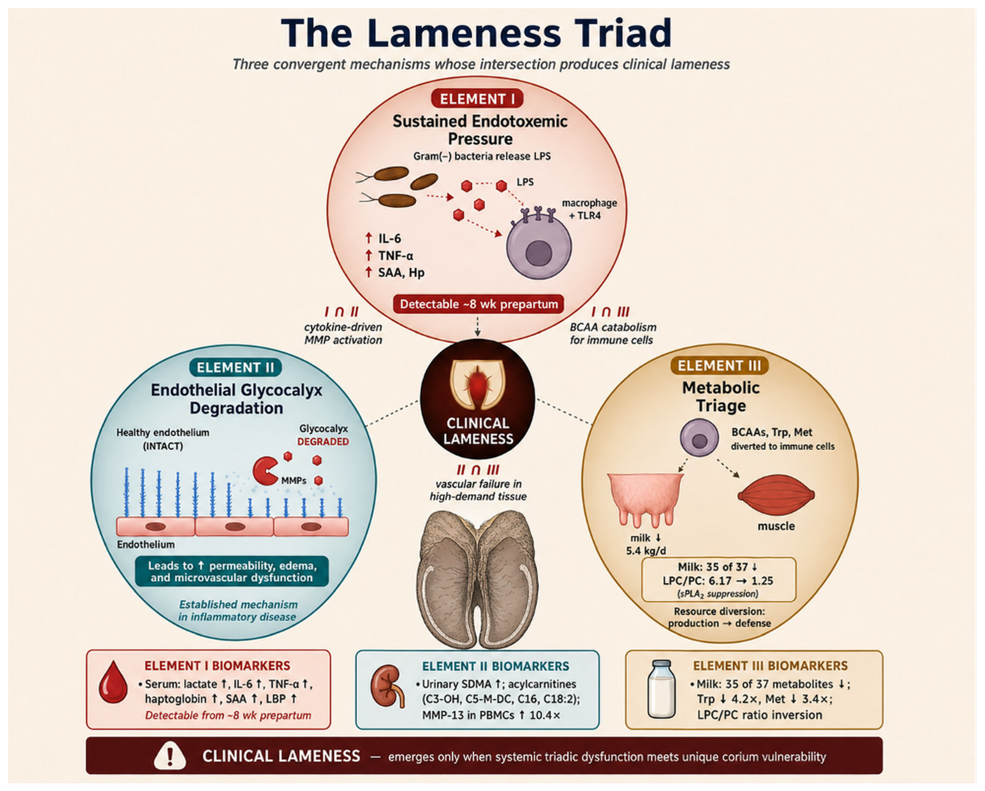
The Lameness Triad comprises three interdependent mechanisms proposed to increase clinical risk when they converge (Figure 3): sustained endotoxemic pressure, endothelial glycocalyx/vascular dysfunction, and metabolic triage. None is considered generally sufficient in isolation.
Each element has distinct kinetics and candidate biomarkers, but their proposed couplings and intersection are central to the framework.

6.1. Element I—Sustained Endotoxemic Pressure

The first element of the triad is sustained endotoxemic pressure: chronic low-grade exposure of the systemic immune system to LPS, sourced variably from the three trigger compartments described in Section 5. The associated inflammatory signature is substantial: serum lactate, IL-6, haptoglobin, and serum amyloid A are all significantly elevated from −8 weeks prepartum through disease diagnosis (quantitative values in Section 3) [30], in a pattern consistent with substantial systemic inflammatory challenge rather than localized hoof inflammation.
Immune activation in Element I may initially represent an appropriate physiological response; the proposed pathology lies in its persistence and cumulative downstream effects. Inflammatory mediators serve as molecular sentinels revealing ongoing tissue threat. The pathology lies not in the activation itself but in its chronicity: while acute LPS exposure produces transient inflammation that resolves with endotoxin clearance, sustained low-grade exposure over weeks produces sustained low-grade activation that may progressively compromise downstream targets. The kinetic distinction between acute and chronic endotoxemia is therefore central to the framework, and explains why single-time-point inflammatory measurements may fail to capture the disease state: what matters is the integrated cytokine exposure over the periparturient window, not its peak amplitude.
The downstream LPS–TLR4 inflammatory pathway is well characterized in other inflammatory contexts, although its proposed role in preclinical dairy-cow lameness remains to be validated. Circulating LPS binds LBP and engages TLR4 on hepatic Kupffer cells and circulating monocytes. Activated immune cells release IL-1β, IL-6, and TNF-α, which stimulate hepatic acute-phase protein synthesis (haptoglobin, SAA, fibrinogen). Cumulative inflammatory mediators activate endothelial cells throughout the vasculature, upregulating selectins (E-selectin, P-selectin) and integrin ligands (ICAM-1, VCAM-1) on endothelial surfaces [72,73]. This endothelial activation is the hand-off point between Element I and Element II: the cytokines of Element I are the proximate stimulus for the matrix metalloproteinase activation that drives Element II.

Element I as Integrated Endotoxin Load: The Staggered Periparturient Sources

Endotoxemic pressure is conceptualized as a temporally evolving flux from anatomically distinct sources rather than a static input. Their staggered contributions may sum into an integrated systemic load.
Four overlapping windows are proposed: mammary vulnerability during dry-off (approximately −8 to −6 weeks), ruminal–intestinal exposure from transition diets (approximately −3 to +6 weeks), uterine exposure after parturition (0 to +6 weeks), and a second mammary window in early lactation (0 to +6 weeks) [27,28,56,74,75,76,77,78,79,80].
This timing may produce a low baseline at dry-off, a rise near calving, and sustained pressure through early lactation. It is compatible with transition-disease clustering and early metabolomic signatures [28,29,30,31,32,33], but the integrated LPS profile itself remains to be measured.
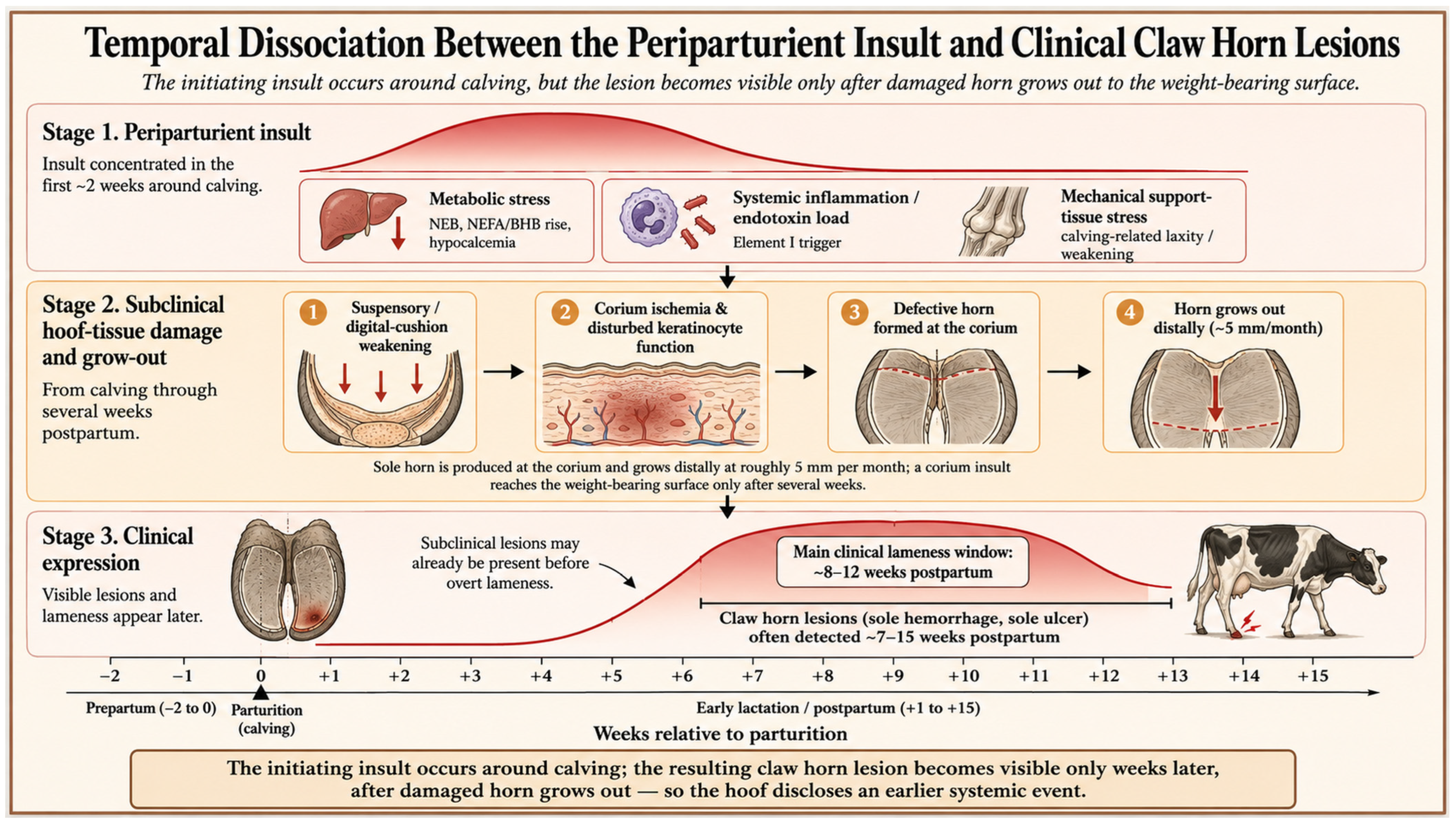
Functional lameness and visible CHDL have different latencies. Locomotor changes may appear within the first postpartum weeks, whereas horn lesions generated after corium injury commonly become visible approximately 7–15 weeks postpartum because horn grows only a few millimeters per month [45,87,88,89,90,91]. Figure 4 illustrates this conceptual dissociation; direct endotoxin exposure was not measured in the cited cohorts.

6.2. Element II—Endothelial Glycocalyx Degradation

Element II is progressive endothelial–vascular dysfunction arising from inflammatory signaling and cellular metabolic stress. Four early, mechanistically distinct findings are relevant: SDMA, acylcarnitines, matrix-degrading enzymes, and urinary xylose.
Urinary SDMA was elevated before diagnosis and at lameness [33,41,42], indicating altered renal–vascular physiology but not specifically glycocalyx injury.
Persistent acylcarnitine accumulation [33] is compatible with mitochondrial stress that could impair endothelial homeostasis and parallels an acylcarnitine–laminitis association in an equine model [62].
Matrix-degrading machinery is supported by 10.44-fold higher MMP-13 expression in PBMCs from lame cows and increased MMP-2, MMP-9, and ADAMTS expression in lamellar tissue [92,93]. In other inflammatory systems, MMPs, heparanase, and hyaluronidase degrade the glycocalyx and impair vascular-barrier function [65]. Whether the same process occurs systemically in pre-lame cows requires direct confirmation.
Urinary xylose, part of the proteoglycan linker, was elevated at −8 and −4 weeks prepartum [61]. Together with SDMA, acylcarnitines, and MMP expression, it provides convergent but indirect evidence relevant to Element II; its tissue source remains unresolved.
A proposed sequence links cytokine-induced matrix-degrading enzymes, reduced nitric-oxide support, and mitochondrial stress to glycocalyx loss, leukocyte adhesion, impaired transmigration, and microvascular dysfunction [94,95,96,97,98]. In the constrained digital corium, such changes could promote ischemia, oxidative stress, and impaired repair.
The measured markers are systemic rather than hoof-specific, while glycocalyx fragments and other damage-associated signals could feed back through TLR pathways [99,100]. This proposed I–II feedback is plausible but not yet demonstrated in dairy-cow lameness.

6.3. Element III—Metabolic Triage and Resource Reallocation

The third element is metabolic triage: the coordinated organism-level reallocation of nutrients and energy from anabolic functions (milk synthesis, growth, reproduction) toward immune cell metabolism and tissue repair. While Elements I and II are well-characterized in human medicine and have direct analogues in human inflammatory and vascular disease, Element III is comparatively distinctive to the periparturient ruminant, and its quantitative magnitude in this species is what makes the corium specifically vulnerable to triadic dysfunction.
The empirical signature—coordinated suppression of most measured milk metabolites, marked depletion of tryptophan and methionine, elevated serum branched-chain amino acids, reduced milk yield, and consistent enrichment of amino-acid and biotin pathways—is presented quantitatively in Section 4.3 [31,32,66] and is not restated here.
A second signature, the inversion of the milk lysophosphatidylcholine-to-phosphatidylcholine ratio and altered phosphatidylcholine and sphingomyelin patterns (Section 4.3 [66]), suggests redirected partitioning of immunomodulatory lipids toward the systemic circulation.
One biologically plausible interpretation is that the organism prioritizes defense and maintenance over production. Branched-chain amino acids are diverted from anabolic to catabolic fate, supplying carbon skeletons and nitrogen to proliferating immune cells. Phosphatidylcholine and sphingomyelin species show patterns consistent with altered partitioning between milk and systemic metabolism, where they exert immunomodulatory effects. Tryptophan and methionine may be preferentially utilized for their specific roles in immune regulation and antioxidant defense. Biotin, the cofactor for four carboxylases central to glucose, fatty acid, and amino acid metabolism, is itself perturbed, placing the entire central metabolic network under triage discipline.
Element III makes the digital corium specifically vulnerable in two distinct ways. First, the corium is a high-metabolic-demand tissue producing keratin continuously; reduced anabolic priority in this tissue cannot be tolerated as it can in skeletal muscle or adipose. Second, the metabolic demands of immune defense compete with the metabolic demands of vascular repair, so substrate availability for glycocalyx maintenance may be constrained when inflammatory degradation is increased, creating a potentially adverse kinetic mismatch that may reinforce Element II. This bidirectional coupling between Elements II and III mirrors the I–II coupling described above, completing a tightly interlocked triadic cycle in which each element may influence and be influenced by the others.
Lean et al. [25] reviewed nutritional impacts on lameness; the framework here clarifies why nutritional interventions have shown variable success. Nutrition that addresses Element III alone (caloric and amino acid sufficiency) without addressing Elements I (endotoxemic pressure) and II (vascular protection) may be insufficient if the triad continues to operate via the unaddressed elements. The framework therefore predicts that effective nutritional intervention should be triad-aware: caloric and amino acid support combined with anti-inflammatory and glycocalyx-protective strategies, deployed during the periparturient window when triadic threshold dynamics permit interruption.

6.4. Triadic Intersection and Threshold Dynamics

Three observations from the longitudinal data support the proposition that lameness does not follow a continuous dose–response relationship with any individual triadic element, but rather a threshold transition determined by triadic intersection.
First, multivariate analyses achieve complete separation between pre-lame and healthy cows at −8 weeks prepartum (PCA and PLS-DA, AUCs 0.992–0.995) [31]. Complete separation is qualitatively different from strong correlation: in that dataset, it indicates that two populations occupied non-overlapping regions of metabolic space. This pattern is consistent with a state-transition interpretation, although validation in larger independent cohorts remains necessary before it can be treated as a universal diagnostic rule.
Second, individual biomarkers from each triadic element show partial separation between groups, but the 5-metabolite panels, which include biomarkers provisionally assigned across the three elements, achieve near-perfect within-cohort separation. The high classification performance of these panels may arise partly because they capture the triadic intersection rather than any single element.
Third, persistence of metabolic perturbations at +4 and +8 weeks postpartum in clinically recovered animals [31] indicates that the measured metabolic perturbations do not resolve simultaneously with clinical signs. The molecular alterations outlast the recognized clinical episode, which is why “clinical recovery” does not equal “biological recovery” and why recurrence rates are high.
These observations support a threshold model: each triadic element accumulates over the periparturient window, and clinical lameness occurs when the joint trajectory crosses a critical surface in three-dimensional triadic space. Cows that remain below this surface despite environmental challenges do not develop lameness; cows that exceed it under any combination of triadic loadings do. The heterogeneity observed by Jewell et al. [14] under broadly comparable husbandry conditions is compatible with the operation of this threshold structure within an underlying heterogeneous population. Genetic variation in inflammatory responsiveness, vascular reserve, and metabolic flexibility, all reviewed below in Section 8, places different individuals at different baseline positions relative to the critical threshold, with environmental stressors potentially providing an additional perturbation that moves some individuals across.
The threshold model has direct intervention implications. Single-element interventions (anti-inflammatory only, vascular protection only, nutritional only) move the system parallel to one axis of triadic space and may therefore have limited effects for individuals positioned far from the threshold along that axis. The model predicts that multi-element interventions could reduce risk more efficiently. Potentially effective interventions include those that target the bidirectional couplings between elements, the I–II coupling (cytokine-driven MMP activation) and the II–III coupling (vascular failure in high-demand tissue), because these couplings are the positive-feedback structures that drive triadic threshold crossing once initiated.

7. Supporting Molecular and Cellular Evidence

Independent molecular and cellular studies support the framework and map onto the three triadic elements (Table 1); they confirm the systemic nature of the pathophysiology rather than a purely local process.

7.1. Transcriptomic Signatures of Systemic Activation

Transcriptomic studies provide evidence of systemic immune activation alongside local hoof-tissue responses: hoof lamellar tissue and circulating leukocytes upregulate matrix-degrading enzymes and inflammatory mediators, corium tissue tracks systemic nutritional and inflammatory status, and even mild sole hemorrhage associates with systemic immune-cell changes (Table 1) [92,93,101,102,103,104].
Table 1. Independent molecular and cellular studies supporting the triadic framework, mapped to the three elements (I, endotoxemic pressure; II, glycocalyx/vascular injury; III, metabolic triage).
StudyApproachKey FindingElement
Transcriptomics
Ding et al. [93]Hoof lamellar biopsy↑ MMP-2, MMP-9, ADAMTSs in induced laminitisII (local)
Almeida et al. [92]PBMC expression↑ IL-2 (12.7×), IL-10 (2.4×), MMP-13 (10.4×), CCR5 (5.3×)I, II
Baranwal et al. [101]Leukocyte DEGs504–991 DEGs incl. IL1B, TNFA, CXCL8, CCL4, MMP-9I, II
Osorio et al. [102,103]Corium tissueHoof markers track systemic nutrition/inflammationI, III
Proteomics
Dong et al. [105]Laminitic-cow
proteome
19 DE proteins: carbohydrate/lipid metabolism, immunity, oxidative stressI, III
Sun et al. [106]Plasma (foot rot)Complement, coagulation, acute-phase activationI
Herzberg et al. [107]Spinal dorsal horn↑ HSP70/HSP90; central sensitizationChronic-pain extension
Metabolomics/Ionomics
Deng et al. [108]Serum LC-MS/MS and ICP-OES/MSHoof deformation associated with systemic metabolic and ionomic alterationsManifestation/systemic coupling
microRNA
Pan et al. [109]Blood miRNAbta-miR-339b discriminates phenotypes; predicts recoveryRegulatory
Note: Up-arrows (↑) indicate increased gene or protein expression relative to the corresponding control or reference group; values in parentheses indicate the reported fold increase.

7.2. Proteomic Landscapes of Immune and Neural Activation

Proteomic studies reveal systemic immune, vascular, coagulation, and nociceptive alterations (Table 1). Laminitic and footrot cows show altered proteins of carbohydrate and lipid metabolism, immune regulation, complement, coagulation, and oxidative stress [105,106,110].
Notably, proteomic profiling of the spinal-cord dorsal horn in chronically lame cows shows upregulation of heat-shock proteins (HSP70, HSP90) involved in central sensitization and neuropathic pain [107], demonstrating that chronic lameness engages the central nervous system and moves beyond purely orthopedic injury into the domain of chronic-pain syndromes, an implication the hoof-centric framework cannot address.
In addition, hoof-deformed cows show systemic metabolomic and ionomic alterations [108], supporting the framework’s prediction that structural hoof changes are accompanied by systemic biochemical changes rather than representing isolated mechanical events.

7.3. MicroRNA Profiling and Regulatory Networks

MicroRNA profiling adds regulatory evidence (Table 1): distinct blood-miRNA patterns discriminate lameness phenotypes and are associated with recovery outcomes [109], consistent with host regulatory state influencing phenotype and resolution.

7.4. Microbiome Dynamics and Host–Pathogen Interactions

Recent investigations into the bovine foot microbiome have revealed that microbial dynamics at the skin-hoof junction play roles in disease susceptibility and lesion evolution [111,112,113]. Using 16S rRNA gene sequencing and metagenomic profiling, several studies have identified microbial shifts that precede or accompany claw horn disruption lesions, digital dermatitis, and foot rot.
In digital dermatitis, lesion microbiota are dominated by Treponema spp. within polymicrobial biofilms, and dysbiosis is detectable before lesion development [111,112,113]. Bay et al. [114] demonstrated host-genotype–microbiome interactions, with GWAS identifying loci correlated with microbial abundance, indicating that the transition from exposure to disease is mediated by host-encoded immune and barrier functions.
Within the framework, these microbiome shifts may contribute to individual susceptibility, but the evidence is primarily associative and requires longitudinal validation.

8. Genetic Architecture Supporting the Triadic Framework

The genetic evidence reviewed in the introduction takes on new significance when interpreted through the triadic framework; heritability estimates for lameness-related traits are summarized in Table 2. Genome-wide association studies have identified QTLs and SNPs for claw horn disruption lesions, digital dermatitis, sole ulcers, and white line disease that map to genes of keratinization, immune regulation, and tissue remodeling, among them TLR2, CD83, IL8, and PLPP3 [37,38,39,115,116]. Genetic variation in these loci predicts variation in triadic threshold position: TLR2 and IL8 variants alter Element I (inflammatory responsiveness); CD83 variants alter immune-cell coordination relevant to Element II; and tissue-remodeling variants alter recovery capacity following triadic intersection.
The discovery that recovery and susceptibility demonstrate distinct genetic control [40] is particularly significant within the framework. Susceptibility (heritability 0.25) may reflect genetic positioning relative to the triadic threshold; recovery (heritability 0.27) may reflect genetic capacity to restore homeostasis after triadic intersection has occurred. These are biologically distinct properties, one operates before threshold crossing, the other after, and the framework predicts that they should be governed by different gene sets and selectable independently.
Heringstad et al. [116] noted that heritabilities for most claw disorders are low to moderate, implying substantial environmental influence. The framework offers a possible reconciliation of low heritability with meaningful genetic contribution: because the triad has a threshold structure, small additive shifts in any element produce large changes in clinical outcome only when the population is positioned near the threshold. Heritability estimates should therefore depend on the environmental severity of the population, consistent with the reported variation across studies.
Chapinal et al. [117] reported genetic correlations among hoof lesions ranging from −0.40 to 0.98, with strongest correlations among sole hemorrhage, sole ulcer, and white line disease. Ødegård et al. [39] reported that digital dermatitis and heel horn erosion showed genetic relationships that differed from several other claw disorders. Within the framework, these patterns are consistent with the idea that lesions with stronger genetic correlations may share more of the same underlying biological vulnerability, whereas infectious lesions such as digital dermatitis may involve additional pathways operating on top of the triadic substrate.
Genetic variants for claw disorders interact with the biological and management context in which animals are evaluated [115,116], consistent with a threshold model in which environmental stress exposes cryptic genetic variation near threshold crossing.
Ring et al. [121] demonstrated that genetic selection for improved locomotion scores can accelerate genetic gain in lameness resistance, whereas feet and leg conformation traits show weak genetic correlations with claw disorders. Current evidence does not support replacing these established selection targets with unvalidated immune or metabolic markers.
Barden et al. [119] reported moderate heritability for digital cushion thickness with negative genetic correlations to sole lesions (Table 2). The Lactanet Hoof Health index provides a national composite reference point for hoof-health selection [118], while additional work confirms physiological and genetic variation in digital cushion thickness [120]. Within the framework, digital cushion thickness is interpreted as both a mechanical buffer and a marker of local tissue reserve. The broader claim that it reflects whole-body metabolic capacity remains a hypothesis, but it is biologically plausible because the digital cushion is affected by body condition, adipose mobilization, and periparturient energy balance, as developed in Section 9.7.

9. The Susceptibility–Resilience Gate: From Pathogenic Convergence to Clinical Disease

9.1. The 75% Question—And Why the Triad Alone Cannot Answer It

Within a common herd environment, cows may share the same barn, diet, flooring, and milking system, yet a predictable subset of cows in a herd develops clinical lameness while their stall-mates remain sound. Annual herd-level incidence consistently centers near 25%, with the upper tail reaching 50% under poor management and the lower tail descending below 15% in well-managed operations [1,2,18]. Within any single herd, lameness clusters in a defined minority. Jewell et al. [14] reported that approximately three quarters of cows housed under broadly comparable conditions did not develop clinically detectable lameness; the same approximate ratio recurs across studies and continents.
The Lameness Triad, as developed in Section 6 and Section 7, proposes a mechanism by which sustained endotoxemic pressure (Element I), endothelial glycocalyx degradation (Element II), and metabolic triage (Element III) may converge to increase the probability of lamellar microvascular dysfunction. Molecular, transcriptomic, proteomic, and metabolomic findings from multiple studies are consistent with parts of this architecture, but they do not yet validate the complete causal sequence. The Triad alone also does not explain why only some cows exposed to broadly comparable conditions progress to clinical disease. Section 6.4 introduced threshold dynamics as one possible explanation; the Gate develops that proposition into a testable host-level model.
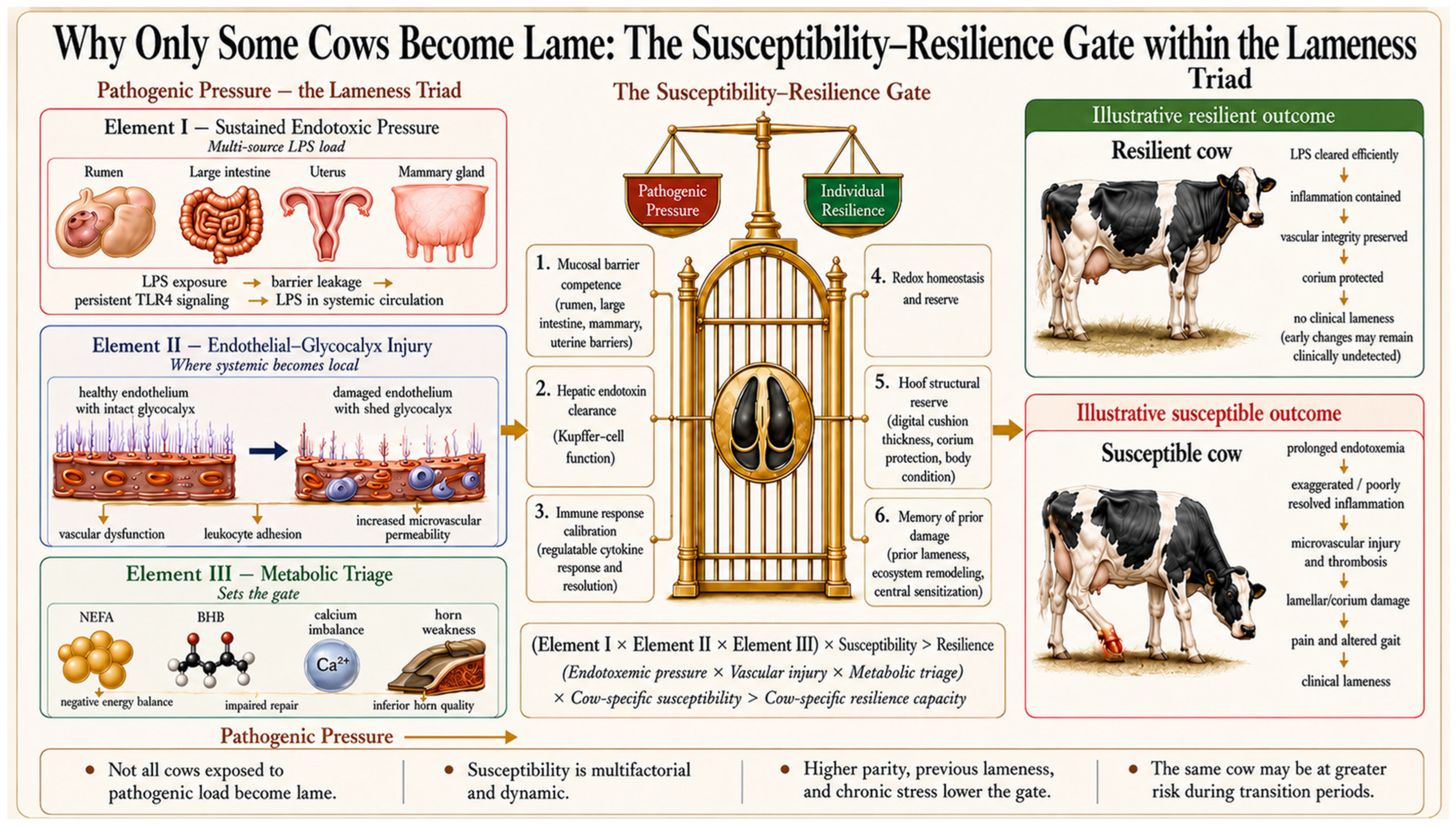
The available evidence suggests that the Triad creates pathogenic pressure, but clinical disease emerges only when that pressure exceeds an individual animal’s resilience threshold. The Triad is therefore not a deterministic switch. It is a risk-amplifying network whose output is filtered through the cow’s individual capacity to neutralize endotoxin, regulate inflammation, maintain vascular integrity, protect the corium, and repair horn tissue. The fact that only a subset of exposed cows develops clinical lameness is not a weakness of the framework; it is an expected feature of a threshold-based systems disease.

9.2. The Susceptibility–Resilience Gate: Formal Definition

We propose that the Lameness Triad operates through a Susceptibility–Resilience Gate: a host-level filter, distinct from but coupled to the three triadic elements, that may influence whether triadic pressure progresses to clinical disease. The Gate comprises six proposed layers that differ substantially in current evidence, measurability, heritability, and modifiability; they are addressed in Section 9.3, Section 9.4, Section 9.5, Section 9.6, Section 9.7 and Section 9.8.
Formally, the framework predicts clinical lameness when:
(Element I × Element II × Element III) × Susceptibility > Resilience
or, in expanded and more accessible form:
(Endotoxemic pressure × Vascular injury × Metabolic triage)
× Cow-specific susceptibility > Cow-specific resilience capacity
The two formulations are equivalent. The first emphasizes the multiplicative interaction of triadic pressure with host susceptibility; small changes in either factor may produce substantial changes in clinical outcome through their product. The second names the constituents explicitly and clarifies that susceptibility and resilience are cow-level properties operating in addition to system-level triadic pressure.
Several features of this formulation deserve note. First, it is not a reparameterization of the Triad; it is an additional dimension. The Triad describes the proposed pathophysiological cascade, whereas the Gate describes host capacities that may influence whether the cascade reaches a clinical endpoint. Second, susceptibility and resilience are not necessarily symmetric opposites. Susceptibility describes the ease with which compensatory systems may be overwhelmed, whereas resilience describes the capacity and rate of restoration after perturbation. Third, Gate state may eventually become measurable, but candidate indicators differ in validation and field feasibility. Several layers remain research constructs rather than sources of established diagnostic biomarkers or proven interventions.
It bears emphasis that the gate and the Triad are dimensionally distinct rather than overlapping. The Triad describes the active pathophysiology that unfolds in the cow’s body during the periparturient window, a process. The Gate describes the cow’s individual capacity to absorb, clear, regulate, buffer, structurally compensate for, and historically remember that process, a set of capacities. Mucosal barrier competence (Layer 1) and hepatic clearance (Layer 2) govern how much triadic pressure is generated in a given animal; immune calibration (Layer 3) and redox reserve (Layer 4) govern how that pressure is shaped and buffered as it propagates; hoof structural reserve (Layer 5) and memory of prior damage (Layer 6) govern whether triadic pressure reaching the target tissue produces clinical disease. The Triad therefore remains a coherent three-element model of pathogenesis; the Gate is the host-level dimension orthogonal to it, and the two architectures together describe both the mechanism by which lameness occurs and the selectivity with which it expresses clinically across a population.
Figure 5 summarizes the six Gate layers and illustrative outcomes.

9.3. Layer 1—Mucosal Barrier Competence

The first determinant of whether triadic pressure becomes clinical disease is whether endotoxin reaches systemic circulation at sufficient concentration and duration to initiate the cascade. Endotoxemic pressure (Element I) depends not only on the size of the mucosal reservoirs but on the integrity of the barriers separating those reservoirs from the bloodstream. The framework predicts that cows with preserved mucosal barriers would experience more limited and transient endotoxin translocation, whereas compromised barriers could permit greater or more sustained systemic exposure.
The ruminal and large-intestinal mucosa, mammary epithelium, and uterine endometrium depend on tight junctions, mucus, antimicrobial defenses, and resident microbiota. Individual variation in intestinal permeability has been documented during subacute ruminal acidosis challenge [80], and multiple anatomical sources of bovine endotoxemia have been reviewed [86].
Early-life microbial composition may be relevant to later host physiology, but direct evidence linking it to adult lameness susceptibility is lacking. Loch et al. [122] reported associations between neonatal calf fecal microbiota, inflammatory markers, and subsequent performance. These findings support further study of developmental contributions to Layer 1 but do not demonstrate that early microbial patterns persist into adulthood or cause later Gate dysfunction.
Candidate assessments of Layer 1 remain research tools. Future studies could combine standardized permeability or challenge-based assays with measures of systemic exposure, but no practical biomarker panel has been validated to predict lameness risk in dairy cows. Management strategies that support ruminal and intestinal barrier function remain biologically relevant, although their direct effects on lameness prevention require testing.

9.4. Layer 2—Hepatic Endotoxin Clearance and Inflammatory Buffering

Once endotoxin enters portal circulation, the liver is the primary site of clearance and inflammatory buffering. Hepatic macrophages (i.e., Kupffer cells), remove portal endotoxin before it reaches the systemic circulation and strongly influence the magnitude of the downstream inflammatory response [100,123]. The hepatic capacity for this task varies substantially between individual animals. Maier and Ulevitch [123] demonstrated, in classic work on isolated rabbit hepatic macrophages, that LPS selectively activates these cells, stimulating mediator release (including a procoagulant factor) alongside direct cellular toxicity, processes they proposed to be central to the endotoxemic shock syndrome.
In ruminants, the hepatic clearance function operates within a narrow window. Below this window, inadequate LPS recognition impairs clearance and permits systemic spillover; above the window, exaggerated LPS presentation to TLR4 amplifies inflammation. Chang et al. [124] demonstrated in a high-concentrate goat model that LPS concentrations rose significantly in both portal and hepatic veins when hepatic clearance was impaired, with downstream upregulation of NF-κB, TRAF6, and acute-phase gene expression. This model supports the principle that hepatic endotoxin clearance capacity is a critical gate between gut-derived endotoxin and systemic inflammation.
LBP and soluble CD14 participate mechanistically in LPS recognition and presentation to TLR4. Their concentrations may vary with both exposure and host response, making isolated values difficult to interpret. Neither is currently established as a practical field biomarker of Gate state or lameness susceptibility; in this framework, they should be regarded as mechanistic research measures rather than routine diagnostic tests.
Endotoxin tolerance, the acquired protection from prior sub-lethal LPS exposure, is a fundamental property of this layer. Filipe et al. [125] demonstrated that bovine peripheral blood mononuclear cells from cows that subsequently remained healthy during the transition period showed lower proliferative and pro-inflammatory responses to in vitro LPS challenge than cells from cows that subsequently developed disease. Healthy cows behaved as endotoxin-tolerant (“high-responders” in the experimental terminology, defined by proliferative response); diseased cows behaved as poorly tolerant low-responders whose unstimulated PBMCs already showed elevated IL-1β, IL-6, and TNF-α expression. The finding overturns the simple intuition that “stronger immune response equals better outcome.” In endotoxin handling, better-regulated response, not larger response, predicts resilience.
Hepatic clearance capacity may be influenced by physiological state and prior exposure history. However, practical measures of this capacity and interventions that reliably modify it in relation to lameness risk have not been established in dairy cows. Layer 2 should therefore remain a mechanistic and testable component of the framework rather than an established management target.

9.5. Layer 3—Immune Response Calibration

Beyond the hepatic clearance window, the systemic immune response itself is calibrated differently between individual cows. The widely held belief that periparturient dairy cows are uniformly immunosuppressed in early lactation does not survive contact with current data. Recent work comparing early- and mid-lactation cows under controlled intravenous LPS challenge has documented that early-lactation animals frequently show amplified rather than suppressed responses, including greater fever, stronger cytokine release, more severe hypocalcemia, augmented neutrophilia, more pronounced platelet reduction, and stronger haptoglobin and LBP responses [126]. Early-lactation immune function is not weak; it is dysregulated. Many components are functionally robust or exaggerated; what is lost is the coordination among components and the rate of resolution.
Individual cows vary in the magnitude, timing, coordination, and resolution of their immune responses. Standardized ex vivo stimulation assays may help characterize this variation [125,126], but their capacity to predict lameness has not been established. Layer 3 is therefore presented as a testable functional concept rather than as a specific commercial classification or selection program.
Within the Susceptibility–Resilience Gate framework, Layer 3 concerns the magnitude, timing, coordination, and resolution of the immune response. A resilient phenotype is hypothesized to show proportionate cytokine activation followed by timely restoration of endothelial and metabolic homeostasis, whereas a susceptible phenotype may show prolonged signaling and delayed recovery. These proposed phenotypes require direct longitudinal validation against subsequent lameness outcomes.
Layer 3 susceptibility is therefore not “weak immunity” but miscalibrated immunity. The relevant distinction concerns the magnitude, timing, coordination, and resolution of the response. Whether functional measures of immune calibration can predict lameness susceptibility requires direct longitudinal testing; no specific immune phenotype is recommended here for clinical use or genetic selection.

9.6. Layer 4—Redox Homeostasis and Reserve

Oxidative stress is a plausible contributor to tissue injury associated with triadic activation. Reactive oxygen species generated during inflammatory activation, lipid peroxidation associated with adipose mobilization, and altered antioxidant defenses during high metabolic demand may reduce tissue tolerance. However, individual variation in redox status has not been shown to predict claw horn disruption lesions reliably.
Studies of transition cows support an association between oxidative stress and disturbances in immune and metabolic function [127,128,129,130,131]. These associations do not establish that any individual redox measure predicts lameness or that targeted antioxidant supplementation prevents claw horn disruption lesions. Accordingly, the cited measures and nutritional strategies should be regarded as background evidence for biological plausibility rather than validated lameness biomarkers or interventions.
Within the Gate framework, redox homeostasis is proposed as a potential buffer against oxidative injury and delayed inflammatory resolution. No individual measure or composite redox panel has been validated as a reliable predictor of lameness susceptibility in dairy cows. Layer 4 is therefore retained as a biologically plausible research construct requiring targeted longitudinal investigation.

9.7. Layer 5—Hoof Structural Reserve

The first four layers operate at the level of systemic biochemistry. The fifth operates at the level of the target tissue itself. Even when triadic pressure has produced microvascular and lamellar dysfunction, clinical lameness emerges only when the hoof’s local structural reserve is insufficient to compensate. This is where individual anatomy matters most directly.
Bicalho et al. [132] demonstrated, in 501 lactating Holstein cows examined cross-sectionally, that the prevalence of sole ulcers and white line disease was strongly associated with digital-cushion thickness measured by ultrasonography at the typical ulcer site. Cows in the upper quartile of digital-cushion thickness had an adjusted lameness prevalence approximately 15 percentage points lower than cows in the lower quartile. These findings support the concept that hoof structural reserve is an important modifier of clinical outcome: under comparable pathogenic pressure, cows with greater digital-cushion reserve may be better protected against lesion development and progression.
Newsome et al. [133] extended this work in a prospective cohort that confirmed the predictive direction: thin sole soft tissue on the lateral claw predicted subsequent lameness, and the digital cushion underwent characteristic mobilization during the period of negative energy balance. The cushion is a depot of adipose tissue, and like other adipose depots it can be mobilized under metabolic demand; the consequence is the thinning of the very tissue that protects the corium from compression injury at precisely the moment in lactation when triadic pressure is maximal.
Digital cushion thickness has demonstrated moderate heritability (0.14–0.44 depending on study and model) and strong negative genetic correlations with claw horn lesions [119,120]. Body condition score correlates with digital cushion thickness, providing a partially modifiable management lever. Lactanet Canada reports approximately 9% heritability for the overall Hoof Health index, with individual lesion heritabilities ranging from 3% to 8% [118].
Layer 5 also includes the suspensory apparatus of the distal phalanx, the corium itself, and horn quality. Each contributes to the structural reserve available to absorb the consequences of upstream triadic dysfunction. The clinical implication is that a cow with thin digital cushion, low body condition, and prior lamellar injury enters the periparturient window with markedly reduced structural reserve; under these conditions, even modest triadic activation may contribute to clinical disease.

9.8. Layer 6—Memory of Prior Damage: Why the Same Cows Get Sick Again

The sixth layer of the gate is perhaps the most consequential for predicting recurrence, and it directly answers a question of substantial epidemiological importance: are the cows lame in this lactation the same cows that will be lame in the next? The literature indicates substantial recurrence within previously affected animals.
Randall et al. [134], examining two large UK herds across multiple lactations, estimated that between 79% and 83% of all lameness events were attributable to previous lameness events, that is, the great majority of clinical cases occur in animals that have already been lame. Cows lame in first lactation showed an approximately two-fold increased hazard of lameness in second lactation [135]. The pattern is not a statistical artifact of repeated detection in chronically affected animals; it is also consistent with durable biological change.
Following lameness, some cows may retain structural and nociceptive alterations that increase their susceptibility to subsequent disease. Three potential mechanisms of this “memory of prior damage” have been documented or strongly supported.
Bone remodeling. Newsome et al. [136], using X-ray micro-computed tomography of culled cows, found new bone formation, including osteophyte development on the caudal aspect of the distal phalanx, in cows with a history of clinical lameness. Such remodeling may alter the local mechanical environment of the corium and digital cushion and thereby contribute to persistent vulnerability after clinical recovery.
Digital-cushion attrition. Digital-cushion adipose tissue is mobilized during negative energy balance [133]. Repeated metabolic stress and lameness may therefore reduce the structural reserve of the cushion, although the extent to which this tissue is restored during subsequent lactations remains insufficiently defined.
Central sensitization. Herzberg et al. [137,138] reported increased spinal dorsal-horn expression of inflammatory mediators, including TNF-α, IL-1α, CXCL10, and CXCL9, together with evidence of oxidative injury in chronically lame cows compared with non-lame controls. These findings support the presence of central neuroinflammatory changes compatible with sensitization and may help explain persistent pain hypersensitivity after prolonged lameness.
The combined effect of these mechanisms is that Layer 6 may progressively lower the threshold for clinical disease across successive lactations in cows with prior lameness. An initial episode can therefore become a biological gateway to recurrence by leaving residual structural, metabolic, and nociceptive vulnerability. This perspective shifts management emphasis beyond treatment after clinical disease has emerged: preventing the first clinical case may be especially important because it may preserve structural and functional resilience. Prompt detection and effective treatment of early lameness also remain essential. Evidence regarding routine NSAID use is context-dependent: a single-herd randomized trial in heifers found a lower future probability of lameness when ketoprofen was administered at calving and at each lameness event [139], whereas a subsequent trial in multiparous cows did not identify a comparable long-term benefit and emphasized therapeutic trimming and orthopedic blocks [140]. Neither trial establishes that NSAID treatment prevents the structural or neurological components proposed for Layer 6.

9.9. Parity, Age, and the Cumulative Erosion of the Gate

Parity is consistently among the strongest risk factors identified in lameness epidemiology. Bicalho et al. [132] reported sole ulcer prevalence of 4.2% in first-parity cows versus 27.8% in higher-parity cows, with corresponding figures of 1.0% versus 6.5% for white line disease and 19.8% versus 48.2% for visually scored lameness. Each year of additional age increases lameness probability by approximately 20% in pasture-based systems, and cows in parity 4 or higher have approximately 2.5-fold greater odds of lameness compared with first-lactation animals according to a 2019 systematic review and meta-analysis [141].
Within the Gate framework, age and parity may function not only as established epidemiological risk factors but also as integrators or proxies for cumulative challenges across successive transition cycles. Successive lactations may involve mucosal disruption, hepatic inflammation, immune challenge, redox depletion, and digital cushion mobilization, and, with non-trivial probability, some clinical damage that activates Layer 6 memory. The cumulative burden of these challenges may increase with age and parity. A first-lactation heifer enters her first transition window with most gate layers intact (though, importantly, with less digital cushion than she will reach in subsequent lactations and less acquired endotoxin tolerance than she will accumulate). A fifth-lactation cow enters her sixth transition window with potentially reduced reserve across several gate layers.
This interpretation predicts, and available data support, that the apparent protection of first-lactation cows is partial and contingent: heifers are less likely to develop sole ulcer or white line lesions than older cows, but they are more likely to be the cohort in which microbiome disturbance, mucosal disruption, or behavioral disadvantage produces the first clinical case, after which Layer 6 begins its accumulation. The framework predicts that interventions focused on protecting the gate during the first lactation may yield disproportionate benefit across the animal’s lifetime, and the limited intervention data available support this prediction.

9.10. Diagnostic and Therapeutic Implications of the Gate

The Susceptibility–Resilience Gate framework has direct consequences for how lameness is diagnosed, monitored, and prevented.
Diagnostic implication. Biomarker panels assessing triadic pressure (Section 4) capture only the upstream side of the proposed framework. A more complete predictive model could combine indicators of triadic pressure with practical measures of Gate state where those measures are validated. At present, digital-cushion thickness measured by ultrasonography and individual lameness and lesion history are the most directly applicable candidate measures for Layers 5 and 6. Laboratory measures of mucosal or hepatic function, immune calibration, and redox state remain research tools and should not be presented as established field biomarkers. The framework predicts that, under comparable triadic pressure, cows with preserved Gate function would have a lower probability of clinical disease than cows with reduced Gate function. Integrated models should therefore be tested prospectively against single-element biomarker panels and environmental risk scores [142].
Therapeutic implication. The Gate framework does not yet support a specific biomarker-directed treatment program. Current prevention should continue to emphasize validated hoof-health practices, adequate lying time and flooring, prompt detection, therapeutic trimming, appropriate off-loading, pain management under veterinary direction, and management of body condition and transition-cow health [16,139,140,143]. Proposed interventions directed at mucosal, hepatic, immune, or redox layers require direct lameness-specific trials before they can be recommended as Gate-targeted therapies. Future breeding studies should likewise test whether validated indicators of systemic resilience add value to established hoof-health and lesion traits.

9.11. A Convergence Model, Not a Deterministic Trigger

The Lameness Triad is therefore best interpreted as a pathogenic convergence model rather than a deterministic trigger: clinical lameness emerges only when sustained endotoxemic pressure, endothelial-glycocalyx injury, and metabolic triage exceed the cow’s individual susceptibility–resilience threshold, shaped by the six gate layers together with body condition, parity, and genetic background. That only a subset of similarly managed cows becomes lame is an expected feature of a threshold-based systems disease, made explicit as a testable proposition by the gate concept.

10. Environmental Modulators Within the Triadic Framework

The framework does not negate the importance of environmental risk factors documented in previous research; it reinterprets their mechanism. Solano et al. [1] demonstrated that cows in freestalls have more than two-fold higher odds of sole ulcers and white line disease compared to deep-bedded systems. Cook [21] showed that rubber flooring reduces some lameness forms compared to concrete. von Keyserlingk et al. [23] identified inadequate lying time as a major risk factor.
These environmental factors act as modulating influences whose effects may depend on both pathogenic pressure and Gate state. Consider two cows housed under broadly comparable conditions. Cow A enters the transition period with low triadic pressure and preserved Gate function, including adequate structural and metabolic reserve. Cow B enters with higher triadic pressure and reduced Gate reserve, potentially including an inflammatory challenge, endothelial–vascular dysfunction, and depleted metabolic capacity. Both experience similar environmental loading, but Cow B may be more likely to develop clinical lameness when biomechanical stress exceeds tissue capacity. This example is conceptual and should be tested prospectively.
This framework may help explain the modest predictive value of risk-assessment tools. van Huyssteen et al. [142] reported moderate associations between lameness risk-assessment scores and prevalence on Alberta farms (r = 0.26 overall; r = 0.45 for noninfectious lesions). Environmental scores capture external challenges but not all dimensions of individual host state. Combining environmental risk assessment with validated molecular or functional measures representing triadic pressure and Gate state may improve predictive accuracy and should be evaluated prospectively.
Altered lying behavior may provide an early behavioral indicator of developing lameness, although it cannot currently be assigned specifically to triadic dysfunction. Solano et al. [144] demonstrated that altered lying patterns associate with lameness even before clinical diagnosis. Cows with lying time ≥ 14 h/day, ≤5 lying bouts daily, or bout duration ≥ 110 min showed 3.7-, 1.7-, and 2.5-fold higher odds of lameness respectively. These behavioral changes may reflect existing discomfort, altered activity, or systemic inflammatory state; the current data do not distinguish these mechanisms definitively. O’Driscoll et al. [145] found that floor surface influenced immune function and locomotion-related outcomes, including neutrophil counts and expression of MMP13 and TAC1. Together with flooring studies [21,22,23], this supports the interpretation that flooring affects more than simple mechanical protection, with consequences that depend on the biological state of the animal.

11. Synthesis and Conclusions

Integrating evidence across immune, vascular, metabolic, genetic, microbial, and environmental domains supports a proposed architecture in which heterogeneous inputs converge on a triadic core and become clinically expressed in the digital corium because of its anatomical and biomechanical constraints. This synthesis generates testable explanations for observations that are not fully accounted for by a hoof-centered framework.
For decades, lameness in dairy cows has been managed primarily through lesion-specific and environmental interventions, including therapeutic trimming, off-loading, flooring improvements, and appropriate treatment of infectious lesions. These strategies remain essential. Multi-omics evidence supports investigation of a broader interpretation in which periparturient CHDL-associated lameness may also involve systemic inflammatory, metabolic, and vascular disturbances whose clinical expression is shaped by the anatomical and biomechanical constraints of the digital corium.
The Lameness Triad, comprising sustained endotoxemic pressure, endothelial glycocalyx degradation, and metabolic triage, provides a convergent-mechanism framework in which the three elements are proposed to increase the probability of clinical lameness. The framework draws on elevations in IL-6, TNF-α, haptoglobin, and SAA before diagnosis (Element I); urinary SDMA and persistent acylcarnitine alterations consistent with renal–vascular and mitochondrial stress (Element II); and coordinated milk-metabolite changes compatible with altered nutrient allocation (Element III). Multivariate models achieved high within-cohort separation between pre-lame and unaffected cows, but these results require independent prospective validation. Collectively, the evidence supports the framework as a set of testable hypotheses rather than a demonstrated causal sequence.
This framework generates hypotheses for diagnosis, prevention, and treatment. Multi-omics findings suggest that cows at increased risk may be identifiable before clinical signs [30,31,32,33,34,35,59,60,61,66], but affordable and externally validated herd-level tools are not yet available. Prevention should retain established hoof-health and environmental practices while testing whether interventions that reduce upstream inflammatory and metabolic pressure provide additional benefit [16,139,140,143]. Future breeding studies should determine whether validated indicators of systemic resilience add predictive value to established hoof-health and lesion traits. Treatment recommendations should remain evidence-based and lesion-specific while systemic strategies are evaluated prospectively.
Above all, the framework reframes the dominant periparturient CHDL phenotype of lameness. It may not be fully explained as a disease of hoof tissue with secondary systemic effects; instead, it may involve systemic immune, vascular, and metabolic disturbances interacting with local anatomical constraints. Management should therefore combine effective hoof care with transition-cow health management during the periparturient period, when the threshold for clinical disease may be crossed and intervention may still be effective. The framework thus generates testable hypotheses for precision prevention while preserving the central role of established hoof-health practices. This analogy is conceptual and does not imply identity with disease mechanisms in other organs.

Funding

This research received no external funding.

Institutional Review Board Statement

Not applicable.

Informed Consent Statement

Not applicable.

Data Availability Statement

No new data were created or analyzed in this study. Data sharing is not applicable to this article.

Acknowledgments

The author thanks the graduate students, postdoctoral fellows, and laboratory technicians whose contributions helped generate the data and publications that served as a basis for this review and conceptualization of lameness. All conceptual figures in this article have been generated with the assistance of ChatGPT (Open AI—GPT-5.6 Sol). The manuscript was written by the author, who is also responsible for the scientific concepts, the interpretation of the literature, and the selection and verification of references. No AI tool was used to generate, analyze, or interpret original research data.

Conflicts of Interest

The author declares no conflicts of interest.

References

  1. Solano, L.; Barkema, H.W.; Pajor, E.A.; Mason, S.; LeBlanc, S.J.; Zaffino Heyerhoff, J.C.; Nash, C.G.; Haley, D.B.; Vasseur, E.; Pellerin, D.; et al. Prevalence of lameness and associated risk factors in Canadian Holstein-Friesian cows housed in freestall barns. J. Dairy Sci. 2015, 98, 6978–6991. [Google Scholar] [CrossRef] [PubMed]
  2. Weaver, M.D.; Jean, G.S.; Steiner, A. Bovine Surgery and Lameness, 3rd ed.; Wiley Blackwell: Hoboken, NJ, USA, 2018. [Google Scholar] [CrossRef]
  3. Bruijnis, M.R.; Hogeveen, H.; Stassen, E.N. Assessing economic consequences of foot disorders in dairy cattle using a dynamic stochastic simulation model. J. Dairy Sci. 2010, 93, 2419–2432. [Google Scholar] [CrossRef] [PubMed]
  4. Cha, E.; Hertl, J.A.; Bar, D.; Gröhn, Y.T. The cost of different types of lameness in dairy cows calculated by dynamic programming. Prev. Vet. Med. 2010, 97, 1–8. [Google Scholar] [CrossRef] [PubMed]
  5. Roche, S.M.; Renaud, D.L.; Saraceni, J.; Kelton, D.F.; DeVries, T.J. Invited review: Prevalence, risk factors, treatment, and barriers to best practice adoption for lameness and injuries in dairy cattle—A narrative review. J. Dairy Sci. 2024, 107, 3347–3366. [Google Scholar] [CrossRef] [PubMed]
  6. Warnick, L.D.; Janssen, D.; Guard, C.L.; Gröhn, Y.T. The effect of lameness on milk production in dairy cows. J. Dairy Sci. 2001, 84, 1988–1997. [Google Scholar] [CrossRef] [PubMed]
  7. Green, L.E.; Hedges, V.J.; Schukken, Y.H.; Blowey, R.W.; Packington, A.J. The impact of clinical lameness on the milk yield of dairy cows. J. Dairy Sci. 2002, 85, 2250–2256. [Google Scholar] [CrossRef] [PubMed]
  8. Hernandez, J.; Shearer, J.K.; Webb, D.W. Effect of lameness on the calving-to-conception interval in dairy cows. J. Am. Vet. Med. Assoc. 2001, 218, 1611–1614. [Google Scholar] [CrossRef] [PubMed]
  9. Garbarino, E.J.; Hernandez, J.A.; Shearer, J.K.; Risco, C.A.; Thatcher, W.W. Effect of lameness on ovarian activity in postpartum Holstein cows. J. Dairy Sci. 2004, 87, 4123–4131. [Google Scholar] [CrossRef] [PubMed]
  10. Booth, C.J.; Warnick, L.D.; Gröhn, Y.T.; Maizon, D.O.; Guard, C.L.; Janssen, D. Effect of lameness on culling in dairy cows. J. Dairy Sci. 2004, 87, 4115–4122. [Google Scholar] [CrossRef] [PubMed]
  11. Huxley, J.N. Impact of lameness and claw lesions in cows on health and production. Livest. Sci. 2013, 156, 64–70. [Google Scholar] [CrossRef]
  12. Whay, H.R.; Waterman, A.E.; Webster, A.J. Associations between locomotion, claw lesions and nociceptive threshold in dairy heifers during the peri-partum period. Vet. J. 1997, 154, 155–161. [Google Scholar] [CrossRef] [PubMed]
  13. Flower, F.C.; Weary, D.M. Effect of hoof pathologies on subjective assessments of dairy cow gait. J. Dairy Sci. 2006, 89, 139–146. [Google Scholar] [CrossRef] [PubMed]
  14. Jewell, M.T.; Cameron, M.; McKenna, S.L.; Cockram, M.S.; Sanchez, J.; Keefe, G.P. Relationships between type of hoof lesion and behavioral signs of lameness in Holstein cows housed in Canadian tiestall facilities. J. Dairy Sci. 2021, 104, 937–946. [Google Scholar] [CrossRef] [PubMed]
  15. Solano, L.; Barkema, H.W.; Mason, S.; Pajor, E.A.; LeBlanc, S.J.; Orsel, K. Prevalence and distribution of foot lesions in dairy cattle in Alberta, Canada. J. Dairy Sci. 2016, 99, 6828–6841. [Google Scholar] [CrossRef] [PubMed]
  16. Leach, K.A.; Tisdall, D.A.; Bell, N.J.; Main, D.C.; Green, L.E. The effects of early treatment for hindlimb lameness in dairy cows on four commercial UK farms. Vet. J. 2012, 193, 626–632. [Google Scholar] [CrossRef] [PubMed]
  17. Murray, R.D.; Downham, D.Y.; Clarkson, M.J.; Faull, W.B.; Hughes, J.W.; Manson, F.J.; Merritt, J.B.; Russell, W.B.; Sutherst, J.E.; Ward, W.R. Epidemiology of lameness in dairy cattle: Description and analysis of foot lesions. Vet. Rec. 1996, 138, 586–591. [Google Scholar] [CrossRef] [PubMed]
  18. Greenough, P.R. Bovine Laminitis and Lameness: A Hands-on Approach; Saunders Elsevier: Philadelphia, PA, USA, 2007. [Google Scholar]
  19. Kloosterman, P. Laminitis—Prevention, diagnosis and treatment. Adv. Dairy Technol. 2007, 19, 157–166. [Google Scholar]
  20. Jewell, M.T.; Cameron, M.; Spears, J.; McKenna, S.L.; Cockram, M.S.; Sanchez, J.; Keefe, G.P. Prevalence of lameness and associated risk factors on dairy farms in the Maritime Provinces of Canada. J. Dairy Sci. 2019, 102, 3392–3405. [Google Scholar] [CrossRef] [PubMed]
  21. Cook, N.B. Prevalence of lameness among dairy cattle in Wisconsin as a function of housing type and stall surface. J. Am. Vet. Med. Assoc. 2003, 223, 1324–1328. [Google Scholar] [CrossRef] [PubMed]
  22. Telezhenko, E.; Bergsten, C. Influence of floor type on the locomotion of dairy cows. Appl. Anim. Behav. Sci. 2005, 93, 183–197. [Google Scholar] [CrossRef]
  23. von Keyserlingk, M.A.G.; Barrientos, A.; Ito, K.; Galo, E.; Weary, D.M. Benchmarking cow comfort on North American freestall dairies: Lameness, leg injuries, lying time, facility design, and management for high-producing Holstein dairy cows. J. Dairy Sci. 2012, 95, 7399–7408. [Google Scholar] [CrossRef] [PubMed]
  24. Sprecher, D.J.; Hostetler, D.E.; Kaneene, J.B. A lameness scoring system that uses posture and gait to predict dairy cattle reproductive performance. Theriogenology 1997, 47, 1179–1187. [Google Scholar] [CrossRef] [PubMed]
  25. Lean, I.J.; Westwood, C.T.; Golder, H.M.; Duffield, T.F. Impact of nutrition on lameness and claw health in cattle. Livest. Sci. 2013, 156, 71–87. [Google Scholar] [CrossRef]
  26. Nocek, J.E. Bovine acidosis: Implications on laminitis. J. Dairy Sci. 1997, 80, 1005–1028. [Google Scholar] [CrossRef] [PubMed]
  27. LeBlanc, S.J. Interactions of metabolism, inflammation, and reproductive tract health in the postpartum period in dairy cattle. Reprod. Domest. Anim. 2012, 47, 18–30. [Google Scholar] [CrossRef] [PubMed]
  28. Mulligan, F.J.; Doherty, M.L. Production diseases of the transition cow. Vet. J. 2008, 176, 3–9. [Google Scholar] [CrossRef] [PubMed]
  29. Daros, R.R.; Eriksson, H.K.; Weary, D.M.; von Keyserlingk, M.A.G. The relationship between transition period diseases and lameness, feeding time, and body condition during the dry period. J. Dairy Sci. 2020, 103, 649–665. [Google Scholar] [CrossRef] [PubMed]
  30. Zhang, G.; Hailemariam, D.; Dervishi, E.; Deng, Q.; Goldansaz, S.A.; Dunn, S.M.; Ametaj, B.N. Alterations of innate immunity reactants in transition dairy cows before clinical signs of lameness. Animals 2015, 5, 1023–1039. [Google Scholar] [CrossRef] [PubMed]
  31. Zhang, G.; Dervishi, E.; Zwierzchowski, G.; Mandal, R.; Wishart, D.S.; Ametaj, B.N. Serum metabolomics identifies metabolite panels that differentiate lame dairy cows from healthy ones. Metabolomics 2020, 16, 73. [Google Scholar] [CrossRef] [PubMed]
  32. Dervishi, E.; Zhang, G.; Zwierzchowski, G.; Mandal, R.; Wishart, D.S.; Ametaj, B.N. Serum metabolic fingerprinting of pre-lameness dairy cows by GC-MS reveals typical profiles that can identify susceptible cows. J. Proteom. 2020, 213, 103620. [Google Scholar] [CrossRef] [PubMed]
  33. Eckel, E.F.; Zhang, G.; Dervishi, E.; Zwierzchowski, G.; Mandal, R.; Wishart, D.S.; Ametaj, B.N. Urinary metabolomics fingerprinting around parturition identifies metabolites that differentiate lame dairy cows from healthy ones. Animal 2020, 14, 2138–2149. [Google Scholar] [CrossRef] [PubMed]
  34. Barden, M.; Phelan, M.M.; Hyde, R.; Anagnostopoulos, A.; Griffiths, B.E.; Bedford, C.; Green, M.; Psifidi, A.; Banos, G.; Oikonomou, G. Serum 1H nuclear magnetic resonance-based metabolomics of sole lesion development in Holstein cows. J. Dairy Sci. 2023, 106, 2667–2684. [Google Scholar] [CrossRef] [PubMed]
  35. Cardoso, A.S.; Martínez-Jarquín, S.; Hyde, R.M.; Green, M.J.; Kim, D.H.; Randall, L.V. Milk lipidome alterations in first-lactation dairy cows with lameness: A biomarker identification approach using untargeted lipidomics and machine learning. J. Dairy Sci. 2025, 108, 6216–6228. [Google Scholar] [CrossRef] [PubMed]
  36. Oberbauer, A.M.; Berry, S.L.; Belanger, J.M.; McGoldrick, R.M.; Pinos-Rodriquez, J.M.; Famula, T.R. Determining the heritable component of dairy cattle foot lesions. J. Dairy Sci. 2013, 96, 605–613. [Google Scholar] [CrossRef] [PubMed]
  37. Lai, E.; Danner, A.L.; Famula, T.R.; Oberbauer, A.M. Genome-wide association studies reveal susceptibility loci for noninfectious claw lesions in Holstein dairy cattle. Front. Genet. 2021, 12, 657375. [Google Scholar] [CrossRef] [PubMed]
  38. van der Spek, D.; van Arendonk, J.A.M.; Bovenhuis, H. Genome-wide association study for claw disorders and trimming status in dairy cattle. J. Dairy Sci. 2015, 98, 1286–1295. [Google Scholar] [CrossRef] [PubMed]
  39. Ødegård, C.; Svendsen, M.; Heringstad, B. Genetic correlations between claw health and feet and leg conformation in Norwegian Red cows. J. Dairy Sci. 2014, 97, 4522–4529. [Google Scholar] [CrossRef] [PubMed]
  40. Barden, M.; Anagnostopoulos, A.; Griffiths, B.E.; Li, B.; Bedford, C.; Watson, C.; Psifidi, A.; Banos, G.; Oikonomou, G. Genetic parameters of sole lesion recovery in Holstein cows. J. Dairy Sci. 2023, 106, 1874–1888. [Google Scholar] [CrossRef] [PubMed]
  41. Gore, M.O.; Lüneburg, N.; Schwedhelm, E.; Ayers, C.R.; Anderssohn, M.; Khera, A.; Atzler, D.; de Lemos, J.A.; Grant, P.J.; McGuire, D.K.; et al. Symmetrical dimethylarginine predicts mortality in the general population: Observations from the Dallas heart study. Arterioscler. Thromb. Vasc. Biol. 2013, 33, 2682–2688. [Google Scholar] [CrossRef] [PubMed]
  42. Tain, Y.L.; Hsu, C.N. Toxic dimethylarginines: Asymmetric dimethylarginine (ADMA) and symmetric dimethylarginine (SDMA). Toxins 2017, 9, 92. [Google Scholar] [CrossRef] [PubMed]
  43. Hirschberg, R.M.; Plendl, J. Pododermal angiogenesis and angioadaptation in the bovine claw. Microsc. Res. Tech. 2005, 66, 145–155. [Google Scholar] [CrossRef] [PubMed]
  44. Shearer, J.K.; van Amstel, S.R. Pathogenesis and treatment of sole ulcers and white line disease. Vet. Clin. N. Am. Food Anim. Pract. 2017, 33, 283–300. [Google Scholar] [CrossRef] [PubMed]
  45. Tarlton, J.F.; Holah, D.E.; Evans, K.M.; Jones, S.; Pearson, G.R.; Webster, A.J. Biomechanical and histopathological changes in the support structures of bovine hooves around the time of first calving. Vet. J. 2002, 163, 196–204. [Google Scholar] [CrossRef] [PubMed]
  46. Ametaj, B.N.; Zebeli, Q.; Saleem, F.; Psychogios, N.; Lewis, M.J.; Dunn, S.M.; Xia, J.; Wishart, D.S. Metabolomics reveals unhealthy alterations in rumen metabolism with increased proportion of cereal grain in the diet of dairy cows. Metabolomics 2010, 6, 583–594. [Google Scholar] [CrossRef]
  47. Ishikawa, Y.; Nakada, K.; Hagiwara, K.; Kirisawa, R.; Iwai, H.; Moriyoshi, M.; Sawamukai, Y. Changes in interleukin-6 concentration in peripheral blood of pre- and post-partum dairy cattle and its relationship to postpartum reproductive diseases. J. Vet. Med. Sci. 2004, 66, 1403–1408. [Google Scholar] [CrossRef] [PubMed]
  48. Hagiwara, K.; Yamanaka, H.; Hisaeda, K.; Taharaguchi, S.; Kirisawa, R.; Iwai, H. Concentrations of IL-6 in serum and whey from healthy and mastitic cows. Vet. Res. Commun. 2001, 25, 99–108. [Google Scholar] [CrossRef] [PubMed]
  49. Pyörälä, S. Indicators of inflammation in the diagnosis of mastitis. Vet. Res. 2003, 34, 565–578. [Google Scholar] [CrossRef] [PubMed]
  50. Kushibiki, S. Tumor necrosis factor-α-induced inflammatory responses in cattle. Anim. Sci. J. 2011, 82, 504–511. [Google Scholar] [CrossRef] [PubMed]
  51. Petersen, H.H.; Nielsen, J.P.; Heegaard, P.M. Application of acute phase protein measurements in veterinary clinical chemistry. Vet. Res. 2004, 35, 163–187. [Google Scholar] [CrossRef] [PubMed]
  52. Fontaine, G.L.; Belknap, J.K.; Allen, D.; Moore, J.N.; Kroll, D.L. Expression of interleukin-1 beta in the digital laminae of horses in the prodromal stage of experimentally induced laminitis. Am. J. Vet. Res. 2001, 62, 714–720. [Google Scholar] [CrossRef] [PubMed]
  53. Goodson, J.M.; Shi, P.; Razzaque, M.S. Dietary phosphorus enhances inflammatory response: A study of human gingivitis. J. Steroid Biochem. Mol. Biol. 2019, 188, 166–171. [Google Scholar] [CrossRef] [PubMed]
  54. Yamada, S.; Tokumoto, M.; Tatsumoto, N.; Taniguchi, M.; Noguchi, H.; Nakano, T.; Masutani, K.; Ooboshi, H.; Tsuruya, K.; Kitazono, T. Phosphate overload directly induces systemic inflammation and malnutrition as well as vascular calcification in uremia. Am. J. Physiol. Ren. Physiol. 2014, 306, F1418–F1428. [Google Scholar] [CrossRef] [PubMed]
  55. Navarro-González, J.F.; Mora-Fernández, C.; Muros, M.; Herrera, H.; García, J. Mineral metabolism and inflammation in chronic kidney disease patients: A cross-sectional study. Clin. J. Am. Soc. Nephrol. 2009, 4, 1646–1654. [Google Scholar] [CrossRef] [PubMed]
  56. Todhunter, D.A.; Smith, K.L.; Hogan, J.S. Growth of Gram-negative bacteria in dry cow secretion. J. Dairy Sci. 1990, 73, 363–372. [Google Scholar] [CrossRef] [PubMed]
  57. Bradley, A.J.; Green, M.J. The importance of the nonlactating period in the epidemiology of intramammary infection and strategies for prevention. Vet. Clin. N. Am. Food Anim. Pract. 2004, 20, 547–568. [Google Scholar] [CrossRef] [PubMed]
  58. Refaai, W.; Gad, M.; Mahmmod, Y. Association of claw disorders with subclinical intramammary infections in Egyptian dairy cows. Vet. World 2017, 10, 358–362. [Google Scholar] [CrossRef] [PubMed]
  59. Randall, L.V.; Kim, D.H.; Abdelrazig, S.M.A.; Bollard, N.J.; Hemingway-Arnold, H.; Hyde, R.M.; Thompson, J.S.; Green, M.J. Predicting lameness in dairy cattle using untargeted liquid chromatography-mass spectrometry-based metabolomics and machine learning. J. Dairy Sci. 2023, 106, 7033–7042. [Google Scholar] [CrossRef] [PubMed]
  60. He, W.; Cardoso, A.S.; Hyde, R.M.; Green, M.J.; Scurr, D.J.; Griffiths, R.L.; Randall, L.V.; Kim, D.H. Metabolic alterations in dairy cattle with lameness revealed by untargeted metabolomics of dried milk spots using direct infusion-tandem mass spectrometry and the triangulation of multiple machine learning models. Analyst 2022, 147, 5537–5545. [Google Scholar] [CrossRef] [PubMed]
  61. Zhang, G.; Dervishi, E.; Zwierzchowski, G.; Mandal, R.; Wishart, D.S.; Ametaj, B.N. Urinary metabolomics around parturition identifies metabolite alterations in dairy cows affected postpartum by lameness: Preliminary study. Dairy 2020, 1, 6–19. [Google Scholar] [CrossRef]
  62. Steelman, S.M.; Johnson, P.; Jackson, A.; Schulze, J.; Chowdhary, B.P. Serum metabolomics identifies citrulline as a predictor of adverse outcomes in an equine model of gut-derived sepsis. Physiol. Genom. 2014, 46, 339–347. [Google Scholar] [CrossRef] [PubMed]
  63. Quinn, M.T.; Parthasarathy, S.; Steinberg, D. Lysophosphatidylcholine: A chemotactic factor for human monocytes and its potential role in atherogenesis. Proc. Natl. Acad. Sci. USA 1988, 85, 2805–2809. [Google Scholar] [CrossRef] [PubMed]
  64. Reitsma, S.; Slaaf, D.W.; Vink, H.; van Zandvoort, M.A.; Oude Egbrink, M.G. The endothelial glycocalyx: Composition, functions, and visualization. Pflug. Arch. 2007, 454, 345–359. [Google Scholar] [CrossRef] [PubMed]
  65. Becker, B.F.; Chappell, D.; Bruegger, D.; Annecke, T.; Jacob, M. Therapeutic strategies targeting the endothelial glycocalyx: Acute deficits, but great potential. Cardiovasc. Res. 2010, 87, 300–310. [Google Scholar] [CrossRef] [PubMed]
  66. Zwierzchowski, G.; Zhang, G.; Mandal, R.; Wishart, D.S.; Ametaj, B.N. Milk metabotyping identifies metabolite alterations in the whole raw milk of dairy cows with lameness. J. Agric. Food Chem. 2020, 68, 4507–4514. [Google Scholar] [CrossRef] [PubMed]
  67. Law, S.H.; Chan, M.L.; Marathe, G.K.; Parveen, F.; Chen, C.H.; Ke, L.Y. An updated review of lysophosphatidylcholine metabolism in human diseases. Int. J. Mol. Sci. 2019, 20, 1149. [Google Scholar] [CrossRef] [PubMed]
  68. Rosenson, R.S.; Gelb, M.H. Secretory phospholipase A2: A multifaceted family of proatherogenic enzymes. Curr. Cardiol. Rep. 2009, 11, 445–451. [Google Scholar] [CrossRef] [PubMed]
  69. Chen, M.; Pan, H.; Dai, Y.; Zhang, J.; Tong, Y.; Huang, Y.; Wang, M.; Huang, H. Phosphatidylcholine regulates NF-κB activation in attenuation of LPS-induced inflammation: Evidence from in vitro study. Anim. Cells Syst. 2018, 22, 7–14. [Google Scholar] [CrossRef]
  70. Grando, F.C.; Felício, C.A.; Twardowschy, A.; Paula, F.M.; Batista, V.G.; Fernandes, L.C.; Curi, R.; Nishiyama, A. Modulation of peritoneal macrophage activity by the saturation state of the fatty acid moiety of phosphatidylcholine. Braz. J. Med. Biol. Res. 2009, 42, 599–605. [Google Scholar] [CrossRef] [PubMed]
  71. Hsu, Y.W.; Chi, K.H.; Huang, W.C.; Lin, W.W. Ceramide inhibits lipopolysaccharide-mediated nitric oxide synthase and cyclooxygenase-2 induction in macrophages: Effects on protein kinases and transcription factors. J. Immunol. 2001, 166, 5388–5397. [Google Scholar] [CrossRef] [PubMed]
  72. Ley, K.; Laudanna, C.; Cybulsky, M.I.; Nourshargh, S. Getting to the site of inflammation: The leukocyte adhesion cascade updated. Nat. Rev. Immunol. 2007, 7, 678–689. [Google Scholar] [CrossRef] [PubMed]
  73. Petri, B.; Phillipson, M.; Kubes, P. The physiology of leukocyte recruitment: An in vivo perspective. J. Immunol. 2008, 180, 6439–6446. [Google Scholar] [CrossRef] [PubMed]
  74. Burvenich, C.; Bannerman, D.D.; Lippolis, J.D.; Peelman, L.; Nonnecke, B.J.; Kehrli, M.E.; Paape, M.J. Cumulative physiological events influence the inflammatory response of the bovine udder to Escherichia coli infections during the transition period. J. Dairy Sci. 2007, 90, E39–E54. [Google Scholar] [CrossRef] [PubMed]
  75. Bannerman, D.D. Pathogen-dependent induction of cytokines and other soluble inflammatory mediators during intramammary infection of dairy cows. J. Anim. Sci. 2009, 87, 10–25. [Google Scholar] [CrossRef] [PubMed]
  76. Schukken, Y.H.; Günther, J.; Fitzpatrick, J.; Fontaine, M.C.; Goetze, L.; Holst, O.; Leigh, J.; Petzl, W.; Schuberth, H.J.; Sipka, A.; et al. Host-response patterns of intramammary infections in dairy cows. Vet. Immunol. Immunopathol. 2011, 144, 270–289. [Google Scholar] [CrossRef] [PubMed]
  77. Emmanuel, D.G.; Dunn, S.M.; Ametaj, B.N. Feeding high proportions of barley grain stimulates an inflammatory response in dairy cows. J. Dairy Sci. 2008, 91, 606–614. [Google Scholar] [CrossRef] [PubMed]
  78. Khafipour, E.; Krause, D.O.; Plaizier, J.C. A grain-based subacute ruminal acidosis challenge causes translocation of lipopolysaccharide and triggers inflammation. J. Dairy Sci. 2009, 92, 1060–1070. [Google Scholar] [CrossRef] [PubMed]
  79. Zhang, B.; Xie, J.; Li, X. High-grain diet-induced ruminal acidosis triggers systemic inflammation and serum metabolic reprogramming in dairy cows. Front. Vet. Sci. 2026, 13, 1775279. [Google Scholar] [CrossRef] [PubMed]
  80. Plaizier, J.C.; Krause, D.O.; Gozho, G.N.; McBride, B.W. Subacute ruminal acidosis in dairy cows: The physiological causes, incidence and consequences. Vet. J. 2008, 176, 21–31. [Google Scholar] [CrossRef] [PubMed]
  81. Gabay, C.; Kushner, I. Acute-phase proteins and other systemic responses to inflammation. N. Engl. J. Med. 1999, 340, 448–454. [Google Scholar] [CrossRef] [PubMed]
  82. Koj, A. Termination of acute-phase response: Role of some cytokines and anti-inflammatory drugs. Gen. Pharmacol. 1998, 31, 9–18. [Google Scholar] [CrossRef] [PubMed]
  83. Saleem, F.; Ametaj, B.N.; Bouatra, S.; Mandal, R.; Zebeli, Q.; Dunn, S.M.; Wishart, D.S. A metabolomics approach to uncover the effects of grain diets on rumen health in dairy cows. J. Dairy Sci. 2012, 95, 6606–6623. [Google Scholar] [CrossRef] [PubMed]
  84. Drackley, J.K. ADSA Foundation Scholar Award: Biology of dairy cows during the transition period: The final frontier? J. Dairy Sci. 1999, 82, 2259–2273. [Google Scholar] [CrossRef] [PubMed]
  85. Sheldon, I.M.; Owens, S.E. Postpartum uterine infection and endometritis in dairy cattle. Anim. Reprod. 2017, 14, 622–629. [Google Scholar] [CrossRef]
  86. Eckel, E.F.; Ametaj, B.N. Invited review: Role of bacterial endotoxins in the etiopathogenesis of periparturient diseases of transition dairy cows. J. Dairy Sci. 2016, 99, 5967–5990. [Google Scholar] [CrossRef] [PubMed]
  87. Proudfoot, K.L.; Weary, D.M.; von Keyserlingk, M.A.G. Behavior during transition differs for cows diagnosed with claw horn lesions in mid lactation. J. Dairy Sci. 2010, 93, 3970–3978. [Google Scholar] [CrossRef] [PubMed]
  88. Sepúlveda-Varas, P.; Lomb, J.; von Keyserlingk, M.A.G.; Held, R.; Bustamante, H.; Tadich, N. Claw horn lesions in mid-lactation primiparous dairy cows under pasture-based systems: Association with behavioral and metabolic changes around calving. J. Dairy Sci. 2018, 101, 9439–9450. [Google Scholar] [CrossRef] [PubMed]
  89. da Silva, L.A.F.; Franco, L.G.; Atayde, I.B.; da Cunha, P.H.J.; de Moura, M.I.; Goulart, D.S. Effect of biotin supplementation on claw horn growth in young, clinically healthy cattle. Can. Vet. J. 2010, 51, 607–610. [Google Scholar] [PubMed]
  90. Blowey, R. Preventing and treating lameness. In Proceedings of the Western Canadian Dairy Seminar, Edmonton, AB, Canada, 8–11 March 2005. [Google Scholar]
  91. Siachos, N.; Griffiths, B.E.; Wilson, J.P.; Bedford, C.; Anagnostopoulos, A.; Neary, J.M.; Smith, R.F.; Oikonomou, G. Evaluation of a fully automated 2-dimensional imaging system for real-time cattle lameness detection using machine learning. J. Dairy Sci. 2025, 108, 4206–4224. [Google Scholar] [CrossRef] [PubMed]
  92. Almeida, P.E.; Weber, P.S.; Burton, J.L.; Tempelman, R.J.; Steibel, J.P.; Zanella, A.J. Gene expression profiling of peripheral mononuclear cells in lame dairy cows with foot lesions. Vet. Immunol. Immunopathol. 2007, 120, 234–245. [Google Scholar] [CrossRef] [PubMed]
  93. Ding, J.; Shi, M.; Wang, L.; Qi, D.; Tao, Z.; Hayat, M.A.; Liu, T.; Zhang, J.T.; Wang, H. Gene expression of metalloproteinases and endogenous inhibitors in the lamellae of dairy heifers with oligofructose-induced laminitis. Front. Vet. Sci. 2020, 7, 597827. [Google Scholar] [CrossRef] [PubMed]
  94. Brinkmann, V.; Reichard, U.; Goosmann, C.; Fauler, B.; Uhlemann, Y.; Weiss, D.S.; Weinrauch, Y.; Zychlinsky, A. Neutrophil extracellular traps kill bacteria. Science 2004, 303, 1532–1535. [Google Scholar] [CrossRef] [PubMed]
  95. Fuchs, T.A.; Abed, U.; Goosmann, C.; Hurwitz, R.; Schulze, I.; Wahn, V.; Weinrauch, Y.; Brinkmann, V.; Zychlinsky, A. Novel cell death program leads to neutrophil extracellular traps. J. Cell Biol. 2007, 176, 231–241. [Google Scholar] [CrossRef] [PubMed]
  96. Hakkim, A.; Fuchs, T.A.; Martinez, N.E.; Hess, S.; Prinz, H.; Zychlinsky, A.; Waldmann, H. Activation of the Raf-MEK-ERK pathway is required for neutrophil extracellular trap formation. Nat. Chem. Biol. 2011, 7, 75–77. [Google Scholar] [CrossRef] [PubMed]
  97. Semple, J.W.; Italiano, J.E.; Freedman, J. Platelets and the immune continuum. Nat. Rev. Immunol. 2011, 11, 264–274. [Google Scholar] [CrossRef] [PubMed]
  98. Karshovska, E.; Zhao, Z.; Blanchet, X.; Schmitt, M.M.; Bidzhekov, K.; Soehnlein, O.; von Hundelshausen, P.; Mattheij, N.J.; Cosemans, J.M.; Megens, R.T.; et al. Hyperreactivity of junctional adhesion molecule A-deficient platelets accelerates atherosclerosis in hyperlipidemic mice. Circ. Res. 2015, 116, 587–599. [Google Scholar] [CrossRef] [PubMed]
  99. Piccinini, A.M.; Midwood, K.S. DAMPening inflammation by modulating TLR signalling. Mediat. Inflamm. 2010, 2010, 672395. [Google Scholar] [CrossRef] [PubMed]
  100. Jenne, C.N.; Kubes, P. Immune surveillance by the liver. Nat. Immunol. 2013, 14, 996–1006. [Google Scholar] [CrossRef] [PubMed]
  101. Baranwal, A.; Ahmad, S.F.; Gandham, R.K.; Celus, C.S.; Gaur, G.K. Gene expression profiling of indigenous Tharparkar and crossbred Vrindavani cattle affected with lameness using PBMC model. Trop. Anim. Health Prod. 2024, 56, 402. [Google Scholar] [CrossRef] [PubMed]
  102. Osorio, J.S.; Fraser, B.C.; Graugnard, D.E.; Singh, S.S.; Drackley, J.K.; Garrett, E.F.; Loor, J.J. Corium tissue expression of genes associated with inflammation, oxidative stress, and keratin formation in relation to lameness in dairy cows. J. Dairy Sci. 2012, 95, 6388–6396. [Google Scholar] [CrossRef] [PubMed]
  103. Osorio, J.S.; Batistel, F.; Garrett, E.F.; Elhanafy, M.M.; Tariq, M.R.; Socha, M.T.; Loor, J.J. Corium molecular biomarkers reveal a beneficial effect on hoof transcriptomics in peripartal dairy cows supplemented with zinc, manganese, and copper from amino acid complexes and cobalt from cobalt glucoheptonate. J. Dairy Sci. 2016, 99, 9974–9982. [Google Scholar] [CrossRef] [PubMed]
  104. O’Driscoll, K.; McCabe, M.; Earley, B. Leukocyte profile, gene expression, acute phase response, and metabolite status of cows with sole hemorrhages. J. Dairy Sci. 2017, 100, 9382–9391. [Google Scholar] [CrossRef] [PubMed]
  105. Dong, S.W.; Zhang, S.D.; Wang, D.S.; Wang, H.; Shang, X.F.; Yan, P.; Yan, Z.T.; Yang, Z.Q. Comparative proteomics analysis provide novel insight into laminitis in Chinese Holstein cows. BMC Vet. Res. 2015, 11, 161. [Google Scholar] [CrossRef] [PubMed]
  106. Sun, D.; Zhang, H.; Guo, D.; Sun, A.; Wang, H. Shotgun proteomic analysis of plasma from dairy cattle suffering from footrot: Characterization of potential disease-associated factors. PLoS ONE 2013, 8, e55973. [Google Scholar] [CrossRef] [PubMed]
  107. Herzberg, D.; Strobel, P.; Müller, H.; Meneses, C.; Werner, M.; Bustamante, H. Proteomic profiling of proteins in the dorsal horn of the spinal cord in dairy cows with chronic lameness. PLoS ONE 2020, 15, e0228134. [Google Scholar] [CrossRef] [PubMed]
  108. Deng, C.; Yue, Y.; Zhang, H.; Liu, M.; Ge, Y.; Xu, E.; Zheng, J. Serum metabolomics and ionomics analysis of hoof-deformed cows based on LC-MS/MS and ICP-OES/MS. Animals 2023, 13, 1440. [Google Scholar] [CrossRef] [PubMed]
  109. Pan, Z.; Li, W.; Bialobzyski, S.; Chen, Y.; O’Hara, E.; Sun, H.Z.; Schwartzkopf-Genswein, K.; Guan, L.L. Profiling of blood miRNAomes revealed the potential regulatory role of miRNAs in various lameness phenotypes in feedlot cattle. BMC Genom. 2024, 25, 1190. [Google Scholar] [CrossRef] [PubMed]
  110. Tølbøll, T.H.; Danscher, A.M.; Andersen, P.H.; Codrea, M.C.; Bendixen, E. Proteomics: A new tool in bovine claw disease research. Vet. J. 2012, 193, 694–700. [Google Scholar] [CrossRef] [PubMed]
  111. Nielsen, M.W.; Strube, M.L.; Isbrand, A.; Al-Medrasi, W.D.; Boye, M.; Jensen, T.K.; Klitgaard, K. Potential bacterial core species associated with digital dermatitis in cattle herds identified by molecular profiling of interdigital skin samples. Vet. Microbiol. 2016, 186, 139–149. [Google Scholar] [CrossRef] [PubMed]
  112. Caddey, B.; Orsel, K.; Naushad, S.; Derakhshani, H.; De Buck, J. Identification and quantification of bovine digital dermatitis-associated microbiota across lesion stages in feedlot beef cattle. mSystems 2021, 6, e00708-21. [Google Scholar] [CrossRef] [PubMed]
  113. Wong, N.S.T.; Malmuthuge, N.; Gellatly, D.; Nordi, W.M.; Alexander, T.W.; Ortega-Polo, R.; Janzen, E.; Jelinski, M.; Schwartzkopf-Genswein, K. Characterization of the hoof bacterial communities of active digital dermatitis lesions in feedlot cattle. Microorganisms 2024, 12, 1470. [Google Scholar] [CrossRef] [PubMed]
  114. Bay, V.; Gillespie, A.; Ganda, E.; Evans, N.J.; Carter, S.D.; Lenzi, L.; Lucaci, A.; Haldenby, S.; Barden, M.; Griffiths, B.E.; et al. The bovine foot skin microbiota is associated with host genotype and the development of infectious digital dermatitis lesions. Microbiome 2023, 11, 4. [Google Scholar] [CrossRef] [PubMed]
  115. Li, B.; Barden, M.; Kapsona, V.; Sánchez-Molano, E.; Anagnostopoulos, A.; Griffiths, B.E.; Bedford, C.; Dai, X.; Coffey, M.; Psifidi, A.; et al. Single-step genome-wide association analyses of claw horn lesions in Holstein cattle using linear and threshold models. Genet. Sel. Evol. 2023, 55, 16. [Google Scholar] [CrossRef] [PubMed]
  116. Heringstad, B.; Egger-Danner, C.; Charfeddine, N.; Pryce, J.E.; Stock, K.F.; Kofler, J.; Sogstad, A.M.; Holzhauer, M.; Fiedler, A.; Müller, K.; et al. Invited review: Genetics and claw health: Opportunities to enhance claw health by genetic selection. J. Dairy Sci. 2018, 101, 4801–4821. [Google Scholar] [CrossRef] [PubMed]
  117. Chapinal, N.; Koeck, A.; Sewalem, A.; Kelton, D.F.; Mason, S.; Cramer, G.; Miglior, F. Genetic parameters for hoof lesions and their relationship with feet and leg traits in Canadian Holstein cows. J. Dairy Sci. 2013, 96, 2596–2604. [Google Scholar] [CrossRef] [PubMed]
  118. Lactanet Canada. Genetic Selection for Improved Hoof Health is Now Possible! Lactanet, 2018. Available online: https://lactanet.ca/en/genetic-selection-for-improved-hoof-health-is-now-possible/ (accessed on 26 June 2026).
  119. Barden, M.; Li, B.; Griffiths, B.E.; Anagnostopoulos, A.; Bedford, C.; Psifidi, A.; Banos, G.; Oikonomou, G. Genetic parameters and genome-wide association study of digital cushion thickness in Holstein cows. J. Dairy Sci. 2022, 105, 8237–8256. [Google Scholar] [CrossRef] [PubMed]
  120. Stambuk, C.R.; Staiger, E.A.; Heins, B.J.; Huson, H.J. Exploring physiological and genetic variation of digital cushion thickness in Holstein and Jersey cows and bulls. J. Dairy Sci. 2020, 103, 9177–9194. [Google Scholar] [CrossRef] [PubMed]
  121. Ring, S.C.; Twomey, A.J.; Byrne, N.; Kelleher, M.M.; Pabiou, T.; Doherty, M.L.; Berry, D.P. Genetic selection for hoof health traits and cow mobility scores can accelerate the rate of genetic gain in producer-scored lameness in dairy cows. J. Dairy Sci. 2018, 101, 10034–10047. [Google Scholar] [CrossRef] [PubMed]
  122. Loch, M.; Dorbek-Sundström, E.; Husso, A.; Pessa-Morikawa, T.; Niine, T.; Kaart, T.; Mõtus, K.; Niku, M.; Orro, T. Associations of neonatal dairy calf faecal microbiota with inflammatory markers and future performance. Animals 2024, 14, 2533. [Google Scholar] [CrossRef] [PubMed]
  123. Maier, R.V.; Ulevitch, R.J. The response of isolated rabbit hepatic macrophages (H-M macrophage) to lipopolysaccharide (LPS). Circ. Shock 1981, 8, 165–181. [Google Scholar] [PubMed]
  124. Chang, G.; Zhang, K.; Xu, T.; Jin, D.; Seyfert, H.-M.; Shen, X.; Zhuang, S. Feeding a high-grain diet reduces the percentage of LPS clearance and enhances immune gene expression in goat liver. BMC Vet. Res. 2015, 11, 67. [Google Scholar] [CrossRef] [PubMed]
  125. Filipe, J.; Inglesi, A.; Amadori, M.; Guarneri, F.; Menchetti, L.; Curone, G.; Brecchia, G.; Vigo, D.; Riva, F. Preliminary evidence of endotoxin tolerance in dairy cows during the transition period. Genes 2021, 12, 1801. [Google Scholar] [CrossRef] [PubMed]
  126. Opgenorth, J.; Mayorga, E.J.; Abeyta, M.A.; Al-Qaisi, M.; Rodriguez-Jimenez, S.; Goetz, B.M.; Piantoni, P.; Baumgard, L.H.; Kvidera, S.K. Intravenous lipopolysaccharide challenge in early- versus mid-lactation dairy cattle. I: The immune and inflammatory responses. J. Dairy Sci. 2024, 107, 6225–6239. [Google Scholar] [CrossRef] [PubMed]
  127. Sordillo, L.M.; Aitken, S.L. Impact of oxidative stress on the health and immune function of dairy cattle. Vet. Immunol. Immunopathol. 2009, 128, 104–109. [Google Scholar] [CrossRef] [PubMed]
  128. Sordillo, L.M.; Mavangira, V. The nexus between nutrient metabolism, oxidative stress and inflammation in transition cows. Anim. Prod. Sci. 2014, 54, 1204–1214. [Google Scholar] [CrossRef]
  129. Sordillo, L.M. Selenium-dependent regulation of oxidative stress and immunity in periparturient dairy cattle. Vet. Med. Int. 2013, 2013, 154045. [Google Scholar] [CrossRef] [PubMed]
  130. Abuelo, A.; Hernández, J.; Benedito, J.L.; Castillo, C. Oxidative stress index (OSi) as a new tool to assess redox status in dairy cattle during the transition period. Animal 2013, 7, 1374–1378. [Google Scholar] [CrossRef] [PubMed]
  131. Abuelo, A.; Hernández, J.; Benedito, J.L.; Castillo, C. The importance of the oxidative status of dairy cattle in the periparturient period: Revisiting antioxidant supplementation. J. Anim. Physiol. Anim. Nutr. 2015, 99, 1003–1016. [Google Scholar] [CrossRef] [PubMed]
  132. Bicalho, R.C.; Machado, V.S.; Caixeta, L.S. Lameness in dairy cattle: A debilitating disease or a disease of debilitated cattle? A cross-sectional study of lameness prevalence and thickness of the digital cushion. J. Dairy Sci. 2009, 92, 3175–3184. [Google Scholar] [CrossRef] [PubMed]
  133. Newsome, R.F.; Green, M.J.; Bell, N.J.; Bollard, N.J.; Mason, C.S.; Whay, H.R.; Huxley, J.N. A prospective cohort study of digital cushion and corium thickness. Part 2: Does thinning of the digital cushion and corium lead to lameness and claw horn disruption lesions? J. Dairy Sci. 2017, 100, 4759–4771. [Google Scholar] [CrossRef] [PubMed]
  134. Randall, L.V.; Green, M.J.; Green, L.E.; Chagunda, M.G.G.; Mason, C.; Archer, S.C.; Huxley, J.N. The contribution of previous lameness events and body condition score to the occurrence of lameness in dairy cows: A study of two herds. J. Dairy Sci. 2018, 101, 1311–1324. [Google Scholar] [CrossRef] [PubMed]
  135. Randall, L.V.; Green, M.J.; Chagunda, M.G.G.; Mason, C.; Archer, S.C.; Green, L.E.; Huxley, J.N. Low body condition predisposes cattle to lameness: An 8-year study of one dairy herd. J. Dairy Sci. 2015, 98, 3766–3777. [Google Scholar] [CrossRef] [PubMed]
  136. Newsome, R.; Green, M.J.; Bell, N.J.; Chagunda, M.G.G.; Mason, C.S.; Rutland, C.S.; Sturrock, C.J.; Whay, H.R.; Huxley, J.N. Linking bone development on the caudal aspect of the distal phalanx with lameness during life. J. Dairy Sci. 2016, 99, 4512–4525. [Google Scholar] [CrossRef] [PubMed]
  137. Herzberg, D.; Strobel, P.; Ramirez-Reveco, A.; Werner, M.; Bustamante, H. Chronic inflammatory lameness increases cytokine concentration in the spinal cord of dairy cows. Front. Vet. Sci. 2020, 7, 125. [Google Scholar] [CrossRef] [PubMed]
  138. Herzberg, D.; Strobel, P.; Chihuailaf, R.; Ramirez-Reveco, A.; Müller, H.; Werner, M.; Bustamante, H. Spinal Reactive Oxygen Species and Oxidative Damage Mediate Chronic Pain in Lame Dairy Cows. Animals 2019, 9, 693. [Google Scholar] [CrossRef] [PubMed]
  139. Wilson, J.P.; Green, M.J.; Randall, L.V.; Rutland, C.S.; Bell, N.J.; Hemingway-Arnold, H.; Thompson, J.S.; Bollard, N.J.; Huxley, J.N. Effects of routine treatment with nonsteroidal anti-inflammatory drugs at calving and when lame on the future probability of lameness and culling in dairy cows: A randomized controlled trial. J. Dairy Sci. 2022, 105, 6041–6054. [Google Scholar] [CrossRef] [PubMed]
  140. Wilson, J.P.; Green, M.J.; Randall, L.V.; Rutland, C.S.; Bell, N.J.; Hemingway-Arnold, H.; Thompson, J.S.; Bollard, N.J.; Huxley, J.N. Effects of nonsteroidal anti-inflammatory drugs, therapeutic hoof trimming, and orthopedic block application on lameness in multiparous dairy cattle: A randomized controlled trial. J. Dairy Sci. 2025, 108, 4194–4205. [Google Scholar] [CrossRef] [PubMed]
  141. Oehm, A.W.; Knubben-Schweizer, G.; Rieger, A.; Stoll, A.; Hartnack, S. A systematic review and meta-analyses of risk factors associated with lameness in dairy cows. BMC Vet. Res. 2019, 15, 346. [Google Scholar] [CrossRef] [PubMed]
  142. van Huyssteen, M.; Barkema, H.W.; Mason, S.; Orsel, K. Association between lameness risk assessment and lameness and foot lesion prevalence on dairy farms in Alberta, Canada. J. Dairy Sci. 2020, 103, 11750–11761. [Google Scholar] [CrossRef] [PubMed]
  143. Thomas, H.J.; Miguel-Pacheco, G.G.; Bollard, N.J.; Archer, S.C.; Bell, N.J.; Mason, C.; Maxwell, O.J.; Remnant, J.G.; Sleeman, P.; Whay, H.R.; et al. Evaluation of treatments for claw horn lesions in dairy cows in a randomized controlled trial. J. Dairy Sci. 2015, 98, 4477–4486. [Google Scholar] [CrossRef] [PubMed]
  144. Solano, L.; Barkema, H.W.; Pajor, E.A.; Mason, S.; LeBlanc, S.J.; Nash, C.G.R.; Haley, D.B.; Pellerin, D.; Rushen, J.; de Passillé, A.M.; et al. Associations between lying behavior and lameness in Canadian Holstein-Friesian cows housed in freestall barns. J. Dairy Sci. 2016, 99, 2086–2101. [Google Scholar] [CrossRef] [PubMed]
  145. O’Driscoll, K.K.; Schutz, M.M.; Lossie, A.C.; Eicher, S.D. The effect of floor surface on dairy cow immune function and locomotion score. J. Dairy Sci. 2009, 92, 4249–4261. [Google Scholar] [CrossRef] [PubMed]
Figure 1. Reframing CHDL as a systemic–local interaction. The traditional model emphasizes local biomechanical initiation. The proposed extension suggests that periparturient inflammatory, vascular, and metabolic disturbances may increase corium vulnerability, while biomechanical loading determines where injury is expressed. Visible CHDL may therefore be delayed manifestations of preceding systemic compromise interacting with mechanical stress. This proposal concerns the predominant periparturient pattern and does not exclude primarily local or unexplained sporadic lesions.
Dairy 07 00061 g001
Figure 2. Conceptual temporal cascade of the Lameness Triad relative to locomotion-defined diagnosis (week 0 ≈ +2 weeks postpartum). Element III-related changes appear from approximately −10 weeks, Element II-related urinary changes from approximately −8 weeks, and Element I-related inflammatory markers peak near diagnosis. Temporally staggered mammary, ruminal–intestinal, and uterine inputs may contribute to sustained pressure. Upward arrows (↑) indicate increased biomarker concentrations relative to baseline; accompanying values denote the approximate fold increase. Curves represent conceptual timing, not measured kinetics; reported AUCs are within-cohort estimates.
Dairy 07 00061 g002
Figure 3. The Lameness Triad. Clinical risk is proposed to be greatest where sustained endotoxemic pressure, endothelial glycocalyx/vascular dysfunction, and metabolic triage intersect. Pairwise couplings are hypothesized to be bidirectional; representative biomarkers are shown. Clinical expression also depends on corium vulnerability and Gate state. Upward arrows (↑) indicate increased biomarker concentrations or abundance, whereas downward arrows (↓) indicate decreased concentrations or abundance relative to the reference group; accompanying values denote the approximate fold change.
Dairy 07 00061 g003
Figure 4. Conceptual dissociation between periparturient insult, early locomotor alteration, and later visible CHDL. Corium injury and disturbed horn formation may begin around calving, while sole horn reaches the weight-bearing surface only after several weeks [45,87,88,89,90]. Curves depict proposed timing, not measured kinetics. The red arrow (horns frows out distally) indicates the distal direction of hoof-horn growth, whereas the red dashed line marks the advancing position of horn formed during the periparturient insult as it grows toward the weight-bearing surface.
Dairy 07 00061 g004
Figure 5. The Susceptibility–Resilience Gate within the Lameness Triad. Triadic pressure is proposed to be filtered through six host layers: mucosal barrier competence, hepatic endotoxin clearance, immune-response calibration, redox reserve, hoof structural reserve, and memory of prior damage. Preserved Gate function is hypothesized to reduce clinical expression, whereas reduced function may increase progression to lameness. The diagram is conceptual and does not establish a causal explanation for herd prevalence.
Dairy 07 00061 g005
Table 2. Heritability estimates for lameness-related and resilience traits, and their interpretation within the triadic/Susceptibility–Resilience-Gate framework.
TraitHeritability (h2)Ref.Interpretation Within Framework
Clinical lameness/locomotion score0.01–0.39
Dairy 07 00061 i001
[36]Wide range reflects threshold structure and herd environment
Sole hemorrhage/sole ulcer (direct lesion)0.02–0.18
Dairy 07 00061 i002
[36,116,117]Low h2 for direct structural lesion traits
Hoof Health index (Lactanet); individual lesions0.09; 0.03–0.08
Dairy 07 00061 i003
[118]Composite index; low direct heritability
Digital cushion thickness0.14–0.44
Dairy 07 00061 i004
[119,120]Local tissue reserve and mechanical buffer (Gate Layer 5)
Lameness susceptibility0.25
Dairy 07 00061 i005
[40]Genetic position relative to triadic threshold
Lameness recovery0.27
Dairy 07 00061 i006
[40]Independent capacity for restoration after threshold crossing
Digital dermatitis and heel horn erosionDifferent genetic relationships[39]Likely includes additional infectious/barrier pathways beyond the triadic substrate
Disclaimer/Publisher’s Note: The statements, opinions and data contained in all publications are solely those of the individual author(s) and contributor(s) and not of MDPI and/or the editor(s). MDPI and/or the editor(s) disclaim responsibility for any injury to people or property resulting from any ideas, methods, instructions or products referred to in the content.