


Abstract
The aim of this study is to synthesize zinc oxide nanoparticles (ZnO NPs) using G. globosa aqueous leaf extract. The preliminary screening of the phytochemicals was performed with the G. globosa aqueous leaf extract. The physical and chemical characteristics of G. globosa-mediated ZnO NPs were evaluated using UV-Vis spectroscopy, Fourier-transform infra-red spectroscopy (FTIR), X-ray diffractometry (XRD), scanning electron microscopy (SEM), energy-dispersive X-ray spectroscopy (EDX), high-resolution transmission electron microscopy (HRTEM), X-ray photoelectron spectroscopy (XPS), nanoanalysis and thermogravimetry. The antibacterial activity of G. globosa-mediated ZnO NPs was assessed against Gram-negative bacteria. Minimum Inhibitory Concentration (MIC) and Minimum Bactericidal Concentration (MBC) analyses were performed to evaluate the antibacterial efficacy of the G. globosa-mediated ZnO NPs. The heavy metal adsorption capacity of the synthesized ZnO NPs was evaluated using atomic adsorption spectroscopy (AAS). The UV-Vis spectrum of G. globosa-mediated ZnO NPs proved the excitonic absorption of the synthesized ZnO NPs. FTIR analysis determined the presence of metal oxide functional groups in the G. globosa-mediated ZnO NPs. The average size of 60 nm and the spherical shape of G. globosa-mediated ZnO NPs were confirmed by HRTEM and SEM analyses. The synthesized ZnO NPs had prominent antibacterial activity against Klebsiella pneumoniae and Escherichia coli. G. globosa-mediated ZnO NPs acted as an important adsorbent of heavy metals such as cadmium and lead. Adsorption kinetic studies (pseudo-first-order and pseudo-second-order kinetics) and isotherm analyses (Langmuir and Freundlich models) were performed. The kinetic studies and isotherm models revealed that the synthesized ZnO NPs showed higher adsorption efficiency for cadmium.
1. Introduction
Nanotechnology is an interdisciplinary field combining physical, chemical, biological and materials sciences [1]. Richard Feyman was the first scientist to introduce nanotechnology in 1958, which is considered to be one of the most promising cutting-edge technologies [2]. In nanoscience, substances are manipulated at an atomic level, making it viable for various purposes, like diagnosis and monitoring of diseases [3], drug delivery [4], vaccine development [5] and regenerative medicine [6].
The unique physicochemical properties of nanomaterials, like their small size and high surface-to-volume ratio, are responsible for their functionalization and stability [7]. Based on their dimensions, nanomaterials are classified into four classes, namely zero-, one-, two- and three-dimensional nanomaterials [8]. Zero-dimensional nanomaterials have all three dimensions in the nanoscale range and are called nanoparticles [9]. Nanomaterials with one dimension in the nanoscale range are known as one-dimensional nanomaterials [10]. Two-dimensional nanomaterials have two dimensions in the nanoscale range, and some of the examples are nanocoatings, nanofilms, etc. [11]. Materials with three dimensions larger than 100 nm are termed 3D nanomaterials or bulk nanoparticles [12].
There are two different approaches to synthesize nanoparticles, namely top-down and bottom-up methods. Bottom-up approaches include combining materials to form a nanoparticle, while top-down approaches involve breaking down bulk substances into nanoparticles [13]. Top-down approaches involve the physical methods of nanoparticle synthesis, like mechanical milling [14], spluttering [15], lithography [16], sonication [17] and laser ablation [18]. Bottom-up approaches involve chemical and biological methods for the synthesis of nanoparticles [19]. Co-precipitation [20], the sol–gel process [21], chemical vapor deposition [22], molecular condensation [23] and hydrothermal methods [24] are some of the chemical approaches used to synthesize nanoparticles [25].
Utilization of bacteria, fungi, algae and plants for the synthesis of nanoparticles is called biological synthesis [26]. Plant extracts are highly significant due to the presence of a plethora of enzymes and capping agents, which reduce agglomeration and cluster formation [27].
Different types of nanoparticles, such as carbon-based, fullerenes, graphene, carbon nanotubes, metal, metal oxide, semiconductor and ceramic nanoparticles, are made via chemical and physical methods. Metal oxide nanoparticles possess a germicidal effect against bacteria [28]. Major applications of nanoparticles are as antimicrobial agents [29], antifungal [30], biomedical agents [31], cosmetics [32], biological sensors [33], photoimaging and targeted drug delivery [34].
Metal nanoparticles include silver, gold, platinum, palladium, and metal oxide nanoparticles, such as copper oxide, zinc oxide, and iron oxide nanoparticles [35]. Silver, platinum, and gold nanoparticles have been employed in the medical industry due to their efficient antibacterial, antioxidant, and anticancer properties [36]. Palladium nanoparticles have excellent electro-catalytic degradation properties, making them an efficient catalyst [37]. The stability and good conductivity of copper oxide nanoparticles are used in electrical applications [38]. Iron oxide nanoparticles are employed in magnetic resonance imaging due to their superparamagnetic properties [39]. Zinc oxide nanoparticles are very good semiconductors, with high thermal and mechanical stability [40], high specific area, piezoelectric properties [41], and biocompatibility [42]. Zinc oxide nanoparticles have a vital role in electronics [43], optometry [44], photocatalysis [45], laser technology [46], and sensors [47].
The green synthesis technique emerged as the most suitable technique to synthesize metallic nanoparticles due to its eco-friendly nature, low toxicity and cost effectiveness. Plant parts, such as leaves, stems, roots and flowers, are rich in phytochemicals that act as capping and reducing agents for the formation of ZnO NPs [27]. Gomphrena globosa is a medicinal plant native to Brazil and South America. G. globosa belongs to the Amaranthaceae family and is widely distributed in the tropic and subtropic regions of India [48]. The usage of G. globosa is extended to curing various ailments, such as diabetes, hypertension, jaundice, kidney problems, whooping cough and bronchitis in traditional medicine [49]. Phytoconstituents present in G. globosa are phytosterols, terpenoids, flavonoids and phenols, and it possesses anti-malarial, antibacterial, antioxidant and diuretic properties [50].
The novelty of this research is the biofabrication of zinc oxide nanoparticles using an aqueous extract of G. globosa leaves, physicochemical characterization, and assessment of its antibacterial activity, antioxidant activity and the heavy metal adsorption properties of the synthesized ZnO NPs.
2. Materials and Methods
2.1. Preparation of G. globosa Leaf Extract
Fresh leaves of G. globosa were collected from the garden of PSG College of Arts & Science, Coimbatore. The collected leaves were thoroughly washed with distilled water to remove soil, dirt and other impurities. Then, 5 g of leaves was carefully measured and homogenized using a mortar and pestle with 100 mL of distilled water. The homogenized mixture was heated in water bath at 70 °C for 15 min and filtered using Whatman No.1 filter paper. The clear aqueous filtrate of G. globosa was stored at 6 °C in a sterile container for further screening and synthesis.
2.1.1. Phytochemical Screening
The preliminary phytochemical screening was performed to determine the presence of secondary metabolites in the aqueous extract of G. globosa using the standard protocol defined by Bagheri et al. [51].
2.1.2. Fabrication of Zinc Oxide Nanoparticles
Next, 100 mL of 0.1 M ZnSO4 and 100 mL of G. globosa leaf extract were mixed and placed on a hot plate magnetic stirrer. After continuous stirring at 90 °C for 30 min, cloudy white precipitation was formed. White precipitate indicated the occurrence of a reduction reaction of ZnSO4 to ZnO NPs. Then, it was centrifuged at 10,000 rpm for 5 min. The supernatant was discarded, and the pellet was washed thrice with distilled water and ethanol and centrifuged to remove the debris and impurities. The residual pellet was dried in a hot air oven at 60 °C for 1 h. The obtained nanoparticles were stored in sterile containers for further characterization.
2.1.3. Characterization of G. globosa-Mediated ZnO NPs
Optical properties of the synthesized ZnO NPs were determined using a UV-Vis spectrometer (Biospec Nano Spectrophotometer-Shimadzu/206-26300-48, Shimadzu Corporation, Kyoto, Japan) between 200 and 800 nm at a resolution of 1 mm. The functional groups of the G. globosa-mediated ZnO NPs were confirmed by Fourier-transform infrared spectrometry [SHIMADZU (Miracle 10), Shimadzu Corporation, Kyoto, Japan] in the transmittance range of 4000–400 cm−1 using the KBr pellet method. The crystalline phase and size of the synthesized NPs were characterized using an X-ray diffractometer (X’Pert Pro PANalytical, Malvern Instruments, Almelo, The Netherlands), operated at a current of 30 mA and a voltage of 40 kV with Cu-Kα (λ = 1.5406 Å) radiation. Debye–Scherrer’s formula was used to calculate the size of G. globosa-mediated ZnO NPs.
The surface morphology of the synthesized ZnO NPs was analyzed using scanning electron microscopy (TESCAN-MIRA3 XMU, Tescan, Brno, Czech Republic) at 10 kV. Talos F200 High-Resolution Scanning Transmission Electron Microscope (Thermo Fisher Scientific, Waltham, MA, USA) was used to evaluate the size and shape of the synthesized NPs. The energy-dispersive X-ray spectrometer (EDX) analysis was carried out using TESCAN-MIRA3 XMU to identify the elemental details of the G. globosa-mediated ZnO NPs.
The elemental mapping of as-synthesized ZnO NPs was accomplished using an X-ray photoelectron spectroscope. The stability of the synthesized nanoparticles was affirmed using a nanoparticle analyzer (nanoPartica SZ-100, HORIBA, Ltd., Kyoto, Japan). Thermal stability of G. globosa-mediated ZnO NPs was verified using an EXSTAR/6300 thermogravimetric analyzer (Hitachi High-tech, Oxfordshire, UK).
2.1.4. Collection of Bacterial Cultures
The bacterial cultures, such as K. pneumoniae and E. coli, were sub-cultured from the glycerol stock maintained by the Department of Biotechnology, PSG College of Arts & Science, Coimbatore. The subcultures were inoculated into the freshly prepared nutrient broth and preserved for biological assays.
2.2. Biological Properties
2.2.1. Antibacterial Activity
The antibacterial activity of G. globosa leaf extract and G. globosa-mediated ZnO NPs was evaluated using the agar well diffusion method. The two Gram-negative pathogens, K. pneumoniae and E. coli, were grown in nutrient broth at 37 °C. The Mueller-Hinton agar was prepared under sterilized conditions, and the pathogens were swabbed [52]. A sterile well puncture was used to make 3 wells of 5 mm. ZnO NPs of different concentrations (75 and 100 µg/mL) were poured into the respective wells. Amoxicillin was used as a positive control. The plates were incubated at 37 °C for 18 h, and the zone of inhibition was measured in mm. Three replicates were maintained for this assay [53]. In addition, MIC and MBC were performed using the slightly modified protocol of Elbassy et al. [54].
2.2.2. Protein Leakage Assay
Protein leakage from K. pneumoniae and E. coli cells was evaluated using the Bradford assay with bovine serum albumin (BSA) as the standard. Then, 10 mL of K. pneumoniae and E. coli cell cultures was taken and centrifuged at 6000 rpm for 10 min. The supernatant was discarded and the pellet was washed, and 10 mL of Phosphate-Buffered Saline was added. Further, 2 mL of the cell culture suspension was treated with 2 mL of synthesized ZnO NPs (100 µg/mL) and incubated at room temperature for 2 h. After incubation, the mixture was centrifuged at 10,000 rpm for 10 min, and 100 µL of supernatant was made up to 1000 µL with distilled water. Then, 1 mL of Bradford’s reagent was added to the treated cell suspension and incubated at room temperature for 5 min. The absorbance was measured in a colorimeter at 595 nm.
2.2.3. Antioxidant Activity
The free radical scavenging property of the G. globosa leaf extract and synthesized ZnO NPs was assessed using DPPH assay. The antioxidant potential of G. globosa-mediated ZnO NPs was measured by its ability to reduce 2,2-diphenyl-1-picrylhydrazyl (DPPH) using standard protocol with ascorbic acid as the standard. Purple-colored DPPH solution displayed an absorption band at 517 nm. Different concentrations (20, 40, 60, 80, 100 and 120 µg/mL) of the synthesized ZnO NPs were mixed with methanol and treated with 2 mL of DPPH solution. The mixture of G. globosa-mediated ZnO NPs and DPPH solution was incubated in dark at room temperature. After 30 min of incubation, the absorbance of the formazan crystals and ascorbic acid was recorded at 517 nm.
2.2.4. Heavy Metal Adsorption
Water from the middle of Singanallur Lake was collected and stored in sterile bottles. Further, 50 mL of the collected water was digested with 20 mL of concentrated nitric acid (HNO3). The levels of cadmium and lead present in the untreated water were evaluated using an Atomic Absorption Spectrometer (AA-2000, Shimadzu Corporation, Kyoto, Japan). The optimum pH, concentration of G. globosa-mediated ZnO NPs and contact time for heavy metal adsorption were identified. The adsorption capacity of synthesized ZnO NPs was evaluated at various pH levels between 4 and 8. Different concentrations (5, 10, 15, 20 and 25 mg/mL) of the G. globosa-mediated ZnO NPs were employed to identify the optimum concentration. The optimum contact time required for adsorption was determined using different time intervals between 3 and 15 h. Then, 20 mg of the synthesized ZnO NPs was mixed with the collected water, and the pH of the mixture was maintained at 6. The mixture was constantly stirred in an orbital shaker at 150 rpm for 12 h. Both the mixtures were filtered using Whatman No.1 filter paper and subjected to acid digestion. The level of heavy metals in treated and untreated water was analyzed using AAS. Adsorption kinetic studies of pseudo-first-order and pseudo-second-order reactions were performed. Langmuir isotherm and Freundlich isotherm models were analyzed to verify the heavy metal adsorption potential of G. globosa-mediated ZnO NPs.
2.2.5. Statistical Analysis
All the experiments were performed in three biological triplicates (n = 3), and the results were expressed as mean ± standard deviation (SD). Statistical tests like two-way ANOVA were performed with the assistance of GraphPad Prism version 10.6 for comparisons. The significance value was determined as p < 0.0001.
3. Results and Discussion
3.1. Phytochemical Screening of G. globosa Leaf Extract
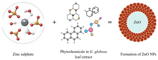
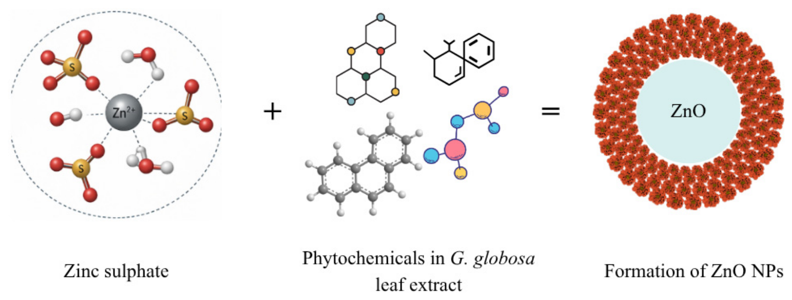
Table 1 represents the phytochemical screening of the aqueous extract of G. globosa leaves. The aqueous extract of G. globosa contained alkaloids, sterols, anthraquinones, proteins, carbohydrates, flavonoids, coumarins and saponins. The phytochemicals, such as alkaloids, flavonoids, anthraquinones and coumarins, in the G. globosa leaf extract acted as the significant reducing and capping agent for the fabrication of ZnO NPs (Figure 1). Similarly, the phytochemical screening performed by Tamilarasi and Meena [55] using the aqueous extract of G. globosa leaves contained significant phytochemicals, namely terpenoids, flavones, aldehydes, phenols, amides, carboxylic acids, ketones and ascorbic acids.
Table 1.
Phytochemical screening of G. globosa aqueous leaf extract.
Figure 1.
Schematic representation of G. globosa-mediated ZnO NP formation.
3.2. Physicochemical Characterization of ZnO NPs
3.2.1. UV-Vis Spectral Analysis
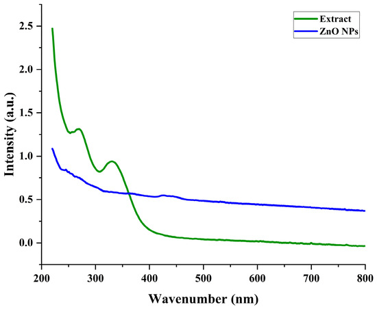
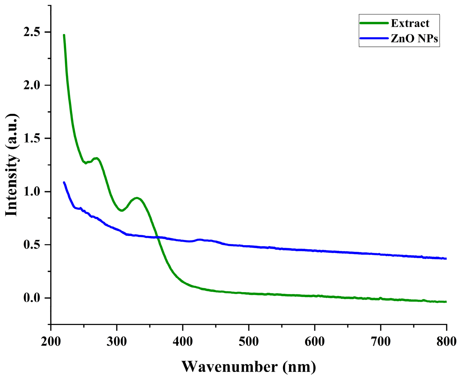
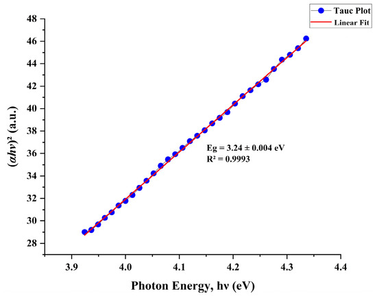
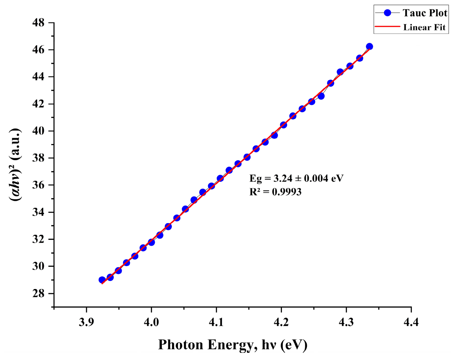
UV-Vis spectra (Figure 2) of the G. globosa aqueous extract showed two significant peaks between 200 and 380 nm, while the UV-Vis spectra (Figure 2) of G. globosa-mediated ZnO NPs displayed a distinct peak at 410 nm. The shift in the peaks denoted the surface modification and bio-capping of Zn2+ ions by phytochemicals present in the G. globosa aqueous extract. The band-gap energy of the G. globosa-mediated ZnO NPs was 3.24 eV, as illustrated by the Tauc plot (Figure 3). The variation in peaks verified the excitonic absorption of the synthesized ZnO NPs. Muhammad et al. [56] performed UV-Vis analysis of Papaver somnifera-mediated ZnO NPs and observed a significant peak at 360 nm. The UV-Vis spectra of Deverra tortuosa displayed a prominent peak at 374 nm [57].
Figure 2.
UV-Vis spectra of G. globosa aqueous extract and G. globosa-mediated ZnO NPs.
Figure 3.
Tauc plot of G. globosa-mediated ZnO NPs.
3.2.2. FTIR Analysis
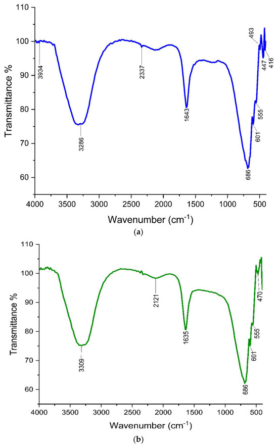
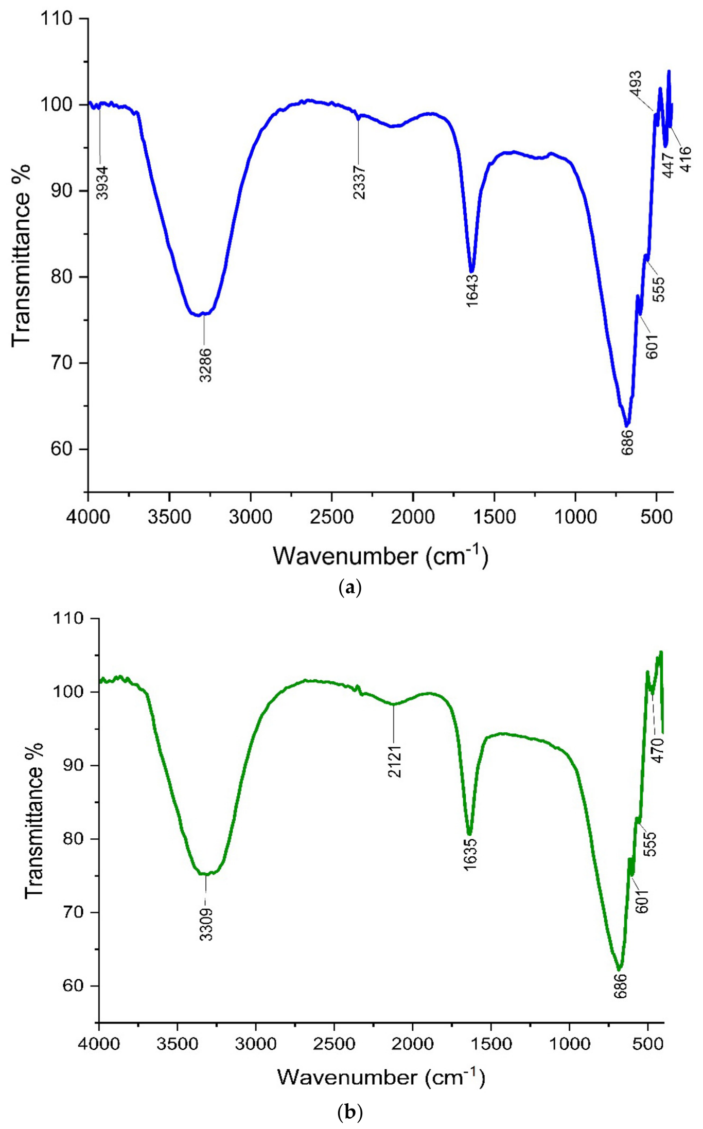
An FTIR spectrum of the G. globosa aqueous extract is displayed in Figure 4a. The peak occurred at 3286 cm−1, corresponding to the presence of alcohols. The peak at 2337 cm−1 indicated the occurrence of carbon dioxide. The peak at 1643 cm−1 confirms that amides were present in the G. globosa aqueous extract. Other peaks at 686, 601, 555, 493, 447 and 416 cm−1 affirmed the existence of alkyl and aryl halides (C-Cl stretch) functional groups. The functional groups such as alcohols and amides represent the coumarins and flavonoids in the G. globosa aqueous extract. Arunachalam et al. [58] reported that the FTIR spectrum of a G. globosa methanolic leaf extract demonstrated the presence of distinct peaks at 656, 665, 685, 687, 735, 737 and 741 cm−1. Figure 4b shows the FTIR spectra of G. globosa-mediated ZnO NPs. The peak observed at 3309 cm−1 determined the incidence of alcohols. The significant peak at 2121 cm−1 verified that alkynes were present. The peak detected at 1635 cm−1 established the prevalence of alkenes. The peaks at 686, 601, 555 and 470 cm−1 denoted the Zn-O groups. Based on the FTIR spectra, it could be confirmed that the coumarins and flavonoids acted as a significant capping and reducing agent for the fabrication of ZnO NPs. Balogun et al. [59] performed FTIR analysis of ZnO NPs fabricated using M. pudica and observed multiple significant peaks at 3903, 3626, 2802, 2395, 2195, 1963, 1905, 1834, 1643, 1494, 1288, 1194, 968, 882, 783 and 626 cm−1 and reported that the peaks at lower wavelengths confirmed the fabrication of ZnO NPs.
Figure 4.
(a) FTIR spectra of G. globosa aqueous extract. (b) FTIR spectra of G. globosa-mediated ZnO NPs.
3.2.3. XRD Analysis
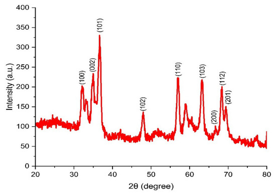
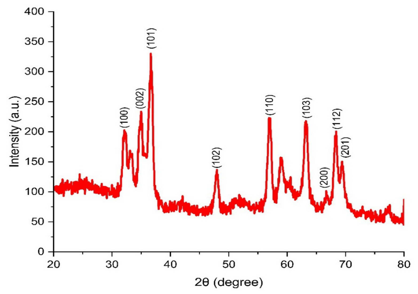
XRD spectra (Figure 5) proved the crystalline nature of G. globosa-mediated ZnO NPs. The crystal lattice peaks were observed (100), (002), (101), (102), (110), (103), (200), (112), (201) at 2θ = 33°, 35°, 36.8°, 47.9°, 56.9°, 63°, 67°, 68.4°, and 69.3°. JCDPS Card No. 36-1451 of the synthesized ZnO NPs revealed their hexagonal wurtzite structure with a d-spacing of 0.244 nm. The crystal lattices (100), (002), (102), (110), (103), and (112) corresponded to the ZnO wurtzite structure. Other planes (101) and (200) depict the formation of ZnO NPs through a biological stabilizing agent. The shifts in the 2θ of wurtzite zinc oxide and the synthesized ZnO NPs were due to the interaction of precursors with plant phytochemicals. The crystalline size of G. globosa-mediated ZnO NPs was 43 nm, as calculated using the Debye–Scherrer equation. The crystal lattice planes at (100), (002), (101), (102), (110), (103), (112) and (201) confirm the formation of Cayratia pedata-mediated ZnO NPs [60]. Muthu Kathija et al. [61] observed the crystalline phase of P. alba leaf extract-mediated ZnO NPs with various diffraction patterns at 2θ = 31.6°, 34.3°, 36.1°, 47.4°, 56.4°, 62.7°, 66.2°, 67.8°, and 69°.
Figure 5.
XRD spectra of G. globosa-mediated ZnO NPs.
3.2.4. SEM and EDX Analysis
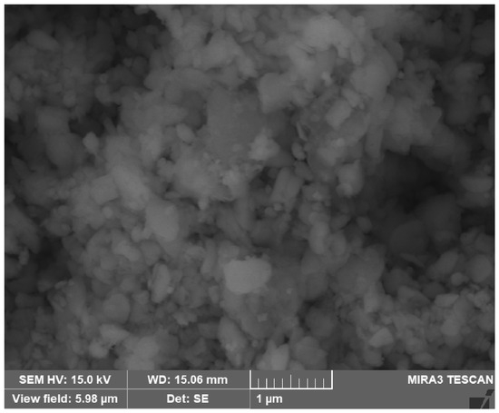
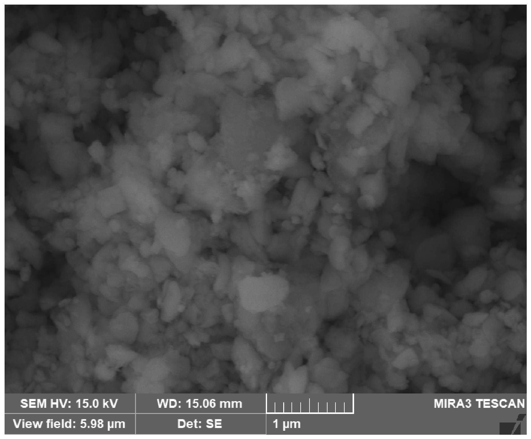
Figure 6 reveals the spherical shape and uniform morphology of G. globosa-mediated ZnO NPs. Vijayakumar et al. [62] reported the spherical shape of Atalantia monophylla leaf-assisted ZnO NPs using SEM images. The average size of ZnO NPs fabricated using Thymbra spicata was 65–75 nm, as recorded by Ceylan et al. [63].
Figure 6.
SEM image of G. globosa-mediated ZnO NPs.
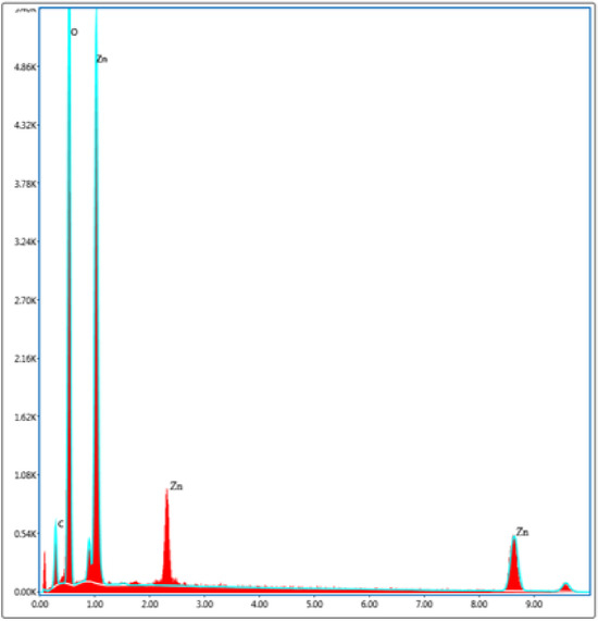
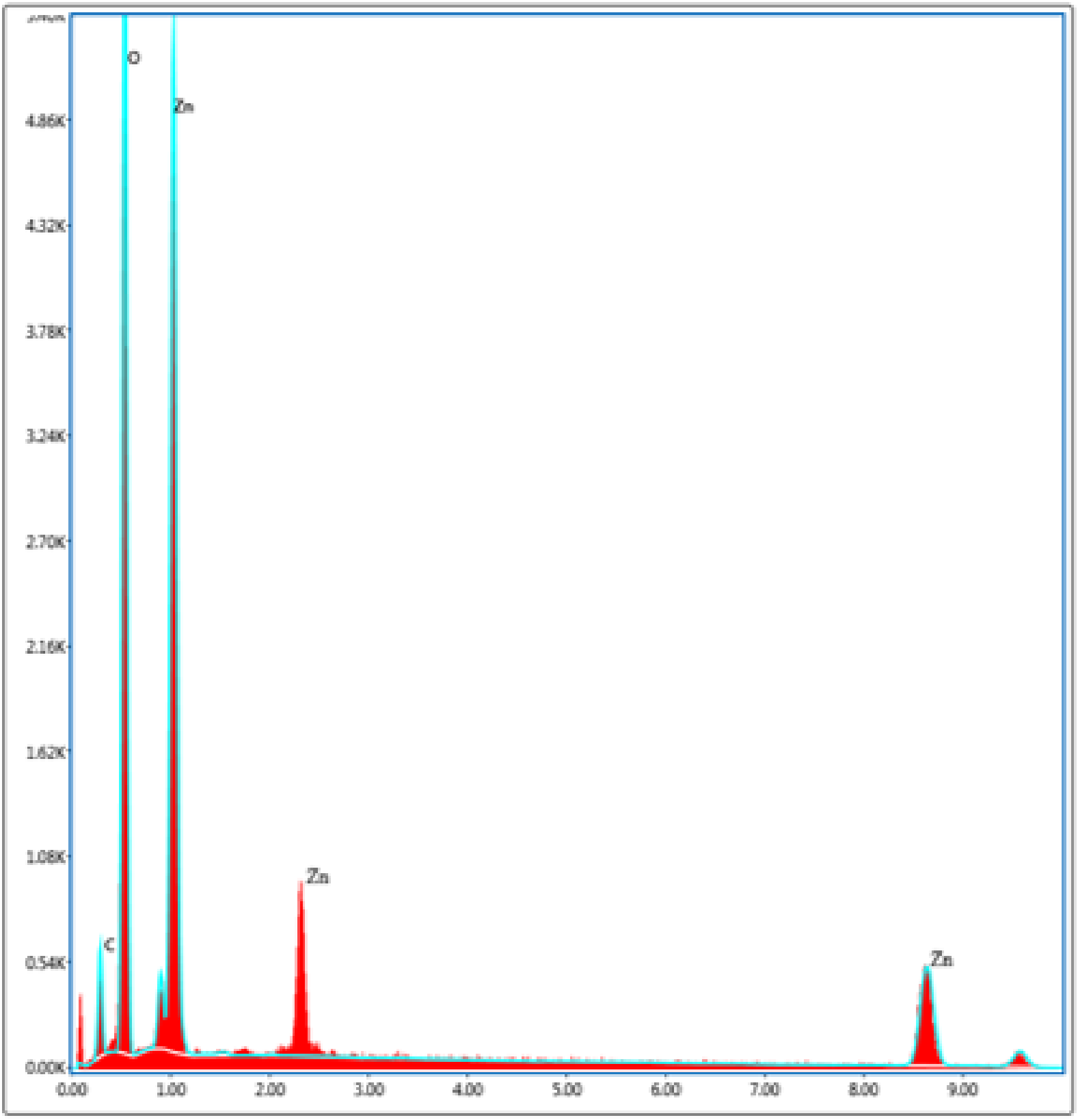
The elemental composition of G. globosa-mediated ZnO NPs was 45% zinc and 35% oxygen with 7% carbon, as exhibited in Figure 7. The occurrence of C bands indicated the carbon elements present in the plant material. ZnO NPs synthesized using S. baicalensis constituted 20% oxygen and 79% zinc [64]. El-Belely et al. [65] investigated the elemental composition of Arthrospira platensis-mediated ZnO NPs, revealing 56% zinc and 20% oxygen.
Figure 7.
EDX spectrum of G. globosa-mediated ZnO NPs.
3.2.5. HRTEM Analysis
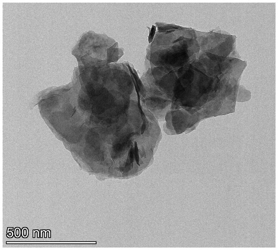
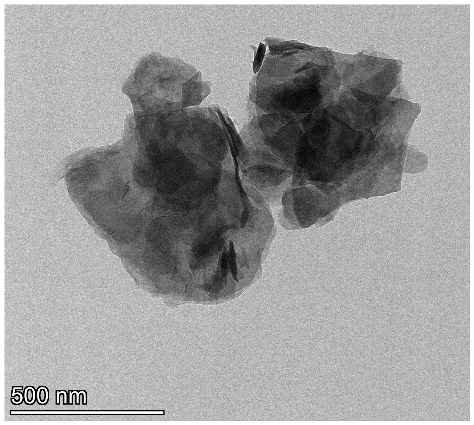
Figure 8 displays the HRTEM micrographs of G. globosa-mediated ZnO NPs. The quasi-spherical shape and average size of 60 nm of the synthesized ZnO NPs were verified by the HRTEM analysis. SAED analysis revealed concentric diffraction rings with bright spots, indicating the polycrystalline nature of the synthesized ZnO NPs. The measured reciprocal spacing corresponded to d-spacing of approximately 0.585, 0.413, 0.256, 0.154 and 0.129 nm. Mushtaq et al. [66] performed HRTEM analysis of Viscum album-mediated ZnO NPs and reported its quasi-spherical shape and average size of 50 nm. TEM images verified the hexagonal shape of ZnO NPs synthesized using Deverra tortuosa [57].
Figure 8.
HRTEM micrograph of G. globosa-mediated ZnO NPs.
3.2.6. XPS Analysis
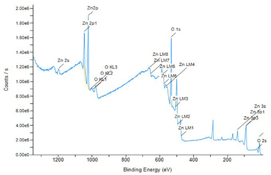
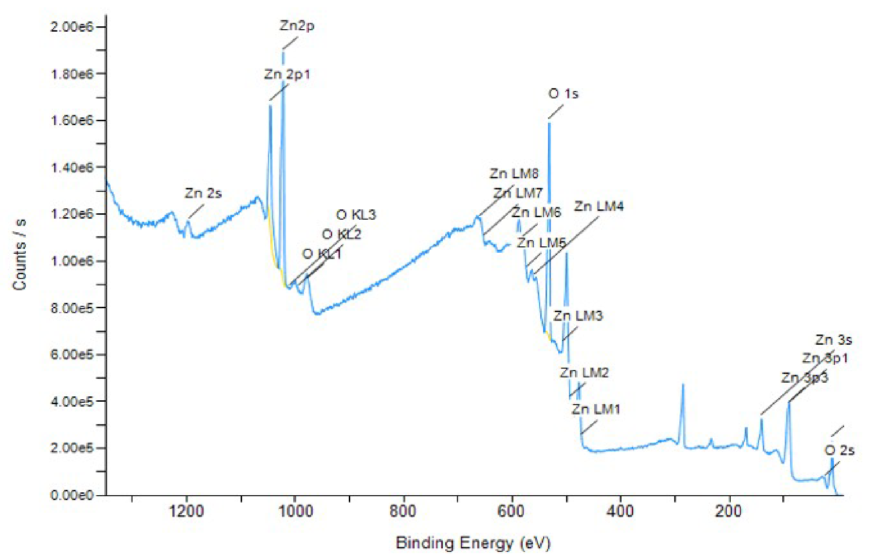
The surface components of the synthesized ZnO NPs were demonstrated in Figure 9. The predominant binding energy peaks of zinc and oxygen between 400 and 600 eV prove the formation of Zn-O bonding. Apart from the surface composition, XPS analysis also revealed that the elements zinc, oxygen and carbon existed in different excitation energies with electrons in different subshells, such as 2s, 2p and 3s. Gaikwad et al. [67] performed XPS analysis with Mirabilis jalapa-mediated ZnO NPs and reported that the Zn peaks at 1021 eV and 1044 eV represented the presence of zinc, while there were O peaks at 529 eV and 530 eV. Wu et al. [68] observed Zn 2p spectra located at 288 and 870 eV, verifying the Zn-O bonding in the XPS analysis of Aloevera leaf-mediated ZnO NPs.
Figure 9.
XPS analysis of G. globosa-mediated ZnO NPs.
3.2.7. DLS Analysis
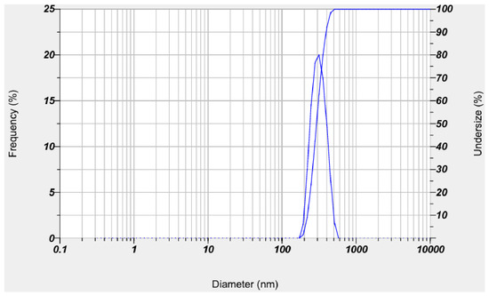
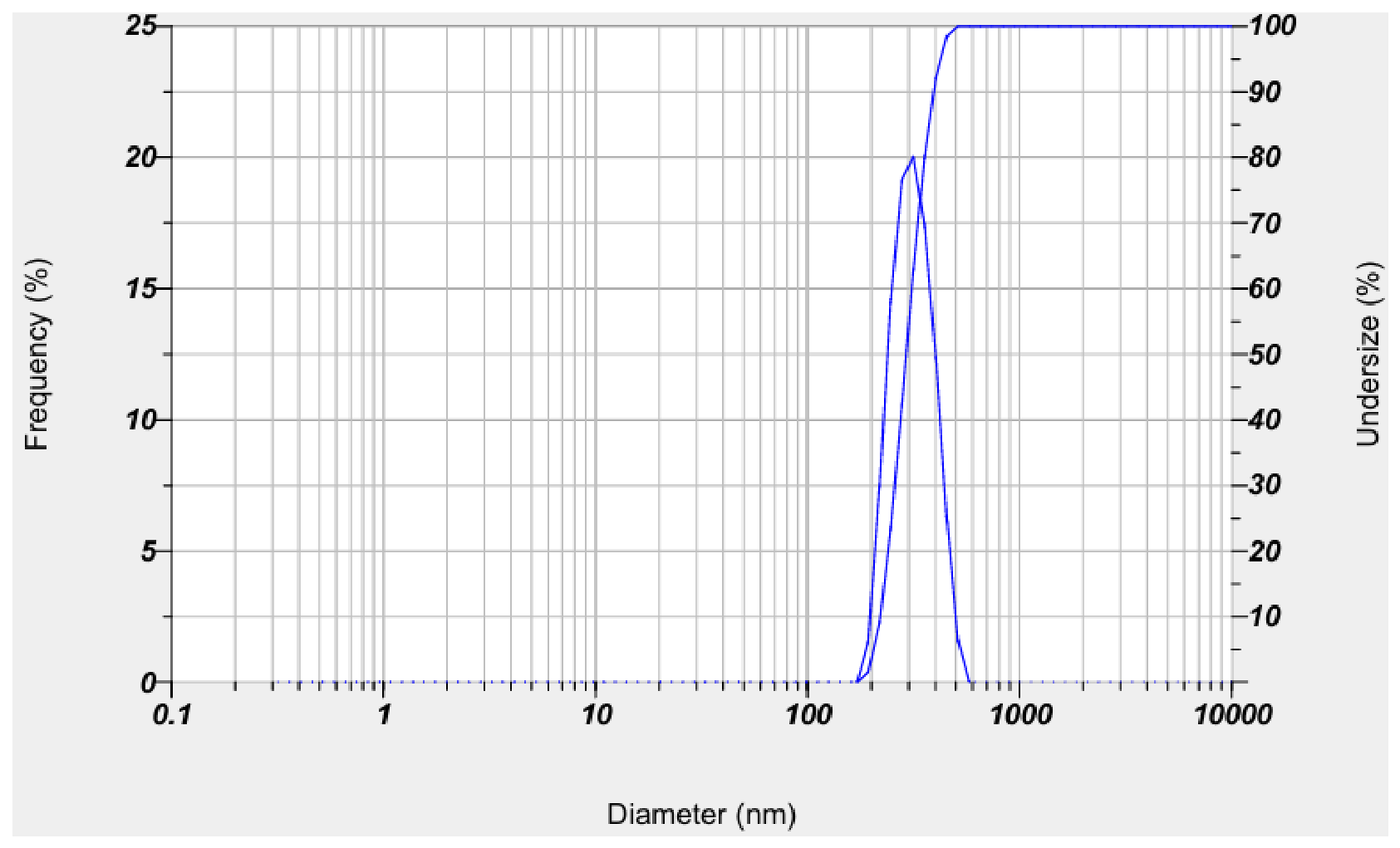
DLS analysis (Figure 10) revealed the particle size distribution of G. globosa-mediated ZnO NPs. The hydrodynamic diameter of 300 nm indicated the size of the synthesized ZnO NPs in Brownian motion and the shell thickness of the capping and reducing agents. The large gap between the average size and the hydrodynamic diameter is due to the agglomeration of the synthesized ZnO NPs and scattering of light accordingly. The polydispersity index of the G. globosa-mediated ZnO NPs was observed to be 0.6 and denoted its polydisperse nature. Acalypha indica leaf-assisted ZnO NPs showed a hydrodynamic diameter of 46 nm [69]. Abdelbaky et al. [70] performed DLS analysis of P. odoratissimum-mediated ZnO NPs and reported a hydrodynamic diameter of 6 nm with a polydispersity index of 0.4.
Figure 10.
DLS analysis of G. globosa-mediated ZnO NPs.
3.2.8. Zeta Potential Analysis
The zeta potential value of −31.6 mV was observed in G. globosa-mediated ZnO NPs. Particles with zeta potential values lower than −30 mV and larger than +30 mV are considered to be stable. Mongy and Shalaby [71] proved the zeta potential of −19.19 mV in zinc oxide nanoparticles fabricated using Rhus coriaria. Myristica fragrans-assisted ZnO NPs had a zeta potential of −22.1 mV and was strongly anionic. The negative surface charge was due to the binding affinity of nanoparticles and the biomolecules present in the plant extract [72].
3.2.9. TG Analysis
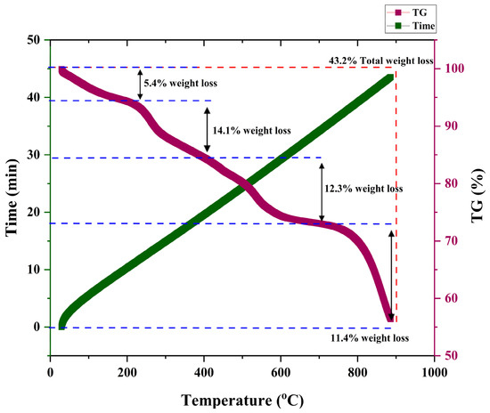
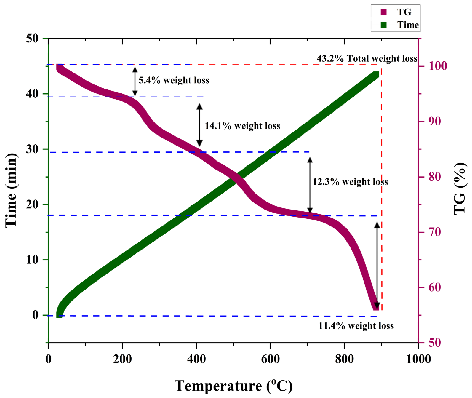
TG analysis exhibited four-step thermal degradation, as shown in Figure 11. The initial weight loss of 5.4% occurred due to the evaporation of water molecules. The second step displayed the degradation of volatile components with 14.1% weight loss. The third weight loss of 12.3% indicated the deterioration of small organic molecules present in the synthesized ZnO NPs. The final weight loss of 11.4% corresponded to the decay of complex organic compounds. The TGA spectra of Ocimum basilicum-assisted ZnO NPs showed 33% weight loss [73]. Naiel et al. [74] performed TG analysis of Limonium pruinosum extract-mediated ZnO NPs and reported its significant thermal stability with 29.08% weight loss.
Figure 11.
TG analysis of G. globosa-mediated ZnO NPs.
3.3. Biological Properties of ZnO NPs
3.3.1. Antibacterial Activity
G. globosa-mediated ZnO NPs had significant antibacterial activity against K. pneumoniae and E. coli (Table 2). G. globosa leaf extract exhibited significantly lower antibacterial activity against K. pneumoniae and E. coli. The results indicated that the bactericidal properties of the synthesized ZnO NPs were more efficient than the G. globosa leaf extract. ZnO NPs induce membrane loss in bacteria through endocytosis [75]. A high concentration of zinc ions causes membrane leakage and proton loss, leading to bacterial cell death [76]. Rambabu et al. [77] assessed the antibacterial activity of Phoenix dactylifera-mediated ZnO NPs and proved its significant antibacterial efficacy against S. aureus, S. pyogenes, P. aeruginosa and P. mirabilis. ZnO NPs fabricated using Brassica oleracea showed antibacterial activity against both Gram-positive and Gram-negative bacteria such as E. coli and S. aureus [78].
Table 2.
Antibacterial activity of G. globosa leaf extract and G. globosa-mediated ZnO NPs.
The synthesized ZnO NPs showed an MIC at 300 µg mL−1 in K. pneumoniae and 200 µg mL−1 in E. coli (Table 3). The higher MIC value in K. pneumoniae may be due to its protective capsule and strong defense mechanisms in comparison with E. coli. MIC and MBC analysis of the G. globosa leaf extract depicted that it inhibited the growth of K. pneumoniae at 900 µL and E. coli at 600 µL. Correspondingly, the G. globosa leaf extract displayed higher MIC and MBC values when compared to G. globosa-mediated ZnO NPs, indicating the enhanced bactericidal activity of the ZnO NPs. The MIC values of Punica granatum-mediated ZnO NPs against E. coli and S. aureus were 50 and 100 µg mL−1 [79]. MBC analysis reported that there was less than 1 colony growth in K. pneumoniae and E. coli bacterial cultures when treated with 400 µg mL−1 and 300 µg mL−1 of G. globosa-mediated ZnO NPs. The MBC of bio-formulated ZnO NPs using Origanum majorana was 20 µg mL−1, as reported by Yassin et al. [80].
Table 3.
MIC and MBC analysis of G. globosa leaf extract and G. globosa-mediated ZnO NPs.
3.3.2. Protein Leakage Assay
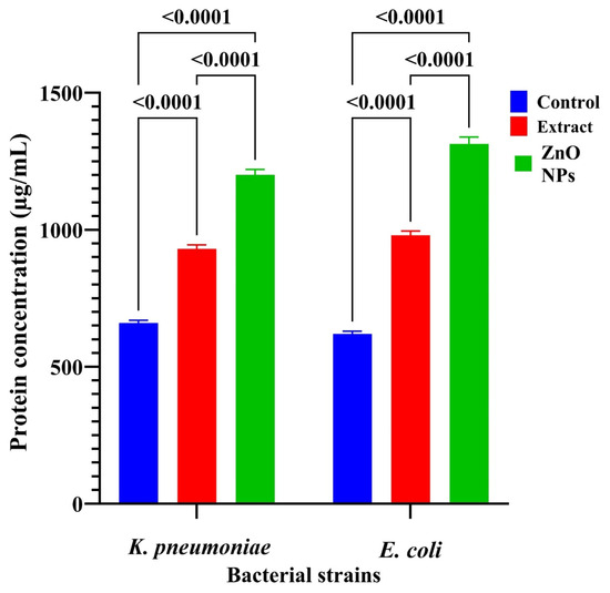
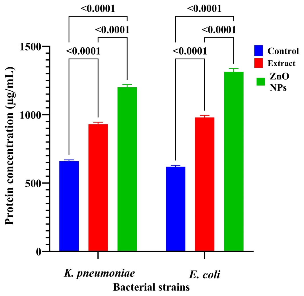
Figure 12 depicts that 1 mL of untreated K. pneumoniae cells comprised 660.7 µg of protein, while the G. globosa leaf extract-treated cells showed 930 µg of protein, and ZnO NP treatment displayed an increase in protein levels up to 1199 µg mL−1. Similarly, untreated E. coli possessed a protein concentration of 617 µg mL−1, while G. globosa leaf extract-treated cells exhibited 980 µg of protein, which was amplified to 1313 µg mL−1 after ZnO NP exposure. The protein leakage assay reported that the synthesized ZnO NPs effectively destroyed bacterial cell walls, leading to increased protein leakage, while the G. globosa leaf extract depicted reduced protein leakage due to less damage on the bacterial cell wall. The escalation in the protein concentration of the ZnO NP-treated cell suspension denoted the disruption of the bacterial cell membrane by the G. globosa-mediated ZnO NPs. The breakdown of cell membrane led to leakage of intracellular proteins into the cell suspension. The observations proved that treatment of synthesized ZnO NPs caused significant damage to the cell membranes of E. coli and K. pneumoniae, resulting in excess protein release. Zhu et al. [81] reported that green synthesized ZnO NPs using Cinnamomum camphora disrupted the membrane integrity of Alternaria alternata. Stevia rebaudiana-mediated ZnO NPs had the capacity to degrade the cell membrane, increasing the level of protein and nucleic acid leakage [57]. Table 4 illustrates the differences in the nature of G. globosa-mediated ZnO NPs and other green synthesized ZnO NPs.
Figure 12.
Protein leakage assay of G. globosa leaf extract and G. globosa-mediated ZnO NP-treated K. pneumoniae and E. coli.
Table 4.
Comparative discussion of green synthesized ZnO NPs and G. globosa-mediated ZnO NPs.
3.3.3. Antioxidant Assay
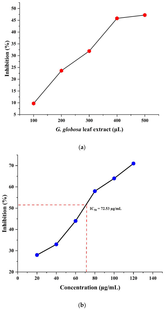
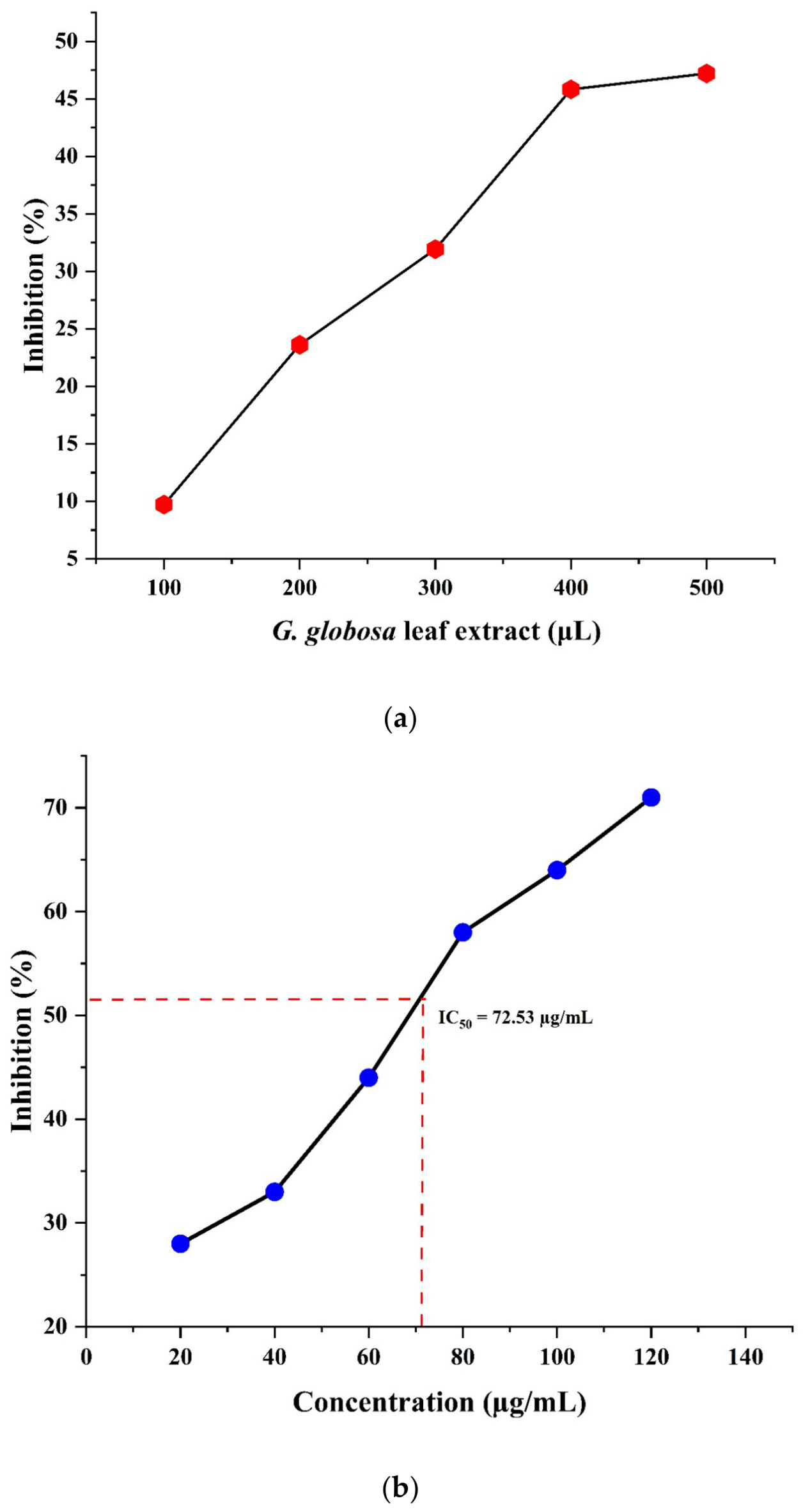
The antioxidant activity of the G. globosa leaf extract is presented in Figure 13a. The G. globosa leaf extract exhibited the lowest antioxidant activity at 100 µL and highest antioxidant activity at 500 µL. As demonstrated in Figure 13b, the synthesized ZnO NPs displayed the lowest antioxidant activity at 20 µg/mL and highest antioxidant activity at a concentration of 120 µg/mL. The IC50 value of the G. globosa-mediated ZnO NPs was observed to be 72.53 µg/mL. The phytochemicals on the surface of the ZnO NPs prohibited the oxidative damage caused by free radicals. The synthesized ZnO NPs facilitated the neutralization of the free radicals with free H+ ions. The G. globosa leaf extract had lower antioxidant activity, while the synthesized ZnO NPs possessed significant antioxidant activity due to the donation of free electrons by the ZnO NPs. Tiwari et al. [82] reported that Calendula officinalis-assisted ZnO NPs possessed free radical scavenging potential at an IC50 of 6600 µL/mL. ZnO NPs synthesized from Acacia nilotica displayed the maximum antioxidant activity at the highest concentration of 250 µg/mL [83].
Figure 13.
(a) Antioxidant activity of G. globosa leaf extract. (b) Antioxidant activity of G. globosa-mediated ZnO NPs.
3.3.4. Heavy Metal Adsorption
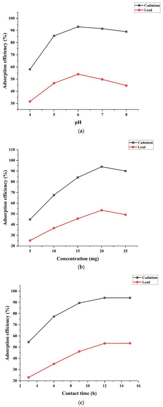
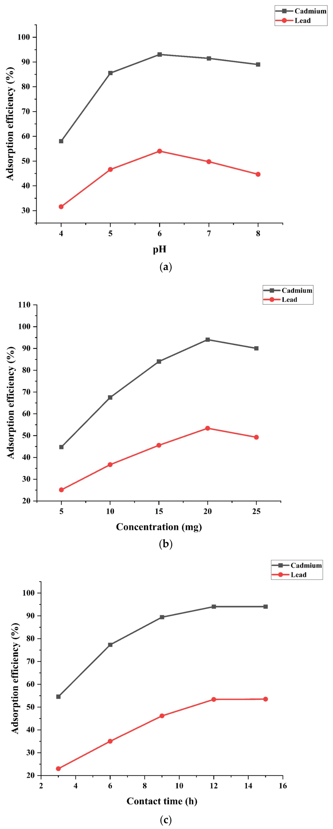
The optimization studies concluded that a pH of 6, concentration of 20 mg and a contact time of 12 h displayed the highest heavy metal removal efficiency for G. globosa-mediated ZnO NPs (Figure 14a–c). The synthesized ZnO NPs showed complete dependence on the pH, concentration and contact time, exhibiting higher removal of cadmium than lead. As the concentration of the ZnO NPs increased, greater efficiency was observed. However, the adsorption efficiency decreased at higher doses beyond 20 mg, which might be explained by particle aggregation and the availability of less active surface sites. The adsorption was found to be rapid and reached equilibrium at about 12 h.
Figure 14.
(a) Effect of pH on adsorption efficiency. (b) Effect of concentration on adsorption efficiency. (c) Effect of contact time on adsorption efficiency.
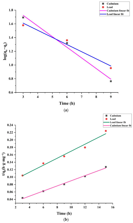
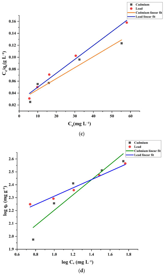
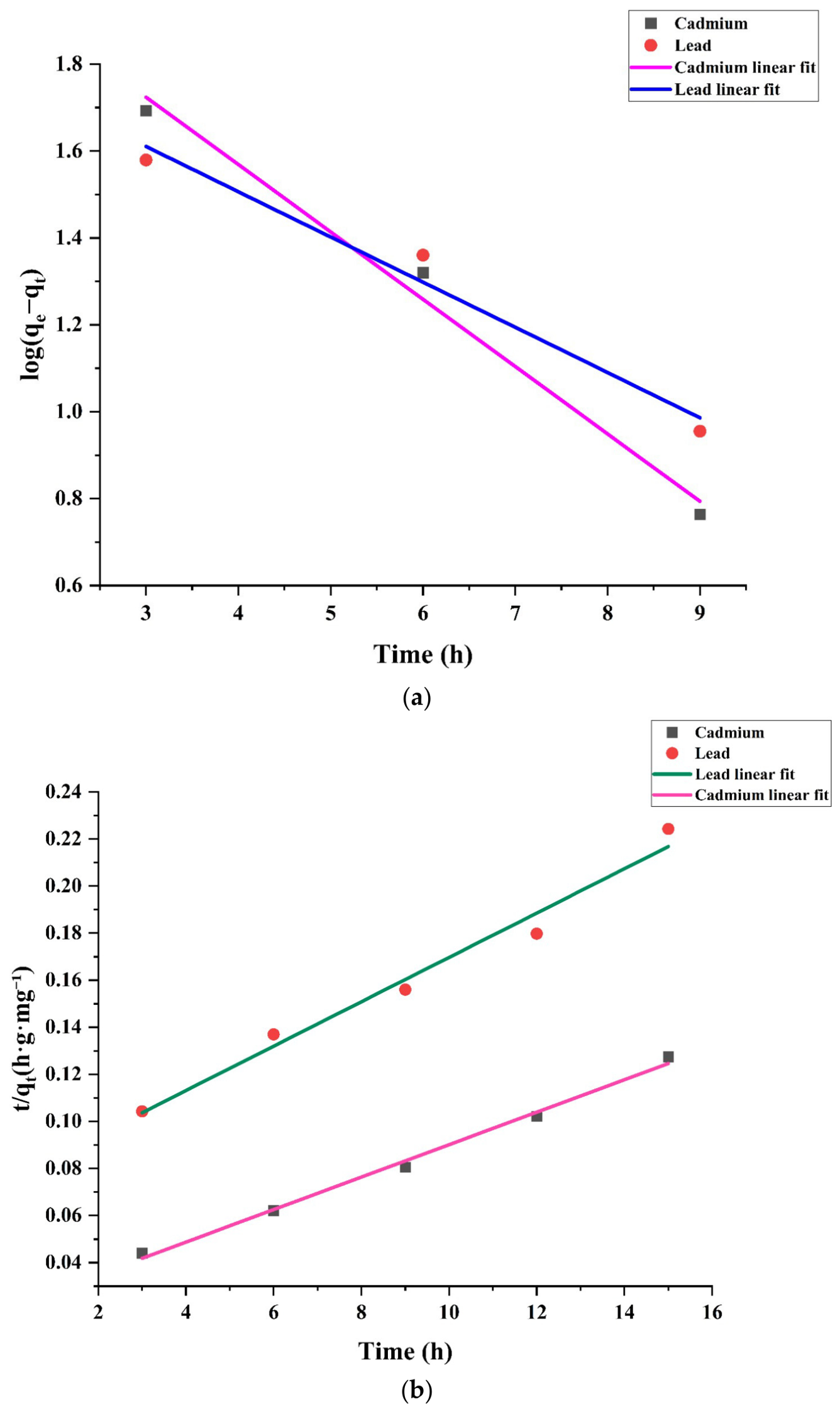
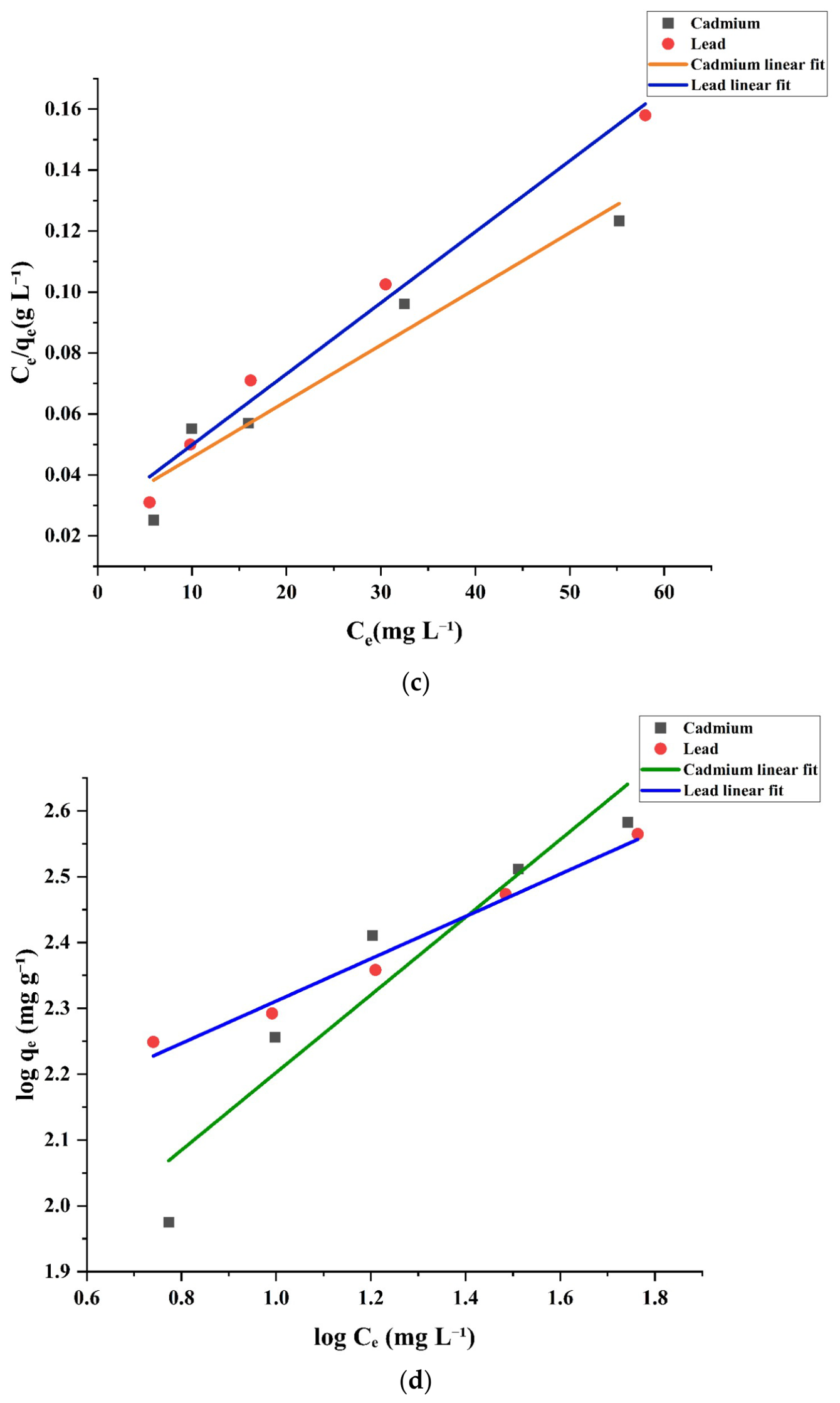
The pseudo-second-order (PSO) model gave slightly better linearized fit of the data, with an R2 = 0.99445 for cadmium and R2 = 0.97846 for lead, than the pseudo-first-order model, R2 = 0.98719 and R2 = 0.97119, respectively (Figure 15a). Figure 15b displays that the adsorption rate behavior is better described by the PSO model. Equilibrium modelling (Figure 15c) showed a better Langmuir fit, R2 = 0.93652 for cadmium and R2 = 0.98463 for lead, than Freundlich fit, R2 = 0.90171 and R2 = 0.98187 (Figure 15d), with calculated Langmuir maximum capacities (Qmax) of 543.48 mg g−1 for cadmium and 429.18 mg g−1 for lead. The Freundlich constants (n = 1.695 for cadmium (n = 3.112) and for lead (n > 1)) also report good adsorption. The results obtained show that the overall adsorption efficiency of the synthesized ZnO NPs was consistent with the Langmuir model with a significantly higher removal efficiency for cadmium than for lead.
Figure 15.
(a) Pseudo-first-order kinetics. (b) Pseudo-second-order kinetics. (c) Langmuir isotherm. (d) Freundlich isotherm.
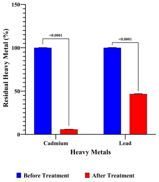
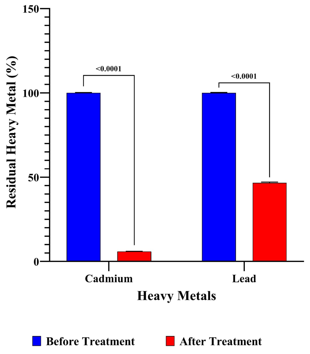
Figure 16 shows that 20 mg of G. globosa-mediated ZnO NPs adsorbed 94% of cadmium and 53% lead in the polluted water, representing its efficient removal. The porosity and large surface area of the G. globosa-mediated ZnO NPs enabled the adsorption of heavy metals onto the NP surface. The observations prove that G. globosa-mediated ZnO NPs efficiently removed cadmium and, moderately, lead from the polluted water. Cadmium has a smaller ionic radius than lead, which assists in the rapid diffusion of cadmium ions on the surfaces of the ZnO NPs. Lead has higher hydration enthalpy, making it more tightly bound to water molecules, leading to reduced adsorption. The contrast in the % of removal is due to the affinity of different metals differing on the NP surface based on the electron binding affinity (Table 5). Elsamra et al. [84] reported ZnO NPs synthesized using Ocimum tenuiflorum displayed 96% of lead and 21% of cadmium removal. The study also stated that the mobility of hydrogen ions influences the affinity for adsorption. Costus afers-assisted ZnO NPs showed efficient removal of cadmium and lead, irrespective of the change in pH and temperature [85].
Figure 16.
Heavy metal adsorption of G. globosa-mediated ZnO NPs.
Table 5.
Comparative discussion of G. globosa-mediated ZnO NPs and Ocimum tenuiflorum-mediated ZnO NPs.
4. Conclusions
The aqueous extract of G. globosa leaves was used to fabricate zinc oxide nanoparticles through the biosynthesis method. The physical, chemical and biological characteristics of the G. globosa-mediated ZnO NPs were evaluated using UV-Vis spectroscopy, FTIR, XRD, SEM, HRTEM, EDX, XPS, DLS, zeta potential analysis, TGA, antibacterial and antioxidant assays. The excitonic absorption of the synthesized ZnO NPs was proved, with a significant peak at 410 nm. The presence of metal oxide functional groups in G. globosa-mediated ZnO NPs was confirmed by the FTIR spectra. The G. globosa-mediated ZnO NPs had an average size of 60 nm, with a spherical shape. The HRTEM analysis proved the quasi-spherical shape of the synthesized ZnO NPs. The elemental composition of G. globosa-mediated ZnO NPs was reported as 45% of zinc and 35% of oxygen by the EDX spectra. The synthesized nanoparticles were stable, with a zeta potential value of −31.6 mV. G. globosa-mediated ZnO NPs had substantial antibacterial activity against K. pneumoniae and E. coli. The protein leakage assay displayed the impact of ZnO NPs on the cell wall of K. pneumoniae and E. coli. G. globosa-mediated ZnO NPs showed higher adsorptive removal of cadmium in comparison with lead. In future, the synthesized ZnO NPs could be potentially used for various applications in medical textiles or as an adsorbent for the removal of heavy metals.
Author Contributions
D.B.: Investigation and writing—original draft preparation; R.P.: conceptualization, supervision, funding acquisition and project administration; J.A.M.: review and editing; N.A.-D.: data curation. All authors have read and agreed to the published version of the manuscript.
Funding
The authors have no relevant financial or non-financial interests to disclose.
Institutional Review Board Statement
Not applicable.
Informed Consent Statement
Not applicable.
Data Availability Statement
The original contributions presented in this study are included in the article. Further inquiries can be directed to the corresponding author.
Conflicts of Interest
The authors declare that they have no conflicts of interest.
References
- Malik, S.; Muhammad, K.; Waheed, Y. Nanotechnology: A revolution in modern industry. Molecules 2023, 28, 661. [Google Scholar] [CrossRef] [Scilit] [PubMed]
- Haleem, A.; Javaid, M.; Singh, R.P.; Rab, S.; Suman, R. Applications of nanotechnology in medical field: A brief review. J. Glob. Health 2023, 7, 70–77. [Google Scholar] [CrossRef] [Scilit]
- Yezdani, U.; Khan, M.G.; Kushwah, N.; Verma, A.; Khan, F. Application of nanotechnology in diagnosis and treatment of various diseases and its future advances in medicine. World J. Pharm. Pharm. Sci. 2018, 7, 1611–1633. [Google Scholar]
- Sahu, T.; Ratre, Y.K.; Chauhan, S.; Bhaskar, L.V.K.S.; Nair, M.P.; Verma, H.K. Nanotechnology based drug delivery system: Current strategies and emerging therapeutic potential for medical science. Drug Deliv. Sci. Technol. 2021, 63, 102487. [Google Scholar] [CrossRef] [Scilit]
- Wang, X.; Gao, X.; Wang, L.; Lin, J.; Liu, Y. Advances of Nanotechnology Toward Vaccine Development Against Animal Infectious Diseases. Adv. Funct. Mater. 2023, 33, 2305061. [Google Scholar] [CrossRef] [Scilit]
- Mitra, S.; Pal, P.; Banerjee, E.R. Nanoscience Research in Regenerative Medicine. In Nanomaterials and Biomedicine; Springer: Singapore, 2020; pp. 67–84. [Google Scholar] [CrossRef] [Scilit]
- Sanzari, I.; Leone, A.; Ambrosone, A. Nanotechnology in plant science: To make a long story short. Front. Bioeng. Biotechnol. 2019, 7, 120. [Google Scholar] [CrossRef] [Scilit] [PubMed]
- Kolahalam, L.A.; Viswanath, I.K.; Diwakar, B.S.; Govindh, B.; Reddy, V.; Murthy, Y.L.N. Review on nanomaterials: Synthesis and applications. Mater. Today Proc. 2019, 18, 2182–2190. [Google Scholar] [CrossRef] [Scilit]
- Yang, T.; Liu, Y.; Wang, H.; Duo, Y.; Zhang, B.; Ge, Y.; Zhang, H.; Chen, W. Recent advances in 0D nanostructure-functionalized low-dimensional nanomaterials for chemiresistive gas sensors. J. Mater. Chem. C 2020, 8, 7272–7299. [Google Scholar] [CrossRef] [Scilit]
- Shin, J.; Kang, N.; Kim, B.; Hong, H.; Yu, L.; Kim, J.; Kang, H.; Kim, J.S. One-dimensional nanomaterials for cancer therapy and diagnosis. Chem. Soc. Rev. 2023, 52, 4488–4514. [Google Scholar] [CrossRef] [Scilit] [PubMed]
- Liu, S.; Pan, X.; Liu, H. Two-dimensional nanomaterials for photothermal therapy. Angew. Chem. Int. Ed. 2020, 59, 5890–5900. [Google Scholar] [CrossRef] [Scilit] [PubMed]
- Verma, C.; Berdimurodov, E.; Verma, D.K.; Berdimuradov, K.; Alfantazi, A.; Hussain, C.M. 3D Nanomaterials: The future of industrial, biological, and environmental applications. Inorg. Chem. Commun. 2023, 156, 111163. [Google Scholar] [CrossRef] [Scilit]
- Salem, S.S.; Hammad, E.N.; Mohamed, A.A.; El-Dougdoug, W. A comprehensive review of nanomaterials: Types, synthesis, characterization, and applications. Biointerface Res. Appl. Chem. 2022, 13, 41. [Google Scholar] [CrossRef] [Scilit]
- El-Eskandarany, M.S.; Al-Hazza, A.; Al-Hajji, L.A.; Ali, N.; Al-Duweesh, A.A.; Banyan, M.; Al-Ajmi, F. Mechanical milling: A superior nanotechnological tool for fabrication of nanocrystalline and nanocomposite materials. Nanomaterials 2021, 11, 2484. [Google Scholar] [CrossRef] [Scilit] [PubMed]
- Dada, A.O.; Adekola, F.A.; Odebunmi, E.O.; Ogunlaja, A.S.; Bello, O.S. Two–three parameters isotherm modeling, kinetics with statistical validity, desorption and thermodynamic studies of adsorption of Cu (II) ions onto zerovalent iron nanoparticles. Sci. Rep. 2021, 11, 16454. [Google Scholar] [CrossRef] [Scilit] [PubMed]
- Lee, E.; Menumerov, E.; Hughes, R.A.; Neretina, S.; Luo, T. Low-cost nanostructures from nanoparticle-assisted large-scale lithography significantly enhance thermal energy transport across solid interfaces. ACS Appl. Mater. Interfaces 2018, 10, 34690–34698. [Google Scholar] [CrossRef] [Scilit] [PubMed]
- Ahmad, M.; Gani, A.; Hassan, I.; Huang, Q.; Shabbir, H. Production and characterization of starch nanoparticles by mild alkali hydrolysis and ultra-sonication process. Sci. Rep. 2020, 10, 3533. [Google Scholar] [CrossRef] [Scilit] [PubMed]
- Balachandran, A.; Sreenilayam, S.P.; Madanan, K.; Thomas, S.; Brabazon, D. Nanoparticle production via laser ablation synthesis in solution method and printed electronic application—A brief review. Results Eng. 2022, 16, 100646. [Google Scholar] [CrossRef] [Scilit]
- Altammar, K.A. A review on nanoparticles: Characteristics, synthesis, applications, and challenges. Front. Microbiol. 2023, 14, 1155622. [Google Scholar] [CrossRef] [Scilit] [PubMed]
- Ba-Abbad, M.M.; Benamour, A.; Ewis, D.; Mohammad, A.W.; Mahmoudi, E. Synthesis of Fe3O4 nanoparticles with different shapes through a co-precipitation method and their application. JOM 2022, 74, 3531–3539. [Google Scholar] [CrossRef] [Scilit]
- Singh, M.; Vadher, D.; Dixit, V.; Jariwala, C. Synthesis, optimization and characterization of zinc oxide nanoparticles prepared by sol–gel technique. Mater. Today Proc. 2022, 48, 690–692. [Google Scholar] [CrossRef] [Scilit]
- Zhao, X.; Wei, C.; Gai, Z.; Yu, S.; Ren, X. Chemical vapor deposition and its application in surface modification of nanoparticles. Chem. Pap. 2020, 74, 767–778. [Google Scholar] [CrossRef] [Scilit]
- Li, H.; Chen, X.; Shen, D.; Wu, F.; Pleixats, R.; Pan, J. Functionalized silica nanoparticles: Classification, synthetic approaches and recent advances in adsorption applications. Nanoscale 2021, 13, 15998–16016. [Google Scholar] [CrossRef] [Scilit] [PubMed]
- Tadic, M.; Trpkov, D.; Kopanja, L.; Vojnovic, S.; Panjan, M. Hydrothermal synthesis of hematite (α-Fe2O3) nanoparticle forms: Synthesis conditions, structure, particle shape analysis, cytotoxicity and magnetic properties. J. Alloys Compd. 2019, 792, 599–609. [Google Scholar] [CrossRef] [Scilit]
- Hachem, K.; Ansari, M.J.; Saleh, R.O.; Kzar, H.H.; Al-Gazally, M.E.; Altimari, U.S.; Hussein, S.A.; Mohammed, H.T.; Hammid, A.T.; Kianfar, E. Methods of chemical synthesis in the synthesis of nanomaterial and nanoparticles by the chemical deposition method: A review. BioNanoScience 2022, 12, 1032–1057. [Google Scholar] [CrossRef] [Scilit]
- Hano, C.; Abbasi, B.H. Plant-based green synthesis of nanoparticles: Production, characterization and applications. Biomolecules 2021, 12, 31. [Google Scholar] [CrossRef] [Scilit] [PubMed]
- Samuel, M.S.; Ravikumar, M.; John, J.A.; Selvarajan, E.; Patel, H.; Chander, P.S.; Soundarya, J.; Vuppala, S.; Balaji, R.; Chandrasekar, N. A review on green synthesis of nanoparticles and their diverse biomedical and environmental applications. Catalysts 2022, 12, 459. [Google Scholar] [CrossRef] [Scilit]
- Sithara, N.V.; Senthil Kumar, J. Green Synthesis and Structural Characterization of Zinc Oxide Nanoparticles Using Aqueous Extract of Musa acuminata colla Bracts. Natl. Acad. Sci. Lett. 2026; in press.
- Ribeiro, A.I.; Dias, A.M.; Zille, A. Synergistic effects between metal nanoparticles and commercial antimicrobial agents: A review. ACS Appl. Nano Mater. 2022, 5, 3030–3064. [Google Scholar] [CrossRef] [Scilit] [PubMed]
- Amutha, V.; Deepak, P.; Kamaraj, C.; Balasubramani, G.; Aiswarya, D.; Arul, D.; Santhanam, P.; Ballamurugan, A.Á.; Perumal, P. Mosquito-larvicidal potential of metal and oxide nanoparticles synthesized from aqueous extract of the seagrass, Cymodocea serrulata. J. Clust. Sci. 2019, 30, 797–812. [Google Scholar] [CrossRef] [Scilit]
- Javed, R.; Zia, M.; Naz, S.; Aisida, S.O.; Ain, N.U.; Ao, Q. Role of capping agents in the application of nanoparticles in biomedicine and environmental remediation: Recent trends and future prospects. J. Nanobiotechnol. 2020, 18, 172. [Google Scholar] [CrossRef] [Scilit] [PubMed]
- Chiari-Andréo, B.G.; de Almeida-Cincotto, M.G.J.; Oshiro, J.A., Jr.; Taniguchi, C.Y.Y.; Chiavacci, L.A.; Isaac, V.L.B. Nanoparticles for cosmetic use and its application. In Nanoparticles in Pharmacotherapy; William Andrew Publishing: Norwich, NY, USA, 2019; pp. 113–146. [Google Scholar] [CrossRef] [Scilit]
- Fritea, L.; Banica, F.; Costea, T.O.; Moldovan, L.; Dobjanschi, L.; Muresan, M.; Cavalu, S. Metal nanoparticles and carbon-based nanomaterials for improved performances of electrochemical (Bio) sensors with biomedical applications. Materials 2021, 14, 6319. [Google Scholar] [CrossRef] [Scilit] [PubMed]
- Zahin, N.; Anwar, R.; Tewari, D.; Kabir, M.T.; Sajid, A.; Mathew, B.; Uddin, M.S.; Aleya, L.; Abdel-Daim, M.M. Nanoparticles and its biomedical applications in health and diseases: Special focus on drug delivery. Environ. Sci. Pollut. Res. 2020, 27, 19151–19168. [Google Scholar] [CrossRef] [Scilit] [PubMed]
- Nair, G.M.; Sajini, T.; Mathew, B. Advanced green approaches for metal and metal oxide nanoparticles synthesis and their environmental applications. Talanta Open 2022, 5, 100080. [Google Scholar] [CrossRef] [Scilit]
- Nishanthi, R.; Malathi, S.; Palani, P. Green synthesis and characterization of bioinspired silver, gold and platinum nanoparticles and evaluation of their synergistic antibacterial activity after combining with different classes of antibiotics. Mater. Sci. Eng. C 2019, 96, 693–707. [Google Scholar] [CrossRef] [Scilit] [PubMed]
- Joudeh, N.; Saragliadis, A.; Koster, G.; Mikheenko, P.; Linke, D. Synthesis methods and applications of palladium nanoparticles: A review. Front. Nanotechnol. 2022, 4, 1062608. [Google Scholar] [CrossRef] [Scilit]
- Chakraborty, N.; Banerjee, J.; Chakraborty, P.; Banerjee, A.; Chanda, S.; Ray, K.; Acharya, K.; Sarkar, J. Green synthesis of copper/copper oxide nanoparticles and their applications: A review. Green Chem. Lett. Rev. 2022, 15, 187–215. [Google Scholar] [CrossRef] [Scilit]
- Ajinkya, N.; Yu, X.; Kaithal, P.; Luo, H.; Somani, P.; Ramakrishna, S. Magnetic iron oxide nanoparticle (IONP) synthesis to applications: Present and future. Materials 2020, 13, 4644. [Google Scholar] [CrossRef] [Scilit] [PubMed]
- Zahra, M.; Ullah, H.; Javed, M.; Iqbal, S.; Ali, J.; Alrbyawi, H.; Alwadai, N.; Basha, B.I.; Waseem, A.; Sarfraz, S.; et al. Synthesis and characterization of polyurethane/zinc oxide nanocomposites with improved thermal and mechanical properties. Inorg. Chem. Commun. 2022, 144, 109916. [Google Scholar] [CrossRef] [Scilit]
- Bhadwal, N.; Ben Mrad, R.; Behdinan, K. Review of zinc oxide piezoelectric nanogenerators: Piezoelectric properties, composite structures and power output. Sensors 2023, 23, 3859. [Google Scholar] [CrossRef] [Scilit] [PubMed]
- Ramesh, P.; Saravanan, K.; Manogar, P.; Johnson, J.; Vinoth, E.; Mayakannan, M. Green synthesis and characterization of biocompatible zinc oxide nanoparticles and evaluation of its antibacterial potential. Sens. Bio-Sens. Res. 2021, 31, 100399. [Google Scholar] [CrossRef] [Scilit]
- Suganthi, K.S.; Harish, K.; Nair, N.M.; Swaminathan, P. Formulation and optimization of a zinc oxide nanoparticle ink for printed electronics applications. Flex. Print. Electron. 2018, 3, 015001. [Google Scholar] [CrossRef] [Scilit]
- Shayani Rad, M.; Sabeti, Z.; Mohajeri, S.A.; Fazly Bazzaz, B.S. Preparation, characterization, and evaluation of zinc oxide nanoparticles suspension as an antimicrobial media for daily use soft contact lenses. Curr. Eye Res. 2020, 45, 931–939. [Google Scholar] [CrossRef] [Scilit] [PubMed]
- Khalafi, T.; Buazar, F.; Ghanemi, K. Phycosynthesis and enhanced photocatalytic activity of zinc oxide nanoparticles toward organosulfur pollutants. Sci. Rep. 2019, 9, 6866. [Google Scholar] [CrossRef] [Scilit] [PubMed]
- Hadi, A.J.; Nayef, U.M.; Mutlak, F.A.H.; Jabir, M.S. Laser-ablated zinc oxide nanoparticles and evaluation of their antibacterial and anticancer activity against an ovarian cancer cell line: In vitro study. Plasmonics 2023, 18, 2091–2101. [Google Scholar] [CrossRef] [Scilit]
- Narayana, A.; Bhat, S.A.; Fathima, A.; Lokesh, S.V.; Surya, S.G.; Yelamaggad, C.V. Green and low-cost synthesis of zinc oxide nanoparticles and their application in transistor-based carbon monoxide sensing. RSC Adv. 2020, 10, 13532–13542. [Google Scholar] [CrossRef] [Scilit] [PubMed]
- Tang, S.R.; Sun, Y.X.; Gu, T.T.; Cao, F.F.; Shen, Y.B.; He, J.P.; Xie, Z.X.; Li, C. Phenolic compounds from Gomphrena globosa L.: Phytochemical analysis, antioxidant, antimicrobial, and enzyme inhibitory activities in vitro. CyTA-J. Food 2022, 20, 218–227. [Google Scholar] [CrossRef] [Scilit]
- Ramadhan, R.; Sari, A.A. The Effect of Extraction Methods on Total Flavonoid Content and Antioxidant Activity of Kenop (Gomphrena globosa). J. Pharm. Sci. 2025, 27, 2356–2361. [Google Scholar] [CrossRef] [Scilit]
- Roriz, C.L.; Barros, L.; Prieto, M.A.; Ćirić, A.; Soković, M.; Morales, P.; Ferreira, I.C. Enhancing the antimicrobial and antifungal activities of a coloring extract agent rich in betacyanins obtained from Gomphrena globosa L. flowers. Food Funct. 2018, 9, 6205–6217. [Google Scholar] [CrossRef] [Scilit] [PubMed]
- Bagheri, G.; Martorell, M.; Ramírez-Alarcón, K.; Salehi, B.; Sharifi-Rad, J. Phytochemical screening of Moringa oleifera leaf extracts and their antimicrobial activities. Cell. Mol. Biol. 2020, 66, 20–26. [Google Scholar] [CrossRef] [Scilit]
- Martin, J.A.; Periakaruppan, R.; Babu, D.; Vijai Selvaraj, K.S.; Al-Dayan, N. Green Synthesis, Characterization, and Antibacterial Activity of Indigofera aspalathoides-Assisted Manganese Oxide-Doped Calcium Oxide Nanocomposites. Biomed. Mater. Devices 2025, 4, 4751–4766. [Google Scholar] [CrossRef] [Scilit]
- Dhandapani, K.V.; Anbumani, D.; Gandhi, A.D.; Annamalai, P.; Muthuvenkatachalam, B.S.; Kavitha, P.; Ranganathan, B. Green route for the synthesis of zinc oxide nanoparticles from Melia azedarach leaf extract and evaluation of their antioxidant and antibacterial activities. Biocatal. Agric. Biotechnol. 2020, 24, 101517. [Google Scholar] [CrossRef] [Scilit]
- Elabbasy, M.T.; El Bayomi, R.M.; Abdelkarim, E.A.; Hafez, A.E.S.E.; Othman, M.S.; Ghoniem, M.E.; Samak, M.A.; Alshammari, M.H.; Almarshadi, F.A.; Elsamahy, T.; et al. Harnessing Stevia rebaudiana for zinc oxide nanoparticle green synthesis: A sustainable solution to combat multidrug-resistant bacterial pathogens. Nanomaterials 2025, 15, 369. [Google Scholar] [CrossRef] [Scilit] [PubMed]
- Tamilarasi, P.; Meena, P. Green synthesis of silver nanoparticles (Ag NPs) using Gomphrena globosa (Globe amaranth) leaf extract and their characterization. Mater. Today Proc. 2020, 33, 2209–2216. [Google Scholar] [CrossRef] [Scilit]
- Muhammad, W.; Ullah, N.; Haroon, M.; Abbasi, B.H. Optical, morphological and biological analysis of zinc oxide nanoparticles (ZnO NPs) using Papaver somniferum L. RSC Adv. 2019, 9, 29541–29548. [Google Scholar] [CrossRef] [Scilit] [PubMed]
- Selim, Y.A.; Azb, M.A.; Ragab, I.; HM Abd El-Azim, M. Green synthesis of zinc oxide nanoparticles using aqueous extract of Deverra tortuosa and their cytotoxic activities. Sci. Rep. 2020, 10, 3445. [Google Scholar] [CrossRef] [Scilit] [PubMed]
- Arunachalam, V.; Salgaonkar, D.C.; Kevat, N.V.; Walawalkar, B.V.; Das, B. Quantification of betacyanin content variation of amaranth varieties by an Android App, colorimeter, and infrared spectroscopy. Chin. J. Anal. Chem. 2022, 50, 100145. [Google Scholar] [CrossRef] [Scilit]
- Balogun, S.W.; James, O.O.; Sanusi, Y.K.; Olayinka, O.H. Green synthesis and characterization of zinc oxide nanoparticles using bashful (Mimosa pudica), leaf extract: A precursor for organic electronics applications. SN Appl. Sci. 2020, 2, 1–8. [Google Scholar] [CrossRef] [Scilit]
- Jayachandran, A.; Aswathy, T.R.; Nair, A.S. Green synthesis and characterization of zinc oxide nanoparticles using Cayratia pedata leaf extract. Biochem. Biophys. Rep. 2021, 26, 100995. [Google Scholar] [CrossRef] [Scilit] [PubMed]
- Muthu Kathija, M.; Badhusha, M.S.M.; Rama, V. Green synthesis of zinc oxide nanoparticles using Pisonia alba leaf extract and its antibacterial activity. Appl. Surf. Sci. Adv. 2023, 15, 100400. [Google Scholar] [CrossRef] [Scilit]
- Vijayakumar, S.; Mahadevan, S.; Arulmozhi, P.; Sriram, S.; Praseetha, P.K. Green synthesis of zinc oxide nanoparticles using Atalantia monophylla leaf extracts: Characterization and antimicrobial analysis. Mater. Sci. Semicond. Proc. 2018, 82, 39–45. [Google Scholar] [CrossRef] [Scilit]
- Ceylan, Y.; Korkmaz, N.; Kaçan, F.N.; Aydın, A. Phyto-fabrication of ZnO-NPs using Thymbra spicata L.; Evaluation of Antibacterial, Antibiofilm and Anticancer Efficacy. Afyon Kocatepe Univ. J. Sci. Eng. 2025, 25, 061201. [Google Scholar]
- Chen, L.; Batjikh, I.; Hurh, J.; Han, Y.; Huo, Y.; Ali, H.; Li, J.F.; Rupa, E.J.; Ahn, J.C.; Mathiyalagan, R.; et al. Green synthesis of zinc oxide nanoparticles from root extract of Scutellaria baicalensis and its photocatalytic degradation activity using methylene blue. Optik 2019, 184, 324–329. [Google Scholar] [CrossRef] [Scilit]
- El-Belely, E.F.; Farag, M.M.; Said, H.A.; Amin, A.S.; Azab, E.; Gobouri, A.A.; Fouda, A. Green synthesis of zinc oxide nanoparticles (ZnO-NPs) using Arthrospira platensis (Class: Cyanophyceae) and evaluation of their biomedical activities. Nanomaterials 2021, 11, 95. [Google Scholar] [CrossRef] [Scilit] [PubMed]
- Mushtaq, W.; Ishtiaq, M.; Maqbool, M.; Mazhar, M.W.; Casini, R.; Abd-ElGawad, A.M.; Elansary, H.O. Green synthesis of zinc oxide nanoparticles using Viscum album extracts: Unveiling bioactive compounds, antibacterial potential, and antioxidant activities. Plants 2023, 12, 2130. [Google Scholar] [CrossRef] [Scilit] [PubMed]
- Gaikwad, D.N.; Gaikwad, S.T.; Manjul, R.K.; Rajbhoj, A.S.; Varade, G.A.; Patil, A.S.; Patane, M.S.; Chandanshive, A.A.; Kamble, S.B. Green synthesis of zinc oxide nanoparticles using Mirabilis jalapa flower extract as a recyclable and heterogeneous nanocatalyst for the microwave assisted synthesis of 1, 8-dioxooctahydroxanthenes. Res. Chem. Intermed. 2026, 52, 241–261. [Google Scholar] [CrossRef] [Scilit]
- Wu, C.; Zhang, T.; Ji, B.; Chou, Y.; Du, X. Green synthesis of zinc oxide nanoparticles using Aloe vera leaf extract and evaluation of the antimicrobial and antioxidant properties of the ZnO/regenerated cellulose film. Cellulose 2024, 31, 4849–4864. [Google Scholar] [CrossRef] [Scilit]
- Kamarajan, D.; Anburaj, B.; Porkalai, V.; Muthuvel, A.; Nedunchezhian, G.; Mahendran, N. Green synthesis of ZnO nanoparticles and their photocatalyst degradation and antibacterial activity. J. Water Environ. Nanotechnol. 2022, 7, 180–193. [Google Scholar]
- Abdelbaky, A.S.; Abd El-Mageed, T.A.; Babalghith, A.O.; Selim, S.; Mohamed, A.M. Green synthesis and characterization of ZnO nanoparticles using Pelargonium odoratissimum (L.) aqueous leaf extract and their antioxidant, antibacterial and anti-inflammatory activities. Antioxidants 2022, 11, 1444. [Google Scholar] [CrossRef] [Scilit] [PubMed]
- Mongy, Y.; Shalaby, T. Green synthesis of zinc oxide nanoparticles using Rhus coriaria extract and their anticancer activity against triple-negative breast cancer cells. Sci. Rep. 2024, 14, 13470. [Google Scholar] [CrossRef] [Scilit] [PubMed]
- Faisal, S.; Jan, H.; Shah, S.A.; Shah, S.; Khan, A.; Akbar, M.T.; Rizwan, M.; Jan, F.; Wajidullah Akhtar, N.; Khattak, A. Green synthesis of zinc oxide (ZnO) nanoparticles using aqueous fruit extracts of Myristica fragrans: Their characterizations and biological and environmental applications. ACS Omega 2021, 6, 9709–9722. [Google Scholar] [CrossRef] [Scilit] [PubMed]
- Solmaz, A.; Turna, T.; Baran, A. Removal of paracetamol from aqueous solution with zinc oxide nanoparticles obtained by green synthesis from purple basil (Ocimum basilicum L.) waste. Biomass Convers. Biorefin. 2024, 14, 10771–10789. [Google Scholar] [CrossRef] [Scilit]
- Naiel, B.; Fawzy, M.; Halmy, M.W.A.; Mahmoud, A.E.D. Green synthesis of zinc oxide nanoparticles using Sea Lavender (Limonium pruinosum L. Chaz.) extract: Characterization evaluation of anti-skin cancer, antimicrobial and antioxidant potentials. Sci. Rep. 2022, 12, 20370. [Google Scholar] [CrossRef] [Scilit] [PubMed]
- Mendes, C.R.; Dilarri, G.; Forsan, C.F.; Sapata, V.D.M.R.; Lopes, P.R.M.; de Moraes, P.B.; Montagnolli, R.N.; Ferreira, H.; Bidoia, E.D. Antibacterial action and target mechanisms of zinc oxide nanoparticles against bacterial pathogens. Sci. Rep. 2022, 12, 2658. [Google Scholar] [CrossRef] [Scilit] [PubMed]
- Agarwal, H.; Menon, S.; Kumar, S.V.; Rajeshkumar, S. Mechanistic study on antibacterial action of zinc oxide nanoparticles synthesized using green route. Chem. Biol. Interact. 2018, 286, 60–70. [Google Scholar] [CrossRef] [Scilit] [PubMed]
- Rambabu, K.; Bharath, G.; Banat, F.; Show, P.L. Green synthesis of zinc oxide nanoparticles using Phoenix dactylifera waste as bioreductant for effective dye degradation and antibacterial performance in wastewater treatment. J. Hazard. Mater. 2021, 402, 123560. [Google Scholar] [CrossRef] [Scilit] [PubMed]
- Pillai, A.M.; Sivasankarapillai, V.S.; Rahdar, A.; Joseph, J.; Sadeghfar, F.; Rajesh, K.; Kyzas, G.Z. Green synthesis and characterization of zinc oxide nanoparticles with antibacterial and antifungal activity. J. Mol. Struct. 2020, 1211, 128107. [Google Scholar] [CrossRef] [Scilit]
- Fouda, A.; Saied, E.; Eid, A.M.; Kouadri, F.; Alemam, A.M.; Hamza, M.F.; Alharbi, M.; Elkelish, A.; Hassan, S.E.D. Green synthesis of zinc oxide nanoparticles using an aqueous extract of Punica granatum for antimicrobial and catalytic activity. J. Funct. Biomater. 2023, 14, 205. [Google Scholar] [CrossRef] [Scilit] [PubMed]
- Yassin, M.T.; Al-Askar, A.A.; Maniah, K.; Al-Otibi, F.O. Green synthesis of zinc oxide nanocrystals utilizing Origanum majorana leaf extract and their synergistic patterns with colistin against multidrug-resistant bacterial strains. Crystals 2022, 12, 1513. [Google Scholar] [CrossRef] [Scilit]
- Zhu, W.; Hu, C.; Ren, Y.; Lu, Y.; Song, Y.; Ji, Y.; Han, C.; He, J. Green synthesis of zinc oxide nanoparticles using Cinnamomum camphora (L.) Presl leaf extracts and its antifungal activity. J. Environ. Chem. Eng. 2021, 9, 106659. [Google Scholar] [CrossRef] [Scilit]
- Tiwari, A.K.; Jha, S.; Tripathi, S.K.; Shukla, R.; Awasthi, R.R.; Bhardwaj, A.K.; Singh, A.K.; Dikshit, A. Spectroscopic investigations of green synthesized zinc oxide nanoparticles (ZnO NPs): Antioxidant and antibacterial activity. Discov. Appl. Sci. 2024, 6, 399. [Google Scholar] [CrossRef] [Scilit]
- Azeem, M.; Siddique, M.H.; Imran, M.; Zubair, M.; Mumtaz, R.; Younas, M.; Abdel-Maksoud, M.A.; El-Tayeb, M.A.; Rizwan, M.; Yong, J.W.H. Assessing anticancer, antidiabetic, and antioxidant capacities in green-synthesized zinc oxide nanoparticles and solvent-based plant extracts. Heliyon 2024, 10, e34073. [Google Scholar] [CrossRef] [Scilit] [PubMed]
- Elsamra, R.M.; Masoud, M.S.; Zidan, A.A.; Zokm, G.M.E.; Okbah, M.A. Green synthesis of nanostructured zinc oxide by Ocimum tenuiflorum extract: Characterization, adsorption modeling, cytotoxic screening, and metal ions adsorption applications. Biomass Convers. Biorefin. 2024, 14, 16843–16856. [Google Scholar] [CrossRef] [Scilit]
- Chukwuemeka-Okorie, H.O.; Ani, J.U.; Agbo, S.C.; Odewole, O.A.; Ojo, F.K.; Alum, O.L.; Akpomie, K.G.; Ofomatah, A.C.; Aralu, C.C. Adsorptive performance of green synthesized zinc oxide nanoparticles for the removal of cadmium (II) and lead (II) ions. IOP Conf. Ser. Earth Environ. Sci. 2023, 1178, 012021. [Google Scholar] [CrossRef] [Scilit]

Disclaimer/Publisher’s Note: The statements, opinions and data contained in all publications are solely those of the individual author(s) and contributor(s) and not of MDPI and/or the editor(s). MDPI and/or the editor(s) disclaim responsibility for any injury to people or property resulting from any ideas, methods, instructions or products referred to in the content. |
© 2026 by the authors. Licensee MDPI, Basel, Switzerland. This article is an open access article distributed under the terms and conditions of the Creative Commons Attribution (CC BY) license.