


Abstract
Zoledronic compounds are widely used as antiresorptive and antitumor agents to prevent bone loss and treat multiple myeloma; however, their pharmacokinetic limitations motivate the search for alternative delivery systems capable of improving their controlled release and bioavailability. In this work, we investigate the stability, structural behavior, and adsorption properties of zoledronic acid (ZOL) and its Ca2+ and Zn2+ salts confined within the interlayer space of the smectite clay mineral montmorillonite by combining empirical force field (FF), density functional theory (DFT), and molecular dynamics (MD) simulations. The main objective of this study is to evaluate the suitability of montmorillonite as a potential drug delivery system (DDS). Specifically, crystal polymorph structures of zoledronic acid [1-(2-hydroxy-2-phosphonate-2-phosphonoethyl)-1H-imidazol-3-ium)] (ZOL) and its Ca2+ and Zn2+ salts were analyzed. Our calculated crystal structures obtained by both methods (FF and DFT) agree well with the known experimental data. Furthermore, the intercalation of ZOL into the confined interlayer space of montmorillonite is energetically favorable. Several interlayer cations (Na+, Ca2+, and Zn2+) were also evaluated. MD simulations showed that ZOL adopts stable confined configurations within the interlayer space of montmorillonite, exhibiting small torsions of the imidazole group. Additionally, the desorption of ZOL in a modelized acidic medium is energetically favorable. Our calculations predict that this clay mineral holds strong potential for the controlled delivery of zoledronic compounds.
1. Introduction
Phosphonates constitute a broad family of organic compounds characterized by the presence of a stable carbon–phosphorus (P–C) bond and are widely used as metal extractants, flame retardants, phytosanitary agents, and pharmaceuticals [1]. Among them, bisphosphonates [2] represent an important subgroup distinguished by a P–C–P backbone, which confers a strong affinity for metal cations and inorganic surfaces such as hydroxyapatite [3]. Owing to these properties, bisphosphonates have found extensive applications in medicine, particularly in the treatment of osteoporosis and other bone resorption disorders.
Zoledronic acid (ZOL) is an aminobisphosphonate belonging to this class. Its molecular structure, composed of two phosphonate groups linked to an imidazole ring, gives rise to multiple ionization states and a remarkable coordination ability toward divalent and trivalent metal cations [4]. In the solid state, ZOL exhibits crystalline polymorphism and forms a wide variety of organic salts and metal–zoledronate coordination compounds, in which the phosphonate groups are involved in intricate hydrogen-bonding networks and metal–oxygen coordination motifs [5].
ZOL displays high affinity for bone tissue together with significant antitumor activity [6] against multiple myeloma and breast and prostate cancers [7]. Despite these advantageous properties, its clinical performance is limited by an unfavorable pharmacokinetic profile. ZOL is commonly administered intravenously, resulting in a rapid decline in plasma concentration following administration [8,9]. Consequently, the development of novel delivery systems capable of enhancing ZOL bioavailability while enabling sustained drug release remains highly desirable. In this context, the structural versatility of ZOL, together with its strong affinity for inorganic matrices [10,11], suggests that hybrid materials may constitute promising platforms for controlled release and the maintenance of sustained therapeutic concentrations in biological media [12,13].
Layered silicate minerals, such as smectites, are especially attractive inorganic supports for drug loading and delivery due to their 2:1 lamellar structure, large specific surface area, swelling properties, and high cation exchange capacity [14,15]. The excess negative charge of the smectite layers is compensated by exchangeable cations (e.g., Na+, Ca2+, or other hydrated species) in the interlayer space, which can be replaced by organic cations or zwitterionic molecules through ion exchange and electrostatic interactions [14,16]. The interaction of drugs with smectite clay minerals can provide useful properties that increase their solubility and bioavailability [17]. The intercalation of drugs into the smectite interlayer region usually leads to an expansion of the interlayer space, resulting in clay–drug hybrids where the release behavior can be tuned by the nature of the interlayer cation, the pH, and the strength of the molecule–clay interactions [18,19].
Smectites have been studied as carriers for a wide variety of active molecules (including antidiabetics, anti-inflammatory drugs, antibiotics, and neuroprotective agents), demonstrating their potential to enhance stability, modulate solubility, and provide sustained or targeted release profiles [20,21]. More broadly, clay-based and smectite-enhanced micro- and nanocomposites are emerging as promising oral drug delivery systems due to their biocompatibility, tunable surface chemistry, and ability to protect guest molecules along the gastrointestinal tract [15,22,23]. Furthermore, drug–smectite systems hold great potential for topical drug delivery in the treatment of skin disorders [22].
In this context, the strong metal-binding capability and polyanionic character of ZOL make it a particularly suitable candidate for incorporation into inorganic matrices for controlled delivery [5]. The interaction of ZOL with divalent cations (Ca2+ or Zn2+) and layered hosts paves the way for designing hybrid systems in which the drug is intercalated or coordinated within an inorganic framework and subsequently released under appropriate environmental conditions [24]. We propose the use of montmorillonite as a delivery carrier for zoledronic acid, intercalating it into the interlayer space of this natural clay mineral. This carrier can protect the drug in a low-pH gastric medium allowing the exploration of other pharmacological formulae for oral administration. However, the physicochemical properties of these drug–clay composites are not yet fully understood. Consequently, the application of molecular modeling methods has become a valuable tool for exploring the properties of these hybrid systems at the atomic scale [25].
The present work focuses on the confinement and atomic-group interactions of ZOL with montmorillonite surfaces through molecular modeling methods. Additionally, this study explores the structural characterization and computational modeling of ZOL and its Ca2+ and Zn2+ salts, as well as their adsorption and intercalation behavior within smectites, with the aim of understanding their performance as bioavailability-enhancing systems. Specifically, special attention is paid to the role of the interlayer cation in stabilizing the clay–ZOL assemblies and to the implications of these interactions for the development of montmorillonite-based controlled delivery systems for ZOL derivatives. Our findings demonstrate that the intercalation of ZOL within the confined interlayer space of montmorillonite is highly probable, a result that supports the design and preparation of ZOL–montmorillonite solids to improve the bioavailability of this drug.
2. Models
A montmorillonite structure from previous works [26] was adopted as the smectite model (SMEC). Its chemical composition per unit cell was Na(Al3Mg)(Si8)O20(OH)4. The octahedral Mg2+ cations substituted isomorphically some Al3+ cations of the octahedral sheet, providing negative charge to the interlayer space of SMEC, which is compensated by the presence of Na+ cations in the interlayer. These Mg2+ cations were arranged in a maximally dispersed configuration along the octahedral sheet [16]. Periodic 3D boundary conditions were applied. Subsequently, a 2 × 2 × 1 supercell containing 12 water molecules was constructed, yielding the formula [Na4 (Al12Mg4)(Si32)O80(OH)16(H2O)12] (NaSMEC). This supercell size (10.42 × 18 Å2) was selected to systematize the long-range intermolecular interactions between adjacent ZOL molecules (6.8 × 4.5 × 3.8 Å3). An alternative SMEC model was created by substituting the interlayer Na+ cations with Ca2+, resulting in the supercell formula Ca2(Al12Mg4)(Si32)O80(OH)16(H2O)20 (CaSMEC). In this case, the interlayer space was filled with 20 water molecules per supercell to achieve the standard density of this clay mineral of 2.3 g cm−3. Analogously, the SMEC model with interlayer Zn2+ cations was also prepared (ZnSMEC).
Several crystal structures of ZOL, 1-(2-hydroxy-2-phosphonato-2-phosphonoethyl)-1H-imidazol-3-ium), were selected from crystallographic data obtained via X-ray diffraction: the triclinic polymorph of zoledronic acid (CCDC ref: 934566), the monoclinic polymorph (CCDC ref: 934565) [27], and the zinc (CCDC ref: 2120920) and calcium (CCDC ref: 2120919) salts of ZOL [28]. Isolated ZOL molecules were extracted from these crystal structures in their zwitterionic and neutral forms and placed in the center of 3D periodic boxes measuring 15 × 15 × 15 Å3. For the hydrated systems 3D periodic boxes of 19 × 19 × 19 Å3 were used. These boxes were filled with water molecules, which were generated using the Monte Carlo method to reach a density of 1 g cm−3 and subsequently optimized.
Zoledronic acid is a polyprotic bisphosphonate with reported pKa values of 0.8, 2.54, 5.99, 8.18, and 11.07, associated with the successive deprotonation of phosphonate groups and the protonation state of the imidazole ring [29]. Therefore, the predominant ionization state of ZOL strongly depends on the pH of the medium. Under physiological conditions (pH ≈ 7.4), zwitterionic and partially deprotonated species are expected to dominate, whereas more protonated cationic forms become relevant under acidic conditions. Our models correspond to low-pH media where the phosphonate groups maintain their H atoms with low dissociation of these groups.
3. Methods
Quantum mechanical calculations were performed within the framework of density functional theory (DFT) using the plane-wave-based CASTEP code [30] and DMol3 code, which is based on a linear combination of atomic orbitals (LCAO) [31]. In both cases, the Generalized Gradient Approximation (GGA) with the Perdew–Burke–Ernzerhof (PBE) exchange-correlation functional [32] was adopted, applying periodic 3D boundary conditions [33]. For the CASTEP calculations, we used ultrasoft pseudopotentials generated on the fly (OTFG) with a Koelling–Harmon relativistic treatment [34], an energy cutoff of 490 eV, and a 1 × 1 × 1 Monkhorst–Pack k-point grid. Empirical dispersion corrections were applied using the Tkatchenko–Scheffler method [35]. For the DMol3 calculations, a double-numerical plus polarization (DNP) basis set and density functional semi-core pseudopotentials (DSPP) were employed [36]. Finally, net atomic charges derived from the electrostatic potential (ESP) [37] were calculated for the ZOL molecule using DMol3.
The Interface force field (FF) [38], based on empirical interatomic potentials, was selected due to its proven success in similar systems [39]. Model optimizations were performed with the Forcite code implemented in the Materials Studio package [33], utilizing the same FF for atomic charge assignment. Van der Waals interactions were described via the Lennard–Jones potential, V(r) = ϵ[(σ/r)12 − 2(σ/r)6]. The Ewald summation method was adopted for both van der Waals and Coulomb non-bonding interactions with a cutoff of 12 Å. For the hydrated systems, 3D periodic boxes of 19 × 19 × 19 Å3 containing water molecules were generated and equilibrated using a Monte Carlo method to achieve a target density of 1 g cm−3 [33]. No constraint in cell parameters was applied in our calculations, except in the water-boxes models, where the cell parameters are fixed.
Similarly, a Monte Carlo simulated annealing method utilizing the Interface FF was employed to construct models with organic molecules intercalated within the mineral interlayer. Ten temperature cycles were executed, sampling 100,000 Monte Carlo steps per cycle, with a convergence tolerance of 2 × 10⁻⁵ kcal mol−1 for energy, and 0.001 kcal mol−1Å−1 for forces. Consequently, various conformations and orientations of ZOL relative to the (001) mineral surface and the interlayer space were randomly sampled, and subsequently optimized using the aforementioned methodology. From these sampled structures, the most stable complex was selected at a density of 2.3 g cm−3. Molecular dynamics (MD) simulations were then conducted in the NVT ensemble using a 1 fs time step for a total duration of 1 ns, maintaining the temperature at 298 K via a Nosé thermostat [40]. Finally, the average relative concentration profiles of ZOL atoms obtained from the MD trajectories were smoothed using the Savitzky–Golay method [41] within the OriginPro software v. 8.5.0 [42].
4. Results
4.1. Optimization of Zoledronic Crystal Structures
As an initial step, this work evaluates the performance of the Interface FF for modeling bisphosphonate compounds by comparing the results with experimental values and DFT calculations. Two crystal polymorphs of ZOL were studied. Optimization of both systems using our FF with its default net atomic charges [43] yielded lattice parameters close to experimental values, with discrepancies under 6% and 12% in the triclinic and monoclinic polymorphs, respectively. Nevertheless, better agreement with experimental data was achieved when applying the same FF potentials combined with DFT-calculated net atomic charges (ESP charges) (Table 1). Subsequently, we extended this study to the crystal structures of Ca2+ and Zn2+ salts of ZOL. In both cases, the experimental cell parameters were reproduced with deviations below 9%, closely matching those obtained via DFT (Table 1). Additionally, we evaluated the concordance between experimental and calculated values for the main chemical bond lengths of the methyl-bisphosphonic moiety in the optimized ZOL crystal structures (Table S1, Supplementary Information). In general, both CASTEP and INTERFACE optimizations exhibited strong agreement with experimental values, maintaining low RMSE (Root Mean Square Error) values of approximately 2.2–3.5%. The error was slightly lower with CASTEP than with INTERFACE (0.027 and 0.033 in the monoclinic polymorph, and 0.022 and 0.035 in the triclinic polymorph, respectively). Therefore, alongside previous studies validating this FF for clay mineral surface interactions [39], our results confirm its suitability for modeling these compounds and their interactions with clay mineral surfaces.
Table 1.
Crystal cell parameters of the experimental and optimized structures of ZOL (distances are in Å and angles in degrees).
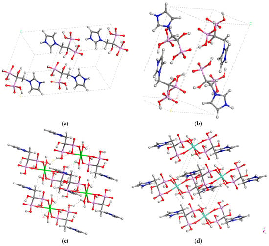
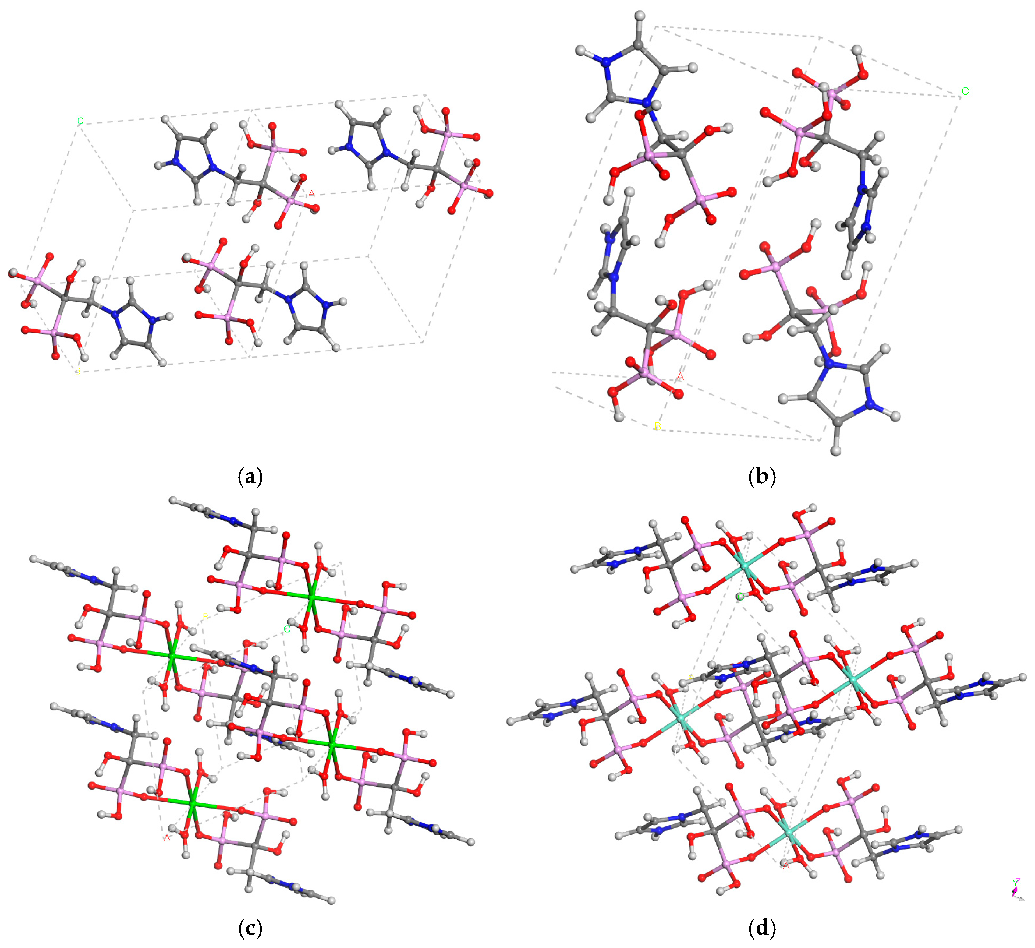
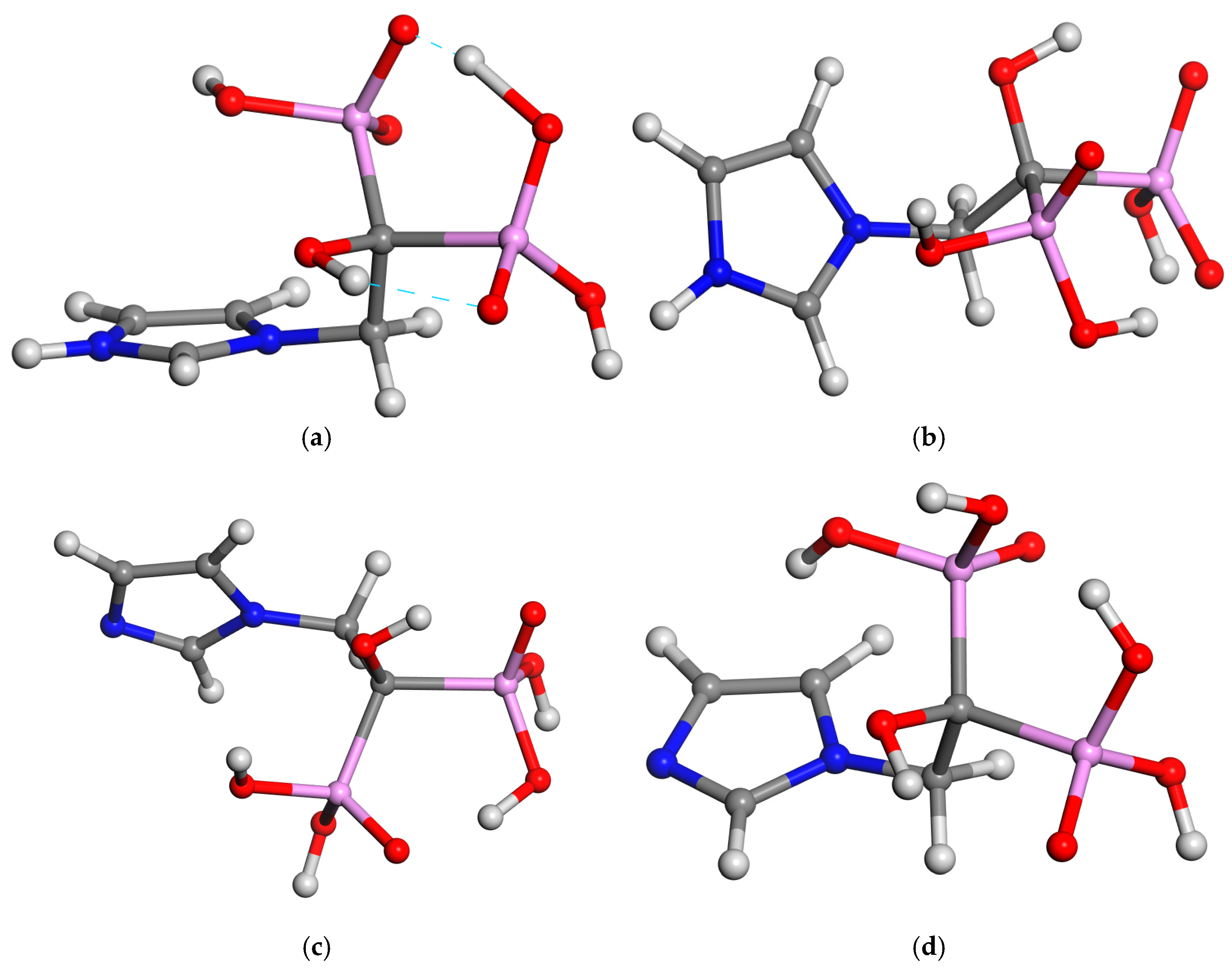
DFT calculations showed that the triclinic polymorph is slightly more stable than the monoclinic one (with a ΔE = 0.07 eV), where the packing energy is −3.71 eV per molecule. ZOL adopts a zwitterion form in both polymorphs. Within both crystals, the molecules organize into layered structures stabilized by strong hydrogen bonding, with the phosphonic groups oriented toward the interior of each layer and the imidazole moieties facing the exterior. Furthermore, the imidazole ring adopts a quasi-perpendicular plane with respect to the methyl-bisphosphonic moiety, θ(C-N-C-CP) = 76.4°. One phosphonate group is oriented opposite to the imidazole moiety, θ(N-C-C-P) = 180°. The molecules are joined by strong hydrogen bonds between the phosphonic groups, with distances of d(P=O…H-O-P) = 1.396–1.502 Å, and between the protonated imidazolium and the P=O bond, d(N-H…O=P) = 1.660 Å. These orientations facilitate electrostatic interactions between the imidazolium cationic part and the anionic phosphonic part. Crucially, the conformation of the bisphosphonic moiety differs between the two polymorphs: in the triclinic form, the P=O bonds are on opposite sides in an anti conformation, whereas in the monoclinic form they are in a syn conformation (Figure 1).
Figure 1.
Crystal structures of the triclinic (a) and monoclinic (b) polymorphs of zoledronic acid, and the Ca2+ (c) and Zn2+ (d) salts of ZOL optimized with Castep. H, C, N, O, P, Ca, and Zn atoms are in white, gray, blue, red, pink, green, and turquoise colors.
The crystal lattices of the Ca2+ and Zn2+ salts of ZOL are similar; the ZOL molecules exist as zwitterions, and each cation exhibits an octahedral coordination with two ZOL ligands [four equatorial M….O-P-C bonds, d(P=O….Ca) = 2.286–2.322 Å, d(P=O….Zn) = 2.042–2.130 Å] and two water molecules [two axial M…OH2 bonds, d(H2O…Ca) = 2.345 Å, d(H2O…Zn) = 2.130 Å] (Figure 1). The optimized structures with FF showed longer P=O…M distances (2.79 Å and 2.65 Å for Ca and Zn, respectively) than experiments and structures optimized with Castep (2.48 Å for Ca, and 2.19 Å for Zn). In both crystals, the ZOL molecule adopts a conformation of the bisphosphonate and imidazole moieties similar to the aforementioned ZOL crystals. The packing forces arise mainly from strong hydrogen bonds between the imidazolium H atom and the cation-coordinating P-O bond, d[N-H…O(Ca)P] = 1.852 Å, d[N-H…O(Zn)P] = 1.806 Å; between P=O bonds with vicinal PO-H groups d(P=O…H-O-P) = 1.856 Å (Ca), 1.756 Å (Zn); between C-OH groups [d(P=O…HOC) = 2.056 Å (Ca), 2.068 Å (Zn)]; and with water H atoms d(P=O…HOH) = 1.856 Å (Ca), 1.855 Å (Zn); and between P-OH groups and water molecules d(P-(H)O….HOH) = 2.213 Å (Ca), 2.379 Å (Zn).
4.2. Intercalation of ZOL into the Interlayer Space of SMEC
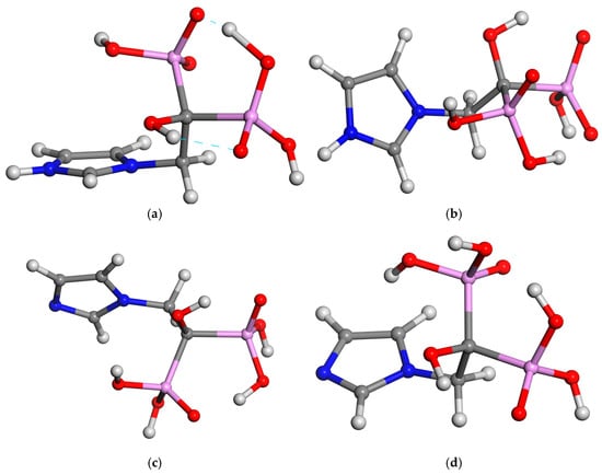
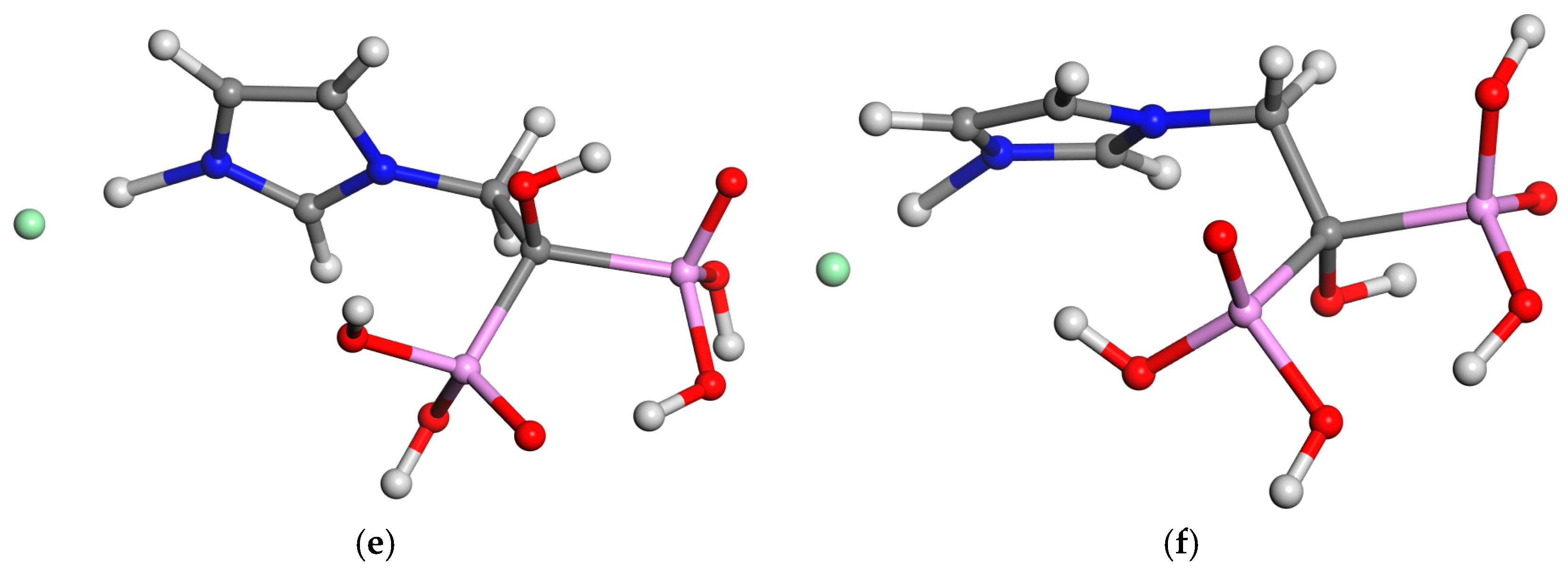
To study their intrinsic properties, ZOL molecules are extracted from the aforementioned crystal structures. In both crystal polymorphs, ZOL is in a zwitterionic form with the imidazole moiety protonated and one P-O group non-protonated to keep the whole molecule electrically neutral; and optimized as isolated zwitterionic molecules within a vacuum box (15 × 15 × 15 Å3) under 3-D periodic boundary conditions. Depending on the crystalline source, the phosphonate groups can adopt different conformations. The optimization of the isolated molecule derived from the monoclinic polymorph yielded a syn-twisted conformation of the P=O bonds (Figure 2a), during which a proton transfer from one P-O to another P=O group was observed. However, the optimization of the molecule originating from the triclinic polymorph maintained the anti conformation of the P=O groups (Figure 2b). In addition, neutral forms of ZOL were modeled by transferring a proton from the heterocycle N atom to the bisphosphonate moiety to maintain the electro-neutrality (Figure 2c,d). Both tautomeric forms were optimized in their respective anti and syn P=O/P=O conformations using DFT. Regarding their relative energies, the zwitterion was 0.055 eV more stable than the neutral form for the anti conformers, whereas the neutral form was 0.155 eV more stable than the zwitterion for the syn conformers. Consequently, within the zwitterionic models, the anti conformer was 0.119 eV more stable than the syn conformation, whereas for the neutral models, the syn conformer (Figure 2c) was 0.091 eV more stable than the anti one (Figure 2d). In all cases, the imidazole ring maintained its quasi-perpendicular orientation with respect to the methyl-bisphosphonic moiety.
Figure 2.
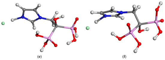
Molecular structures of ZOL optimized at DFT-CASTEP level of zwitterion form in anti (a) and syn (b) conformations; the neutral form, syn-twisted conformation (c), anti conformation (d), and chloride cationic ZOL salts syn (e) and anti (f) forms. The H, C, N, O, P, and Cl atoms are in white, gray, blue, red, pink, and green color.
On the other hand, at low pH, ZOL can exist in a cationic form, electrostatically neutralized by a chloride counterion, where the imidazole ring and both phosphonic groups are protonated (Figure 2e,f). After DFT optimization, we found that the syn form was 0.178 eV more stable than the anti conformation. In both cases, the electrostatic attraction between the acidic imidazolium N-H group and the Cl− anion elongated the corresponding N-H bond.
The SMEC models featuring Na+, Ca2+, and Zn2+ interlayer cations were optimized yielding cell parameters of the crystal structures according to experimental data [26] (Table 2). As expected, the d(001) spacing values of CaSMEC and ZnSMEC are larger than that of NaSMEC. The optimized CaSMEC and ZnSMEC models yielded similar lattice parameters. In all cases, the d(001) interlayer spacings calculated with CASTEP were slightly larger than those calculated with the FF.
Table 2.
Geometrical features of the crystal structures of SMEC models optimized using Castep. Values in brackets are calculated using the Interface FF (distances are in Å and angles in degrees).
The ZOL molecule, in either its neutral or zwittterionic form, was placed in the center of the interlayer space of CaSMEC, and both systems were fully optimized. In the complex with the intercalated ZOL zwitterion, the interlayer d(001) spacing increased slightly from 14.07 Å to 14.48 Å (Castep) and from 13.55 Å to 14.18 Å (FF). In contrast, with neutral ZOL, the increase in spacing was more pronounced, moving from 14.07 Å to 14.85 Å (Castep) and from 13.55 Å to 14.09 Å (FF). In both cases, water molecules form hydrogen bonds with the basal O atoms of the mineral surface [d(HOH…O-Si) = 1.777–1.959 Å], a network that also involves the acidic imidazole N-H proton. The P-O- groups engage in very strong hydrogen bonds with water molecules [d(HOH….O-P) = 1.581 Å and 1.713 Å] distorting the water symmetry [d(HO-H) = 0.977 Å and 1.02 Å]. Additionally, the P-OH groups form hydrogen bonds with the basal O atoms of the mineral surface. The Ca2+ cations are coordinated by water molecules [d(Ca…OH) = 2.307 Å] and one P=O group [d(Ca…O=P) = 2.48–2.62 Å].
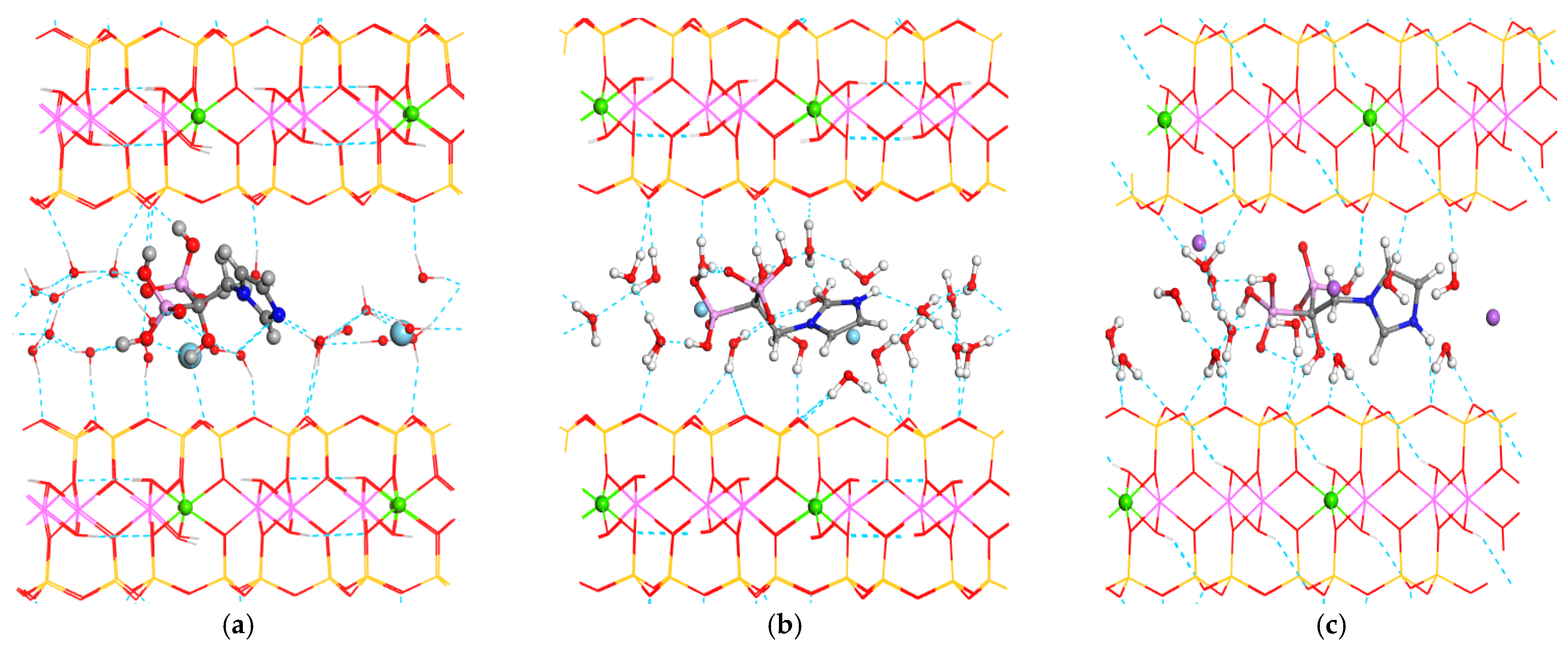
The conformation of ZOL did not change drastically upon intercalation. However, the ZOL imidazole ring is slightly more coplanar with the mineral surface in the zwitterion [θ(C-N-C-CP) = 75.6°] than in the neutral ZOL form [θ(C-N-C-CP) = 77.8°] (Figure 3). This structural rigidity within the confined interlayer space arises from its expansibility and the relative mild host–guest interactions. Although this preserved configuration could initially be attributed to a bias from the starting structure, the internal interactions are simply not strong enough to induce major conformational changes, causing only minor rotations in the imidazole ring. Nevertheless, to definitively rule out any initial configuration bias, a molecular dynamics sampling procedure was subsequently performed (vide infra).
Figure 3.
Optimized (with CASTEP) crystal structures of the intercalation complexes of CaSMECZOL (a), CaSMECZOLzwit (b), SMECZOL+ (c). The H, O, C, N, Si, Al, Mg, Na, and Ca atoms are white, red, gray, blue, yellow, pink, green, violet, and light blue colors. The intercalated atoms and Mg substitutions in the octahedral sheet of SMEC have been highlighted as balls and the hydrogen bonds are shown as dashed lines.
In NaSMEC the intercalation of ZOL via cation exchange was simulated, where one Na+ cation was replaced by a ZOL+ cation (NaSMECZOL+). A large increase in the interlayer d(001) spacing was observed, shifting from 11.98 to 14.46 Å (Castep) and from 11.75 to 13.66 Å (FF). Within this complex, the ZOL imidazole ring was oriented completely perpendicular to the mineral surface (Figure 3), allowing the H atom of the acidic imidazolium N-H group to form a hydrogen bond with a basal O atom of the mineral surface. The P=O groups are arranged in an anti conformation, whereas the PO–H groups engage in hydrogen bonding interactions with O atoms from both water molecules and the basal mineral surface.
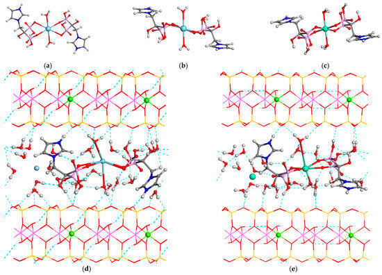
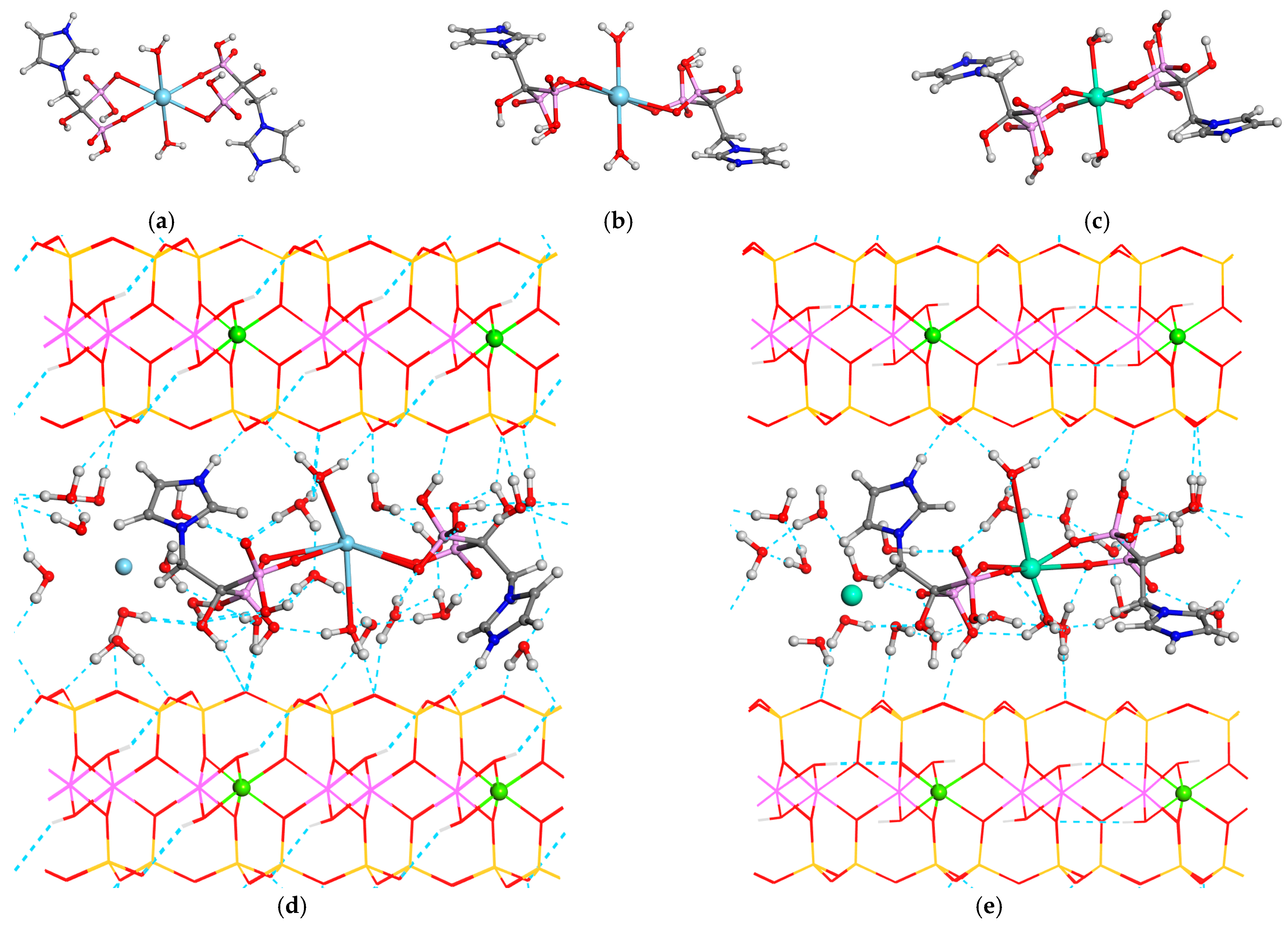
These intercalation mechanisms are expected to occur under low-pH conditions where the phosphonate groups remain protonated. At higher pH, however, some phosphonate groups will form salts with interlayer cations. Therefore, taking into account the crystal structures of the Ca2+ and Zn2+ ZOL salts studied above, we simulated the intercalation of two ZOL molecules forming coordination complexes with a single interlayer Ca2+ or Zn2+ cation, mimicking the arrangement observed in their respective crystal structures. The imidazole ring was protonated, and two P-O groups are dissociated for coordinating with interlayer cations and the H+ proton is joined to neighboring water molecules to maintain charge neutrality. This intercalation provoked a remarkable increase in the interlayer d(001) spacing of CaSMEC from 14.07 to 16.0 Å (Castep) and from 13.55 to 15.44 Å (FF). A similar effect was observed for ZnSMEC. In each coordination complex, the Ca2+ and Zn2+ cations exhibited a quasi-octahedral coordination sphere, bound to four P-O groups of ZOL and two water molecules, [d(Zn…O=P) = 2.0 Å].
These coordinating water molecules formed strong hydrogen bonds with the basal O atoms of the mineral surface, while the acidic N-H group of the imizadolium ring also formed a hydrogen bond with the mineral interlayer surface (Figure 4).
Figure 4.
Optimized structures of the Ca2+ ((a,b), at different orientations) and Zn2+ (c) salts of ZOL, and the intercalation complexes of these salts in CaSMEC (d) and ZnSMEC (e). The H, O, C, N, Si, Al, Mg, Zn, and Ca atoms are white, red, gray, blue, yellow, pink, green, violet, and light blue colors. The Mg substitutions in the octahedral sheet of SMEC have been highlighted as balls and the hydrogen bonds are shown as dashed lines.
We can compare the total energy values of these optimized structures and calculate adsorption energies. These energies were calculated following the adsorption reaction process:
where MSMEC is the optimized structure of the smectite minerals (NaSMEC, CaSMEC, or ZnSMEC); ZOL is the optimized structure of ZOL in its neutral or zwitterionic form; and MSMECZOL is the optimized clay mineral with intercalated ZOL. Consequently, the adsorption energy will be the difference between product and reactants:
where Eads is the adsorption energy; EMSMECZOL is the energy of the optimized structure of the complex of the SMEC model with intercalated ZOL; EZOL is the energy of the optimized molecular structure of ZOL; and EMSMEC is the energy of the optimized SMEC model without ZOL. The reaction will be favorable when the adsorption energy is negative.
MSMEC + ZOL = MSMECZOL,
Eads= EMSMECZOL − EZOL − EMSMEC
In the adsorption by cation exchange process, we used the scheme:
where NaSMEC is the optimized structure of smectite; ZOLCl is the optimized structure of ZOL in its cationic form with the Cl counteranion; ZOLSMEC is the optimized clay mineral with an intercalated ZOL+ cation; and NaCl is the exchanged Na+ interlayer cation with the Cl− counteranion. Consequently, the adsorption energy will be the difference between product and reactant energies:
where Eads is the adsorption energy; EZOLSMEC is the energy of the optimized structure of the complex of the SMEC model with an intercalated ZOL cation; EZOLCl is the energy of the optimized molecular structure of ZOL along with the chloride counteranion; ENaCl is the energy of the exchanged Na+ interlayer cation with the Cl counterion; and ENaSMEC is the energy of the optimized SMEC model without ZOL. The reaction will be favorable when the adsorption energy is negative.
NaSMEC + ZOLCl = ZOLSMEC + NaCl,
Eads = EZOLSMEC + ENaCl − EZOLCl − ENaSMEC
The intercalation of ZOL into CaSMEC showed multiple possible orientations. To avoid possible bias arising from the starting configurations, we explored different conformational and orientation states through molecular dynamics simulations. Several of the lowest-energy configurations were subsequently selected and fully optimized.
The intercalation of neutral ZOL into CaSMEC (CaSMECZOL) was found to be energetically favorable with an adsorption energy of −2.58 eV using the FF method (−2.43 eV with DFT). Similarly, the intercalation of ZOL in its zwitterionic form into CaSMEC was also energetically favorable with adsorption energies of −4.50 eV calculated using the Interface FF and −4.96 eV using DFT. Both computational approaches yielded comparable intercalation energies, indicating that the zwitterionic form of ZOL intercalates more favorably than the neutral form.
The intercalation of ZOL in NaSMEC through cation exchange evolved the exchange of one Na+ interlayer cation by one ZOL+ cation, with an adsorption energy of −7.57 eV. This process requires a low-pH medium to maintain the ZOL molecule as a cation. This result indicates that the cation exchange process is also probable and energetically favorable.
In the intercalation of ZOL through the formation of Ca2+ or Zn2+ interlayer salts, two ZOL molecules are intercalated per the same supercell. In this case the adsorption energy will be:
where Eads is the adsorption energy; EMSMECZOL2 is the energy of the optimized structure of the complex of the MSMEC model (M = Ca or Zn) with two intercalated ZOL molecules; EZOL is the energy of the optimized molecular structure of ZOL; and EMSMEC is the energy of the optimized SMEC model without ZOL.
Eads= EMSMECZOL2 − 2 × EZOL − EMSMEC
This intercalation was also energetically favorable, with adsorption energies of −1.18 eV for CaSMECZOL2H2 and −1.21 eV for ZnSMECZOL2H2. These values are lower than those reported above, probably due to steric effects, where the ZOL molecules are at 4.85 Å to those of the vicinal cell. All intercalation models were more stable than separated reactants, showing that the intercalation is energetically favorable, indicating that the internal interactions within the confined phase are weak but sufficient to produce stable materials.
4.3. Desorption of ZOL
Since the release of ZOL is governed by its desorption from the confined interlayer space of montmorillonite, the individual stages of the desorption process from CaSMEC were investigated. The calculations trace the pathway from the intercalated state within the clay structure to the fully solvated state in an aqueous medium. For this purpose, a bulk water model (waterbox) was constructed by filling a 3D periodic box (12.4 × 12.4 × 12.4 Å3) with 60 water molecules randomly placed via the Monte Carlo method at 298 K, reaching a density of 1 g cm−3. This model was subsequently optimized using the INTERFACE FF. Similarly, a second periodic box containing 60 water molecules and one ZOL molecule was generated, yielding a 13.2 × 13.2 × 13.2 Å3 box (ZOLw), which was also optimized using the same FF.
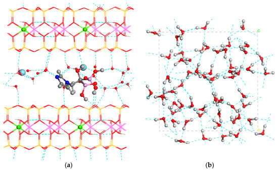
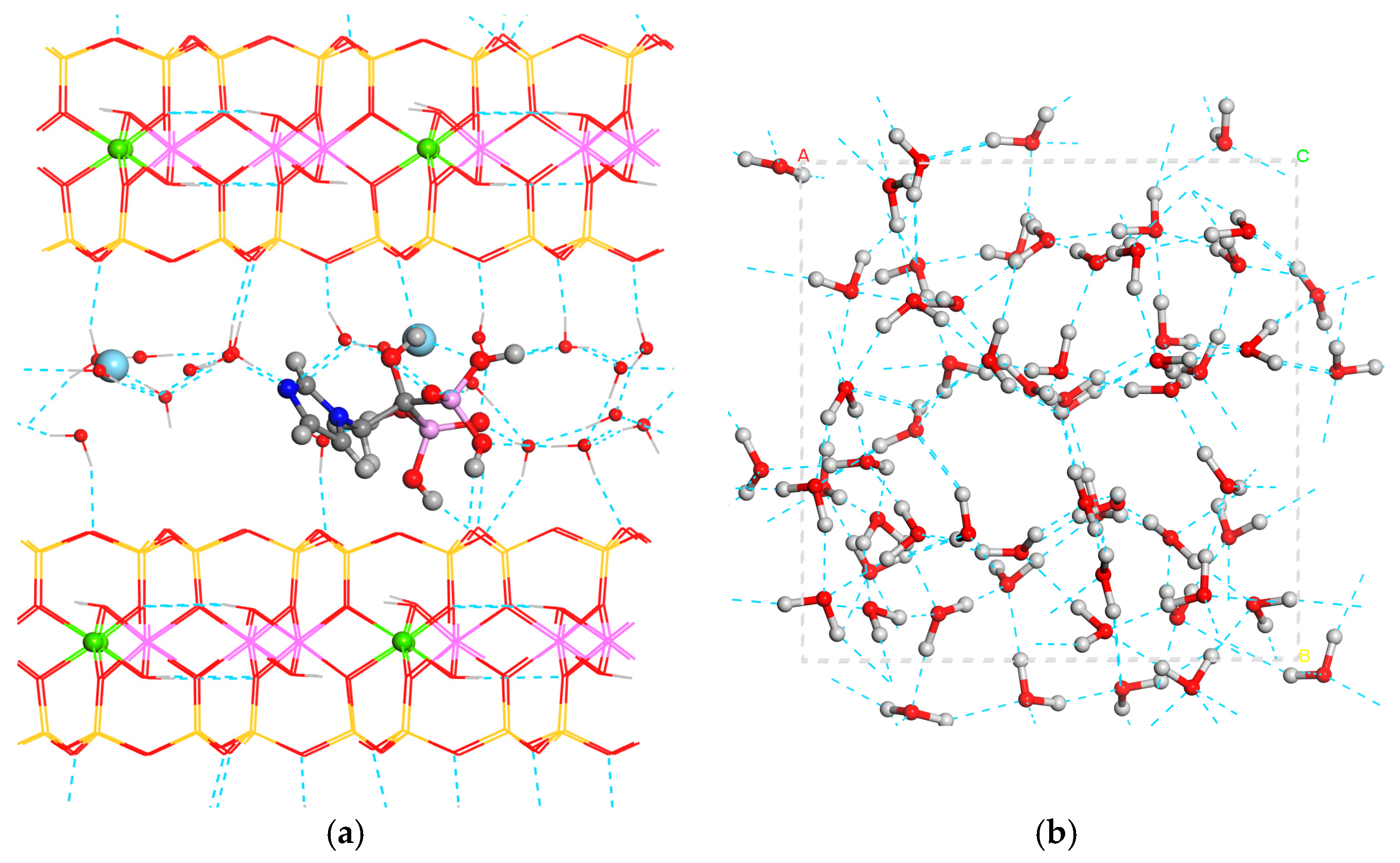
The desorption reaction was modeled by combining the smectite-containing intercalated ZOL (CaSMECZOL) with an aqueous medium (waterbox), obtaining the pristine smectite (CaSMEC) and solvated ZOL in the aqueous phase (ZOLw) (Figure 5):
CaSMECZOL + waterbox = CaSMEC + ZOLw
Figure 5.
Desorption of intercalated ZOL from smectite to an aqueous medium with the optimized structures involved in the reaction. CaSMECZOL (a) + waterbox (b) = CaSMEC (c) + ZOLw (d). The H, O, C, N, P, Si, Al, Mg, and Ca atoms are in white, red, gray, blue, pink, yellow, light pink, green, and light blue colors. Hydrogen bonds are shown as dashed lines.
The desorption energy can be calculated from the energies of the respective reactant and product species with the following equation:
Edes = E(CaSMEC) + E(ZOLw) − E(waterbox) − E(CaSMECZOL)
Based solely on the energy difference between the initial and final states, this process was energetically unfavorable (Edes = +0.916 eV).
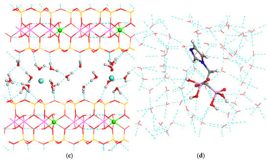
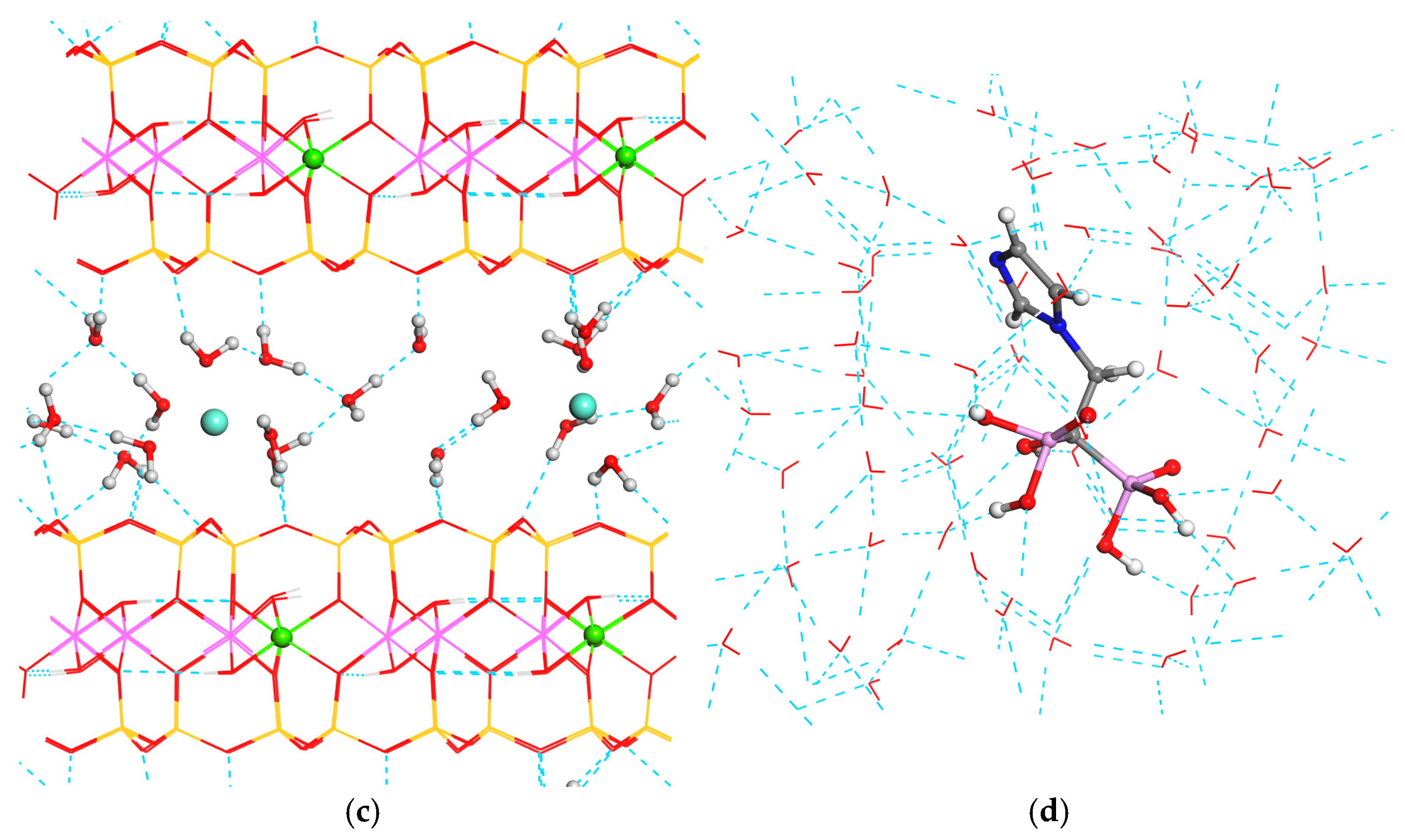
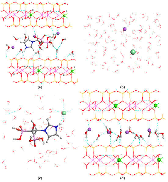
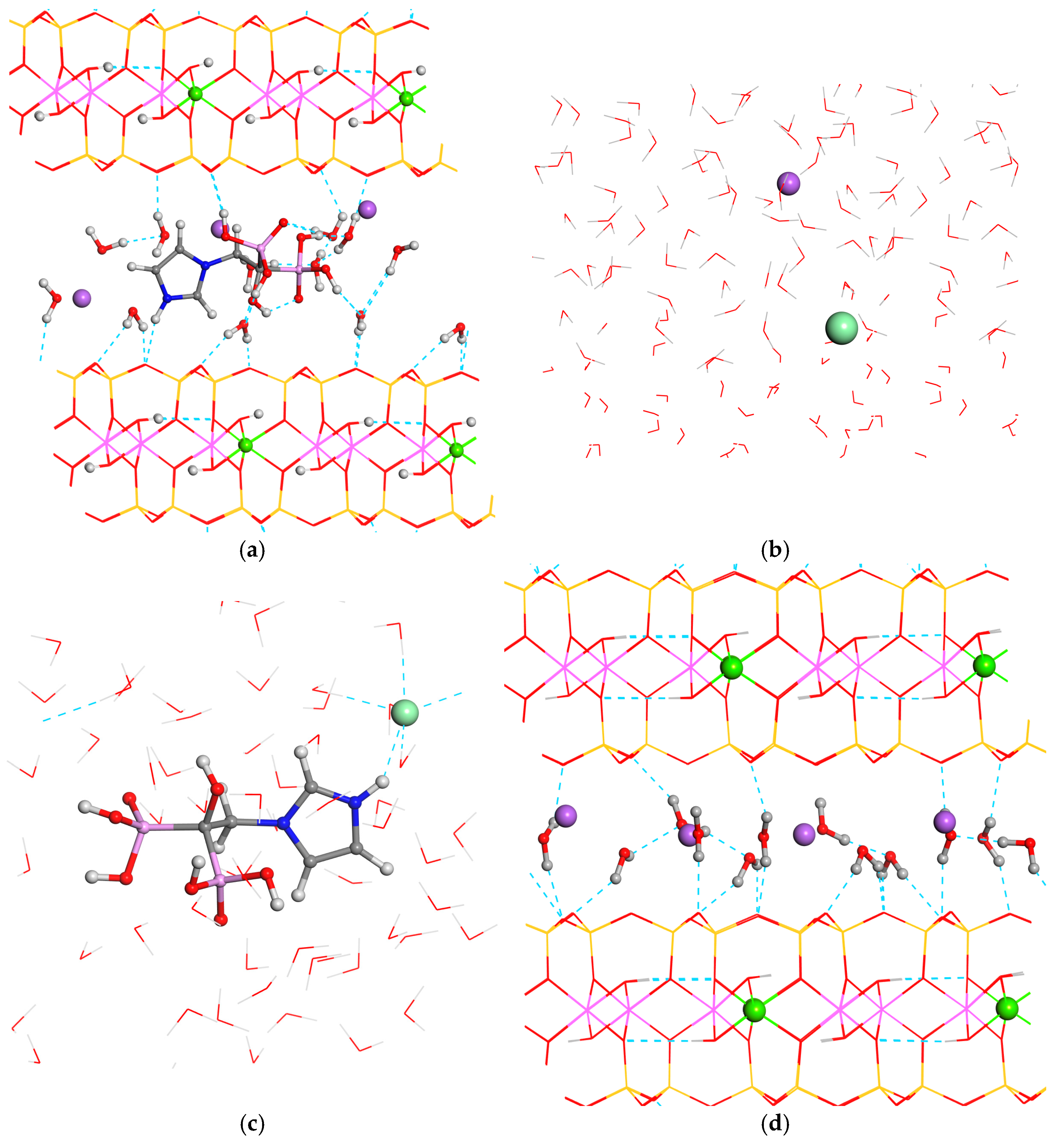
Alternatively, we modeled the release of ZOL into a simplified aqueous NaCl medium intended to mimic selected features of physiological environments. This model does not account for the full complexity of biological fluids, which contain inorganic ions, organic molecules, proteins, and other components that may influence adsorption, desorption, and diffusion processes. For this purpose, a periodic box containing 60 water molecules, one Na+ cation and one Cl− anion was generated via random Monte Carlo method mixing at a density of 1 g/cm3, resulting in dimensions of 12.4 × 12.4 × 12.4 Å3 (NaClw). The final product was modeled in a similar 3D periodic box, filled with 60 water molecules, one Cl− anion, and one ZOL+ cation distributed randomly by the Monte Carlo method at the same density (ZOLClw). The release energy was determined by the optimized reactant and product structures participating in the reaction (Figure 6):
SMECZOL+ + NaClw = ZOLClw + NaSMEC
Edes = E(NaSMEC) + E(ZOLClw) − E(NaClw) − E(SMECZOL+)
Figure 6.
Release of intercalated ZOL from smectite to a NaCl aqueous medium with the optimized structures involved in the reaction. NaSMECZOL+ (a) + NaClw (b) = ZOLClw (c) + NaSMEC (d). The H, O, C, N, P, Cl, Si, Al, Mg, Cl, and Na atoms are in white, red, gray, blue, pink, yellow, light pink, green, light green, and violet colors. Hydrogen bonds are shown as dashed lines.
In this case the release energy was −0.726 eV, indicating that the release of ZOL into this saline medium is energetically favorable.
4.4. Molecular Dynamics
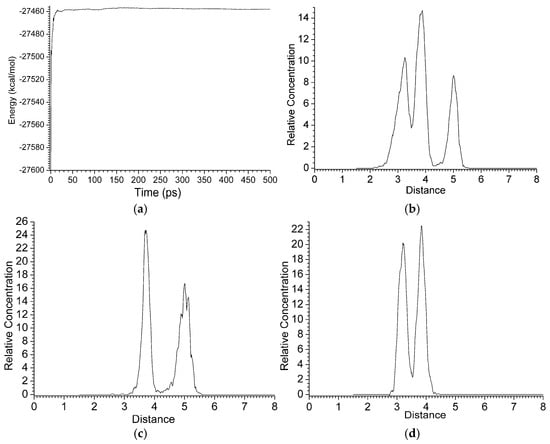
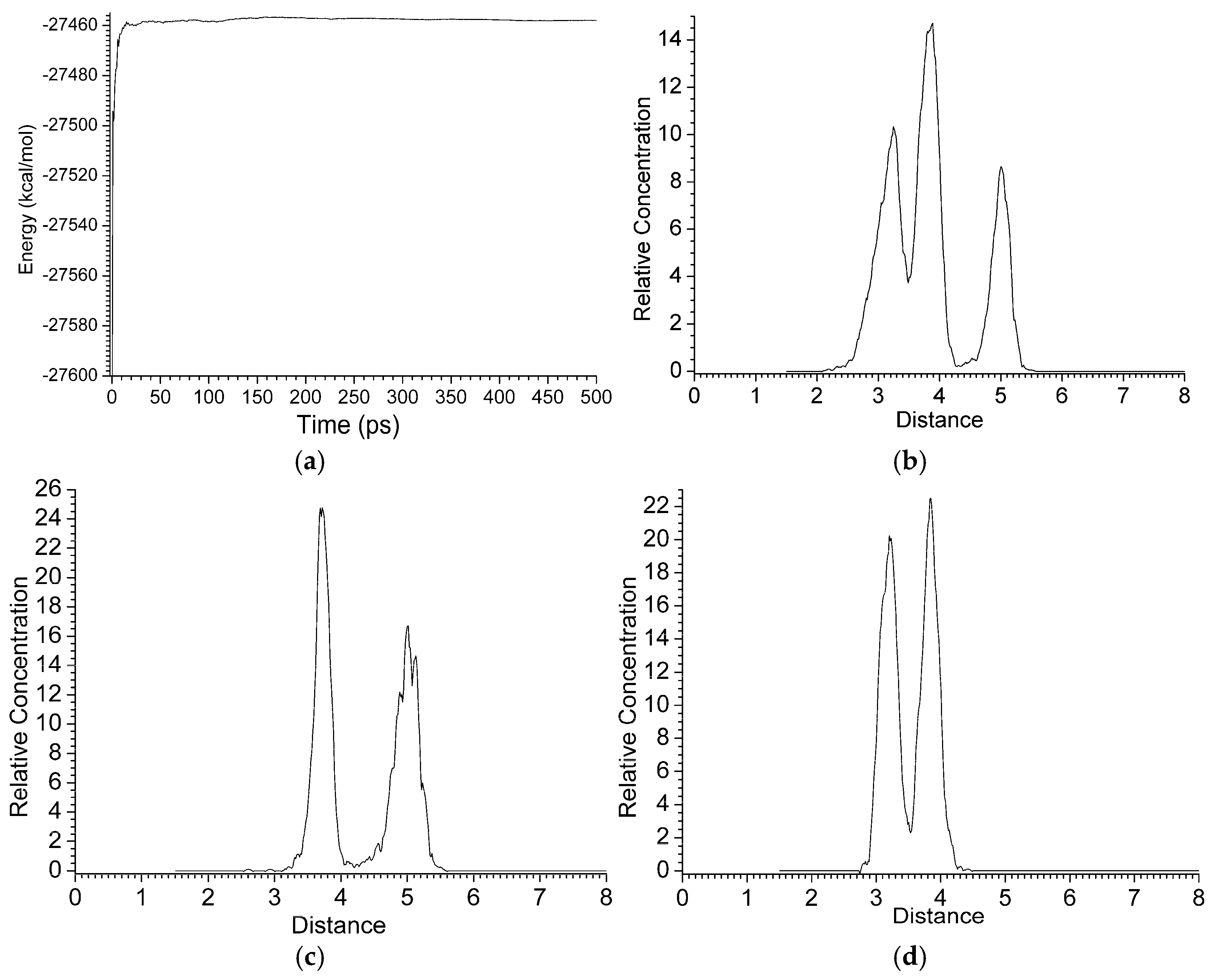
During the MD simulations, the CaSMEC + zwitterion ZOL system reached equilibrium after 10 ps. The potential energy converged after the first 10–30 ps, fluctuating over a constant average energy throughout the simulation time (Figure 7a). We observed that the smectite host support is not rigid; instead, the structure exhibits certain flexibility, especially within the octahedral AlOHAl hydroxyl bonds. After reaching equilibrium, the ZOL molecule adopts a very stable position in the interlayer space, indicating high stability of the drug within the host clay mineral. This is consistent with the energetically unfavorable desorption of ZOL from the CaSMEC interlayer (vide supra). Consequently, an acidic medium is necessary to stimulate ZOL release. The imidazole ring lies in a plane perpendicular to the mineral (001) surface, forming a hydrogen bond between its acidic N-H group and the surface basal O atoms. The conformation of the P=O groups is maintained throughout the entire simulation. However, the conformation of the imidazole ring changes with respect to the bisphosphonate moiety (see Movie S1 in Supplementary Materials).
Figure 7.
Relative average energy throughout MD time (a). Relative concentration of C + N + P (b), N (c), and P (d) atoms of ZOL inside the interlayer space of SMEC. Average values from 990 samples of 106 structures generated along the MD simulations. Distances (Å) represent the interlayer space in the [001] direction of SMEC crystal.
From the 1 ns MD simulation, 106 structures were generated and a sampling of one every 103 frames procedure was applied. Following the equilibrium phase, we calculated the mean square displacement (MSD) of the ZOL molecule intercalated into the confined interlayer of CaSMEC over the last 990 frames. Although the potential energy converged after the first 30 ps to reach a constant value, this simulation time is too short to provide enough statistical reliability to calculate the diffusion coefficient (D) accurately. However, we can estimate approximately the relative differences in the diffusion coefficient between the atoms intercalated into the interlayer space in a qualitative sense. Furthermore, there is a certain anisotropy in the diffusion of intercalated atoms, taking into account the layered structure of smectites. Considering this anisotropy, we calculated the diffusion components for each selected atom. Water molecules exhibited an isotropic diffusion coefficient of 2.36 × 10⁻³ Å2 ps−1; however, this diffusion was mainly in the xy plane (Dxx = 7.07 × 10⁻³ Å2 ps−1; Dxy = 3.05 × 10⁻³ Å2 ps−1), while it was much lower in the z direction. The whole ZOL molecule exhibited lower mobility with an isotropic diffusion coefficient of 4.26 × 10⁻⁴ Å2/ps. On the contrary, this molecule showed lower anisotropy due to the twisting movements of the methylbisphosphonate moiety and the heterocycle ring within the interlayer throughout the simulation (Dxx = 4.29 × 10⁻⁴ Å2 ps−1; Dyy = 2.77 × 10⁻⁴ Å2 ps−1; Dzz = 5.7 × 10⁻⁴ Å2 ps−1). A slightly higher mobility showed the Ca2+ cations with an isotropic diffusion coefficient of 5.7 × 10⁻⁴ Å2 ps−1, where the main components were along the xy plane due to the electrostatic energy balance of these cations and both mineral interlayer surfaces (Dxx = 7.08 × 10⁻⁴ Å2 ps−1; Dyy = 6.65 × 10⁻⁴ Å2 ps−1; Dzz = 3.37 × 10⁻⁴ Å2 ps−1).
Analysis of the average sampled frames from the MD simulations revealed that the ZOL molecule remained predominantly in the center of the interlayer space throughout the simulations, balancing between the interactions from both mineral surfaces. The average concentration of C, N, and P atoms of ZOL inside the interlayer space provides a good approximation for describing the behavior of the whole molecule. We observed that ZOL is equidistant to both mineral surfaces of the SMEC interlayer space along the [001] direction, where the surface limits are normalized from 0 and 8 Å. Three maxima are observed at 3.3, 3.9, and 5 Å (Figure 7b), corresponding to N atoms with maxima at 3.8 and 5 Å (Figure 7c), and P atoms with maxima at 3.2 and 3.9 Å (Figure 7d). The double peak in the N- atom plot is due the two N atoms of the imidazole ring, which adopts a perpendicular disposition with respect to the (001) mineral surface and twists during the MD simulations. The peak at 3.8 Å from one surface corresponds to the N atom bonded to the rest of the molecule, whereas the other N atom is primarily responsible for the peak at 5 Å (or 3 Å from the opposite surface); the latter displays a fine structure due to its higher mobility, which is enhanced by its H atom interacting with the basal O atoms of the mineral surface. The P atoms also show two peaks very close to the interlayer center in the [001] direction. Each peak corresponds to a different P atom, depending on the orientation of the P-C-P plane, which is not parallel to the mineral surface and oscillates with respect to the (001) surface.
5. Discussion
The application of the INTERFACE FF for the study of clay minerals has proven to be an effective approach in several previous reports. After validating the INTERFACE FF for bisphosphonates by successfully reproducing the crystal structures of ZOL polymorphs, we applied it to study the intercalation of ZOL into montmorillonite. Consequently, this work demonstrates that the INTERFACE FF is fully capable of modeling bisphosphonates and their corresponding interactions during intercalation into the interlayer space of clay minerals. Nevertheless, while DFT calculations yielded higher accuracy, they required a drastically greater computational effort.
Although the bisphosphonate moiety exhibits several conformers depending on the relative disposition of the P=O groups, the energy differences remain low across its zwitterionic, neutral, and cationic forms, which does not significantly affect its interactions with clay minerals.
Our molecular modeling calculations predict that montmorillonite can readily intercalate ZOL through an energetically favorable process, with the delivery of ZOL from the carrier being facilitated under acidic conditions. However, these calculations describe only the initial and final steps of the adsorption and desorption processes, without accounting for temperature effects. Consequently, our models do not include physiological conditions or the complex solvent environments typical of practical drug delivery scenarios. Furthermore, the atomic-scale study of thermodynamics, kinetic parameters, and the influence of buffers or skin mimetics was outside the scope of this work. Nevertheless, these results may guide future experiments focused on the oral administration of ZOL, given the acidic environment of the digestive system.
An interesting finding is that our calculations predict an increase in the powder X-ray diffraction (XRD) d(001) spacing of the clay mineral upon drug intercalation. This structural shift can be verified experimentally, meaning our prediction can serve as a useful benchmark for future studies involving smectites and ZOL.
Therefore, our molecular modeling results can enhance future experimental designs for the delivery of ZOL and its derivatives, particularly when exploring delivery kinetics and the bioactivity of the post-release media.
6. Conclusions
The intercalation of ZOL into montmorillonite induces a significant increase in the d(001) spacing, regardless of whether ZOL is in its neutral, zwitterionic, or cationic form. In all cases, the intercalation process was energetically favorable, and the cation exchange mechanism was also favorable. This theoretical study predicts that ZOL intercalation into montmorillonite is facile and serves as an effective method for stabilizing ZOL in an amorphous state, rendering it more soluble and bioavailable than its crystalline counterpart. These predictions will be highly useful for future experimental work, specifically for monitoring intercalation experiments via XRD and for designing drug–clay composites for pharmaceutical applications.
Furthermore, while ZOL desorption into a pure aqueous medium is not energetically favorable, its release into a low-pH aqueous medium appears highly probable. These predictions are promising for future experimental studies, indicating that these materials can be excellent candidates for the controlled release of ZOL into environments.
Finally, our MD simulations demonstrated that molecular dynamics is a robust method for searching and sampling minimal-energy structures, effectively avoiding the local minima often encountered in static optimizations. These simulations also qualitatively indicated a low diffusion coefficient for the intercalated ZOL, reflecting the high stability of this drug within the interlayer space of montmorillonite.
Overall, this theoretical work lays the foundation for future experimental research focused on the preparation of ZOL–montmorillonite delivery systems, which hold great promise for therapeutic applications.
Supplementary Materials
The following supporting information can be downloaded at: https://www.mdpi.com/article/10.3390/surfaces9030073/s1, Movie S1: Molecular dynamics simulation of ZOL zwitterion intercalated into the CaSMEC interlayer space (with INTERFACE FF, NVT, 1ns). Table S1: Main chemical bond lengths (in Å) of the methyl-bisphosphonic moiety in ZOL crystal structures. Comparison of experimental and calculated values. Table S1a: Atomic coordinates of the crystal structure of the monoclinic polymorph of ZOL optimized with Interface FF (fractional coordinates in Å, angles in degrees). Table S1b: Atomic coordinates of the crystal structure of the monoclinic polymorph of ZOL optimized using CASTEP (fractional coordinates in Å, angles in degrees). Table S2a: Atomic coordinates of the crystal structure of the triclinic polymorph of ZOL optimized with Interface FF (fractional coordinates in Å, angles in degrees). Table S2b: Atomic coordinates of the crystal structure of the triclinic polymorph of ZOL optimized using CASTEP (fractional coordinates in Å, angles in degrees). Table S3: Atomic coordinates of ZOL2Zn optimized with Interface FF (fractional coordinates in Å, angles in degrees). Table S4: Atomic coordinates of ZOL2Ca optimized with Interface FF (fractional coordinates in Å, angles in degrees). Table S5: Atomic coordinates of ZOL in zwitterionic form optimized with Interface FF (Cartesian coordinates in Å). Table S6: Atomic coordinates of ZOL in neutral form optimized with Interface FF (Cartesian coordinates in Å). Table S7: Atomic coordinates of ZOL in cationic form neutralized by a chloride counter-ion optimized with Interface FF (Cartesian coordinates in Å). Table S8: Atomic coordinates of CaSMEC optimized with Interface FF (fractional coordinates in Å, angles in degrees). Table S9: Atomic coordinates of ZnSMEC optimized with Interface FF (fractional coordinates in Å, angles in degrees). Table S10: Atomic coordinates of NaSMECZOL+ optimized with Interface FF (fractional coordinates in Å, angles in degrees). Table S11: Atomic coordinates of CaSMECZOLzwit optimized with Interface FF (fractional coordinates in Å, angles in degrees). Table S12: Atomic coordinates of ZnSMECZOL2H2 optimized with Interface FF (fractional coordinates in Å, angles in degrees). Table S13: Atomic coordinates of waterbox optimized with Interface FF (fractional coordinates in Å, angles in degrees). Table S14: Atomic coordinates of ZOLw (ZOL in a waterbox) optimized with Interface FF (fractional coordinates in Å, angles in degrees).
Author Contributions
Conceptualization, C.I.S.-D.; methodology, C.I.S.-D.; software, C.I.S.-D. and J.O.-C.; investigation, M.L.-L., and C.I.S.-D.; resources, A.H.-L.; writing—original draft preparation, M.L.-L. and C.I.S.-D.; writing—review and editing, C.I.S.-D. and J.O.-C.; supervision, C.I.S.-D.; All authors have read and agreed to the published version of the manuscript.
Funding
This work has been funded by the European MSCA-SE-NANOTRIOSTEO 101182658 project and the projects PID2024-160443NB-I00, and PID2022-137889OB-I00 funded by MCIN/AEI/10.13039/501100011033/ and FEDER ‘una manera de hacer Europa’. J.O.-C. acknowledges the financial support from the MCIN/AEI/10.13039/501100011033 and FEDER, UE (project PID2023-148860OB-I00).
Data Availability Statement
Data will be provided upon request to the corresponding authors.
Acknowledgments
The authors thank the High-Performance Computation Center of CSIC, and M.L.-L. thanks the JAE-Intro program of CSIC.
Conflicts of Interest
The authors declare no conflict of interest.
References
- Hernández-Laguna, A.; Sainz-Díaz, C.I.; Smeyers, J.L.G.; De Paz, E.; Galvez-Ruano, E. Electronic Structure and Conformational Properties of Vinylphosphonic Acid and Some Related Derivatives. J. Phys. Chem. 1994, 98, 1109–1116. [Google Scholar] [CrossRef] [Scilit]
- Xu, X.-L.; Gou, W.-L.; Wang, A.-Y.; Wang, Y.; Guo, Q.-Y.; Lu, Q.; Lu, S.-B.; Peng, J. Basic Research and Clinical Applications of Bisphosphonates in Bone Disease: What Have We Learned over the Last 40 Years? J. Transl. Med. 2013, 11, 303. [Google Scholar] [CrossRef] [Scilit] [PubMed]
- Russell, R.G.G. Bisphosphonates: The First 40 years. Bone 2011, 49, 2–19. [Google Scholar] [CrossRef] [Scilit] [PubMed]
- Ebetino, F.H.; Sun, S.; Cherian, P.; Roshandel, S.; Neighbors, J.D.; Hu, E.; Dunford, J.E.; Sedghizadeh, P.P.; McKenna, C.E.; Srinivasan, V.; et al. Bisphosphonates: The Role of Chemistry in Understanding Their Biological Actions and Structure-Activity Relationships, and New Directions for Their Therapeutic Use. Bone 2022, 156, 116289. [Google Scholar] [CrossRef] [Scilit] [PubMed]
- Vassaki, M.; Lazarou, S.; Turhanen, P.; Choquesillo-Lazarte, D.; Demadis, K. Drug-Inclusive Inorganic–Organic Hybrid Systems for the Controlled Release of the Osteoporosis Drug Zoledronate. Molecules 2022, 27, 6212. [Google Scholar] [CrossRef] [Scilit] [PubMed]
- Qiu, L.; Lv, G.; Guo, L.; Chen, L.; Luo, S.; Zou, M.; Lin, J. Synthesis, Crystal Structure and Antitumor Effect of a Novel Copper(II) Complex Bearing Zoledronic Acid Derivative. Eur. J. Med. Chem. 2015, 89, 42–50. [Google Scholar] [CrossRef] [Scilit] [PubMed]
- Liu, Q.; Li, K.; Lv, G.; Li, X.; Peng, Y.; Lin, J.; Qiu, L. Density Functional Theory Studies on a Non-Covalent Interaction System: Hydrogen-Bonded Dimers of Zoledronate. J. Mol. Model. 2018, 24, 310. [Google Scholar] [CrossRef] [Scilit] [PubMed]
- Chen, T.; Berenson, J.; Vescio, R.; Swift, R.; Gilchick, A.; Goodin, S.; LoRusso, P.; Ma, P.; Ravera, C.; Deckert, F.; et al. Pharmacokinetics and Pharmacodynamics of Zoledronic Acid in Cancer Patients with Bone Metastases. J. Clin. Pharmacol. 2002, 42, 1228–1236. [Google Scholar] [CrossRef] [Scilit] [PubMed]
- Marra, M.; Santini, D.; Tonini, G.; Meo, G.; Zappavigna, S.; Facchini, G.; Morabito, A.; Abbruzzese, A.; Cartenì, G.; Budillon, A.; et al. Molecular and Preclinical Models Enhancing Anti-Tumour Activity of Zoledronic Acid. Eur. J. Cancer Suppl. 2008, 6, 79–85. [Google Scholar] [CrossRef] [Scilit]
- Povoroznyuk, V.V.V.; Grygorieva, V.N.; Pekhnyo, V.I.; Om, K.; Nv, T. Zoledronic Acid and Its Calcium-Contain Complexes in Treatment of Experimental Osteoporosis in Wistar Female Rats. Biochem. Anal. Biochem. 2017, 6, 1000340. [Google Scholar] [CrossRef]
- Vassaki, M.; Kotoula, C.; Turhanen, P.; Choquesillo-Lazarte, D.; Demadis, K.D. Calcium and Strontium Coordination Polymers as Controlled Delivery Systems of the Anti-Osteoporosis Drug Risedronate and the Augmenting Effect of Solubilizers. Appl. Sci. 2021, 11, 11383. [Google Scholar] [CrossRef] [Scilit]
- Papathanasiou, K.E.; Vassaki, M.; Spinthaki, A.; Alatzoglou, F.-E.G.; Tripodianos, E.; Turhanen, P.; Demadis, K.D. Phosphorus Chemistry: From Small Molecules, to Polymers, to Pharmaceutical and Industrial Applications. Pure Appl. Chem. 2019, 91, 421–441. [Google Scholar] [CrossRef] [Scilit]
- Papathanasiou, K.E.; Turhanen, P.; Brückner, S.I.; Brunner, E.; Demadis, K.D. Smart, Programmable and Responsive Injectable Hydrogels for Controlled Release of Cargo Osteoporosis Drugs. Sci. Rep. 2017, 7, 4743. [Google Scholar] [CrossRef] [Scilit] [PubMed]
- Park, J.-H.; Shin, H.-J.; Kim, M.H.; Kim, J.-S.; Kang, N.; Lee, J.-Y.; Kim, K.-T.; Lee, J.I.; Kim, D.-D. Application of Montmorillonite in Bentonite as a Pharmaceutical Excipient in Drug Delivery Systems. J. Pharma. Investig. 2016, 46, 363–375. [Google Scholar] [CrossRef] [Scilit] [PubMed]
- Xiao, Z.; Gu, X.; Huang, J.; Liu, L.; Yuan, S.; Jiang, P.; Zhang, Y.; Zhao, Y.; Wei, S.; Tao, Q.; et al. Montmorillonite-Enhanced Micro- and Nanocomposites for Targeted and Controlled Oral Drug Delivery Systems. Appl. Clay Sci. 2025, 267, 107713. [Google Scholar] [CrossRef] [Scilit]
- Francisco-Márquez, M.; Soriano-Correa, C.; Sainz-Díaz, C.I. Adsorption of Sulfonamides on Phyllosilicate Surfaces by Molecular Modeling Calculations. J. Phys. Chem. C 2017, 121, 2905–2914. [Google Scholar] [CrossRef] [Scilit]
- Borrego-Sánchez, A.; Sánchez-Espejo, R.; García-Villén, F.; Viseras, C.; Sainz-Díaz, C.I. Praziquantel–Clays as Accelerated Release Systems to Enhance the Low Solubility of the Drug. Pharmaceutics 2020, 12, 914. [Google Scholar] [CrossRef] [Scilit] [PubMed]
- Khatoon, N.; Chu, M.Q.; Zhou, C.H. Nanoclay-Based Drug Delivery Systems and Their Therapeutic Potentials. J. Mater. Chem. B 2020, 8, 7335–7351. [Google Scholar] [CrossRef] [Scilit] [PubMed]
- Rebitski, E.P.; Darder, M.; Sainz-Diaz, C.I.; Carraro, R.; Aranda, P.; Ruiz-Hitzky, E. Theoretical and Experimental Investigation on the Intercalation of Metformin into Layered Clay Minerals. Appl. Clay Sci. 2020, 186, 105418. [Google Scholar] [CrossRef] [Scilit]
- Bello, M.L.; Junior, A.M.; Freitas, C.A.; Moreira, M.L.A.; Da Costa, J.P.; De Souza, M.A.; Santos, B.A.M.C.; De Sousa, V.P.; Castro, H.C.; Rodrigues, C.R.; et al. Development of Novel Montmorillonite-Based Sustained Release System for Oral Bromopride Delivery. Eur. J. Pharm. Sci. 2022, 175, 106222. [Google Scholar] [CrossRef] [Scilit] [PubMed]
- Kaur, M.; Datta, M. Diclofenac Sodium Adsorption onto Montmorillonite: Adsorption Equilibrium Studies and Drug Release Kinetics. Adsorpt. Sci. Technol. 2014, 32, 365–387. [Google Scholar] [CrossRef] [Scilit]
- Massaro, M.; Borrego-Sánchez, A.; Sánchez-Espejo, R.; Viseras Iborra, C.; Cavallaro, G.; García-Villén, F.; Guernelli, S.; Lazzara, G.; Miele, D.; Sainz-Díaz, C.I.; et al. Ciprofloxacin Carrier Systems Based on Hectorite/Halloysite Hybrid Hydrogels for Potential Wound Healing Applications. Appl. Clay Sci. 2021, 215, 106310. [Google Scholar] [CrossRef] [Scilit]
- Swain, R.; Nandi, S.; Mohapatra, S.; Mallick, S. Engineered Clay-Polymer Composite for Biomedical Drug Delivery andFuture Challenges: A Survey. Curr. Drug Deliv. 2024, 21, 645–661. [Google Scholar] [CrossRef] [Scilit] [PubMed]
- Vassaki, M.; Papathanasiou, K.E.; Hadjicharalambous, C.; Chandrinou, D.; Turhanen, P.; Choquesillo-Lazarte, D.; Demadis, K.D. Self-Sacrificial MOFs for Ultra-Long Controlled Release of Bisphosphonate Anti-Osteoporotic Drugs. Chem. Commun. 2020, 56, 5166–5169. [Google Scholar] [CrossRef] [Scilit] [PubMed]
- Silva, D.T.C.; Fonseca, M.G.; Borrego-Sánchez, A.; Soares, M.F.R.; Viseras, C.; Sainz-Díaz, C.I.; Soares- Sobrinho, J.L. Adsorption of Tamoxifen on Montmorillonite Surface. Micropor. Mesopor. Mater. 2020, 297, 110012. [Google Scholar] [CrossRef] [Scilit]
- Borrego-Sánchez, A.; Viseras, C.; Sainz-Díaz, C.I. Molecular Interactions of Praziquantel Drug with Nanosurfaces of Sepiolite and Montmorillonite. Appl. Clay Sci. 2020, 197, 105774. [Google Scholar] [CrossRef] [Scilit]
- Chernyshev, V.V.; Shkavrov, S.V.; Paseshnichenko, K.A.; Puryaeva, T.P.; Velikodny, Y.A. Zoledronic Acid: Monoclinic and Triclinic Polymorphs from Powder Diffraction Data. Acta Crystallogr. Sect. C 2013, 69, 263–266. [Google Scholar] [CrossRef] [Scilit] [PubMed]
- Quiñones Vélez, G.; Carmona-Sarabia, L.; Rivera Raíces, A.A.; Hu, T.; Peterson-Peguero, E.A.; López-Mejías, V. High Affinity Zoledronate-Based Metal Complex Nanocrystals to Potentially Treat Osteolytic Metastases. Mater. Adv. 2022, 3, 3251–3266. [Google Scholar] [CrossRef] [Scilit] [PubMed]
- Petrovici, A.-R.; Silion, M.; Simionescu, N.; Kallala, R.; Pinteala, M.; Maier, S.S. Quantification of Low Amounts of Zoledronic Acid by HPLC-ESI-MS Analysis: Method Development and Validation. Int. J. Mol. Sci. 2022, 23, 5944. [Google Scholar] [CrossRef] [Scilit] [PubMed]
- Segall, M.D.; Lindan, P.J.D.; Probert, M.J.; Pickard, C.J.; Hasnip, P.J.; Clark, S.J. First-Principles Simulation: Ideas, Illustrations and the CASTEP Code. J. Phys. Condens. Matter. 2002, 14, 2717–2744. [Google Scholar] [CrossRef] [Scilit]
- Delley, B. An All-Electron Numerical Method for Solving the Local Density Functional for Polyatomic Molecules. J. Chem. Phys. 1990, 92, 508–517. [Google Scholar] [CrossRef] [Scilit]
- Perdew, J.P.; Burke, K.; Ernzerhof, M. Generalized Gradient Approximation Made Simple. Phys. Rev. Lett. 1996, 77, 3865–3868. [Google Scholar] [CrossRef] [Scilit] [PubMed]
- Biovia. Materials Studio; Dassault Systems Inc.: Waltham, MA, USA, 2025. [Google Scholar]
- Koelling, D.; Harmon, B. A Technique for Relativistic Spin-Polarised Calculations. J. Phys. C Solid State Phys. 2001, 10, 3107. [Google Scholar] [CrossRef] [Scilit]
- Tkatchenko, A.; Scheffler, M. Accurate Molecular Van Der Waals Interactions from Ground-State Electron Density and Free-Atom Reference Data. Phys. Rev. Lett. 2009, 102, 073005. [Google Scholar] [CrossRef] [Scilit] [PubMed]
- Delley, B. Hardness Conserving Semilocal Pseudopotentials. Phys. Rev. B 2002, 66, 1551251–1551259. [Google Scholar] [CrossRef] [Scilit]
- Besler, B.H.; Merz, K.M., Jr.; Kollman, P.A. Atomic Charges Derived from Semiempirical Methods. J. Comput. Chem. 1990, 11, 431–439. [Google Scholar] [CrossRef] [Scilit]
- Heinz, H.; Lin, T.-J.; Kishore Mishra, R.; Emami, F.S. Thermodynamically Consistent Force Fields for the Assembly of Inorganic, Organic, and Biological Nanostructures: The INTERFACE Force Field. Langmuir 2013, 29, 1754–1765. [Google Scholar] [CrossRef] [Scilit] [PubMed]
- Meruvia-Rojas, Y.V.; Molina-Montes, E.; Hernández-Laguna, A.; Sainz-Díaz, C.I. Intercalation of the Anticancer Drug Lenalidomide into Montmorillonite for Bioavailability Improvement: A Computational Study. J. Mol. Model. 2025, 31, 5. [Google Scholar] [CrossRef] [Scilit] [PubMed]
- Nosé, S. Constant Temperature Molecular Dynamics Methods. Prog. Theor. Phys. Suppl. 1991, 103, 1–46. [Google Scholar] [CrossRef] [Scilit]
- Savitzky, A.; Golay, M.J.E. Smoothing and Differentiation of Data by Simplified Least Squares Procedures. Anal. Chem. 1964, 36, 1627–1639. [Google Scholar] [CrossRef] [Scilit]
- OriginLab Corporation. OriginPro v. 8.5.0; OriginPro: Northampton, MA, USA, 2010. [Google Scholar]
- Heinz, H.; Suter, U.W. Atomic Charges for Classical Simulations of Polar Systems. J. Phys. Chem. B 2004, 108, 18341–18352. [Google Scholar] [CrossRef] [Scilit]

Disclaimer/Publisher’s Note: The statements, opinions and data contained in all publications are solely those of the individual author(s) and contributor(s) and not of MDPI and/or the editor(s). MDPI and/or the editor(s) disclaim responsibility for any injury to people or property resulting from any ideas, methods, instructions or products referred to in the content. |
© 2026 by the authors. Licensee MDPI, Basel, Switzerland. This article is an open access article distributed under the terms and conditions of the Creative Commons Attribution (CC BY) license.