


Abstract
This research is based on the eco-friendly biogenic synthesis of titanium dioxide (TiO2) and manganese oxide (MnO) nanoparticles using Lawsonia inermis (henna) leaf extract. The biosynthesized NPs were examined via UV–visible spectroscopy, FTIR, FESEM, EDX, TGA, Zeta potential, and DLS to study their optical characteristics, functional group, structural nature, surface morphology, elemental composition, thermal stability, and surface charge. FTIR peaks confirmed the functional groups responsible for nanoparticle formation. FESEM micrographs indicated spherical TiO2 nanoparticles and irregular MnO nanoparticles. The biosynthesized nanoparticles revealed antibacterial activity against pathogens, including Pseudomonas aeruginosa, Escherichia coli, Klebsiella pneumoniae, and Bacillus subtilis. Antioxidant potential was demonstrated using the DPPH assay, with MnO nanoparticles exhibiting higher activity (IC50: 30 µg/mL) than TiO2 nanoparticles. Cytotoxicity studies on L929 cell lines revealed dose-dependent effects, while wound-healing assays indicated enhanced cell migration, particularly with MnO nanoparticles. This study highlights the L. inermis-mediated nanoparticles as sustainable and biocompatible with biomedical and environmental applications.
1. Introduction
Nanotechnology is a study that is associated with the engineering, fabrication, and implementation of materials at nanoscale range [1]. Nanotechnology and nanoscience are used in the production of new materials that are applied in diverse domains such as agriculture [2], medicine [3], the food industry [4], environmental technology, biosensors [5], and transport [6]. Nanoparticles (NPs) are materials ranging between 1 and 100 nm and express unique physical and chemical properties [7]. NPs are mainly developed based on carbon structures, lipids, polymers, metal, metal oxides, and semiconductors [8]. The prospective use of unique physicochemical features is exhibited in its optical, chemical, mechanical, magnetic, and electric qualities [9,10].
Metal oxide nanoparticles (MONPs), due to their adaptable nature, can be modified easily and possess enhanced applications, like a large surface area and significant thermal stability. MONPs can be produced by three methods, namely physical, chemical, and biological methods, in different forms and sizes [11]. MONPs express different physical and chemical properties liable to their structure, composition, and doping, resulting in varied optical, magnetic, electrical, and chemical features, and also have definite characteristics such as catalytic, ferroelectric, dielectric, piezoelectric, and superconducting behaviour [12,13]. MONPs are appropriate agents for producing nano-fertilizers [14], nano-pesticides [15], antimicrobial agents [16], and nano-biosensors [17], and have different biomedical applications [18,19]. MONPs can be synthesized by countless methods, including green synthesis, sol–gel processes, thermal decomposition, hydrothermal process, and chemical precipitation [20].
Green nanotechnology, an eco-friendly approach, combines biotechnology and nanotechnology. The objective of green synthesis is to remove all the hazardous substances in a single process, and it is a simple, biosafe, low-risk, and cost-effective method [21]. Polyphenols, proteins, and other bioactive substances present in green biosources act as reducing and capping agents, which replace chemical reagents [22]. Biosynthesis is an economically friendly approach that is emerging as a sustainable alternative to other methods [23].
Titanium dioxide (TiO2 NPs) is a commercially available, popular nanoscale material. TiO2 NPs are different from their bulk counterparts as they possess excellent optical, electrical, and magnetic properties and are present in both crystalline and amorphous forms. Crystalline form occurs in three polymorphous states, such as anatase, rutile, and brookite [24]. TiO2 NPs are semiconductive and photocatalytic in nature. The excellent durability, ability to absorb UV light, nontoxicity, electrical conductivity, and antimicrobial properties of the TiO2 NPs result in multifaceted applications [25]. TiO2 NPs are used as ultraviolet radiation filters in cosmetics [26]. The crystalline shape, size, and phase of synthesized TiO2 NPs decide the physicochemical nature of the particles. They have several functions to different extents due to their availability, affordability, non-hazardous nature, stability, and biocompatibility [27].
Manganese oxide nanoparticles (MnO NPs) are environmentally friendly, affordable, and exist in various forms of manganese. Manganese, a transition metal, occurs in −3 to +7 oxidation states [28]. MnO NPs present enzyme-like activities, including peroxidase, glutathione peroxidase, oxidase, catalase, dismutase, and superoxide [29]. The biogenic synthesis of MnO NPs uses biodegradable materials and confers numerous advantages in cooperation with non-toxic, eco-friendly, biocompatible, safe, and low-cost alternative and modified applications [30].
Lawsonia inermis is a perennial shrub of the Lythraceae family. It is native to Asia and Africa and grows in warm, semi-arid climatic conditions [31]. L. inermis is a glabrous, branched, and deciduous tree with greyish-brown bark, and is 2.4–5 m in height [32]. L. inermis is native to subtropical regions with valuable medicinal properties. L. inermis is commonly known as Henna. The parts of L. inermis, such as stem, bark, flowers, seed, and roots, contain numerous bioactive components with medicinal and industrial uses. In traditional medicine, the L. inermis plant is used to treat conditions such as ulcers, hair loss, jaundice, and bronchitis. The staining of leaves happens due to the presence of lawsone [33]. It is a renowned therapeutic and ornamental plant with antibacterial, antifungal, anti-inflammatory, anticancer, antioxidant, and antidiabetic potential [34,35]. The enhanced medicinal characteristics of L. inermis leaf are due to the presence of lawsone, betulinic acid, gallic acid, tannic acid, mannitol, laxanthone, botulin, fraxetin, lupeol, lacoumarin, and triterpens [36,37]. The leaf is used as a cosmetic dye and also as a treatment for skin disorders, ophthalmia, jaundice, bronchitis, smallpox, diarrhea, spermatorrhoea, and sexually transmitted diseases [38].
According to a previous investigation of phytochemical analysis of L. inermis leaf extract, it specified the existence of phenolic compounds, alkaloids, flavonoids, glycosides, and tannins [39,40]. Malaikozhundan et al. [41] studied and confirmed the presence of bactericidal effect on L. inermis leaf-assisted ZnO NPs. L. inermis leaf synthesized TiO2-doped GO NPs signified its antimicrobial and antioxidant properties [42]. Lawsonia inermis leaf biogenically synthesized silver nanoparticles indicated their anticancer and antioxidant potential [43]. CO3O4 NPs synthesized using L. inermis plant extract quantified their antibacterial and antifungal effect [44]. Said et al. [45] demonstrated the bactericidal activity of the L. inermis leaf-synthesized silver NPs. L. Inermis leaf-synthesized silver-copper NPs, depicted its functions in nanomedicine and pharmacology. L. inermis leaf-mediated zinc oxide NPs verified their wound-healing responsiveness [46].
Stegarescu et al. [47] produced MnO2 NPs as a catalyst for producing biofuel via microwave-assisted transesterification of grape residue and seed oil. Biogenic synthesis of MnO2 NPs was produced with acerola leaf extract (Malpighia emarginata) by da Silva et al. [48]. Manganese dioxide nanoparticles were green-synthesized by leaf extract of Plectranthus amboinicus and their bactericidal activity against C. albicans, A. niger, and E. coli. Antioxidant activity of MnO2 nanoparticles was determined by Revathi et al. [49]. MnO2 NPs were green-synthesized using a freshwater red alga and their photocatalytic activity was studied by Borah et al. [50]. Rahmat et al. [51] prepared an Au-MnO2 via the green synthesis route and evaluated its antimicrobial activity. The interaction between MnO2 nanoparticles synthesized using Sapindus mukorossi-mediated biosynthesis and aromatic amines was evaluated by Jassal et al. [52]. From Gardenia resinifera leaves, manganese oxide nanoparticles were green-synthesized and characterized by Manjula et al. [53]. Patra et al. [54] determined the calcination temperature and the steps required for reactive dye adsorption in MnO2 nanoparticles. Mahlangeni et al. [55] described the biogenic synthesis, antioxidant, and antimicrobial activities of Au-MnO2 NPs produced by Cussonia zuluensis. Thangapushbam et al. [56] evaluated the potential anti-inflammatory and antioxidant activities of MnO2 NPs synthesized via phytosynthesis using the extract of Martynia annua. MnO2 nanorods were green-synthesized, and their fluorescence was studied by Prasad and Patra [57]. MnO NPs and their energy storage, biomedical, and environmental applications were evaluated by Abuzeid et al. [58]. Majani et al. [59] used Saracaasoca leaf extract and an eco-friendly method to produce MnO2 NPs and evaluated in vitro anticancer activity. Using aqueous extract of Bryophyllum pinnatum leaf, Ullah et al. [60] synthesized ferromagnetic α-MnO2 NPs. Biogenic synthesis of bimetallic silver–manganese nanomaterials and its antibacterial activity were evaluated by Deshpande et al. [61]. Biological synthesis of silver, manganese, and silver-doped manganese dioxide nanoparticles, mediated by plant extract, and their antibacterial activity against pathogens were reported by Krishnaraj et al. [62].
The green synthesis methods were used by Aravind et al. to prepare TiO2 nanoparticles with multifaceted properties [63]. TiO2 nanoparticles synthesis and their photocatalytic degradation of dyes were evaluated by Shimi et al. [64]. TiO2 nanoparticles were green-synthesized, and their antibacterial effect was studied by Ahmad et al. [65]. TiO2 nanoparticles were synthesized by Rao et al. [66] using Aloe vera extract. Using Jatropha curcas L. aqueous extract, TiO2 nanoparticles were synthesized by Hudlikar et al. [67]. Santhosh kumar et al. [68] determined the efficiency of antibacterial and antioxidant activities of aqueous leaf extract of Psidium guajava-mediated biosynthesis of TiO2 NPs. Titanium dioxide (TiO2) nanoparticles were synthesized using marine extracts of a brown alga (Sargassum filipendula), a green alga (Halimeda tuna), and two red algae (Bostrychia tenella and Laurencia obtusa). They were studied by Alarif et al. [69] for antifouling activity. Bio-mediated TiO2 NPs were synthesized by Kashale et al. [70] for lithium-ion battery application. At different pH values, titanium dioxide (TiO2) nanoparticles were synthesized and characterized using Aloe vera extract by Hanafy et al. [71]. Ahmad et al. [72] investigated the antimicrobial activity of the green-synthesized titanium dioxide (TiO2) nanoparticles using the leaf extract of Mentha arvensis.
Naseem et al. [73] synthesized Fe NPs using Lawsonia inermis and Gardenia jasminoides leaf extracts, and their antibacterial activity was studied against Salmonella enterica, Escherichia coli, Staphylococcus aureus, and Proteus mirabilis using the well-diffusion method. The green synthesis of Ag-NPs using Lawsonia inermis extract and their antimicrobial properties were investigated by Alhomaidi et al. [74], who tested them against Salmonella typhimurium, Bacillus cereus, Escherichia coli, and Staphylococcus aureus. Amuthavalli et al. [75] evaluated zinc oxide nanoparticles synthesis using L. inermis. They found that the growth of several microbial pathogens, like Bacillus subtilis, Escherichia coli, and fungi (Aspergillus flavus), was inhibited. L. inermis-encased Au-Cu NPs were synthesized, and their antibacterial, antioxidant, and cytotoxic activity were determined by Periakaruppan et al. [76]. Metallic gold nanoparticles (Au NPs) were synthesized using henna (Lawsonia inermis) extract, and Au NPs showed high degradation rates for DDT (dichlorodiphenyltrichloroethane). Potential applications of metal nanoparticles were evaluated by Abd El-Aziz et al. [77]. Using plant derivatives of L. inermis, FeO NPs were biosynthesized, and the functionalized nanoparticles have great potential as antimicrobial compounds, as indicated by Chauhan et al. [78]. L. inermis plant-based cobalt oxide (Co3O4) nanoparticles were synthesized, characterized, and their biological applications were studied by Kolahalam et al. [44]. Iron nanoparticles (Fe NPs) were synthesized from L. inermis, and the biodegradation efficiency (BE) of crude oil was evaluated by Muthukumar et al. [79]. Synthesis of silver nanoparticles (Ag-NPs) and their efficacy against human pathogenic bacteria and fungi (Candida albicans, Microsporum canis, Propionibacterium acnes, and Trichophyton mentagrophytes) were mediated using L. inermis, and an antimicrobial nanogel was formulated by Gupta et al. [80]. L. inermis aqueous extract was used for the biogenic synthesis and characterization of Ag and ZnO NPs, and for acute oral toxicity studies of the synthesized nanoparticles, as reported by Jayarambabu et al. [81]. This present research emphasizes eco-friendly synthesis of L. inermis leaf-assisted TiO2 and MnO NPs. TiO2 was selected because of its well-established antimicrobial, antioxidant, and biomedical applications, while MnO nanoparticles were chosen due to their emerging importance in wound healing, antimicrobial activity, redox regulation, and biomedical research. The uniqueness of the study is the biogenic synthesis of TiO2 and MnO NPs using L. inermis leaf and depiction of its physical and chemical potential, and evaluation of bactericidal, cell viability, and wound-healing properties.
2. Materials and Methods
2.1. Collection of Leaves and Chemicals
Pest-free L. inermis leaves were collected from the garden of the Department of Botany, PSG College of Arts & Science, Coimbatore—641014, Tamil Nadu, India. The chemicals, such as Titanium Tetra Isopropoxide (TTIP), Manganese sulphate (MnSO4), absolute ethanol, and required chemicals, were bought from Sigma Aldrich, Mumbai, India.
2.2. Preparation of L. inermis Leaf Extract and Phytochemical Screening
The obtained leaves were cleaned with running tap water to eradicate dust and other impurities. Then 5 g of leaves was weighed and ground with a motor and a pestle. The resultant mixture was added to 100 mL of distilled water and boiled at 100 °C for 40 min in a water bath. The mixture was allowed to cool down to room temperature and filtered using Whatman No. 1 filter paper (Cytiva, Danaher Corporation, Buckinghamshire, UK). The obtained clear filtrate was stored at 4 °C for further analysis. Phytochemical analysis was carried out using a standard protocol to verify the presence of bioactive secondary metabolites [82].
2.3. Synthesis of L. inermis Leaf-Mediated TiO2 NPs
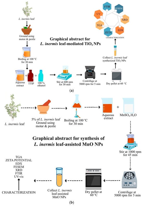
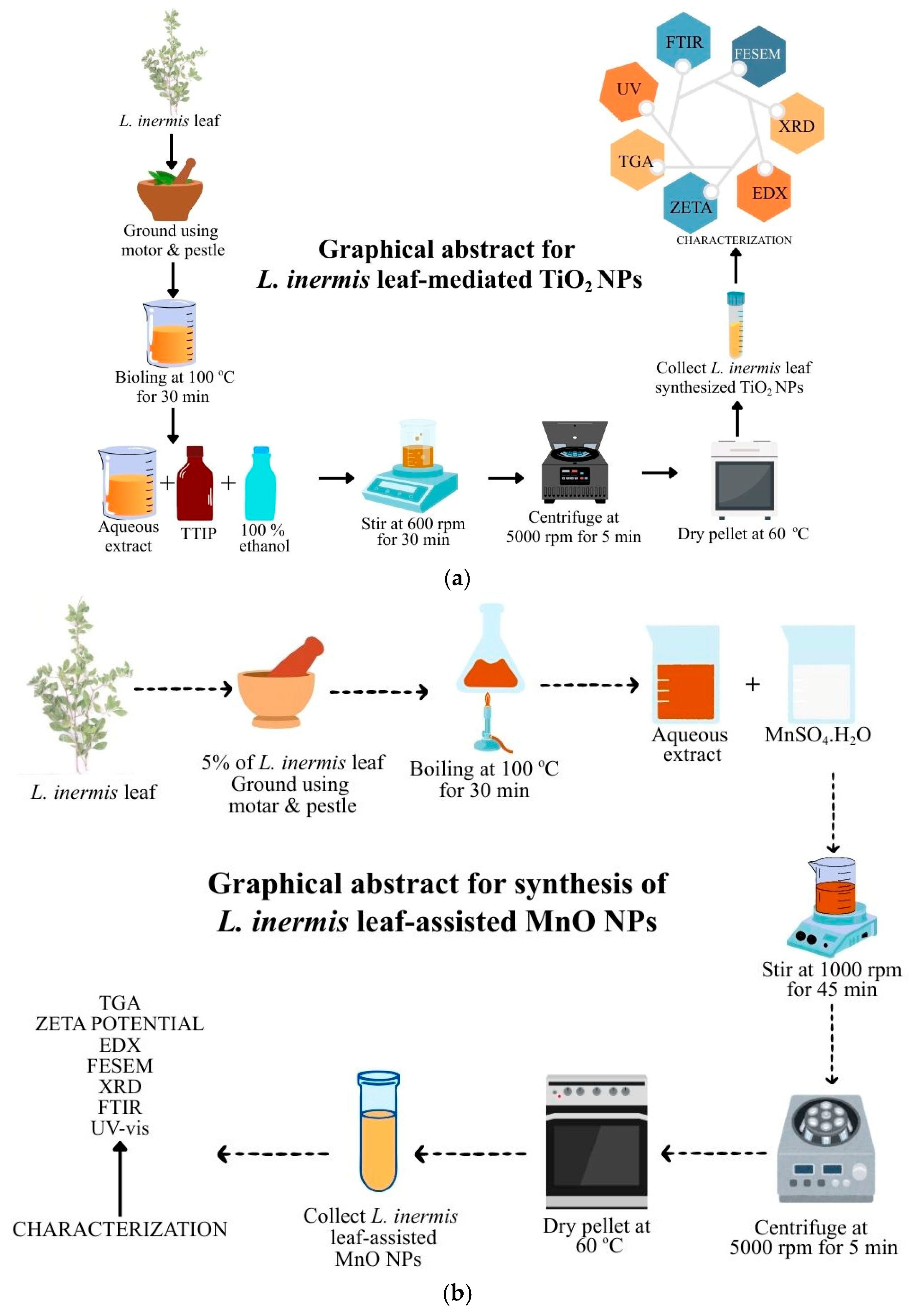
Next, 2 mL of TTIP solution and 10 mL of ethanol (100%) were added to 20 mL of L. inermis leaf extract, stirred for 20 min at 700 rpm using a magnetic stirrer, and left undisturbed overnight. The precipitated mixture was centrifuged at 5000 rpm for 5 min, and the supernatant was discarded. Then the pellet mixture was washed twice using distilled water and 70% ethanol. The obtained pellet was air dried and transferred to a Petri dish, then dried at 100 °C in a hot air oven for 20 min. The dried TiO2 NPs were ground using a motor and pestle to acquire fine TiO2 NPs and stored for subsequent characterization. Figure 1a represented the graphical abstract of L. inermis leaf-mediated TiO2 NPs synthesis.
Figure 1.
(a) Graphical abstract of L. inermis leaf-mediated TiO2 NPs synthesis. (b) Graphical abstract of L. inermis leaf-assisted MnO NPs.
2.4. Synthesis of L. inermis Leaf-Assisted MnO NPs
Firstly, 100 mL of filtered L. inermis leaf extract was mixed with 100 mL of 0.1 M Manganese sulphate (MnSO4) and allowed to stir on the magnetic stirrer (DLAB Scientific, Beijing, China), at 1000 rpm for 45 min, and kept overnight for precipitation. After precipitation, it was centrifuged at 5000 rpm for 5 min and the supernatant was discarded. The obtained pellet was washed twice with distilled water, followed by a 70% ethanol wash. The pellet was transferred to a Petri plate and dried at 100 °C in a hot air oven for 20 min. To obtain fine particles, the MnO NPs are ground using a motor and pestle and stored for further use. Figure 1b demonstrates the L. inermis leaf-mediated MnO NPs green synthesis.
2.5. Characterization
2.5.1. UV–Visible Spectroscopy
The UV-vis spectroscopy of L. inermis leaf-assisted TiO2 and MnO NPs was performed to demonstrate their optical properties. The UV-vis spectrum was documented using a Shimadzu UV–visible spectrophotometer (Shimadzu Corporation, Kyoto, Japan) in a wavelength range of 200–800 nm.
2.5.2. Fourier Transform Infrared Spectroscopy
FTIR analysis was performed to verify the presence of the functional groups present in L. inermis leaf extract, L. inermis leaf-assisted TiO2, and MnO NPs. The FTIR spectra were documented using a Shimadzu (MIRACLE 10) FTIR spectrophotometer (Shimadzu Corporation, Kyoto, Japan) in the range of 400–4000 cm−1.
2.5.3. FESEM Analysis
FESEM was performed to validate the morphological characteristics of TiO2 and MnO NPs synthesized using L. inermis leaf. The TESCAN MIRA3 XMU (TESCAN Orsay Holding, Brno, Czech Republic) field emission scanning electron microscope analyzed the synthesized TiO2 and MnO NPs, and the images at different magnifications were captured.
2.5.4. Energy-Dispersive X-Ray Spectrum
Energy-Dispersive X-ray analysis was accomplished to depict the elemental composition of the L. inermis leaf bio-fabricated TiO2 and MnO NPs. EDX analysis was conducted using a TESCAN MIRA3 XMU microscope (TESCAN Orsay Holding, Brno, Czech Republic)) attached with an EDX detector (AMETEK, Mahwah, NJ, USA).
2.5.5. Thermogravimetric Analysis
TGA was conducted to evaluate the thermal stability of the L. inermis environmentally friendly synthesized TiO2 and MnO NPs. The thermal stability was calculated in a temperature range of 30 °C to 985 °C in an inert atmosphere of Nitrogen gas (N2) by a thermogravimetric analyzer EXSTAR\6300 (Seiko Instruments Inc., Chiba, Japan).
2.5.6. Zeta Potential Analysis
Zeta potential analysis was performed to assess the surface charge and stability of the synthesized TiO2 and MnO NPs. The zeta potential of TiO2 and MnO NPs was assessed by the HORIBA SZ-100 analyzer (HORIBA, Kyoto, Japan), which exhibited the mean zeta potential value based on its electrophoretic mobility.
2.5.7. Dynamic Light-Scattering Analysis
DLS was performed to assess the size distribution and polydispersity index of the synthesized TiO2 and MnO NPs. A HORIBA SZ-100 analyzer (HORIBA, Kyoto, Japan) was employed to assess the average particle size and PDI of the synthesized nanoparticles.
2.6. Biological Properties
2.6.1. Bactericidal Activity
The agar well diffusion method was used to assess the antibacterial activity of the TiO2 and MnO NPs using L. inermis leaf. The bactericidal activity of the biogenic TiO2 NPs was determined against Escherichia coli, Pseudomonas aeruginosa, Klebsiella pneumonia, and Bacillus subtilis. The synthesized MnO NPs were tested against Bacillus subtilis, Pseudomonas aeruginosa, and Klebsiella pneumoniae at varying concentrations, such as 120, 140, 160, and 180 µg/mL, and incubated at 37 °C for 24 h. The zone of inhibition was measured in mm and compared against the standard antibiotic amoxicillin of concentration µg/mL to establish its bactericidal effect.
2.6.2. Antioxidant Activity
DPPH free Radical Scavenging Assay was performed to depict the antioxidant potential of synthesized TiO2 and MnO NPs. Varying concentrations of TiO2 and MnO NPs were taken and mixed with the DPPH solution and incubated at 37 °C for 30 min in the dark. The absorbance was recorded at 517 nm. The percentage of inhibition at different concentrations was calculated using the formulae
where
% inhibition = (A control − A sample)/A control × 100
A control = Absorbance of DPPH control (DPPH + methanol).
A sample = Absorbance of DPPH + particle.
A sample + control = Absorbance of particle with methanol (no DPPH).
2.6.3. Cytotoxicity Assay
MTT assay was performed on L929 cell lines to disclose the cytotoxicity of the biogenically synthesized TiO2 and MnO NPs. The cells were grown in 96-well plates containing DMEM medium and incubated at 37 °C for 24 h in 5% CO2. Different concentrations of the synthesized MnO NPs were introduced into the grown L929 cell lines and incubated. The MTT reagent was added to the cell lines containing TiO2 and MnO NPs and incubated for 4 h. The medium was drained after incubation. To dissolve the formazan crystals, DMSO was added, and the absorbance was measured at 570 nm.
Cell viability (%) = Absorbance of treated cells/Absorbance of control × 100
% Inhibition = 100 − cell viability %
2.6.4. Wound-Healing Assay
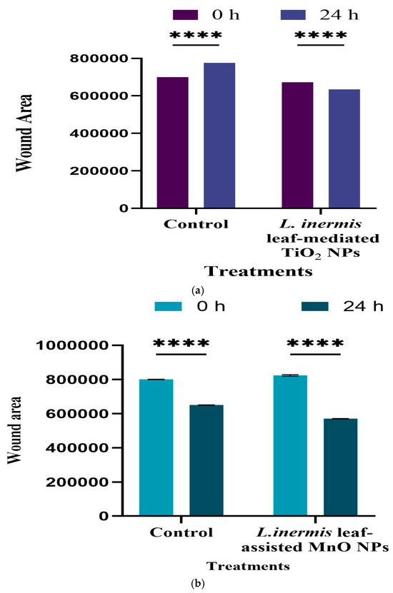
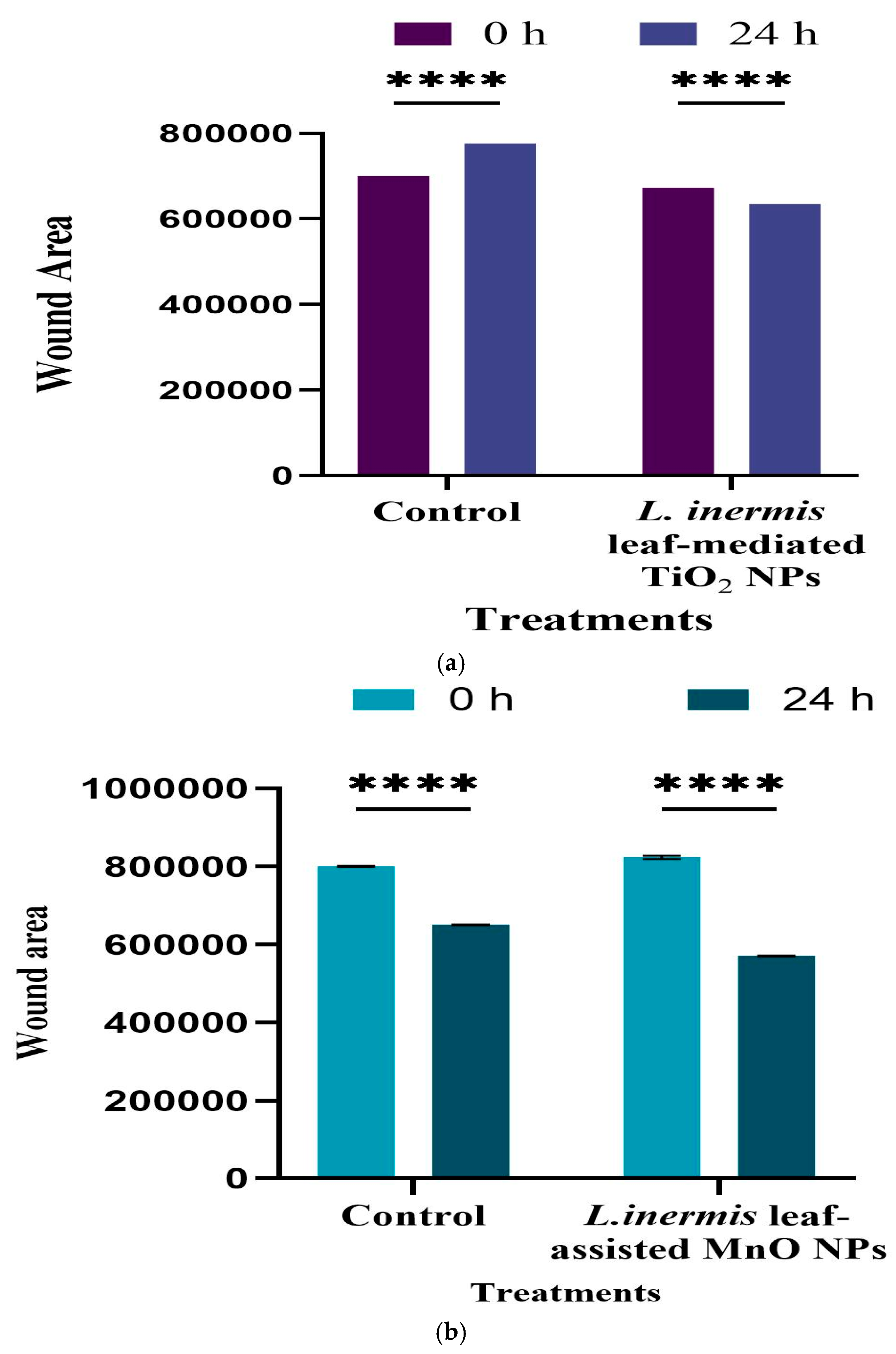
The in vitro wound-healing scratch assay was carried out to reveal the migration potential of the L929 cell line treated with bio-fabricated TiO2 and MnO NPs. L929 cells were cultured in cell culture plates containing DMEM medium and incubated for 24 h in 5% CO2 at 37 °C until a monolayer of cells was formed. A wound was created by using a sterile pipette tip on the monolayer of culture and was treated and washed with phosphate-buffered saline. DMEM media containing synthesized TiO2 and MnO NPs was added. Untreated cells in DMEM media serve as a negative control. The cells were incubated at 37 °C in 5% CO2 for 24 h and photographed at 0 h and 24 h.
Wound Closure (%) = Wound Area0h − Wound Area24h/Wound Area0h × 100
2.6.5. Statistical Analysis
The two-way analysis of variance (ANOVA) was performed in GraphPad Prism 10.6.1, followed by a Bonferroni post-test. It was used to statistically examine the data collected for this study, which is demonstrated as the mean of three independent replicates. Significant values were represented by **** (p < 0.0001); n = 3.
3. Results and Discussion
3.1. Phytochemical Analysis
Table 1 proved the presence of secondary metabolites such as flavonoids, sterols, anthraquinone, proteins, carbohydrates, cardiac glycosides, glycosides, and coumarins. The phytochemical analysis of L. inermis leaf extract revealed the presence of alkaloids, terpenoids, saponins, quinones, flavonoids, phenolic compounds, and tannins [83].
Table 1.
Phytochemical analysis of L. inermis leaf extract.
3.2. UV–Visible Spectroscopy
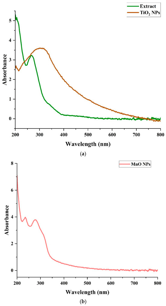
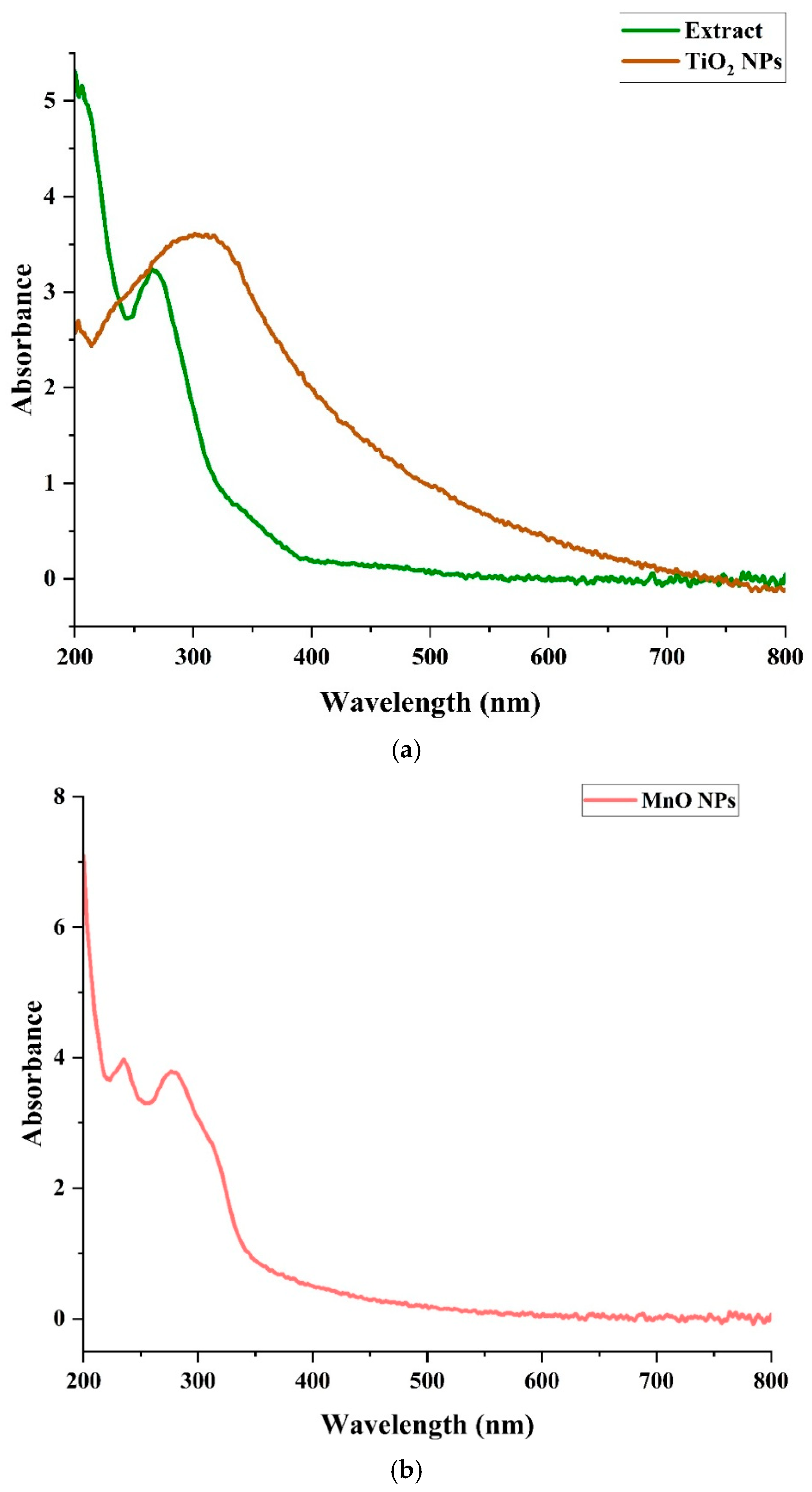
In Figure 2a the UV-vis spectra of the L. inermis leaf extract displayed an absorption peak at 280 and the synthesized TiO2 NPs displayed an absorption peak at 320 nm. The shift in surface plasmon resonance at 320 nm showed the successful synthesis of TiO2 NPs by the reducing and capping of Ti4+ ions using L. inermis leaf. Sakina [27] reported that A. calamus leaf-synthesized TiO2 NPs demonstrated a characteristic peak at 355 nm. The UV absorption spectrum of Mentha arvensis leaf-mediated TiO2 NPs depicted an absorption band at 400 nm [72].
Figure 2.
(a) UV-vis spectra of L. inermis leaf extract and synthesized TiO2 NPs. (b) UV-vis spectra of L. inermis leaf-assisted MnO NPs.
The L. inermis leaf-assisted MnO NPs demonstrated a peak at 295 nm (Figure 2b). The shift in surface plasmon resonance at 295 nm proved the successful synthesis of MnO NPs. The Abutilon indicum leaf synthesized MnO NPs demonstrated a characteristic peak at 460 nm [84].
3.3. FTIR Analysis
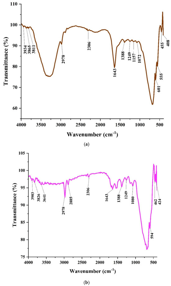
Figure 3a displayed the FTIR spectra of L. inermis leaf extract, which showed characteristic peaks at 3934, 3865, 3811, 2978, 2306, 1643, 1388, 1249, 1157, 1072, 601, 555, 455, and 408 cm−1. The peaks at 3934, 3865, and 3811 cm−1 denoted the O-H stretching vibrations of phenolic compounds. The peaks at 2978 and 2306 cm−1 reflected the C-H stretching vibrations of alkanes. The peak at 1643 cm−1 corresponds to C=O stretching of amide groups. The peak at 1388 cm−1 is attributed to C-H bending vibrations of the methyl group. The peaks at 1249, 1157, and 1072 cm−1 resemble the C-O stretching vibrations of phenols, alcohols, ethers, and esters. The peaks at 601, 555, 455, and 408 cm−1 indicated the aryl-halide groups. In a study on green tea leaf extract, the FTIR spectra disclosed peaks at 3566, 2918, 2850, 1653, 1384, 1107, 1053, and 997 cm−1 [32]. The FTIR analysis of the thyme plant leaf showed distinct peaks with a wavenumber range of 400–4000 cm−1 [85].
Figure 3.
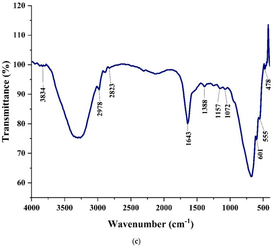
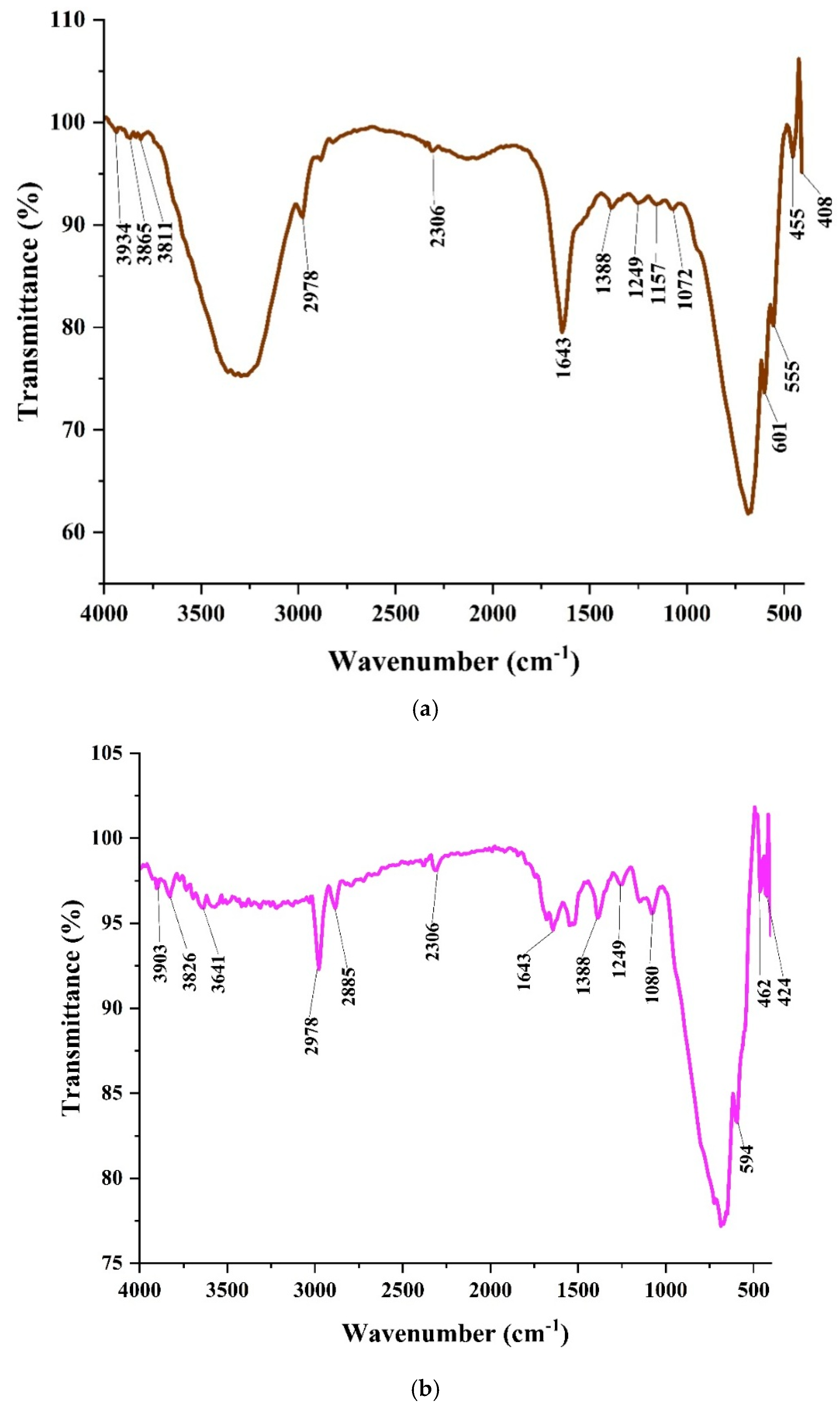
(a) FTIR spectra of L. inermis leaf extract. (b) FTIR spectra of L. inermis leaf-mediated TiO2 NPs. (c) FTIR spectra of L. inermis leaf-assisted MnO NPs.
Figure 3b exhibited the FTIR spectrum of the L. inermis leaf-mediated TiO2 NPs that presented peaks at 3903, 3826, 3641, 2978, 2885, 2306, 1643, 1388, 1249, 1080, 594, 462, and 424 cm−1. The peaks at 3903, 3826, and 3641 cm−1 disclosed the O-H stretching vibrations of alcohols, phenols, and flavonoids. The peak at 2885 cm−1 represented the C-H stretching vibrations of alkanes. The peak at 2306 cm−1 symbolized the O=C=O stretching vibrations of carbon dioxide. The peak at 1643 cm−1 represented the C=C stretching vibrations of aromatic compounds. The peaks at 1388, 1249, and 1080 cm−1 signified the C-N stretching vibrations of amide groups. The peaks at 594, 462, and 424 cm−1 signify the O-Ti-O stretching of synthesized TiO2 NPs. In a previous study on Phyllanthus emblica leaf green synthesized TiO2 NPs, the FTIR analysis displayed a band at 675 cm−1 that demonstrated the stretching vibrations of the Ti-O bond [86].
The L. inermis leaf-assisted MnO NPs presented characteristic peaks at 3834, 2978, 2823, 1643, 1388, 1157, 1072, 601, 555, and 478 cm−1 (Figure 3c). The peak at 3834 cm−1 indicated the O-H stretching vibrations of hydroxyl groups. The peaks at 2978, 2823, 1643 cm−1 corresponded to C-H stretching vibrations of alkanes. Peaks at 1388, 1157, and 1072 cm−1 denoted the C-N stretching vibrations of amide groups. The metal oxygen peaks at 601, 555, and 478 cm−1 represented Mn-O bonding. Islam et al. [30] stated that Ocimum basilicum leaf synthesized displayed bands at 470 and 522 cm−1 that established the formation of MnO NPs.
3.4. FESEM Analysis
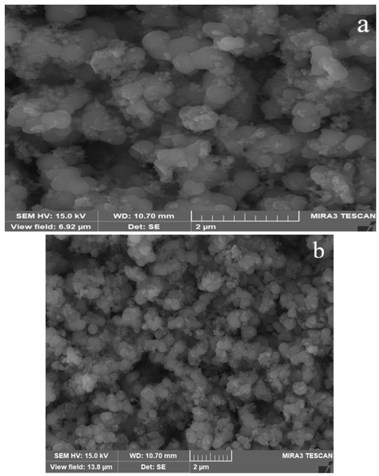
The spherical and aggregated morphology of the L. inermis leaf-mediated TiO2 NPs was confirmed by FESEM images (Figure 4a,b). The FESEM images of Phyllanthus niruri leaf-synthesized TiO2 NPs represented spherical morphology [87]. Nasreen [88] reported that the round shape was observed in the bio-fabricated TiO2 NPs using mulberry leaf.
Figure 4.
(a,b) FESEM images of L. inermis leaf-mediated TiO2 NPs.
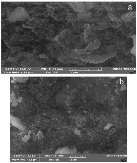
The FESEM analysis of L. inermis leaf-assisted MnO NPs exposed the irregular and aggregated morphology with agglomeration (Figure 5a,b). Manjula et al. [53] validated the spherical shape of the Gardenia resinifera leaf-mediated MnO2 NPs. In a previous study on Terminalia catappa leaf green-synthesized MnO2 NPs, the rectangular shape was observed [89].
Figure 5.
(a,b) FESEM images of L. inermis leaf-assisted MnO NPs.
3.5. EDX Analysis
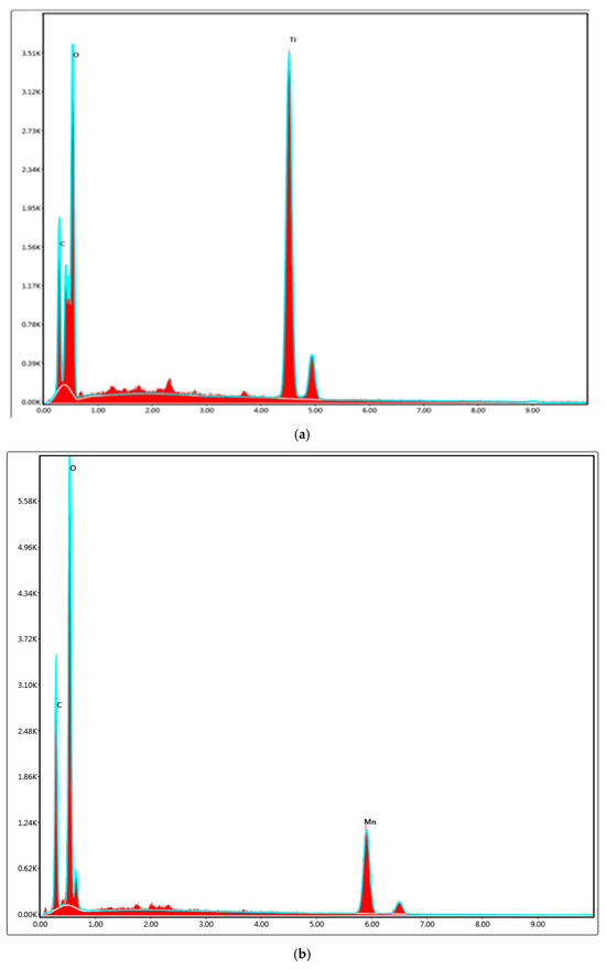
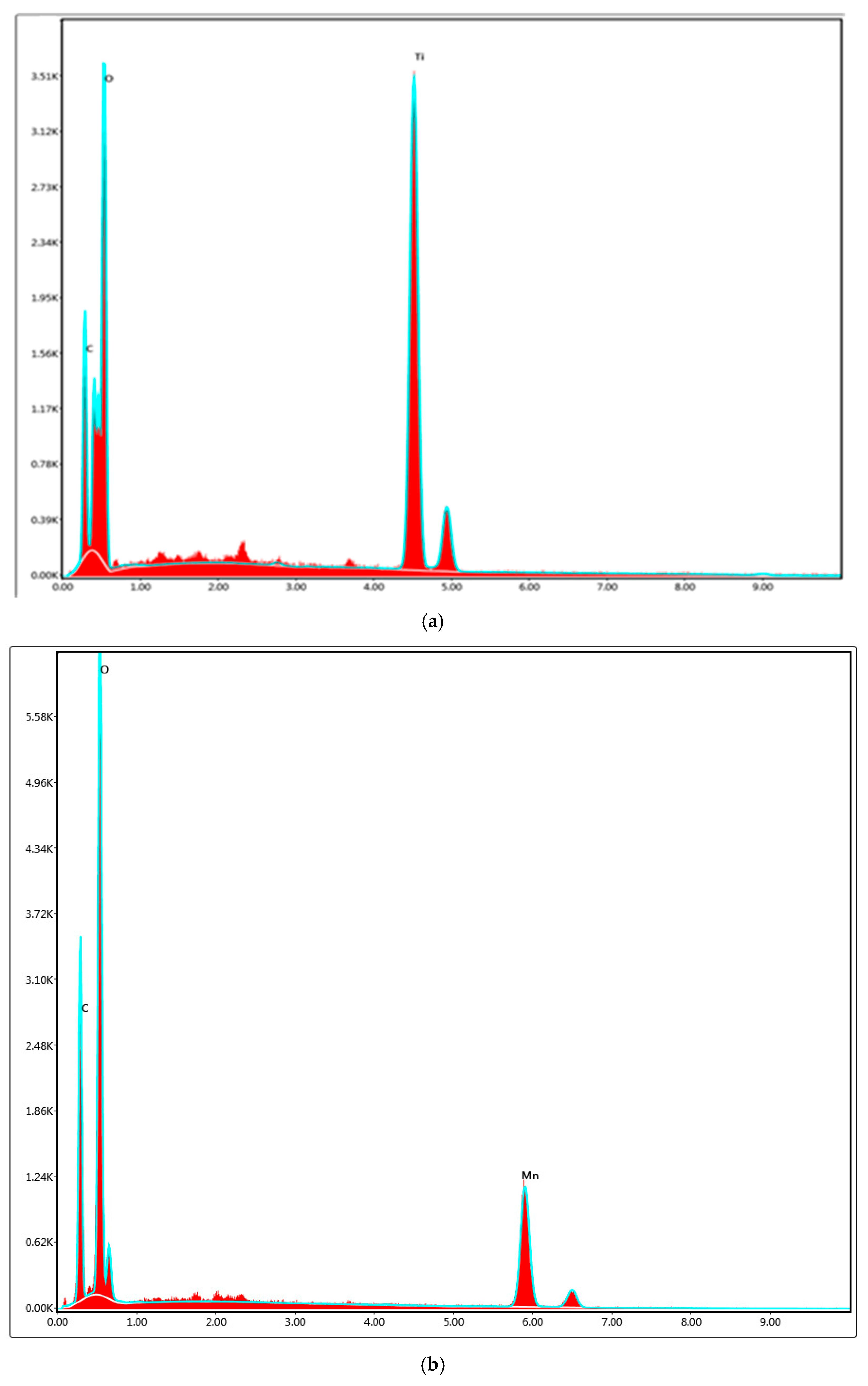
Figure 6a depicts the EDX spectra of the L. inermis leaf-mediated TiO2 NPs and confirms the presence of 51.40% of titanium, 40.60% of oxygen, and 8% of carbon. The EDX analysis of the Ocimum basilicum leaf-assisted TiO2 NPs verified the presence of 47.2% titanium and 34.2% oxygen [90]. Yitagesu et al. [91] signified that 54.33% titanium and 45.67% oxygen were found in Impatiens rothii leaf biosynthesized TiO2 NPs.
Figure 6.
(a) EDX spectra of L. inermis leaf-mediated TiO2 NPs. (b) EDX spectra of L. inermis leaf-assisted MnO NPs.
Figure 6b revealed the presence of 40.81% manganese, 35.11% oxygen, and 24.08% carbon in L. inermis leaf-assisted MnO NPs. 67.41% of manganese and 32.59% of oxygen were found in Matricaria chamomilla leaf-assisted MnO2 NPs [92]. EDX analysis of Ficus benjamina leaf-synthesized MnO NPs indicated the presence of 60.78% of Mn and 39.22% of oxygen [93].
3.6. Zeta Potential Analysis
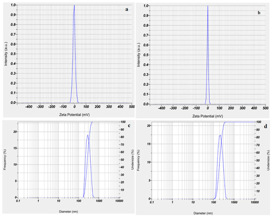
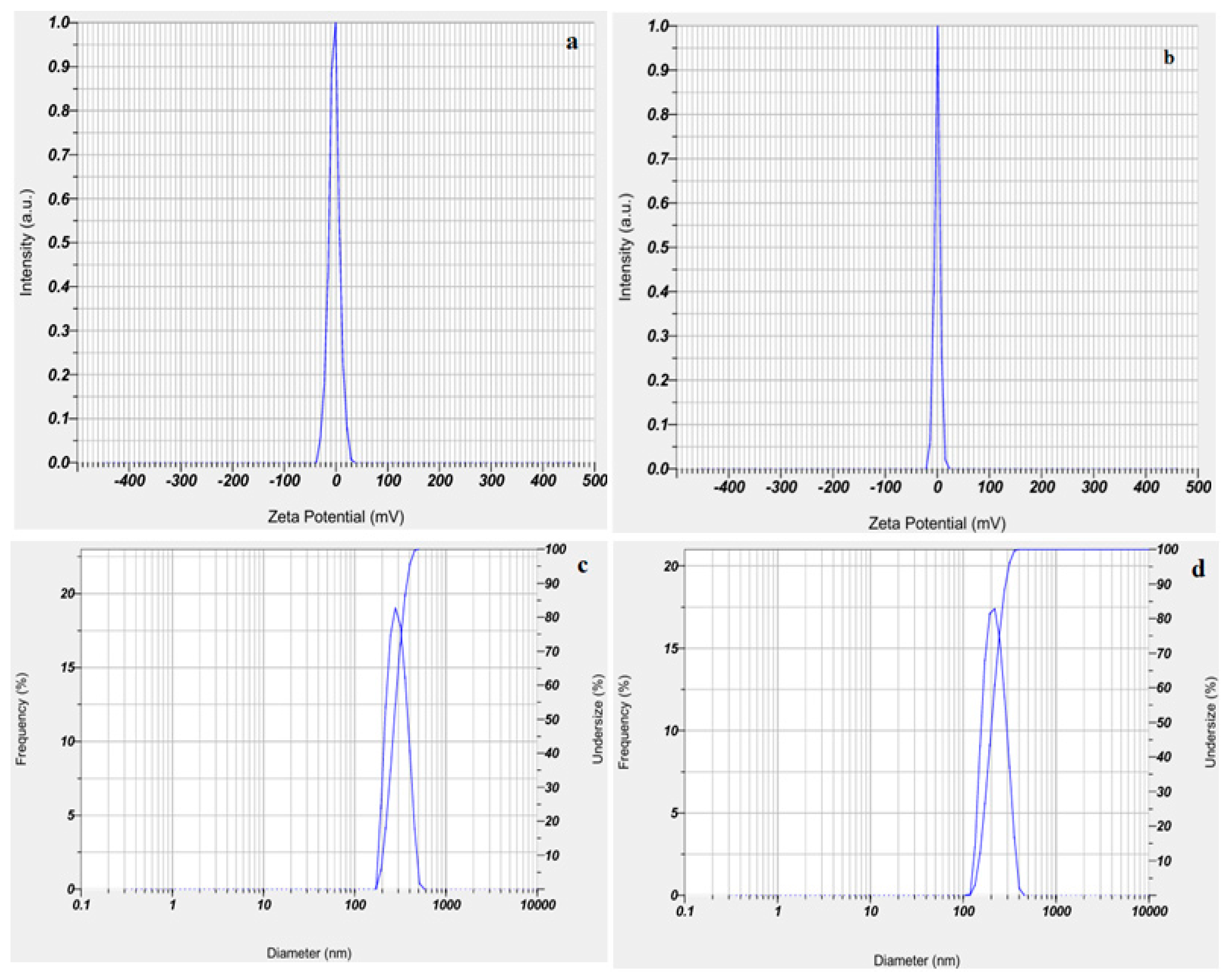
The mean value of −3.5 mV was identified in the zeta potential analysis of L. inermis leaf-mediated TiO2 NPs. The values in the series of −30 mV to +30 mV signify the stability of NPs. The value of −3.5 mV indicated the stable nature of the synthesized TiO2 NPs (Figure 7a). The zeta potential value of the Ocimum sanctum leaf-mediated TiO2 NPs was found to be −11.5 mV [94]. The mean zeta potential value of synthesized MnO NPs was obtained to be −0.1 mV (Figure 7b). The mean zeta potential value of mV is associated with the stable nature of biogenically synthesized MnO NPs. The green synthesized MnO2 NPs indicated the zeta potential value of −11.2 mV [95]. The aggregation of the nanoparticles in the FESEM images was due to the low colloidal stability of the nanoparticles.
Figure 7.
(a,b) Zeta potential analysis of TiO2 NPs and MnO NPs and (c,d) DLS analysis of TiO2 NPs and MnO NPs.
3.7. DLS Analysis
The average particle size of 286 nm and PDI value of 0.300 were observed for the TiO2 NPs (Figure 7c). The DLS analysis performed by Ahmad et al. [94] using green-synthesized TiO2 NPs revealed the mean particle size of 130 nm and PDI of 0.237. For MnO NPs, an average particle size of 210 nm and 0.46 PDI was reported (Figure 7d). The DLS analysis executed by Johnston et al. [95] reported an average particle size of 266 nm and a PDI of 0.364. DLS analysis demonstrated that the MnO NPs were relatively smaller than TiO2 NPs. The smaller size and high surface area may contribute to enhancement of antibacterial activity, antioxidant potential, cytotoxic response, and wound-healing efficiency observed for MnO NPs. The zeta potential values (−0.1 mV and −3.5 mV) indicate the electrostatic stability and a tendency toward aggregation. The phytochemical capping agents from L. inermis extract might improve the cell interactions and biocompatibility. The biological activities are influenced by the combined effects of particle size, surface charge, dispersity, and surface functionalization.
3.8. TG Analysis
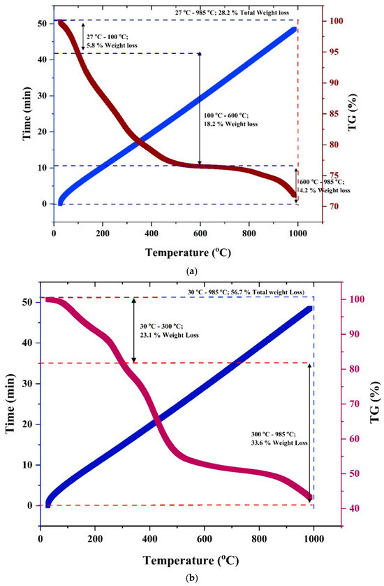
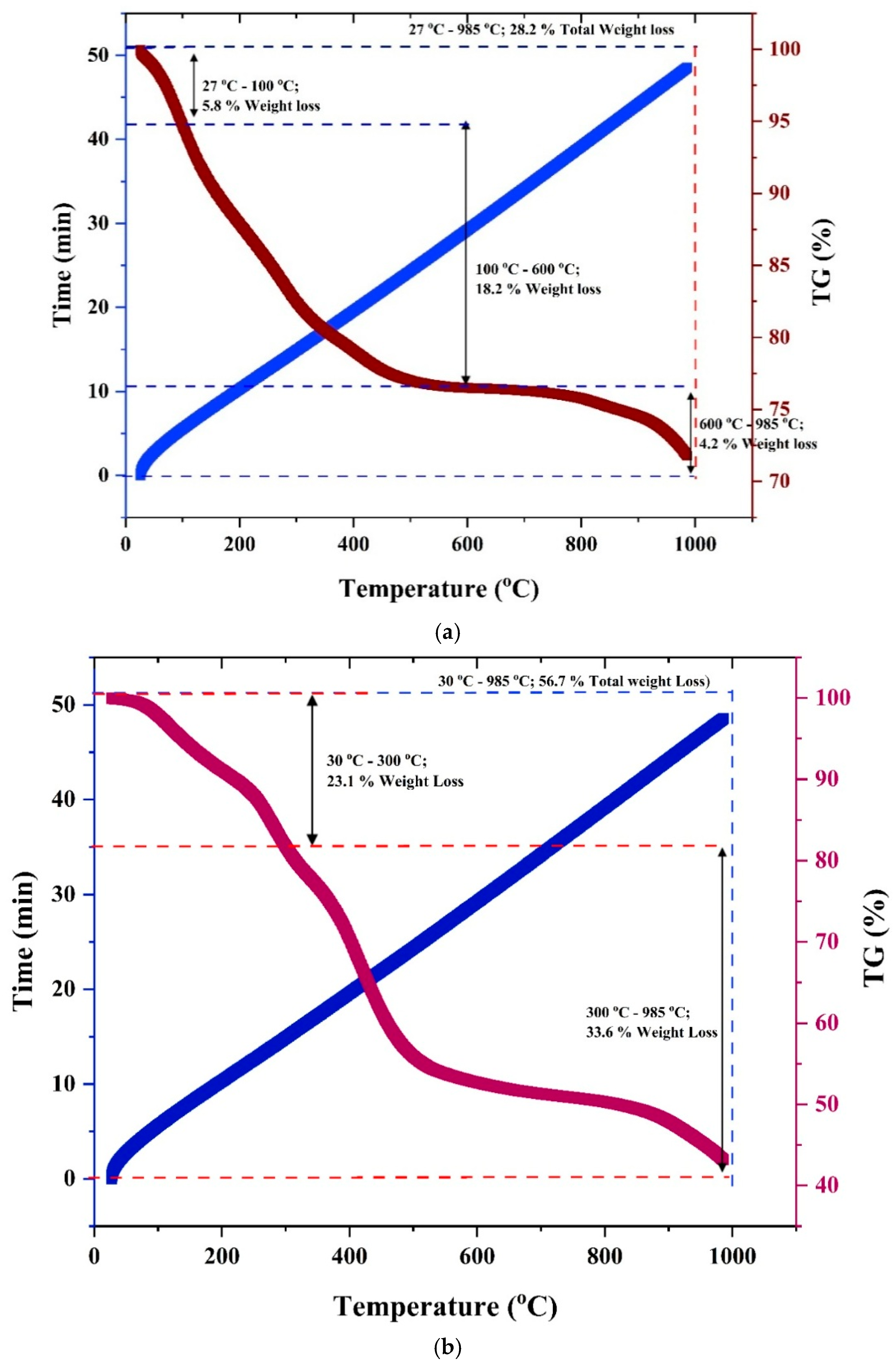
Figure 8a illustrates the thermal stability of the L. inermis leaf-mediated TiO2 NPs. The TG analysis of TiO2 NPs demonstrated the total weight loss of 28.2% in the temperature range of 27–985 °C. The 5.8% of the first weight loss is attributed to the temperature range of 27–100 °C. 18.2% of the second weight loss occurred in the temperature range of 100–600 °C. The final weight loss of 4.2% occurred in the range of 600–985 °C. TiO2 NPs synthesized using the leaf of Andrographis paniculata showed 18.76% weight loss in the temperature range of 20–800 °C [96].
Figure 8.
(a) TG analysis of L. inermis leaf-mediated TiO2 NPs. (b) TG Analysis of L. inermis leaf-assisted MnO NPs.
The TG analysis revealed the thermal stability of the L. inermis leaf-assisted MnO NPs (Figure 8b). The total weight loss of 56.7% occurred in a temperature range of 30–985 °C. The first weight loss of 23.1% due to evaporation of water occurred in the temperature range of 30–300 °C. The second weight loss of 33.6% due to evaporation of organic molecules occurred in the 300–985 °C temperature range. Tagetes erecta leaf biosynthesized Mn3O4 NPs established a total weight loss of 9.6% in a 0–600 °C temperature range [97].
3.9. Biological Properties
3.9.1. Bactericidal Activity
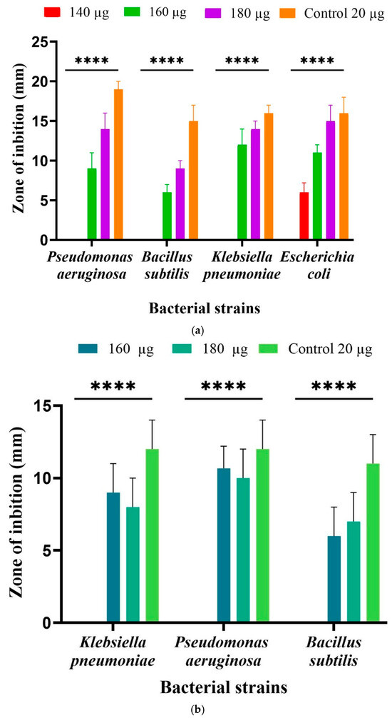
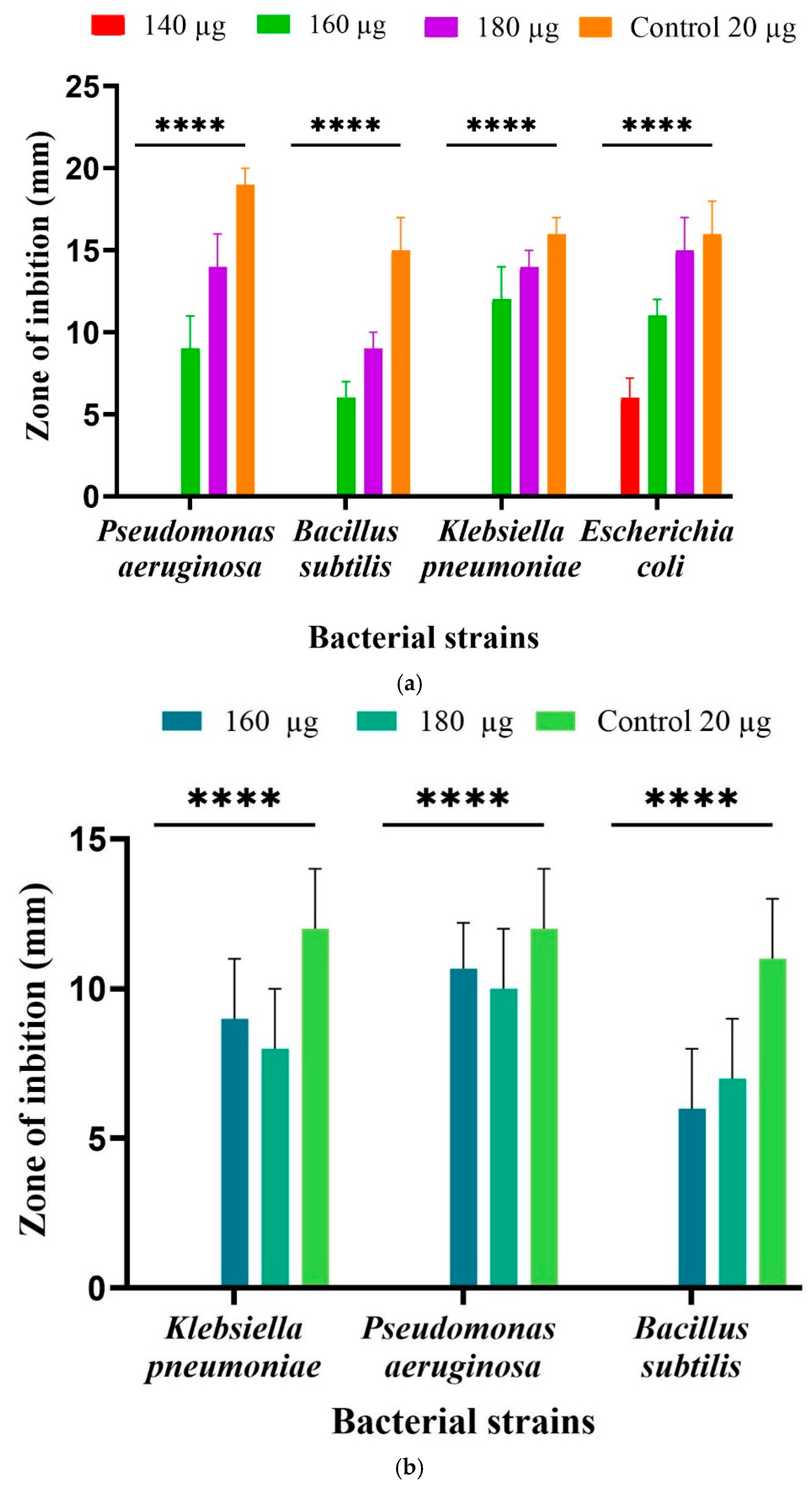
The L. inermis leaf-mediated TiO2 NPs exhibited inhibition activity against E. coli, P. aeruginosa, K. pneumoniae, and B. subtilis (Figure 9a). The biogenically synthesized TiO2 NPs showed maximum inhibition against Escherichia coli and low inhibition against Bacillus subtilis. The TiO2 NPs interacts with the cell membrane, causing rupture of the bacterial cell membrane, releasing intracellular components, and resulting in apoptosis of the cell. In a preceding study, green-synthesized TiO2 NPs using Piper Longum leaf demonstrated antibacterial activity against Pseudomonas aeruginosa and Klebsiella pneumoniae [98]. Inhibition against P. aeruginosa, E. coli, and K. pneumoniae was observed in Luffa acutangula leaf-synthesized TiO2 NPs [99].
Figure 9.
(a) Antibacterial activity of L. inermis leaf-mediated TiO2 NPs. (b) Antibacterial activity of L. inermis leaf-assisted MnO NPs. ****—resembles high significance of results.
L. inermis leaf-assisted MnO NPs showed inhibition against K. pneumoniae, P. aeruginosa, and B. subtilis (Figure 9b). The MnO NPs synthesized using L. inermis leaf-mediated MnO NPs depicted bactericidal effects due to cell wall damage, release of intracellular contents, electrolyte leakage, and generation of Reactive Oxygen Species. MnO2 NPs synthesized using Viola betonicifolia leaf showed inhibition against Staphylococcus aureus and K. pneumoniae [100]. In a preceding study, the biofabrication using Plectranthus amboinicus leaf MnO2 NPs exhibited antibacterial activity against P. aeruginosa, B. subtilis, E. coli, and K. pneumoniae [49].
3.9.2. Antioxidant Activity
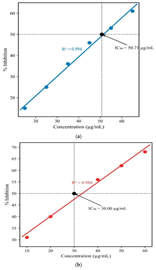
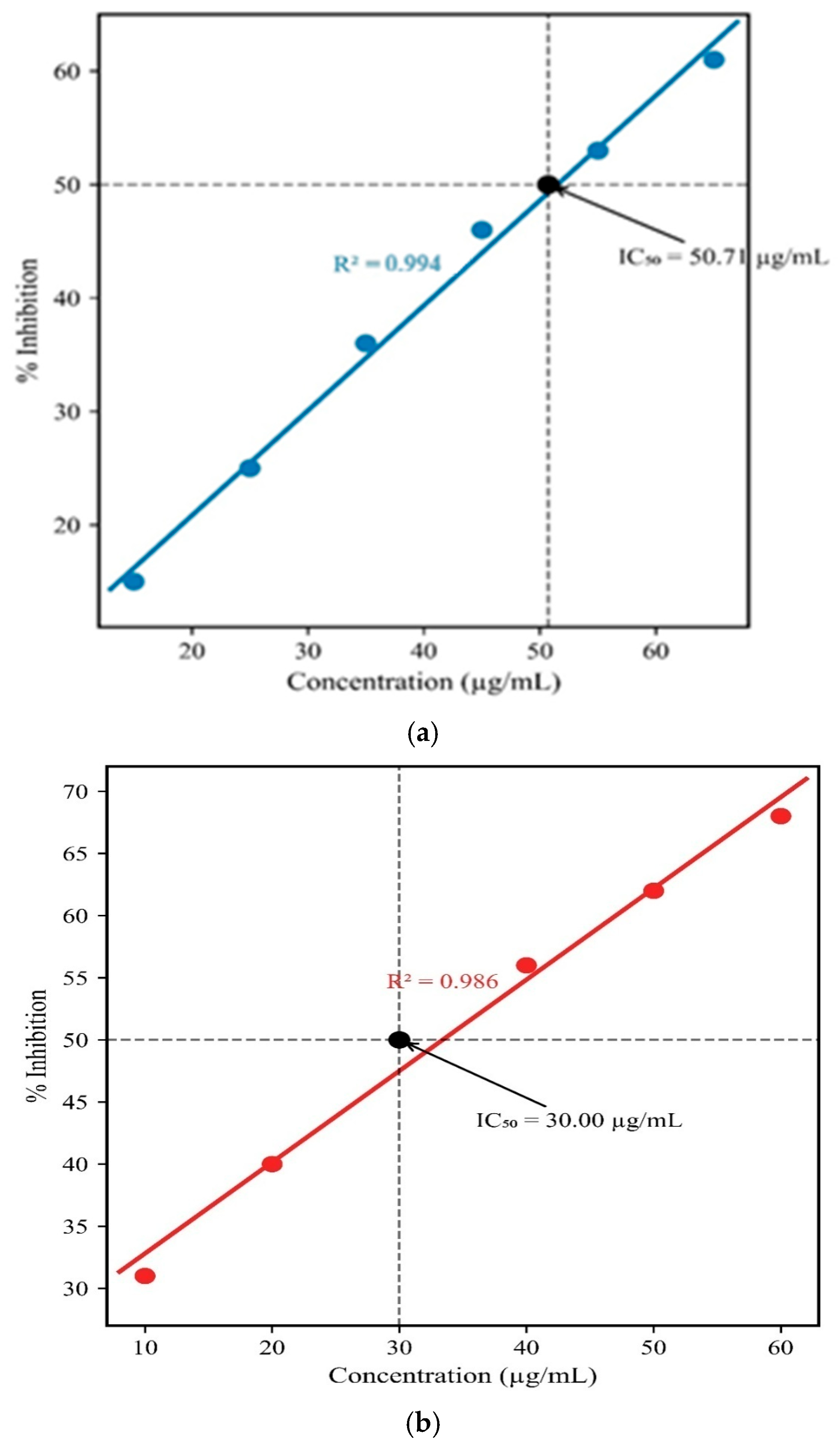
L. inermis leaf-mediated TiO2 NPs presented concentration-dependent antioxidant activity. The synthesized TiO2 NPs exhibited an IC50 value of 50.71 µg/mL, which depicted its antioxidant activity (Figure 10a). The secondary metabolites present in the L. inermis leaf are represented as an electron donor in the DPPH reduction reaction. DPPH produced free radicals, which were neutralized by the free electrons of phytochemicals on the surface of the synthesized TiO2 NPs [101]. The synthesized TiO2 NPs using Calotropis procera leaf showed antioxidant potential of 58.7% at 40 mg/mL. Terenna asiatica fruit mediated TiO2 NPs depicted an IC50 value of 42.71 µg [102].
Figure 10.
(a) Antioxidant activity of L. inermis leaf-mediated TiO2 NPs. (b) Antioxidant activity of L. inermis leaf-assisted MnO NPs.
Figure 10b depicts that the L. inermis leaf-assisted MnO NPs presented concentration-dependent antioxidant activity. The antioxidant activity of L. inermis leaf-synthesized MnO NPs showed the IC50 value of 30 µg/mL. The free electrons donated by the secondary metabolites present on the surface of synthesized MnO NPs neutralized the free radicals produced by DPPH. Syzygium aromaticum extract-mediated MnO NPs portrayed an IC50 value of 51.18 µg/mL [103]. Mahdavi et al. [104] stated that the Ziziphora clinopodioides Lam leaf biosynthesized MnO NPs displayed an IC50 value of 236 µg/mL.
3.9.3. Cytotoxicity Assay
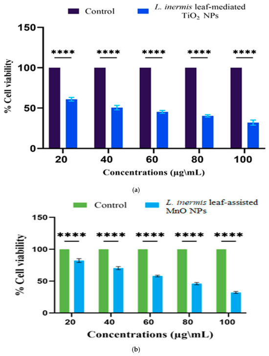
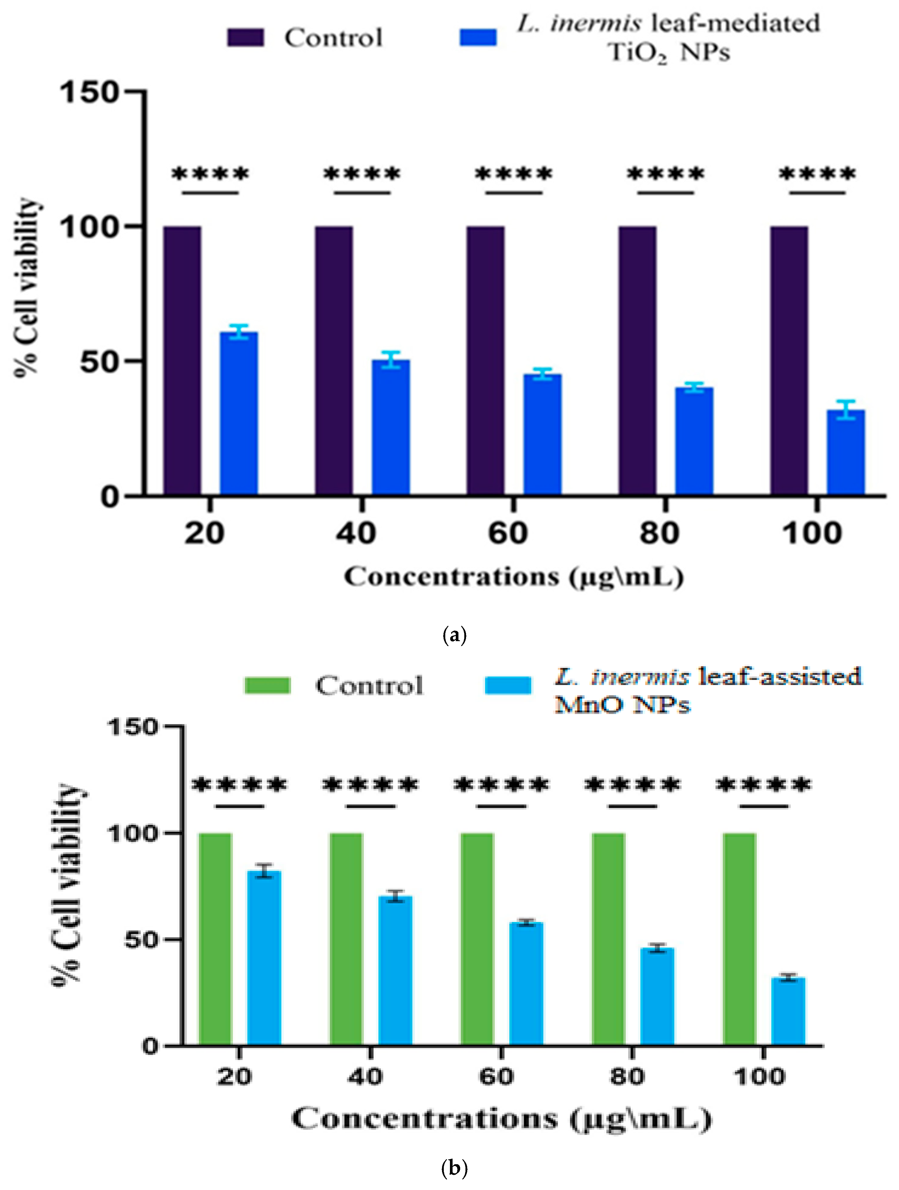
The L. inermis leaf-mediated TiO2 NPs described 60% cell viability at the concentrations of 20 µg/mL and 100 µg/mL (Figure 11a). The synthesized TiO2 NPs promote the generation of Reactive Oxygen Species (ROS), which cause oxidative damage that results in mitochondrial dysfunction and leads to cell cytotoxicity. Al-Shabib et al. [105] reported that bio-fabricated TiO2 NPs using Withania somnifera root showed 40% of cell viability at the concentration of 100 μg/mL. Prosopis Cineraria leaf-synthesized TiO2 NPs showed 95.65% cell viability at 0.125 mg/mL [106].
Figure 11.
(a) Cytotoxicity assay of L. inermis leaf-mediated TiO2 NPs. (b) Cytotoxicity assay of L. inermis leaf-assisted MnO NPs. ****—resembles high significance of results.
The L. inermis leaf-assisted MnO NPs represented dose-dependent cytotoxicity (Figure 11b). The eco-friendly synthesized MnO NPs illustrated 82.28% and 32.24% cell viability at the concentrations of 20 µg/mL and 100 µg/mL. The green synthesized MnO NPs induce the generation of ROS that lead to DNA damage and result in cell death. The cytotoxic effects of the biogenic MnO NPs are based on their physical characteristics and interaction with secondary metabolites. Ag-MnO2 NPs synthesized using Vinca minor showed 46.4% of cytotoxicity on HaCaT cells [107]. Tinospora cardifoliastem-synthesized MnO NPs portrayed an IC50 of 1.1 mg/mL [108].
3.9.4. Wound-Healing Assay
The wound-healing potential of the L. inermis leaf-mediated TiO2 NPs was verified by 5.68% wound closure after 24 h in the L929 cell lines treated with the synthesized TiO2 NPs (Figure 12a). The biogenically synthesized TiO2 NPs limit the Reactive Oxygen Species and promote fibroblast migration, thereby enhancing the wound-healing potential. Thammaacheep et al. [93] stated that TiO2 NPs synthesized using Morus alba leaf showed 80% wound closure on 3T3 mouse fibroblast cells after 24 h. Neem flower-mediated TiO2 NPs revealed 62% wound closure on L929 cell lines by 12 h [109].
Figure 12.
(a) Wound-healing assay of L. inermis leaf-mediated TiO2 NPs. (b) Wound-healing assay of L. inermis leaf-assisted MnO NPs. ****—resembles high significance of results.
The L. inermis leaf-assisted MnO NPs treated L929 cell lines showed an enhanced cell migration with a wound closure of 60.4% at the concentration of 20 µg/mL (Figure 12b). MnO NPs limit the ROS, prevent bacterial infections in the wound site, and enhance cell proliferation, which promotes wound closure. Chemically synthesized K-MnO NPs revealed significant wound healing with resistance to MRSA [110]. Chitosan nanovesicles loaded with Mn showed 57.84% wound closure after 16 h [111].
4. Conclusions
The successful biogenic synthesis of L. inermis leaf-mediated TiO2 and MnO NPs was verified by the current study. L. inermis leaf extract used as a reducing and capping agent for the green synthesis of TiO2 and MnO NPs. The FTIR spectra confirmed the presence of the O-Ti-O group and Mn-O functional group. The FESEM images revealed the spherical morphology of the green-synthesized TiO2 NPs and an irregular shape with agglomeration for MnO NPs. The elemental composition of the synthesized TiO2 and MnO NPs was depicted by the EDX spectra. Zeta potential analysis depicted the stability of the eco-friendly synthesized TiO2 and MnO NPs. The DLS analysis portrayed the aggregation of the synthesized TiO2 and MnO NPs. The TG analysis showed the thermal stability of the bio-fabricated TiO2 and MnO NPs. The antibacterial activity against E. coli, P. aeruginosa, K. pneumoniae, and B. subtilis was observed. The green-synthesized MnO NPs showed inhibition against bacterial pathogens. MTT assay depicted concentration-dependent cytotoxicity in both TiO2 and MnO NPs. In vitro wound-healing assay denoted the wound-healing properties of the biosynthesized TiO2 and MnO NPs. Future studies should focus on large-scale sustainable synthesis of TiO2 and MnO NPs using L. inermis leaf and exploring its applications in medicine, energy, and environmental fields.
Author Contributions
Conceptualization, Supervision, Funding acquisition, and Project administration, R.P.; Investigation and Writing—Original draft preparation, Investigation and Writing—Original draft preparation, K.K.; Review and editing, V.P. and J.A.M.; Data Curation, N.A.-D. All authors have read and agreed to the published version of the manuscript.
Funding
This research received no external funding.
Informed Consent Statement
Not applicable.
Data Availability Statement
Data available on request due to restrictions (e.g., privacy, legal or ethical reasons).
Conflicts of Interest
The authors declare that they have no known competing financial interests or personal relation-ships that could have appeared to influence the work reported in this paper. The authors declare no conflicts of interest.
References
- Malik, S.; Muhammad, K.; Waheed, Y. Nanotechnology: A revolution in modern industry. Molecules 2023, 28, 661. [Google Scholar] [CrossRef] [Scilit]
- Ray, M.K.; Mishra, A.K.; Mohanta, Y.K.; Mahanta, S.; Chakrabartty, I.; Kungwani, N.A.; Avula, S.K.; Panda, J.; Pudake, R.N. Nanotechnology as a promising tool against phytopathogens: A futuristic approach to agriculture. Agriculture 2023, 13, 1856. [Google Scholar] [CrossRef] [Scilit]
- Deng, S.; Gu, J.; Jiang, Z.; Cao, Y.; Mao, F.; Xue, Y.; Wang, J.; Dai, K.; Qin, L.; Liu, K.; et al. Application of nanotechnology in the early diagnosis and comprehensive treatment of gastrointestinal cancer. J. Nanobiotechnol. 2022, 20, 415. [Google Scholar] [CrossRef] [Scilit]
- Gupta, R.K.; Abd El Gawad, F.; Ali, E.A.; Karunanithi, S.; Yugiani, P.; Srivastav, P.P. Nanotechnology: Current applications and future scope in food packaging systems. Meas. Food 2024, 13, 100131. [Google Scholar] [CrossRef] [Scilit]
- Fattahi, Z.; Hasanzadeh, M. Nanotechnology-assisted microfluidic systems for chemical sensing, biosensing, and bioanalysis. TrAC Trends Anal. Chem. 2022, 152, 116637. [Google Scholar] [CrossRef] [Scilit]
- Kumar, S.; Tirlangi, S.; Kumar, A.; Imran, M.; HP, J.S.P.; Koshariya, A.K.; Sathish, T.; Ubaidullah, M.; Ayub, R.; Reddy, V.R.M.; et al. A review on the contribution of nanotechnology for biofuel production from algal biomass: A bridge to the reduction of carbon footprint. Sustain. Energy Technol. Assess. 2023, 60, 103498. [Google Scholar] [CrossRef] [Scilit]
- Haris, M.; Hussain, T.; Mohamed, H.I.; Khan, A.; Ansari, M.S.; Tauseef, A.; Khan, A.A.; Akhtar, N. Nanotechnology–A new frontier of nano-farming in agricultural and food production and its development. Sci. Total Environ. 2023, 857, 159639. [Google Scholar] [CrossRef] [Scilit]
- Woźniak, M.; Płoska, A.; Siekierzycka, A.; Dobrucki, L.W.; Kalinowski, L.; Dobrucki, I.T. Molecular imaging and nanotechnology—Emerging tools in diagnostics and therapy. Int. J. Mol. Sci. 2022, 23, 2658. [Google Scholar] [CrossRef] [Scilit] [PubMed]
- Martin, J.A.; Periakaruppan, R.; Babu, D.; Vijai Selvaraj, K.S.; Al-Dayan, N. Green Synthesis, Characterization, and Antibacterial Activity of Indigofera aspalathoides-Assisted Manganese Oxide-Doped Calcium Oxide Nanocomposites. Biomed. Mater. Devices 2025, 1–16. [Google Scholar] [CrossRef] [Scilit]
- Yusuf, A.; Almotairy, A.R.Z.; Henidi, H.; Alshehri, O.Y.; Aldughaim, M.S. Nanoparticles as drug delivery systems: A review of the implication of nanoparticles’ physicochemical properties on responses in biological systems. Polymers 2023, 15, 1596. [Google Scholar] [CrossRef] [Scilit]
- Alprol, A.E.; Mansour, A.T.; Abdelwahab, A.M.; Ashour, M. Advances in green synthesis of metal oxide nanoparticles by marine algae for wastewater treatment by adsorption and photocatalysis techniques. Catalysts 2023, 13, 888. [Google Scholar] [CrossRef] [Scilit]
- Samhitha, S.S.; Raghavendra, G.; Quezada, C.; Bindu, P.H. Green synthesized TiO2 nanoparticles for anticancer applications: Mini review. Mater. Today Proc. 2022, 54, 765–770. [Google Scholar] [CrossRef] [Scilit]
- Sharifi, R.; Vatani, A.; Sabzi, A.; Safaei, M. A narrative review on application of metal and metal oxide nanoparticles in endodontics. Heliyon 2024, 10, e34673. [Google Scholar] [CrossRef] [Scilit] [PubMed]
- Liu, W.; Zeb, A.; Lian, J.; Wu, J.; Xiong, H.; Tang, J.; Zheng, S. Interactions of metal-based nanoparticles (MBNPs) and metal-oxide nanoparticles (MONPs) with crop plants: A critical review of research progress and prospects. Environ. Rev. 2020, 28, 294–310. [Google Scholar] [CrossRef] [Scilit]
- Maity, D.; Gupta, U.; Saha, S. Biosynthesized metal oxide nanoparticles for sustainable agriculture: Next-generation nanotechnology for crop production, protection and management. Nanoscale 2022, 14, 13950–13989. [Google Scholar] [CrossRef] [Scilit]
- Jafarzadeh, S.; Salehabadi, A.; Jafari, S.M. Metal nanoparticles as antimicrobial agents in food packaging. In Handbook of Food Nanotechnology; Academic Press: Cambridge, MA, USA, 2020; pp. 379–414. [Google Scholar]
- Liu, B.; Liu, J. Sensors and biosensors based on metal oxide nanomaterials. TrAC Trends Anal. Chem. 2019, 121, 115690. [Google Scholar] [CrossRef] [Scilit]
- Senthil Rathi, B.; Ewe, L.S.; S, S.; S, S.; Yew, W.K.; R, B.; Tiong, S.K. Recent trends and advancement in metal oxide nanoparticles for the degradation of dyes: Synthesis, mechanism, types and its application. Nanotoxicology 2024, 18, 272–298. [Google Scholar] [CrossRef] [Scilit]
- Shreyash, N.; Bajpai, S.; Khan, M.A.; Vijay, Y.; Tiwary, S.K.; Sonker, M. Green synthesis of nanoparticles and their biomedical applications: A review. ACS Appl. Nano Mater. 2021, 4, 11428–11457. [Google Scholar] [CrossRef] [Scilit]
- Patil, N.A.; Udgire, S.; Shinde, D.R.; Patil, P.D. Green synthesis of gold nanoparticles using extract of Vitis vinifera, Buchananialanzan, Juglandaceae, phoenix dactylifera plants, and evaluation of antimicrobial activity. Chem. Methodol. 2023, 7, 15–27. [Google Scholar]
- Alqarni, L.S.; Alghamdi, M.D.; Alshahrani, A.A.; Nassar, A.M. Green nanotechnology: Recent research on bioresource-based nanoparticle synthesis and applications. J. Chem. 2022, 2022, 4030999. [Google Scholar] [CrossRef] [Scilit]
- Ying, S.; Guan, Z.; Ofoegbu, P.C.; Clubb, P.; Rico, C.; He, F.; Hong, J. Green synthesis of nanoparticles: Current developments and limitations. Environ. Technol. Innov. 2022, 26, 102336. [Google Scholar] [CrossRef] [Scilit]
- Salem, S.S. A mini review on green nanotechnology and its development in biological effects. Arch. Microbiol. 2023, 205, 128. [Google Scholar] [CrossRef] [Scilit] [PubMed]
- Subhapriya, S.; Gomathipriya, P.J.M.P. Green synthesis of titanium dioxide (TiO2) nanoparticles by Trigonella foenum-graecum extract and its antimicrobial properties. Microb. Pathog. 2018, 116, 215–220. [Google Scholar] [CrossRef] [Scilit]
- Rajaram, P.; Jeice, A.R.; Jayakumar, K. Review of green synthesized TiO2 nanoparticles for diverse applications. Surf. Interfaces 2023, 39, 102912. [Google Scholar] [CrossRef] [Scilit]
- Gul, H.; Javed, H.M.A.; Awais, M.; Javaid, M.Y.; Khan, M.I.; Arif, M.; Alshahrani, M.Y.; Khalil, R.M.A.; Khan, F.S.; Galal, A.M. TiO2 nanoparticles functionalized with marigold for antioxidant role to enhance the skin protection. Biomass Convers. Biorefinery 2023, 13, 16025–16035. [Google Scholar] [CrossRef] [Scilit]
- Sakina, B. The Impact of TiO2 Nanoparticles Treatment, Developmental, and Environmental Factors on Phenolic Content and Antioxidant Capacity of Grapevine (Vitis vinifera L.) Leaves. Ph.D. Thesis, University of Pécs, Pécs, Hungary, 2022. [Google Scholar]
- Sobańska, Z.; Roszak, J.; Kowalczyk, K.; Stępnik, M. Applications and biological activity of nanoparticles of manganese and manganese oxides in in vitro and in vivo models. Nanomaterials 2021, 11, 1084. [Google Scholar] [CrossRef] [Scilit]
- Boulaares, I.; Derouiche, S.; Niemann, J. Greenly Synthesized Manganese Oxide Nanoparticles (MnO NPs) In Tumor Therapy: A Narrative Review. Arch. Razi Inst. 2024, 79, 1135. [Google Scholar]
- Islam, B.; Samir, D.; Sara, C.; Janetta, N. Ocimum basilicum L. leaves extract-mediated green synthesis of MnO NPs: Phytochemical profile, characterization, catalytic and thrombolytic activities. Results Surf. Interfaces 2024, 17, 100284. [Google Scholar] [CrossRef] [Scilit]
- Hosseiny, Z.; Meftahizadeh, H.; Ghorbanpour, M.; Gholamnejad, J.; Zareshahi, R. Influence of harvesting dates and genotypes on secondary metabolites status and antioxidant activity in Lawsonia inermis L. (Henna). Acta Ecol. Sin. 2023, 43, 891–898. [Google Scholar] [CrossRef] [Scilit]
- Moutawalli, A.; Benkhouili, F.Z.; Doukkali, A.; Benzeid, H.; Zahidi, A. The biological and pharmacologic actions of Lawsonia inermis L. Phytomed. Plus 2023, 3, 100468. [Google Scholar] [CrossRef] [Scilit]
- Mohammed, D.M.; Ahmed, K.A.; Desoukey, M.A.; Sabry, B.A. Assessment of the antiulcer properties of Lawsonia inermis L. leaves and its nano-formulation against prolonged effect of acute ulcer in rats. Toxicol. Rep. 2022, 9, 337–345. [Google Scholar] [CrossRef] [Scilit] [PubMed]
- Maksoud, S.A.; Gad, K.I.; Hamed, E.Y. The potentiality of biostimulant (Lawsonia inermis L.) on some morpho-physiological, biochemical traits, productivity and grain quality of Triticum aestivum L. BMC Plant Biol. 2023, 23, 95. [Google Scholar] [CrossRef] [Scilit] [PubMed]
- Singh, N. Studies on antimicrobial activity of Lawsonia inermis L. against different strains of bacteria and fungi. J. Appl. Nat. Sci. 2022, 14, 247–253. [Google Scholar] [CrossRef] [Scilit]
- Fapetu, A.P.; Akintimehin, E.S.; Karigidi, K.O.; Jibayo, S.; Adetuyi, F.O. Effects of boiling on in vitro antioxidant and antidiabetic activity of aqueous extract of Lawsonia inermis leaves. Herba Pol. 2023, 69, 13–22. [Google Scholar]
- Kavepour, N.; Bayati, M.; Rahimi, M.; Aliahmadi, A.; Ebrahimi, S.N. Optimization of aqueous extraction of henna leaves (Lawsonia inermis L.) and evaluation of biological activity by HPLC-based profiling and molecular docking techniques. Chem. Eng. Res. Des. 2023, 195, 332–343. [Google Scholar] [CrossRef] [Scilit]
- Sen, S.; Borthakur, M.S.; Chetia, D. Lawsonia inermis Linn: A breakthrough in cosmeceuticals. Sci. Phytochem. 2023, 2, 99–120. [Google Scholar] [CrossRef] [Scilit]
- Revathi, S.; Nasser, E.K.; Wafaa, H.K.; Altemimi, A.B.; Sutikno, S.; Kartika, T.; Guswenrivo, I.; Awlqadr, F.H.; Abedelmaksoud, T.G. Lawsonia inermis extract: Antibacterial, anticancer and antioxidant properties. Food Syst. 2025, 8, 73–80. [Google Scholar] [CrossRef] [Scilit]
- El Massoudi, S.; Zinedine, A.; Rocha, J.M.; Benidir, M.; Najjari, I.; El Ghadraoui, L.; Benjelloun, M.; Errachidi, F. Phenolic composition and wound healing potential assessment of Moroccan henna (Lawsonia inermis) aqueous extracts. Cosmetics 2023, 10, 92. [Google Scholar] [CrossRef] [Scilit]
- Malaikozhundan, B.; Mohandoss, S.; Krishnamoorthi, R.; Bharathi, P.V.; Palanisamy, S.; Vinodhini, J. Enhanced bactericidal, antibiofilm and antioxidative response of Lawsonia inermis leaf extract synthesized ZnO NPs loaded with commercial antibiotic. Bioprocess Biosyst. Eng. 2024, 47, 1241–1257. [Google Scholar] [CrossRef] [Scilit]
- Kaviyarasu, K. Investigate the biological activities of Lawsonia inermis extract synthesized from TiO2 doped graphene oxide nanoparticles. Microsc. Res. Tech. 2024, 87, 2425–2436. [Google Scholar] [CrossRef] [Scilit] [PubMed]
- Zhao, S.; Wang, Y.; Hao, P. Green mediation, characterization, and treatment of prostate cancer by the Lawsonia inermis-based silver nanoparticles. Inorg. Chem. Commun. 2025, 181, 115242. [Google Scholar] [CrossRef] [Scilit]
- Kolahalam, L.A.; Prasad, K.R.S.; Krishna, P.M.; Supraja, N. Lawsonia inermis plant-based cobalt oxide nanoparticles: Synthesis, characterization and their biological studies. Results Chem. 2024, 7, 101367. [Google Scholar] [CrossRef] [Scilit]
- Said, A.; Abu-Elghait, M.; Atta, H.M.; Salem, S.S. Antibacterial activity of green synthesized silver nanoparticles using Lawsonia inermis against common pathogens from urinary tract infection. Appl. Biochem. Biotechnol. 2024, 196, 85–98. [Google Scholar] [CrossRef] [Scilit] [PubMed]
- Ragavendran, C.; Kamaraj, C.; Natarajan, D.; Al-Ghanim, K.A.; Magesh, M.; Nicoletti, M.; Govindarajan, M. Synthesis of Lawsonia inermis-encased silver–copper bimetallic nanoparticles with antioxidant, antibacterial, and cytotoxic activity. Green Process. Synth. 2024, 13, 20230194. [Google Scholar] [CrossRef] [Scilit]
- Stegarescu, A.; Lung, I.; Leoștean, C.; Kacso, I.; Opriș, O.; Lazăr, M.D.; Copolovici, L.; Guțoiu, S.; Stan, M.; Popa, A.; et al. Green synthesis, characterization and test of MnO2 nanoparticles as catalyst in biofuel production from grape residue and seeds oil. Waste Biomass Valorization 2020, 11, 5003–5013. [Google Scholar] [CrossRef] [Scilit]
- da Silva, J.D.O.; dos Santos, H.C.; Bento, G.S.; Oliveira, J.F.R.; de Souza Abud, A.K.; de Fatima Gimenez, I. Green synthesis of manganese dioxide (MnO2) nanoparticles produced with acerola (Malpighia emarginata) leaf extract. Mater. Chem. Phys. 2024, 315, 128963. [Google Scholar] [CrossRef] [Scilit]
- Revathi, N.; Sankarganesh, M.; Raja, J.D.; Rajakanna, J.; Senthilkumar, O. Green synthesis of Plectranthusamboinicus leaf extract incorporated fine-tuned manganese dioxide nanoparticles: Antimicrobial and antioxidant activity. Inorg. Chem. Commun. 2023, 154, 110935. [Google Scholar] [CrossRef] [Scilit]
- Borah, D.; Rout, J.; Gogoi, D.; Ghosh, N.N.; Bhattacharjee, C.R. Composition controllable green synthesis of manganese dioxide nanoparticles using an edible freshwater red alga and its photocatalytic activity towards water soluble toxic dyes. Inorg. Chem. Commun. 2022, 138, 109312. [Google Scholar] [CrossRef] [Scilit]
- Rahmat, M.; Bhatti, H.N.; Rehman, A.; Chaudhry, H.; Yameen, M.; Iqbal, M.; Al-Mijalli, S.H.; Alwadai, N.; Fatima, M.; Abbas, M. Bionanocomposite of Au decorated MnO2 via in situ green synthesis route and antimicrobial activity evaluation. Arab. J. Chem. 2021, 14, 103415. [Google Scholar] [CrossRef] [Scilit]
- Jassal, V.; Shanker, U.; Gahlot, S.; Kaith, B.S.; Kamaluddin; Iqubal, M.A.; Samuel, P. Sapindus mukorossi-mediated green synthesis of manganese oxide nanoparticles and their interaction with aromatic amines. Appl. Phys. A 2016, 122, 271. [Google Scholar] [CrossRef] [Scilit]
- Manjula, R.; Thenmozhi, M.; Thilagavathi, S.; Srinivasan, R.; Kathirvel, A. Green synthesis and characterization of manganese oxide nanoparticles from Gardenia resinifera leaves. Mater. Today Proc. 2020, 26, 3559–3563. [Google Scholar] [CrossRef] [Scilit]
- Patra, T.; Mohanty, A.; Singh, L.; Muduli, S.; Parhi, P.K.; Sahoo, T.R. Effect of calcination temperature on morphology and phase transformation of MnO2 nanoparticles: A step towards green synthesis for reactive dye adsorption. Chemosphere 2022, 288, 132472. [Google Scholar] [CrossRef] [Scilit] [PubMed]
- Mahlangeni, N.T.; Magura, J.; Moodley, R.; Baijnath, H.; Chenia, H. Biogenic synthesis, antioxidant and antimicrobial activity of silver and manganese dioxide nanoparticles using Cussoniazuluensis Strey. Chem. Pap. 2020, 74, 4253–4265. [Google Scholar] [CrossRef] [Scilit]
- Thangapushbam, V.; Rama, P.; Sivakami, S.; Jothika, M.; Muthu, K.; Almansour, A.I.; Arumugam, N.; Perumal, K. Potential in vitro antioxidant and anti-inflammatory activity of Martynia annua extract-mediated phytosynthesis of MnO2 nanoparticles. Heliyon 2024, 10, e29457. [Google Scholar] [CrossRef] [Scilit]
- Prasad, K.S.; Patra, A. Green synthesis of MnO2 nanorods using Phyllanthus amarus plant extract and their fluorescence studies. Green Process. Synth. 2017, 6, 549–554. [Google Scholar] [CrossRef] [Scilit]
- Abuzeid, H.M.; Julien, C.M.; Zhu, L.; Hashem, A.M. Green synthesis of nanoparticles and their energy storage, environmental, and biomedical applications. Crystals 2023, 13, 1576. [Google Scholar] [CrossRef] [Scilit]
- Majani, S.S.; Sathyan, S.; Manoj, M.V.; Vinod, N.; Pradeep, S.; Shivamallu, C.; K.N, V.; Kollur, S.P. Eco-friendly synthesis of MnO2 nanoparticles using Saracaasoca leaf extract and evaluation of in vitro anticancer activity. Curr. Res. Green Sustain. Chem. 2023, 6, 100367. [Google Scholar] [CrossRef] [Scilit]
- Ullah, A.A.; Haque, M.M.; Akter, M.; Hossain, A.; Tamanna, A.N.; Hosen, M.M.; Kibria, A.F.; Khan, M.N.I.; Khan, M.K.A. Green synthesis of Bryophyllumpinnatum aqueous leaf extract-mediated biomolecule-capped dilute ferromagnetic α-MnO2 nanoparticles. Mater. Res. Express 2020, 7, 015088. [Google Scholar] [CrossRef] [Scilit]
- Deshpande, P.K.; Gothalwal, R.; Chandra, S. Green Synthesis of Bimetallic Silver-Manganese Nanoparticle and Evalaution of It’s Antibacterial Activity. Int. J. Pharm. Sci. Innov. 2023, 3, 1–8. [Google Scholar]
- Krishnaraj, C.; Ji, B.J.; Harper, S.L.; Yun, S.I. Plant extract-mediated biogenic synthesis of silver, manganese dioxide, silver-doped manganese dioxide nanoparticles and their antibacterial activity against food-and water-borne pathogens. Bioprocess Biosyst. Eng. 2016, 39, 759–772. [Google Scholar] [CrossRef] [Scilit] [PubMed]
- Aravind, M.; Amalanathan, M.; Mary, M.S.M. Synthesis of TiO2 nanoparticles by chemical and green synthesis methods and their multifaceted properties. SN Appl. Sci. 2021, 3, 409. [Google Scholar] [CrossRef] [Scilit]
- Shimi, A.K.; Ahmed, H.M.; Wahab, M.; Katheria, S.; Wabaidur, S.M.; Eldesoky, G.E.; Islam, M.A.; Rane, K.P. Synthesis and applications of green synthesized TiO2 nanoparticles for photocatalytic dye degradation and antibacterial activity. J. Nanomater. 2022, 2022, 7060388. [Google Scholar] [CrossRef] [Scilit]
- Ahmad, R.; Khatoon, N.; Sardar, M. Antibacterial effect of green synthesized TiO2 nanoparticles. Adv. Sci. Lett. 2014, 20, 1616–1620. [Google Scholar] [CrossRef] [Scilit]
- Rao, K.G.; Ashok, C.H.; Rao, K.V.; Chakra, C.S.; Tambur, P. Green synthesis of TiO2 nanoparticles using Aloe vera extract. Int. J. Adv. Res. Phys. Sci. 2015, 2, 28–34. [Google Scholar]
- Hudlikar, M.; Joglekar, S.; Dhaygude, M.; Kodam, K. Green synthesis of TiO2 nanoparticles by using aqueous extract of Jatropha curcas L. latex. Mater. Lett. 2012, 75, 196–199. [Google Scholar] [CrossRef] [Scilit]
- Santhoshkumar, T.; Rahuman, A.A.; Jayaseelan, C.; Rajakumar, G.; Marimuthu, S.; Kirthi, A.V.; Velayutham, K.; Thomas, J.; Venkatesan, J.; Kim, S.K. Green synthesis of titanium dioxide nanoparticles using Psidium guajava extract and its antibacterial and antioxidant properties. Asian Pac. J. Trop. Med. 2014, 7, 968–976. [Google Scholar] [CrossRef] [Scilit]
- Alarif, W.M.; Shaban, Y.A.; Orif, M.I.; Ghandourah, M.A.; Turki, A.J.; Alorfi, H.S.; Tadros, H.R. Green synthesis of TiO2 nanoparticles using natural marine extracts for antifouling activity. Mar. Drugs 2023, 21, 62. [Google Scholar] [CrossRef] [Scilit]
- Kashale, A.A.; Gattu, K.P.; Ghule, K.; Ingole, V.H.; Dhanayat, S.; Sharma, R.; Chang, J.Y.; Ghule, A.V. Biomediated green synthesis of TiO2 nanoparticles for lithium-ion battery application. Compos. Part B Eng. 2016, 99, 297–304. [Google Scholar] [CrossRef] [Scilit]
- Hanafy, M.S.; Abdel Fadeel, D.A.; Elywa, M.A.; Kelany, N.A. Green synthesis and characterization of TiO2 nanoparticles Using (Aloe vera) Extract at Different pH Value. Sci. J. King Faisel Univ. 2020, 21, 103–110. [Google Scholar] [CrossRef] [Scilit]
- Ahmad, W.; Jaiswal, K.K.; Soni, S. Green synthesis of titanium dioxide (TiO2) nanoparticles by using Mentha arvensis leaves extract and its antimicrobial properties. Inorg. Nano-Met. Chem. 2020, 50, 1032–1038. [Google Scholar] [CrossRef] [Scilit]
- Naseem, T.; Farrukh, M.A. Antibacterial activity of green synthesis of iron nanoparticles using Lawsonia inermis and Gardenia jasminoides leaves extract. J. Chem. 2015, 2015, 912342. [Google Scholar] [CrossRef] [Scilit]
- Alhomaidi, E.; Jasim, S.A.; Amin, H.I.M.; Lima Nobre, M.A.; Khatami, M.; Jalil, A.T.; Hussain Dilfy, S. Biosynthesis of silver nanoparticles using Lawsonia inermis and their biomedical application. IET Nanobiotechnol. 2022, 16, 284–294. [Google Scholar] [CrossRef] [Scilit]
- Amuthavalli, P.; Hwang, J.S.; Dahms, H.U.; Wang, L.; Anitha, J.; Vasanthakumaran, M.; Gandhi, A.D.; Murugan, K.; Subramaniam, J.; Paulpandi, M.; et al. Zinc oxide nanoparticles using plant Lawsonia inermis and their mosquitocidal, antimicrobial, anticancer applications showing moderate side effects. Sci. Rep. 2021, 11, 8837. [Google Scholar] [CrossRef] [Scilit]
- Periakaruppan, R.; Kannan, R.; Martin, J.A.; Al-Dayan, N. Fabrication, characterization of Ficus benghalensis bark-assisted calcium oxide nanoparticles and investigation of its antibacterial, cytotoxicity and wound healing activity. Inorg. Chem. Commun. 2026, 188, 116599. [Google Scholar] [CrossRef] [Scilit]
- Abd El-Aziz, A.R.; Al-Othman, M.R.; Mahmoud, M.A. Degradation of DDT by gold nanoparticles synthesised using Lawsonia inermis for environmental safety. Biotechnol. Biotechnol. Equip. 2018, 32, 1174–1182. [Google Scholar] [CrossRef] [Scilit]
- Chauhan, S.; Upadhyay, L.S.B. Biosynthesis of iron oxide nanoparticles using plant derivatives of Lawsonia inermis (Henna) and its surface modification for biomedical application. Nanotechnol. Environ. Eng. 2019, 4, 8. [Google Scholar] [CrossRef] [Scilit]
- Muthukumar, B.; Duraimurugan, R.; Parthipan, P.; Rajamohan, R.; Rajagopal, R.; Narenkumar, J.; Rajasekar, A.; Malik, T. Synthesis and characterization of iron oxide nanoparticles from Lawsonia inermis and its effect on the biodegradation of crude oil hydrocarbon. Sci. Rep. 2024, 14, 11335. [Google Scholar] [CrossRef] [Scilit]
- Gupta, A.; Bonde, S.R.; Gaikwad, S.; Ingle, A.; Gade, A.K.; Rai, M. Lawsonia inermis-mediated synthesis of silver nanoparticles: Activity against human pathogenic fungi and bacteria with special reference to formulation of an antimicrobial nanogel. IET Nanobiotechnol. 2014, 8, 172–178. [Google Scholar] [CrossRef] [Scilit]
- Jayarambabu, N.; Rao, K.V.; Rajendar, V. Biogenic synthesis, characterization, acute oral toxicity studies of synthesized Ag and ZnO nanoparticles using aqueous extract of Lawsonia inermis. Mater. Lett. 2018, 211, 43–47. [Google Scholar] [CrossRef] [Scilit]
- Periakaruppan, R.; Krishna, S.N.; Samuel, V.J.; Martin, J.A.; Babu, D.; Selvaraj, K.S.V.; Al-Dayan, N. Polyphenol-Mediated Iron Oxide Nanomaterials: Biosynthesis, Physicochemical Characterizations, and Analysis of Functional Groups: Periakaruppan, Krishna, Samuel, Martin, Babu, Selvaraj, and Al-Dayan. JOM 2026, 78, 2610–2621. [Google Scholar] [CrossRef] [Scilit]
- Salma, R.; Vijay, V.; Vijayakumar, A.R.; Deepa, N. A Review on Phytochemical, Pharmacological, and Toxicological Properties of Lawsonia inermis L. J. Pharma Insights Res. 2024, 2, 155–166. [Google Scholar] [CrossRef] [Scilit]
- Khan, S.A.; Shahid, S.; Shahid, B.; Fatima, U.; Abbasi, S.A. Green synthesis of MnO nanoparticles using abutilon indicum leaf extract for biological, photocatalytic, and adsorption activities. Biomolecules 2020, 10, 785. [Google Scholar] [CrossRef] [Scilit] [PubMed]
- Karam, S.T.; Abdulrahman, A.F. Green synthesis and characterization of ZnO nanoparticles by using thyme plant leaf extract. Photonics 2022, 9, 594. [Google Scholar] [CrossRef] [Scilit]
- Singh, A.; Goyal, V.; Singh, J.; Rawat, M. Structural, morphological, optical and photocatalytic properties of green synthesized TiO2 NPs. Curr. Res. Green Sustain. Chem. 2020, 3, 100033. [Google Scholar] [CrossRef] [Scilit]
- Shimi, A.K.; Wabaidur, S.M.; Siddiqui, M.R.; Islam, M.A.; Rane, K.P.; Jeevan, T.A. Photocatalytic activity of green construction TiO2 nanoparticles from Phyllanthus niruri leaf extract. J. Nanomater. 2022, 2022, 7011539. [Google Scholar] [CrossRef] [Scilit]
- Nasreen, Z. Green Synthesis of TiO2 Nanoparticle in Morus nigra Leaves; Characterization and Biological Potential. Pol. J. Environ. Stud. 2025, 33, 2707. [Google Scholar]
- Sankarganesh, M.; Raja, J.D.; Revathi, N.; Sivaranjana, P.; Karthikeyan, N.; Karthik, P.; Gurusamy, S. Green synthesis, morphological characterization, antimicrobial, antioxidant, biomolecular interaction of manganese dioxide nanoparticles using Terminalia catappa Leaf Extract. Mater. Res. Bull. 2025, 194, 113733. [Google Scholar] [CrossRef] [Scilit]
- Thammaacheep, P.; Suriwong, T.; Jannoey, P.; Khanitchaidecha, W.; Nakaruk, A.; Channei, D. Bio-synthesis of TiO2 photocatalyst: A reduced step approach using leaf extract. Green Chem. Lett. Rev. 2024, 17, 2433607. [Google Scholar] [CrossRef] [Scilit]
- Yitagesu, G.B.; Leku, D.T.; Workneh, G.A. Green synthesis of TiO2 using Impatiens rothii hook. f. Leaf Extract for Efficient Removal of Methylene Blue Dye. ACS Omega 2023, 8, 43999–44012. [Google Scholar] [CrossRef] [Scilit]
- Ogunyemi, S.O.; Zhang, F.; Abdallah, Y.; Zhang, M.; Wang, Y.; Sun, G.; Qiu, W.; Li, B. Biosynthesis and characterization of magnesium oxide and manganese dioxide nanoparticles using Matricaria chamomilla L. extract and its inhibitory effect on Acidovorax oryzae strain RS-2. Artif. Cells Nanomed. Biotechnol. 2019, 47, 2230–2239. [Google Scholar] [CrossRef] [Scilit]
- Hasan, I.M.A.; Salman, H.M.; Hafez, O.M. Ficus-mediated green synthesis of manganese oxide nanoparticles for adsorptive removal of malachite green from surface water. Environ. Sci. Pollut. Res. 2023, 30, 28144–28161. [Google Scholar] [CrossRef] [Scilit]
- Ahmad, M.Z.; Alasiri, A.S.; Ahmad, J.; Alqahtani, A.A.; Abdullah, M.M.; Abdel-Wahab, B.A.; Pathak, K.; Saikia, R.; Das, A.; Sarma, H.; et al. Green synthesis of titanium dioxide nanoparticles using Ocimum sanctum leaf extract: In vitro characterization and its healing efficacy in diabetic wounds. Molecules 2022, 27, 7712. [Google Scholar] [CrossRef] [Scilit]
- Johnston, L.J.; Du, X.; Zborowski, A.; Kennedy, D.C. Characterization and cellular toxicity studies of commercial manganese oxide nanoparticles. Nanomaterials 2024, 14, 198. [Google Scholar] [CrossRef] [Scilit]
- Rajeshkumar, S.; Santhoshkumar, J.; Jule, L.T.; Ramaswamy, K. Phytosynthesis of titanium dioxide nanoparticles using king of bitter Andrographis paniculata and its embryonic toxicology evaluation and biomedical potential. Bioinorg. Chem. Appl. 2021, 2021, 6267634. [Google Scholar] [CrossRef] [Scilit]
- Hussain, S.; Aslam, A.; Tajammal, A.; Othman, F.; Mustafa, Z.; Alsuhaibani, A.M.; Refat, M.S.; Shahid, M.; Sagir, M.; Zakaria, Z.A. Tagetes erecta-mediated biosynthesis of Mn3O4 nanoparticles: Structural, electrochemical, and biological investigations. ACS Omega 2024, 9, 35408–35419. [Google Scholar] [CrossRef] [Scilit]
- Shemona, A.M.G.; Kanagaprabha, S.; Johnson Jeyakumar, H.; Vathana, S.A. Green Synthesis of TiO2 Nanoparticles Using Piper Longum Leaf Extract: Morphological, Optical and Antibacterial Characterization. In Materials Science Forum; Trans Tech Publications Ltd.: Bäch, Switzerland, 2024; Volume 1143, pp. 73–88. [Google Scholar]
- Anbumani, D.; vizhi Dhandapani, K.; Manoharan, J.; Babujanarthanam, R.; Bashir, A.K.H.; Muthusamy, K.; Alfarhan, A.; Kanimozhi, K. Green synthesis and antimicrobial efficacy of titanium dioxide nanoparticles using Luffa acutangula leaf extract. J. King Saud. Univ.-Sci. 2022, 34, 101896. [Google Scholar] [CrossRef] [Scilit]
- Lu, H.; Zhang, X.; Khan, S.A.; Li, W.; Wan, L. Biogenic synthesis of MnO2 nanoparticles with leaf extract of Viola betonicifolia for enhanced antioxidant, antimicrobial, cytotoxic, and biocompatible applications. Front. Microbiol. 2021, 12, 761084. [Google Scholar] [CrossRef] [Scilit] [PubMed]
- Oluwaniyi, O.O.; Oyewo, B.T. Green Synthesis of Zinc Oxide nanoparticles via Dennettiatripetala extracts: Optimization, characterization, and biological activity evaluation. Nano Sel. 2025, 6, e202400110. [Google Scholar] [CrossRef] [Scilit]
- Venkatappa, M.M.; Udagani, C.; Hanume Gowda, S.M.; Venkataramaiah, S.; Casini, R.; Moussa, I.M.; Achur, R.; Sannaningaiah, D.; Elansary, H.O. Green synthesised TiO2 nanoparticles-mediated Terenna asiatica: Evaluation of their role in reducing oxidative stress, inflammation and human breast cancer proliferation. Molecules 2023, 28, 5126. [Google Scholar] [CrossRef] [Scilit] [PubMed]
- Ahmad, A.; Noor, S.; Muhammad, S.A.; Hussain, S.B. Eco-Friendly synthesis of manganese nanoparticles from Syzygium aromaticum and study of their biological activities. Inorg. Nano-Met. Chem. 2025, 55, 444–457. [Google Scholar] [CrossRef] [Scilit]
- Mahdavi, B.; Paydarfard, S.; Zangeneh, M.M.; Goorani, S.; Seydi, N.; Zangeneh, A. Assessment of antioxidant, cytotoxicity, antibacterial, antifungal, and cutaneous wound healing activities of green synthesized manganese nanoparticles using Ziziphoraclinopodioides Lam leaves under in vitro and in vivo condition. Appl. Organomet. Chem. 2020, 34, e5248. [Google Scholar] [CrossRef] [Scilit]
- Al-Shabib, N.A.; Husain, F.M.; Qais, F.A.; Ahmad, N.; Khan, A.; Alyousef, A.A.; Arshad, M.; Noor, S.; Khan, J.M.; Alam, P.; et al. Phyto-mediated synthesis of porous titanium dioxide nanoparticles from Withaniasomnifera root extract: Broad-spectrum attenuation of biofilm and cytotoxic properties against HepG2 cell lines. Front. Microbiol. 2020, 11, 1680. [Google Scholar] [CrossRef] [Scilit]
- Yadav, V.; Gupta, V. Green Synthesis of Titanium Nanoparticle from Leaf Extract of Prosopis Cineraria and Cytotoxicity Analysis. Int. J. Environ. Sci. 2025, 11, 232–247. [Google Scholar] [CrossRef] [Scilit]
- Ciorîță, A.; Suciu, M.; Macavei, S.; Kacso, I.; Lung, I.; Soran, M.L.; Parvu, M. Green synthesis of Ag-MnO2 nanoparticles using chelidonium majus and vinca minor extracts and their in vitro cytotoxicity. Molecules 2020, 25, 819. [Google Scholar] [CrossRef] [Scilit]
- Dixit, A.; Bala, R.; Pareek, B.; Chaudhary, A.; Jaswal, V.S. An analysis of green synthesis of manganese oxide nanoparticles including applications and prospects as anticancer activity (MTT, ROS & MMP). Int. J. Bot. Stud. 2021, 6, 1452–1456. [Google Scholar]
- Mohanasundaram, P.; Saral, M. In vitro scratch assay, cytotoxicity, DNA/BSA binding properties, and antimicrobial activity of green synthesized TiO2 nanoparticles via neem flowers. Mater. Adv. 2025, 6, 3533–3545. [Google Scholar] [CrossRef] [Scilit]
- Wei, J.; Zhang, X.; Sui, B.; Ding, X.; Li, Y.; Liu, B.; Wang, J.; Lv, X.; Zhang, Y.; Jiang, X.; et al. Potassium-Doped MnO2 Nanoparticles Reprogram Neutrophil Calcium Signaling to Accelerate Healing of Methicillin-Resistant Staphylococcus aureus -Infected Diabetic Wounds. ACS Nano 2025, 19, 11807–11822. [Google Scholar] [CrossRef] [Scilit]
- Davidson, E.; Pereira, J.; Gan Giannelli, G.; Murphy, Z.; Anagnostopoulos, V.; Santra, S. Multi-functional chitosan nanovesicles loaded with bioactive manganese for potential wound healing applications. Molecules 2023, 28, 6098. [Google Scholar] [CrossRef] [Scilit]

Disclaimer/Publisher’s Note: The statements, opinions and data contained in all publications are solely those of the individual author(s) and contributor(s) and not of MDPI and/or the editor(s). MDPI and/or the editor(s) disclaim responsibility for any injury to people or property resulting from any ideas, methods, instructions or products referred to in the content. |
© 2026 by the authors. Licensee MDPI, Basel, Switzerland. This article is an open access article distributed under the terms and conditions of the Creative Commons Attribution (CC BY) license.