


Abstract
Portable digital microscopy (PDM) has become standard equipment in archaeological and restoration campaigns. In recent years, several studies have proposed combining PDM and micro-photogrammetry for preliminary documentation and in situ three-dimensional (3D) digitization, testing its applicability on a variety of materials and artifacts, including stone, bone, mural paintings, and small jewelry. However, limited data is available on its use with some of the most common types of artworks, such as paintings on canvas and wooden panels. In this study, the performance of micro-photogrammetry using a Dino-Lite digital microscope (Dino-Lite Europe, Almere, The Netherlands) was evaluated on five representative artistic materials: metal, stone, and paintings, including mural, canvas, and panel works. Textured mesh models were generated from the acquired datasets, and the quality of the 3D reconstruction was assessed through analysis of dense cloud confidence and Digital Elevation Model (DEM) accuracy. The metrical reliability of the micro-photogrammetric results was validated by comparing the 3D data obtained with the digital microscope against height measurements acquired using an optical microprofilometer. The results indicate that the performance of the method is strongly influenced by the physical properties of the substrates examined. Accurate and metrically consistent reconstructions were achieved for matte and textured surfaces (e.g., fresco and stone), whereas significant limitations emerged on dark or glossy painted surfaces, such as canvas and panel paintings.
1. Introduction
Over the past few decades, three-dimensional (3D) digitization has become an increasingly common practice in the field of cultural heritage. Its applications are numerous, ranging from documenting the degradation of individual artworks over time or during conservation campaigns to monitoring architectural heritage, entire historic urban centers, and cultural landscapes [1,2,3,4,5,6].
The creation of digital models of artworks supports the production of exhibition replicas, the development of digital platforms for virtual museum visits, and the design of interactive environments, including inclusive tactile pathways [7,8,9,10,11,12]. These models also facilitate the dissemination of digital catalogues and the creation of digital twins [13,14,15].
Despite the widespread adoption of 3D digitization techniques, conventional approaches may present limitations when the object of study requires microscopic-scale analysis or when operational constraints demand rapid, non-invasive interventions. This has prompted the development and adoption of alternative methodologies [16,17].
Over the past six to seven years, several studies have focused on generating 3D models of small objects through micro-photogrammetry using images acquired with portable digital microscopes (PDMs) [16,17,18,19,20,21,22,23]. These compact and affordable devices are increasingly adopted by archaeologists, conservators, and conservation scientists, enabling high-magnification and high-resolution imaging [24,25,26,27,28]. Their increasing popularity, ease of use, and favorable combination of cost, size, and performance have made them attractive for 3D model generation, a task traditionally performed with more expensive or bulky systems, such as structured-light scanners, conventional photogrammetry setups, or laser scanners. In addition to their compact size, these systems simplify logistical requirements and, as noted by Previti et al. [17], enable higher-resolution photogrammetric reconstructions. This is because the PDM can approach within a few centimeters (up to 1 cm, depending on the microscope model) of the object surface, whereas a camera equipped with a macro lens requires a minimum distance of 18 cm, thereby limiting the achievable magnification [17].
One of the earliest applications of micro-photogrammetry based on Structure from Motion (SfM) using a portable digital microscope was reported in 2019 by Ronchi et al., who focused on the stratigraphic analysis of Roman plaster fragments [22]. Micro-photograms were acquired using a Dino-Lite AM4113ZT microscope (Dino-Lite Europe, Almere, The Netherlands) at a working distance of 2 cm (30× magnification). The images were processed into scaled 3D models and orthophotos using Agisoft Metashape, RealityCapture, and CloudCompare 2.10.2 software [22]. Layer thicknesses were subsequently measured on the orthophotos and compared with manual caliper measurements, which are time-consuming, operator-dependent, and require direct contact with the sample. The results showed good agreement between the two approaches, supporting the reliability of the micro-photogrammetric method for cross-sectional studies of plaster fragments [22].
Rivero and Garate (2020) applied the same approach to the study of motifs engraved on plaster and bovine ribs using dihedral burins, with the aim of investigating Paleolithic artistic skills and gesture [23]. The analyses were performed using a Dino-Lite AD7013MZT digital microscope (Dino-Lite Europe, Almere, The Netherlands). The resulting scaled 3D reconstructions enabled visualization of the engraved grooves in cross-section and measurement of their depth, width, and length. According to the authors, short-range photogrammetry may represent one of the most effective methodologies for studying and documenting this particular typology of art [23].
In a 2021 work, Antinozzi et al. highlighted that the reduced field of view, limited depth of field, and constrained acquisition geometry of portable digital microscopes may limit the effectiveness of very-close-range photogrammetry, particularly in complex 3D configurations [16]. To address these limitations, the authors proposed combining Dino-Lite PDMs with a dedicated three-dimensional calibration device [18,20,29]. The proposed calibrator, termed the 3Dino-plate, is a 3D-printed PLA (polylactic acid) plate featuring a geometrically and chromatically non-repetitive pattern with 99 truncated conical holes having known angles, dimensions, and spatial coordinates. This accessory improves camera alignment through a dense grid of control points, eliminating the need to introduce external dimensional references into the acquisition scene [16,29]. The system was tested on small objects, including geopolymer reproductions of cuneiform tablets (approximately the size of a 50-cent euro coin) and bronze coins [16,29]. The results demonstrated particular effectiveness for objects with limited thickness (approximately 2–3 mm). The authors also compared Dino-Lite devices from different price ranges (a lower-cost Universal model and a Higher-speed model priced above €1000) against a Nikon digital camera and a structured-light Scan-in-a-Box system. Their findings indicated that the lower-cost Dino-Lite device offered a favorable balance between performance and cost [16]. In 2022, Antinozzi et al. introduced an additional calibration device designed for small, non-flat objects. This system is based on a support block used to secure the object, partially covered by a calibrated pattern to improve spatial referencing during acquisition [20].
Di Paola et al. (2023) applied PDM micro-photogrammetry using a Dino-Lite AM7013MZT to investigate small, high-value artifacts: the gold earrings of Constance of Aragon (Regional Gallery of Sicily in Palermo) [21]. The artifacts were mounted on a rotating support, while the Dino-Lite microscope was placed on a micrometric stage, allowing radial translation and tilt adjustment to maintain perpendicular alignment with respect to the object surfaces. Similar to the approach proposed by Antinozzi et al. [16,29], a metric reference system was employed. In this case, it consisted of an adhesive marker bearing coded targets, which enabled the definition of a coordinate system for scaling and aligning the image acquisitions [21].
According to the authors, although the overall results were satisfactory, the 3D model of the earrings exhibited gaps along the edges and significant noise. These limitations were attributed to the complex geometry of the decorative elements and to the reflective properties of the gilded surface [21].
In 2024, Previti et al. applied Dino-Lite micro-photogrammetry to the study of an inscription engraved with a cord groove on a Bardiglio marble slab, as well as on a comparable artifact from the Leopoli-Cencelle archaeological site (Viterbo) [17]. They evaluated the performance of the Dino-Lite Edge AM7915MZTL under varying object-to-instrument distances and lighting conditions.
In a controlled dark-room environment, the authors used only one of the four integrated LEDs, with polarization set at 50% power. Image acquisition was performed in two steps: the first by shooting perpendicular to the surface, and the second with a 20° vertical inclination. Each configuration was repeated at working distances of 4 cm and 8 cm [17].
The experiment was subsequently replicated under ambient conditions with natural and diffused indirect light to simulate in situ surveying. In this phase, the Dino-Lite MSAA502 adapter (Dino-Lite Europe, Almere, The Netherlands) (a truncated-cone nozzle placed around the lens and in contact with the object surface to shield it from ambient light) was employed. Measurements were carried out both with the adapter in contact with the surface and positioned 2 cm away, corresponding to total object-to-instrument distances of 12 cm and 14 cm, respectively.
According to the authors, the most accurate and detailed 3D models for the analysis of stone engravings were obtained from images acquired at a 4 cm distance under dark-room conditions. However, due to the higher number of micro-frames required at shorter distances, they recommend performing a preliminary survey at 8–12 cm and reserving 4 cm acquisitions for detailed measurements [17].
In a subsequent study, Previti et al. compared the method’s performance on different lithotypes, including peperino, white marble, pink marble, and gray marble [19]. They concluded that epigraphic analysis can be successfully conducted on all tested substrates, provided that lighting conditions are appropriately adjusted: maximum polarization combined with LED illumination for highly reflective marbles, and raking lighting with two lamps for peperino [19].
As this brief overview demonstrates, PDM micro-photogrammetry is gaining considerable popularity due to its strong in situ performance and its excellent cost-performance ratio. Studies conducted to date have also highlighted that the effectiveness of PDM micro-photogrammetry strongly depends on lighting control, acquisition geometry, and substrate properties. Specifically, reflective or transparent materials, homogeneous textures, and, more broadly, non-Lambertian surfaces have been identified as critical challenges in SfM-based 3D reconstruction [30,31]. This limitation stems from the underlying principles of these methods, which rely on the extraction and matching of distinct visual features across overlapping images. Consequently, surfaces lacking sufficient texture or affected by specular reflections systematically compromise the reliability of feature-matching algorithms [32]. In response, several mitigation strategies have been explored, including controlled illumination setups and surface pre-treatments aimed at enhancing texture and reducing reflectance artifacts, and, more recently, AI-driven and deep learning approaches have been proposed, although this research topic remains the subject of ongoing debate [30,31,33,34].
Within the field of cultural heritage, PDM micro-photogrammetry has proven generally effective for the study of various materials, including bones, jewelry, stone, and wall paintings; its application to some of the most common art objects, such as paintings on canvas and panel, remains largely unexplored. However, these art objects often feature complex surface micro-relief, such as craquelure, cracks, and paint lifting and flaking, the documentation of which can provide valuable information for conservation and restoration.
In this study, we tested the performance of a low-cost Dino-Lite device for micro-photogrammetry on small areas of paintings on canvas and panels, as well as frescoes, and compared the results with those obtained on bronze and peperino stone, which have already been investigated in the literature. Furthermore, we compared 3D data acquired using the Dino-Lite microscope with those obtained using an Optical Microprofilometer (OM, Heritage Science Group, National Research Council–National Institute of Optics, Italy, Florence) to verify the accuracy of the results.
2. Materials and Methods
2.1. Samples
Five laboratory mock-ups covering different heritage material classes were investigated using a Dino-Lite digital microscope. The investigated materials included replicas of fresco paintings, patinated bronze, acrylic paintings on panel and canvas, and peperino tuff.
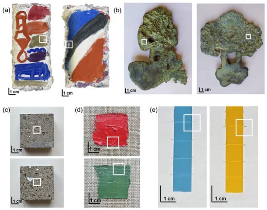
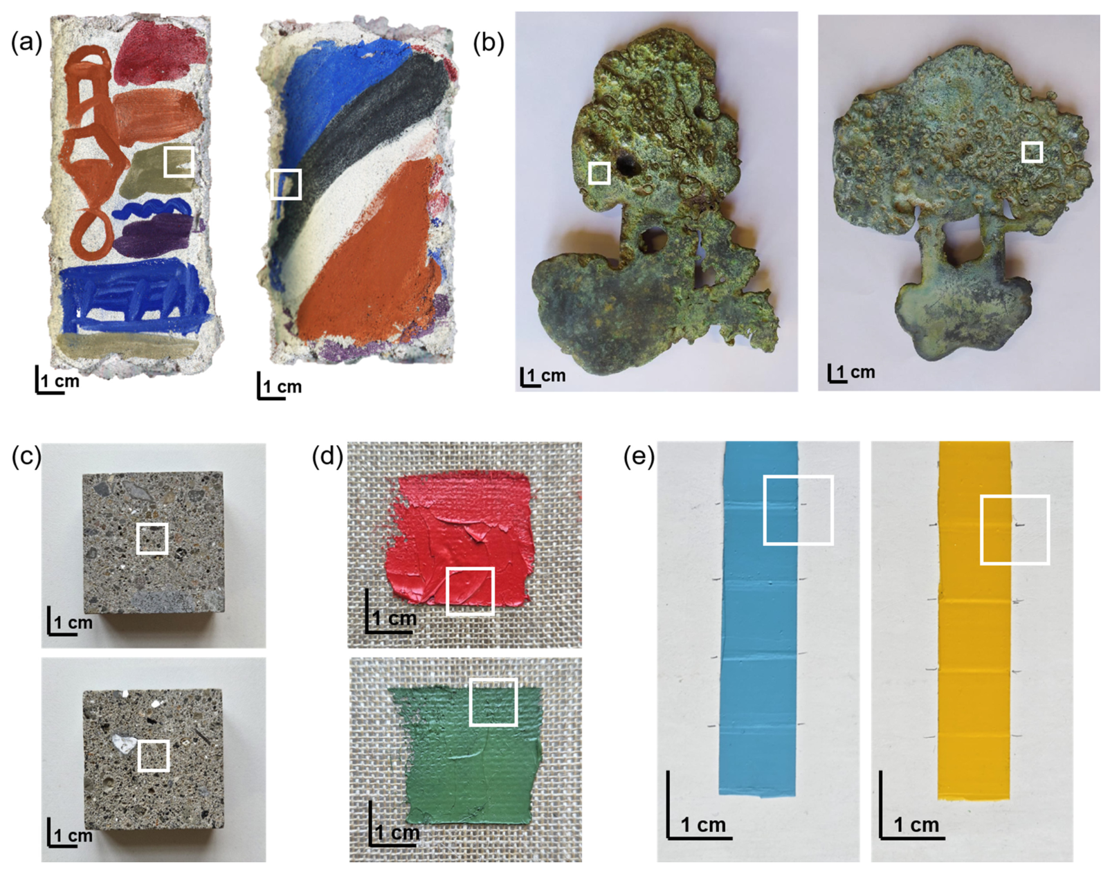
A total of ten independent measurement areas (1 × 1 cm2 each) were acquired on different specimens and/or regions belonging to the five investigated material categories. Two measurement areas were analysed for each material category. Figure 1 provides an overview of all the investigated specimens and highlights the corresponding acquisition areas. Whenever available, multiple specimens and/or regions characterized by different surface morphologies and pigment/substrate combinations were included in the analysis to assess the consistency of the observed results across the investigated samples. A detailed description of the analysed specimens and acquisition areas is provided in the following sections.
Figure 1.
Investigated specimens with the 1 × 1 cm2 acquisition areas highlighted by the white squares: fresco mock-ups (a), bronze samples (b), peperino samples (c), and acrylic paints on canvas (d) and primed wooden panel (e).
For conciseness, only selected results are presented in the following sections. The reported examples were chosen because they illustrate the trends consistently observed across the analysed specimens and measurement areas. The complete dataset was considered in the interpretation of the results and in assessing the consistency of the observed trends.
2.1.1. Fresco Painting
Two fresco mock-ups (Figure 1a) were prepared by the art conservator Ezio Buzzegoli following the traditional buon fresco technique. The first mock-up simulates a complete mural painting stratigraphy, consisting of an arriccio rough plaster layer composed of coarse sand and slaked lime in a 2:1 ratio, overlaid by a thinner intonaco fine plaster layer made with fine sand and slaked lime (2:1 ratio). The surface was painted with green earth pigment (Verona green, an iron oxide and silicate compound) while the plaster was still fresh.
The second mock-up reproduces a fresco surface subsequently covered by an additional fine plaster layer. The investigated area was selected at the boundary between the two fine plaster layers.
2.1.2. Bronze
Two bronze samples (MO 3) with different surface morphologies were investigated (Figure 1b). The samples were supplied by the Opificio delle Pietre Dure (Florence, Italy). The metal substrate was cast from a Cu90/Sn10 bronze alloy by Fonderia Marinelli (Barberino Val d’Elsa, Italy). An artificial patina was subsequently applied using a multi-step process: first, a bronzing solution (comprising phosphoric acid, zinc sulfate, copper sulfate pentahydrate, selenium dioxide, and hydrochloric acid), followed by an application of iron perchloride, and finally, a greening solution (containing water, ammonium salts, vinegar, copper sulfate, and iron perchloride).
2.1.3. Peperino Stone
Two peperino samples were investigated. Each sample consisted of a 50 × 50 × 20 mm3 block with a smooth upper surface and was provided by the Stone Materials Laboratory of the Istituto Centrale per il Restauro (ICR, Rome, Italy). Peperino is a volcanic tuff, commonly characterized by a lithic fragment matrix including basalt and limestone, along with biotite and leucite crystals [35,36]. This lithotype is characteristic of both Roman archaeological heritage and modern architecture.
2.1.4. Acrylic Painting
The panel painting mock-up was prepared by applying an acrylic primer (Lefranc Bourgeois™, Paris, France) onto a wooden support. The primed surface was smoothed with fine-grit sandpaper. Subsequently, two acrylic paints (yellow and light blue) were applied with different controlled layer thicknesses. The extra-fine acrylic paints (Maimeri Brera™, Milan, Italy) were Primary Yellow (PY, Arylide Yellow, PY97-11767) and Permanent Blue Light (PBL, PB15:3-74160). Each paint was applied in five adjacent 1 cm2 areas with increasing controlled thicknesses, as detailed in a previous study [37]. For the present investigation, only the two thickest paint layers (200 µm and 250 µm, measured from the primed surface) were considered.
The canvas mock-up consisted of two acrylic paints (red and green) applied directly onto unprimed linen canvas. One measurement area was acquired on each paint layer.
2.2. Micro-Photogrammetry
For each sample, a 1 × 1 cm2 region of interest was delimited using a paper mask. Micro-photogrammetric acquisition was performed using a Dino-Lite AM7115MT-FUW digital microscope (Dino-Lite Europe, Almere, The Netherlands). The instrument is equipped with a 5.0 Mpx CMOS Edge sensor (2592 × 1944 pixels), four white LEDs, and four UV lights (λ~375 nm), all provided by Dino-Lite Europe, Almere, The Netherlands. It provides a magnification range of 20× to 220×, corresponding to working distances of 60.2 mm and 11.9 mm, respectively.
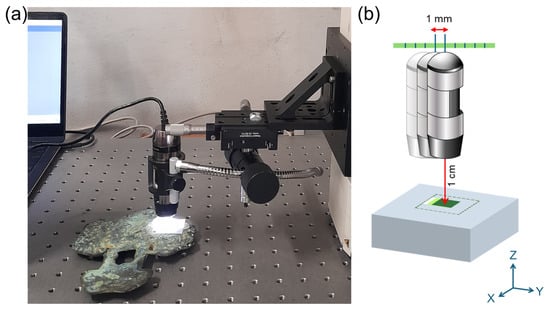
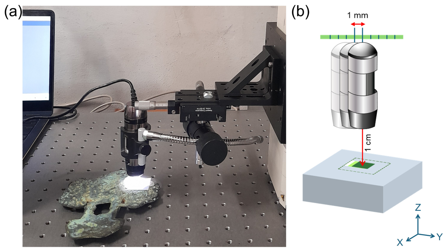
During acquisition, the samples were placed on a flat surface, while the microscope was positioned at a fixed working distance of approximately 1 cm using a quick-mount linear stage attached to a height-adjustable arm. Images were captured by translating the microscope with 1 mm increments along both the X and Y axes using micrometric screws. A whiskbroom (raster) acquisition pattern was employed with a fixed focus, ensuring a minimum overlap of 70% between consecutive images. The total number of images acquired per sample ranged between 100 and 200. The same measurement setup was consistently applied across all samples (Figure 2). Image capture was controlled using DinoCapture 2.0 software (https://www.dinolite.us/features/dinocapture/, accessed on 8 June 2025).
Figure 2.
(a) Experimental setup for micro-photogrammetric acquisition using the Dino-Lite digital microscope; (b) detail of the acquisition geometry: working distance of 1 cm and sample translation along the Y-axis in 1 mm steps.
Following the advanced three-dimensional calibration plate used by Antinozzi et al. [16,29], we adopted a reference paper mask specifically designed to match the dimensions of the acquisition area (1 × 1 cm2). This mask provided a metric reference within the scene and was used for both alignment and scaling procedures. The coordinates of the mask were imported into the photogrammetric project and used as control points and scalebars. This approach enabled us to scale and reference the sparse cloud to an XY coordinate system, using the mask dimensions as known distances (errors in scale-bar distances estimated by Agisoft Metashape software (v.2.0.4, https://www.agisoft.com/, accessed on 10 June 2025) range from 0.2 to 1 mm). To optimize the camera alignment phase, a thick magenta line was printed along the perimeter of the mask.
As demonstrated by Previti [17], parameters such as working distance and lighting conditions exert a substantial influence on reconstruction quality. Consequently, the acquisition protocol presented and applied herein represents the final optimization derived from multiple trials conducted with systematic variations in the setup parameters. Working distances were tested across a range from one to five centimetres; ultimately, the shortest distance was selected to prioritize higher spatial resolution over acquisition speed and coverage area. Acquisition areas of 1 × 1 cm2 were chosen to capture quasi-planar regions, where variation along the Z-axis is negligible in comparison to the X and Y axes. This optimization enables the PDM system to operate at its maximum efficacy, mitigating the inherent limitations of its shallow depth of field at high magnification. The number of photographs and their spatial overlap were optimized to balance acquisition time against confidence in the resulting model reconstruction. Notably, in cases where the final model proved geometrically unreliable, this protocol was maintained, as further increases in the number of photographs or in the degree of overlap did not yield improved outcomes. With regard to lighting conditions, no modifications were introduced owing to the lack of advanced equipment (e.g., lighting domes for shadow-free illumination [20] or conical shields to protect the lens from external light [17]). It should therefore be acknowledged that the possibility of achieving superior results under alternative lighting configurations cannot be entirely excluded.
Image processing and 3D model generation were conducted using Agisoft Metashape software (v.2.0.4, https://www.agisoft.com/, accessed on 10 June 2025), which implements Structure from Motion (SfM) and Dense Multi-View 3D Reconstruction (DMVR) algorithms. The software performs image alignment by identifying homologous points (tie points) across consecutive photograms. By incorporating known reference distances, the software generates both sparse and dense point clouds scaled to real-world dimensions and referenced to an XY coordinate system. The dense point cloud was subsequently triangulated to generate a polygonal mesh, onto which a texture was projected to produce a realistic 3D model.
Agisoft Metashape was further utilized to generate orthomosaics (high-resolution, geometrically correct, and georeferenced composite images), as well as to compute dense point cloud confidence values and to visualize photogrammetric overlap and camera position relative to the sample surface (Figure 3).
Figure 3.
Standard image acquisition configuration for the sample surfaces, processed with Agisoft Metashape software.
For each sample, a Digital Elevation Model (DEM) was derived using the open-source software CloudCompare (v.2.12.4, https://www.danielgm.net/cc/, accessed on 15 June 2025).
2.3. Laser Scanner Microprofilometry
Microprofilometric measurements were performed using an optical microprofilometer developed in-house by the Heritage Science Group (HSG) of the National Research Council–National Institute of Optics (CNR-INO), Florence [38,39]. The system comprises a ConoPoint-10 (Optimet, Jerusalem, Israel) laser probe mounted on two motorized linear stages with a positioning precision of 0.1 µm. The probe was equipped with a 50 mm focal length lens (f50), providing a working distance of 44 mm, a depth of field of 8 mm, and a spot diameter of 37 µm at the beam waist. High-resolution 3D surface models were acquired with a sampling step of 50 µm along both the X and Y axes. The acquired data can be visualized as 3D models, grayscale maps, or color-coded topographies; simulated raking light images can also be generated to enhance the visualization of surface morphology [3].
To assess the accuracy of the micro-photogrammetric reconstructions, the 3D data obtained with the digital microscope were compared against height measurements acquired with the optical microprofilometer, which served as the reference instrument. This analysis was conducted on two samples: the fresco and the panel-painting mock-ups, which yielded the best and worst results from micro-photogrammetry, respectively.
The comparison was performed by analyzing the Digital Elevation Models (DEMs) derived from the meshes, as well as specific profiles extracted from the digital models. Profile extraction represents an effective criterion for evaluating instrument performance, as it highlights complex geometric features [40]. Prior to profile extraction, the 3D models from both instruments were aligned in CloudCompare using the Iterative Closest Point (ICP) algorithm. Subsequently, DEMs were generated within the same software, setting a pixel size of 50 µm for the microprofilometric models and 10 µm for the digital microscope models. For the microprofilometric data, the pixel size was set to match the 50 µm sampling step. For the digital microscope, we deliberately adopted a smaller pixel size to ensure it remained below the instrument’s undeclared spatial resolution, thereby preventing the software from interpolating across low-confidence regions. Finally, the DEMs were imported into MATLAB (v. R2024a) as matrices for profile extraction.
3. Results and Discussion
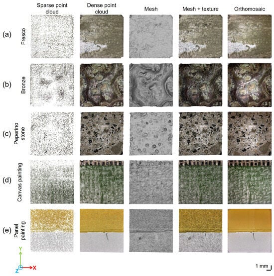
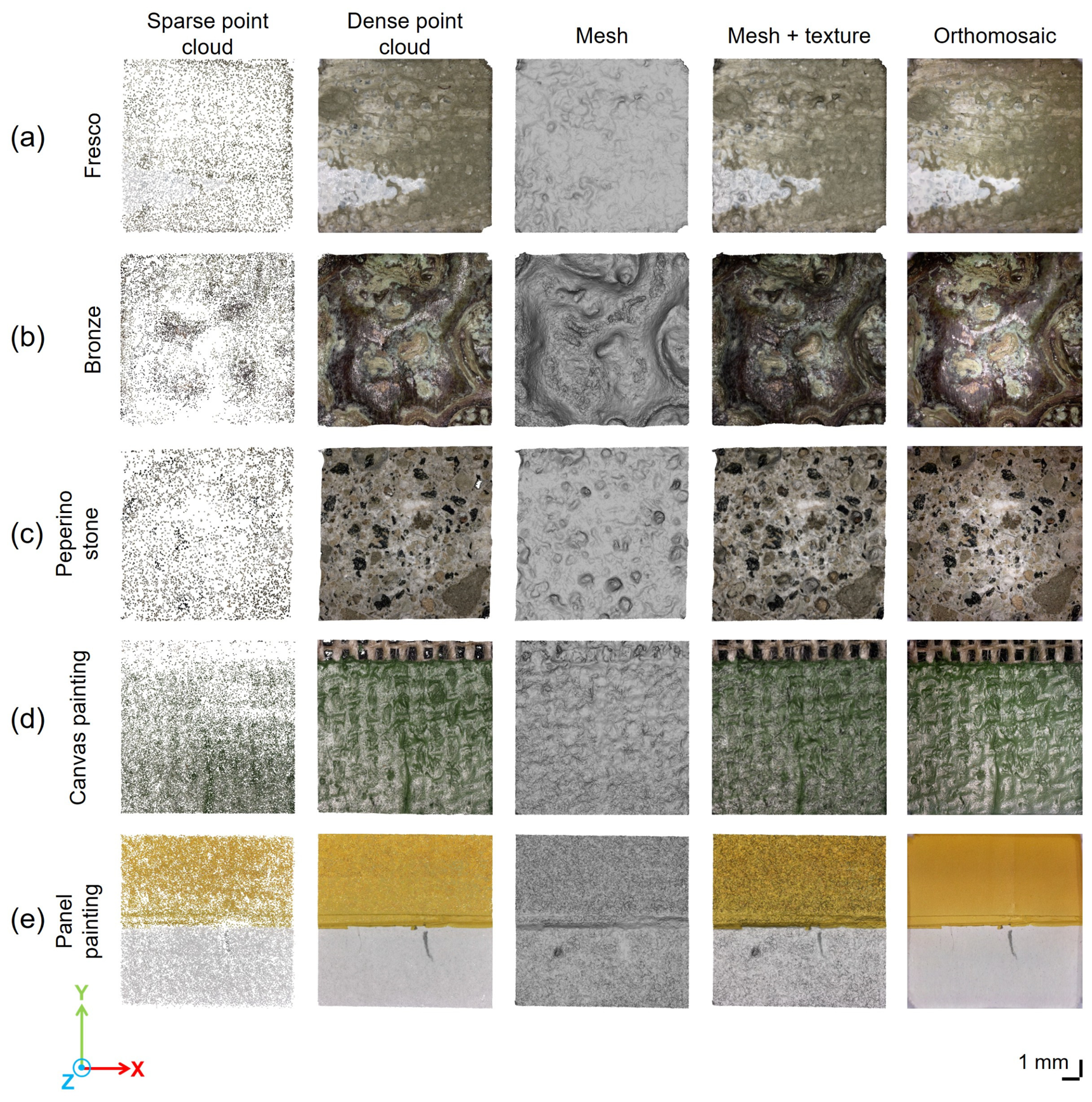
Image processing and 3D reconstruction performed with Agisoft Metashape followed the typical photogrammetric pipeline, producing sparse and dense point clouds, meshes, textured meshes, and orthomosaics. The corresponding results are displayed in Figure 4.
Figure 4.
Results of the 3D survey conducted on samples of fresco (a); bronze (b); peperino stone (c); canvas painting (d); and panel painting (e). From left to right, the columns display the sparse point cloud, dense point cloud, mesh, textured mesh, and orthomosaic, respectively. Each image covers a 1 × 1 cm2 area.
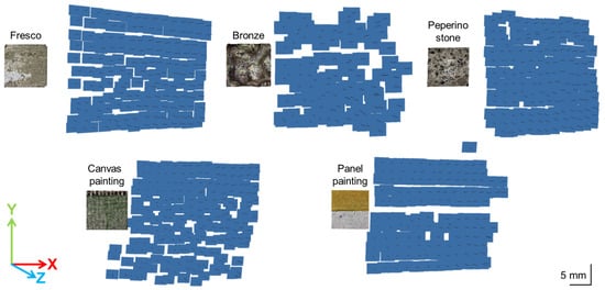
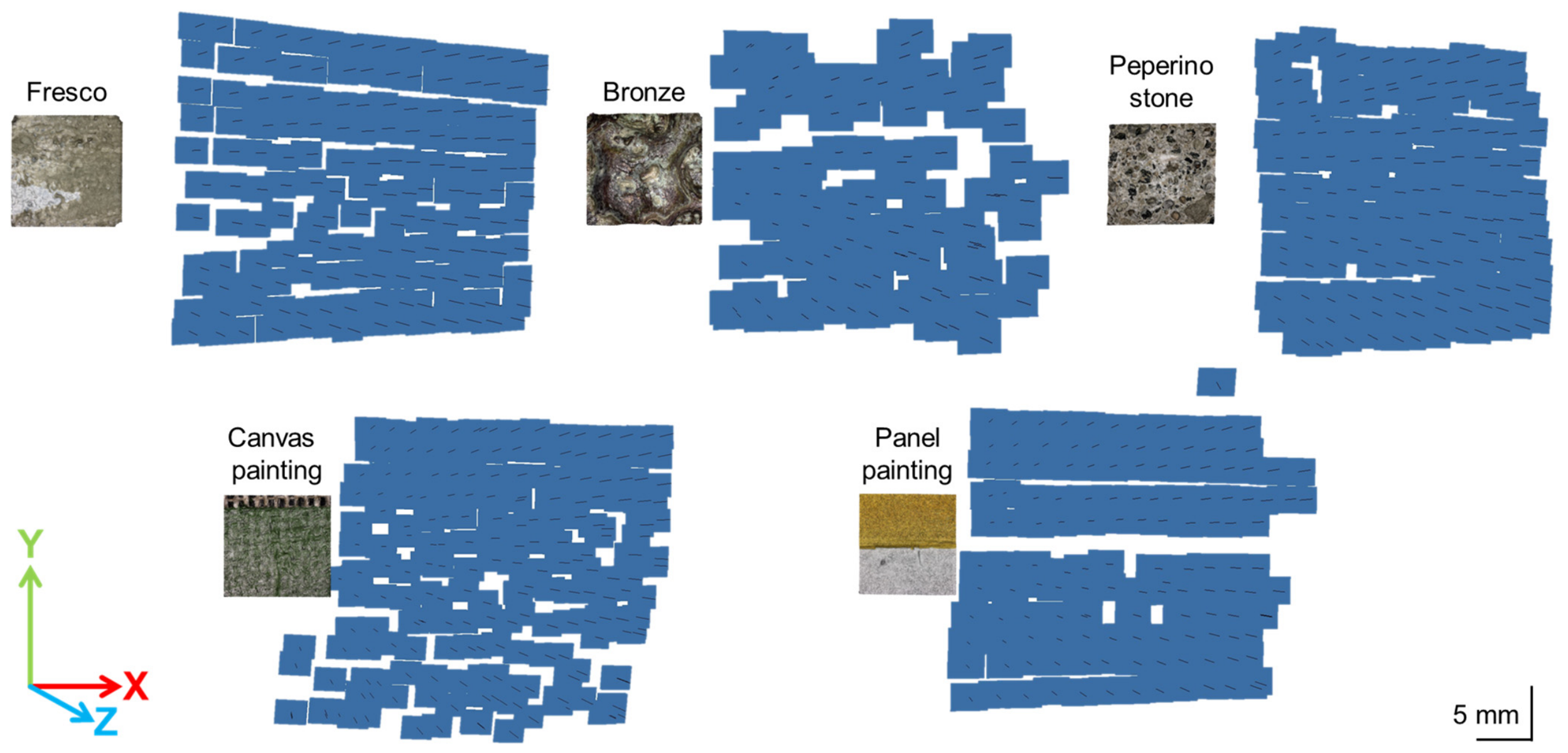
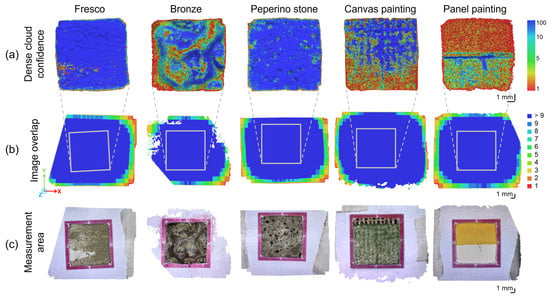
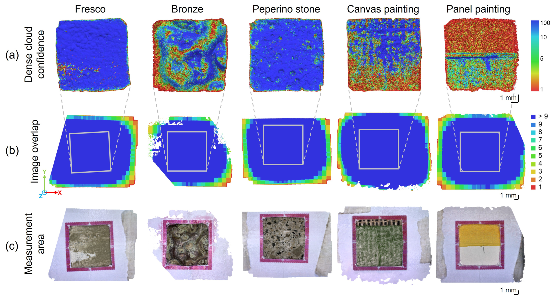
The performance of the Dino-Lite microscope in the 3D digitization of different artistic substrates was evaluated using the dense cloud confidence metric computed in Agisoft Metashape (Figure 5a). The algorithm assigns each point a confidence value ranging from 1 to 100, reflecting the reliability of its reconstruction: values approaching 100 indicate high confidence, whereas values near 1 denote low confidence. Dense cloud confidence is visualized through a color-coded scale ranging from red (low reliability) to blue (high reliability). As illustrated in Figure 5a, the fresco and peperino stone samples exhibit high confidence values across nearly the entire surface. The bronze and canvas painting samples display greater variability in confidence values, with several low-confidence areas (red). Conversely, the panel painting presents the lowest confidence values over more than half of its surface, particularly within the yellow paint layer.
Figure 5.
Visualization of: (a) dense cloud confidence (scale: 1–100); (b) image overlap degree (scale: 1 to >9); and (c) measurement areas delimited by a paper mask for the five materials tested.
Figure 5b shows the image overlap for each sample as a color-coded map, where red indicates points captured in a single image and blue denotes points appearing in multiple images. This representation enables assessment of whether sufficient data were collected for accurate surface reconstruction and, if necessary, facilitates planning additional acquisitions in regions with lower coverage. All samples exhibit an overlap of at least nine images in the measurement area (highlighted by the gray square and shown in the reconstructed images in Figure 5c), providing sufficient redundancy for reliable photogrammetric reconstruction.
Comparison of dense cloud confidence and the image overlap indicates that the observed variability in reconstruction quality is not attributable to differences in image coverage, which is sufficient for reliable reconstruction of each sample. Instead, differences in the physical properties of the samples—such as color, surface reflectance, tonal brightness, and the presence of pores or surface unevenness—appear to significantly influence 3D reconstruction performance. Highly reflective or extreme lightness or darkness can compromise image acquisition by the Dino-Lite microscope, resulting in data gaps that reduce the quality of the 3D model.
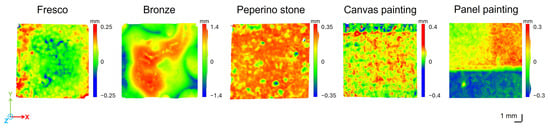
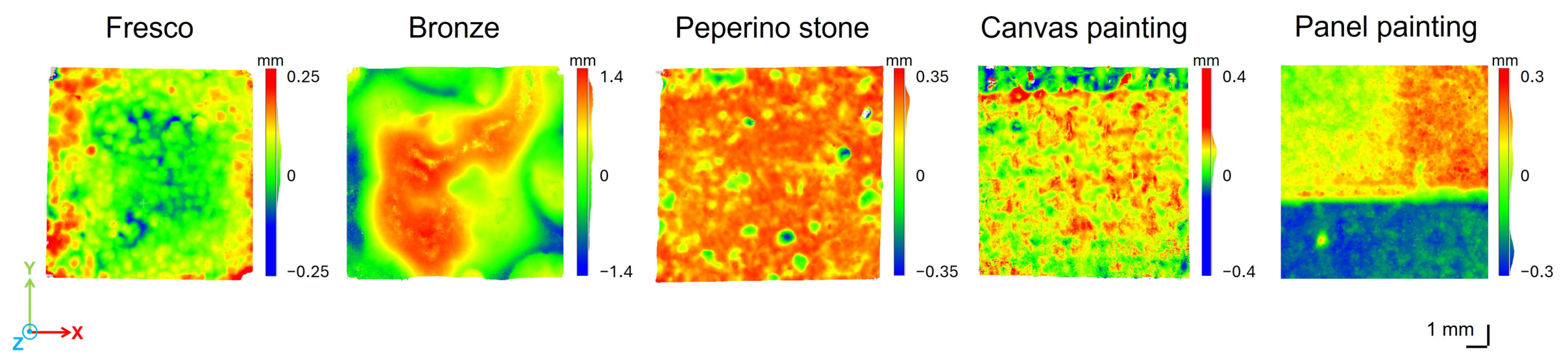
Figure 6 displays the Digital Elevation Model (DEM) generated from the Dino-Lite 3D model for each material sample. The DEM is represented using a color scale in which zero elevation (green) corresponds to the reference plane employed for height measurements. Areas raised above the plane are indicated by positive elevation values (red), whereas areas below the reference plane are represented by negative values (blue).
Figure 6.
Digital Elevation Models (DEMs) of the samples. From left to right: fresco, bronze, peperino stone, canvas painting, and panel painting.
The DEM of the panel painting sample clearly reveals the height difference between the white preparation layer and the two thick yellow paint layers (approximately 200 and 250 µm, respectively). However, the thickness values derived from the DEMs are not quantitatively comparable with the known layer thicknesses, probably due to the limited dense cloud confidence and the resulting noise in the 3D reconstruction.
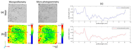
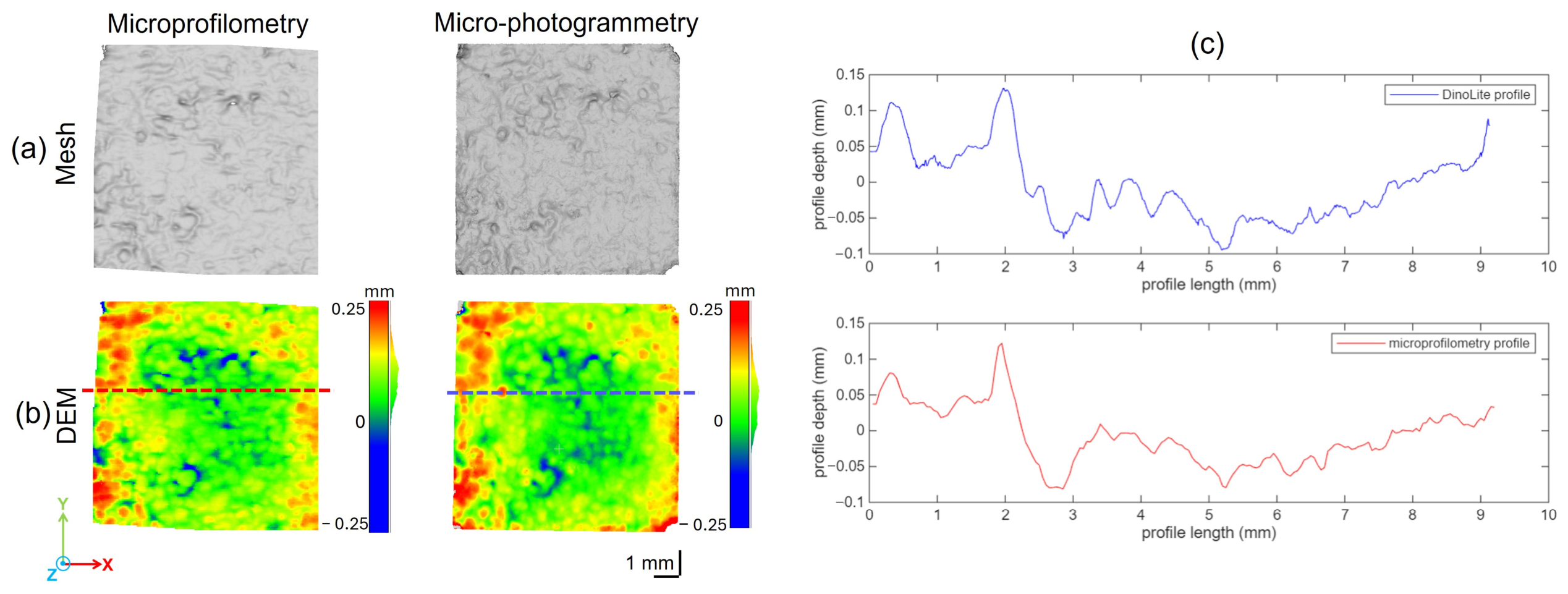
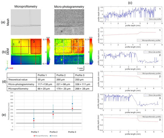
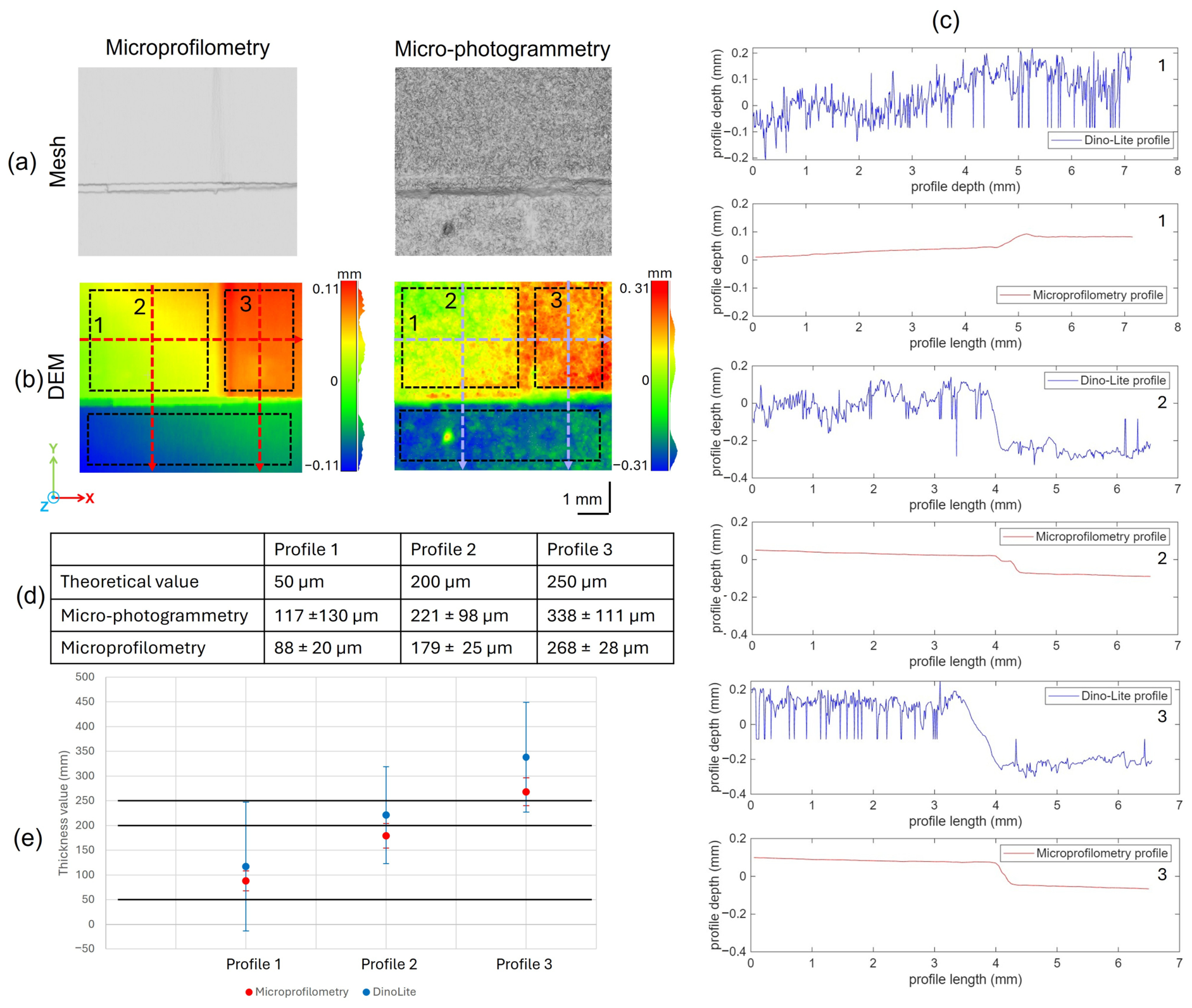
The micro-photogrammetric 3D data of the fresco and panel painting mock-ups were compared with reference height values obtained using an optical microprofilometer (Heritage Science Group, National Research Council–National Institute of Optics, Italy, Florence). Specifically, the digital elevation models (DEMs) derived from the meshes were compared, along with specific profiles extracted from the digital models (Figure 7 and Figure 8).
Figure 7.
(a) Untextured meshes of the fresco sample obtained using the optical microprofilometer and the Dino-Lite digital microscope. (b) Digital elevation models (DEMs) derived from the meshes. The red and blue dotted lines indicate the locations from which the profiles shown in (c) were extracted.
Figure 8.
(a) Untextured meshes of the panel painting sample captured by the optical microprofilometer and the Dino-Lite digital microscope. (b) Digital elevation models (DEMs) obtained from the meshes. The red and blue dotted lines indicate the traces of the profiles presented in (c). (d) Table summarizing the mean value and standard deviation for each thickness step, calculated from the regions delimited by the black dotted lines in (b). The three measured thicknesses are numbered from 1 to 3, consistent with the profiles. Mean values and standard deviation are plotted in (e); the black lines correspond to theoretical reference values of 50, 200, and 250 microns.
The comparison of the fresco mock-up confirmed the high accuracy of the micro-photogrammetric model. The extracted profiles are nearly perfectly superimposed; any minor discrepancies are most likely attributable to slight alignment errors during the superimposition process rather than actual differences between the models (Figure 7c). Furthermore, the profile derived from the Dino-Lite model appears more precise than that obtained from microprofilometry, as it captures sharper peaks. It is worth noting that the microprofilometric model was generated using a 50 µm sampling step as a compromise between acquisition time and metric accuracy; a finer sampling step would have yielded higher resolution.
In contrast, comparison of the painted panel models revealed substantial differences, particularly highlighting the high noise levels in the Dino-Lite model (Figure 8a,b). The height color scales of the DEMs differ significantly, as the Dino-Lite data span considerably larger values. The extracted profiles confirm this discrepancy, showing that the photogrammetric data are substantially noisier than the microprofilometric data (Figure 8c). To further validate these results, the thickness steps of the paint layer were calculated. To this end, the mean height values extracted from the side of each painted region were subtracted from those obtained from its center, and the standard deviation was subsequently computed (Figure 8d). The standard deviation confirms the high noise level inherent to the micro-photogrammetric approach (Figure 8e), which renders it unreliable for computing step thickness. This is further corroborated by the dense cloud confidence map in Figure 5a; values below 5 cover more than half of the panel surface, indicating that no geometrically interpretable output was obtained. Given the handcrafted nature of the mock-up, the microprofilometric values are consistent with the expected results.
4. Conclusions
Low-cost, compact size, and high performance in terms of resolution and magnification have established Dino-Lite digital microscopy and similar tools as standard equipment for restorers, conservators, and archeologists. Their application in portable micro-photogrammetry has increased in recent years, driven by growing interest in the three-dimensional documentation of cultural heritage objects. While recent studies have demonstrated the potential of this approach on materials such as stone, bone, and metals, data on painted surfaces remain scarce.
In this study, five representative artistic substrates—fresco wall painting, bronze, peperino stone, canvas painting, and panel painting samples—were analyzed through surface 3D surveys using Dino-Lite micro-photogrammetry. The performance of this combined approach across different substrates was assessed by comparing the resulting 3D models in terms of dense cloud confidence and Digital Elevation Model (DEM) accuracy.
In particular, the accuracy of the DEMs obtained using Dino-Lite scanning was verified by comparison with those acquired with an optical microprofilometer.
The results demonstrate that the performance of Dino-Lite micro-photogrammetry is strongly influenced by the physical properties of the investigated surfaces, including color, reflectance, tonal brightness, and surface morphology.
On the one hand, for opaque and textured substrates such as frescoes and stone, reliable and metrically consistent reconstructions were obtained. The profiles extracted from the mesh are almost perfectly superimposed for the two techniques. The profile derived from the Dino-Lite model even exhibits sharper peaks than that obtained with microprofilometry, suggesting potentially higher precision for the micro-photogrammetric approach.
On the other hand, dark or highly reflective surfaces, such as those of canvas and panel paintings, negatively affect image acquisition and, consequently, the reliability of dense point clouds. In the case of the painted panel sample, the generated DEM was highly noisy, and comparison with the microprofilometry-based model revealed that this noise resulted in an overestimation of the paint layer thickness.
Although the number of investigated specimens does not allow a complete statistical characterization of the variability within each material category, the analysis of different specimens and measurement areas, selected to include distinct surface morphologies and pigment/substrate combinations, revealed consistent trends. This agreement among the investigated samples supports the reproducibility of the observed features under the examined conditions.
Overall, micro-photogrammetry based on the Dino-Lite digital microscope can be considered a valid method for constructing 3D models of small objects in situ. Extending this technique to paintings—which are often glossy, reflective, or very dark—for the study of surface micromorphology remains promising, as it enables the creation of low-cost 3D models. However, for these materials, the lack of reliable metric information restricts the method to primarily qualitative applications, which must be supplemented with additional metric measurements.
Author Contributions
Conceptualization, R.F. and M.G.; methodology, R.F., D.P., and E.V.; validation, R.F., D.P., and E.V.; formal analysis, D.P., E.V., and A.D.F.; investigation, D.P. and E.V.; resources, R.F., A.D.F., and M.G.; data curation, D.P. and E.V.; writing—original draft preparation, D.P.; writing—review and editing, E.V., A.D.F., M.G., and R.F.; visualization, D.P.; supervision, R.F.; project administration, R.F.; funding acquisition, R.F. All authors have read and agreed to the published version of the manuscript.
Funding
This research was funded by PE_00000020—CHANGES: Cultural Heritage Active Innovation for Sustainable Society project, PIANO NAZIONALE DI RIPRESA E RESILIENZA (PNRR)—MISSIONE 4 COMPONENTE 2, INVESTIMENTO 1.3—CUP D53C22002560006, funded by Next Generation EU. PNRR H2IOSC (Humanities and Cultural Heritage Italian Open Science Cloud) Project (IR0000029)-CUP_B63C22000730005, funded by Next Generation EU. PRIN-PNRR AIPAD (Artificial Intelligence and Physics for Art Diagnostics) Project (P2022SFEPN), CUP_B53D23029240001, and funded by the European Union–Next Generation EU M4.C2.1.1.
Data Availability Statement
The raw data supporting the conclusions of this article will be made available by the authors on request.
Acknowledgments
The authors thank Ezio Buzzegoli for the fresco mock-up, and Eleonora Gioventù for the peperino sample.
Conflicts of Interest
The authors declare no conflicts of interest.
References
- Haddad, N. 3D and 2D Visual Digital Technologies and Cultural Heritage Documentation for Conservation and Monitoring: A Critical Review and Assessment. In Optoelectronic Devices in Robotic Systems; Sergiyenko, O., Ed.; Springer International Publishing: Cham, Switzerland, 2022; pp. 257–288. [Google Scholar]
- Vannini, E.; Lunghi, I.; Grifoni, E.; Farioli, P.; Ginanni, M.; Santacesaria, A.; Fontana, R. Three-Dimensional Analysis for the Documentation of the Restoration of an Earthquake-Damaged Triptych. Heritage 2024, 7, 2176–2194. [Google Scholar] [CrossRef] [Scilit]
- Grifoni, E.; Vannini, E.; Lunghi, I.; Faraioli, P.; Ginanni, M.; Santacesarea, A.; Fontana, R. 3D Multi-Modal Point Clouds Data Fusion for Metrological Analysis and Restoration Assessment of a Panel Painting. J. Cult. Herit. 2024, 66, 356–366. [Google Scholar] [CrossRef] [Scilit]
- Li, Y.; Zhao, L.; Chen, Y.; Zhang, N.; Fan, H.; Zhang, Z. 3D LiDAR and Multi-Technology Collaboration for Preservation of Built Heritage in China: A Review. Int. J. Appl. Earth Obs. Geoinf. 2023, 116, 103156. [Google Scholar] [CrossRef] [Scilit]
- Parrinello, S.; Picchio, F.; La Placa, S. The Construction of an Informative 3D Model for the Monitoring of City Heritage Risk. In Reviving Aleppo: Urban, Legal and Digital Approaches for Post-War Recovery; Thiel, F., Orabi, R., Eds.; Springer International Publishing: Cham, Switzerland, 2024; pp. 243–274. [Google Scholar]
- Jia, S.; Liao, Y.; Xiao, Y.; Zhang, B.; Meng, X.; Qin, K. Methods of Conserving and Managing Cultural Heritage in Classical Chinese Royal Gardens Based on 3D Digitalization. Sustainability 2022, 14, 4108. [Google Scholar] [CrossRef] [Scilit]
- Barszcz, M.; Dziedzic, K.; Skublewska-Paszkowska, M.; Powroznik, P. 3D Scanning Digital Models for Virtual Museums. Comput. Animat. Virtual Worlds 2023, 34, e2154. [Google Scholar] [CrossRef]
- Balletti, C.; Ballarin, M. An Application of Integrated 3D Technologies for Replicas in Cultural Heritage. ISPRS Int. J. Geo-Inf. 2019, 8, 285. [Google Scholar] [CrossRef] [Scilit]
- Carmo, M.B.; Cláudio, A.P. 3D Virtual Exhibitions. DJLIT 2013, 33, 222–235. [Google Scholar] [CrossRef] [Scilit]
- Wilson, P.F.; Stott, J.; Warnett, J.M.; Attridge, A.; Smith, M.P.; Williams, M.A. Evaluation of Touchable 3D-Printed Replicas in Museums. Curator Mus. J. 2017, 60, 445–465. [Google Scholar] [CrossRef] [Scilit]
- Loaiza Carvajal, D.A.; Morita, M.M.; Bilmes, G.M. Virtual Museums. Captured Reality and 3D Modeling. J. Cult. Herit. 2020, 45, 234–239. [Google Scholar] [CrossRef] [Scilit]
- Mazzocato, S.; Daffara, C. Experiencing the Untouchable: A Method for Scientific Exploration and Haptic Fruition of Artworks Microsurface Based on Optical Scanning Profilometry. Sensors 2021, 21, 4311. [Google Scholar] [CrossRef] [Scilit] [PubMed]
- Guo, M.; Shang, X.; Zhao, J.; Shi, Y.; Sun, M.; Huang, M.; Wang, G.; Liao, L. Digital Twin Models for Architectural Heritage Conservation. J. Build. Eng. 2025, 112, 113792. [Google Scholar] [CrossRef] [Scilit]
- Mazzetto, S. Integrating Emerging Technologies with Digital Twins for Heritage Building Conservation: An Interdisciplinary Approach with Expert Insights and Bibliometric Analysis. Heritage 2024, 7, 6432–6479. [Google Scholar] [CrossRef] [Scilit]
- Hosamo, H.H.; Hosamo, M.H. Digital Twin Technology for Bridge Maintenance Using 3D Laser Scanning: A Review. Adv. Civ. Eng. 2022, 2022, 2194949. [Google Scholar] [CrossRef] [Scilit]
- Antinozzi, S.; Ronchi, D.; Fiorillo, F.; Barba, S. 3Dino: Configuration for a Micro-Photogrammetric Survey—Applying Dino-Lite Microscope for the Digitalization of a Cuneiform Tablet. In Towards a New, Configurable Architecture. Proceedings of the 39th eCAADe Conference; University of Novi Sad: Novi Sad, Serbia, 2021. [Google Scholar]
- Previti, G.; Luci, B.; Lemorini, C. Micro-Photogrammetry and Traceology: New on-Site Documentation Approaches Using Portable Digital Microscopes. J. Archaeol. Sci. 2024, 168, 106004. [Google Scholar] [CrossRef] [Scilit]
- Antinozzi, S.; Fiorillo, F.; Surdi, M. Geometrical Feature Identification of Cuneiform Signs on Micro-Survey Reconstruction. In Proceedings of the ArtsIT, Interactivity and Game Creation; Brooks, A.L., Ed.; Springer Nature: Cham, Switzerland, 2023; pp. 78–88. [Google Scholar]
- Previti, G.; Kusumastuti, S.; Luci, B.; Malatesta, L.; Lemorini, C. From Chisel to Inscription: Affordable Protocols for the Digital Documentation of Stone Carving Techniques. An Experimental Archaeology and Traceological Approach Applied to Epigraphy. PLoS ONE 2025, 20, e0327303. [Google Scholar] [CrossRef] [Scilit] [PubMed]
- Antinozzi, S.; Fiorillo, F. Optimized configurations for micro-photogrammetric surveying adaptable to macro optics and digital microscope. Int. Arch. Photogramm. Remote Sens. Spat. Inf. Sci. 2022, 46, 25–32. [Google Scholar] [CrossRef] [Scilit]
- Di Paola, F.; Morena, S.; Raimondi, G.; Antinozzi, S. The Survey of the ‘Precious Ones of Constance’. In Proceedings of the IMEKO TC-4, Rome, Italy, 19–21 October 2023. [Google Scholar]
- Ronchi, D.; Fortunati, S.; Limongiello, M. Roman Fragmentary Painting: Surveying Technologies and Methodological Approaches. In Proceedings of the IMEKO TC-4, Florence, Italy, 4–6 December 2019. [Google Scholar]
- Rivero, O.; Garate, D. Motion and Gesture: Analysing Artistic Skills in Palaeolithic Art. J. Archaeol. Method Theory 2020, 27, 561–584. [Google Scholar] [CrossRef] [Scilit]
- Herrero-Cortell, M.A.; Samaniego-Jiménez, I.; Belenguer-Salvador, C.; Raïch-Creus, M.; Osete-Cortina, L.; Abbafati, A.; Vila, A.; Picollo, M.; Fuster-López, L. Micro-Multiband Imaging (µMBI) in the Technical Study and Condition Assessment of Paintings: An Insight into Its Potential and Limitations. Heritage 2026, 9, 54. [Google Scholar] [CrossRef] [Scilit]
- Luci, B. Unveiling the Hidden Stories: The Impact of Digital Technologies in Medieval Latin Epigraphic Field Documentation. Digit. Appl. Archaeol. Cult. Herit. 2026, 40, e00488. [Google Scholar] [CrossRef] [Scilit]
- Tserevelakis, G.J.; Pirgianaki, E.; Melessanaki, K.; Zacharakis, G.; Fotakis, C. Non-Invasive Optoacoustic Imaging for In-Depth Cultural Heritage Diagnostics. Photonics 2024, 11, 902. [Google Scholar] [CrossRef] [Scilit]
- Ricca, M.; Alberghina, M.F.; Randazzo, L.; Schiavone, S.; Donato, A.; Albanese, M.P.; La Russa, M.F. A Combined Non-Destructive and Micro-Destructive Approach to Solving the Forensic Problems in the Field of Cultural Heritage: Two Case Studies. Appl. Sci. 2021, 11, 6951. [Google Scholar] [CrossRef] [Scilit]
- Serino, M. Tracing Ancient Artisanal Gestures: Digital Applications and Experimental Archaeology on South Italian Red-Figure Pottery Production. Digit. Appl. Archaeol. Cult. Herit. 2025, 39, e00468. [Google Scholar] [CrossRef] [Scilit]
- Antinozzi, S.; Ronchi, D. Salvatore Barba 3Dino System, come accorciare le distanze nei rilievi di precisione. In 42th International Conference of Representation Disciplines Teachers; FrancoAngeli: Milan, Italy, 2021. [Google Scholar]
- Siddique, A.; Cignoni, P.; Corsini, M.; Banterle, F. Evaluating Image-Based Interactive 3D Modeling Tools. IEEE Access 2024, 12, 104138–104152. [Google Scholar] [CrossRef] [Scilit]
- Billi, D.; Caroti, G.; Piemonte, A. 3D Reconstruction of Transparent and Reflective Surfaces, Through the Use of SfM Processes Supported by 3D Gaussian-Splatting and 2D Gaussian-Splatting. Sensors 2025, 25, 4410. [Google Scholar] [CrossRef] [Scilit] [PubMed]
- Um, D.; Lee, S. Microscopic Structure from Motion (SfM) for Microscale 3D Surface Reconstruction. Sensors 2020, 20, 5599. [Google Scholar] [CrossRef] [Scilit] [PubMed]
- Maté-González, M.Á.; Yali, R.; Rodríguez-Hernández, J.; González-González, E.; Aguirre de Mata, J. Comparison of NeRF- and SfM-Based Methods for Point Cloud Reconstruction for Small-Sized Archaeological Artifacts. Remote Sens. 2025, 17, 2535. [Google Scholar] [CrossRef] [Scilit]
- Iakovaki, E.; Konstantakis, M.; Giaourtsakis, I.; Rentoumi, E.; Protopapas, D.; Psarras, C.; Koskeridou, E. When Reality Meets Practice: Challenges and Pitfalls in 3D Digitization Using Structured Light Scanning and Photogrammetry in Cultural Heritage. Information 2026, 17, 237. [Google Scholar] [CrossRef] [Scilit]
- Capobianco, G.; Pelosi, C.; Lanteri, L.; Bonifazi, G.; Trotta, O.; Serranti, S. Monitoring of Protective Products on Peperino Stone Using Portable Devices. In Proceedings of the 2003 IMEKO TC, Rome, Italy, 19–21 October 2023; pp. 603–608. [Google Scholar]
- Marra, F.; Palladino, D.M.; Licht, O.A.B. The Peperino Rocks: Historical and Volcanological Overview. Bull. Volcanol. 2022, 84, 69. [Google Scholar] [CrossRef] [Scilit]
- Dal Fovo, A.; Martínez-Weinbaum, M.; Oujja, M.; Castillejo, M.; Fontana, R. Reflectance Spectroscopy as a Novel Tool for Thickness Measurements of Paint Layers. Molecules 2023, 28, 4683. [Google Scholar] [CrossRef] [Scilit] [PubMed]
- Carcagni, P.; Daffara, C.; Fontana, R.; Gambino, M.C.; Mastroianni, M.; Mazzotta, C.; Pampaloni, E.; Pezzati, L. Optical Micro-Profilometry for Archaeology. In Proceedings of the Optical Methods for Arts and Archaeology; SPIE: Bellingham, WA, USA, 2005; Volume 5857, pp. 118–128. [Google Scholar]
- Fontana, R.; Gambino, M.C.; Greco, M.; Marras, L.; Materazzi, M.; Pampaloni, E.; Pezzati, L. High-Precision Surface Analysis of the Roughness of Michelangelo’s David. In Proceedings of the Optical Metrology for Arts and Multimedia; SPIE: Bellingham, WA, USA, 2003; Volume 5146, pp. 236–243. [Google Scholar]
- Lunghi, I.; Vannini, E.; Dal Fovo, A.; Di Sarno, V.; Rocco, A.; Fontana, R. A Performance Comparison of 3D Survey Instruments for Their Application in the Cultural Heritage Field. Sensors 2024, 24, 3876. [Google Scholar] [CrossRef] [Scilit] [PubMed]
Disclaimer/Publisher’s Note: The statements, opinions and data contained in all publications are solely those of the individual author(s) and contributor(s) and not of MDPI and/or the editor(s). MDPI and/or the editor(s) disclaim responsibility for any injury to people or property resulting from any ideas, methods, instructions or products referred to in the content. |
© 2026 by the authors. Licensee MDPI, Basel, Switzerland. This article is an open access article distributed under the terms and conditions of the Creative Commons Attribution (CC BY) license.