


Abstract
Background and Clinical Significance: Sarcoidosis and lymphoma may precede, follow or accompany each other, and lymphoma can also induce a sarcoid-like granulomatous reaction. Case Presentation: We describe a 62-year-old woman with exertional dyspnea, abdominal pain and fluctuating cervical lymphadenopathy. Computed tomography showed mediastinal, hilar and abdominal lymphadenopathy with hepatosplenomegaly; a subcarinal lymph node obtained by mediastinoscopy showed non-necrotizing granulomas, and sarcoidosis was diagnosed and managed with observation. Four months later, worsening dyspnea and progressive lymphadenopathy led to FDG-PET/CT, which showed markedly hypermetabolic cervical nodes (SUVmax 20.5) but only moderate mediastinal uptake (SUVmax 4.8). Excisional cervical lymph node biopsy established diffuse large B-cell lymphoma (DLBCL), non-germinal-center B-cell phenotype, and Ann Arbor stage IIIS-A. After six cycles of R-CHOP, she achieved a complete metabolic response and remains in remission three years after treatment. Because both diagnoses were established within one year and lymphoma had initially been excluded on a single mediastinal node, a lymphoma already present at first presentation with an accompanying sarcoid-like reaction cannot be ruled out. Conclusions: In patients labeled as having sarcoidosis, peripheral or abdominal lymphadenopathy, splenomegaly and discordant FDG uptake should prompt excisional biopsy of the most accessible FDG-avid node, ideally before corticosteroids are started. Thirty-nine published cases of sarcoidosis associated with DLBCL are summarized.
1. Introduction and Clinical Significance
Since Brincker described the sarcoidosis–lymphoma syndrome (SLS) in 1986, on the basis of 17 personal cases and 29 cases from the literature, an association between sarcoidosis and lymphoproliferative disease has been recognized; in that series, malignant lymphoproliferative disease developed at least 5.5 times more often than expected in middle-aged patients with chronic active sarcoidosis [1]. Population-based studies and meta-analyses have since confirmed an increased risk of malignancy, including lymphoma, in patients with sarcoidosis [2,3].
The association may follow three temporal patterns: sarcoidosis preceding lymphoma, sarcoidosis following lymphoma, or both diseases diagnosed concurrently [4,5]. In a recent multicenter series of 46 patients, in which a simultaneous diagnosis was defined as both diagnoses within one year, 10 patients (22%) had a simultaneous presentation [5]. The interval between the two diagnoses varies widely: a median interval of 24 months was reported in Brincker’s series [1], whereas in the series of Charrière et al., the median interval was 132 months when sarcoidosis preceded lymphoma [5]. Diffuse large B-cell lymphoma (DLBCL) is the most frequently reported lymphoma subtype in recent series [5,6].
A further scenario is the sarcoid-like reaction (SLR): non-necrotizing epithelioid-cell granulomas arising in lymph nodes or organs of patients with malignancy, without systemic sarcoidosis [7,8]. Such reactions have been described in 13.8% of patients with Hodgkin lymphoma and 7.3% of patients with non-Hodgkin lymphoma [8], including DLBCL [9], and granulomas may be the only finding in a sampled lymph node while lymphoma is present elsewhere [10,11]. Because sarcoidosis is a diagnosis of exclusion [12], granulomas in a single lymph node do not exclude a coexisting lymphoma.
We report a patient in whom granulomatous mediastinal lymphadenitis, initially diagnosed as sarcoidosis, was followed within months by a diagnosis of DLBCL. We use the case to illustrate this diagnostic pitfall, to outline the features that should prompt re-biopsy in a patient labeled as having sarcoidosis, and to summarize the published cases of sarcoidosis associated with DLBCL.
1.1. Sarcoidosis
Sarcoidosis is a multisystem granulomatous disease of unknown etiology, characterized by non-necrotizing granulomas that predominantly affect the lungs and intrathoracic lymph nodes; cardiac, ocular, cutaneous, neurological, hepatic and splenic involvement may also occur [12,13,14,15]. Ten to fifteen percent of patients are asymptomatic at diagnosis [15]; others present with non-specific systemic symptoms such as fatigue, fever, weight loss and night sweats, or with organ-specific symptoms, most commonly cough, dyspnea and chest pain [12]. Abdominal involvement, including hepatosplenomegaly and abdominal lymphadenopathy, is less specific and may mimic lymphoma, metastatic disease or mycobacterial infection [16,17,18].
Chest imaging typically shows hilar and mediastinal lymphadenopathy, with or without perilymphatic micronodules, fibrotic changes and, less commonly, ground-glass opacities. Pulmonary function may be normal or may show a restrictive pattern—reduced forced vital capacity (FVC) with a normal or increased ratio of forced expiratory volume in one second to FVC (FEV1/FVC)—or airflow obstruction, often with a reduced diffusing capacity of the lung for carbon monoxide (DLCO) [12,13].
1.2. Diffuse Large B-Cell Lymphoma
Lymphomas are broadly classified as Hodgkin and non-Hodgkin lymphomas [19,20]. DLBCL is the most common non-Hodgkin lymphoma, accounting for 30–40% of cases, and typically presents as a rapidly enlarging nodal or extranodal mass [21,22]. In the 5th edition of the World Health Organization classification (WHO-HAEM5), most cases are classified as DLBCL, not otherwise specified (NOS), after exclusion of specific entities such as DLBCL/high-grade B-cell lymphoma with MYC and BCL2 rearrangements [23].
Gene-expression profiling distinguishes germinal-center B-cell-like (GCB) and activated B-cell-like (ABC) DLBCL, which differ in biology and outcome; in routine practice, immunohistochemical algorithms such as the Hans algorithm (CD10, BCL6 and MUM1) are used as surrogates and classify cases as GCB or non-GCB [22,24]. Co-expression of MYC (≥40% of tumor cells) and BCL2 (≥50%) by immunohistochemistry (“double-expressor” lymphoma) is associated with inferior outcomes [21]. Prognosis is estimated with the International Prognostic Index (IPI), and the risk of central nervous system (CNS) relapse with the CNS-IPI [25].
1.3. Proposed Pathogenetic Links
Several mechanisms have been proposed to link the two diseases, but none has been established. Both diseases are characterized by impaired delayed-type hypersensitivity (cutaneous anergy), peripheral lymph node enlargement and tissue infiltration by T-helper cells [26]. Chronic immune stimulation and dysregulation, pro-inflammatory and pro-proliferative cytokines, M2-like (tumor-associated) macrophage polarization and oxidative stress may favor lymphomagenesis in chronic active sarcoidosis [27]. Th17 cells are increased in bronchoalveolar lavage fluid in active sarcoidosis, and a decline in Th17 activity during remission has been hypothesized to create a more permissive environment for tumor development [28]. Circulating microRNAs such as miR-146a are altered in sarcoidosis [29] and have been associated with outcome in DLBCL [30]. Immunosuppressive treatment of sarcoidosis, as well as environmental and infectious exposures common to both diseases, may also contribute [27,31,32,33].
1.4. Differential Diagnosis
Systemic symptoms, widespread lymph node enlargement and involvement of the liver and spleen occur in both diseases and therefore help little in distinguishing them [34,35]. The possibility of lymphoma should be kept in mind in every patient with presumed sarcoidosis, especially when the presentation is atypical (compressive or unexplained progressive lymphadenopathy, unusual sites of involvement or splenomegaly), the response to treatment is poor, or hematological abnormalities persist [3,36].
FDG-PET/CT is not required to diagnose sarcoidosis but is useful to assess disease extent and activity, to detect occult extrathoracic or cardiac involvement, to monitor treatment response, and to identify accessible active sites for biopsy [11,17,18]. FDG uptake does not discriminate between sarcoidosis and lymphoma, as both may be intensely avid [11,37]. However, markedly discordant uptake between nodal regions should prompt targeted biopsy of the most avid accessible lesion; in one reported case, this revealed lymphoma, whereas the moderately avid mediastinal nodes contained only granulomas [11]. Combining FDG with amino-acid tracers such as L-[3−18F]-α-methyltyrosine (FAMT) has been proposed, since sarcoid lesions may occasionally also take up FAMT [38,39].
Serum angiotensin-converting enzyme (ACE), produced mainly by epithelioid cells and macrophages of granulomas, is often elevated in sarcoidosis [13,40]; the higher levels reported in SLS have been hypothesized to reflect lymphocytic activation [34]. Its specificity is limited, and serum ACE activity is lowered by ACE inhibitors [41]. Histological confirmation therefore remains essential [12,34].
2. Case Presentation
A 62-year-old Caucasian woman who had never smoked, with a history of arterial hypertension, coronary artery disease, diabetes mellitus and mild Parkinson’s disease, presented with exertional dyspnea of three months’ duration, abdominal pain and cervical lymph node enlargement. Her regular medications were alogliptin, metformin, acetylsalicylic acid, amlodipine, olmesartan, hydrochlorothiazide, pravastatin, escitalopram, mirtazapine and pramipexole. Her family history was unremarkable. An abdominal computed tomography (CT) scan performed five months earlier to investigate abdominal pain had shown no abnormal findings. The main clinical events are summarized in Table 1.
Table 1.
Timeline of the main clinical events (time relative to the first presentation).
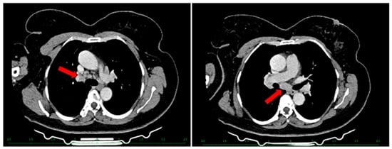
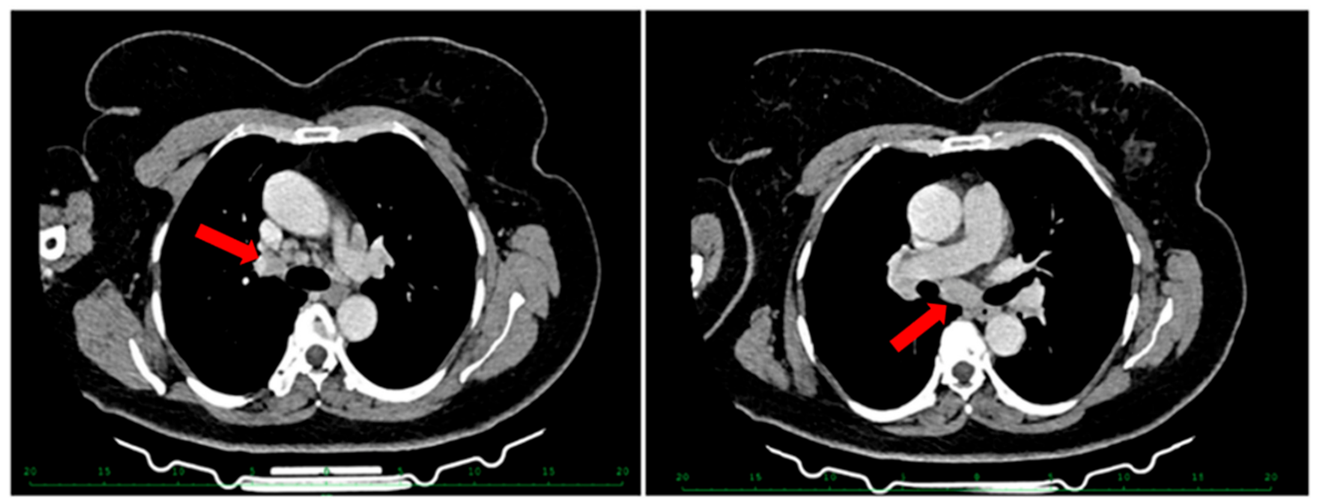
Physical examination was unremarkable except for cervical lymphadenopathy that had fluctuated in size. A CT of the thorax and abdomen showed mediastinal and bilateral hilar lymphadenopathy with ground-glass opacities (Figure 1), and mild pre-aortic, para-aortic and mesenteric lymphadenopathy with hepatosplenomegaly, diffuse hepatic steatosis and a focal calcified hepatic lesion. At mediastinoscopy, a 1.2 cm subcarinal lymph node was excised. Histopathological examination showed non-necrotizing granulomas, and staining for acid-fast bacilli was negative. Immunohistochemistry and flow cytometry were not performed. The cervical lymph nodes were not sampled at this time. An interferon-γ release assay (QuantiFERON) was negative.
Figure 1.
Chest computed tomography at first presentation showing perihilar, subcarinal and mediastinal lymphadenopathy (arrows).
Serum ACE was 62 U/L (laboratory reference range for patients aged >20 years, 8–52 U/L). Antinuclear antibodies were positive at a titer of 1:320 (fine speckled pattern), and Epstein–Barr virus (EBV) IgG was positive. Serum and urinary calcium levels were normal, and aspartate aminotransferase (AST) and γ-glutamyl transferase levels were mildly elevated. The white blood cell count was 6.76 × 10³/µL, with an absolute lymphocyte count of 0.76 × 10³/µL, and lactate dehydrogenase (LDH) was 203 U/L. Bronchoalveolar lavage, β2-microglobulin and soluble interleukin-2 receptor measurements were not performed.
Pulmonary function tests were normal. On a six-minute walk test (6MWT), she walked 450 m, with oxygen saturation decreasing from 98% to 96%. Ophthalmological examination showed no signs of ocular involvement. Cardiac evaluation with electrocardiography and transthoracic echocardiography showed mild diastolic dysfunction with a left ventricular ejection fraction (LVEF) of 74% and no findings suggestive of cardiac sarcoidosis.
Because there was no evidence of threatened organ function and her symptoms were mild, corticosteroid therapy was not started, and the patient was followed up. This approach is consistent with current guidance that treatment of sarcoidosis is indicated for patients at risk of mortality or permanent organ impairment, or to improve quality of life [42].
Four months after the first presentation, she returned with worsening exertional dyspnea; the cervical lymphadenopathy was less prominent. She was hemodynamically stable (blood pressure 140/80 mmHg, heart rate 90 beats/min) and afebrile (36.5 °C), with a respiratory rate of 14 breaths/min and an oxygen saturation of 96% on room air. Painless lymph nodes were palpable in the right cervical triangle and in both supraclavicular regions. She reported a 5 kg weight loss over approximately 40 days (about 6.3% of her baseline weight of 80 kg), which she attributed to dietary restriction, and denied fever and night sweats. There was no history of travel to Central or South America, Africa or Asia.
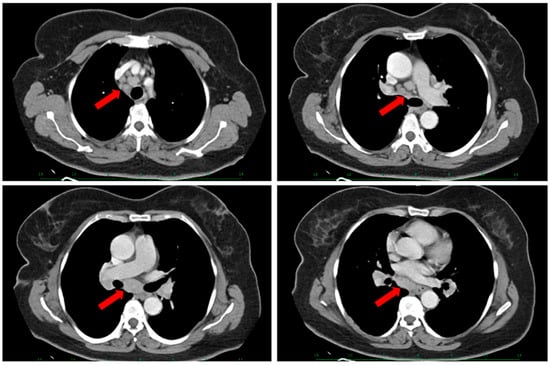
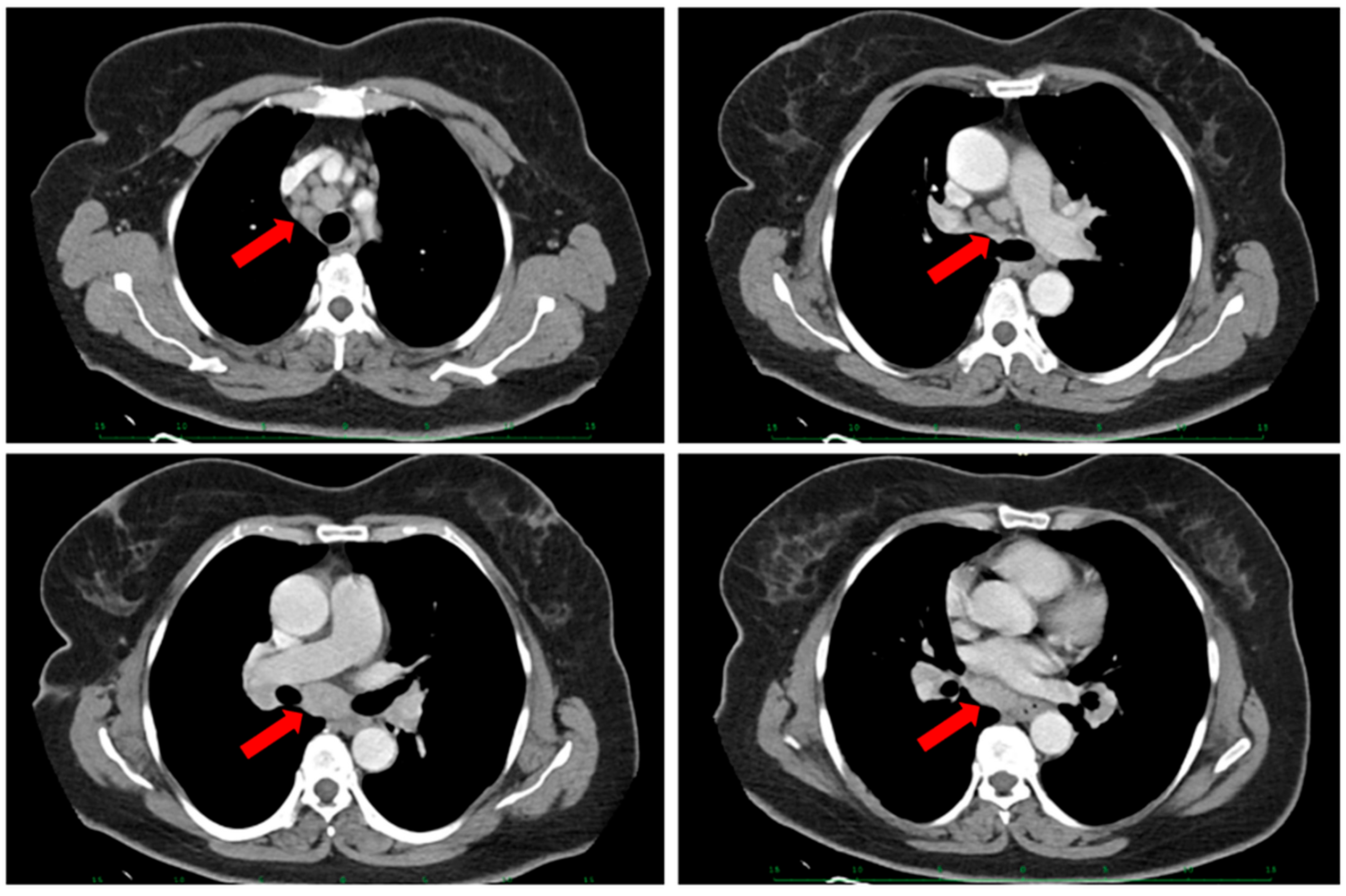
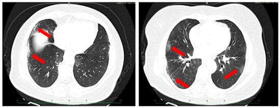
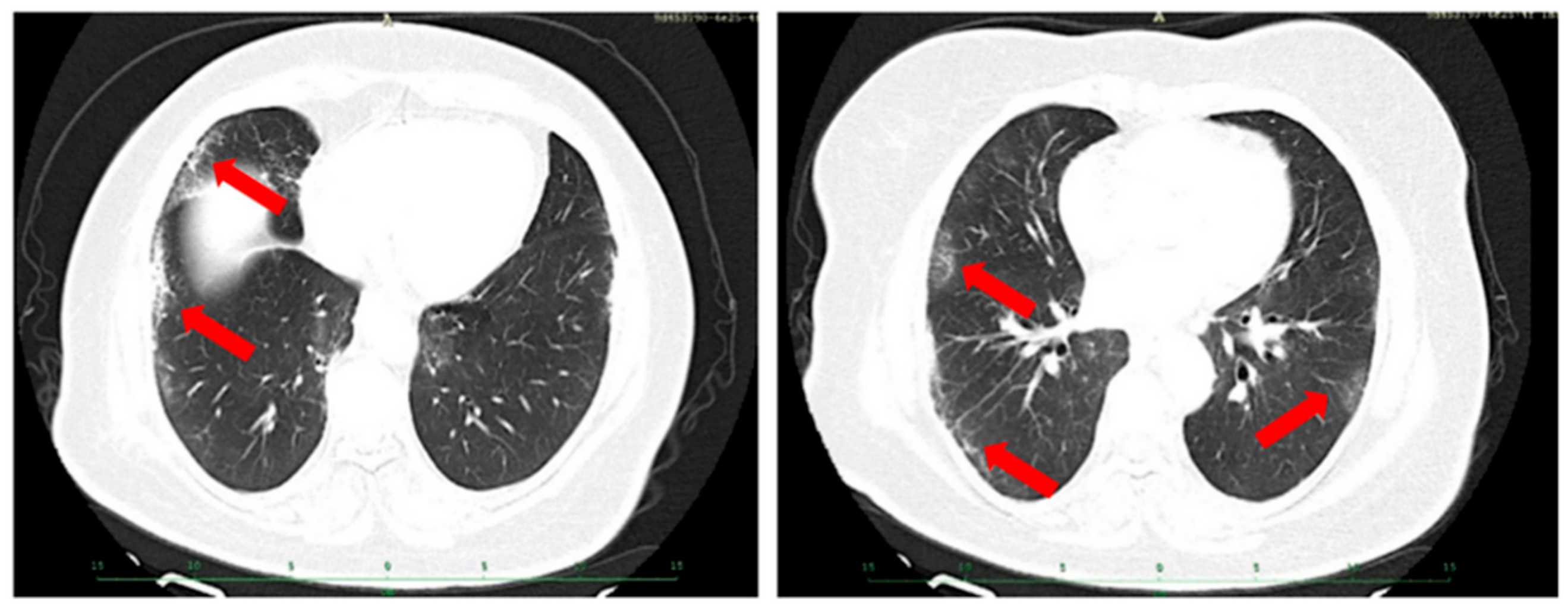
A CT of the neck, thorax and abdomen showed cervical and intra-abdominal lymphadenopathy, progression of the mediastinal lymphadenopathy (Figure 2), and increased ground-glass opacities with a micronodular pattern (Figure 3). Spirometry was normal (FEV1 2.97 L, 104% predicted; FVC 3.90 L, 108% predicted; FEV1/FVC 76%), and DLCO was 72% of predicted. On the 6MWT, she walked 540 m, with oxygen saturation decreasing from 96% to 92%.
Figure 2.
Chest computed tomography four months after the first presentation showing progression of the perihilar, subcarinal and mediastinal lymphadenopathy (arrows). (Top left) The arrow indicates soft tissue beside the right side of the trachea, consistent with right paratracheal lymph nodes. (Top right) The arrow highlights nodal tissue near the lower trachea and its division into the main bronchi—the carinal region. (Bottom left) The arrow indicates prominent subcarinal nodal tissue, beneath the tracheal division and between the main bronchi. (Bottom right) A lower section shows the apparent continuation of the subcarinal nodal tissue, behind the central airways.
Figure 3.
Chest computed tomography four months after the first presentation showing subpleural ground-glass opacities and a micronodular pattern (arrows). (Left) The arrows indicate fine, irregular linear opacities and mild hazy opacity near the outer surface of the right lung, involving the right middle and lower lobe regions. (Right) The arrows highlight scattered peripheral hazy and fine reticular opacities in both lungs, more conspicuous on the right in this image.
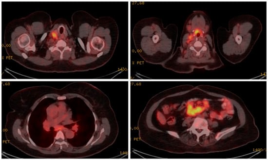
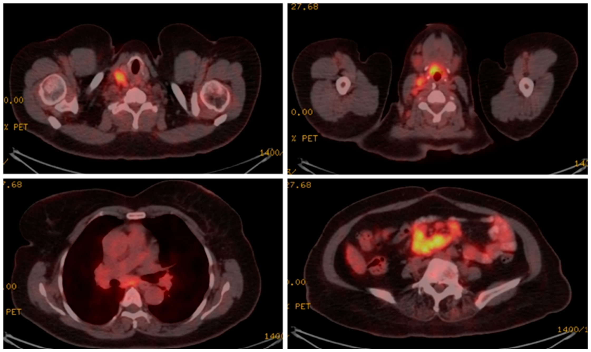
18F-FDG PET/CT, performed a month after the second presentation, showed hypermetabolic lymphadenopathy, with a maximum standardized uptake value (SUVmax) of 20.5 in the cervical nodes, 4.8 in the mediastinal nodes, and 5.6 and 9.7 in the para-aortic and mesenteric nodes, respectively (Figure 4). The spleen showed diffuse uptake exceeding that of the liver (SUVmax 4.7 vs. 3.7).
Figure 4.
18F-FDG PET/CT showing hypermetabolic cervical (upper panels; SUVmax 20.5), mediastinal (lower left; SUVmax 4.8) and mesenteric (lower right; SUVmax 9.7) lymphadenopathy.
A few days after the 18F-FDG PET/CT, oral methylprednisolone 15 mg/day was started by her primary care physician for symptomatic, painful cervical lymph node enlargement and was continued for two weeks.
An excisional cervical lymph node biopsy was planned. Because of patient-related delay, the biopsy was performed approximately three months after PET/CT and more than two months after corticosteroids had been discontinued.
The excised cervical lymph node measured 2.5 cm × 1.5 cm × 0.8 cm. Histopathological examination showed necrosis involving most of the specimen and a diffuse infiltrate of large lymphoid cells. On immunohistochemistry, the neoplastic cells were CD20+, CD79a+, PAX5+, CD10−, BCL6+, MUM1+ and BCL2+, with MYC expression in approximately 20% of cells, and were negative for CD3, CD4, CD5, CD8, CD15, CD23, CD30 and PGM1 (CD68). The Ki-67 (MIB-1) proliferation index was approximately 80% in viable areas. EBV-encoded RNA in situ hybridization (EBER) was negative, as were Ziehl–Neelsen staining, polymerase chain reaction (PCR) and culture for Mycobacterium tuberculosis. The histopathology report did not describe granulomas or epithelioid-cell clusters. Fluorescence in situ hybridization (FISH) for MYC, BCL2 and BCL6 rearrangements was not performed. The pathologists diagnosed DLBCL, NOS, according to WHO-HAEM5 [23], with a non-GCB phenotype according to the Hans algorithm [24]; with MYC expression below 40%, the lymphoma did not meet the criteria for double-expressor status.
A bone marrow trephine biopsy showed neither lymphomatous infiltration nor granulomas. Serum LDH was 244 U/L (reference range 120–246 U/L). Diffuse splenic uptake exceeding hepatic uptake was regarded as splenic involvement under the Lugano criteria [43,44]. With nodal involvement on both sides of the diaphragm, splenic involvement, no extranodal involvement and no B symptoms (weight loss of less than 10% within six months, no fever or night sweats), the disease was classified as Ann Arbor stage IIIS-A (Lugano stage III) [43]. The Eastern Cooperative Oncology Group (ECOG) performance status was 1, the IPI score was 2 (age > 60 years and stage III), and the CNS-IPI score was 2 [25].
Echocardiography was repeated before anthracycline therapy and showed an LVEF of 70%. The patient received six cycles of R-CHOP (rituximab, cyclophosphamide, doxorubicin, vincristine and prednisone) every 21 days at full dose, without a pre-phase. Polatuzumab vedotin with R-CHP [45] was not available at our institution at that time. CNS prophylaxis was not given, in view of the intermediate CNS-IPI and the absence of high-risk extranodal sites. Treatment was completed without treatment-related toxicity, including pulmonary toxicity. End-of-treatment PET/CT showed a complete metabolic response according to the Lugano criteria (Deauville score 3, compared with 5 at baseline) [43]. The mediastinal and hilar lymph nodes had decreased markedly in size and FDG uptake, and the parenchymal abnormalities had improved. Serial ACE measurements were not available. Three years after completion of treatment, the patient remains in complete remission.
3. Discussion
3.1. Sarcoidosis Preceding DLBCL, or Lymphoma with a Sarcoid-like Reaction?
Sarcoidosis was diagnosed histologically approximately 7 months before DLBCL, and the case was initially regarded as sarcoidosis followed by lymphoma. On critical review, however, the available data do not allow this sequence to be distinguished from a lymphoma that was already present at first presentation and accompanied by a sarcoid-like reaction in the sampled mediastinal node. Several features favor the latter possibility. First, fluctuating cervical lymphadenopathy was present from the outset, yet only a single 1.2 cm subcarinal node was examined, and the cervical nodes—the most accessible site and the one in which DLBCL was eventually found—were not sampled. Second, multi-compartment lymphadenopathy (pre-aortic, para-aortic and mesenteric) with hepatosplenomegaly appeared within five months of a normal abdominal CT. Third, splenomegaly and peripheral lymphadenopathy have been identified as features suggesting lymphoma in patients with sarcoidosis [3,40]. Fourth, both diagnoses were established within one year, which would be classified as a simultaneous presentation in recent series [5]. Finally, sarcoidosis is a diagnosis of exclusion [12], and in our patient the exclusion of lymphoma rested on a single mediastinal node.
Other findings are compatible with genuine sarcoidosis coexisting with, or preceding, the lymphoma. Granulomas were found in the mediastinal nodes, which showed only moderate FDG uptake (SUVmax 4.8), whereas DLBCL was found in the intensely avid cervical nodes (SUVmax 20.5). Such discordance may reflect two distinct processes, as in the case reported by Kis et al., in which intensely avid lymphoma coexisted with moderately avid mediastinal nodes containing only sarcoid granulomas [11]; it is, however, equally compatible with a sarcoid-like reaction in nodes adjacent to or draining a lymphoma [8,10]. The elevated serum ACE and the pulmonary ground-glass opacities and micronodules are compatible with either interpretation. The improvement of the thoracic findings after R-CHOP does not discriminate between the two possibilities either, because the regimen includes high-dose prednisone.
We therefore regard this case as a concurrent presentation of granulomatous inflammation and DLBCL in which the true sequence cannot be determined. Its main value lies in illustrating a diagnostic pitfall rather than in documenting early-onset lymphoma after sarcoidosis.
3.2. Lessons for Clinical Management
In retrospect, several aspects of the diagnostic pathway could have been improved. First, at the first presentation, the most accessible abnormal nodes were the fluctuating cervical nodes; excisional biopsy of a peripheral node is preferred when lymphoma is in the differential diagnosis, because it provides adequate tissue for architecture, immunohistochemistry and molecular studies [19,43]. Second, FDG-PET/CT was not performed at the initial evaluation; in a patient with multi-compartment lymphadenopathy and hepatosplenomegaly, it could have identified the most metabolically active site for biopsy [11,17], which, judging from the later scan, would probably have been a cervical node. Third, corticosteroids were started, albeit for a short period, when lymphoma was already a likely differential diagnosis and before re-biopsy, which could have compromised the histological diagnosis. Fourth, the interval of approximately three months between PET/CT and lymph node biopsy, due to patient-related reasons, delayed the diagnosis.
Corticosteroids given before biopsy may induce apoptosis and nodal shrinkage and can compromise the histological diagnosis of lymphoma [46]. In B-cell lymphoma, prebiopsy corticosteroids have been associated with a prominent histiocytic infiltrate, loss of the diffuse pattern of infiltration and atypical cytoplasmic or extracellular CD20 staining, effects related to the type, dose, route and duration of treatment [47]. A recent retrospective study of 320 patients with DLBCL found no significant association between prebiopsy corticosteroids and diagnostic yield, whereas the biopsy technique had a greater influence [48]; such data, however, cannot exclude an effect in individual patients. In our patient, the low dose, the short duration and the interval of more than two months before biopsy probably explain why the specimen remained diagnostic. Nevertheless, when lymphoma is suspected, corticosteroids should be withheld until adequate tissue has been obtained, unless they are urgently required.
The decision not to treat at first presentation was consistent with the European Respiratory Society guideline, which recommends treatment for patients at risk of mortality or permanent organ impairment, or to improve quality of life [42]; observation is appropriate for patients with mild symptoms and preserved organ function. In retrospect, withholding treatment also avoided corticosteroid exposure that could have obscured the subsequent diagnosis of lymphoma.
3.3. When Should Lymphoma Be Suspected in a Patient Labeled as Having Sarcoidosis?
On the basis of this case and the literature, the following features should prompt re-evaluation and re-biopsy in a patient with presumed sarcoidosis: peripheral (cervical, supraclavicular, axillary or inguinal) or abdominal lymphadenopathy, particularly if fluctuating or progressive; splenomegaly or focal splenic lesions; markedly high or regionally discordant FDG uptake; new or enlarging lymphadenopathy, or disease progression without or despite treatment; and persistent hematological abnormalities [3,11,36,40,49]. In such patients, excisional biopsy of the most accessible FDG-avid lymph node is preferred to sampling of a single mediastinal node, and corticosteroids should be deferred until a histological diagnosis has been obtained whenever possible [43,46,47]. FDG-PET/CT is useful to map disease extent and to select the biopsy site, but FDG uptake alone cannot distinguish sarcoidosis from lymphoma [11,37].
The distribution of lesions overlaps considerably between the two diseases: both may involve mediastinal, hilar, peripheral and abdominal lymph nodes, the spleen, the liver, the bone marrow and other extranodal sites [16,17,18,50]. Bilateral, usually symmetric hilar and mediastinal lymphadenopathy, often with perilymphatic micronodules, is the most common thoracic pattern of sarcoidosis [12,13,51], whereas compressive or bulky lymphadenopathy, unexplained progressive lymphadenopathy, atypical localizations and splenomegaly favor lymphoma [3,40]; in a series of enlarged mediastinal nodes, the short-axis diameter was more informative than SUVmax [52]. In our patient, the distribution was mixed: bilateral hilar and mediastinal lymphadenopathy with micronodules, typical of sarcoidosis, coexisted with fluctuating cervical and supraclavicular nodes, mesenteric and para-aortic lymphadenopathy and splenomegaly, which in retrospect were atypical for sarcoidosis. Even experienced readers classified about one in four patients with sarcoidosis as having lymphoma on FDG-PET/CT in a recent series (specificity 0.75) [50], so the pattern of involvement can guide, but not replace, biopsy.
Absolute SUVmax values do not reliably separate the two diseases. Aggressive non-Hodgkin lymphomas, including DLBCL, are usually intensely FDG-avid (mean SUVmax 19.6 ± 9.3 in newly diagnosed aggressive lymphoma; range 3.2–43.0) [53], but active sarcoidosis can be equally avid: in a series of enlarged mediastinal lymph nodes, the SUVmax of sarcoidosis was 15.9 ± 5.1 and did not differ significantly from that of lymphoma [52], and sarcoid lesions in lymph nodes and muscle with SUV values of up to 26.1 have been reported after treatment of DLBCL [54]. SUVmax has therefore not proved discriminatory [50,52]. Within individual patients with both diseases, however, lymphoma-involved sites have often shown higher uptake than granulomatous sites: SUVmax 18.3–19.5 in hepatic lymphoma versus 2.7 in sarcoid lymph nodes [38], intense uptake in pulmonary lymphoma versus moderate uptake in sarcoid mediastinal nodes [11], and, in our patient, 20.5 in the DLBCL-involved cervical nodes versus 4.8 in the mediastinal nodes. Intra-individual discordance in FDG uptake is therefore more useful than any absolute cut-off, mainly to select the biopsy site.
3.4. Possible Pathogenetic Links in the Present Case
Of the mechanisms proposed to link sarcoidosis and lymphoma, impaired immune surveillance due to immunosuppressive treatment [27] does not apply to our patient, who had received no immunosuppressive treatment before lymphoma was suspected. A role for EBV in connecting lymphoma with sarcoid-like granulomas has been proposed, and half of classic Hodgkin lymphomas with a marked granulomatous reaction were EBV-positive in one series [33]; our patient had serological evidence of past EBV infection, but the lymphoma was EBER-negative. Splenic and bone marrow involvement by sarcoidosis has been associated with hematological malignancy [40,55]. In our patient, however, the splenic enlargement and FDG uptake cannot be attributed to either sarcoidosis or lymphoma without histology and are therefore not regarded as a pre-existing risk factor, and the bone marrow biopsy showed no granulomas.
3.5. Published Cases of Sarcoidosis Associated with DLBCL
We searched PubMed and Google Scholar in December 2025, without date restrictions, for published cases of sarcoidosis associated with lymphoma, using combinations of the terms “sarcoidosis”, “lymphoma”, “sarcoidosis-lymphoma syndrome”, “diffuse large B-cell lymphoma”, “DLBCL” and “case report”; the PubMed search was updated in October 2026 and identified no additional eligible reports. Reference lists of relevant articles and reviews were also screened. Reports were eligible if they described individual patients with sarcoidosis, as diagnosed by the original authors, and DLBCL; no language restriction was applied. From cohort studies, only individually described DLBCL cases were extracted. For reports available to us only as abstracts, including several non-English articles, data were extracted from the abstract, and missing items are indicated as not reported. Demographics, sequence and interval between diagnoses, previous immunosuppression, lymphoma histology, treatment and outcome were tabulated (Table 2).
We identified 39 published cases from 25 reports. As categorized by the original authors, sarcoidosis preceded DLBCL in 20 cases [6,10,49,56,57,58,59,60,61,62,63,64,65], followed by DLBCL in 16 [6,54,66,67,68,69,70,71,72,73,74,75] and was diagnosed concurrently in 3 [6,76]. Short intervals were not exceptional: in at least 14 cases (36%), including the 3 concurrent cases, both diagnoses were established within 12 months (Table 2). Applying the one-year definition used by Charrière et al. [5], our patient’s presentation is classified as concurrent. In that multicenter series of 46 patients with sarcoidosis and lymphoma, DLBCL was the most frequent subtype (20 patients); sarcoidosis preceded DLBCL in 8, followed it in 11 and was diagnosed simultaneously in 1, and lymphoma was diagnosed at an older age when sarcoidosis came first [5]. Because individual patient data were not reported, these patients are not included in Table 2.
Whether corticosteroid treatment of a preceding sarcoidosis delays the onset of DLBCL cannot be determined from the published cases. Among the 20 patients in whom sarcoidosis preceded DLBCL, 8 had received corticosteroids before the lymphoma was diagnosed, with a median interval of approximately 12 years (range 2 months to 15 years), compared with 7 years (range 4–52 years) in the 9 patients without reported immunosuppression and a documented interval (Table 2). The ranges overlapped widely, and the two shortest intervals (approximately 2 months and less than one year) both occurred in patients treated with corticosteroids for presumed sarcoidosis [49,65]; in one of them, initial improvement on prednisone was followed by generalized lymphadenopathy due to DLBCL [49]. Because corticosteroids can shrink lymphomatous nodes and alter their histology [46,47], they may delay the recognition of a coexisting lymphoma rather than its biological onset; conversely, long-term immunosuppression has been proposed to promote lymphomagenesis [27], and corticosteroids are usually given to patients with chronic active sarcoidosis, the setting in which lymphoma is most often reported [1]. These observations derive from a small, heterogeneous set of case reports subject to reporting bias. In our patient, no corticosteroids were given before the second presentation, so this mechanism cannot explain the course of her disease.
Among the cases categorized as sarcoidosis preceding DLBCL, two had intervals of less than one year [49,65]. Jbeli et al. described a 54-year-old man with hypercalcemia and acute kidney injury who was treated with prednisone for sarcoidosis and later presented with generalized lymphadenopathy, in which excisional biopsy revealed DLBCL [49]. That patient shared with ours the short interval between the two diagnoses but differed in renal involvement, hypercalcemia and prior corticosteroid treatment. As the authors emphasized, new or enlarging lymphadenopathy in a patient with sarcoidosis should be biopsied [49].
DLBCL associated with sarcoidosis does not appear to have a different prognosis from DLBCL without sarcoidosis [6], and our patient achieved a durable complete remission with standard R-CHOP.
Table 2.
Published cases of sarcoidosis associated with diffuse large B-cell lymphoma (DLBCL), grouped according to the sequence of diagnoses reported by the original authors, and the present case. Abbreviations: ABC, activated B-cell-like; ACVBP, doxorubicin, cyclophosphamide, vindesine, bleomycin and prednisone; AZA, azathioprine; CHOP, cyclophosphamide, doxorubicin, vincristine and prednisone; CNS, central nervous system; CS, corticosteroids; GCB, germinal-center B-cell-like; HCQ, hydroxychloroquine; HD-MTX, high-dose methotrexate; HSCT, hematopoietic stem cell transplantation; i.v., intravenous; IVLBCL, intravascular large B-cell lymphoma; MTX, methotrexate; NOS, not otherwise specified; NSAID, nonsteroidal anti-inflammatory drug; p.o., oral; PTCL, peripheral T-cell lymphoma; R-ACVBP, rituximab and ACVBP; R-CHOP, rituximab and CHOP; R-COP, rituximab, cyclophosphamide, vincristine and prednisone; R-COPADM, rituximab, cyclophosphamide, vincristine, prednisone, doxorubicin and methotrexate; R-CYM, rituximab, cytarabine and methotrexate; R-DHAP, rituximab, dexamethasone, high-dose cytarabine and cisplatin; R-miniCEOP, rituximab and reduced-dose cyclophosphamide, epirubicin, vincristine and prednisone; R-miniCHOP, rituximab and reduced-dose CHOP; R-MTX-CHOP, rituximab, methotrexate and CHOP; RTX, rituximab; R-THP-COP, rituximab, pirarubicin, cyclophosphamide, vincristine and prednisone.
3.6. Limitations
This report has limitations. The initial subcarinal specimen was not examined by immunohistochemistry or clonality testing, so a small lymphomatous infiltrate cannot be excluded. PET/CT was not performed at the first presentation. Serial ACE measurements and systematic imaging follow-up of the pulmonary findings were not available, and the CT images available for publication do not allow measurements at strictly matched anatomical levels. Finally, part of the literature summary relied on abstracts of non-English reports.
4. Conclusions
In a patient with granulomatous lymphadenitis, sarcoidosis remains a diagnosis of exclusion that must be revisited when the clinical course is atypical. In our patient, DLBCL was diagnosed within one year of the finding of mediastinal granulomas, and a lymphoma present from the outset with an accompanying sarcoid-like reaction cannot be excluded. Peripheral or abdominal lymphadenopathy, splenomegaly and discordant FDG uptake should prompt excisional biopsy of the most accessible FDG-avid node, preferably before corticosteroids are started.
5. Key Points
- Sarcoidosis is a diagnosis of exclusion; granulomas in a single mediastinal lymph node do not exclude a coexisting lymphoma, which may itself induce a sarcoid-like reaction;
- When sarcoidosis and lymphoma are diagnosed within one year of each other, the sequence may not be determinable, and such presentations are best regarded as concurrent;
- Peripheral or abdominal lymphadenopathy, splenomegaly, very high or regionally discordant FDG uptake, and progression without or despite treatment should prompt re-biopsy in patients labeled as having sarcoidosis;
- Excisional biopsy of the most accessible FDG-avid node is preferred, and corticosteroids should be deferred until a histological diagnosis has been obtained whenever possible.
Author Contributions
Conceptualization, F.D. and P.S.; data curation, F.D., C.R. and L.I.; writing—original draft preparation, S.V., F.D. and L.I.; writing—review and editing, F.D., P.S. and C.R.; supervision, P.S. and F.D. All authors have read and agreed to the published version of the manuscript.
Funding
This research received no external funding.
Institutional Review Board Statement
Ethical review and approval of this study were not required by the Scientific Council of the University General Hospital of Alexandroupolis, because case reports describing the routine clinical care of a single patient are not considered research. The patient’s information has been de-identified.
Informed Consent Statement
Written informed consent has been obtained from the patient to publish this paper.
Data Availability Statement
The original contributions presented in this study are included in the article. Further inquiries can be directed to the corresponding author. The patient’s medical records are not publicly available due to privacy restrictions.
Acknowledgments
During the revision of this manuscript, the authors used Claude (Anthropic) to edit the English language and to assist in redrafting the text in response to the reviewers’ comments. The authors reviewed and edited all output and take full responsibility for the content of this publication.
Conflicts of Interest
The authors declare no conflicts of interest.
Abbreviations
The following abbreviations are used in this manuscript:
| 6MWT | six-minute walk test |
| ABC | activated B-cell-like |
| ACE | angiotensin-converting enzyme |
| AST | aspartate aminotransferase |
| CNS | central nervous system |
| CNS-IPI | Central Nervous System International Prognostic Index |
| CT | computed tomography |
| DLBCL | diffuse large B-cell lymphoma |
| DLCO | diffusing capacity of the lung for carbon monoxide |
| EBER | Epstein–Barr virus-encoded RNA |
| EBV | Epstein–Barr virus |
| ECOG | Eastern Cooperative Oncology Group |
| FAMT | L-[3−18F]-α-methyltyrosine |
| FDG-PET/CT | 18F-fluorodeoxyglucose positron emission tomography/computed tomography |
| FEV1 | forced expiratory volume in one second |
| FISH | fluorescence in situ hybridization |
| FVC | forced vital capacity |
| GCB | germinal-center B-cell-like |
| IPI | International Prognostic Index |
| LDH | lactate dehydrogenase |
| LVEF | left ventricular ejection fraction |
| NOS | not otherwise specified |
| PCR | polymerase chain reaction |
| R-CHOP | rituximab, cyclophosphamide, doxorubicin, vincristine and prednisone |
| SLR | sarcoid-like reaction |
| SLS | sarcoidosis–lymphoma syndrome |
| SUVmax | maximum standardized uptake value |
| WHO-HAEM5 | 5th edition of the World Health Organization Classification of Haematolymphoid Tumours |
References
- Brincker, H. The Sarcoidosis-Lymphoma Syndrome. Br. J. Cancer 1986, 54, 467–473. [Google Scholar] [CrossRef] [Scilit] [PubMed]
- Askling, J.; Grunewald, J.; Eklund, A.; Hillerdal, G.; Ekbom, A. Increased Risk for Cancer Following Sarcoidosis. Am. J. Respir. Crit. Care Med. 1999, 160, 1668–1672. [Google Scholar] [CrossRef] [Scilit] [PubMed]
- El Jammal, T.; Pavic, M.; Gerfaud-Valentin, M.; Jamilloux, Y.; Sève, P. Sarcoidosis and Cancer: A Complex Relationship. Front. Med. 2020, 7, 594118. [Google Scholar] [CrossRef] [Scilit] [PubMed]
- García Casares, E.; Mateo Soria, L.; García Melchor, E.; Mínguez Blasco, S.; Vall-Llobera Calmet, F.; Cañellas Oliver, G.; Holgado Pérez, S. Sarcoidosis-lymphoma syndrome. Reumatol. Clin. 2009, 5, 31–33. [Google Scholar] [CrossRef] [Scilit] [PubMed]
- Charrière, R.; El Jammal, T.; Bert, A.; Chalayer, E.; Kilian, M.; Gerfaud-Valentin, M.; Jamilloux, Y.; Ghesquieres, H.; Sève, P. The Association between Sarcoidosis and Lymphoma: A Retrospective and Multicentre Study of 46 Patients. Sarcoidosis Vasc. Diffus. Lung Dis. 2025, 42, 16866. [Google Scholar] [CrossRef] [Scilit] [PubMed]
- Chalayer, É.; Bachy, E.; Occelli, P.; Weiler, L.; Faurie, P.; Ghesquieres, H.; Pavic, M.; Broussolle, C.; Sève, P. Sarcoidosis and Lymphoma: A Comparative Study. QJM 2015, 108, 871–878. [Google Scholar] [CrossRef] [Scilit] [PubMed]
- Cohen, P.R.; Kurzrock, R. Sarcoidosis and Malignancy. Clin. Dermatol. 2007, 25, 326–333. [Google Scholar] [CrossRef] [Scilit] [PubMed]
- Brincker, H. Sarcoid Reactions in Malignant Tumours. Cancer Treat. Rev. 1986, 13, 147–156. [Google Scholar] [CrossRef] [Scilit] [PubMed]
- Mei, F.; Maripudi, S.; Hogan, R.N.; Cao, J.; Mancini, R. Sarcoid-like Reaction of the Orbit in Diffuse Large B-Cell Lymphoma. Orbit 2025, 44, 104–107. [Google Scholar] [CrossRef] [Scilit] [PubMed]
- Masuda, R.; Toyoshima, H.; Bandou, T.; Isoyama, T.; Matsui, Y.; Takemura, T. Malignant Lymphoma of the Stomach Associated with Systemic Sarcoidosis. Cancer 1992, 70, 2592–2596. [Google Scholar] [CrossRef] [Scilit] [PubMed]
- Kis, A.; Eszes, N.; Tamasi, L.; Losonczy, G.; Csekeo, A.; Csomor, J.; Muller, V. Sarcoidosis Lymphoma Syndrome-the Value of PET-CT in the Diagnosis. World J. Surg. Oncol. 2013, 11, 235. [Google Scholar] [CrossRef] [Scilit] [PubMed]
- Crouser, E.D.; Maier, L.A.; Wilson, K.C.; Bonham, C.A.; Morgenthau, A.S.; Patterson, K.C.; Abston, E.; Bernstein, R.C.; Blankstein, R.; Chen, E.S.; et al. Diagnosis and Detection of Sarcoidosis. An Official American Thoracic Society Clinical Practice Guideline. Am. J. Respir. Crit. Care Med. 2020, 201, e26–e51. [Google Scholar] [CrossRef] [Scilit] [PubMed]
- Belperio, J.A.; Fishbein, M.C.; Abtin, F.; Channick, J.; Balasubramanian, S.A.; Lynch, J.P., III. Pulmonary Sarcoidosis: A Comprehensive Review: Past to Present. J. Autoimmun. 2024, 149, 103107. [Google Scholar] [CrossRef] [Scilit] [PubMed]
- Cooper, D.; Suau, S. Sarcoidosis. Immunol. Allergy Clin. N. Am. 2023, 43, 583–591. [Google Scholar] [CrossRef] [Scilit] [PubMed]
- Rossides, M.; Darlington, P.; Kullberg, S.; Arkema, E.V. Sarcoidosis: Epidemiology and Clinical Insights. J. Intern. Med. 2023, 293, 668–680. [Google Scholar] [CrossRef] [Scilit] [PubMed]
- Prabhakar, H.B.; Rabinowitz, C.B.; Gibbons, F.K.; O’Donnell, W.J.; Shepard, J.-A.O.; Aquino, S.L. Imaging Features of Sarcoidosis on MDCT, FDG PET, and PET/CT. Am. J. Roentgenol. 2008, 190, S1–S6. [Google Scholar] [CrossRef] [Scilit] [PubMed]
- Akaike, G.; Itani, M.; Shah, H.; Ahuja, J.; Yilmaz Gunes, B.; Assaker, R.; Behnia, F. PET/CT in the Diagnosis and Workup of Sarcoidosis: Focus on Atypical Manifestations. RadioGraphics 2018, 38, 1536–1549. [Google Scholar] [CrossRef] [Scilit] [PubMed]
- Soussan, M.; Augier, A.; Brillet, P.-Y.; Weinmann, P.; Valeyre, D. Functional Imaging in Extrapulmonary Sarcoidosis: FDG-PET/CT and MR Features. Clin. Nucl. Med. 2014, 39, e146–e159. [Google Scholar] [CrossRef] [Scilit] [PubMed]
- Lewis, W.D.; Lilly, S.; Jones, K.L. Lymphoma: Diagnosis and Treatment. Am. Fam. Physician 2020, 101, 34–41. [Google Scholar] [CrossRef] [Scilit]
- Matasar, M.J.; Zelenetz, A.D. Overview of Lymphoma Diagnosis and Management. Radiol. Clin. N. Am. 2008, 46, 175–198. [Google Scholar] [CrossRef] [Scilit] [PubMed]
- Li, S.; Young, K.H.; Medeiros, L.J. Diffuse Large B-Cell Lymphoma. Pathology 2018, 50, 74–87. [Google Scholar] [CrossRef] [Scilit] [PubMed]
- Takahara, T.; Nakamura, S.; Tsuzuki, T.; Satou, A. The Immunology of DLBCL. Cancers 2023, 15, 835. [Google Scholar] [CrossRef] [Scilit] [PubMed]
- Alaggio, R.; Amador, C.; Anagnostopoulos, I.; Attygalle, A.D.; de Oliveira Araujo, I.B.; Berti, E.; Bhagat, G.; Borges, A.M.; Boyer, D.; Calaminici, M.; et al. The 5th Edition of the World Health Organization Classification of Haematolymphoid Tumours: Lymphoid Neoplasms. Leukemia 2022, 36, 1720–1748. [Google Scholar] [CrossRef] [Scilit] [PubMed]
- Hans, C.P.; Weisenburger, D.D.; Greiner, T.C.; Gascoyne, R.D.; Delabie, J.; Ott, G.; Müller-Hermelink, H.K.; Campo, E.; Braziel, R.M.; Jaffe, E.S.; et al. Confirmation of the Molecular Classification of Diffuse Large B-Cell Lymphoma by Immunohistochemistry Using a Tissue Microarray. Blood 2004, 103, 275–282. [Google Scholar] [CrossRef] [Scilit] [PubMed]
- Schmitz, N.; Zeynalova, S.; Nickelsen, M.; Kansara, R.; Villa, D.; Sehn, L.H.; Glass, B.; Scott, D.W.; Gascoyne, R.D.; Connors, J.M.; et al. CNS International Prognostic Index: A Risk Model for CNS Relapse in Patients with Diffuse Large B-Cell Lymphoma Treated with R-CHOP. J. Clin. Oncol. 2016, 34, 3150–3156. [Google Scholar] [CrossRef] [Scilit] [PubMed]
- Gediz, F.; Yilmaz, A.F.; Payzin, B.; Yuksel, T.; Calli, A.O.; Kobak, S. Development of Hodgkin Lymphoma in a Patient with Sarcoidosis. Reumatismo 2017, 69, 84–87. [Google Scholar] [CrossRef] [Scilit] [PubMed][Green Version]
- Voutidou, S.; Eleftheriadis, D.; Drakopanagiotakis, F.; Papanikolaou, I.C.; Steiropoulos, P. Pathogenetic Mechanisms Linking Sarcoidosis to Lymphoma. Int. J. Mol. Sci. 2025, 26, 594. [Google Scholar] [CrossRef] [Scilit] [PubMed]
- Hachisu, Y.; Koga, Y.; Kasama, S.; Kaira, K.; Uno, S.; Yatomi, M.; Aoki-Saito, H.; Tsurumaki, H.; Jingu, A.; Sunaga, N.; et al. The Relationship between Tumor Development and Sarcoidosis in Aspects of Carcinogenesis before and after the Onset of Sarcoidosis. Medicina 2022, 58, 768. [Google Scholar] [CrossRef] [Scilit] [PubMed]
- Arger, N.K.; O’Connor, B.; Koth, L.L. Molecular Profiling in Sarcoidosis. Curr. Opin. Pulm. Med. 2020, 26, 562–567. [Google Scholar] [CrossRef] [Scilit] [PubMed]
- Huang, X.; Gui, A.; Zhou, Y.; Xia, Z.; Liu, W.; Zuo, J.; Yang, L.; Zhang, Q. Serum miR-146a Level Is a Potential Biomarker in Predicting the Outcome of Diffuse Large B-Cell Lymphoma. Asia Pac. J. Clin. Oncol. 2023, 19, e283–e290. [Google Scholar] [CrossRef] [Scilit] [PubMed]
- Bassig, B.A.; Lan, Q.; Rothman, N.; Zhang, Y.; Zheng, T. Current Understanding of Lifestyle and Environmental Factors and Risk of Non-Hodgkin Lymphoma: An Epidemiological Update. J. Cancer Epidemiol. 2012, 2012, 978930. [Google Scholar] [CrossRef] [Scilit] [PubMed]
- Judson, M.A. Environmental Risk Factors for Sarcoidosis. Front. Immunol. 2020, 11, 1340. [Google Scholar] [CrossRef] [Scilit] [PubMed]
- Xu, J.; Li, S.; Yin, C.C.; Patel, K.P.; Tang, G.; Wang, W.; Miranda, R.N.; Garces, S.; Tang, Z.; Lin, P.; et al. Classic Hodgkin Lymphoma with Marked Granulomatous Reaction: A Clinicopathologic Study of 20 Cases. Hum. Pathol. 2023, 134, 114–123. [Google Scholar] [CrossRef] [Scilit] [PubMed]
- Cerri, S.; Fontana, M.; Balduzzi, S.; Potenza, L.; Faverio, P.; Luppi, M.; D’Amico, R.; Spagnolo, P.; Clini, E.; Luppi, F. Clinical Differences in Sarcoidosis Patients with and without Lymphoma: A Single-Centre Retrospective Cohort Analysis. Eur. Respir. J. 2019, 54, 1802470. [Google Scholar] [CrossRef] [Scilit] [PubMed]
- Ji, B.; Khatun, N.; Mostafidi, E.; Gupta, R.; McFarlane, S.I. Sarcoidosis Versus Lymphoma: A Clinical Diagnostic Dilemma in a Patient with Extensive Lymphadenopathy. Cureus 2023, 15, e43281. [Google Scholar] [CrossRef] [Scilit] [PubMed]
- Dal Lago, L.; Sarrand, J.; Woff, E.; Awada, A.; Vouche, M.; Pepersack, T. Sarcoidosis Versus Lymphoma? Eur. J. Case Rep. Intern. Med. 2021, 8, 002250. [Google Scholar] [CrossRef] [Scilit] [PubMed]
- Love, C.; Tomas, M.B.; Tronco, G.G.; Palestro, C.J. FDG PET of Infection and Inflammation. RadioGraphics 2005, 25, 1357–1368. [Google Scholar] [CrossRef] [Scilit] [PubMed]
- Yudistiro, R.; Arisaka, Y.; Tokue, A.; Nakajima, T. Differentiation of Sarcoidosis-Lymphoma Syndrome Lesions: A Case Report on the Use of Two Different Positron Emission Tomography Tracers. BMC Med. Imaging 2016, 16, 1. [Google Scholar] [CrossRef] [Scilit] [PubMed]
- Kaira, K.; Oriuchi, N.; Otani, Y.; Yanagitani, N.; Sunaga, N.; Hisada, T.; Ishizuka, T.; Endo, K.; Mori, M. Diagnostic Usefulness of Fluorine–18-α–Methyltyrosine Positron Emission Tomography in Combination with 18F-Fluorodeoxyglucose in Sarcoidosis Patients. Chest 2007, 131, 1019–1027. [Google Scholar] [CrossRef] [Scilit] [PubMed]
- Papanikolaou, I.C.; Sharma, O.P. The Relationship between Sarcoidosis and Lymphoma. Eur. Respir. J. 2010, 36, 1207–1209. [Google Scholar] [CrossRef] [Scilit] [PubMed]
- Krasowski, M.D.; Savage, J.; Ehlers, A.; Maakestad, J.; Schmidt, G.A.; La’ulu, S.; Rasmussen, N.N.; Strathmann, F.G.; Genzen, J.R. Ordering of the Serum Angiotensin-Converting Enzyme Test in Patients Receiving Angiotensin-Converting Enzyme Inhibitor Therapy: An Avoidable but Common Error. Chest 2015, 148, 1447–1453. [Google Scholar] [CrossRef] [Scilit] [PubMed]
- Baughman, R.P.; Valeyre, D.; Korsten, P.; Mathioudakis, A.G.; Wuyts, W.A.; Wells, A.; Rottoli, P.; Nunes, H.; Lower, E.E.; Judson, M.A.; et al. ERS Clinical Practice Guidelines on Treatment of Sarcoidosis. Eur. Respir. J. 2021, 58, 2004079. [Google Scholar] [CrossRef] [Scilit] [PubMed]
- Cheson, B.D.; Fisher, R.I.; Barrington, S.F.; Cavalli, F.; Schwartz, L.H.; Zucca, E.; Lister, T.A. Recommendations for Initial Evaluation, Staging, and Response Assessment of Hodgkin and Non-Hodgkin Lymphoma: The Lugano Classification. J. Clin. Oncol. 2014, 32, 3059–3068. [Google Scholar] [CrossRef] [Scilit] [PubMed]
- Barrington, S.F.; Mikhaeel, N.G.; Kostakoglu, L.; Meignan, M.; Hutchings, M.; Müeller, S.P.; Schwartz, L.H.; Zucca, E.; Fisher, R.I.; Trotman, J.; et al. Role of Imaging in the Staging and Response Assessment of Lymphoma: Consensus of the International Conference on Malignant Lymphomas Imaging Working Group. J. Clin. Oncol. 2014, 32, 3048–3058. [Google Scholar] [CrossRef] [Scilit] [PubMed]
- Tilly, H.; Morschhauser, F.; Sehn, L.H.; Friedberg, J.W.; Trněný, M.; Sharman, J.P.; Herbaux, C.; Burke, J.M.; Matasar, M.; Rai, S.; et al. Polatuzumab Vedotin in Previously Untreated Diffuse Large B-Cell Lymphoma. N. Engl. J. Med. 2022, 386, 351–363. [Google Scholar] [CrossRef] [Scilit] [PubMed]
- Kan, E.; Levi, I.; Benharroch, D. Alterations in the Primary Diagnosis of Lymphomas Pretreated with Corticosteroid Agents. Leuk. Lymphoma 2011, 52, 425–428. [Google Scholar] [CrossRef] [Scilit] [PubMed]
- Krishnamurthy, K.; Li, X.; Choudhuri, J.; Shi, Y.; Wang, Y. The Impact of Type, Dose, and Duration of Prebiopsy Corticosteroids Use on Histomorphology and Immunohistochemical Stains in B-Cell Lymphoma: A Case-Control Study to Highlight the “Atypical” Cytoplasmic and Extra-Cellular Granular Staining Pattern of CD20. Appl. Immunohistochem. Mol. Morphol. 2025, 33, 164–169. [Google Scholar] [CrossRef] [Scilit] [PubMed]
- Madireddy, S.; Pou, S.; Mandi, K.; Pan, K.; Friefeld, A.; Ding, H.H.; Raker, C.; Terada, R.; Milrod, C.J.; Pelcovits, A. Prebiopsy Steroids and Diagnostic Yield in Patients with Diffuse Large B-Cell Lymphoma. JAMA Netw. Open 2025, 8, e2548617. [Google Scholar] [CrossRef] [Scilit] [PubMed]
- Jbeli, A.H.; Sunassee, A.; Sanford, A.; Elshami, A.; Bleeker, J. A Very Rare Case of Sarcoid-Lymphoma Syndrome Imposing an Intriguing Diagnostic Challenge. S. D. Med. 2018, 71, 108–111. [Google Scholar] [PubMed]
- Lovinfosse, P.; Ferreira, M.; Withofs, N.; Jadoul, A.; Derwael, C.; Frix, A.-N.; Guiot, J.; Bernard, C.; Diep, A.N.; Donneau, A.-F.; et al. Distinction of Lymphoma from Sarcoidosis on 18F-FDG PET/CT: Evaluation of Radiomics-Feature-Guided Machine Learning versus Human Reader Performance. J. Nucl. Med. 2022, 63, 1933–1940. [Google Scholar] [CrossRef] [Scilit] [PubMed]
- Criado, E.; Sánchez, M.; Ramírez, J.; Arguis, P.; de Caralt, T.M.; Perea, R.J.; Xaubet, A. Pulmonary Sarcoidosis: Typical and Atypical Manifestations at High-Resolution CT with Pathologic Correlation. RadioGraphics 2010, 30, 1567–1586. [Google Scholar] [CrossRef] [Scilit] [PubMed]
- Yu, C.; Xia, X.; Qin, C.; Sun, X.; Zhang, Y.; Lan, X. Is SUVmax Helpful in the Differential Diagnosis of Enlarged Mediastinal Lymph Nodes? A Pilot Study. Contrast Media Mol. Imaging 2018, 2018, 3417190. [Google Scholar] [CrossRef] [Scilit] [PubMed]
- Schöder, H.; Noy, A.; Gönen, M.; Weng, L.; Green, D.; Erdi, Y.E.; Larson, S.M.; Yeung, H.W.D. Intensity of 18Fluorodeoxyglucose Uptake in Positron Emission Tomography Distinguishes between Indolent and Aggressive Non-Hodgkin’s Lymphoma. J. Clin. Oncol. 2005, 23, 4643–4651. [Google Scholar] [CrossRef] [Scilit] [PubMed]
- Ichimaru, K.; Nakata, J.; Matsuura, A.; Yasuhara, Y.; Nakatsuka, S.-I.; Shibano, M. Systemic sarcoidosis developed after chemoradiotherapy for localized thyroid diffuse large B-cell lymphoma. [Rinsho Ketsueki] Jpn. J. Clin. Hematol. 2021, 62, 1598–1603. [Google Scholar] [CrossRef] [Scilit] [PubMed]
- Brito-Zerón, P.; Flores-Chávez, A.; González-de-Paz, L.; Feijoo-Massó, C.; de Escalante, B.; González-García, A.; Gómez-de-la-Torre, R.; Policarpo-Torres, G.; Alguacil, A.; García-Morillo, J.S.; et al. Temporal Relationship between Sarcoidosis and Malignancies in a Nationwide Cohort of 1942 Patients. Postgrad. Med. J. 2024, 100, 898–907. [Google Scholar] [CrossRef] [Scilit] [PubMed]
- Furusawa, H.; Shimada, S.; Sawada, A.; Tanaka, K.; Yamamoto, K.; Miyazaki, Y. Composite Lymphoma after Treatment with Infliximab for Sarcoidosis and Ankylosing Spondylitis. Intern. Med. 2025, 64, 129–132. [Google Scholar] [CrossRef] [Scilit] [PubMed]
- Iizuka, K.; Ando, J.; Masuda, A.; Ochiai, T.; Tomomasa, R.; Yasuda, H.; Takaku, T.; Ota, Y.; Yao, T.; Gotoh, A.; et al. Acute exacerbation of organizing pneumonia leading to sudden death in a patient with sarcoidosis-lymphoma syndrome. [Rinsho Ketsueki] Jpn. J. Clin. Hematol. 2017, 58, 927–932. [Google Scholar] [CrossRef] [PubMed]
- Marks, R.; Engelhardt, M.; Hager, S.; Füllgraf, H.; Südkamp, N.P.; Herget, G.W. Diffuse Large B Cell Lymphoma (DLBCL): Bilateral Vanishing Tibiae. Ann. Hematol. 2018, 97, 1497–1500. [Google Scholar] [CrossRef] [Scilit] [PubMed]
- Matsuo, T.; Tanaka, T.; Omote, R.; Okada, T.; Notohara, K.; Okada, K. Diffuse Large B-Cell Lymphoma in the Course of Systemic Sarcoidosis: A Case Report and Review of 30 Japanese Patients with Sarcoidosis-Lymphoma Syndrome. J. Clin. Exp. Hematop. 2022, 62, 226–237. [Google Scholar] [CrossRef] [Scilit] [PubMed]
- Nakano, K.; Ito, Y.; Takagi, F.; Uchiyama, S.; Yoda, A.; Sakamoto, N.; Takaba, M.; Adachi, M.; Takemura, T.; Nagata, Y.; et al. Sarcoidosis-lymphoma syndrome showing abnormal FDG uptake in lymph nodes and muscles upon post-chemotherapy PET/CT imaging. [Rinsho Ketsueki] Jpn. J. Clin. Hematol. 2019, 60, 302–307. [Google Scholar] [CrossRef] [PubMed]
- Oda, Y.; Ishioka, K.; Ohtake, T.; Sato, S.; Tamai, Y.; Oki, R.; Matsui, K.; Mochida, Y.; Moriya, H.; Hidaka, S.; et al. Intravascular Lymphoma Forming Massive Aortic Tumors Complicated with Sarcoidosis and Focal Segmental Glomerulosclerosis: A Case Report and Literature Review. BMC Nephrol. 2018, 19, 300. [Google Scholar] [CrossRef] [Scilit] [PubMed]
- Suvajdzic, N.; Milenkovic, B.; Perunicic, M.; Stojsic, J.; Jankovic, S. Two Cases of Sarcoidosis-Lymphoma Syndrome. Med. Oncol. 2007, 24, 469–471. [Google Scholar] [CrossRef] [Scilit] [PubMed]
- Wartique, L.; Jamilloux, Y.; De Parisot De Bernecourt, A.; Kodjikian, L.; Ghesquieres, H.; Ide, C.; Sève, P. Development of Vitreoretinal Lymphoma in a Patient with Sarcoid Uveitis. Ocul. Immunol. Inflamm. 2020, 28, 647–650. [Google Scholar] [CrossRef] [Scilit] [PubMed]
- Berkowitz, S.T.; Brock, A.L.; Reichstein, D.A. Chorioretinal Biopsy-Proven Ocular Sarcoidosis in a Patient with a History of B-Cell Lymphoma. Case Rep. Ophthalmol. 2021, 12, 438–445. [Google Scholar] [CrossRef] [Scilit] [PubMed]
- Yamanaka, T.; Kanai, H.; Aihara, N.; Ohno, T.; Mase, M. A Case of Sarcoidosis-Lymphoma Syndrome: Importance of Brain Biopsy. NMC Case Rep. J. 2019, 6, 61–64. [Google Scholar] [CrossRef] [Scilit] [PubMed][Green Version]
- Ahn, J.H.; Jang, M.H. Delayed Sarcoidosis Onset Mimicking Mediastinal Lymphoma Recurrence after Complete Remission of Diffuse Large B Cell Lymphoma: A Case Report. Thorac. Cancer 2021, 12, 1231–1233. [Google Scholar] [CrossRef] [Scilit] [PubMed]
- London, J.; Grados, A.; Fermé, C.; Charmillon, A.; Maurier, F.; Deau, B.; Crickx, E.; Brice, P.; Chapelon-Abric, C.; Haioun, C.; et al. Sarcoidosis Occurring after Lymphoma: Report of 14 Patients and Review of the Literature. Medicine 2014, 93, e121. [Google Scholar] [CrossRef] [Scilit] [PubMed]
- Nakamura, Y.; Kurihara, N.; Sato, A.; Nakamura, M.; Koyama, K.; Suzuki, H.; Saito, H.; Ishibashi, T.; Takahashi, S. Muscle Sarcoidosis Following Malignant Lymphoma: Diagnosis by MR Imaging. Skelet. Radiol. 2002, 31, 702–705. [Google Scholar] [CrossRef] [Scilit] [PubMed]
- Saha, B.K.; Burns, S.L.; Foulke, L.A.; Judson, M.A. Rare Case of Parathyroid Gland Sarcoidosis Presenting with Hypercalcaemia. BMJ Case Rep. 2019, 12, e230598. [Google Scholar] [CrossRef] [Scilit] [PubMed]
- Whooley, P.D.; Dorer, R.K.; Aboulafia, D.M. The Fear of Lymphadenopathy: A Cautionary Case of Sarcoidosis Masquerading as Recurrent Diffuse Large B-Cell Lymphoma (DLBCL). Leuk. Res. Rep. 2018, 9, 48–53. [Google Scholar] [CrossRef] [Scilit] [PubMed]
- Yoshida, S.; Temmoku, J.; Saito, K.; Matsumoto, H.; Mukaiyama, R.; Suzuki, E.; Ogawa, S.; Sumichika, Y.; Asano, T.; Sato, S.; et al. Cutaneous Sarcoidosis Following Complete Remission of Diffuse Large B-Cell Lymphoma: A Case Report and Literature Review. Immunol. Med. 2026, 49, 244–252. [Google Scholar] [CrossRef] [Scilit] [PubMed]
- Di, L.; Wang, C.P.; Tang, J.; Macaulay, R.; Tran, N. Sarcoidosis-Lymphoma Syndrome Presenting As Bony Vertebral Metastasis: A Case Report and Literature Review. Cureus 2021, 13, e13227. [Google Scholar] [CrossRef] [Scilit] [PubMed]
- Iino, M.; Iizuka, C. Contingent diagnosis of sarcoidosis by 18F-fluorodeoxyglucose-positron emission tomography (FDG-PET) after completion of chemotherapy for malignant lymphoma. [Rinsho Ketsueki] Jpn. J. Clin. Hematol. 2012, 53, 540–542. [Google Scholar]
- Ozer, O.; Eskazan, A.E.; Ar, M.C.; Beköz, H.; Tabak, F.; Ongen, G.; Ferhanoglu, B. Sarcoidosis Mimicking Lymphoma on Positron Emission Tomography-Computed Tomography in Two Patients Treated for Lymphoma: Two Case Reports. J. Med. Case Rep. 2009, 3, 7306. [Google Scholar] [CrossRef] [Scilit] [PubMed]
- Kobayashi, H.; Kato, Y.; Hakamada, M.; Hattori, Y.; Sato, A.; Shimizu, N.; Imamura, A.; Mihara, H.; Kato, H.; Oki, Y.; et al. Malignant Lymphoma of the Bone Associated with Systemic Sarcoidosis. Intern. Med. 2001, 40, 435–438. [Google Scholar] [CrossRef] [Scilit] [PubMed][Green Version]
- Nakajima, M.; Watanabe, T.; Nakagawa, M.; Sado, S.; Doi, S.; Mukougawa, T.; Takahama, Y.; Ikeda, N.; Hongo, S.; Miyamoto, Y.; et al. A Case of Gastric Sarcoidosis Associated with a Gastric Lymphoma. J. Jpn. Pract. Surg. Soc. 1996, 57, 1374–1378. [Google Scholar] [CrossRef] [Scilit][Green Version]

Disclaimer/Publisher’s Note: The statements, opinions and data contained in all publications are solely those of the individual author(s) and contributor(s) and not of MDPI and/or the editor(s). MDPI and/or the editor(s) disclaim responsibility for any injury to people or property resulting from any ideas, methods, instructions or products referred to in the content. |
© 2026 by the authors. Licensee MDPI, Basel, Switzerland. This article is an open access article distributed under the terms and conditions of the Creative Commons Attribution (CC BY) license.