


Abstract
Skin disorders are one of the commonest causes of children’s referral to the emergency department and include changes in skin structure and appearance due to allergic reaction, infection, trauma or systemic disease. Skin infection can also be attributed to viral, bacterial, fungal or parasitic pathogens and varying lesions might often lead to diagnostic overshadowing. Herein, we report an interesting case of a young infant with a bug bite that was later infected by Staphylococcus aureus and rapidly progressed to eczema herpeticum despite no prior history of known atopic dermatitis. Challenges in diagnosis and investigations will be analyzed.
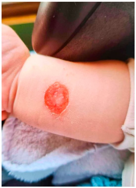
A previously healthy 4-month-old male infant was referred by the community pediatrician due to a worsening skin lesion on the right forearm that was accompanied by systemic symptoms. The initial skin lesion was a large round pustule appearing 2 days after a bug bite. The community pediatrician examined the baby, prescribed topical antibiotic cream (amikacin) and a pus culture was sent for analysis. Just 4 days after the examination, the lesion developed into ecthyma and the culture tested positive for Methicillin-sensitive Staphylococcus aureus (MSSA) (Figure 1). Targeted antibiotic treatment with cloxacillin by mouth (100 mg/kg/d in 4 divided doses) was administered, but shortly after the initiation of antibiotic treatment (2 days), the infant presented with new skin lesions and systemic symptoms, therefore referred to our emergency department for further investigation.
Figure 1.
Ecthyma due to methicillin-sensitive Staphylococcus aureus.
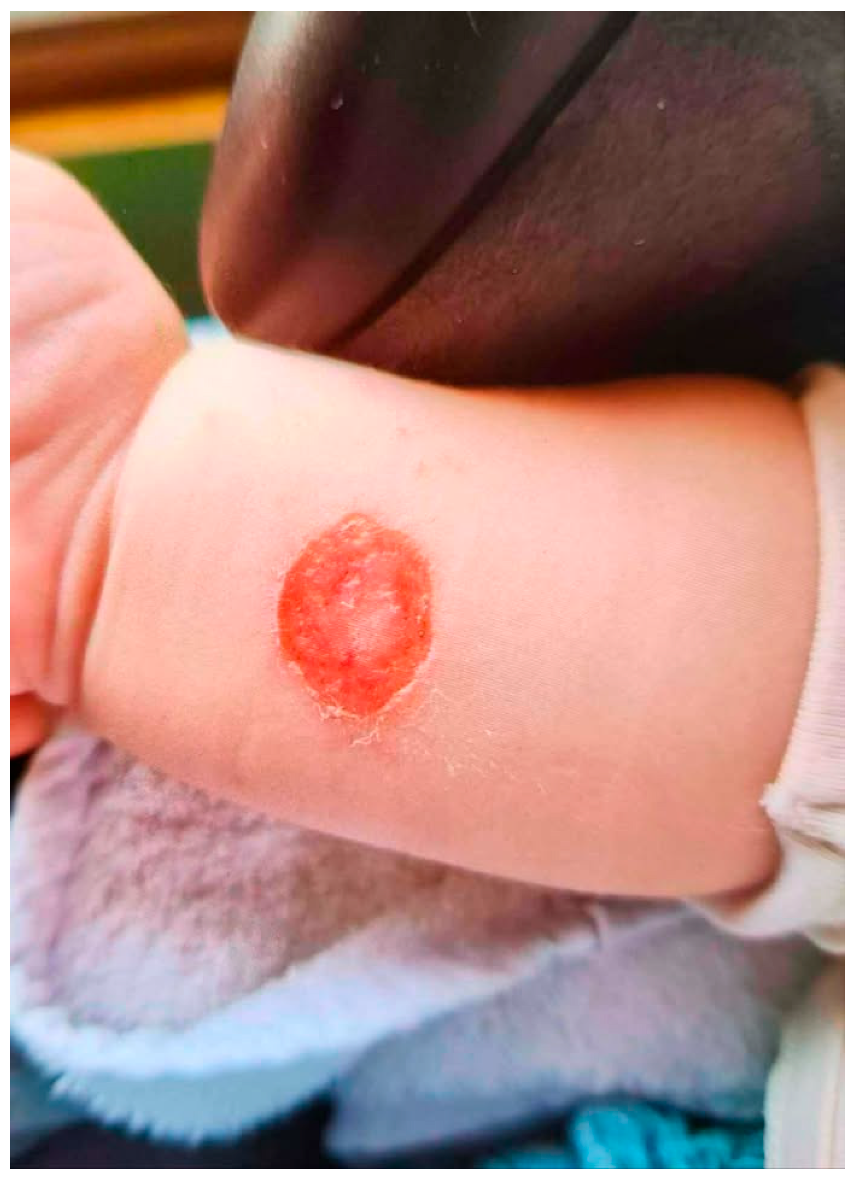
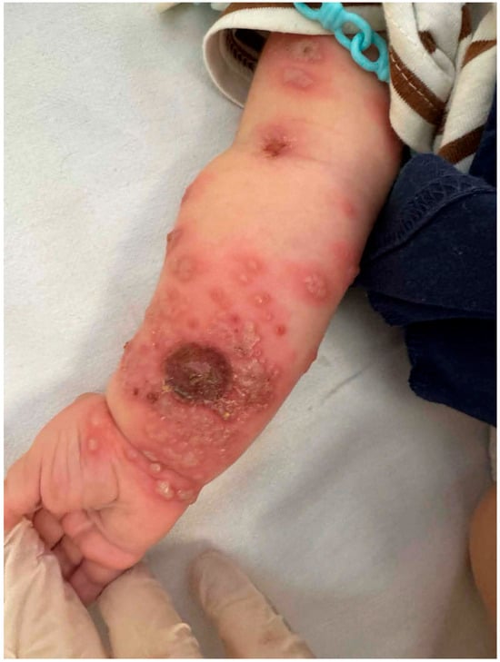
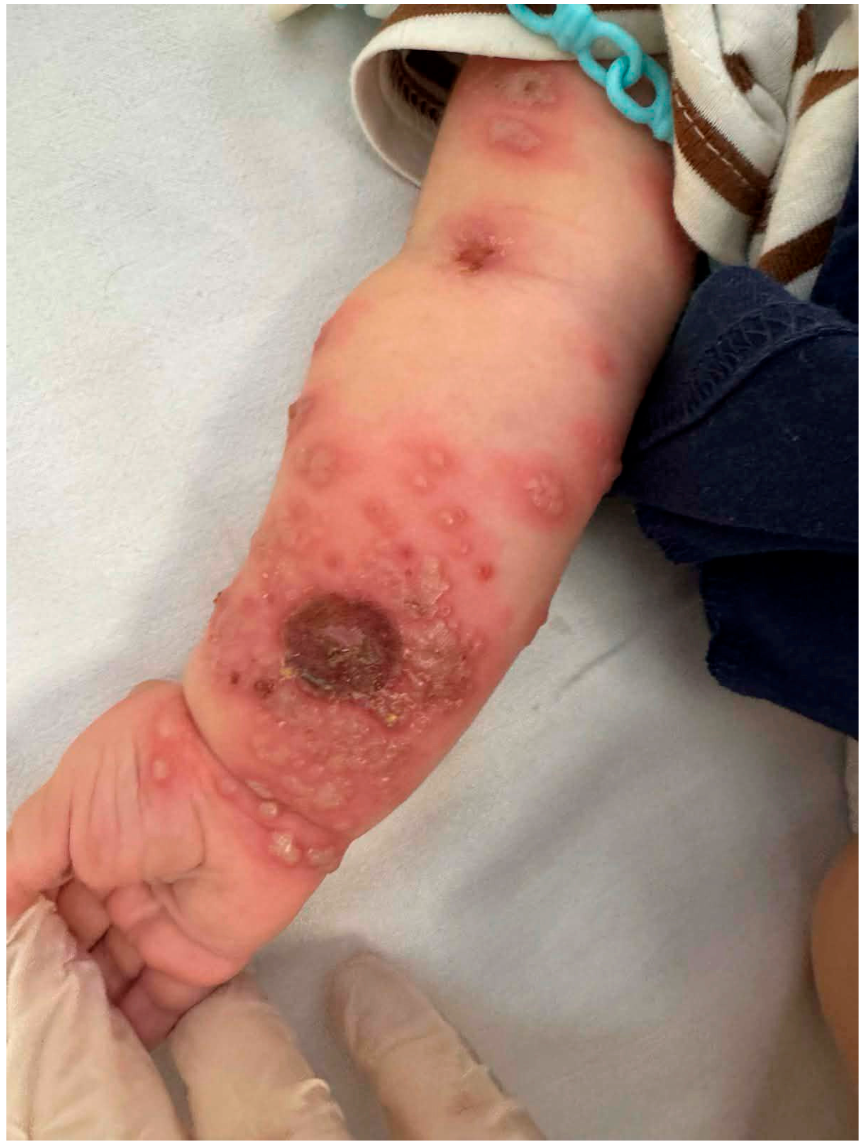
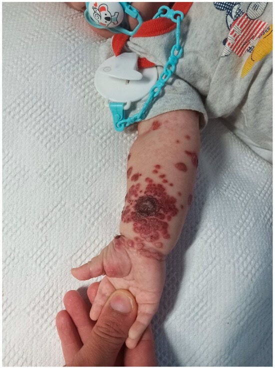
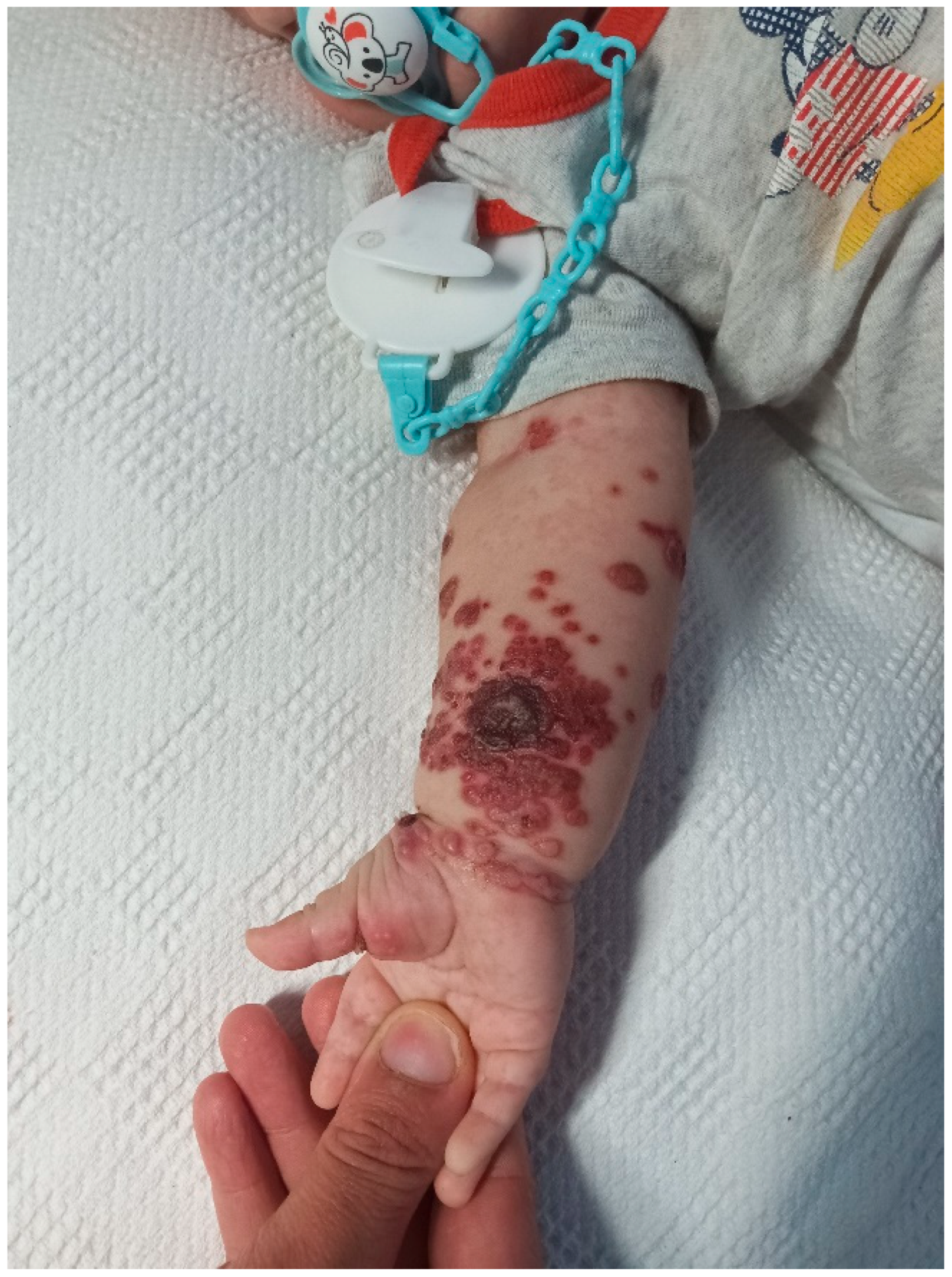
Upon arrival, the infant had mild fever, malaise and diarrhea, unilateral cervical lymphadenopathy (right side) with no signs of dehydration and normal observations. Blood investigations revealed no inflammatory markers and blood and skin cultures were all negative. The skin appeared with a central necrotic ulcer surrounded by continuous monomorphic vesicles that were developing into pustules with hemorrhagic crusts and completely disrupted epidermal integrity (Figure 2 and Figure 3). Polymerase Chain Reaction testing of the vesicle’s fluid for Herpes Simplex Virus (HSV)1-2 was performed, and the result was positive for HSV-1. The diagnosis of eczema herpeticum was made and intravenous acyclovir (10 mg/kg/d/8 hourly) was added to the initial antistaphylococcal course, with gradual improvement of lesions within a week of treatment.
Figure 2.
Central necrotic ulcer accompanied by painful, uniform-sized (monomorphic) clusters of blisters and pustules on the right upper limb. There was a rapid accumulation of bumps across forearm and fewer lesions extending up to the arm.
Figure 3.
The blisters rupture, then dry out and are covered with bloody crusts (hemorrhagic crusts).
This is a case of severe eczema herpeticum in a very young infant presenting primarily with an infected bug bite, with paradoxically no history or clinical evidence of atopic dermatitis, and manifesting with a sudden appearance of monomorphic vesicles and erosions with hemorrhagic crusts alongside systemic symptoms. Eczema herpeticum, initially described by Kaposi in 1887 and hence also known as Kaposi varicelliform eruption, is a disseminated herpes simplex infection of the skin that most commonly occurs in patients with atopic dermatitis [1]. Rarely, it can develop in any patient with a compromised skin barrier after skin trauma or burns [2,3]. Data highlight that patients with atopic eczema and a known history of Staphylococcus aureus skin infection are at increased risk for eczema herpeticum, with authors reporting sixfold odds [4]. The best studied virulence factors are staphylococcal enterotoxins, also known as superantigens, which induce robust T-cell-mediated inflammation and signal the release of proinflammatory cytokines. Moreover, strains that produce toxic shock syndrome toxin-1 (TSST-1) along with HSV activation can cause enormous epidermal inflammation [5]. In our case, the young infant had no signs or symptoms of known atopic dermatitis and the family reported no signs or symptoms of allergic rhinitis, atopy or asthma. We thus hypothesize that atopic dermatitis might present as a subclinical form at this young age and parents were advised for prompt detection of future signs. A second hypothesis is that the severity of skin infection can be attributed to the loss of skin barrier due to the bug bite along with the MSSA infection. However, this severe skin infection at such an early age requires evaluation of the host’s immune system. Thus, serum immunoglobulins tests, immunophenotyping, and a dihydrorhodamine (DHR) test for Chronic Granulomatous Disease were performed, investigations that are all standard. This case presentation highlights that diagnosis may be challenging in clinical settings, as all puzzle pieces might not always fit. When initial diagnosis seems uncertain and unreliable, careful consideration of progressive clinical images can lead to prompt and optimal therapeutic management.
Author Contributions
Conceptualization, A.S.; attending pediatricians, A.S., S.Z. and A.P.; writing—original draft preparation, A.S. and D.M.; writing—review and editing, A.P. All authors have read and agreed to the published version of the manuscript.
Funding
This research received no external funding.
Institutional Review Board Statement
Ethical review and approval of this study were not required by the Ethics Committee of ‘P&A Kyriakou’ Children’s Hospital because anonymized data used on case reports are not considered research in our jurisdiction. The patient’s information has been de-identified.
Informed Consent Statement
Informed consent was obtained from the patient’s caregivers for publication of this paper.
Data Availability Statement
The original contributions presented in this study are included in the article. Further inquiries can be directed to the corresponding author.
Conflicts of Interest
The authors declare no conflicts of interest.
References
- Moran, M.C.; Brewer, M.G.; Schlievert, P.M.; Beck, L.A.S. aureus virulence factors decrease epithelial barrier function and increase susceptibility to viral infection. Microbiol. Spectr. 2023, 11, e0168423. [Google Scholar] [CrossRef] [Scilit] [PubMed]
- Naumeri, F.; Rijal, S.; Rashid, A.R.; Ahmad, H.M. Eczema Herpeticum in children with burns. Pak. J. Med. Sci. 2021, 37, 421–425. [Google Scholar] [CrossRef] [Scilit] [PubMed]
- Bestue, M.; Cordero, A. Kaposi’s Varicelliform Eruption in a Patient with Healing Peribucal Dermabrasion. Dermatol. Surg. 2000, 26, 939–940. [Google Scholar] [CrossRef] [Scilit] [PubMed]
- Moran, M.C.; Klose, A.; Yoshida, T.; De Benedetto, A.; Schneider, L.C.; Ong, P.Y.; Simpson, E.L.; Leung, D.Y.M.; Miller, B.L.; Seplaki, C.L.; et al. History of S. aureus Skin Infection Significantly Associates with History of Eczema Herpeticum in Patients with Atopic Dermatitis. Dermatol. Ther. 2023, 13, 2417–2429. [Google Scholar] [CrossRef] [Scilit] [PubMed]
- Napolitano, M.; Esposito, M.; Fargnoli, M.C.; Girolomoni, G.; Romita, P.; Nicoli, E.; Matruglio, P.; Foti, C. Infections in Patients with Atopic Dermatitis and the Influence of Treatment. Am. J. Clin. Dermatol. 2025, 26, 183–197. [Google Scholar] [CrossRef] [Scilit] [PubMed]
Disclaimer/Publisher’s Note: The statements, opinions and data contained in all publications are solely those of the individual author(s) and contributor(s) and not of MDPI and/or the editor(s). MDPI and/or the editor(s) disclaim responsibility for any injury to people or property resulting from any ideas, methods, instructions or products referred to in the content. |
© 2026 by the authors. Licensee MDPI, Basel, Switzerland. This article is an open access article distributed under the terms and conditions of the Creative Commons Attribution (CC BY) license.